Epistasis between neurochemical gene polymorphisms and risk for ADHD
Upload
khangminh22Category
view
4download
0
HAL Id: hal-03486715https://hal.archives-ouvertes.fr/hal-03486715
Submitted on 20 Dec 2021
HAL is a multi-disciplinary open accessarchive for the deposit and dissemination of sci-entific research documents, whether they are pub-lished or not. The documents may come fromteaching and research institutions in France orabroad, or from public or private research centers.
L’archive ouverte pluridisciplinaire HAL, estdestinée au dépôt et à la diffusion de documentsscientifiques de niveau recherche, publiés ou non,émanant des établissements d’enseignement et derecherche français ou étrangers, des laboratoirespublics ou privés.
Distributed under a Creative Commons Attribution - NonCommercial| 4.0 InternationalLicense
Neurochemical impact of the 5-HT2C receptor agonistWAY-163909 on monoamine tissue content in the rat
brainAbdeslam Chagraoui, Sara Whitestone, Lynn Baassiri, Julien Manem,
Giuseppe Di Giovanni, Philippe de Deurwaerdère
To cite this version:Abdeslam Chagraoui, Sara Whitestone, Lynn Baassiri, Julien Manem, Giuseppe Di Giovanni,et al.. Neurochemical impact of the 5-HT2C receptor agonist WAY-163909 on monoamine tis-sue content in the rat brain. Neurochemistry International, Elsevier, 2019, 124, pp.245 - 255.�10.1016/j.neuint.2019.01.019�. �hal-03486715�

1
Neurochemical impact of the 5-HT2C receptor agonist WAY-163909 on
monoamine tissue content in the rat brain
Abdeslam Chagraoui1*, Sara Whitestone2*, Lynn Baassiri2, Julien Manem2, Giuseppe Di
Giovanni3,4, Philippe De Deurwaerdère2
1Normandie Univ, UNIROUEN, INSERM, U1239, CHU Rouen, Neuronal and Neuroendocrine
Differentiation and Communication Laboratory, Institute for Research and Innovation in
Biomedicine of Normandy (IRIB), Rouen, France.; Department of Medical Biochemistry, Rouen
University Hospital, Rouen, France. 2Centre National de la Recherche Scientifique (Unité Mixte
de Recherche 5293) 33076 Bordeaux Cedex, France). 3Department of Physiology &
Biochemistry, Faculty of Medicine and Surgery, University of Malta, Msida, Malta.
4Neuroscience Division, School of Biosciences, Cardiff University, Cardiff, UK.
*These authors have equally participated in this work
Footnotes to the title: WAY-163909 and brain monoamines
Corresponding author: Philippe De Deurwaerdère
Centre National de la Recherche scientifique; UMR CNRS 5287
146, rue Léo Saignat; 33076 Bordeaux; France
Tel. +33 (0) 557 57 12 90
E-mail: [email protected]
© 2019 published by Elsevier. This manuscript is made available under the CC BY NC user licensehttps://creativecommons.org/licenses/by-nc/4.0/
Version of Record: https://www.sciencedirect.com/science/article/pii/S0197018618306909Manuscript_6508bbe74e2d7e565ba0587dc9b165ca

2
Figures: 4; Tables: 1
Abbreviations footnote: aCg (anterior cingulate cortex), ains (anterior insular cortex), BLA
(basolateral nucleus of the amygdala), CE (central nucleus of the amygdala), CNS (central
nervous system), core (core of the nucleus accumbens), DA (dopamine), DLO (dorsolateral
orbitofrontal cortex), dHP (dorsal hippocampus), DLS (dorsolateral striatum), DMS
(dorsomedial striatum), DR (dorsal raphe nucleus), dHY (dorsal hypothalamus), EPN
(entodepuncular nucleus), HPLC (high pressure liquid chromatography), IL (infralimbic cortex),
LO (lateral orbitofrontal cortex), M2 (motor cortex M2), MO (medial orbitofrontal cortex), MR
(median raphe nucleus), NA (noradrenalin), OFC (orbitofrontal cortices), pCg (posterior
cingulate cortex), pIns (posterior insular cortex), PL (prelimbic cortex), 5-HT (5-
hydroxytryptamine; serotonin), 5-HT2C receptors (Serotonin2C receptors), shell (shell of the
nucleus accumbens), SN (substantia nigra), SNc (substantia nigra pars compacta), SNr
(substantia nigra pars reticulata), STN (subthalamic nucleus), vHP (ventral hippocampus), vHY
(ventral hypothalamus), VCS (ventrocaudal striatum), VLS (ventrolateral striatum), VMS
(ventromedial striatum), VTA (ventral tegmental area), WAY-163909 ((7bR,10aR)-1,2,
3,4,8,9,10,10a-octahydro-7bH-cyclopenta-[b][1,4]diazepino[6,7, 1hi]indole]).

3
ABSTRACT
Serotonin2C receptor (5-HT2C) agonists are promising drugs for the treatment of
neuropsychiatric diseases. However, their effect is not completely understood in part because
they possibly affect several neurobiological networks simultaneously. We studied the effect of
the 5-HT2C receptor agonist WAY-163909 (0.3 and 3 mg/kg; i.p.) on the tissue concentration of
dopamine (DA), 5-HT and noradrenaline (NA) in 29 rat brain regions related to motor, cognitive,
mood and vegetative networks. We found that WAY-163909, without altering the tissue
concentration of NA, increased 5-HT concentrations in the medial orbitofrontal cortex and the
motor cortex M2 at 3 mg/kg and decreased it in the dorsolateral orbitofrontal cortex at 0.3 mg/kg.
WAY-163909 enhanced DA concentrations in the central nucleus of the amygdala at 0.3 mg/kg
and reduced it in the dorsal hypothalamus at 3 mg/kg. Using correlative analysis of the tissue
content of monoamines, WAY-163909 dramatically changed the profile and the pattern of the
correlations within and between monoaminergic systems without drastically changing the total
number of these correlations. The profile of these changes in correlations was dose-dependent as
it was very different between the two doses within and among monoaminergic systems. In
conclusion, the data indicated that the 5-HT2C receptor agonist WAY-163909 quantitatively alters
monoamine content in very few regions but promotes multiple changes of monoaminergic
connectivity in the brain.

4
1. INTRODUCTION
The serotonin2C (5-hydroxytryptamin2C, 5-HT2C) receptor agonists have potential interest
in the treatment of various neuropsychiatric diseases (Chagraoui et al., 2016; Di Giovanni and De
Deurwaerdere, 2016; Higgins and Fletcher, 2015; Howell and Cunningham, 2015; Kostrzewa et
al., 2007). The 5-HT2C receptor agonists have been tested in humans for schizophrenia and drug
addiction, although the only drug that has received the FDA approval so far is the 5-HT2A/2C
receptor agonist lorcaserin for the treatment of obesity (Di Giovanni and De Deurwaerdère,
2016). However, the mechanism by which 5-HT2C receptor agonists are effective is still unclear.
The 5-HT2C receptor subtype is diffusely expressed in the CNS (Abramowski et al., 1995;
Clemett et al., 2000; Pasqualetti et al., 1999; Pazos and Palacios, 1985). According to this large
pattern of expression, the administration of 5-HT2C receptor agonists in rodents is associated with
a vast array of behavioral effects. They decrease locomotor activity, feeding behavior, and the
reinforcing properties of drug abuse or impulsive responses (Anastasio et al., 2015; Dalton et al.,
2004; Di Giovanni and De Deurwaerdere, 2016; Fletcher et al., 2011; Fletcher et al., 2006;
Fletcher et al., 2013; Fletcher et al., 2009; Higgins et al., 2016; Howell and Cunningham, 2015).
On the other hand, they increase purposeless oral movements, penile erection and grooming
(Graf, 2006; Kreiss and De Deurwaerdere, 2017; Millan et al., 1997; Navailles et al., 2013b) and
have opposite effects on anxiety or epilepsy depending on the agonist and the nature of the
trouble (Di Giovanni and De Deurwaerdere, 2016). Specific mechanisms of action and circuits
have been proposed to account for the above-mentioned behavioral effects of 5-HT2C receptor
agonists but not conclusive proofs are avalaible yet. For instance, it has been largely documented
that 5-HT2C receptors inhibit dopamine (DA) neuron activity and DA release (Di Giovanni et al.,

5
2001) in the control of motor behaviors. Yet, this action does not appear essential in the ability of
5-HT2C receptor agonists to reduce behaviors associated with enhanced DA (Cathala et al., 2015;
De Deurwaerdere and Di Giovanni, 2017). Alternatively, we have proposed that the effects
resulting from the brain 5-HT2C receptor stimulation, such as narrowing DA-dependent
behavioral effects or food intake, might emerge from a simultaneous action on the activity of
several neurobiological networks and from a diffuse interaction and competition between these
neurobiological networks (Di Giovanni and De Deurwaerdere, 2016).
The DA, 5-HT and noradrenaline (NA) systems innervate the brain from the cell bodies
located in the substantia nigra (SN) and ventral tegmental area (VTA), dorsal and median raphe
nuclei (DR and MR), and locus coeruleus (LC), respectively. Monoamines tissue measurement in
the brain gives rough information on the biochemical status of their terminals in one brain region,
or in multiple brain regions. The content of monoamines is regulated by local neurons connected
to distinct networks. Therefore, the tissue content measurement ultimately permits to address the
connectivity of neurochemical, monoaminergic markers within one monoaminergic system and
between them through correlational approaches (Fitoussi et al., 2013; Klouche et al., 2015). We
hypothesized that pharmacological activation of the 5-HT2C receptors may disrupt the
monoaminergic connectivity in the brain.
In the present study, we studied the effect of the intraperitoneal (i.p.) injection of the
preferential 5-HT2C receptor agonist WAY-163909 (0.3 and 3 mg/kg, i.p.) on the tissue
concentration of DA, 5-HT, and NA in 29 brain regions in rats. WAY-163909 elicited various
behaviors (Dunlop et al., 2006; Navailles et al., 2013b) that have no clear association with DA
release, c-Fos expression in basal ganglia or changes in electrophysiological activities in the basal

6
ganglia (Lagière et al., 2017). The chosen brain regions included various frontal cortices, some
basal ganglia regions, the hippocampus, the hypothalamus, and the amygdala.
MATERIALS AND METHODS
Animals
Male Sprague Dawley rats weighing 300-400 g were used. They were kept in the animal
facility (University of Bordeaux, France) with free access to food and water, in a constant
temperature (21 ± 2° C) and humidity (60%) levels, under a 12-h day/night cycle. All the
animals’ procedures were in accordance with the European Council Directive 2010/63/EU and
the French National Committee (décret 2001-464), and local committee for the care and use of
laboratory animals. All efforts were made to minimize animal suffering and to reduce the number
of animals used.
Tissue sampling and conditioning
The rats were sacrificed 45 min after drug injection (see pharmacological treatment) and
the brain was quickly removed. The brains were immersed in isopentane (2-methyl butane) (-35 ±
5° C) and stored in a freezer at -80° C. The brains were cut using a cryostat at -24° C, and
bilateral “punches” were made of various brain structures of interest using steel cannulae of 500
or 800 μm in diameter except for the subthalamic nucleus (STN) (Fitoussi et al., 2013). The STN
was gently taken with the smaller cannula used as a spoon to collect the surface (usually around
200 µm thickness) of the tissue from the medial to the lateral extension of the STN. The
operation was performed three to four times to account for the rostro-caudal length of the STN.
7
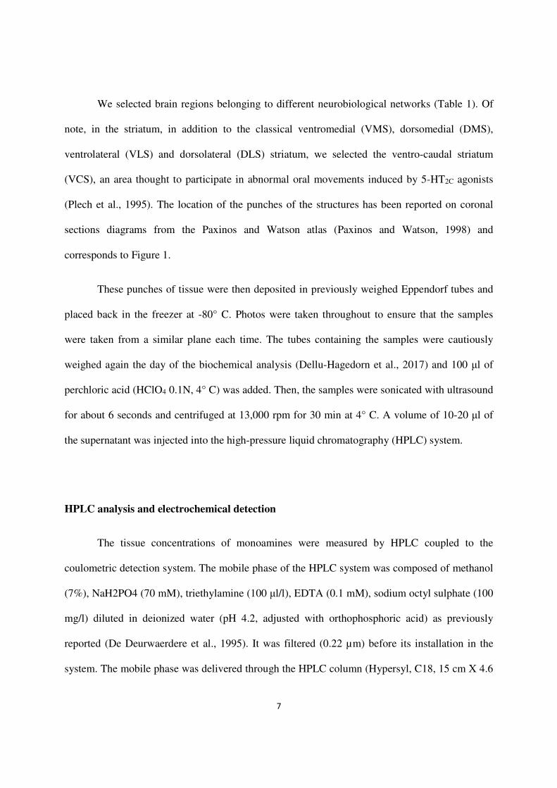
We selected brain regions belonging to different neurobiological networks (Table 1). Of
note, in the striatum, in addition to the classical ventromedial (VMS), dorsomedial (DMS),
ventrolateral (VLS) and dorsolateral (DLS) striatum, we selected the ventro-caudal striatum
(VCS), an area thought to participate in abnormal oral movements induced by 5-HT2C agonists
(Plech et al., 1995). The location of the punches of the structures has been reported on coronal
sections diagrams from the Paxinos and Watson atlas (Paxinos and Watson, 1998) and
corresponds to Figure 1.
These punches of tissue were then deposited in previously weighed Eppendorf tubes and
placed back in the freezer at -80° C. Photos were taken throughout to ensure that the samples
were taken from a similar plane each time. The tubes containing the samples were cautiously
weighed again the day of the biochemical analysis (Dellu-Hagedorn et al., 2017) and 100 μl of
perchloric acid (HClO4 0.1N, 4° C) was added. Then, the samples were sonicated with ultrasound
for about 6 seconds and centrifuged at 13,000 rpm for 30 min at 4° C. A volume of 10-20 μl of
the supernatant was injected into the high-pressure liquid chromatography (HPLC) system.
HPLC analysis and electrochemical detection
The tissue concentrations of monoamines were measured by HPLC coupled to the
coulometric detection system. The mobile phase of the HPLC system was composed of methanol
(7%), NaH2PO4 (70 mM), triethylamine (100 μl/l), EDTA (0.1 mM), sodium octyl sulphate (100
mg/l) diluted in deionized water (pH 4.2, adjusted with orthophosphoric acid) as previously
reported (De Deurwaerdere et al., 1995). It was filtered (0.22 µm) before its installation in the
system. The mobile phase was delivered through the HPLC column (Hypersyl, C18, 15 cm X 4.6

8
mm, particle size 5 μm, C.I.L.) at a flow rate of 1.2 mL/min using an HPLC pump (LC10Ad Vp,
Schimadzu, France). The column was protected by a Brownlee-Newgard precolumn (RP-8, 15 X
3.2 mm, 7 µm; C.I.L.). The injection of the samples (10-20 μL) was carried out by a manual
injection valve (Rheodyne, model 7725i, C.I.L.) equipped with a loop of 20 μl. The monoamines
exit the column at different retention times (approximately NA: 3’40”; DA: 8’; 5-HT: 18’) and
passed into the coulometric detection cell (Cell 5011, ESA, Paris, France) equipped with two
electrodes. The potential of these two electrodes was fixed via the coulometric detector
(CoulochemII, ESA, Paris, France) at +350 mV (oxidation) and -270 mV (reduction),
respectively. The coulometric detector was connected to a computer through an interface (Ulyss,
Azur system, Toulouse, France).
The calibration curves were performed once the pics in a standard solution (1 ng/10 µl)
were well separated in the chromatogram. The calibration curves were adapted according to the
brain areas investigated, because the quantities of monoamines are heterogeneous (Fitoussi et al.,
2013), requiring different gains set at the level of the detector using a timeline method. The gains
used ranged from 5 nA (for the DA in the hippocampus) to 1 µA (for DA in the striatum).
Standard solutions were used before each series of 10/12 samples to verify the good
correspondence of the chromatographic conditions to both the elution time and quantities
calculated from the calibration curves. Similarly, the quantities of monoamines in the standards
varied according to the brain region investigated. The overall sensitivity for the compounds
ranged from 2 pg/10 µl for DA to 7 pg/10 µl for 5-HT with a signal/noise ratio of 3:1.
Pharmacological Treatment and Experimental Design

9
WAY 163909 (7bR,10aR)-1,2, 3,4,8,9,10,10a-octahydro-7bH-cyclopenta-
[b][1,4]diazepino[6,7, 1hi]indole]), freshly diluted as free base in NaCl 0.9%, was injected i.p.
(0.3 or 3 mg/kg). For each experimental group, the animals were randomized and received either
the drug or appropriate vehicle (in 1 ml/kg).
WAY 163909 is one of the most selective 5-HT2C receptor agonists available on the
market, it is brain penetrant and summarizes the main behavioral effects elicited by 5-HT2CR
agonists in rodents (Dunlop et al., 2006; Dunlop et al., 2005). The doses of 0.3 and 3 mg/, i.p.
have been chosen based on previous data showing that it reduced locomotor activity and induced
purposeless oral movements (Marquis et al., 2007; Navailles et al., 2013). These studies
reported that the behavioral effects elicited by WAY-163909 were observed 30-45 min after
its injection. Therefore, we choose to observe the behavioral effect of WAY-163909 at 45 min
after its injection with the aim of monitoring the abnormal oral movements and penile
grooming. Then, quickly we collected the brain areas in order to get the monoamine status at
a time corresponding to the highest behavioral activity. Their brains were finally stored at -
80° C for successive neurochemical post-mortem analysis.
Statistical analysis of the data
The tissue levels of NA, DA, and 5-HT for each structure were expressed in pg/mg. These
levels are presented as the mean ± the standard error of the mean (SEM) according to their
treatment group. Outlier data were discarded on the basis of the value outside the range of the
average mean ± two standard deviations (Dellu-Hagedorn et al., 2017). The values for NA, DA,
and 5-HT were compared between experimental groups (saline, WAY 163909 at 0.3 or 3 mg/kg,

10
i.p.) with a one-way ANOVA, followed by the Fisher protected least significant difference
(PLSD) post-hoc test. A similar analysis was performed for the weight of the tissue between
groups for each structure. In all comparisons, p < 0.05 was used as the criterion for significance.
The qualitative analysis corresponds to multiple correlative analyses using the Bravais-
Pearson’s correlation coefficient. These analyzes were performed within and between NA, DA
and 5-HT systems in the 29 brain regions investigated. The correlations have been separately
performed in rats receiving saline, 0.3 mg/kg WAY 163909 and 3 mg/kg WAY 163909 to
estimate monoamine connectivity in each group with no attempt to statistically compare the
profiles. As previously reported (Fitoussi et al., 2013), p-values were adjusted using the
False Discovering Rate (FDR) controlling procedures (Benjamini and Hochberg, 1995).
Correlations were then considered as significant at the 5% level and were reported in the
corresponding figures.

11
Results
1. Quantitative analysis of monoamine tissue content after WAY-163909 administration
All rats treated with WAY 163909 exhibited classical behaviors induced by 5-HT2C
receptor agonists including abnormal oral movements and few episodes of penile grooming
(Navailles et al., 2013b). WAY 163909 was administered at 0.3 or 3 mg/kg i.p. and the results of
tissue monoamines of the 29 brain areas of interest were compared to those of saline-treated rats.
The size of the tissue and the number of observations kept after removing outliers (or simply non-
detected parameters) are indicated in Table 1. For all brain regions, the size of the tissue did not
significantly vary between groups. The quantitative analysis of the effects of WAY 163909 for
NA, DA, and 5-HT systems is reported in Figure 2.
The tissue levels of DA were largely heterogeneous across the brain of the saline group.
Briefly, DA levels were very high along the nigrostriatal and mesolimbic areas reaching up to
6118 ± 655 pg/mg of tissue in the DMS (almost equivalent in DLS). Conversely, it was very low
and sometimes undetectable in the hippocampus (dHP and vHP with 3.4 ± 0.38 pg/mg and 3.8 ±
0.56 pg/mg of tissue, respectively) or LO. WAY 163909 treatments did not affect DA levels
except for the dorsal hypothalamus (dHY) [one-way ANOVA F(2,20)=3.92, p = 0.039], the CE
amygdala [F(2,23)=5.38, p = 0.013] and the MR [F(2,19)=4.07, p = 0.036]. Precisely, DA levels
were significantly decreased after the administration of 3 mg/kg WAY 163909 compared to
saline treatment (Figure 2). In the amygdala (CE), DA tissue content was significantly increased
above values of the saline group after 0.3 mg/kg WAY 163909 only. DA content in the MR of
0.3 mg/kg WAY-163909 treated rats was higher compared to that of the 3 mg/kg group but not
that of the saline group (Figure 2).

12
Tissue levels of 5-HT were less heterogeneous compared to DA in investigated brain
regions, although important and expected differences were reported. DA levels were highest in
the SN and very high in DR, VTA, EPN, NAc shell and parts of the cortex. Conversely, 5-HT
tissue content was low in the hippocampus (dHP and vHP), in the hypothalamus (dHY and vHY)
and the lateral orbitofrontal cortices. WAY 163909 treatment did not alter 5-HT tissue content
except for MO, DLO, and M2 [one-way ANOVA F(2,23)=6.32, p=0.007; F(2,23)=3.6, p=0.045
and F(2,21)=5.95, p=0.01 respectively]. The 5-HT content was increased in MO and M2 at the
dose of 3 mg/kg WAY 163909 and reduced in DLO at the dose of 0.3 mg/kg WAY 163909.
Tissue levels of NA in the saline-treated group were heterogeneous as expected from
previous works. The highest concentration was observed in the DR. High concentrations were
observed in VTA, MR, SN, and an unexpectedly high level was found in the EPN. Some cortical
territories (MO, M2), hippocampus, and hypothalamus had moderate values. The lowest
concentrations were found in the striatal quadrants, LO and dHY. As illustrated in Figure 2,
WAY 163909 did not alter NA tissue content at the two doses used here in any of the brain
regions considered (one-way ANOVA, ns for all comparisons).
2. Qualitative analysis of monoamine tissue content
2.1 Within monoaminergic systems
To get a deeper analysis of the data profile, we then evaluated possible relationships of
monoamines between the 29 investigated brain areas using a correlative approach. The main
important point is to indicate that the correlations are few and their nature and number changed
upon WAY 163909 administration at increasing doses (0.3 mg/kg and 3 mg/kg).

13
Correlations for DA content between brain structures upon saline administration appear to be
limited and equilibrated between positive and negative correlations (15 and 14, respectively).
Some brain regions differently from other established several correlations with the DA content at
distal regions. Notably, DA content in the pins or the VMS had six correlations with the DA
content in other regions; the vHP, the EPN, and the DR had five correlations. Conversely, DA
content in the shell, DLS or VCS poorly correlated. The injection of WAY-163909 reduced the
number of correlations at 0.3 mg/kg (19 comprising 4 negative correlations only) and 3 mg/kg
(20 comprising 8 negative correlations). None of the correlations observed in saline-treated rats
were present in WAY 163909-treated rats and they were different between 0.3 and 3 mg/kg
condition. DA content in vHP and DR did not correlate after WAY-163909 injection. The pins
and the VMS DA content still had a few correlations (1 to 3) though with different brain areas.
The correlations of DA content after 3 mg/kg concerned mainly the cortex and the basal ganglia
regions when compared to 0.3 mg/kg results.
Few correlations (17) appear for 5-HT content in the 29 brain areas upon saline
administration, including 10 negative correlations. Several correlations concerned the NAc or the
striatum with the orbitofrontal cortices or the hippocampus. The 5-HT content in the BLA, IL,
aCg, LO, DR or VTA did not correlate. The profile of correlations was dramatically changed in
WAY-163909-treated rats with very few correlations in saline-treated rats reported in WAY-
163909 groups. WAY-163909 at 0.3 mg/kg induced a similar number of correlations (19) but
they were mainly positive (15/19). The 5-HT content in MO and VCS was correlated. The
correlations were dispatched between prefrontal cortices, amygdala with other territories while 5-
HT content of several brain regions (dHP, vHY, M2, VTA) did not correlate with other brain
regions. At 3 mg/kg, the number of correlations increased (25 including 15 negative correlations).

14
Several correlations were established between frontal cortices (particularly DLO, LO and M2)
and basal ganglia, often positive, while correlations within the basal ganglia tended to be negative
(Figure 3).
Very few correlations (18 comprising an almost equal number of positive and negative
correlations) were seen between the NA content of the 29 brain regions in the saline group. The
profile of correlations was totally different from that of DA. This profile dramatically changed
after WAY-163909 with a number of correlations reaching 29 and 16 after 0.3 and 3 mg/kg,
respectively. NA content persisted in 0.3 mg/kg WAY-163909 between IL and LO. In addition,
NA content in PL established correlations with NA content in seven other brain regions, all
positive except with LO. NA content in pins, pCg, VLS, or MR also established some
correlations (from 4 to 6). The number of correlations for NA content was reduced at 3 mg/kg
WAY-163909. At variance with DA, it is marked by low cortical/basal ganglia associations. It is
noteworthy that NA content in several brain regions including MO, M2, PL, ains or BLA did not
correlate with other brain regions. However, NA content in the hypothalamus correlated more
with that in the cortices, shell, and the STN.
2.1 Between monoaminergic systems
5-HT/DA tissue content
A total of 48 correlations were observed between 5-HT and DA contents, including 8 in the
same brain area (all positive). The proportion of negative correlations was slightly higher
compared to positive correlations in saline-treated rats. One of the most striking patterns is the
number of correlations between the 5-HT content in the EPN and the SN with DA content in

15
diverse brain regions including within these two, and between the orbitofrontal cortices for the
EPN and the frontal cortex for the SN. DA content in the SN negatively correlated with 5-HT
content from the orbitofrontal cortices and the CE whereas DA content in the DR negatively
correlated with 5-HT in the SN. DA and 5-HT contents strongly correlated within the shell and
core. The STN or hypothalamic regions slightly correlated, including some striatal quadrants. The
number of correlations after WAY-163909 was quite similar (48 after 0.3 and 42 after 3 mg/kg).
However, the pattern was very different compared to saline-treated rats. The correlations were
more positive, particularly in the case of 0.3 mg/kg. The number of correlations of 5-HT and DA
content in single brain regions was increased to 14. In addition, the correlations reported at the
level of the EPN and the SN were reduced. At 0.3 mg/kg, the DA content in the STN correlated
with 5-HT content of some orbitofrontal/prefrontal regions. Contents (DA or 5-HT) were
correlating more after 0.3 mg/kg between cortical regions or in the basal ganglia, particularly the
VMS. No striking pattern was noticed after 3 mg/kg except a slightly higher proportion of
correlations for both DA or 5-HT content within brain regions of the basal ganglia and the lower
number of correlations of 5-HT and DA content in the same brain regions.
DA/NA tissue content
DA and NA contents correlated in 39 brain areas including 6 positive correlations within the
same regions (shell, core, VLS, VCS, EPN, and SN) in saline-treated rats. The number of positive
and negative correlations were approximately even in distal comparisons. The brain regions in
which a higher number of correlations is found for DA and/or NA content are the VCS, the SN,
the EPN, the core. NA or DA content in MO negatively correlated with DA and NA content in
the EPN, respectively. Interestingly, the content of NA in the SN, DR, and VTA had correlations
with the CE, BLA, or dHP. WAY-163909 drastically modified the pattern of correlations, slightly

16
increasing the number of correlations to 49 and 53 at 0.3 and 3 mg/kg respectively. The most
noticeable changes at 0.3 mg/kg concern the numerous (5 to 9) correlations of DA contents in the
LO, IL, or STN with NA content of diverse cortices and subcortical areas for the two later. NA
content in the core or the aCg had multiple correlations with DA content of 4 to 6 areas. Finally,
0.3 mg/kg WAY-163909 increased NA/DA correlations in regions belonging to basal ganglia and
decreased them in amygdala/hippocampus. At 3 mg/kg WAY-163909, DA/NA tissue correlations
were observed between subcortical structures belonging to basal ganglia and limbic regions
including amygdala and hippocampus. Of note, vHY NA or DA content correlated with the core,
CE or M2. In fact, NA content in M2 displayed 5 correlations with DA content in other brain
regions. The NA/DA correlativity of STN was low at this dose.
5-HT/NA tissue content
NA and 5-HT contents diffusively correlated in saline-treated rats with 51 correlations
including 7 positive ones within the same regions. The correlations were found between cortical
regions, between cortical and striatal regions, and slightly with the mesencephalon. The regions
establishing the most distal correlations were pins and ains (6), LO, VLS, dHP and VCS (5)
followed by MO, SN, core, EPN, BLA, DLS, DMS, MR, and the STN (4 each). No
correlation was reported with NA or 5-HT content of aCg. Again, WAY-163909 changed the
pattern of correlations with 58 and 53 correlations at 0.3 and 3 mg/kg, respectively. In both cases,
a net, higher proportion of positive correlations were reported. The most evident change at 0.3
mg/kg was the number of NA/5-HT correlations in cortices involving LO, IL, PL, pCg towards
the other cortices, hippocampus/amygdala, NAc, and striatum. The second important change is
the increase of correlations within same the brain regions (from 7 to 13), including the shell, the
core, VMS, DMS, VLS, or STN. Orbitofrontal/frontal cortex also displayed few correlations with

17
the basal ganglia (notably the EPN). The hypothalamus regions displayed more correlations
compared to saline-treated rats. At 3 mg/kg, fewer correlations between the two parameters (10)
were observed within brain regions notably in the cortex and striatum (when compared with the
pattern obtained at 0.3 mg/kg). Cortical 5-HT and NA content correlated with NA and 5-HT
content in subcortical regions. In other words, the strong correlations centered in the cortex at 0.3
mg/kg were no longer present at 3 mg/kg.

18
DISCUSSION
Peripheral administration of the 5-HT2C receptor agonist WAY-163909 affected 5-HT and
DA tissue contents only in a few brain areas without altering the NA tissue content in rats.
However, WAY-163909 produced numerous changes in the correlations profiles within and
between monoamines across the various regions analyzed. These changes in the connectivity of
the monoaminergic system likely suggest new interactions between neurobiological networks
which are postulated to be simultaneously altered by central 5-HT2C receptor stimulation.
The tissue concentrations of endogenous monoamines here measured are comparable with
the density of monoaminergic fibers in the brain (Aston-Jones, 2004; Björklund and Lindvall,
1986; Steinbusch, 1984; Steinbusch et al., 1981) with the highest heterogeneity for the DA
system (Fitoussi et al., 2013). In addition to DA, 5-HT, and NA fibers coming from SN/VTA,
DR/MR and LC, local neuronal systems likely contribute for tissue DA in the hypothalamus and
DR (Bjorklund and Dunnett, 2007; Cho et al., 2017) whereas NA tissue content in the
hypothalamus (dHY notably) also originate from A5 and A7 (Aston-Jones, 2004). The NA
content in the dHY was low compared to the expectations (Brownstein and Palkovits, 1984) and
it could come from the punching procedure (see below).
We report that monoamines tissue correlates in few instances within a monoaminergic
system across multiple brain areas corresponding here to about 10% of the entire analyses for
each monoamine content. This finding is consistent with previous findings (Fitoussi et al., 2013),
although the proportion of negative correlations reported here is higher. Nonetheless, the two
studies are not comparable on numerous criteria (distinct strain, handling, age, depth of tissue
punched). Thus, we had no specific expectation as regards the pattern of monoamines

19
connectivity in control animals, as it should probably depend on the experimental conditions that
are always different, and the interindividual differences which are important for monoamine
tissue content (Dellu-Hagedorn et al., 2018). As postulated previously (Fitoussi et al., 2013), the
content of one monoamine in the terminal region does not correlate or rarely with the content of
the same monoamine at the level of the cell bodies, at least for DA and 5-HT. Nevertheless, the
lack of correlations could be due to the punching procedure. Indeed, we used the larger punches
for the SN, VTA, DR and the MR, with higher depth for raphe region, with the aim of to
abolishing the anatomo-functional specificities of midbrain monoaminergic centers toward
projections sites (Bjorklund and Dunnett, 2007; Björklund and Lindvall, 1986; Hale and Lowry,
2011; Kiyasova et al., 2011). On the other hand, the lack of correlations with midbrain contents
strengthens the idea that the biochemical regulation of the content of one monoamine in terminal
fields is in part independent from the incoming activity from cell bodies, and dependent on local
interaction with neighboring cells (Fitoussi et al., 2013).
WAY-163909 did not dramatically change the quantities of monoamines in numerous
brain regions analyzed but it revealed a specific pattern of effect on 5-HT content tissue content
in the orbitofrontal cortex. The lack of effect in other brain regions is consistent with a study in
mice reporting that the 5-HT2B/2C agonist Ro60-0175, over a large range of doses, did not modify
5-HT tissue content taken from the hippocampus, dorsal frontal part of the cortex, NAc and
VTA/SN (Mongeau et al., 2010). A pronounced effect in the orbitofrontal cortex is congruent
with the ability of 5-HT2C receptor agonists to trigger compulsive forms of behaviors including
grooming (Graf, 2006; Graf et al., 2003), penile grooming (Chagraoui et al., 2003; Millan et al.,
1997), and purposeless oral movements (Gong and Kostrzewa, 1992; Kreiss et al., 2013;
Navailles et al., 2013b; Stewart et al., 1989), the latter which could model compulsive emanation

20
(Kreiss and De Deurwaerdere, 2017) rather than dyskinesia (Kostrzewa et al., 2007; Kreiss and
De Deurwaerdere, 2017). The compulsive property of 5-HT2C receptor agonists has been
hypothesized to be located in the OFC by appropriate behavioral tests reults (Alsio et al., 2015;
Flaisher-Grinberg et al., 2008).
The low effect of WAY-163909 on DA tissue content agrees with the data in the
literature. Indeed, although the tissue measurement is considered less sensitive compared to the
extracellular levels measured by microdialysis, our results are consistent with the finding that
WAY-163909 did not modify DA release at 0.3, 1 or 3 mg/kg in the NAc or the striatum (Lagière
et al., 2017; Marquis et al., 2007). Other 5-HT2C receptor agonists have been shown to inhibit DA
release in the NAc and the firing rate of DA neurons in the VTA (De Deurwaerdere and Di
Giovanni, 2017; Di Giovanni and De Deurwaerdere, 2016; Di Matteo et al., 2000; Prisco et al.,
1994). Inhibitory effects of these agonists have been also reported at the level of the striatum (De
Deurwaerdere et al., 2004; Di Giovanni and De Deurwaerdere, 2016; Gobert et al., 2000),
without inhibition of SNc presumed-DA neurons electrical activity (Di Giovanni et al., 2000; Di
Matteo et al., 2000). The lack of parallel outcomes between the firing rate and the extracellular
levels at terminal fields, even described in the NAc, is in part due to the existence of several
populations of 5-HT2C receptors which could impact, sometimes oppositely, the biochemical
activity at terminals (Di Giovanni and De Deurwaerdere, 2016; Leggio et al., 2009a; Leggio et
al., 2009b; Navailles et al., 2006). Moreover, some of the agonists previously used including
Ro60-0175 are less selective compared to WAY-163909 and trigger non-selective effects
(Fletcher et al., 2006; Navailles et al., 2013a).
Overall, the quantitative analysis revealed a poor influence of WAY-163909 on DA and
5-HT system and a total absence of effect on NA content over the regions analyzed. It is

21
noteworthy that the tissue content of biogenic amines represents a complex balance between
synthesis, storage capacity and degradation (Commissiong, 1985; De Deurwaerdere et al.,
2017; Eisenhofer et al., 2004). Tissue measurement of metabolites could also witness
changes in synthesis/metabolism of monoaminergic terminals in some regions. For instance,
we succeed to measure homovanillic acid, a metabolite of DA, in some brain regions and
found a specific increase in MO at both 0.3 and 3 mg/kg WAY-163909 (data not shown). On
the other hand, the correlative analyses revealed profound changes for all monoamines within
and between monoaminergic systems including the NA one. Even though the number of non-
correlating parameters remains the great majority and somehow similar, WAY-163909
dramatically changed the pattern of correlations of DA content toward low (0.3 mg/kg) to the
high proportion (3 mg/kg) of the cortico-striatal pattern. The general profile for 5-HT content was
similar with several correlations found in the basal ganglia. The content of NA followed an
opposite pattern. The comparisons between pairs of monoamines highlight different points. The
relationships between 5-HT content in the EPN and SN with cortical DA content in saline-treated
rats were absent in WAY-163909. The DA content in the STN established correlations with
diverse brain regions at 0.3 mg/kg whereas DA/5-HT pattern of correlations at 3 mg/kg was more
cortico-striatal. Therefore, without quantitative modifications of DA tissue content or DA release
at the regimen used in this study, one may suggest that the changes in cortico-striatal connectivity
could also narrow DA tone. In line with this evidence, both Ro 60-0175 and WAY-163909 (1
mg/kg) have been reported to enhance the electrophysiological response of the cortico-striatal
indirect pathway on SNr neuronal activity (Beyeler et al., 2010; Lagière et al., 2017), basically
opposed to DA tone (Nambu, 2008).

22
The absence of dose-dependent effect (quantitative and correlative through a hypothetical
reinforcement of correlations) is compatible with the recruitment of behavioral effects as the dose
of the 5-HT2C receptor agonist increases (Lagiere et al., 2013). WAY-163909 preferentially
affects oral movements/penile grooming at low doses (i.e., 0.3 mg/kg) and reduces food intake at
higher doses (i.e., 3 mg/kg) (Dunlop et al., 2005; Marquis et al., 2007; Navailles et al., 2013b).
Interestingly, we report a quantitative reduction of DA content in the dHY at 3 mg/kg while the
correlations for all monoamines changed for dHY and vHY depending on the doses.
The change in connectivity induced by WAY-163909 across the brain regions is
widespread. It is in line with accumulating evidence that several brain regions mediate the
efficacy of 5-HT2C receptor agonists in food intake (D'Agostino et al., 2018; Xu et al., 2017; Xu
et al., 2008), drug addiction (Filip and Cunningham, 2002, 2003; Fletcher and Higgins, 2011;
Fletcher et al., 2004; Higgins and Fletcher, 2015; Higgins et al., 2012; Howell and Cunningham,
2015), impulsive/compulsive behavior (Anastasio et al., 2015; Carli and Invernizzi, 2014;
Fletcher et al., 2007) or anxiety (Di Giovanni and De Deurwaerdere, 2016; Heisler et al., 2007;
Millan, 2003). The circuits overlap and interact which is likely the meaning of the different
patterns of correlations obtained at the two doses. Speculatively, the level of activity of
monoaminergic terminals could be kept almost constant (homologous and heterologous controls),
but the changes of activity of neurobiological networks induced by WAY-163909 impact local
partners of monoaminergic terminals. The changes of correlations profile of monoamines tissue
content would witness distinct modes of activities of neurobiological networks triggered by
WAY-163909. Unfortunately, we cannot propose now a specific fingerprint of the pattern of
WAY-163909’s correlations, and additional studies are warranted with other 5-HT2C receptor
agonists to determine if there is a complete change of the correlation profile with respect to

23
saline-treated rats and between the doses. Therefore, the neurochemical approach is still
interesting in view of the poor ability of 5-HT2C receptor agonists to trigger c-Fos expression to
map their brain effect (De Deurwaerdere et al., 2013; Devroye et al., 2015; Lagière et al., 2017).
In conclusion, WAY 163909 slightly modified 5-HT and DA, but not NA tissue content
in a very few regions. However, it completely changed the pattern of correlations of monoamines
in the brain. This result suggests that the change of brain excitability induced by WAY-163909
impacts the organization of monoaminergic connectivity rather than inducing quantitative
changes.

24
ACKNOWLEDGMENTS
This work was supported by grants from Centre National de la Recherche Scientifique. The
authors wish to thank Dr J. Dunlop for the generous gift of the 5-HT2C receptor agonist, WAY
163909. Clément Jourdin is acknowledged for his technical support.

25
References
Abramowski, D., Rigo, M., Duc, D., Hoyer, D., Staufenbiel, M., 1995. Localization of the 5-
hydroxytryptamine2C receptor protein in human and rat brain using specific antisera.
Neuropharmacology 34, 1635-1645.
Alsio, J., Nilsson, S.R., Gastambide, F., Wang, R.A., Dam, S.A., Mar, A.C., Tricklebank, M.,
Robbins, T.W., 2015. The role of 5-HT2C receptors in touchscreen visual reversal learning in the
rat: a cross-site study. Psychopharmacology (Berl) 232, 4017-4031.
Anastasio, N.C., Stutz, S.J., Fink, L.H., Swinford-Jackson, S.E., Sears, R.M., DiLeone, R.J.,
Rice, K.C., Moeller, F.G., Cunningham, K.A., 2015. Serotonin (5-HT) 5-HT2A Receptor (5-
HT2AR):5-HT2CR Imbalance in Medial Prefrontal Cortex Associates with Motor Impulsivity.
ACS Chem Neurosci 6, 1248-1258.
Aston-Jones, G., 2004. Locus Coeruleus, A5 and A7 noradrenergic cell groups. The rat nervous
system third edition, 259-284.
Benjamini, Y., Hochberg, Y., 1995. Controlling the False Discovery Rate: a practical and
powerful approach to multiple testing. J R Stat Soc [Ser. B] 57, 289-300.
Beyeler, A., Kadiri, N., Navailles, S., Boujema, M.B., Gonon, F., Moine, C.L., Gross, C., De
Deurwaerdere, P., 2010. Stimulation of serotonin2C receptors elicits abnormal oral movements
by acting on pathways other than the sensorimotor one in the rat basal ganglia. Neuroscience 169,
158-170.
Bjorklund, A., Dunnett, S.B., 2007. Dopamine neuron systems in the brain: an update. Trends
Neurosci 30, 194-202.
Björklund, A., Lindvall, O., 1986. Catecholaminergic brain stem regulatory systems, In:
Mountcastle, V.B., Bloom, F.E., Geiger, S.R. (Eds.), Catecholaminergic brain stem regulatory
systems. In: Handbook of physiology. The nervous system. IV. Intrinsic regulatory systems of the
brain., American Physiological Society, pp. 677-700.

26
Brownstein, M., Palkovits, M., 1984. Catecholamines, serotonin, acetylcholine and
γαµµα−αµινοβυτψριχ αχιδ ιν τηε ρατ βραιν: βιοχηεµιχαλ στυδιεσ, In: Bjorklund, A.,
Hokfelt, T. (Eds.), Classical transmitters in the CNS. Vol. 2 part 1,Elsevier, Amsterdam, pp. 23-
54.
Carli, M., Invernizzi, R.W., 2014. Serotoninergic and dopaminergic modulation of cortico-striatal
circuit in executive and attention deficits induced by NMDA receptor hypofunction in the 5-
choice serial reaction time task. Front Neural Circuits 8, 58.
Cathala, A., Devroye, C., Maitre, M., Piazza, P.V., Abrous, D.N., Revest, J.M., Spampinato, U.,
2015. Serotonin2C receptors modulate dopamine transmission in the nucleus accumbens
independently of dopamine release: behavioral, neurochemical and molecular studies with
cocaine. Addict Biol 20, 445-457.
Chagraoui, A., Protais, P., Filloux, T., Mocaer, E., 2003. Agomelatine(S 20098) antagonizes the
penile erections induced by the stimulation of 5-HT2C receptors in Wistar rats.
Psychopharmacology (Berl) 170, 17-22.
Chagraoui, A., Thibaut, F., Skiba, M., Thuillez, C., Bourin, M., 2016. 5-HT2C receptors in
psychiatric disorders: A review. Prog Neuropsychopharmacol Biol Psychiatry 66, 120-135.
Cho, J.R., Treweek, J.B., Robinson, J.E., Xiao, C., Bremner, L.R., Greenbaum, A., Gradinaru, V.,
2017. Dorsal Raphe Dopamine Neurons Modulate Arousal and Promote Wakefulness by Salient
Stimuli. Neuron 94, 1205-1219.e1208.
Clemett, D.A., Punhani, T., Duxon, M.S., Blackburn, T.P., Fone, K.C., 2000.
Immunohistochemical localisation of the 5-HT2C receptor protein in the rat CNS.
Neuropharmacology 39, 123-132.
Commissiong, J.W., 1985. Monoamine metabolites: their relationship and lack of relationship to
monoaminergic neuronal activity. Biochem Pharmacol 34, 1127-1131.
D'Agostino, G., Lyons, D., Cristiano, C., Lettieri, M., Olarte-Sanchez, C., Burke, L.K.,
Greenwald-Yarnell, M., Cansell, C., Doslikova, B., Georgescu, T., Martinez de Morentin, P.B.,

27
Myers, M.G., Jr., Rochford, J.J., Heisler, L.K., 2018. Nucleus of the Solitary Tract Serotonin 5-
HT2C Receptors Modulate Food Intake. Cell Metab 28, 619-630.e615.
Dalton, G.L., Lee, M.D., Kennett, G.A., Dourish, C.T., Clifton, P.G., 2004. mCPP-induced
hyperactivity in 5-HT2C receptor mutant mice is mediated by activation of multiple 5-HT
receptor subtypes. Neuropharmacology 46, 663-671.
De Deurwaerdere, P., Bonhomme, N., Le Moal, M., Spampinato, U., 1995. d-fenfluramine
increases striatal extracellular dopamine in vivo independently of serotonergic terminals or
dopamine uptake sites. J Neurochem 65, 1100-1108.
De Deurwaerdere, P., Di Giovanni, G., 2017. Serotonergic modulation of the activity of
mesencephalic dopaminergic systems: Therapeutic implications. Prog Neurobiol 151, 175-236.
De Deurwaerdere, P., Di Giovanni, G., Millan, M.J., 2017. Expanding the repertoire of L-
DOPA's actions: A comprehensive review of its functional neurochemistry. Prog Neurobiol 151,
57-100.
De Deurwaerdere, P., Lagiere, M., Bosc, M., Navailles, S., 2013. Multiple controls exerted by 5-
HT2C receptors upon basal ganglia function: from physiology to pathophysiology. Exp Brain Res
230, 477-511.
De Deurwaerdere, P., Navailles, S., Berg, K.A., Clarke, W.P., Spampinato, U., 2004. Constitutive
activity of the serotonin2C receptor inhibits in vivo dopamine release in the rat striatum and
nucleus accumbens. J Neurosci 24, 3235-3241.
Dellu-Hagedorn, F., Fitoussi, A., De Deurwaerdere, P., 2017. Correlative analysis of
dopaminergic and serotonergic metabolism across the brain to study monoaminergic function and
interaction. J Neurosci Methods.
Dellu-Hagedorn, F., Rivalan, M., Fitoussi, A., De Deurwaerdere, P., 2018. Inter-individual
differences in the impulsive/compulsive dimension: deciphering related dopaminergic and
serotonergic metabolisms at rest. Philos Trans R Soc Lond B Biol Sci 373.

28
Devroye, C., Cathala, A., Maitre, M., Piazza, P.V., Abrous, D.N., Revest, J.M., Spampinato, U.,
2015. Serotonin2C receptor stimulation inhibits cocaine-induced Fos expression and DARPP-32
phosphorylation in the rat striatum independently of dopamine outflow. Neuropharmacology 89,
375-381.
Di Giovanni, G., De Deurwaerdere, P., 2016. New therapeutic opportunities for 5-HT2C receptor
ligands in neuropsychiatric disorders. Pharmacol Ther 157, 125-162.
Di Giovanni, G., Di Matteo, V., Di Mascio, M., Esposito, E., 2000. Preferential modulation of
mesolimbic vs. nigrostriatal dopaminergic function by serotonin(2C/2B) receptor agonists: a
combined in vivo electrophysiological and microdialysis study. Synapse 35, 53-61.
Di Giovanni, G., Di Matteo, V., La Grutta, V., Esposito, E., 2001. m-Chlorophenylpiperazine
excites non-dopaminergic neurons in the rat substantia nigra and ventral tegmental area by
activating serotonin-2C receptors. Neuroscience 103, 111-116.
Di Matteo, V., Di Giovanni, G., Di Mascio, M., Esposito, E., 2000. Biochemical and
electrophysiological evidence that RO 60-0175 inhibits mesolimbic dopaminergic function
through serotonin(2C) receptors. Brain Res 865, 85-90.
Dunlop, J., Marquis, K.L., Lim, H.K., Leung, L., Kao, J., Cheesman, C., Rosenzweig-Lipson, S.,
2006. Pharmacological profile of the 5-HT(2C) receptor agonist WAY-163909; therapeutic
potential in multiple indications. CNS Drug Rev 12, 167-177.
Dunlop, J., Sabb, A.L., Mazandarani, H., Zhang, J., Kalgaonker, S., Shukhina, E., Sukoff, S.,
Vogel, R.L., Stack, G., Schechter, L., Harrison, B.L., Rosenzweig-Lipson, S., 2005. WAY-
163909 [(7bR, 10aR)-1,2,3,4,8,9,10,10a-octahydro-7bH-cyclopenta-
[b][1,4]diazepino[6,7,1hi]indol e], a novel 5-hydroxytryptamine 2C receptor-selective agonist
with anorectic activity. J Pharmacol Exp Ther 313, 862-869.
Eisenhofer, G., Kopin, I.J., Goldstein, D.S., 2004. Catecholamine metabolism: a contemporary
view with implications for physiology and medicine. Pharmacol Rev 56, 331-349.

29
Filip, M., Cunningham, K.A., 2002. Serotonin 5-HT(2C) receptors in nucleus accumbens regulate
expression of the hyperlocomotive and discriminative stimulus effects of cocaine. Pharmacol
Biochem Behav 71, 745-756.
Filip, M., Cunningham, K.A., 2003. Hyperlocomotive and discriminative stimulus effects of
cocaine are under the control of serotonin(2C) (5-HT(2C)) receptors in rat prefrontal cortex. J
Pharmacol Exp Ther 306, 734-743.
Fitoussi, A., Dellu-Hagedorn, F., De Deurwaerdere, P., 2013. Monoamines tissue content
analysis reveals restricted and site-specific correlations in brain regions involved in cognition.
Neuroscience 255, 233-245.
Flaisher-Grinberg, S., Klavir, O., Joel, D., 2008. The role of 5-HT2A and 5-HT2C receptors in
the signal attenuation rat model of obsessive-compulsive disorder. Int J Neuropsychopharmacol
11, 811-825.
Fletcher, A., Higgins, G.A., 2011. Serotonin and reward-related behaviour: focus on 5-HT2C
receptors, In: Di Giovanni, G., Esposito, E., Di Matteo, V. (Eds.), 5-HT2C Receptors in the
Pathophysiology of CNS Disease,Springer, New York, pp. 293-324.
Fletcher, P.J., Chintoh, A.F., Sinyard, J., Higgins, G.A., 2004. Injection of the 5-HT2C receptor
agonist Ro60-0175 into the ventral tegmental area reduces cocaine-induced locomotor activity
and cocaine self-administration. Neuropsychopharmacology 29, 308-318.
Fletcher, P.J., Rizos, Z., Noble, K., Higgins, G.A., 2011. Impulsive action induced by
amphetamine, cocaine and MK801 is reduced by 5-HT(2C) receptor stimulation and 5-HT(2A)
receptor blockade. Neuropharmacology 61, 468-477.
Fletcher, P.J., Sinyard, J., Higgins, G.A., 2006. The effects of the 5-HT(2C) receptor antagonist
SB242084 on locomotor activity induced by selective, or mixed, indirect serotonergic and
dopaminergic agonists. Psychopharmacology (Berl) 187, 515-525.
Fletcher, P.J., Soko, A.D., Higgins, G.A., 2013. Impulsive action in the 5-choice serial reaction
time test in 5-HT(2)c receptor null mutant mice. Psychopharmacology (Berl) 226, 561-570.

30
Fletcher, P.J., Tampakeras, M., Sinyard, J., Higgins, G.A., 2007. Opposing effects of 5-HT(2A)
and 5-HT(2C) receptor antagonists in the rat and mouse on premature responding in the five-
choice serial reaction time test. Psychopharmacology (Berl) 195, 223-234.
Fletcher, P.J., Tampakeras, M., Sinyard, J., Slassi, A., Isaac, M., Higgins, G.A., 2009.
Characterizing the effects of 5-HT(2C) receptor ligands on motor activity and feeding behaviour
in 5-HT(2C) receptor knockout mice. Neuropharmacology 57, 259-267.
Gobert, A., Rivet, J.M., Lejeune, F., Newman-Tancredi, A., Adhumeau-Auclair, A., Nicolas, J.P.,
Cistarelli, L., Melon, C., Millan, M.J., 2000. Serotonin(2C) receptors tonically suppress the
activity of mesocortical dopaminergic and adrenergic, but not serotonergic, pathways: a
combined dialysis and electrophysiological analysis in the rat. Synapse 36, 205-221.
Gong, L., Kostrzewa, R.M., 1992. Supersensitized oral responses to a serotonin agonist in
neonatal 6-OHDA-treated rats. Pharmacol Biochem Behav 41, 621-623.
Graf, M., 2006. 5-HT2c receptor activation induces grooming behaviour in rats: possible
correlations with obsessive-compulsive disorder. Neuropsychopharmacol Hung 8, 23-28.
Graf, M., Kantor, S., Anheuer, Z.E., Modos, E.A., Bagdy, G., 2003. m-CPP-induced self-
grooming is mediated by 5-HT2C receptors. Behav Brain Res 142, 175-179.
Hale, M.W., Lowry, C.A., 2011. Functional topography of midbrain and pontine serotonergic
systems: implications for synaptic regulation of serotonergic circuits. Psychopharmacology (Berl)
213, 243-264.
Heisler, L.K., Zhou, L., Bajwa, P., Hsu, J., Tecott, L.H., 2007. Serotonin 5-HT(2C) receptors
regulate anxiety-like behavior. Genes Brain Behav 6, 491-496.
Higgins, G.A., Fletcher, P.J., 2015. Therapeutic Potential of 5-HT2C Receptor Agonists for
Addictive Disorders. ACS Chem Neurosci 6, 1071-1088.
Higgins, G.A., Silenieks, L.B., Altherr, E.B., MacMillan, C., Fletcher, P.J., Pratt, W.E., 2016.
Lorcaserin and CP-809101 reduce motor impulsivity and reinstatement of food seeking behavior

31
in male rats: Implications for understanding the anti-obesity property of 5-HT2C receptor
agonists. Psychopharmacology (Berl) 233, 2841-2856.
Higgins, G.A., Silenieks, L.B., Rossmann, A., Rizos, Z., Noble, K., Soko, A.D., Fletcher, P.J.,
2012. The 5-HT2C receptor agonist lorcaserin reduces nicotine self-administration,
discrimination, and reinstatement: relationship to feeding behavior and impulse control.
Neuropsychopharmacology 37, 1177-1191.
Howell, L.L., Cunningham, K.A., 2015. Serotonin 5-HT2 receptor interactions with dopamine
function: implications for therapeutics in cocaine use disorder. Pharmacol Rev 67, 176-197.
Kiyasova, V., Fernandez, S.P., Laine, J., Stankovski, L., Muzerelle, A., Doly, S., Gaspar, P.,
2011. A genetically defined morphologically and functionally unique subset of 5-HT neurons in
the mouse raphe nuclei. J Neurosci 31, 2756-2768.
Klouche, M.S., De Deurwaerdere, P., Dellu-Hagedorn, F., Lakhdar-Ghazal, N., Benomar, S.,
2015. Monoamine content during the reproductive cycle of Perna perna depends on site of origin
on the Atlantic Coast of Morocco. Sci Rep 5, 13715.
Kostrzewa, R.M., Huang, N.Y., Kostrzewa, J.P., Nowak, P., Brus, R., 2007. Modeling tardive
dyskinesia: predictive 5-HT2C receptor antagonist treatment. Neurotox Res 11, 41-50.
Kreiss, D.S., Coffman, C.F., Fiacco, N.R., Granger, J.C., Helton, B.M., Jackson, J.C., Kim, L.V.,
Mistry, R.S., Mizer, T.M., Palmer, L.V., Vacca, J.A., Winkler, S.S., Zimmer, B.A., 2013.
Ritualistic chewing behavior induced by mCPP in the rat is an animal model of obsessive
compulsive disorder. Pharmacol Biochem Behav 104, 119-124.
Kreiss, D.S., De Deurwaerdere, P., 2017. Purposeless oral activity induced by meta-
chlorophenylpiperazine (m-CPP): Undefined tic-like behaviors? J Neurosci Methods.
Lagière, M., Bosc, M., Whitestone, S., Manem, J., Elboukhari, H., Benazzouz, A., Di Giovanni,
G., De Deurwaerdère, P., 2017. Does the Serotonin2C receptor segregate circuits of the basal
ganglia responding to cingulate cortex stimulation? . CNS Neuroscience and Therapeutics In
press.

32
Lagiere, M., Navailles, S., Bosc, M., Guthrie, M., De Deurwaerdère, P., 2013. Serotonin2C
receptors and the motor control of oral activity. Curr Neuropharmacol.
Leggio, G.M., Cathala, A., Moison, D., Cunningham, K.A., Piazza, P.V., Spampinato, U., 2009a.
Serotonin2C receptors in the medial prefrontal cortex facilitate cocaine-induced dopamine release
in the rat nucleus accumbens. Neuropharmacology 56, 507-513.
Leggio, G.M., Cathala, A., Neny, M., Rouge-Pont, F., Drago, F., Piazza, P.V., Spampinato, U.,
2009b. In vivo evidence that constitutive activity of serotonin2C receptors in the medial
prefrontal cortex participates in the control of dopamine release in the rat nucleus accumbens:
differential effects of inverse agonist versus antagonist. J Neurochem 111, 614-623.
Marquis, K.L., Sabb, A.L., Logue, S.F., Brennan, J.A., Piesla, M.J., Comery, T.A., Grauer, S.M.,
Ashby, C.R., Jr., Nguyen, H.Q., Dawson, L.A., Barrett, J.E., Stack, G., Meltzer, H.Y., Harrison,
B.L., Rosenzweig-Lipson, S., 2007. WAY-163909 [(7bR,10aR)-1,2,3,4,8,9,10,10a-octahydro-
7bH-cyclopenta-[b][1,4]diazepino[6,7,1hi ]indole]: A novel 5-hydroxytryptamine 2C receptor-
selective agonist with preclinical antipsychotic-like activity. J Pharmacol Exp Ther 320, 486-496.
Millan, M.J., 2003. The neurobiology and control of anxious states. Prog Neurobiol 70, 83-244.
Millan, M.J., Peglion, J.L., Lavielle, G., Perrin-Monneyron, S., 1997. 5-HT2C receptors mediate
penile erections in rats: actions of novel and selective agonists and antagonists. Eur J Pharmacol
325, 9-12.
Mongeau, R., Martin, C.B., Chevarin, C., Maldonado, R., Hamon, M., Robledo, P., Lanfumey,
L., 2010. 5-HT2C receptor activation prevents stress-induced enhancement of brain 5-HT
turnover and extracellular levels in the mouse brain: modulation by chronic paroxetine treatment.
J Neurochem 115, 438-449.
Nambu, A., 2008. Seven problems on the basal ganglia. Curr Opin Neurobiol 18, 595-604.
Navailles, S., Lagiere, M., Le Moine, C., De Deurwaerdere, P., 2013a. Role of 5-HT2C receptors
in the enhancement of c-Fos expression induced by a 5-HT2B/2C inverse agonist and 5-HT 2
agonists in the rat basal ganglia. Exp Brain Res 230, 525-535.

33
Navailles, S., Lagiere, M., Roumegous, A., Polito, M., Boujema, M.B., Cador, M., Dunlop, J.,
Chesselet, M.F., Millan, M.J., De Deurwaerdere, P., 2013b. Serotonin2C ligands exhibiting full
negative and positive intrinsic activity elicit purposeless oral movements in rats: distinct effects
of agonists and inverse agonists in a rat model of Parkinson's disease. Int J
Neuropsychopharmacol 16, 593-606.
Navailles, S., Moison, D., Ryczko, D., Spampinato, U., 2006. Region-dependent regulation of
mesoaccumbens dopamine neurons in vivo by the constitutive activity of central serotonin2C
receptors. J Neurochem 99, 1311-1319.
Pasqualetti, M., Ori, M., Castagna, M., Marazziti, D., Cassano, G.B., Nardi, I., 1999. Distribution
and cellular localization of the serotonin type 2C receptor messenger RNA in human brain.
Neuroscience 92, 601-611.
Paxinos, G., Watson, C., 1998. The Rat Brain in Stereotaxic Coordinates, fourth ed. Academic
Press, San Diego ed, San Diego.
Pazos, A., Palacios, J.M., 1985. Quantitative autoradiographic mapping of serotonin receptors in
the rat brain. I. Serotonin-1 receptors. Brain Res 346, 205-230.
Plech, A., Brus, R., Kalbfleisch, J.H., Kostrzewa, R.M., 1995. Enhanced oral activity responses
to intrastriatal SKF 38393 and m-CPP are attenuated by intrastriatal mianserin in neonatal 6-
OHDA-lesioned rats. Psychopharmacology (Berl) 119, 466-473.
Prisco, S., Pagannone, S., Esposito, E., 1994. Serotonin-dopamine interaction in the rat ventral
tegmental area: an electrophysiological study in vivo. J Pharmacol Exp Ther 271, 83-90.
Steinbusch, H.W., 1984. Serotonin-immunoreactive neurons and their projections in the CNS, In:
Björklund, A.H., T.; Kuhar, M.J. (Ed.), Handbook of Chemical Neuroanatomy – Classical
transmitters and transmitters receptors in the CNS Part II, Amsterdam, pp. 68-125.
Steinbusch, H.W., Nieuwenhuys, R., Verhofstad, A.A., Van der Kooy, D., 1981. The nucleus
raphe dorsalis of the rat and its projection upon the caudatoputamen. A combined
cytoarchitectonic, immunohistochemical and retrograde transport study. J Physiol (Paris) 77, 157-
174.

34
Stewart, B.R., Jenner, P., Marsden, C.D., 1989. Induction of purposeless chewing behaviour in
rats by 5-HT agonist drugs. Eur J Pharmacol 162, 101-107.
Xu, P., He, Y., Cao, X., Valencia-Torres, L., Yan, X., Saito, K., Wang, C., Yang, Y., Hinton, A.,
Jr., Zhu, L., Shu, G., Myers, M.G., Jr., Wu, Q., Tong, Q., Heisler, L.K., Xu, Y., 2017. Activation
of Serotonin 2C Receptors in Dopamine Neurons Inhibits Binge-like Eating in Mice. Biol
Psychiatry 81, 737-747.
Xu, Y., Jones, J.E., Kohno, D., Williams, K.W., Lee, C.E., Choi, M.J., Anderson, J.G., Heisler,
L.K., Zigman, J.M., Lowell, B.B., Elmquist, J.K., 2008. 5-HT2CRs expressed by pro-
opiomelanocortin neurons regulate energy homeostasis. Neuron 60, 582-589.

35
Figure legends
Figure 1: The approximate position of punched tissue aliquots from coronal sections of the
rat brain (adapted from (Paxinos and Watson, 1998)). Tissue extracts were taken from left and
right cerebral hemispheres in a cryostat. Cortical areas: medial (MO), lateral (LO) and
dorsolateral (DLO) orbitofrontal cortex, prelimbic (PL), and infralimbic (IL) cortices, anterior
(aCg) and posterior (pCg) cingulate cortices, anterior (aIns) and posterior (pIns) insular cortices;
motor cortex (M2) and subcortical areas including striatum (dorsomedial (DMS), ventromedial
(VMS), ventrolateral (VLS), dorsolateral (DLS) striatum, ventrocaudal (VCS) striatum; nucleus
accumbens (shell and core), dorsal and ventral hippocampus (dHP and vHP), amygdala
[basolateral nucleus (BLA) and central nucleus (CE)], entopeduncular nucleus (EPN), dorsal and
ventral parts of the hypothalamus (dHY and vHY), substantia nigra (SN), ventral tegmental area
(VTA), dorsal raphe nucleus (DR); median raphe nucleus (MR), subthalamic nucleus (STN). This
latter region was the only one selected without punching due to its shape and its small size. Two
punched tissue in each side were taken from the dHP regions to be able to measure the
concentrations of DA. The photomicrographs illustrate punched brain region.
Figure 2: Effect of WAY 163909 on DA, 5-HT and NA across the brain. Upper, middle and
lower panels correspond to DA, 5-HT, and NA contents, respectively. The left, medial and right
panels correspond to saline-, WAY-163909 0.3 mg/kg, and 3 mg/kg treated rats, respectively.
The results correspond to the mean ± s.e.m of monoamine content (pg/mg tissue) in the 29
different rat brain regions. WAY 163909 has been intraperitoneally administered and the tissue
values correspond to 45 minutes after the administration. WAY-163909’s effects have been

36
compared to saline-treated rats using a one-way ANOVA (see Table 1 for the number of
observations). *p < 0.05 with respect to the saline group; +p < 0.05 with respect to the other
group of WAY-163909 (PLSD’s test).
Figure 3: Correlative analysis of monoamine content across rat brain regions.
Representation of the range of Pearson’s r values for each linear regression, of dopamine,
serotonin, and noradrenaline tissue content (pg/mg) between the 29 brain areas in saline, WAY-
163909 0.3 mg/kg, and 3 mg/kg treated rats. Colored boxes correspond to the existence of a
correlation between the two parameters (red: positive; blue: negative) considered after
correction for multiple comparisons. The grey boxes correspond to values that were not studied
due to the small numbers of values (see Table 1).
Figure 4: Correlative analysis of monoamine content across rat brain regions.
Representation of the range of Pearson’s r values for each linear regression of
serotonin/dopamine contents (first row), dopamine/noradrenaline contents (second row) and
serotonin/noradrenaline contents (third row) within and between the 29 brain areas in saline (first
column), WAY-163909 0.3 mg/kg (second column) and 3 mg/kg (third column) treated rats.
Colored boxes correspond to the existence of a correlation between the two parameters (red:
positive; blue: negative) considered after correction for multiple comparisons. The grey boxes
correspond to values that were not studied due to the small numbers of values (see Table 1).

Interaural 13.20 mm Bergma 4.20 mm
Interaural 5.86 mmBergma -3.14 mm
Interaural 5.20 mmBergma -3.80 mm
Interaural 3.70 mmBergma -5.30 mm
Interaural 1.70 mmBergma -7.30 mm
Interaural 12.70 mm Bergma 3.7 mm
Interaural 10.60 mm Bergma 1.60 mm
Interaural 9.70 mmBergma 0.70 mm
Interaural 8.74 mmBergma -0.26 mm
Interaural 6.88 mmBergma -2.12
PL
IL
aCg
shellcore
ains
DMS
VMS VLS
DLS
pCg
VCS pins
dHP dHP
CEBLA
dHY
vHY
VTA SNvHP
DR
MR
EPN
STN
M2
MOLO
DLO

VTA core
shell
SN VMSVLS DLS DMS
CVS
MO LO DLO M2 IL PL aC
g
pCgains
pinsdHP
vHP
BLA CE EPNST
N
vHY
dHY
DR MR0
50
150
100
200
250DA
con
tent
(pg/
mg)
*
*0
2000
4000
6000
8000
10000
Mesolimbic Nigrostriatal
saline0.3 WAY3 WAY
MO
LO
DLO M2
PL
aCg
pCg ai
ns
pins
dHP
vH
P BLA C
Esh
ell co
re VMS
VLS DMS ST
N
IL
DLS VCS EPN
vHY
dHY S
N V
TA
D
R M
R
*
*
*
+
saline0.3 WAY3 WAY
100
200
300
400
500
5-HT
con
tent
(pg/
mg)
MO
LO
DLO M2
PL
aCg pCg
ains
pins dHP
vHP
BLA CE
shell
core
VMS VLS DMS
STN
IL DLS VCS
EPN vH
Y dHY
SN
VTA
DR
MR
100
0
200
300
400
600
500
NA
cont
ent (
pg/m
g)
saline0.3 WAY3 WAY

MoDLOLOM2PLILaCgpCgainspinsdHP vHP BLACEshellcoreVMSVLSDMSDLSVCS EPNSTNvHYdHYSNVTADRMR
Saline
NoradenalineMoDLOLOM2PLILaCgpCgainspinsdHP vHP BLACEshellcoreVMSVLSDMSDLSVCS EPNSTNvHYdHYSNVTADRMR
MoDLOLOM2PLILaCgpCgainspinsdHP vHP BLACEshellcoreVMSVLSDMSDLSVCS EPNSTNvHYdHYSNVTADRMR
WAY 0.3 WAY 3
MoDLOLOM2PLILaCgpCgainspinsdHP vHP BLACEshellcoreVMSVLSDMSDLSVCS EPNSTNvHYdHYSNVTADRMR
Saline
Serotonin
WAY 0.3 WAY 3
MoDLOLOM2PLILaCgpCgainspinsdHP vHP BLACEshellcoreVMSVLSDMSDLSVCS EPNSTNvHYdHYSNVTADRMR
MoDLOLOM2PLILaCgpCgainspinsdHP vHP BLACEshellcoreVMSVLSDMSDLSVCS EPNSTNvHYdHYSNVTADRMR
MoDLOLOM2PLILaCgpCgainspinsdHP vHP BLACEshellcoreVMSVLSDMSDLSVCS EPNSTNvHYdHYSNVTADRMR
Saline
Dopamine> 0.850.800.750.7
WAY 0.3 WAY 3
MoDLOLOM2PLILaCgpCgainspinsdHP vHP BLACEshellcoreVMSVLSDMSDLSVCS EPNSTNvHYdHYSNVTADRMR
MoDLOLOM2PLILaCgpCgainspinsdHP vHP BLACEshellcoreVMSVLSDMSDLSVCS EPNSTNvHYdHYSNVTADRMR
MoDLO
LOM2
PLIL
aCgpCg
ainspins
dHP vHP
BLACE
shellcore
VMSVLS
DMSDLS
VCS EPN
STNvHY
dHYSN
VTADR
MR
MoDLO
LOM2
PLIL
aCgpCg
ainspins
dHP vHP
BLACE
shellcore
VMSVLS
DMSDLS
VCS EPN
STNvHY
dHYSN
VTADR
MR
MoDLO
LOM2
PLIL
aCgpCg
ainspins
dHP vHP
BLACE
shellcore
VMSVLS
DMSDLS
VCS EPN
STNvHY
dHYSN
VTADR
MR
MoDLO
LOM2
PLIL
aCgpCg
ainspins
dHP vHP
BLACE
shellcore
VMSVLS
DMSDLS
VCS EPN
STNvHY
dHYSN
VTADR
MR
MoDLO
LOM2
PLIL
aCgpCg
ainspins
dHP vHP
BLACE
shellcore
VMSVLS
DMSDLS
VCS EPN
STNvHY
dHYSN
VTADR
MR
MoDLO
LOM2
PLIL
aCgpCg
ainspins
dHP vHP
BLACE
shellcore
VMSVLS
DMSDLS
VCS EPN
STNvHY
dHYSN
VTADR
MR
MoDLO
LOM2
PLIL
aCgpCg
ainspins
dHP vHP
BLACE
shellcore
VMSVLS
DMSDLS
VCS EPN
STNvHY
dHYSN
VTADR
MR
MoDLO
LOM2
PLIL
aCgpCg
ainspins
dHP vHP
BLACE
shellcore
VMSVLS
DMSDLS
VCS EPN
STNvHY
dHYSN
VTADR
MR
MoDLO
LOM2
PLIL
aCgpCg
ainspins
dHP vHP
BLACE
shellcore
VMSVLS
DMSDLS
VCS EPN
STNvHY
dHYSN
VTADR
MR
r values> - 0.85- 0.80- 0.75- 0.7

values (n)/group (saline,
0.3 and 3) Brain regions Abbreviation weight
(mg ± sem)
DA
5-HT
NA
Orbital Cortex OFC
medial orbital MO 1.36 ± 0.06 8,8,7 8,8,8 8,8,8
Lateral orbital LO 1.2 ± 0.08 4*,7,6 7,6,8 8,8,8
Dorsolateral orbital DLO 1.4 ± 0.08 8,8,8 8,8,8 8,8,8
Motor M2 M2 6.6 ± 0.44 8,8,8 8,7,7 8,7,7
Frontal Cortex
prelimbic PL 1.8 ± 0.1 8,8,8 8,8,8 8,8,8
infralimbic IL 2 ± 0.14 8,8,8 8,8,8 8,8,8
Anterior cingulate aCg 2.1 ± 0.08 8,7,8 8,7,8 8,8,8
Posterior cingulate pCg 2.1 ± 0.09 8,8,8 8,8,8 8,8,8
Anterior insular ains 5.8 ± 0.4 6,7,6 6,7,6 6,7,6
Posterior insular pins 5.9 ± 0.16 6,8,8 6,8,8 6,8,8
Nucleus accumbens
Striatum NAc
Shell Shell 1.9 ± 0.07 8,8,8 8,8,8 8,8,8
core core 2.1 ± 0.1 8,7,8 8,7,8 8,7,8
Dorsomedial striatum DMS 2 ± 0.1 8,8,8 8,8,8 8,8,8
Ventromedial striatum VMS 2.1 ± 0.14 8,8,8 8,8,8 8,8,8
Dorsolateral striatum DLS 2 ± 0.07 8,8,8 8,8,8 8,8,8
Ventrolateral striatum VLS 1.7 ± 0.08 8,8,8 8,8,8 8,8,8
Ventrocaudal striatum VCS 3.1 ± 0.23 8,8,8 7,8,8 8,8,8
Basal ganglia
mesencephalon
Entopuncular nucleus EPN 4.5 ± 0.23 8,8,8 8,7,8 8,8,8
Subthalamic nucleus STN 2.23 ± 0.09 8,8,8 8,8,8 8,8,8
Substantia nigra SN 2.08 ± 0.09 8,8,8 8,8,8 8,8,8
Ventral tegmental area VTA 2.48 ± 0.1 8,8,8 8,8,8 8,8,8
Dorsal raphe nucleus DR 3.6 ± 0.18 7,8,8 7,8,8 7,8,8
Median raphe nucleus MR 2.3 ± 0.11 7,7,6 8,8,8 8,8,8
Amygdala
Basolateral nucleus BLA 1.38 ± 0.33 8,8,8 8,8,8 8,8,8
Central nucleus CE 0.97 ± 0.08 8,8,8 8,8,7 8,7,8
Hippocampus
Dorsal, anterior parts dHP 3.48 ± 0.26 8,6,3* 7,7,7 8,8,8
Ventral, posterior parts vHP 3.07 ± 0.18 6,6,5 6,8,8 7,8,8
Hypothalamus
dorsal parts dHY 2.58 ± 0.13 7,6,8 7,8,8 5,6,6
Ventral parts vHY 2.63 ± 0.16 7,8,7 8,8,8 8,8,8
Table 1: Experimental details. It reports the name of the brain structures (and the corresponding
abbreviation) that have been chosen for their involvement in neurobiological networks
controlling cognition, mood, motor behavior, food intake. The mean size of punched brain region
is reported (in mg ± sem) (see also Figure 1). No significant differences were observed between
the weight of tissue between the groups saline-, WAY-163909 0.3- and WAY-163909 3 mg/kg-
treated rats (data not shown). The 3 numbers reported in the columns DA, 5-HT and NA

correspond to the number of values considered for the statistical analysis for each brain regions.
Starting from 8 observations/group, lower numbers are due to loss of tissue, accidental
manipulations, loss of chromatographic signal and/or outliers. The asterisk highlights two groups
of data that were not considered for correlations due to the low number of data.
Copyright © 2022 FDOKUMEN