
Dual Agonist: An In Vitro and In Silico Study - MDPI
20
International Journal of Molecular Sciences Article Cheonggukjang-Specific Component 1,3-Diphenyl-2-Propanone as a Novel PPARα/γ Dual Agonist: An In Vitro and In Silico Study Radha Arulkumar 1 , Hee-Jin Jung 2, *, Sang-Gyun Noh 1 , Daeui Park 3 and Hae-Young Chung 1,2, * Citation: Arulkumar, R.; Jung, H.-J.; Noh, S.-G.; Park, D.; Chung, H.-Y. Cheonggukjang-Specific Component 1,3-Diphenyl-2-Propanone as a Novel PPARα/γ Dual Agonist: An In Vitro and In Silico Study. Int. J. Mol. Sci. 2021, 22, 10884. https://doi.org/ 10.3390/ijms221910884 Academic Editors: Yoshikazu Higami, Masaki Kobayashi and Akira Sato Received: 8 September 2021 Accepted: 5 October 2021 Published: 8 October 2021 Publisher’s Note: MDPI stays neutral with regard to jurisdictional claims in published maps and institutional affil- iations. Copyright: © 2021 by the authors. Licensee MDPI, Basel, Switzerland. This article is an open access article distributed under the terms and conditions of the Creative Commons Attribution (CC BY) license (https:// creativecommons.org/licenses/by/ 4.0/). 1 Interdisciplinary Research Program of Bioinformatics and Longevity Science, Pusan National University, Busan 46241, Korea; [email protected] (R.A.); [email protected] (S.-G.N.) 2 Department of Pharmacy, College of Pharmacy, Pusan National University, Busan 46241, Korea 3 Department of Predictive Toxicology, Korea Institute of Toxicology, Daejeon 34114, Korea; [email protected] * Correspondence: [email protected] (H.-J.J.); [email protected] (H.-Y.C.); Tel.: +82-51-510-2814 (H.-Y.C.) Abstract: Background: Cheonggukjang is a traditional fermented soybean paste that is mostly consumed in Korea. However, the biological activities of Cheonggukjang specific compounds have not been studied. Thus, we aimed to discover a novel dual agonist for PPARα/γ from dietary sources such as Cheonggukjang specific volatile compounds and explore the potential role of PPARα/γ dual agonists using in vitro and in silico tools. Methods: A total of 35 compounds were selected from non- fermented and fermented soybean products cultured with Bacillus subtilis, namely Cheonggukjang, for analysis by in vitro and in silico studies. Results: Molecular docking results showed that 1,3- diphenyl-2-propanone (DPP) had the lowest docking score for activating PPARα (1K7L) and PPARγ (3DZY) with non-toxic effects. Moreover, DPP significantly increased the transcriptional activities of both PPARα and PPARγ and highly activated its expression in Ac2F liver cells, in vitro. Here, we demonstrated for the first time that DPP can act as a dual agonist of PPARα/γ using in vitro and in silico tools. Conclusions: The Cheonggukjang-specific compound DPP could be a novel PPARα/γ dual agonist and it is warranted to determine the therapeutic potential of PPARα/γ activation by dietary intervention and/or supplementation in the treatment of metabolic disorders without causing any adverse effects. Keywords: Cheonggukjang volatile compounds; fermented soybean; molecular docking; PPARα/γ dual agonist; 1,3-diphenyl-2-propanone 1. Introduction Soybean is a functional dietary dish in Asian countries such as Japan and Korea be- cause of its rich protein and oil contents [1]. Fermented soybeans have higher nutritional components than non-fermented soybeans and are easily digestible. Cheonggukjang (CGJ) is a commonly consumed fermented soybean paste in South Korea [2,3], which may en- hance immune activity, inhibit murine allergic asthma, regulate lipid metabolism, and fight against neurodegenerative diseases [4–7]. CGJ is a steamed fermented soybean manufac- tured using Bacillus subtilis culture that can produce various bioactive constituents, includ- ing organic acids, amino acids, fatty acids, and volatile compounds [3]. Recently, volatile compound and fatty acid profiles during CGJ fermentation have been reported [2]. How- ever, the biological activities of CGJ-specific volatile compounds in age-related metabolic disorders and their underlying mechanisms have not been studied. Peroxisome proliferator-activated receptors (PPARs) are ligand-dependent intracel- lular proteins that act as transcription factors by binding to specific DNA sequences of appropriate genes and stimulating transcription activity upon ligand activation. The acti- vated transcription factors are mainly involved in cellular differentiation, development, Int. J. Mol. Sci. 2021, 22, 10884. https://doi.org/10.3390/ijms221910884 https://www.mdpi.com/journal/ijms
-
Upload
khangminh22 -
Category
Documents
-
view
0 -
download
0
Transcript of Dual Agonist: An In Vitro and In Silico Study - MDPI
International Journal of
Molecular Sciences
Article
Cheonggukjang-Specific Component 1,3-Diphenyl-2-Propanoneas a Novel PPARα/γ Dual Agonist: An In Vitro and InSilico Study
Radha Arulkumar 1, Hee-Jin Jung 2,*, Sang-Gyun Noh 1, Daeui Park 3 and Hae-Young Chung 1,2,*
�����������������
Citation: Arulkumar, R.; Jung, H.-J.;
Noh, S.-G.; Park, D.; Chung, H.-Y.
Cheonggukjang-Specific Component
1,3-Diphenyl-2-Propanone as a Novel
PPARα/γ Dual Agonist: An In Vitro
and In Silico Study. Int. J. Mol. Sci.
2021, 22, 10884. https://doi.org/
10.3390/ijms221910884
Academic Editors: Yoshikazu Higami,
Masaki Kobayashi and Akira Sato
Received: 8 September 2021
Accepted: 5 October 2021
Published: 8 October 2021
Publisher’s Note: MDPI stays neutral
with regard to jurisdictional claims in
published maps and institutional affil-
iations.
Copyright: © 2021 by the authors.
Licensee MDPI, Basel, Switzerland.
This article is an open access article
distributed under the terms and
conditions of the Creative Commons
Attribution (CC BY) license (https://
creativecommons.org/licenses/by/
4.0/).
1 Interdisciplinary Research Program of Bioinformatics and Longevity Science, Pusan National University,Busan 46241, Korea; [email protected] (R.A.); [email protected] (S.-G.N.)
2 Department of Pharmacy, College of Pharmacy, Pusan National University, Busan 46241, Korea3 Department of Predictive Toxicology, Korea Institute of Toxicology, Daejeon 34114, Korea;
[email protected]* Correspondence: [email protected] (H.-J.J.); [email protected] (H.-Y.C.);
Tel.: +82-51-510-2814 (H.-Y.C.)
Abstract: Background: Cheonggukjang is a traditional fermented soybean paste that is mostlyconsumed in Korea. However, the biological activities of Cheonggukjang specific compounds havenot been studied. Thus, we aimed to discover a novel dual agonist for PPARα/γ from dietary sourcessuch as Cheonggukjang specific volatile compounds and explore the potential role of PPARα/γ dualagonists using in vitro and in silico tools. Methods: A total of 35 compounds were selected from non-fermented and fermented soybean products cultured with Bacillus subtilis, namely Cheonggukjang,for analysis by in vitro and in silico studies. Results: Molecular docking results showed that 1,3-diphenyl-2-propanone (DPP) had the lowest docking score for activating PPARα (1K7L) and PPARγ(3DZY) with non-toxic effects. Moreover, DPP significantly increased the transcriptional activities ofboth PPARα and PPARγ and highly activated its expression in Ac2F liver cells, in vitro. Here, wedemonstrated for the first time that DPP can act as a dual agonist of PPARα/γ using in vitro and insilico tools. Conclusions: The Cheonggukjang-specific compound DPP could be a novel PPARα/γdual agonist and it is warranted to determine the therapeutic potential of PPARα/γ activation bydietary intervention and/or supplementation in the treatment of metabolic disorders without causingany adverse effects.
Keywords: Cheonggukjang volatile compounds; fermented soybean; molecular docking; PPARα/γdual agonist; 1,3-diphenyl-2-propanone
1. Introduction
Soybean is a functional dietary dish in Asian countries such as Japan and Korea be-cause of its rich protein and oil contents [1]. Fermented soybeans have higher nutritionalcomponents than non-fermented soybeans and are easily digestible. Cheonggukjang (CGJ)is a commonly consumed fermented soybean paste in South Korea [2,3], which may en-hance immune activity, inhibit murine allergic asthma, regulate lipid metabolism, and fightagainst neurodegenerative diseases [4–7]. CGJ is a steamed fermented soybean manufac-tured using Bacillus subtilis culture that can produce various bioactive constituents, includ-ing organic acids, amino acids, fatty acids, and volatile compounds [3]. Recently, volatilecompound and fatty acid profiles during CGJ fermentation have been reported [2]. How-ever, the biological activities of CGJ-specific volatile compounds in age-related metabolicdisorders and their underlying mechanisms have not been studied.
Peroxisome proliferator-activated receptors (PPARs) are ligand-dependent intracel-lular proteins that act as transcription factors by binding to specific DNA sequences ofappropriate genes and stimulating transcription activity upon ligand activation. The acti-vated transcription factors are mainly involved in cellular differentiation, development,
Int. J. Mol. Sci. 2021, 22, 10884. https://doi.org/10.3390/ijms221910884 https://www.mdpi.com/journal/ijms
Int. J. Mol. Sci. 2021, 22, 10884 2 of 20
metabolism, inflammation, and tumorigenesis [8–10]. There are three PPAR subtypes:PPARα, PPARγ, and PPARβ/δ. Generally, PPARs heterodimerize with another nuclear re-ceptor, the retinoid X receptor (RXR), and the PPAR-RXR complex was translocated into thenucleus, where it can bind to peroxisome proliferator hormone response elements (PPREs)with the promotor of the target DNA [10]. A heterodimer complex recruits a transcriptioncoactivator when activated by an agonist and controls the transcription of genes, whichregulate the lipid and carbohydrate digestion systems. PPARα is highly expressed in manytissues with a higher capacity for fatty acid oxidation, such as the liver, kidney, and heartmuscle; it regulates the genes involved in lipid catabolism [8–10]. PPARα activation canincrease the high-density lipoprotein (HDL) cholesterol synthesis and cholesterol transportand reduce the triglyceride levels [11–13]. Similarly, PPARγ plays a pivotal role in cellularproliferation and differentiation, stimulating lipid storage, and subsequently improving in-sulin sensitivity indirectly, and augmenting glucose disposal in adipose tissues and skeletalmuscles [13]. Moreover, improving insulin sensitivity and increasing HDL levels throughligand activation of PPARβ/δ has been reported to be a potential target in the treatment ofobesity and dyslipidemias [14]. Although PPARγ agonists from thiazolidinedione havebeen used clinically, they have serious side effects [15–18]. Elafibranor and GFT505, whichact as dual PPARα and PPARδ agonists, have shown a good response in the treatmentof non-alcoholic steatohepatitis/non-alcoholic fatty liver disease (NASH/NAFLD) andits associated metabolic syndrome (MetS) [19,20]. Elafibranor has been reported to exertfavorable effects on glucose levels, lipid profiles, liver enzymes, and the inflammatoryresponse in patients with NASH; however, it failed to alleviate hepatic fibrosis in phase3 clinical trials [21]. Thus, the discovery of agonists from natural herbs and dietary sourcesfor the activation of PPARs could be useful for improving lipid metabolism and insulinsensitivity and tackling aging and cancer, without causing any adverse effects.
In this study, we aimed to explore the biological activities of CGJ-specific volatilecompounds using in vitro and in silico tools. Thus far, 35 compounds have been screenedand docked with various molecular targets. Among them, six compounds showed lowerdocking scores for PPARα and PPARγ. More specifically, 1,3-diphenyl-2-propanone (DPP)had the lowest docking score for activating PPARα (1K7L) and PPARγ (3DZY); the in silicoapproach was used to analyse the physicochemical and pharmacokinetic properties of thechosen compounds using the ProTox-II and PreADMET servers. Furthermore, the effect ofDPP on PPARα/γ activation in Ac2F liver cells was examined. These results suggest thatthe CGJ-specific compound DPP is a novel PPARα/γ dual agonist.
2. Results2.1. In Silico Screening of Volatile Compounds from Cheonggukjang by Culturing with B. subtilis
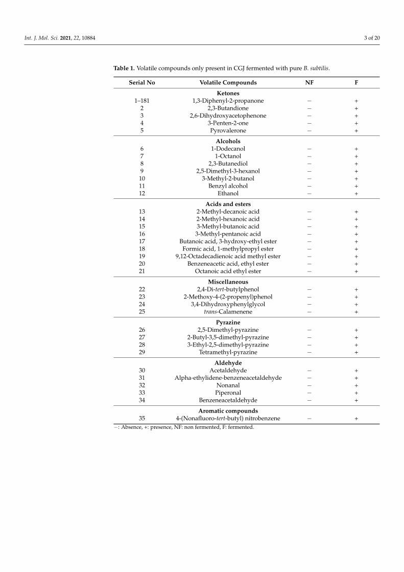
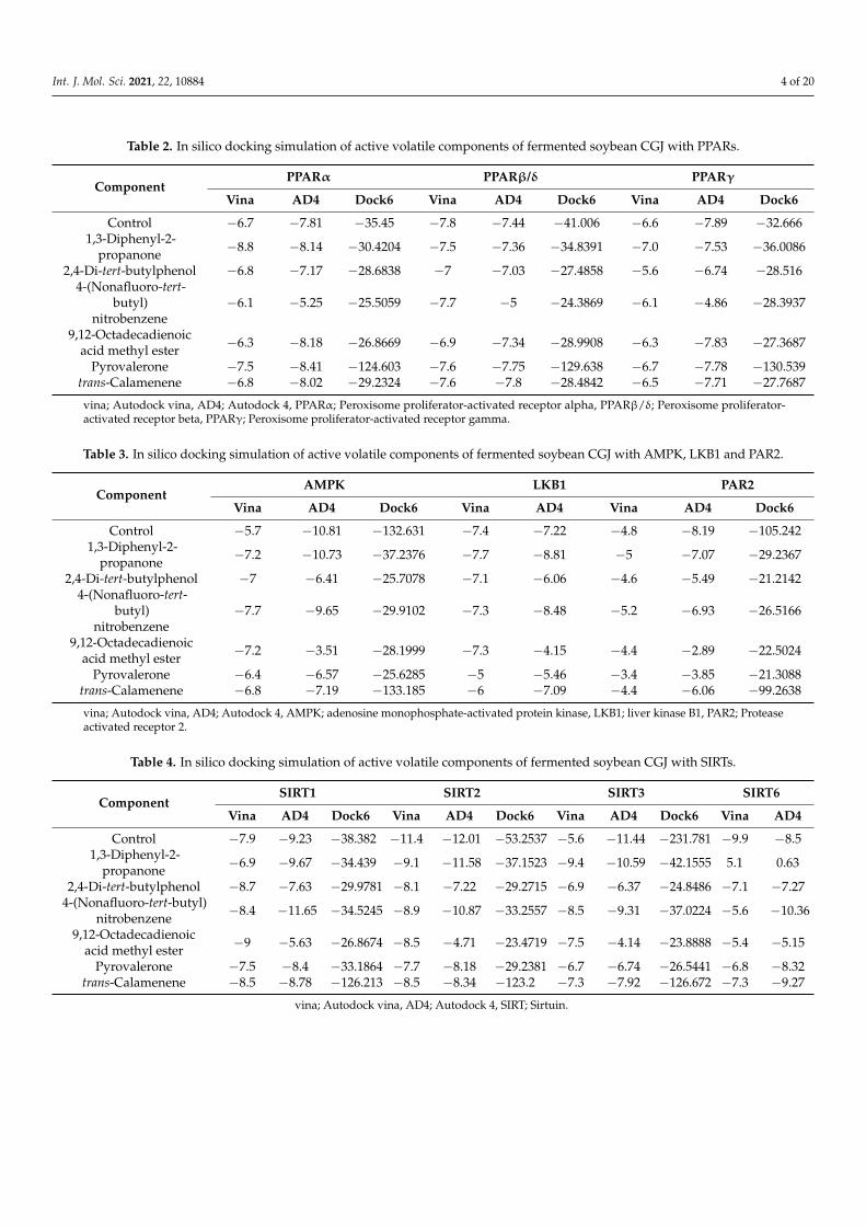
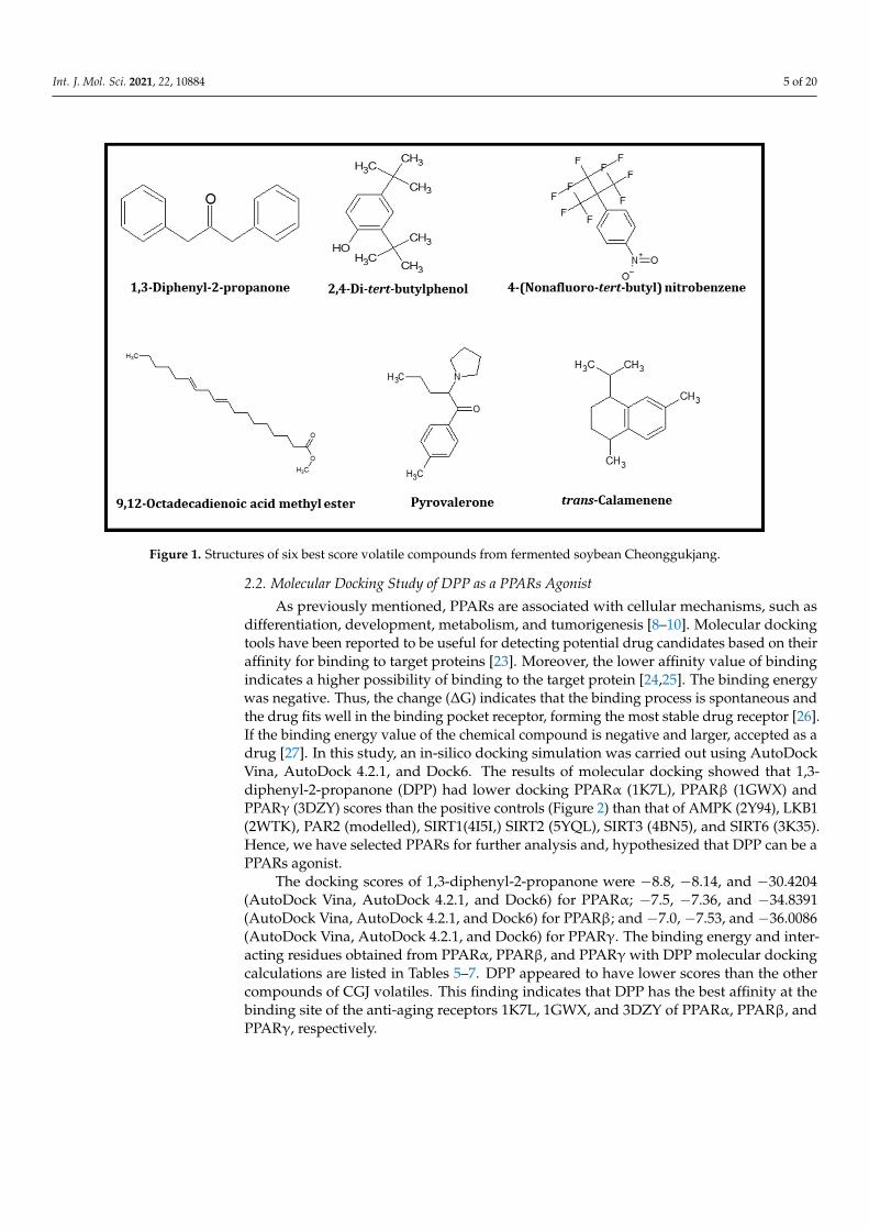
Chukeatirote et al. (2017) [22] identified 67 volatile compounds from non-fermentedand fermented soybean products cultured with B. subtilis. Among them, 35 volatiles wereidentified only in fermented soybean products, including seven alcohols, five aldehydes,one aromatic, five ketones, nine acids and esters, four pyrazines, and four miscellaneouscompounds (Table 1). Thirty-five compounds were selected for further analysis and screen-ing by conducting in vitro and in silico studies. Molecular docking studies of 35 volatileswere performed to investigate their binding status to the active site of 10 distinctive pro-teins, namely PPARα (1K7L), PPARβ (1GWX), PPARγ (3DZY), AMPK(2Y94), LKB1(2WTK),PAR2 (modelled), SIRT1(4I5I,) SIRT2(5YQL), SIRT3 (4BN5), and SIRT6 (3K35). A total of35 compounds were tested for distinctive proteins, while six compounds with a lowerdocking score (Tables 2–4), and high-affinity binding were found. The six compounds were1,3-diphenyl-2-propanone, 2.4-di-tert-butylphenol, 4-(nonafluoro-tert-butyl)-nitrobenzene,9,12-octadecadienoic acid methyl ester, pyrovalerone, and trans-calamenene. The chemicalstructures of these six volatiles are shown in Figure 1.
Int. J. Mol. Sci. 2021, 22, 10884 3 of 20
Table 1. Volatile compounds only present in CGJ fermented with pure B. subtilis.
Serial No Volatile Compounds NF F
Ketones1–181 1,3-Diphenyl-2-propanone − +
2 2,3-Butandione − +3 2,6-Dihydroxyacetophenone − +4 3-Penten-2-one − +5 Pyrovalerone − +
Alcohols6 1-Dodecanol − +7 1-Octanol − +8 2,3-Butanediol − +9 2,5-Dimethyl-3-hexanol − +
10 3-Methyl-2-butanol − +11 Benzyl alcohol − +12 Ethanol − +
Acids and esters13 2-Methyl-decanoic acid − +14 2-Methyl-hexanoic acid − +15 3-Methyl-butanoic acid − +16 3-Methyl-pentanoic acid − +17 Butanoic acid, 3-hydroxy-ethyl ester − +18 Formic acid, 1-methylpropyl ester − +19 9,12-Octadecadienoic acid methyl ester − +20 Benzeneacetic acid, ethyl ester − +21 Octanoic acid ethyl ester − +
Miscellaneous22 2,4-Di-tert-butylphenol − +23 2-Methoxy-4-(2-propenyl)phenol − +24 3,4-Dihydroxyphenylglycol − +25 trans-Calamenene − +
Pyrazine26 2,5-Dimethyl-pyrazine − +27 2-Butyl-3,5-dimethyl-pyrazine − +28 3-Ethyl-2,5-dimethyl-pyrazine − +29 Tetramethyl-pyrazine − +
Aldehyde30 Acetaldehyde − +31 Alpha-ethylidene-benzeneacetaldehyde − +32 Nonanal − +33 Piperonal − +34 Benzeneacetaldehyde − +
Aromatic compounds35 4-(Nonafluoro-tert-butyl) nitrobenzene − +
−: Absence, +: presence, NF: non fermented, F: fermented.
Int. J. Mol. Sci. 2021, 22, 10884 4 of 20
Table 2. In silico docking simulation of active volatile components of fermented soybean CGJ with PPARs.
ComponentPPARα PPARβ/δ PPARγ
Vina AD4 Dock6 Vina AD4 Dock6 Vina AD4 Dock6
Control −6.7 −7.81 −35.45 −7.8 −7.44 −41.006 −6.6 −7.89 −32.6661,3-Diphenyl-2-
propanone −8.8 −8.14 −30.4204 −7.5 −7.36 −34.8391 −7.0 −7.53 −36.0086
2,4-Di-tert-butylphenol −6.8 −7.17 −28.6838 −7 −7.03 −27.4858 −5.6 −6.74 −28.5164-(Nonafluoro-tert-
butyl)nitrobenzene
−6.1 −5.25 −25.5059 −7.7 −5 −24.3869 −6.1 −4.86 −28.3937
9,12-Octadecadienoicacid methyl ester −6.3 −8.18 −26.8669 −6.9 −7.34 −28.9908 −6.3 −7.83 −27.3687
Pyrovalerone −7.5 −8.41 −124.603 −7.6 −7.75 −129.638 −6.7 −7.78 −130.539trans-Calamenene −6.8 −8.02 −29.2324 −7.6 −7.8 −28.4842 −6.5 −7.71 −27.7687
vina; Autodock vina, AD4; Autodock 4, PPARα; Peroxisome proliferator-activated receptor alpha, PPARβ/δ; Peroxisome proliferator-activated receptor beta, PPARγ; Peroxisome proliferator-activated receptor gamma.
Table 3. In silico docking simulation of active volatile components of fermented soybean CGJ with AMPK, LKB1 and PAR2.
ComponentAMPK LKB1 PAR2
Vina AD4 Dock6 Vina AD4 Vina AD4 Dock6
Control −5.7 −10.81 −132.631 −7.4 −7.22 −4.8 −8.19 −105.2421,3-Diphenyl-2-
propanone −7.2 −10.73 −37.2376 −7.7 −8.81 −5 −7.07 −29.2367
2,4-Di-tert-butylphenol −7 −6.41 −25.7078 −7.1 −6.06 −4.6 −5.49 −21.21424-(Nonafluoro-tert-
butyl)nitrobenzene
−7.7 −9.65 −29.9102 −7.3 −8.48 −5.2 −6.93 −26.5166
9,12-Octadecadienoicacid methyl ester −7.2 −3.51 −28.1999 −7.3 −4.15 −4.4 −2.89 −22.5024
Pyrovalerone −6.4 −6.57 −25.6285 −5 −5.46 −3.4 −3.85 −21.3088trans-Calamenene −6.8 −7.19 −133.185 −6 −7.09 −4.4 −6.06 −99.2638
vina; Autodock vina, AD4; Autodock 4, AMPK; adenosine monophosphate-activated protein kinase, LKB1; liver kinase B1, PAR2; Proteaseactivated receptor 2.
Table 4. In silico docking simulation of active volatile components of fermented soybean CGJ with SIRTs.
ComponentSIRT1 SIRT2 SIRT3 SIRT6
Vina AD4 Dock6 Vina AD4 Dock6 Vina AD4 Dock6 Vina AD4
Control −7.9 −9.23 −38.382 −11.4 −12.01 −53.2537 −5.6 −11.44 −231.781 −9.9 −8.51,3-Diphenyl-2-
propanone −6.9 −9.67 −34.439 −9.1 −11.58 −37.1523 −9.4 −10.59 −42.1555 5.1 0.63
2,4-Di-tert-butylphenol −8.7 −7.63 −29.9781 −8.1 −7.22 −29.2715 −6.9 −6.37 −24.8486 −7.1 −7.274-(Nonafluoro-tert-butyl)
nitrobenzene −8.4 −11.65 −34.5245 −8.9 −10.87 −33.2557 −8.5 −9.31 −37.0224 −5.6 −10.36
9,12-Octadecadienoicacid methyl ester −9 −5.63 −26.8674 −8.5 −4.71 −23.4719 −7.5 −4.14 −23.8888 −5.4 −5.15
Pyrovalerone −7.5 −8.4 −33.1864 −7.7 −8.18 −29.2381 −6.7 −6.74 −26.5441 −6.8 −8.32trans-Calamenene −8.5 −8.78 −126.213 −8.5 −8.34 −123.2 −7.3 −7.92 −126.672 −7.3 −9.27
vina; Autodock vina, AD4; Autodock 4, SIRT; Sirtuin.
Int. J. Mol. Sci. 2021, 22, 10884 5 of 20Int. J. Mol. Sci. 2021, 22, x FOR PEER REVIEW 5 of 21
Figure 1. Structures of six best score volatile compounds from fermented soybean Cheonggukjang.
2.2. Molecular Docking Study of DPP as a PPARs Agonist As previously mentioned, PPARs are associated with cellular mechanisms, such as
differentiation, development, metabolism, and tumorigenesis [8–10]. Molecular docking tools have been reported to be useful for detecting potential drug candidates based on their affinity for binding to target proteins [23]. Moreover, the lower affinity value of bind-ing indicates a higher possibility of binding to the target protein [24,25]. The binding en-ergy was negative. Thus, the change (ΔG) indicates that the binding process is spontane-ous and the drug fits well in the binding pocket receptor, forming the most stable drug receptor [26]. If the binding energy value of the chemical compound is negative and larger, accepted as a drug [27]. In this study, an in-silico docking simulation was carried out using AutoDock Vina, AutoDock 4.2.1, and Dock6. The results of molecular docking showed that 1,3-diphenyl-2-propanone (DPP) had lower docking PPARα (1K7L), PPARꞵ (1GWX) and PPARγ (3DZY) scores than the positive controls (Figure 2) than that of AMPK (2Y94), LKB1 (2WTK), PAR2 (modelled), SIRT1(4I5I,) SIRT2 (5YQL), SIRT3 (4BN5), and SIRT6 (3K35). Hence, we have selected PPARs for further analysis and, hypothesized that DPP can be a PPARs agonist.
Figure 1. Structures of six best score volatile compounds from fermented soybean Cheonggukjang.
2.2. Molecular Docking Study of DPP as a PPARs Agonist
As previously mentioned, PPARs are associated with cellular mechanisms, such asdifferentiation, development, metabolism, and tumorigenesis [8–10]. Molecular dockingtools have been reported to be useful for detecting potential drug candidates based on theiraffinity for binding to target proteins [23]. Moreover, the lower affinity value of bindingindicates a higher possibility of binding to the target protein [24,25]. The binding energywas negative. Thus, the change (∆G) indicates that the binding process is spontaneous andthe drug fits well in the binding pocket receptor, forming the most stable drug receptor [26].If the binding energy value of the chemical compound is negative and larger, accepted as adrug [27]. In this study, an in-silico docking simulation was carried out using AutoDockVina, AutoDock 4.2.1, and Dock6. The results of molecular docking showed that 1,3-diphenyl-2-propanone (DPP) had lower docking PPARα (1K7L), PPARβ (1GWX) andPPARγ (3DZY) scores than the positive controls (Figure 2) than that of AMPK (2Y94), LKB1(2WTK), PAR2 (modelled), SIRT1(4I5I,) SIRT2 (5YQL), SIRT3 (4BN5), and SIRT6 (3K35).Hence, we have selected PPARs for further analysis and, hypothesized that DPP can be aPPARs agonist.
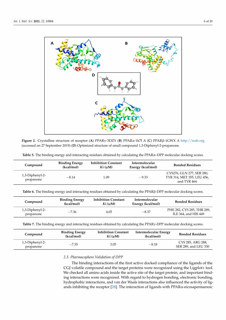
The docking scores of 1,3-diphenyl-2-propanone were −8.8, −8.14, and −30.4204(AutoDock Vina, AutoDock 4.2.1, and Dock6) for PPARα; −7.5, −7.36, and −34.8391(AutoDock Vina, AutoDock 4.2.1, and Dock6) for PPARβ; and −7.0, −7.53, and −36.0086(AutoDock Vina, AutoDock 4.2.1, and Dock6) for PPARγ. The binding energy and inter-acting residues obtained from PPARα, PPARβ, and PPARγ with DPP molecular dockingcalculations are listed in Tables 5–7. DPP appeared to have lower scores than the othercompounds of CGJ volatiles. This finding indicates that DPP has the best affinity at thebinding site of the anti-aging receptors 1K7L, 1GWX, and 3DZY of PPARα, PPARβ, andPPARγ, respectively.
Int. J. Mol. Sci. 2021, 22, 10884 6 of 20Int. J. Mol. Sci. 2021, 22, x FOR PEER REVIEW 6 of 21
Figure 2. Crystalline structure of receptor (A) PPARγ-3DZY (B) PPARα-1k7l A (C) PPARβ-1GWX A http://rcsb.org (ac-cessed on 27 September 2019) (D) Optimized structure of small compound 1,3-Diphenyl-2-propanone.
The docking scores of 1,3-diphenyl-2-propanone were −8.8, −8.14, and −30.4204 (Au-toDock Vina, AutoDock 4.2.1, and Dock6) for PPARα; −7.5, −7.36, and −34.8391 (AutoDock Vina, AutoDock 4.2.1, and Dock6) for PPARβ; and −7.0, −7.53, and −36.0086 (AutoDock Vina, AutoDock 4.2.1, and Dock6) for PPARγ. The binding energy and interacting resi-dues obtained from PPARα, PPARβ, and PPARγ with DPP molecular docking calcula-tions are listed in Tables 5–7. DPP appeared to have lower scores than the other com-pounds of CGJ volatiles. This finding indicates that DPP has the best affinity at the binding site of the anti-aging receptors 1K7L, 1GWX, and 3DZY of PPARα, PPARβ, and PPARγ, respectively.
Table 5. The binding energy and interacting residues obtained by calculating the PPARα–DPP molecular docking scores.
Compound Binding Energy (kcal/mol)
Inhibition Constant Ki (μM)
Intermolecular En-ergy (kcal/mol)
Bonded Residues
1,3-Diphenyl-2-propanone −8.14 1.09 −9.33
CYS276, GLN 277, SER 280, TYR 314, MET 355, LEU
456, and TYR 464
Table 6. The binding energy and interacting residues obtained by calculating the PPARβ-DPP molecular docking scores.
Compound Binding Energy (kcal/mol)
Inhibition Constant Ki (μM)
Intermolecular En-ergy (kcal/mol)
Bonded Residues
1,3-Diphenyl-2-propanone
−7.36 4.05 −8.37 PHE 282, CYS 285, THR 289, ILE 364, and HIS 449
Table 7. The binding energy and interacting residues obtained by calculating the PPARγ-DPP molecular docking scores.
Compound Binding Energy
(kcal/mol) Inhibition Constant
Ki (μM) Intermolecular En-
ergy (kcal/mol) Bonded Residues
1,3-Diphenyl-2-propanone −7.35 3.05 −8.18
CYS 285, ARG 288, SER 289, and LEU 330
Figure 2. Crystalline structure of receptor (A) PPARγ-3DZY (B) PPARα-1k7l A (C) PPARβ-1GWX A http://rcsb.org(accessed on 27 September 2019) (D) Optimized structure of small compound 1,3-Diphenyl-2-propanone.
Table 5. The binding energy and interacting residues obtained by calculating the PPARα–DPP molecular docking scores.
Compound Binding Energy(kcal/mol)
Inhibition ConstantKi (µM)
IntermolecularEnergy (kcal/mol) Bonded Residues
1,3-Diphenyl-2-propanone −8.14 1.09 −9.33
CYS276, GLN 277, SER 280,TYR 314, MET 355, LEU 456,
and TYR 464
Table 6. The binding energy and interacting residues obtained by calculating the PPARβ-DPP molecular docking scores.
Compound Binding Energy(kcal/mol)
Inhibition ConstantKi (µM)
IntermolecularEnergy (kcal/mol) Bonded Residues
1,3-Diphenyl-2-propanone −7.36 4.05 −8.37 PHE 282, CYS 285, THR 289,
ILE 364, and HIS 449
Table 7. The binding energy and interacting residues obtained by calculating the PPARγ-DPP molecular docking scores.
Compound Binding Energy(kcal/mol)
Inhibition ConstantKi (µM)
Intermolecular Energy(kcal/mol) Bonded Residues
1,3-Diphenyl-2-propanone −7.35 3.05 −8.18 CYS 285, ARG 288,
SER 289, and LEU 330
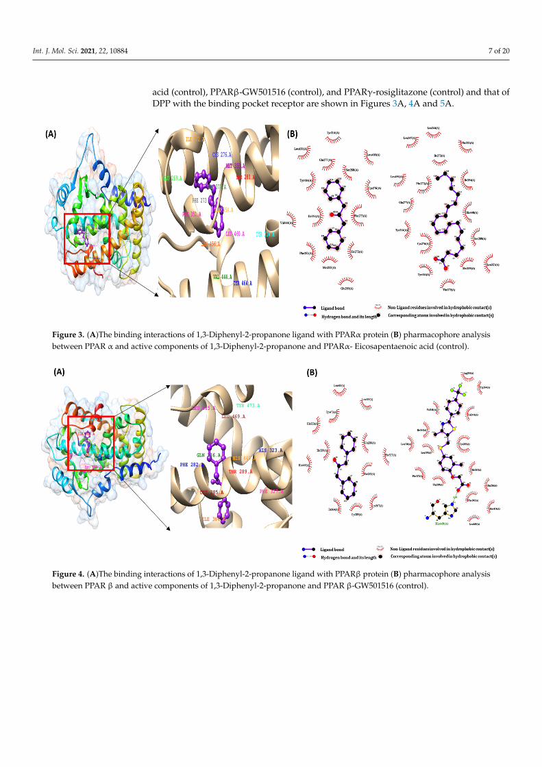
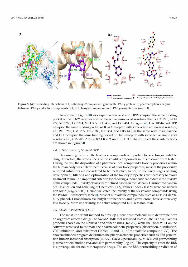
2.3. Pharmacophore Validation of DPP
The binding interactions of the first active docked compliance of the ligands of theCGJ volatile compound and the target proteins were recognized using the Ligplot+ tool.We checked all amino acids inside the active site of the target protein, and important bind-ing interactions were recognized. With regard to hydrogen bonding, electronic bonding,hydrophobic interactions, and van der Waals interactions also influenced the activity of lig-ands inhibiting the receptor [28]. The interaction of ligands with PPARα-eicosapentaenoic
Int. J. Mol. Sci. 2021, 22, 10884 7 of 20
acid (control), PPARβ-GW501516 (control), and PPARγ-rosiglitazone (control) and that ofDPP with the binding pocket receptor are shown in Figures 3A, 4A and 5A.
Int. J. Mol. Sci. 2021, 22, x FOR PEER REVIEW 7 of 21
1,3-Diphenyl-2-propanone −7.35 3.05 −8.18
CYS 285, ARG 288, SER 289, and LEU 330
2.3. Pharmacophore Validation of DPP The binding interactions of the first active docked compliance of the ligands of the
CGJ volatile compound and the target proteins were recognized using the Ligplot+ tool. We checked all amino acids inside the active site of the target protein, and important bind-ing interactions were recognized. With regard to hydrogen bonding, electronic bonding, hydrophobic interactions, and van der Waals interactions also influenced the activity of ligands inhibiting the receptor [28]. The interaction of ligands with PPARα-eicosapentae-noic acid (control), PPARβ-GW501516 (control), and PPARγ-rosiglitazone (control) and that of DPP with the binding pocket receptor are shown in Figures 3A, 4A and 5A.
Figure 3. (A)The binding interactions of 1,3-Diphenyl-2-propanone ligand with PPARα protein (B) pharmacophore anal-ysis between PPAR α and active components of 1,3-Diphenyl-2-propanone and PPARα- Eicosapentaenoic acid (control).
Figure 4. (A)The binding interactions of 1,3-Diphenyl-2-propanone ligand with PPARβ protein (B) pharmacophore anal-ysis between PPAR β and active components of 1,3-Diphenyl-2-propanone and PPAR β-GW501516 (control).
Figure 3. (A)The binding interactions of 1,3-Diphenyl-2-propanone ligand with PPARα protein (B) pharmacophore analysisbetween PPAR α and active components of 1,3-Diphenyl-2-propanone and PPARα- Eicosapentaenoic acid (control).
Int. J. Mol. Sci. 2021, 22, x FOR PEER REVIEW 7 of 21
1,3-Diphenyl-2-propanone −7.35 3.05 −8.18
CYS 285, ARG 288, SER 289, and LEU 330
2.3. Pharmacophore Validation of DPP The binding interactions of the first active docked compliance of the ligands of the
CGJ volatile compound and the target proteins were recognized using the Ligplot+ tool. We checked all amino acids inside the active site of the target protein, and important bind-ing interactions were recognized. With regard to hydrogen bonding, electronic bonding, hydrophobic interactions, and van der Waals interactions also influenced the activity of ligands inhibiting the receptor [28]. The interaction of ligands with PPARα-eicosapentae-noic acid (control), PPARβ-GW501516 (control), and PPARγ-rosiglitazone (control) and that of DPP with the binding pocket receptor are shown in Figures 3A, 4A and 5A.
Figure 3. (A)The binding interactions of 1,3-Diphenyl-2-propanone ligand with PPARα protein (B) pharmacophore anal-ysis between PPAR α and active components of 1,3-Diphenyl-2-propanone and PPARα- Eicosapentaenoic acid (control).
Figure 4. (A)The binding interactions of 1,3-Diphenyl-2-propanone ligand with PPARβ protein (B) pharmacophore anal-ysis between PPAR β and active components of 1,3-Diphenyl-2-propanone and PPAR β-GW501516 (control).
Figure 4. (A)The binding interactions of 1,3-Diphenyl-2-propanone ligand with PPARβ protein (B) pharmacophore analysisbetween PPAR β and active components of 1,3-Diphenyl-2-propanone and PPAR β-GW501516 (control).
Int. J. Mol. Sci. 2021, 22, 10884 8 of 20Int. J. Mol. Sci. 2021, 22, x FOR PEER REVIEW 8 of 21
Figure 5. (A)The binding interactions of 1,3–Diphenyl-2-propanone ligand with PPARγ protein (B) pharmacophore anal-ysis between PPARγ and active components of 1,3-Diphenyl-2-propanone and PPARγ-rosiglitazone (control).
As shown in Figure 3B, eicosapentaenoic acid and DPP occupied the same binding pocket of the 3DZY receptor with some active amino acid residues, that is, CYS276, GLN 277, SER 280, TYR 314, MET 355, LEU 456, and TYR 464. In Figure 4B, GW501516 and DPP occupied the same binding pocket of 1GWX receptor with some active amino acid resi-dues, i.e., PHE 282, CYS 285, THR 289, ILE 364, and HIS 449; in the same way, rosiglitazone and DPP occupied the same binding pocket of 1K7L receptor with some active amino acid residues, i.e., CYS 285, ARG 288, SER 289, and LEU 330. The results of these interactions are shown in Figure 5B.
2.4. In Silico Toxicity Study of DPP Determining the toxic effects of these compounds is important for selecting a candi-
date drug. Therefore, the toxic effects of the volatile compounds in this research were tested. During the test, the disposition of a pharmaceutical compound’s toxicity properties within the human body was determined. Because of poor toxic properties, most of the previously reported inhibitors are considered to be ineffective; hence, in the early stages of drug development, filtering and optimization of the toxicity properties are necessary to avoid treatment failure. An important criterion for choosing a therapeutic candidate is the toxicity of the compounds. Toxicity classes were defined based on the Globally Harmo-nized System of Classification and Labelling of Chemicals. LD50 values under Class VI were considered non-toxic (LD50 > 5000). Hence, we tested the toxicity of the six volatile compounds using the ProTox-II webserver (Table 8). Most of our volatile compounds, such as DPP, 2.4-di-tert-butylphenol, 4-(nonafluoro-tert-butyl) nitrobenzene, and pyro-valerone, have shown very low toxicity. More importantly, the active compound DPP was non-toxic.
Table 8. Toxicity prediction based on the docking scores of active volatile components of CGJ us-ing the ProTox-II tool.
Serial No Volatile Compounds Toxicity LD50 (mg/Kg) 1 1,3-Diphenyl-2-propanone 2000 2 2,4-Di-tert-butylphenol 700 3 4-(Nonafluoro-tert-butyl) nitrobenzene 1000 4 9,12-Octadecadienoic acid methyl ester 20,000
Figure 5. (A)The binding interactions of 1,3–Diphenyl-2-propanone ligand with PPARγ protein (B) pharmacophore analysisbetween PPARγ and active components of 1,3-Diphenyl-2-propanone and PPARγ-rosiglitazone (control).
As shown in Figure 3B, eicosapentaenoic acid and DPP occupied the same bindingpocket of the 3DZY receptor with some active amino acid residues, that is, CYS276, GLN277, SER 280, TYR 314, MET 355, LEU 456, and TYR 464. In Figure 4B, GW501516 and DPPoccupied the same binding pocket of 1GWX receptor with some active amino acid residues,i.e., PHE 282, CYS 285, THR 289, ILE 364, and HIS 449; in the same way, rosiglitazoneand DPP occupied the same binding pocket of 1K7L receptor with some active amino acidresidues, i.e., CYS 285, ARG 288, SER 289, and LEU 330. The results of these interactionsare shown in Figure 5B.
2.4. In Silico Toxicity Study of DPP
Determining the toxic effects of these compounds is important for selecting a candidatedrug. Therefore, the toxic effects of the volatile compounds in this research were tested.During the test, the disposition of a pharmaceutical compound’s toxicity properties withinthe human body was determined. Because of poor toxic properties, most of the previouslyreported inhibitors are considered to be ineffective; hence, in the early stages of drugdevelopment, filtering and optimization of the toxicity properties are necessary to avoidtreatment failure. An important criterion for choosing a therapeutic candidate is the toxicityof the compounds. Toxicity classes were defined based on the Globally Harmonized Systemof Classification and Labelling of Chemicals. LD50 values under Class VI were considerednon-toxic (LD50 > 5000). Hence, we tested the toxicity of the six volatile compounds usingthe ProTox-II webserver (Table 8). Most of our volatile compounds, such as DPP, 2.4-di-tert-butylphenol, 4-(nonafluoro-tert-butyl) nitrobenzene, and pyrovalerone, have shown verylow toxicity. More importantly, the active compound DPP was non-toxic.
2.5. ADMET Prediction of DPP
The most important method to develop a new drug molecule is to determine howan organism affects a drug. The SwissADME tool was used to calculate its drug-likenessproperties based on the Lipinski’s and Veber’s rules (Table 9), while the PreADMET onlinesoftware was used to estimate the pharmacokinetic properties (absorption, distribution,CYP inhibition, and substrate) (Tables 10 and 11) of the volatile compound CGJ. Theabovementioned program determines the pharmacokinetic properties such as BBB penetra-tion human intestinal absorption (HIA%), CaCo-2 permeability, MDCK cell permeability,plasma protein binding (%), and skin permeability (log kp). The capacity to enter the BBBis a prerequisite for neurotherapeutic drugs. The online BBB permeability prediction of
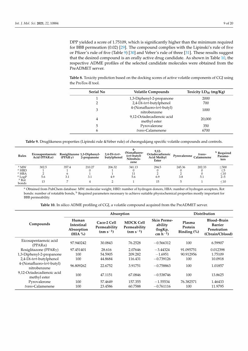
Int. J. Mol. Sci. 2021, 22, 10884 9 of 20
DPP yielded a score of 1.75109, which is significantly higher than the minimum requiredfor BBB permeation (0.02) [29]. The compound complies with the Lipinski’s rule of fiveor Pfizer’s rule of five (Table 9) [30] and Veber’s rule of three [31]. These results suggestthat the desired compound is an orally active drug candidate. As shown in Table 10, therespective ADME profiles of the selected candidate molecules were obtained from thePreADMET server.
Table 8. Toxicity prediction based on the docking scores of active volatile components of CGJ usingthe ProTox-II tool.
Serial No Volatile Compounds Toxicity LD50 (mg/Kg)
1 1,3-Diphenyl-2-propanone 20002 2,4-Di-tert-butylphenol 700
3 4-(Nonafluoro-tert-butyl)nitrobenzene 1000
4 9,12-Octadecadienoic acidmethyl ester 20,000
5 Pyrovalerone 3506 trans-Calamenene 6700
Table 9. Druglikeness properties (Lipinski rule &Veber rule) of cheongukjang specific volatile compounds and controls.
Rules EicosapentaenoicAcid (PPARα)
Rosiglitazone(PPARγ)
1,3-Diphenyl-2-propanone
2,4-Di-tert-butylphenol
4-(Nonafluoro-tert-butyl)Nitroben-
zene
9,12-Octadecadienoic
Acid MethylEster
Pyrovalerone trans-Calamenene
b RequiredParame-
ters
a MW 302.5 357.4 210.27 206.32 341.13 294.5 245.36 202.33 ≤500a HBD 1 1 0 1 0 0 0 0 ≤5a HBA 2 6 1 1 11 2 2 0 ≤10a LogP 5.6 3.1 3.1 4.9 5.6 6.9 3.8 5.1 2–5a Rotbonds 13 7 4 2 1 15 5 1 ≤10
a Obtained from PubChem database: MW: molecular weight, HBD: number of hydrogen donors, HBA: number of hydrogen acceptors, Rotbonds: number of rotatable bonds, b Required parameters necessary to achieve suitable physiochemical properties mostly important forBBB permeability.
Table 10. In silico ADME profiling of CGJ, a volatile compound acquired from the PreADMET server.
Compounds
Absorption Distribution
HumanIntestinal
Absorption(HIA %)
Caco-2 CellPermeability
(nm s−1)
MDCK CellPermeability
(nm s−1)
Skin Perme-ability(logKp,cm h−1)
PlasmaProtein
Binding (%)
Blood–BrainBarrier
Penetration(Cbrain/Cblood)
Eicosapentaenoic acid(PPARα) 97.940242 30.0843 76.2528 −0.566312 100 6.59907
Rosiglitazone (PPARγ) 97.451401 28.616 2.07646 −3.44324 91.095751 0.0123981,3-Diphenyl-2-propanone 100 54.5905 209.282 −1.6951 90.912956 1.75109
2,4-Di-tert-butylphenol 100 44.8684 116.431 −0.739126 100 10.09184-(Nonafluoro-tert-butyl)
nitrobenzene 96.809262 22.6752 3.91751 −0.758863 100 1.01857
9,12-Octadecadienoic acidmethyl ester 100 47.1151 67.0846 −0.538746 100 13.8625
Pyrovalerone 100 57.4649 157.355 −1.55534 76.382571 1.46433trans-Calamenene 100 23.4586 60.7588 −0.761116 100 11.9795
Int. J. Mol. Sci. 2021, 22, 10884 10 of 20
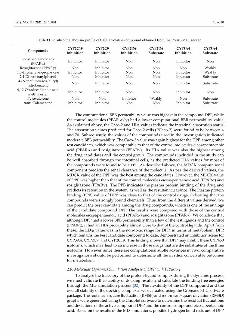
Table 11. In silico metabolism profile of CGJ, a volatile compound obtained from the PreADMET server.
Compounds CYP2C19Inhibition
CYP2C9Inhibition
CYP2D6Inhibition
CYP2D6Substrate
CYP3A4Inhibition
CYP3A4Substrate
Eicosapentaenoic acid(PPARα) Inhibitor Inhibitor Non Non Inhibitor Non
Rosiglitazone (PPARγ) Non Inhibitor Non Non Non Weakly1,3-Diphenyl-2-propanone Inhibitor Inhibitor Non Non Inhibitor Weakly
2,4-Di-tert-butylphenol Non Inhibitor Non Non Inhibitor Substrate4-(Nonafluoro-tert-butyl)
nitrobenzene Non Inhibitor Non Non Inhibitor Substrate
9,12-Octadecadienoic acidmethyl ester Inhibitor Inhibitor Non Non Inhibitor Non
Pyrovalerone Non Non Inhibitor Weakly Non Substratetrans-Calamenene Inhibitor Inhibitor Non Non Inhibitor Substrate
The computational BBB permeability value was highest in the compound DPP, whilethe control molecules (PPAR α/γ) had a lower computational BBB permeability value.As explained above, the Caco-2 and HIA values indicate the intestinal absorption status.The absorption values predicted for Caco-2 cells (PCaco-2) were found to be between 4and 70. Subsequently, the values of the compounds used in the investigation indicatedmoderate BBB permeability. The Caco-2 value was again highest for the DPP, among othertest candidates, which was comparable to that of the control molecules eicosapentaenoicacid (PPARα) and rosiglitazone (PPARγ). Its HIA value was also the highest amongthe drug candidates and the control group. The compounds included in the study canbe well absorbed through the intestinal cells, as the predicted HIA values for most ofthe compounds were found to be 100%. As described above, the MDCK computationalcomponent predicts the renal clearance of the molecule. As per the derived values, theMDCK value of the DPP was the best among the candidates. However, the MDCK valueof DPP was higher than that of the control molecules eicosapentaenoic acid (PPARα) androsiglitazone (PPARγ). The PPB indicates the plasma protein binding of the drug andpredicts its retention in the system, as well as the resultant clearance. The Plasma proteinbinding (PPB) value of DPP was close to that of the control drugs, indicating that thecompounds were strongly bound chemicals. Thus, from the different values derived, wecan predict the best candidate among the drug compounds, which is one of the analogsof the candidate compound DPP. The results were compared with those of the controlmolecules eicosapentaenoic acid (PPARα) and rosiglitazone (PPARγ). We conclude thatalthough DPP had a lower BBB permeability than a few of the test ligands and the control(PPARα), it had an HIA probability almost close to that of the control ligands. Apart fromthese, the LD50 value was in the non-toxic range for DPP; in terms of metabolism, DPP,which remains the best candidate compound to date, demonstrated an inhibition scene forCYP3A4, CYP2C9, and CYP2C19. This finding shows that DPP may inhibit these CYP450isoforms, which may lead to an increase in those drugs that are the substrates of the threeisoforms. However, since these are computational subtle advances in vivo, comparativeinvestigations should be performed to determine all the in silico conceivable outcomesfor metabolism.
2.6. Molecular Dynamics Simulation Analyses of DPP with PPARα/γ
To analyse the trajectory of the protein-ligand complex during the dynamic process,we must validate the stability of docking results and calculate the binding free energiesthrough the MD simulation process [32]. The flexibility of the DPP compound and theoverall stability of the docking complexes we evaluated using the Gromacs 5.1.2 softwarepackage. The root mean square fluctuation (RMSF) and root-mean-square deviation (RMSD)graphs were generated using the Gnuplot software to determine the residual fluctuationsand deviations of the active compound DPP and the control compound eicosapentaenoicacid. Based on the results of the MD simulations, possible hydrogen bond residues of DPP
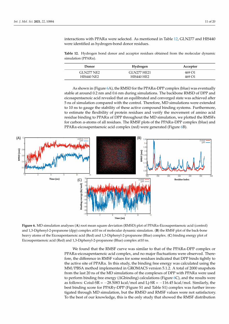
Int. J. Mol. Sci. 2021, 22, 10884 11 of 20
interactions with PPARα were selected. As mentioned in Table 12, GLN277 and HIS440were identified as hydrogen-bond donor residues.
Table 12. Hydrogen bond donor and acceptor residues obtained from the molecular dynamicsimulation (PPARα).
Donor Hydrogen Acceptor
GLN277 NE2 GLN277 HE21 469 O1HIS440 NE2 HIS440 HE2 469 O1
As shown in (Figure 6A), the RMSD for the PPARα-DPP complex (blue) was eventuallystable at around 0.2 nm and 0.6 nm during simulations. The backbone RMSD of DPP andeicosapentaenoic acid revealed that an equilibrated and converged state was achieved after5 ns of simulation compared with the control. Therefore, MD simulations were extendedto 10 ns to gauge the stability of these active compound binding systems. Furthermore,to estimate the flexibility of protein residues and verify the movement of amino acidresidue binding to PPARα of DPP throughout the MD simulation, we plotted the RMSFsfor carbon α-atoms of all residues. The RMSF plots of the PPARα-DPP complex (blue) andPPARα-eicosapentaenoic acid complex (red) were generated (Figure 6B).
Int. J. Mol. Sci. 2021, 22, x FOR PEER REVIEW 12 of 21
Figure 6. MD simulation analyses (A) root mean square deviation (RMSD) plot of PPARα-Eicosapentaenoic acid (control) and 1,3-Diphenyl-2-propanone (dpp) complex at10 ns of molecular dynamic simulation. (B) the RMSF plot of the back-bone heavy atoms of the Eicosapentaenoic acid (Red) and 1,3-Diphenyl-2-propanone (Blue) complex. (C) binding energy plot of Eicosapentaenoic acid (Red) and 1,3-Diphenyl-2-propanone (Blue) complex at10 ns.
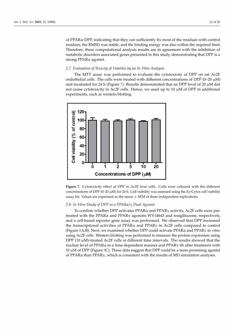
2.7. Evaluation of Toxicity of Volatiles by an In Vitro Analysis The MTT assay was performed to evaluate the cytotoxicity of DPP on rat Ac2F endo-
thelial cells. The cells were treated with different concentrations of DPP (0–20 µM) and incubated for 24 h (Figure 7). Results demonstrated that an DPP level of 20 µM did not cause cytotoxicity in Ac2F cells. Hence, we used up to 10 µM of DPP in additional exper-iments, such as western blotting.
Figure 7. Cytotoxicity effect of DPP in Ac2F liver cells. Cells were cultured with the different con-centrations of DPP (0–20 µM) for 24 h. Cell viability was assessed using the Ez-Cytox cell viability assay kit. Values are expressed as the mean ± SEM of three independent replications.
2.8. In Vitro Study of DPP as a PPARα/γ Dual Agonist To confirm whether DPP activates PPARα and PPARγ activity, Ac2F cells were pre-
treated with the PPARα and PPARγ agonists WY14643 and rosiglitazone, respectively, and a cell-based reporter gene assay was performed. We observed that DPP increased the transcriptional activities of PPARα and PPARγ in Ac2F cells compared to control (Figure 8A,B). Next, we examined whether DPP could activate PPARα and PPARγ in vitro using Ac2F cells. Western blotting was performed to measure the protein expression using DPP
Figure 6. MD simulation analyses (A) root mean square deviation (RMSD) plot of PPARα-Eicosapentaenoic acid (control)and 1,3-Diphenyl-2-propanone (dpp) complex at10 ns of molecular dynamic simulation. (B) the RMSF plot of the back-boneheavy atoms of the Eicosapentaenoic acid (Red) and 1,3-Diphenyl-2-propanone (Blue) complex. (C) binding energy plot ofEicosapentaenoic acid (Red) and 1,3-Diphenyl-2-propanone (Blue) complex at10 ns.
We found that the RMSF curve was similar to that of the PPARα-DPP complex orPPARα-eicosapentaenoic acid complex, and no major fluctuations were observed. There-fore, the difference in RMSF values for some residues indicated that DPP binds tightly tothe active site of PPARα. In this study, the binding free energy was calculated using theMM/PBSA method implemented in GROMACS version 5.1.2. A total of 2000 snapshotsfrom the last 20 ns of the MD simulations of the complexes of DPP with PPARα were usedto perform binding free energy (∆Gbinding) calculations (Figure 6C), and the results wereas follows: Coiul-SR = −28.5083 kcal/mol and Lj-SR = −116.45 kcal/mol. Similarly, thebest binding score for PPARγ-DPP (Figure S1 and Table S1) complex was further inves-tigated through MD simulation, but the RMSD and RMSF values were not satisfactory.To the best of our knowledge, this is the only study that showed the RMSF distribution
Int. J. Mol. Sci. 2021, 22, 10884 12 of 20
of PPARα-DPP, indicating that they can sufficiently fix most of the residues with controlresidues; the RMSD was stable, and the binding energy was also within the required limit.Therefore, these computational analysis results are in agreement with the inhibition ofmetabolic disorders associated genes presented in this study, demonstrating that DPP is astrong PPARα agonist.
2.7. Evaluation of Toxicity of Volatiles by an In Vitro Analysis
The MTT assay was performed to evaluate the cytotoxicity of DPP on rat Ac2Fendothelial cells. The cells were treated with different concentrations of DPP (0–20 µM)and incubated for 24 h (Figure 7). Results demonstrated that an DPP level of 20 µM didnot cause cytotoxicity in Ac2F cells. Hence, we used up to 10 µM of DPP in additionalexperiments, such as western blotting.
Int. J. Mol. Sci. 2021, 22, x FOR PEER REVIEW 12 of 21
Figure 6. MD simulation analyses (A) root mean square deviation (RMSD) plot of PPARα-Eicosapentaenoic acid (control) and 1,3-Diphenyl-2-propanone (dpp) complex at10 ns of molecular dynamic simulation. (B) the RMSF plot of the back-bone heavy atoms of the Eicosapentaenoic acid (Red) and 1,3-Diphenyl-2-propanone (Blue) complex. (C) binding energy plot of Eicosapentaenoic acid (Red) and 1,3-Diphenyl-2-propanone (Blue) complex at10 ns.
2.7. Evaluation of Toxicity of Volatiles by an In Vitro Analysis The MTT assay was performed to evaluate the cytotoxicity of DPP on rat Ac2F endo-
thelial cells. The cells were treated with different concentrations of DPP (0–20 µM) and incubated for 24 h (Figure 7). Results demonstrated that an DPP level of 20 µM did not cause cytotoxicity in Ac2F cells. Hence, we used up to 10 µM of DPP in additional exper-iments, such as western blotting.
Figure 7. Cytotoxicity effect of DPP in Ac2F liver cells. Cells were cultured with the different con-centrations of DPP (0–20 µM) for 24 h. Cell viability was assessed using the Ez-Cytox cell viability assay kit. Values are expressed as the mean ± SEM of three independent replications.
2.8. In Vitro Study of DPP as a PPARα/γ Dual Agonist To confirm whether DPP activates PPARα and PPARγ activity, Ac2F cells were pre-
treated with the PPARα and PPARγ agonists WY14643 and rosiglitazone, respectively, and a cell-based reporter gene assay was performed. We observed that DPP increased the transcriptional activities of PPARα and PPARγ in Ac2F cells compared to control (Figure 8A,B). Next, we examined whether DPP could activate PPARα and PPARγ in vitro using Ac2F cells. Western blotting was performed to measure the protein expression using DPP
Figure 7. Cytotoxicity effect of DPP in Ac2F liver cells. Cells were cultured with the differentconcentrations of DPP (0–20 µM) for 24 h. Cell viability was assessed using the Ez-Cytox cell viabilityassay kit. Values are expressed as the mean ± SEM of three independent replications.
2.8. In Vitro Study of DPP as a PPARα/γ Dual Agonist
To confirm whether DPP activates PPARα and PPARγ activity, Ac2F cells were pre-treated with the PPARα and PPARγ agonists WY14643 and rosiglitazone, respectively,and a cell-based reporter gene assay was performed. We observed that DPP increasedthe transcriptional activities of PPARα and PPARγ in Ac2F cells compared to control(Figure 8A,B). Next, we examined whether DPP could activate PPARα and PPARγ in vitrousing Ac2F cells. Western blotting was performed to measure the protein expression usingDPP (10 µM)-treated Ac2F cells at different time intervals. The results showed that thenuclear level of PPARα in a time-dependent manner and PPARγ 6h after treatment with10 µM of DPP (Figure 8C). These data suggest that DPP could be a more promising agonistof PPARα than PPARγ, which is consistent with the results of MD simulation analyses.
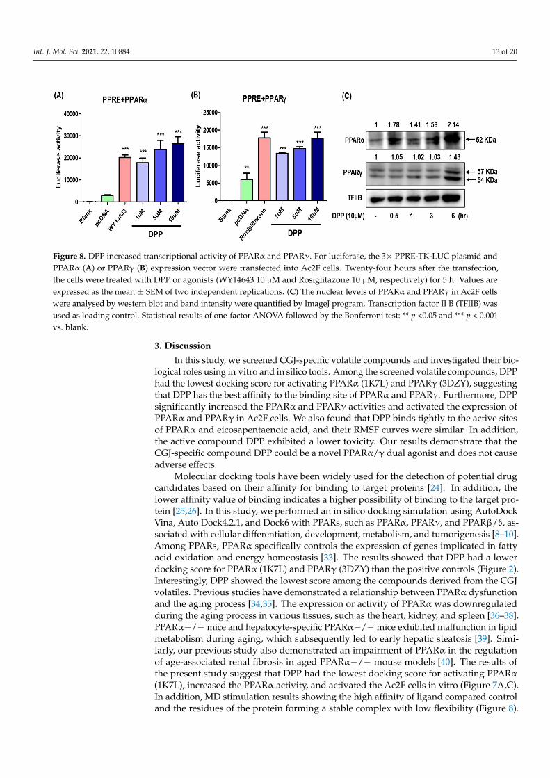
Int. J. Mol. Sci. 2021, 22, 10884 13 of 20
Int. J. Mol. Sci. 2021, 22, x FOR PEER REVIEW 13 of 21
(10 µM)-treated Ac2F cells at different time intervals. The results showed that the nuclear level of PPARα in a time-dependent manner and PPARγ 6h after treatment with 10 µM of DPP (Figure 8C). These data suggest that DPP could be a more promising agonist of PPARα than PPARγ, which is consistent with the results of MD simulation analyses.
Figure 8. DPP increased transcriptional activity of PPARα and PPARγ. For luciferase, the 3× PPRE-TK-LUC plasmid and PPARα (A) or PPARγ (B) expression vector were transfected into Ac2F cells. Twenty-four hours after the transfection, the cells were treated with DPP or agonists (WY14643 10 µM and Rosiglitazone 10 µM, respectively) for 5 h. Values are ex-pressed as the mean ± SEM of two independent replications. (C) The nuclear levels of PPARα and PPARγ in Ac2F cells were analysed by western blot and band intensity were quantified by ImageJ program. Transcription factor II B (TFIIB) was used as loading control. Statistical results of one-factor ANOVA followed by the Bonferroni test: ** p <0.05 and ***p < 0.001 vs. blank.
3. Discussion In this study, we screened CGJ-specific volatile compounds and investigated their
biological roles using in vitro and in silico tools. Among the screened volatile compounds, DPP had the lowest docking score for activating PPARα (1K7L) and PPARγ (3DZY), sug-gesting that DPP has the best affinity to the binding site of PPARα and PPARγ. Further-more, DPP significantly increased the PPARα and PPARγ activities and activated the ex-pression of PPARα and PPARγ in Ac2F cells. We also found that DPP binds tightly to the active sites of PPARα and eicosapentaenoic acid, and their RMSF curves were similar. In addition, the active compound DPP exhibited a lower toxicity. Our results demonstrate that the CGJ-specific compound DPP could be a novel PPARα/γ dual agonist and does not cause adverse effects.
Molecular docking tools have been widely used for the detection of potential drug candidates based on their affinity for binding to target proteins [24]. In addition, the lower affinity value of binding indicates a higher possibility of binding to the target protein [25,26]. In this study, we performed an in silico docking simulation using AutoDock Vina, Auto Dock4.2.1, and Dock6 with PPARs, such as PPARα, PPARγ, and PPARβ/δ, associ-ated with cellular differentiation, development, metabolism, and tumorigenesis [8–10]. Among PPARs, PPARα specifically controls the expression of genes implicated in fatty acid oxidation and energy homeostasis [33]. The results showed that DPP had a lower docking score for PPARα (1K7L) and PPARγ (3DZY) than the positive controls (Figure 2). Interestingly, DPP showed the lowest score among the compounds derived from the CGJ volatiles. Previous studies have demonstrated a relationship between PPARα dysfunction and the aging process [34,35]. The expression or activity of PPARα was downregulated during the aging process in various tissues, such as the heart, kidney, and spleen [36–38]. PPARα−/− mice and hepatocyte-specific PPARα−/− mice exhibited malfunction in lipid metabolism during aging, which subsequently led to early hepatic steatosis [39]. Similarly, our previous study also demonstrated an impairment of PPARα in the regulation of age-
Figure 8. DPP increased transcriptional activity of PPARα and PPARγ. For luciferase, the 3× PPRE-TK-LUC plasmid andPPARα (A) or PPARγ (B) expression vector were transfected into Ac2F cells. Twenty-four hours after the transfection,the cells were treated with DPP or agonists (WY14643 10 µM and Rosiglitazone 10 µM, respectively) for 5 h. Values areexpressed as the mean ± SEM of two independent replications. (C) The nuclear levels of PPARα and PPARγ in Ac2F cellswere analysed by western blot and band intensity were quantified by ImageJ program. Transcription factor II B (TFIIB) wasused as loading control. Statistical results of one-factor ANOVA followed by the Bonferroni test: ** p <0.05 and *** p < 0.001vs. blank.
3. Discussion
In this study, we screened CGJ-specific volatile compounds and investigated their bio-logical roles using in vitro and in silico tools. Among the screened volatile compounds, DPPhad the lowest docking score for activating PPARα (1K7L) and PPARγ (3DZY), suggestingthat DPP has the best affinity to the binding site of PPARα and PPARγ. Furthermore, DPPsignificantly increased the PPARα and PPARγ activities and activated the expression ofPPARα and PPARγ in Ac2F cells. We also found that DPP binds tightly to the active sitesof PPARα and eicosapentaenoic acid, and their RMSF curves were similar. In addition,the active compound DPP exhibited a lower toxicity. Our results demonstrate that theCGJ-specific compound DPP could be a novel PPARα/γ dual agonist and does not causeadverse effects.
Molecular docking tools have been widely used for the detection of potential drugcandidates based on their affinity for binding to target proteins [24]. In addition, thelower affinity value of binding indicates a higher possibility of binding to the target pro-tein [25,26]. In this study, we performed an in silico docking simulation using AutoDockVina, Auto Dock4.2.1, and Dock6 with PPARs, such as PPARα, PPARγ, and PPARβ/δ, as-sociated with cellular differentiation, development, metabolism, and tumorigenesis [8–10].Among PPARs, PPARα specifically controls the expression of genes implicated in fattyacid oxidation and energy homeostasis [33]. The results showed that DPP had a lowerdocking score for PPARα (1K7L) and PPARγ (3DZY) than the positive controls (Figure 2).Interestingly, DPP showed the lowest score among the compounds derived from the CGJvolatiles. Previous studies have demonstrated a relationship between PPARα dysfunctionand the aging process [34,35]. The expression or activity of PPARα was downregulatedduring the aging process in various tissues, such as the heart, kidney, and spleen [36–38].PPARα−/− mice and hepatocyte-specific PPARα−/− mice exhibited malfunction in lipidmetabolism during aging, which subsequently led to early hepatic steatosis [39]. Simi-larly, our previous study also demonstrated an impairment of PPARα in the regulationof age-associated renal fibrosis in aged PPARα−/− mouse models [40]. The results ofthe present study suggest that DPP had the lowest docking score for activating PPARα(1K7L), increased the PPARα activity, and activated the Ac2F cells in vitro (Figure 7A,C).In addition, MD stimulation results showing the high affinity of ligand compared controland the residues of the protein forming a stable complex with low flexibility (Figure 8).
Int. J. Mol. Sci. 2021, 22, 10884 14 of 20
These findings indicate that DPP can activate PPARα, which is associated with alterationsin lipid metabolism in MetS and aging.
Another PPAR subtype, PPARγ, plays an important role in lipid metabolism, improv-ing insulin sensitivity, and augmenting glucose disposal in adipose tissues and skeletalmuscles [13]. In addition, PPARγ agonists, namely, pioglitazone and rosiglitazone, arethe currently approved drugs for the management of hyperglycaemia in patients withtype 2 diabetes mellitus [41]. In addition, many PPARγ agonists are effective in loweringtriglyceride levels in the plasma; regulating lipid accumulation in various tissues suchas the liver, heart, and skeletal muscle; and controlling insulin sensitivity [42,43]. Hence,we performed an in-silico docking simulation with PPARγ and examined whether DPPcan activate PPARγ in vitro using Ac2F cells. The docking results revealed that DPP hasthe best binding affinity for the anti-aging receptor 3DZY of PPARγ. Furthermore, thein silico physicochemical and pharmacokinetic properties indicate that lead compoundscan be good predictors of nontoxicity and have good absorption, penetration, and perme-ability abilities in the human body, thus indicating high potential for BBB permeability,HIA, PPB, and Caco-2 permeability. Moreover, in vitro results showed that DPP increasedPPARγ activity and triggered its expression in Ac2F cells compared to that in controlcells (Figure 8B,C). These data suggest that DPP could be a PPARγ agonist; however,results of the MD simulation analyses showed the lower affinity of this ligand when com-pared with that of the control. In addition, elafibranor acts as a dual PPARα/δ agonistand improves the cardio metabolic risk profiles of patients with NASH [19]. Furthermore,elafibranor is still under phase 3 trials and is being tested in patients with moderate tosevere symptoms. Our findings suggest that DPP could be a stronger agonist of PPARαthan PPARγ, whose dysregulation is associated with MetS.
4. Materials and Methods4.1. Tools
This study utilized a personalized computer with an Intel(R) Core (TM) i7 CPU,X86-64-bit, 6 GB RAM to illustrate the experiment. The software used were as follows:RCSB PDB (RRID: SCR_012820), NCBI PubChem (RRID:SCR_004284), AutoDock Vinav.1.1.2, (RRID:SCR_011958), AutoDock v.4.2.6, (RRID:SCR_012746), UCSF Dock v.6.7, UCSFChimera v.1.14 (RRID: SCR_004097), Ligplot+ v.2.2. (RRID: SCR_018249), PreADMET,ProTox-II, GROMACS v.5.1.2, (RRID:SCR_014565), and Gnuplot v.5.4.2 (RRID:SCR_008619).
4.2. Materials
Thirty-five three-dimensional (3D) structures of CGJ compounds were discovered inour investigation [22], and a crystalline form of receptor with the following PDB Ids: 1K7L(PPARα), 1GWX (PPARβ), 3DZY (PPARγ) MPK (2Y94), LKB1 (2WTK), PAR2 (modelled),SIRT1 (4I5I,) SIRT2 (5YQL), SIRT3 (4BN5), and SIRT6 (3K35) were taken for the experiment.
4.3. Protein and Ligand Preparation
Protein and ligand arrangements were performed utilizing the UCSF Chimera soft-ware version 1.13.1 (construct 41965). Protein targets with PDB IDs 1K7L.pdb (PPARα),1GWX.pdb (PPARβ), and 3DZY.pdb (PPARγ), 2Y94.pdb (AMPK), 2WTK.pdb (LKB1),PAR2.pdb (PAR2 modelled), 4I5I.pdb (SIRT1), 5YQL.pdb (SIRT2), 4BN5.pdb (SIRT3), and3K35.pdb (SIRT6) were downloaded from the RCSB database, a Protein Data Bank poweredby UCSF Chimera. Proteins and local ligands were separated and saved with the followingfilenames: protein.pdb. mol2, ligand.pdb, and ligand. mol2. Local ligands were diligentlyarranged, while new ligands (volatile compounds) were downloaded from the PubChemdatabase https://pubchem.ncbi.nlm.nih.gov/ (accessed on 27 September 2019) to prepareconformations utilizing the Marvin Sketch software version 17.1.30 and saved using thefilename ligand.pdb and ligand. mol2.
Int. J. Mol. Sci. 2021, 22, 10884 15 of 20
4.4. Docking Protocol Validation
The root mean square deviation (RMSD) values between the local ligands with PDBIDs were calculated with the affirmation of the docked ligand used for docking protocolvalidation. The docking protocol is considered to be remarkable and can be utilized for thefurther docking process.
4.5. Molecular Docking
The docking simulation was performed to examine the authoritative mode in the activesite of different proteins: PPARα: 1K7L [44], PPARβ: 1GWX [45], and PPARγ: 3DZY [46].The data regarding the experimental resolution of each protein could be consulted in theProtein Data Bank web site. The 3D structures of the compounds or ligands were subjectedto a geometrical optimization at ACD/ChemSketch/3D. All optimized conformations wereconfirmed to have the lowest potential energy.
4.6. AutoDock 4.2.6
Molecular docking for the set of optimized ligands was performed using the AutoDock program version 4.2.6 [47]. Auto Dock combines a quick energy assessment throughpre-calculated grids of affinity conceivable outcomes with medicinal chemistry research,thus allowing grouping of calculations to determine the appropriate binding positions fora ligand on a given macromolecule. This program solidifies the van der Waals attractionpotential, geometric collision, screened electrostatic potential, and Lazaridis–Karplus de-solvation energy into the score. Thus, all nuclear docking results shown in this study arethe global docking scores. Within the course of action of the proteins for docking recreation,the water atoms, cofactors, and particles were disallowed from each X-ray crystallographicstructure. The polar hydrogen particles of the proteins were included, the atomic chargeswere computed using the Gasteiger technique, and the nonpolar hydrogen particles werecombined. Finally, the chemical was treated as a rigid body. Atomic docking calculationswere performed inside the active site of each protein. Nuclear docking frequently requiresa user-defined docking space in which the conceivable ligand definitive conformationsare examined. A small search space can produce an insufficient number of conformations,although a generously expansive space may deliver various inconsequential interactionstances. Consequently, in a perfect world, limited docking look space is significant tothe success of ligand-protein coupling. The default box size can be calculated usingexperimentally resolved protein–ligand complex structures. To start with, an initial boxis constructed to enclose the ligand, and then the size of the box is increased in randomdirections to ensure that the minimum length in any dimension is at least 22.5 Å. Thegrid maps of interaction energy for various atom types with each macromolecule werecalculated by the auxiliary program Auto Grid choosing a grid box centered at: (−17, −14,−4) for 1K7L, and (−11, 19.5, 15) for 3DZY, with dimensions of 40 × 40 × 40 Å aroundthe active site, and a grid point spacing of 0.375 Å. All these conditions are sufficiently toinclude the most important residues of each enzyme. The docking searches for the bestorientations of the molecules to the active site of each protein were per formed using theLamarckian genetic algorithm (LGA) [48]. The LGA protocol applied a population size of2000 individuals, while 2,500,000 energy valuations were used for the 200 LGA runs. Theleading docking complex arrangements (postures) were analysed based on the potentialintermolecular interactions (ligand/enzyme), such as hydrogen bonding, hydrophobicinteractions, and cation–π, π–π stacking.
4.7. AutoDock Vina
Vina utilized the progressed angle optimization technique in the local optimizationstrategy. It is the improved version with more docking accuracy, including a new scor-ing function, and has efficient optimization [49]. The molecular docking studies wereperformed to predict and understand binding of macromolecules and small moleculesefficiently. The AutoDock apparatus was used to determine the polar hydrogen in the
Int. J. Mol. Sci. 2021, 22, 10884 16 of 20
protein structure. The grid box was utilized for the arrangement of the framework outline,and the size measure was set to 30 × 30 × 30 XYZ focuses. Encourage, an adaptation recordthat contained protein, ligand, and grid data, was arranged to allow docking investigation.AutoDock Vina provides nine authoritative modes for each ligand, and the poses arepositioned in agreement with the binding affinity. Compounds with the most favorableand most noteworthy binding affinity were chosen for further investigation.
4.8. Dock6
Hydrogen atom orientations were optimized using the sander module in AMBER16for a maximum of 100 cycles of minimization, with heavy restraints (1000.0 kcal mol–1 Å–2)on all non-hydrogen atoms [50]. DOCK6 required the MOL2 format of the protein, andligand coordinates were extricated and saved.
4.9. Docking Visualization
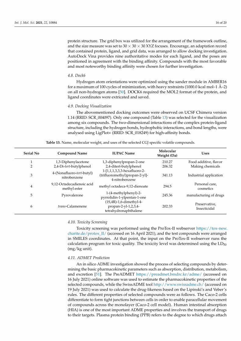
The abovementioned docking outcomes were observed on UCSF Chimera version1.14 (RRID: SCR_004097). Only one compound (Table 13) was selected for the visualizationamong six compounds. The two-dimensional interactions of the complex protein-ligandstructure, including the hydrogen bonds, hydrophobic interactions, and bond lengths, wereanalysed using LigPlot+ (RRID: SCR_018249) for high-affinity bonds.
Table 13. Name, molecular weight, and uses of the selected CGJ specific volatile compounds.
Serial No Compound Name IUPAC Name MolecularWeight (Da) Uses
1 1,3-Diphenylacetone 1,3-diphenylpropan-2-one 210.27 Food additive, flavor2 2,4-Di-tert-butylphenol 2,4-ditert-butylphenol 206.32 Making chemicals
3 4-(Nonafluoro-tert-butyl)nitrobenzene
1-[1,1,1,3,3,3-hexafluoro-2-(trifluoromethyl)propan-2-yl]-
4-nitrobenzene341.13 Industrial application
4 9,12-Octadecadienoic acidmethyl ester methyl octadeca-9,12-dienoate 294.5 Personal care,
cosmetics
5 Pyrovalerone 1-(4-methylphenyl)-2-pyrrolidin-1-ylpentan-1-one 245.36 manufacturing of drugs
6 trans-Calamenene(1S,4R)-1,6-dimethyl-4-
propan-2-yl-1,2,3,4-tetrahydronaphthalene
202.33 Preservative,Insecticidal
4.10. Toxicity Screening
Toxicity screening was performed using the ProTox-II webserver https://tox-new.charite.de/protox_II/ (accessed on 16 April 2021), and the test compounds were arrangedin SMILES coordinates. At that point, the input on the ProTox-II webserver runs thecalculation program for toxic quality. The toxicity level was determined using the LD50(mg/kg unit).
4.11. ADMET Prediction
An in silico ADME investigation showed the process of selecting compounds by deter-mining the basic pharmacokinetic parameters such as absorption, distribution, metabolism,and excretion [51]. The PreADMET https://preadmet.bmdrc.kr/adme/ (accessed on16 July 2021) online software was used to estimate the pharmacokinetic properties of theselected compounds, while the SwissADME tool http://www.swissadme.ch/ (accessed on19 July 2021) was used to calculate the drug-likeness based on the Lipinski’s and Veber’srules. The different properties of selected compounds were as follows. The Caco-2 cellsdifferentiate to form tight junctions between cells in order to enable paracellular movementof compounds across the monolayer (Caco-2 cell model). Human intestinal absorption(HIA) is one of the most important ADME properties and involves the transport of drugsto their targets. Plasma protein binding (PPB) refers to the degree to which drugs attach
Int. J. Mol. Sci. 2021, 22, 10884 17 of 20
to proteins within the blood. Moreover, the blood-brain barrier (BBB) prevents the brainuptake of most drugs. Meanwhile, an ADME property based on Madin-Darby CanineKidney (MDCK) cell line involves the intestinal drug absorption of small molecules and iscorrelated to human intestinal permeability (MDCK cell permeability); furthermore, skinpermeability was evaluated.
4.12. Molecular Dynamics Simulation
The Gromacs 5.1.2 program (RRID:SCR_014565) was utilized to perform moleculardynamic (MD) simulations of the PPARα-DPP or eicosapentaenoic corrosive (control) com-plex structures. PPARα atomic drive field parameters were written from CHARMM36 [52],which is an all-atom drive lipid force field, while the compound DPP or eicosapentaenoiccorrosive (control) atomic drive field parameters were obtained from the Gromos54a7force field using the Automated Topology Builder ATB, https://atb.uq.edu.au/index.py(accessed on 27 April 2021), which were then converted into the GROMACS file format.Initially, energy minimization was executed by employing a steep descent strategy of50,000 steps to achieve steady compliance. After minimization, the isobar isothermalensembles (NPT) and canonical ensembles (NVT) were applied. Individually, a consistentweight of 1 atm per 100 ps and a consistent temperature of 300 K for NPT and a consistenttemperature of 300 K for NVT were maintained over a period of 100 ps. The generationMD runs were performed for 10 ns, maintaining the temperature at 300 K and the weightat 1 bar. The RMSD, root mean square change (RMSF), and the distance between PPARαand DPP or eicosapentaenoic acid were calculated after the runs. Similarly, PPARγ andDPP were also calculated. These parameters were outlined using the Gnuplot program(RRID:SCR_008619).
4.13. Chemicals and Reagents
DPP and dimethyl sulfoxide (DMSO) were purchased from Sigma-Aldrich (St. Louis,MO, USA). Antibody against TFIIB (sc-271736) was purchased from Santa Cruz Biotech-nology (Santa Cruz, CA, USA). PPARγ (PA3-821A) and PPARα (ab24509) antibodies wereobtained from Thermo Fisher Scientific (Waltham, MA, USA) and Abcam Inc. (Cambridge,MA, USA), respectively.
4.14. Cell Culture and Cell Viability Assay
Rat liver (Ac2F) cells were obtained from the American Type Culture Collection(Rockville, MD, USA) and cultured in Dulbecco’s Modified Eagle Medium (Gyeongsan-si,Daegu, South Korea) containing 2 mM L-glutamine, 100 µg/mL streptomycin (HyClone,Logan, UT, USA), and 10% heat-inactivated fetal bovine serum (Gibco, Grand Island, NE,USA) at 37 ◦C in a humidified atmosphere containing 5% CO2. Cell viability was assessedusing the Ez-Cytox cell viability assay kit (Daeil Lab Service Co. Ltd, Seoul, Korea). Thecompounds were then broken down in DMSO.
4.15. Transfection and Luciferase Assay
For luciferase assays, 100 µL of Ac2F cells were seeded at a density of 5 × 10³ cells in a96-well plate. The cells were transfected with Lipofectamine transfection reagent (ThermoFisher Scientific, Rockford, IL, USA) and then transfected with the PPRE-X3-TK-LUC plas-mid (Dr. Christopher K. Glass, College of California, San Diego, CA, USA) and full-lengthhuman PPARα and PPARγ expression vectors (Dr. Han Geuk Seo, Konkuk College, Seoul,South Korea). After transfection for 24 h, the cells were treated with WY14643 (a PPARαagonist), rosiglitazone (a PPARγ agonist), or DPP for 5 h. Luciferase activity was mea-sured utilizing the ONE-Glo Luciferase Assay System (Promega, Madison, WI, USA) and aluminescence plate reader (Berthold Advances GmbH & Co., Bad Wildbad, Germany).

Int. J. Mol. Sci. 2021, 22, 10884 18 of 20
4.16. Western Blot Analysis
The Ac2F cells were incubated with DPP using the abovementioned concentrationsfor 24 h. Cell lysates were added; 30 µg of proteins was resolved by sodium dodecylsulfate-polyacrylamide gel electrophoresis (SDS-PAGE) using an 8–10% gel and transferredto a polyvinylidene fluoride (PVDF) membrane (Millipore, Burlington, MA, USA). Themembrane was blocked with 5% skim milk for 1 h at room temperature and incubatedovernight with primary antibody (1:1000 dilution), followed by incubation with horseradishperoxidase-conjugated secondary antibodies (Santa Cruz Biotechnology). Counter-actingagent labeling was identified using WesternBright peroxide solution (Advansta, CA, USA)and Davinci-Chemi CAS-400 (Davinch-K, Seoul, Korea), in accordance with the manufac-turers’ instructions.
4.17. Statistical Analysis
Statistical analysis was performed using GraphPad Prism 5 (GraphPad Software,La Jolla, CA, USA). Student’s t-test was used to determine the differences between the twogroups. Statistical significance was set at p < 0.05.
5. Conclusions
In conclusion, we demonstrated that DPP has a high binding affinity and can activatePPARα and PPARγ without causing toxicity. Concomitant with the in silico results, weconfirmed that DPP significantly increased the transcriptional activities of both PPARα andPPARγ and activated PPARα and PPARγ in Ac2F liver cells. To the best of our knowledge,this is the first in vitro and in silico study to report that DPP can act as a dual agonist ofPPARα/γ. The findings of this study suggest that the CGJ-specific compound DPP couldbe a novel PPARα/γ dual agonist; however, further studies are warranted to examine itstherapeutic potential in various metabolic disorders linked with PPARα/γ dysregulation.
Supplementary Materials: The supplementary materials are available online at https://www.mdpi.com/article/10.3390/ijms221910884/s1.
Author Contributions: H.-Y.C. and R.A. designed the study. R.A. conducted the computationalanalysis and wrote the manuscript. H.-J.J. performed the in vitro experiments and reviewed themanuscript. S.-G.N. and D.P. helped analyse the docking and MD simulation results and reviewed themanuscript. H.-Y.C. was involved in the conceptualization, supervision, project administration, andfunding acquisition. All authors have read and agreed to the published version of the manuscript.
Funding: This work was supported by a National Research Foundation of Korea (NRF) grant fundedby the Korean government (NRF-2018R1A2A3075425).
Institutional Review Board Statement: Not applicable.
Informed Consent Statement: Not applicable.
Conflicts of Interest: The authors declare no conflict of interest.
References1. Natarajan, S.; Luthria, D.; Bae, H.; Lakshman, D.; Mitra, A. Transgenic soybeans and soybean protein analysis: An overview.
J. Agric. Food Chem. 2013, 61, 11736–11743. [CrossRef] [PubMed]2. Cho, K.M.; Lim, H.-J.; Kim, M.-S.; Kim, D.S.; Hwang, C.E.; Nam, S.H.; Joo, O.S.; Lee, B.W.; Kim, J.K.; Shin, E.-C. Time course
effects of fermentation on fatty acid and volatile compound profiles of Cheonggukjang using new soybean cultivars. J. Food DrugAnal. 2016, 25, 637–653. [CrossRef]
3. Baek, J.G.; Shim, S.-M.; Kwon, D.Y.; Choi, H.-K.; Lee, C.H.; Kim, Y.-S. Metabolite profiling of Cheonggukjang, a fermented soybeanpaste, inoculated with various Bacillus strains during fermentation. Biosci. Biotechnol. Biochem. 2010, 74, 1860–1868. [CrossRef]
4. Cho, C.-w.; Han, C.-J.; Rhee, Y.K.; Lee, Y.-C.; Shin, K.-S.; Shin, J.-S.; Lee, K.-T.; Hong, H.-D. Cheonggukjang polysaccharidesenhance immune activities and prevent cyclophosphamide-induced immunosuppression. Int. J. Biol. Macromol. 2015, 72, 519–525.[CrossRef]
5. Bae, M.-J.; Shin, H.S.; See, H.-J.; Chai, O.H.; Shon, D.-H. Cheonggukjang ethanol extracts inhibit a murine allergic asthma via.suppression of mast cell-dependent anaphylactic reactions. J. Med. Food 2014, 17, 142–149. [CrossRef]
Int. J. Mol. Sci. 2021, 22, 10884 19 of 20
6. Kim, J.; Choi, J.N.; Choi, J.H.; Cha, Y.S.; Muthaiya, M.J.; Lee, C.H. Effect of fermented soybean product (Cheonggukjang) intake onmetabolic parameters in mice fed a high-fat diet. Mol. Nutr. Food Res. 2013, 57, 1886–1891. [CrossRef]
7. Go, J.; Kim, J.E.; Kwak, M.H.; Koh, E.K.; Song, S.H.; Sung, J.E.; Kim, D.S.; Hong, J.T.; Hwang, D.Y. Neuroprotective effects offermented soybean products (Cheonggukjang) manufactured by mixed culture of Bacillus subtilis MC31 and Lactobacillus sakei 383on trimethyltin-induced cognitive defects mice. Nutr. Neurosci. 2016, 19, 247–259. [CrossRef]
8. Belfiore, A.; Genua, M.; Malaguarnera, R. PPAR-gamma agonists and their effects on IGF-I receptor signaling: Implications forcancer. PPAR Res. 2009, 2009, 830501. [CrossRef]
9. Tenenbaum, A.; Fisman, E.Z.; Motro, M. Metabolic syndrome and type 2 diabetes mellitus: Focus on peroxisome proliferatoractivated receptors (PPAR). Cardiovasc. Diabetol. 2003, 2, 4. [CrossRef]
10. Kota, B.P.; Huang, T.H.-W.; Roufogalis, B.D. An overview on biological mechanisms of PPARs. Pharmacol. Res. 2005, 51, 85–94.[CrossRef]
11. Fruchart, J.-C.; Staels, B.; Duriez, P. The role of fibric acids in atherosclerosis. Curr. Atheroscler. Rep. 2001, 3, 83–92. [CrossRef]12. Desvergne, B.; Michalik, L.; Wahli, W. Be fit or be sick: Peroxisome proliferator-activated receptors are down the road. Mol.
Endocrinol. 2004, 18, 1321–1332. [CrossRef]13. Auwerx, J. PPARγ, the ultimate thrifty gene. Diabetologia 1999, 42, 1033–1049. [CrossRef]14. Burdick, A.D.; Kim, D.J.; Peraza, M.A.; Gonzalez, F.J.; Peters, J.M. The role of peroxisome proliferator-activated receptor-beta/delta
in epithelial cell growth and differentiation. Cell Signal 2006, 18, 9–20. [CrossRef]15. Al-Salman, J.; Arjomand, H.; Kemp, D.G.; Mittal, M. Hepatocellular injury in a patient receiving rosiglitazone. A case report. Ann.
Intern. Med. 2000, 132, 121–124. [CrossRef]16. Forman, L.M.; Simmons, D.A.; Diamond, R.H. Hepatic failure in a patient taking rosiglitazone. Ann. Intern. Med. 2000, 132,
118–121. [CrossRef]17. Cariou, B.; Charbonnel, B.; Staels, B. Thiazolidinediones and PPAR gamma agonists: Time for a reassessment. Trends Endocrinol.
Metab. 2012, 23, 205–215. [CrossRef]18. Bongartz, T.; Coras, B.; Vogt, T.; Scholmerich, J.; Muller-Ladner, U. Treatment of active psoriatic arthritis with the PPAR gamma
ligand pioglitazone: An open-label pilot study. Rheumatology 2005, 44, 126–129. [CrossRef]19. Ratziu, V.; Harrison, S.A.; Francque, S.; Bedossa, P.; Lehert, P.; Serfaty, L.; Romero-Gomez, M.; Boursier, J.; Abdelmalek, M.;
Caldwell, S.; et al. Elafibranor, an agonist of the peroxisome proliferator-activated receptor-alpha and -delta, induces resolutionof nonalcoholic steatohepatitis without fibrosis worsening. Gastroenterology 2016, 150, 1147–1159.e5. [CrossRef]
20. Staels, B.; Rubenstrunk, A.; Noel, B.; Rigou, G.; Delataille, P.; Millatt, L.J.; Baron, M.; Lucas, A.; Tailleux, A.; Hum, D.W.; et al.Hepatoprotective effects of the dual peroxisome proliferator-activated receptor alpha/delta agonist, GFT505, in rodent models ofnonalcoholic fatty liver disease/nonalcoholic steatohepatitis. Hepatology 2013, 58, 1941–1952. [CrossRef]
21. Tacke, F.; Weiskirchen, R. Non-alcoholic fatty liver disease (NAFLD)/non-alcoholic steatohepatitis (NASH)-related liver fibrosis:Mechanisms, treatment and prevention. Ann. Transl. Med. 2021, 9, 729. [CrossRef]
22. Chukeatirote, E.; Eungwanichayapant, P.; Kanghae, A. Determination of volatile components in fermented soybean prepared bya co-culture of Bacillus subtilis and Rhizopus oligosporus. Food Res. 2017, 1, 225–233. [CrossRef]
23. Kastritis, P.; Bonvin, A.M.J.J. On the binding affinity of macromolecular interactions: Daring to ask why proteins interact. J. R. Soc.Interface 2013, 10, 20120835. [CrossRef] [PubMed]
24. Baker, J.R.; Woolfson, D.N.; Muskett, F.W.; Stoneman, R.G.; Urbaniak, M.D.; Caddick, S. Protein-small molecule interactions inneocarzinostatin, the prototypical enediyne chromoprotein antibiotic. Chembiochem 2007, 8, 704–717. [CrossRef] [PubMed]
25. Tassa, C.; Duffner, J.L.; Lewis, T.; Weissleder, R.; Schreiber, S.L.; Koehler, A.N.; Shaw, S.Y. Binding affinity and kinetic analysis oftargeted small molecule-modified nanoparticles. Bioconjugate Chem. 2010, 21, 14–19. [CrossRef] [PubMed]
26. Kumar, K.S.; Rao, A.L.; Rao, M.B. Design, synthesis, biological evaluation and molecular docking studies of novel 3-substituted-5-[(indol-3-yl)methylene]-thiazolidine-2,4-dione derivatives. Heliyon 2018, 4, e00807. [CrossRef] [PubMed]
27. Balavignesh, V.; Srinivasan, E.; Ramesh Babu, N.G.; Saravanan, N. Molecular docking study ON NS5B polymerase of hepatitis cvirus by screening of volatile compounds from Acacia concinna and ADMET prediction. Int. J. Pharm. Life Sci. 2013, 4, 2548–2558.
28. Khalid, S.; Hanif, R.; Jabeen, I.; Mansoor, Q.; Ismail, M. Pharmacophore modeling for identification of anti-IGF-1R drugs andin-vitro validation of fulvestrant as a potential inhibitor. PLoS ONE 2018, 13, e0196312. [CrossRef] [PubMed]
29. Liu, H.; Wang, L.; Lv, M.; Pei, R.; Li, P.; Pei, Z.; Wang, Y.; Su, W.; Xie, X.-Q. AlzPlatform: An Alzheimer’s disease domain-specificchemogenomics knowledgebase for polypharmacology and target identification research. J. Chem. Inf. Model. 2014, 54, 1050–1060.[CrossRef]
30. Lipinski, C.A. Lead- and drug-like compounds: The rule-of-five revolution. Drug Discov. Today Technol. 2004, 1, 337–341.[CrossRef]
31. Veber, D.F.; Johnson, S.R.; Cheng, H.-Y.; Smith, B.R.; Ward, K.W.; Kopple, K.D. Molecular properties that influence the oralbioavailability of drug candidates. J. Med. Chem. 2002, 45, 2615–2623. [CrossRef] [PubMed]
32. Chun, Y.C.; Yih, H.; Shwu, J.L.; Hsuan, L.L. Discovery of Novel N-Glycoside and Non-Glycoside hSGLT2 Inhibitors for theTreatment of Type 2 Diabetes Mellitus. J. Diabetes Mellit. 2019, 9, 77–104. [CrossRef]
33. Van Raalte, D.H.; Li, M.; Pritchard, P.H.; Wasan, K.M. Peroxisome proliferator-activated receptor (PPAR)-α: A pharmacologicaltarget with a promising future. Pharm. Res. 2004, 21, 1531–1538. [CrossRef] [PubMed]
Int. J. Mol. Sci. 2021, 22, 10884 20 of 20
34. Chung, J.H.; Seo, A.Y.; Chung, S.W.; Kim, M.K.; Leeuwenburgh, C.; Yu, B.P.; Chung, H.Y. Molecular mechanism of PPAR in theregulation of age-related inflammation. Ageing Res. Rev. 2008, 7, 126–136. [CrossRef] [PubMed]
35. Howroyd, P.; Swanson, C.; Dunn, C.; Cattley, R.C.; Corton, J.C. Decreased longevity and enhancement of age-dependent lesionsin mice lacking the nuclear receptor peroxisome proliferator-activated receptor alpha (PPARalpha). Toxicol. Pathol. 2004, 32,591–599. [CrossRef] [PubMed]
36. Sung, B.; Park, S.; Yu, B.P.; Chung, H.Y. Modulation of PPAR in aging, inflammation, and calorie restriction. J. Gerontol. Ser. A Boil.Sci. Med. Sci. 2004, 59, B997–B1006. [CrossRef] [PubMed]
37. Iemitsu, M.; Miyauchi, T.; Maeda, S.; Tanabe, T.; Takanashi, M.; Irukayama-Tomobe, Y.; Sakai, S.; Ohmori, H.; Matsuda, M.;Yamaguchi, I. Aging-induced decrease in the PPAR-α level in hearts is improved by exercise training. Am. J. Physiol. Circ. Physiol.2002, 283, H1750–H1760. [CrossRef]
38. Sanguino, E.; Roglans, N.; Alegret, M.; Sanchez, R.M.; Vazquez-Carrera, M.; Laguna, J.C. Atorvastatin reverses age-relatedreduction in rat hepatic PPAR alpha and HNF-4. Br. J. Pharmacol. 2005, 145, 853–861. [CrossRef]
39. Montagner, A.; Polizzi, A.; Fouche, E.; Ducheix, S.; Lippi, Y.; Lasserre, F.; Barquissau, V.; Regnier, M.; Lukowicz, C.; Benhamed,F.; et al. Liver PPAR alpha is crucial for whole-body fatty acid homeostasis and is protective against NAFLD. Gut 2016, 65,1202–1214. [CrossRef]
40. Chung, K.W.; Lee, E.K.; Lee, M.K.; Oh, G.T.; Yu, B.P.; Chung, H.Y. Impairment of PPAR alpha and the fatty acid oxidation pathwayaggravates renal fibrosis during aging. J. Am. Soc. Nephrol. 2018, 29, 1223–1237. [CrossRef]
41. Chiarelli, F.; Di Marzio, D. Peroxisome proliferator-activated receptor-gamma agonists and diabetes: Current evidence and futureperspectives. Vasc. Health Risk Manag. 2008, 4, 297–304. [CrossRef]
42. Shiomi, Y.; Yamauchi, T.; Iwabu, M.; Okada-Iwabu, M.; Nakayama, R.; Orikawa, Y.; Yoshioka, Y.; Tanaka, K.; Ueki, K.; Kadowaki,T. A novel peroxisome proliferator-activated receptor (PPAR) alpha agonist and PPAR gamma antagonist, Z-551, ameliorateshigh-fat diet-induced obesity and metabolic disorders in mice. J. Biol. Chem. 2015, 290, 14567–14581. [CrossRef]
43. Khuchua, Z.; Glukhov, A.I.; Strauss, A.W.; Javadov, S. Elucidating the beneficial role of PPAR agonists in cardiac diseases. Int. J.Mol. Sci. 2018, 19, 3464. [CrossRef] [PubMed]
44. Xu, H.E.; Lambert, M.H.; Montana, V.G.; Plunket, K.D.; Moore, L.B.; Collins, J.L.; Oplinger, J.A.; Kliewer, S.A.; Gampe, R.T.;McKee, D.D.; et al. Structural determinants of ligand binding selectivity between the peroxisome proliferator-activated receptors.Proc. Natl. Acad. Sci. USA 2001, 98, 13919–13924. [CrossRef] [PubMed]
45. Xu, H.E.; Lambert, M.H.; Montana, V.G.; Parks, D.J.; Blanchard, S.G.; Brown, P.J.; Sternbach, D.D.; Lehmann, J.M.; Wisely, G.B.;Willson, T.M.; et al. Molecular recognition of fatty acids by peroxisome proliferator-activated receptors. Mol. Cell 1999, 3, 397–403.[CrossRef]
46. Chandra, V.; Huang, P.; Hamuro, Y.; Raghuram, S.; Wang, Y.; Burris, T.P.; Rastinejad, F. Structure of the intact PPAR-gamma-RXR-nuclear receptor complex on DNA. Nature 2008, 456, 350–356. [CrossRef] [PubMed]
47. Morris, G.M.; Huey, R.; Lindstrom, W.; Sanner, M.F.; Belew, R.K.; Goodsell, D.S.; Olson, A.J. AutoDock4 and AutoDockTools4:Automated docking with selective receptor flexibility. J. Comput. Chem. 2009, 30, 2785–2791. [CrossRef] [PubMed]
48. Fuhrmann, J.; Rurainski, A.; Lenhof, H.-P.; Neumann, D. A new lamarckian genetic algorithm for flexible ligand-receptor docking.J. Comput. Chem. 2010, 31, 1911–1918. [CrossRef]
49. Trott, O.; Olson, A.J. AutoDock vina: Improving the speed and accuracy of docking with a new scoring function, efficientoptimization, and multithreading. J. Comput. Chem. 2010, 31, 455–461. [CrossRef]
50. Kuntz, I.D.; Blaney, J.M.; Oatley, S.J.; Langridge, R.; Ferrin, T.E. A geometric approach to macromolecule-ligand interactions.J. Mol. Biol. 1982, 161, 269–288. [CrossRef]
51. Guan, L.; Yang, H.; Cai, Y.; Sun, L.; Di, P.; Li, W.; Liu, G.; Tang, Y. ADMET-score—a comprehensive scoring function for evaluationof chemical drug-likeness. Med. Chem. Commmun. 2019, 10, 148–157. [CrossRef] [PubMed]
52. Basith, S.; Manavalan, B.; Shin, T.H.; Lee, G. A molecular dynamics approach to explore the intramolecular signal transduction ofPPAR-α. Int. J. Mol. Sci. 2019, 20, 1666. [CrossRef] [PubMed]


![in silico (BFA thesis), 2002 [español]](https://static.fdokumen.com/doc/165x107/631f4913dbf756400702aca8/in-silico-bfa-thesis-2002-espanol.jpg)


