Non-Cannabinoid Constituents From a High Potency Cannabis Sativa Variety

Neurochemical changes in the striatum of dyskinetic ratsafteradministration of the cannabinoid agonist WIN55,212-2
M. G. Morgese1,2,*, T. Cassano1,*, S. Gaetani3, T. Macheda1, L. Laconca1,3, P.Dipasquale3, L. Ferraro4, T. Antonelli4, V. Cuomo3, and A. Giuffrida2
1 Department of Biomedical Sciences, University of Foggia, Italy
2 Department of Pharmacology, University of Texas Health Science Center, San Antonio, USA
3 Department of Physiology and Pharmacology, University “La Sapienza”, Rome, Italy
4 Department of Clinical and Experimental Medicine, University of Ferrara, Italy
AbstractChronic use of levodopa, the most effective treatment for Parkinson’s disease, causes abnormalinvoluntary movements named dyskinesias, which are linked to maladaptive changes in plasticityand disturbances of dopamine and glutamate neurotransmission in the basal ganglia. Dyskinesias canbe modeled in rats with unilateral 6-hydroxydopamine lesions by repeated administration of lowdoses of levodopa (6 mg/kg, s.c.). Previous studies from our lab showed that sub-chronic treatmentwith the cannabinoid agonist WIN55,212-2 attenuates levodopa-induced dyskinesias at doses thatdo not interfere with physiological motor function. To investigate the neurochemical changesunderlying WIN55,212-2 anti-dyskinetic effects, we used in vivo microdialysis to monitorextracellular dopamine and glutamate in the dorsal striatum of the both hemisphere of freely-moving6-hydroxydopamine-treated, SHAM-operated and intact rats receiving levodopa acutely orchronically (11 days), and studied how sub-chronic WIN55,212-2 (1 injection × 3 days, 20 min beforelevodopa) affected these neurochemical outputs.
Our data indicate that: 1) the 6-hydroxydopamine lesion decreases dopamine turnover in thedenervated striatum; 2) levodopa injection reduces extracellular glutamate in the side ipsilateral tothe lesion of dyskinetic rats; 3) sub-chronic WIN55,212-2 prevents levodopa-induced glutamatevolume transmission unbalances across the two hemispheres; 4) levodopa-induced dyskinesias areinversely correlated with glutamate levels in the denervated striatum. These data indicate that theanti-dyskinetic properties of WIN55,212-2 are accompanied by changes of dopamine and glutamateoutputs in the two brain hemispheres of 6-hydroxydopamine-treated rats.
Parkinson’s disease (PD) is a progressive neurodegenerative disorder characterized by the lossof nigrostriatal neurons. Although the dopamine (DA) precursor levodopa represents thegolden standard for PD therapy, its long-term use causes disabling abnormal involuntarymovements (AIMs) known as dyskinesias. In rodents, levodopa-induced dyskinesias can bemimicked by chronic administration of low doses of levodopa following nigrostriataldenervation by unilateral injection of the neurotoxin 6-hydroxydopamine (6-OHDA). This
Corresponding author: Tommaso Cassano, PhD, Department of Biomedical Sciences, University of Foggia, Foggia, Italy, 71100, Phone:+39 0881 588042, Fax: +39 0881 588037, E-mail: [email protected].*These authors contributed equally to the present studyPublisher's Disclaimer: This is a PDF file of an unedited manuscript that has been accepted for publication. As a service to our customerswe are providing this early version of the manuscript. The manuscript will undergo copyediting, typesetting, and review of the resultingproof before it is published in its final citable form. Please note that during the production process errors may be discovered which couldaffect the content, and all legal disclaimers that apply to the journal pertain.
NIH Public AccessAuthor ManuscriptNeurochem Int. Author manuscript; available in PMC 2010 January 1.
Published in final edited form as:Neurochem Int. 2009 January ; 54(1): 56–64. doi:10.1016/j.neuint.2008.10.007.
NIH
-PA Author Manuscript
NIH
-PA Author Manuscript
NIH
-PA Author Manuscript

model has been pharmacologically validated (Lundblad et al. 2002) and displays behavioralphenotypes and cellular responses similar to those observed in dyskinetic non-human primatesand PD patients (Di Monte et al. 2000; Lundblad et al. 2004).
Although the pathophysiology of dyskinesias is not completely understood, experimentalevidence indicates that non-physiological stimulation of DA receptors, as well as alterationsin the plasticity and neurochemistry of the basal ganglia (Carta et al. 2006; Cenci and Lindgren2007), may contribute to the expression of these motor disturbances.
Numerous studies point to the endocannabinoid system as an important neuromodulator ofbasal ganglia function (Giuffrida et al. 1999; Beltramo et al. 2000; Gerdeman et al. 2002; Sidloet al. 2008; Kofalvi et al. 2005), and stimulation of CB1 cannabinoid receptors has been shownto alleviate levodopa-induced dyskinesias in animal models of PD and PD patients (Sieradzanet al. 2001; Ferrer et al. 2003; Morgese et al. 2007). CB1 receptors are highly expressed inbrain areas regulating motor behaviors, including the basal ganglia (Tsou et al. 1998; Mackie2005), and co-expressed with D1 and D2 dopamine receptors in the striatum. This co-localization has functional implications as (1) CB1 and D2 receptors share common pools ofG-proteins (Glass and Felder 1997) and may form heterodimers and change their original signaltransduction pathways (Kearn et al. 2005); (2) CB1 receptor stimulation inhibits D1-mediatedcellular (Meschler and Howlett 2001) and behavioral (Martin et al. 2008) responses. CB1receptors also exert a modulatory control on striatal glutamate (GLU) release, GABAergicactivity and synaptic plasticity (Gubellini et al. 2002; Kofalvi et al. 2005; Kreitzer and Malenka2007).
Previous studies showed that sub-chronic administration of the cannabinoid agonistWIN55,212-2 (WIN) reduces levodopa-induced AIMs in 6-OHDA-treated rats in a CB1receptor-dependent fashion (Ferrer et al. 2003; Morgese et al. 2007), and causes changes ofGLU and DA transmission in intact animals. Specifically, WIN inhibits GLU (Domenici et al.2006; Takahashi and Castillo 2006) and DA (Cadogan et al. 1997; Sidlo et al. 2008) release inthe striatum ex vivo, whereas it induces GLU release in rat cerebral cortex (Ferraro et al.2001) and increases DA output in the dorsal striatum in vivo (Price et al. 2007). To date, thereis no information on the effects of systemic administration of WIN on striatal GLU and DArelease in animal models of dykinesia. In this study we used in vivo microdialysis to monitorstriatal DA and GLU extracellular levels in both hemispheres of dyskinetic rats, before andafter sub-chronic treatment with WIN. We also correlated changes in dopaminergic andglutamatergic transmission with the severity of levodopa-induced AIMs to identify possibleneurochemical correlates accompanying the anti-dyskinetic activity of WIN.
1. Experimental procedures1.1. Materials
Desipramine hydrochloride, levodopa methyl ester, 6-OHDA hydrochloride, benserazide andamphetamine were purchased from Sigma Chemicals Co. (St. Louis, MO); WIN55,212-2mesylate was from Tocris Bioscience (Ellisville, MI). All HPLC chemicals were from Sigma-Aldrich (Milan, Italy).
1.2. Animals and 6-OHDA LesionAnimal care and all experiments were conducted in accordance with the guidelines of theNational Institutes of Health (“Guide for the Care and Use of Laboratory Animals”), theEuropean Communities Council Directive of 24 November 1986 (86/609/EEC) and the ItalianDepartment of Health (D.L. 116/92), and approved by the Institutional Animal Care and UseCommittees of the University of Texas Health Science Center at San Antonio and the
Morgese et al. Page 2
Neurochem Int. Author manuscript; available in PMC 2010 January 1.
NIH
-PA Author Manuscript
NIH
-PA Author Manuscript
NIH
-PA Author Manuscript
University of Foggia, Italy. All efforts were made to minimize animal suffering and to reducethe number of animals used in the study.
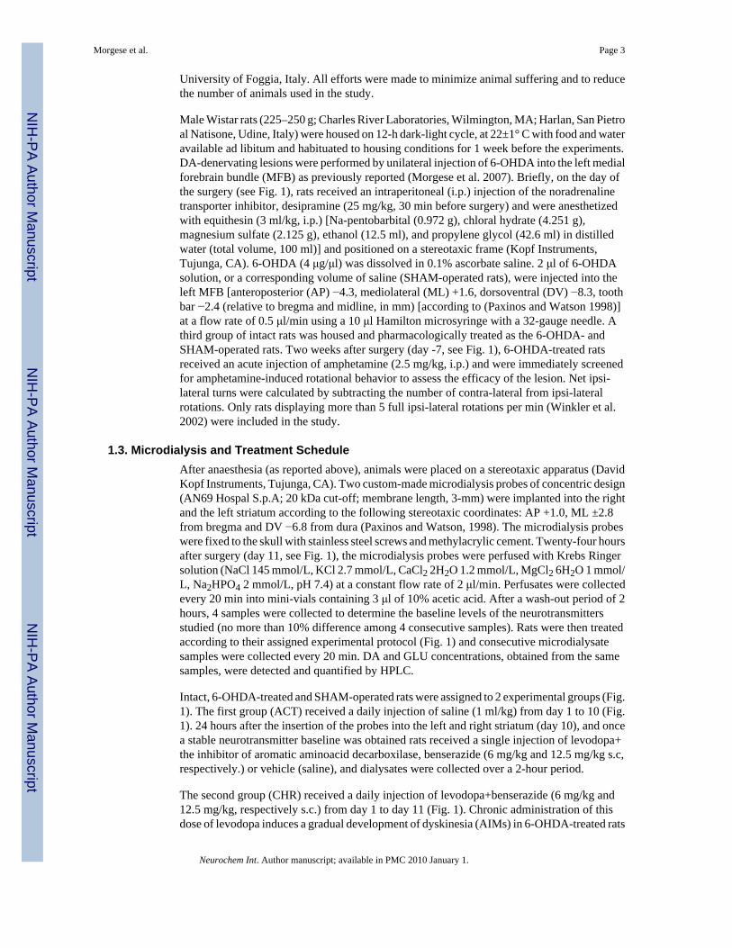
Male Wistar rats (225–250 g; Charles River Laboratories, Wilmington, MA; Harlan, San Pietroal Natisone, Udine, Italy) were housed on 12-h dark-light cycle, at 22±1° C with food and wateravailable ad libitum and habituated to housing conditions for 1 week before the experiments.DA-denervating lesions were performed by unilateral injection of 6-OHDA into the left medialforebrain bundle (MFB) as previously reported (Morgese et al. 2007). Briefly, on the day ofthe surgery (see Fig. 1), rats received an intraperitoneal (i.p.) injection of the noradrenalinetransporter inhibitor, desipramine (25 mg/kg, 30 min before surgery) and were anesthetizedwith equithesin (3 ml/kg, i.p.) [Na-pentobarbital (0.972 g), chloral hydrate (4.251 g),magnesium sulfate (2.125 g), ethanol (12.5 ml), and propylene glycol (42.6 ml) in distilledwater (total volume, 100 ml)] and positioned on a stereotaxic frame (Kopf Instruments,Tujunga, CA). 6-OHDA (4 μg/μl) was dissolved in 0.1% ascorbate saline. 2 μl of 6-OHDAsolution, or a corresponding volume of saline (SHAM-operated rats), were injected into theleft MFB [anteroposterior (AP) −4.3, mediolateral (ML) +1.6, dorsoventral (DV) −8.3, toothbar −2.4 (relative to bregma and midline, in mm) [according to (Paxinos and Watson 1998)]at a flow rate of 0.5 μl/min using a 10 μl Hamilton microsyringe with a 32-gauge needle. Athird group of intact rats was housed and pharmacologically treated as the 6-OHDA- andSHAM-operated rats. Two weeks after surgery (day -7, see Fig. 1), 6-OHDA-treated ratsreceived an acute injection of amphetamine (2.5 mg/kg, i.p.) and were immediately screenedfor amphetamine-induced rotational behavior to assess the efficacy of the lesion. Net ipsi-lateral turns were calculated by subtracting the number of contra-lateral from ipsi-lateralrotations. Only rats displaying more than 5 full ipsi-lateral rotations per min (Winkler et al.2002) were included in the study.
1.3. Microdialysis and Treatment ScheduleAfter anaesthesia (as reported above), animals were placed on a stereotaxic apparatus (DavidKopf Instruments, Tujunga, CA). Two custom-made microdialysis probes of concentric design(AN69 Hospal S.p.A; 20 kDa cut-off; membrane length, 3-mm) were implanted into the rightand the left striatum according to the following stereotaxic coordinates: AP +1.0, ML ±2.8from bregma and DV −6.8 from dura (Paxinos and Watson, 1998). The microdialysis probeswere fixed to the skull with stainless steel screws and methylacrylic cement. Twenty-four hoursafter surgery (day 11, see Fig. 1), the microdialysis probes were perfused with Krebs Ringersolution (NaCl 145 mmol/L, KCl 2.7 mmol/L, CaCl2 2H2O 1.2 mmol/L, MgCl2 6H2O 1 mmol/L, Na2HPO4 2 mmol/L, pH 7.4) at a constant flow rate of 2 μl/min. Perfusates were collectedevery 20 min into mini-vials containing 3 μl of 10% acetic acid. After a wash-out period of 2hours, 4 samples were collected to determine the baseline levels of the neurotransmittersstudied (no more than 10% difference among 4 consecutive samples). Rats were then treatedaccording to their assigned experimental protocol (Fig. 1) and consecutive microdialysatesamples were collected every 20 min. DA and GLU concentrations, obtained from the samesamples, were detected and quantified by HPLC.
Intact, 6-OHDA-treated and SHAM-operated rats were assigned to 2 experimental groups (Fig.1). The first group (ACT) received a daily injection of saline (1 ml/kg) from day 1 to 10 (Fig.1). 24 hours after the insertion of the probes into the left and right striatum (day 10), and oncea stable neurotransmitter baseline was obtained rats received a single injection of levodopa+the inhibitor of aromatic aminoacid decarboxilase, benserazide (6 mg/kg and 12.5 mg/kg s.c,respectively.) or vehicle (saline), and dialysates were collected over a 2-hour period.
The second group (CHR) received a daily injection of levodopa+benserazide (6 mg/kg and12.5 mg/kg, respectively s.c.) from day 1 to day 11 (Fig. 1). Chronic administration of thisdose of levodopa induces a gradual development of dyskinesia (AIMs) in 6-OHDA-treated rats
Morgese et al. Page 3
Neurochem Int. Author manuscript; available in PMC 2010 January 1.
NIH
-PA Author Manuscript
NIH
-PA Author Manuscript
NIH
-PA Author Manuscript

(Lundblad et al. 2002). On day 8 of levodopa, AIMs were scored using a severity scale aspreviously described (Morgese et al. 2007). For the following three consecutive days (days 9to 11), all rats received an injection of WIN (1 mg/kg, i.p.) or vehicle (5/5/90% of Tween80/PEG/saline) 20 minutes before levodopa. On day 11 (Fig. 1), after receiving the last injectionof WIN or vehicle followed by levodopa+benserazide (20 min later), microdialysates werecollected over a 2 hour period, while dyskinetic behaviors were monitored simultaneously. Atthe end of each experiment, the correct placement of dialysis probes was verified histologically.
1.4. HPLC AnalysesThe levels of DA and its metabolite, homovanillic acid (HVA), were determined by microboreHPLC using a SphereClone 150-mm × 2-mm column (3-μm packing) with a Unijet cell (BAS,Bioanalytical Systems, Kenilworth Warwickshire, United Kingdom) equipped with a 6-mm-diameter glassy carbon electrode (set at + 650 mV) and connected to an electrochemicalamperometric detector (INTRO, Antec Leyden, The Netherlands). The flow rate of the mobilephase [85 mM sodium acetate, 0.34 mM EDTA, 15 mM sodium chloride, 0.81 mMoctanesulphonic acid sodium salt, 5% methanol (v/v), pH 4.85] was 220 μl/min and the totalruntime 15 min.
GLU and Aspartate (ASP) levels were quantified using a HPLC/fluorimetric detection system,including pre-column derivatization with o-phtaldialdehyde reagent and a Chromsep 5 (C18)column as previously described (Ferraro et al. 2001). The flow rate of the mobile phase (0.1M sodium acetate, 10% methanol and 2.5% tetrahydrofurane, pH 6.5) was 1 ml/min and thetotal runtime 6 min.
1.5. AIMs recordingsAxial, limb, locomotive and oro-facial AIMs were monitored between 10:00 a.m. and 4:00p.m. as described (Lundblad et al. 2002). Briefly, animals were placed individually in aPlexiglas box and observations were carried out by a trained researcher blind to the treatmentschedule. AIMs were scored on a severity scale ranging from 0 to 4 (Lundblad et al. 2002;Morgese et al. 2007). Only animals displaying AIMs were included in the study.
In the case of the animals undergoing microdialysis, AIMs were scored for 1 min every 20 minafter levodopa injection throughout the microdialysis experiment (2 hours).
1.6. Statistical AnalysisMicrodialysis data were analyzed by one- or two-way analysis of variance (ANOVA) forrepeated measures with “treatment” (6-OHDA injection, or SHAM operation, or no-surgicalprocedure) or “time” as the between variables and “hemisphere” as the within variable (basalneurotransmitter comparisons or post-challenge comparisons). Dunnett’s and Tukey’s post hoccomparisons were used where appropriate. Violations of the sphericity assumption werecorrected using the Greenhouse-Geisser epsilon correction to adjust the degrees of freedomfor each test. Basal extracellular values of each neurotransmitter were defined as the averageof 4 consecutive samples before drug administration. Comparisons of basal neurotransmitterlevels between levodopa-treated (CHR) and saline-treated (ACT) groups were analyzed bytwo-way ANOVA, followed by the Tukey’s multiple comparison test. The overall effect ofdrug treatments on neurotransmitter outputs in each brain hemisphere was estimated by two-way ANOVA and by comparing the areas under the curve (AUC) by the paired Student’s t-test. The AUC was calculated using the standard trapezoid method (Gibaldi and Perrier1975) using neurotransmitter levels over a time window of 20–120 min for the ACT group,and 40–140 min for the CHR group.
Morgese et al. Page 4
Neurochem Int. Author manuscript; available in PMC 2010 January 1.
NIH
-PA Author Manuscript
NIH
-PA Author Manuscript
NIH
-PA Author Manuscript

DA turnover was expressed as HVA/DA ratio. Comparisons of DA turnover between the ipsi-and contra-lateral hemispheres in each experimental group were analyzed using the Studentpaired t-test. Neurochemical data were expressed as mean ± SEM. Dyskinesias were expressedas median of total AIMs score and analyzed using the Kruskal-Wallis test followed by theDunn’s multiple comparison test. Pearson’s linear correlation analyses were performedbetween GLU levels in the ipsi-lateral striatum of 6-OHDA-treated rats (CHR) and the severityof AIMs. The threshold for statistical significance was set at p<0.05.
2. Results2.1. Effects of 6-OHDA lesion on striatal DA and GLU outputs
Table 1 shows the basal levels of DA, HVA and GLU in the dorsal striatum of each hemisphere.ASP levels were omitted for clarity. These values reflect the mean neurotransmitterconcentrations of 4 consecutive microdialysates collected on day 11, 2 hours before the acuteinjection of levodopa or its vehicle (saline, ACT).
As expected, the 6-OHDA lesion produced a significant DA depletion in the striatum ipsi-lateral to the lesion compared to the contra-lateral side (p<0.05, Tukey’s test), whereas nodifferences were found across the two hemispheres of intact and SHAM-operated rats (Table1). Basal HVA levels followed a similar pattern (Table 1).
The 6-OHDA lesion did not affect the basal output of GLU and ASP (Table 1 and data notshown).
2.2. Effects of acute (ACT) and chronic (CHR) Levodopa on DA and GLU outputsTo investigate the effect of acute levodopa administration on DA and GLU outputs, 6-OHDA-treated, SHAM-operated and intact rats received a single injection of levodopa+benserazideor vehicle (saline) (ACT rats) on day 11, immediately after the collection of 4 basalmicrodialysates (baseline). Sample collection was continued for the following 120 min. Two-way ANOVA showed that DA levels were significantly lower in the denervated striatum,independently from acute levodopa (Fig. 2a; F(time)5,70=0.164, n.s., F(time × hemi)5,70=0.092,n.s., F(hemi)1,14=10.267, p<0.01) or vehicle (Fig. 2b; F(time)5,55=1.507, n.s.,F(time × hemi)5,55=0.860, n.s., F(hemi)1,11=25.544, p<0.001) administration. Moreover, acutelevodopa did not modify DA efflux in SHAM-operated or intact rats (data not shown).
As the difference in DA levels across the two hemispheres of 6-OHDA-treated rats was time-independent, we estimated and compared the AUCs from the ipsi- and contra-lateral striata bypaired Student’s t-test (Fig. 2a, b inserts) and confirmed that the difference in DA outputbetween the two hemispheres (p<0.01 and p<0.001, respectively) was unaffected by levodopaor vehicle.
DA turnover was decreased in the ipsi-lateral striatum of 6-OHDA-treated rats (p<0.05,Student’s paired t test) following acute levodopa or vehicle (Fig 2c, d), whereas no changeswere observed in SHAM-operated or intact animals (data not shown). Similarly, ASP or GLUefflux were unchanged in all experimental groups (data not shown).
6-OHDA-treated rats receiving chronic levodopa (CHR rats) showed a reduction of DA basallevels (day 11, before the last injection of levodopa) in both hemispheres and a decrease ofbasal HVA in the hemisphere ipsi-lateral to the lesion, (p<0.05, compared to 6-OHDA-treatedanimals receiving saline only) (Table 1). Chronic levodopa had no effect on basal DA outputof intact and SHAM-operated rats (Table 1), while it significantly increased basal DA turnover(SHAM, CHR: 513±130, 541±130, vs ACT: 240±37, 267±50; intact: 468±59, 404±73 vs ACT:254±32, 230±48, left and right, respectively) in both hemispheres, suggesting that this effect
Morgese et al. Page 5
Neurochem Int. Author manuscript; available in PMC 2010 January 1.
NIH
-PA Author Manuscript
NIH
-PA Author Manuscript
NIH
-PA Author Manuscript

is independent of the 6-OHDA lesion. In CHR 6-OHDA-treated rats, the last injection oflevodopa did not modify the difference in DA output observed between the two brainhemispheres (40–140 min sampling period), (Fig. 3a) (two-way ANOVA: F(hemi)1,12=9.152,p<0.05; F(time)5,60=0.726, n.s., F(time × hemi)5,60=1,059, n.s.). The analysis of the correspondingAUCs further confirmed this result (p<0.05, Student’s paired t-test, Fig. 3a, insert). Similarly,the last injection of levodopa did not affect DA output in CHR SHAM-operated and intact rats(data not shown).
Chronic levodopa further increased the disparity in DA turnover across the two hemispheresof 6-OHDA-treated rats (Fig. 3b) (p<0.05, Student’s paired t test) (see Fig. 2c for comparisonwith ACT rats), and elevated DA turnover by ~80% in CHR SHAM-operated rats (data notshown).
Concerning the effects of chronic levodopa on basal aminoacid neurotransmission (day 11,before last levodopa injection), two-way ANOVA revealed a significant effect of treatment onGLU output in CHR 6-OHDA-treated rats (Table 1) (F(treatment)1,48=7.136, p<0.05,F(hemi)1,48=0.926, n.s., F(treatment × hemi)1,48=1.058, n.s.). Although the GLU increase occurredin both hemispheres, statistical significance was reached only in the intact striatum (Table 1)(p<0.05, Tukey’s test). Chronic levodopa had no effect on basal ASP output in all CHRexperimental groups (data not shown).
In CHR 6-OHDA-treated rats, the last injection of levodopa produced a disparity in GLU effluxbetween the two hemispheres (Fig. 3c; two-way ANOVA: F(time)5,95=0.761, n.s.,F(time × hemi)5,95=0.162, n.s., F(hemi)1,19=6.984, p<0.05), as revealed by the smaller AUC of theipsi-lateral vs the contra-lateral striatum (p<0.01 Student’s paired t test, Fig. 3c, insert).Comparison of the post-challenge vs baseline GLU values revealed that such difference(p<0.05, Dunnett’s test) resulted from GLU decrease in the denervated side beginning at 40min after levodopa administration (Fig. 3c). No changes were observed in CHR SHAM-operated and intact rats (data not shown).
2.3. Sub-chronic WIN reduces neurotransmitter unbalances in dyskinetic ratsAs previously reported (Morgese et al. 2007), rats with unilateral 6-OHDA lesion chronicallytreated with levodopa develop severe AIMs, whereas no dyskinetic behaviors are observed ineither SHAM-operated or intact animals. Fig. 4 shows the time course of levodopa-inducedAIMs (total score) in CHR 6-OHDA-treated rats measured during microdialysis (day 11). Inagreement with our previous study, sub-chronic administration of WIN had a significant anti-dyskinetic effect (Fig. 4) at a dose that is most effective and does not produce motor suppression(1 mg/kg, i.p., 1 injection per day, for 3 days) (Morgese et al. 2007).
To investigate the neurochemical changes underlying the behavioral effects of WIN, wemeasured DA and aminoacid outputs in the same group of 6-OHDA-treated animals, as wellas in CHR SHAM-operated and intact rats. All experimental groups received an injection ofWIN (1 mg/kg, i.p.) or vehicle (5/5/90% of Tween80/PEG/saline) on days 9–11, 20 minutesbefore levodopa. On day 11, WIN was injected after the collection of 4 microdialysates(baseline), and the measurements of neurochemical outputs were carried out for a total of 2hours.
Table 1 shows the basal DA, GLU and ASP levels collected on day 11. The last injection ofWIN had no effect per se on DA output in both striata of CHR 6-OHDA-treated rats (Fig. 5a),but completely abolished the difference in DA levels between the two sides (Fig. 5a). No effectin either CHR SHAM-operated or intact rats were observed (data not shown). WIN also reducedthe disparity in DA turnover between left and right hemispheres induced by the last injection
Morgese et al. Page 6
Neurochem Int. Author manuscript; available in PMC 2010 January 1.
NIH
-PA Author Manuscript
NIH
-PA Author Manuscript
NIH
-PA Author Manuscript

of levodopa in CHR 6-OHDA-treated rats (Fig. 5b), whereas it did not change DA turnover inthe two hemispheres of either CHR SHAM-operated or intact rats (data not shown).
Finally, WIN prevented levodopa-induced GLU reduction in the denervated side of CHR 6-OHDA-treated rats (Fig. 5c and insert; see Fig. 3c for comparison) (F(time)5,100=5.048, p<0.01,F(time × hemi)5,100=0.361, n.s., F(hemi)1,20=0.479, n.s., two-way ANOVA). We also found thatthe GLU levels in this side were inversely correlated with levodopa-induced AIMs (Fig. 6)(p<0.05, rs=0.0586, Pearson’s correlation), whereas no significant correlation was observedcontralaterally (data not shown). In this side, sub-chronic WIN significantly reduced GLUlevels beginning at 80 min after the last injection of levodopa (Fig 5c) (p<0.05 vs baseline,Dunnett’s test). No differences in GLU and ASP outputs were found in CHR SHAM-operatedand intact animals following WIN+levodopa treatment (data not shown).
3. DiscussionThis study shows that systemic administration of levodopa and/or levodopa+WIN change DAand GLU outputs in the striatum of 6-OHDA-treated rats. In particular, we found that chroniclevodopa significantly decreases GLU levels in the denervated striatum of dyskinetic rats andthat this effect is correlated to the severity of levodopa-induced dyskinesias. We also showedthat sub-chronic WIN attenuates (1) the differences in DA-turnover across the two hemispheresand (2) levodopa-induced GLU decrease in the striatum ipsilateral to the lesion.
As expected, basal DA levels were significantly decreased in the denervated striatum of 6-OHDA-treated rats. In these animals we found no increase in DA efflux immediately afteracute levodopa injection as well as after chronic levodopa treatment, which is accompanied byovert dyskinetic behavior. Although some reports have shown increased extracellular DA afterchronic levodopa in dyskinetic rats (Meissner et al. 2006; Buck and Ferger 2008), thediscrepancy with our study may depend on the dose and/or route or schedule of levodopaadministration, which were different from those used in our experiments. For example,Meissner et al. used a dose that was 4-fold higher than ours (25 vs 6 mg/kg) and deliveredintraperitoneally. All our experiments were carried out using 6 mg/kg (s.c.) of levodopa, sincethis dose is clinically relevant and produces dyskinesias which gradually develop over time(Cenci and Lundblad 2005; Cenci and Lundblad 2007; Lindgren et al. 2007). In addition, inthe study of Buck and Ferger, levodopa was administered per os and for a longer period of time(21 days), whereas we used subcutaneous injections for 11 days. As different routes ofadministration are known to affect levodopa absorption and produce fluctuations in the severityof dyskinesia and wearing-off phenomena (Lindgren et al. 2007), it is likely that variations inlevodopa pharmacokinetics may account for the differences in striatal DA output reported inthese studies. Nevertheless, our results are in line with a recent study showing that high dosesof levodopa (33 and 66 mg/kg, i.p.) do not increase DA levels in 6-OHDA-treated rats(Rodriguez et al. 2007). Interestingly, we found that the 6-OHDA lesion decreased basal DAturnover in the denervated striatum, suggesting an ongoing compensatory mechanism for thelower DA production. A similar result is obtained when calculating the HVA/DA ratio usingthe DA and HVA basal levels reported by Buck and Ferger. By contrast, chronic levodopaenhanced DA metabolism in all experimental groups, without affecting the difference in DAturnover across the two hemispheres of 6-OHDA-treated rats. These data indicate that theconversion of levodopa into DA is accompanied by a higher DA metabolism, which mayexplain the gradual loss of levodopa efficacy over time (Marsden and Parkes 1976; Carey1991). Furthermore, the increased DA metabolism, as well as the reduction in DA transporter(DAT) expression in both hemispheres of dyskinetic rats (unpublished observations), is in linewith the observation that DAT function and DA turnover are inversely correlated in PD patients(Sossi et al. 2007).
Morgese et al. Page 7
Neurochem Int. Author manuscript; available in PMC 2010 January 1.
NIH
-PA Author Manuscript
NIH
-PA Author Manuscript
NIH
-PA Author Manuscript

DA denervation did not change GLU basal levels in 6-OHDA-treated rats, whereas chroniclevodopa increased basal GLU output in both hemispheres. This elevation, however, reachedstatistical significance only in the intact striatum. No changes in GLU output were observedin SHAM-operated or intact rats, suggesting that the GLU increase results from thecombination of DA denervation and chronic levodopa administration, two conditions thatpromote the development of dyskinesias (Lundblad 2004; Carta et al. 2006). The GLUelevation may also underlie and/or participate to the dyskinesia-priming process attributed tochronic levodopa (Cenci et al. 1998; Blanchet et al. 2004). Interestingly, the last levodopainjection in chronically-treated animals produced a significant reduction in GLU output in thedenervated side. This finding suggests that levodopa, at the dose used in our experimentalsetting, has an immediate depressing effect over GLU efflux and is followed by a long-lastingrebound, as indicated by GLU elevation upon chronic levodopa administration (Table 1). Ourdata differ from those reported by Robelet and colleagues (Robelet et al. 2004), which showeda significant increase in GLU levels in 6-OHDA-treated rats after the last injection of chroniclevodopa. In this study, however, the dose of levodopa was ~ 17 fold higher than ours, and thelength of treatment almost double (21 vs 11 days). The decrease of GLU observed by us in theipsi-lateral striatum may result from the stimulation of dopamine D2 receptors, which are highlyexpressed in rat striatum (Graybiel et al. 1994; Surmeier et al. 2007), become supersensitiveafter DA denervation (Pierot et al. 1988; Lisovoski et al. 1992; Cai et al. 2000; Ishida et al.2004) and negatively control GLU transmission. Indeed, (1) ex vivo studies show thatstimulation of D2 receptors reduces GLU release presynaptically (Bamford et al. 2004); (2)dopaminergic and glutamatergic systems work antagonistically in the striatum to controlcognitive and motor functions (Carlsson and Carlsson 1990; Riederer et al. 1992; Amalric etal. 1994). This hypothesis, however, remains controversial (Morari et al. 1998). Although inour study levodopa administration did not increase DA output, we cannot rule out thatlevodopa-induced increase of DA turnover might have masked DA elevation, thus making thetonic stimulation of D2 receptors still possible. Alternatively, levodopa may directly bind toD2 receptors as postulated by Silva and colleagues in PD patients (Silva et al. 2006), andconfirmed by PET studies in 6-OHDA-treated rats (Ishida et al. 2004).
Our study shows that the unilateral 6-OHDA lesion causes neurochemical changes also in theintact hemisphere. Indeed, chronic levodopa elevated basal GLU output in the intact side, thusindicating that the use of the contra-lateral hemisphere as control in experimental protocolsimplicating unilateral lesions is generally biased. In keeping with our observations, molecularalterations have been reported in the intact hemisphere of 6-OHDA-treated rats, such aschanges in striatal NMDA receptor and [3H]-Mazindol binding (O’Dell and Marshall 1996)and hypertrophy of dendritic cells in the neocortex (Miklyaeva et al. 2007). Similarly, othermodels utilizing unilateral lesions, such as intra-hippocampal kainic acid injections to mimicchronic seizures, produce neurochemical alterations contra-laterally (Arabadzisz et al. 2005).
In previous reports, we showed that sub-chronic administration of WIN at a dose unable tocause general motor suppression, significantly reduced levodopa-induced AIMs via a CB1-mediated mechanism (Morgese et al. 2007). To assess whether WIN also inducedneurochemical changes across the brain hemispheres of 6-OHDA-treated rats, we measuredDA, HVA and aminoacid outputs in dyskinetic rats receiving sub-chronic WIN from day 9 today 11 of levodopa. Our data indicate that WIN completely reversed the disparity in DA andGLU efflux across the two hemispheres of these animals. In particular, the analysis of the AUCsin each striatum showed that WIN increased DA output by 49% in the denervated striatum anddecreased it by 21% in the contra-lateral side. On the other hand, sub-chronic WIN preventedlevodopa-induced GLU decrease ipsi-laterally and significantly decreased GLU output contra-laterally.
Morgese et al. Page 8
Neurochem Int. Author manuscript; available in PMC 2010 January 1.
NIH
-PA Author Manuscript
NIH
-PA Author Manuscript
NIH
-PA Author Manuscript

The WIN-induced increase of DA output in the denervated striatum may result from a CB1-dependent inhibition of GABA release from striatonigral terminals, producing a disinhibitionof the SNc (Wallmichrath and Szabo 2002). In support of this hypothesis, CB1 receptors areexpressed on striatonigral terminals (Herkenham et al. 1991) and their activation has beenshown to excite dopaminergic neurons in the SNc (French et al. 1997). This phenomenon maybe particularly relevant in dyskinetic animals, as chronic levodopa increases CB1 receptormRNA levels in the denervated hemisphere (Zeng et al. 1999), thus explaining the oppositeeffect on DA output in the intact side. In particular, the DA decrease observed in thishemisphere cannot be attributed to either: (1) inhibition of DAT activity by WIN (Price et al.2007), which would prevent levodopa uptake, as this effect occurs in vivo only when WIN isadministered at higher doses (4 mg/kg) (Price et al. 2007); or (2) changes in DA turnover, sinceWIN did not affect DA metabolism in the contra-lateral hemisphere. Therefore, as previouslypostulated, WIN-induced DA decrease may be an indirect consequence of a cannabinoid-mediated reduction of GLU release, possibly from glutamatergic terminals projecting to theSN from the subthalamicus nucleus, which in turn may decrease the firing of nigrostriatalneurons (Freiman and Szabo 2005). Alternatively, WIN may decrease striatal DA release viamultiple non-synaptic mechanisms, including GABA release inhibition (Sidlo et al. 2008).
CB1 receptor stimulation has been associated with a decrease of GLU release in rat striatum(Gerdeman and Lovinger 2001; Kofalvi et al. 2005; Kreitzer and Malenka 2005; Adermarkand Lovinger 2007). In agreement with these studies, we found that WIN significantlydecreased GLU output in the intact striatum of 6-OHDA-treated rats receiving chroniclevodopa. By contrast, WIN attenuated levodopa-induced GLU decrease in the denervatedside. This unexpected result suggests that, following DA denervation and chronic levodopaadministration, cannabinoid agonists may evoke different responses due to changes in CB1receptor function. CB1 receptors are coupled to Gi/o proteins and they are co-expressed withD1 and D2 receptors in the striatum. This co-expression has functional consequences as CB1and D2 receptors share common pools of G-proteins (Meschler and Howlett 2001; Brotchie2003). In particular, in vitro studies have shown that concomitant application of cannabinoidand dopaminergic agonists promotes the formation of CB1 and D2 heterodimers (Kearn et al.2005) and switches the CB1 receptor coupling from Gi (Howlett and Fleming 1984; Childersand Deadwyler 1996) to stimulatory Gs proteins (Glass and Felder 1997), which in turn mayaffect cannabinoid-mediated responses. This hypothesis fits with the pharmacologicaltreatment received by dyskinetic rats; indeed, the concomitant stimulation of DA and CB1receptors by levodopa and WIN may change the coupling of CB1 receptors and prevent in turna further decrease of GLU efflux in the denervated striatum. This phenomenon, however,appears to be relevant only in this side, where D2 receptors are supersensitive (Pierot et al.1988; Ishida et al. 2004), have altered coupling (Hossain and Weiner 1993) and CB1 receptorsare over-expressed following chronic levodopa treatment (Zeng et al. 1999). Nevertheless, wecannot rule out that other receptor systems in the striatum, i.e. GABA receptors, might affectCB1 receptor signaling as already shown in the hippocampus (Cinar et al. 2008). Further studiesaiming at identifying changes in CB1 receptor coupling under our experimental conditions arenecessary to test this hypothesis.
Our findings show that the levodopa-dependent decrease of GLU efflux in the denervatedstriatum of dyskinetic rats is positively correlated with the severity of levodopa-induced AIMs.By contrast, no correlation between these two variables was observed in the intact side. Bypreventing levodopa-induced GLU reduction in the denervated side, WIN may positively affectstriatal synaptic plasticity, which is dysfunctional in 6-OHDA-treated rats (Picconi et al.2003; Cenci 2007). In particular, these animals show decreased concentrations of theendocannabinoid anandamide (Ferrer et al. 2003) and lack of endocannabinoid-mediated long-term depression (LTD) (Kreitzer and Malenka 2007). Since activation of metabotropic GLUreceptors is required to trigger endocannabinoid-mediated LTD, the GLU decrease induced by
Morgese et al. Page 9
Neurochem Int. Author manuscript; available in PMC 2010 January 1.
NIH
-PA Author Manuscript
NIH
-PA Author Manuscript
NIH
-PA Author Manuscript

chronic levodopa may explain its pro-dyskinetic effect. On the other end, WIN-inducedreduction of GLU efflux in the intact side may contribute to its anti-dyskinetic properties.Indeed, in intact animals, the motor suppressive effects of cannabinoid agonists have beenlinked to stimulation of CB1 receptors located on glutamatergic cortico-striatal terminals(Monory et al. 2007), which in turn inhibits GLU release (Huang et al. 2001). Finally, wecannot exclude that WIN might modulate other neurotransmitter/neuromodulator systemsregulating basal ganglia function, such as acetylcholine, serotonin, nitric oxide and GABA(Malone and Taylor 1999; Kofalvi et al. 2005; Bambico et al. 2007; Narushima et al. 2007;Sergeeva et al. 2007; Balazsa et al. 2008). In particular, physical manifestations of AIMs havebeen linked with enhanced GABA overflow in the output nuclei of the basal ganglia (Mela etal. 2007) and stimulation of CB1 receptors may affect the excitability of striatal medium spinyneurons by acting at GABAergic interneurons in the striatum (Narushima et al. 2007) wherethey are highly expressed (Uchigashima et al. 2007).
In conclusion, the ability of WIN to attenuate neurochemical disturbances in the denervatedside of 6-OHDA-treated rats may contribute to the anti-dyskinetic properties of this drug.
AcknowledgementsThis study was supported by a grant from the Italian Department of Research (FIRB) to V.C., by the National Instituteof Health NS 050401-04 (to A.G.) and by the National Parkinson Foundation (to A.G.).
ReferencesAdermark L, Lovinger DM. Retrograde endocannabinoid signaling at striatal synapses requires a
regulated postsynaptic release step. Proc Natl Acad Sci U S A 2007;104:20564–20569. [PubMed:18077376]
Amalric M, Ouagazzal A, Baunez C, Nieoullon A. Functional interactions between glutamate anddopamine in the rat striatum. Neurochemistry international 1994;25:123–131. [PubMed: 7994193]
Arabadzisz D, Antal K, Parpan F, Emri Z, Fritschy JM. Epileptogenesis and chronic seizures in a mousemodel of temporal lobe epilepsy are associated with distinct EEG patterns and selective neurochemicalalterations in the contralateral hippocampus. Exp Neurol 2005;194:76–90. [PubMed: 15899245]
Balazsa T, Biro J, Gullai N, Ledent C, Sperlagh B. CB1-cannabinoid receptors are involved in themodulation of non-synaptic [3H]serotonin release from the rat hippocampus. Neurochemistryinternational 2008;52:95–102. [PubMed: 17719142]
Bambico FR, Katz N, Debonnel G, Gobbi G. Cannabinoids elicit antidepressant-like behavior and activateserotonergic neurons through the medial prefrontal cortex. J Neurosci 2007;27:11700–11711.[PubMed: 17959812]
Bamford NS, Zhang H, Schmitz Y, Wu NP, Cepeda C, Levine MS, Schmauss C, Zakharenko SS, ZablowL, Sulzer D. Heterosynaptic dopamine neurotransmission selects sets of corticostriatal terminals.Neuron 2004;42:653–663. [PubMed: 15157425]
Beltramo M, de Fonseca FR, Navarro M, Calignano A, Gorriti MA, Grammatikopoulos G, Sadile AG,Giuffrida A, Piomelli D. Reversal of dopamine D(2) receptor responses by an anandamide transportinhibitor. J Neurosci 2000;20:3401–3407. [PubMed: 10777802]
Blanchet PJ, Calon F, Morissette M, Hadj Tahar A, Belanger N, Samadi P, Grondin R, Gregoire L, MeltzerL, Di Paolo T, Bedard PJ. Relevance of the MPTP primate model in the study of dyskinesia primingmechanisms. Parkinsonism Relat Disord 2004;10:297–304. [PubMed: 15196509]
Brotchie JM. CB1 cannabinoid receptor signalling in Parkinson’s disease. Current Opinion inPharmacology 2003;3:54–61. [PubMed: 12550742]
Buck K, Ferger B. Intrastriatal inhibition of aromatic amino acid decarboxylase prevents l-DOPA-induced dyskinesia: a bilateral reverse in vivo microdialysis study in 6-hydroxydopamine lesionedrats. Neurobiol Dis 2008;29:210–220. [PubMed: 17920284]
Morgese et al. Page 10
Neurochem Int. Author manuscript; available in PMC 2010 January 1.
NIH
-PA Author Manuscript
NIH
-PA Author Manuscript
NIH
-PA Author Manuscript

Cadogan AK, Alexander SP, Boyd EA, Kendall DA. Influence of cannabinoids on electrically evokeddopamine release and cyclic AMP generation in the rat striatum. J Neurochem 1997;69:1131–1137.[PubMed: 9282935]
Cai G, Zhen X, Uryu K, Friedman E. Activation of extracellular signal-regulated protein kinases isassociated with a sensitized locomotor response to D(2) dopamine receptor stimulation in unilateral6-hydroxydopamine-lesioned rats. J Neurosci 2000;20:1849–1857. [PubMed: 10684886]
Carey RJ. Chronic L-dopa treatment in the unilateral 6-OHDA rat: evidence for behavioral sensitizationand biochemical tolerance. Brain Res 1991;568:205–214. [PubMed: 1814568]
Carlsson M, Carlsson A. Interactions between glutamatergic and monoaminergic systems within the basalganglia--implications for schizophrenia and Parkinson’s disease. Trends Neurosci 1990;13:272–276.[PubMed: 1695402]
Carta M, Lindgren HS, Lundblad M, Stancampiano R, Fadda F, Cenci MA. Role of striatal L-DOPA inthe production of dyskinesia in 6-hydroxydopamine lesioned rats. J Neurochem 2006;96:1718–1727.[PubMed: 16539687]
Cenci M, Lindgren H. Advances in understanding l-DOPA-induced dyskinesia. Curr Opin Neurobiol2007;17:665–671. [PubMed: 18308560]
Cenci MA. Dopamine dysregulation of movement control in L-DOPA-induced dyskinesia. TrendsNeurosci 2007;30:236–243. [PubMed: 17400300]
Cenci, MA.; Lundblad, M. Utility of 6-Hydroxydopamine lesioned rats in the preclinical screening ofnovel treatment for Parkinson Disease. In: LeDoux, M., editor. Animal Models of MovementsDisorders. Elselvier Academic Press; Burlington: 2005. p. 193-208.
Cenci MA, Lundblad M. Ratings of L-DOPA-induced dyskinesia in the unilateral 6-OHDA lesion modelof Parkinson’s disease in rats and mice. Curr Protoc Neurosci. 2007Chapter 9, Unit 9 25
Cenci MA, Lee CS, Bjorklund A. L-DOPA-induced dyskinesia in the rat is associated with striataloverexpression of prodynorphin- and glutamic acid decarboxylase mRNA. The European journal ofneuroscience 1998;10:2694–2706. [PubMed: 9767399]
Childers SR, Deadwyler SA. Role of cyclic AMP in the actions of cannabinoid receptors. BiochemPharmacol 1996;52:819–827. [PubMed: 8781498]
Cinar R, Freund TF, Katona I, Mackie K, Szucs M. Reciprocal inhibition of G-protein signaling is inducedby CB(1) cannabinoid and GABA(B) receptor interactions in rat hippocampal membranes.Neurochemistry international 2008;52:1402–1409. [PubMed: 18407377]
Di Monte DA, McCormack A, Petzinger G, Janson AM, Quik M, Langston WJ. Relationship amongnigrostriatal denervation, parkinsonism, and dyskinesias in the MPTP primate model. Mov Disord2000;15:459–466. [PubMed: 10830409]
Domenici MR, Azad SC, Marsicano G, Schierloh A, Wotjak CT, Dodt HU, Zieglgansberger W, Lutz B,Rammes G. Cannabinoid receptor type 1 located on presynaptic terminals of principal neurons in theforebrain controls glutamatergic synaptic transmission. J Neurosci 2006;26:5794–5799. [PubMed:16723537]
Ferraro L, Tomasini MC, Gessa GL, Bebe BW, Tanganelli S, Antonelli T. The cannabinoid receptoragonist WIN 55,212–2 regulates glutamate transmission in rat cerebral cortex: an in vivo and in vitrostudy. Cereb Cortex 2001;11:728–733. [PubMed: 11459762]
Ferrer B, Asbrock N, Kathuria S, Piomelli D, Giuffrida A. Effects of levodopa on endocannabinoid levelsin rat basal ganglia: implications for the treatment of levodopa-induced dyskinesias. The Europeanjournal of neuroscience 2003;18:1607–1614. [PubMed: 14511339]
Freiman I, Szabo B. Cannabinoids depress excitatory neurotransmission between the subthalamic nucleusand the globus pallidus. Neuroscience 2005;133:305–313. [PubMed: 15893652]
French ED, Dillon K, Wu X. Cannabinoids excite dopamine neurons in the ventral tegmentum andsubstantia nigra. Neuroreport 1997;8:649–652. [PubMed: 9106740]
Gerdeman G, Lovinger DM. CB1 cannabinoid receptor inhibits synaptic release of glutamate in ratdorsolateral striatum. J Neurophysiol 2001;85:468–471. [PubMed: 11152748]
Gerdeman GL, Ronesi J, Lovinger DM. Postsynaptic endocannabinoid release is critical to long-termdepression in the striatum. Nat Neurosci 2002;5:446–451. [PubMed: 11976704]
Gibaldi, M.; Perrier, D. Pharmacokinetics. Vol. 1. Vol. 1. Marcel Dekker; New York: 1975. p. 37-40.
Morgese et al. Page 11
Neurochem Int. Author manuscript; available in PMC 2010 January 1.
NIH
-PA Author Manuscript
NIH
-PA Author Manuscript
NIH
-PA Author Manuscript

Giuffrida A, Parsons LH, Kerr TM, Rodriguez de Fonseca F, Navarro M, Piomelli D. Dopamine activationof endogenous cannabinoid signaling in dorsal striatum. Nat Neurosci 1999;2:358–363. [PubMed:10204543]
Glass M, Felder CC. Concurrent stimulation of cannabinoid CB1 and dopamine D2 receptors augmentscAMP accumulation in striatal neurons: evidence for a Gs linkage to the CB1 receptor. J Neurosci1997;17:5327–5333. [PubMed: 9204917]
Graybiel AM, Aosaki T, Flaherty AW, Kimura M. The basal ganglia and adaptive motor control. Science1994;265:1826–1831. [PubMed: 8091209]
Gubellini P, Picconi B, Bari M, Battista N, Calabresi P, Centonze D, Bernardi G, Finazzi-Agro A,Maccarrone M. Experimental parkinsonism alters endocannabinoid degradation: implications forstriatal glutamatergic transmission. J Neurosci 2002;22:6900–6907. [PubMed: 12177188]
Herkenham M, Lynn AB, de Costa BR, Richfield EK. Neuronal localization of cannabinoid receptors inthe basal ganglia of the rat. Brain Res 1991;547:267–274. [PubMed: 1909204]
Hossain MA, Weiner N. Dopaminergic functional supersensitivity: effects of chronic L-dopa andcarbidopa treatment in an animal model of Parkinson’s disease. J Pharmacol Exp Ther1993;267:1105–1111. [PubMed: 8263772]
Howlett AC, Fleming RM. Cannabinoid inhibition of adenylate cyclase. Pharmacology of the responsein neuroblastoma cell membranes. Mol Pharmacol 1984;26:532–538. [PubMed: 6092901]
Huang CC, Lo SW, Hsu KS. Presynaptic mechanisms underlying cannabinoid inhibition of excitatorysynaptic transmission in rat striatal neurons. The Journal of physiology 2001;532:731–748. [PubMed:11313442]
Ishida Y, Kawai K, Magata Y, Takeda R, Hashiguchi H, Abe H, Mukai T, Saji H. Changes in dopamineD2 receptors and 6-[18F]fluoro-L-3,4-dihydroxyphenylalanine uptake in the brain of 6-hydroxydopamine-lesioned rats. Neurodegener Dis 2004;1:109–112. [PubMed: 16908982]
Kearn CS, Blake-Palmer K, Daniel E, Mackie K, Glass M. Concurrent stimulation of cannabinoid CB1and dopamine D2 receptors enhances heterodimer formation: a mechanism for receptor cross-talk?Mol Pharmacol 2005;67:1697–1704. [PubMed: 15710746]
Kofalvi A, Rodrigues RJ, Ledent C, Mackie K, Vizi ES, Cunha RA, Sperlagh B. Involvement ofcannabinoid receptors in the regulation of neurotransmitter release in the rodent striatum: a combinedimmunochemical and pharmacological analysis. J Neurosci 2005;25:2874–2884. [PubMed:15772347]
Kreitzer AC, Malenka RC. Dopamine modulation of state-dependent endocannabinoid release and long-term depression in the striatum. J Neurosci 2005;25:10537–10545. [PubMed: 16280591]
Kreitzer AC, Malenka RC. Endocannabinoid-mediated rescue of striatal LTD and motor deficits inParkinson’s disease models. Nature 2007;445:643–647. [PubMed: 17287809]
Lindgren HS, Rylander D, Ohlin KE, Lundblad M, Cenci MA. The “motor complication syndrome” inrats with 6-OHDA lesions treated chronically with L-DOPA: relation to dose and route ofadministration. Behav Brain Res 2007;177:150–159. [PubMed: 17157933]
Lisovoski F, Haby C, Borrelli E, Schleef C, Revel MO, Hindelang C, Zwiller J. Induction of D2 dopaminereceptor mRNA synthesis in a 6-hydroxydopamine parkinsonian rat model. Brain Res Bull1992;28:697–701. [PubMed: 1535533]
Lundblad M. A model of L-DOPA-induced dyskinesia in 6-hydroxydopamine lesioned mice: relation tomotor and cellular parameters of nigrostriatal function. Neurobiology of Disease 2004;16:110–123.[PubMed: 15207268]
Lundblad M, Picconi B, Lindgren H, Cenci MA. A model of L-DOPA-induced dyskinesia in 6-hydroxydopamine lesioned mice: relation to motor and cellular parameters of nigrostriatal function.Neurobiol Dis 2004;16:110–123. [PubMed: 15207268]
Lundblad M, Andersson M, Winkler C, Kirik D, Wierup N, Cenci MA. Pharmacological validation ofbehavioural measures of akinesia and dyskinesia in a rat model of Parkinson’s disease. Eur J Neurosci2002;15:120–132. [PubMed: 11860512]
Mackie K. Distribution of cannabinoid receptors in the central and peripheral nervous system. HandbExp Pharmacol 2005:299–325. [PubMed: 16596779]
Morgese et al. Page 12
Neurochem Int. Author manuscript; available in PMC 2010 January 1.
NIH
-PA Author Manuscript
NIH
-PA Author Manuscript
NIH
-PA Author Manuscript

Malone DT, Taylor DA. Modulation by fluoxetine of striatal dopamine release following Delta9-tetrahydrocannabinol: a microdialysis study in conscious rats. Br J Pharmacol 1999;128:21–26.[PubMed: 10498830]
Marsden CD, Parkes JD. “On-off” effects in patients with Parkinson’s disease on chronic levodopatherapy. Lancet 1976;1:292–296. [PubMed: 55599]
Martin AB, Fernandez-Espejo E, Ferrer B, Gorriti MA, Bilbao A, Navarro M, Rodriguez de Fonseca F,Moratalla R. Expression and Function of CB(1) Receptor in the Rat Striatum: Localization andEffects on D(1) and D(2) Dopamine Receptor-Mediated Motor Behaviors.Neuropsychopharmacology 2008;33:1667–1679. [PubMed: 17957223]
Meissner W, Ravenscroft P, Reese R, Harnack D, Morgenstern R, Kupsch A, Klitgaard H, Bioulac B,Gross CE, Bezard E, Boraud T. Increased slow oscillatory activity in substantia nigra pars reticulatatriggers abnormal involuntary movements in the 6-OHDA-lesioned rat in the presence of excessiveextracellular striatal dopamine. Neurobiol Dis 2006;22:586–598. [PubMed: 16531050]
Mela F, Marti M, Dekundy A, Danysz W, Morari M, Cenci MA. Antagonism of metabotropic glutamatereceptor type 5 attenuates l-DOPA-induced dyskinesia and its molecular and neurochemicalcorrelates in a rat model of Parkinson’s disease. J Neurochem 2007;101:483–497. [PubMed:17359492]
Meschler JP, Howlett AC. Signal transduction interactions between CB1 cannabinoid and dopaminereceptors in the rat and monkey striatum. Neuropharmacology 2001;40:918–926. [PubMed:11378162]
Miklyaeva EI, Whishaw IQ, Kolb B. A golgi analysis of cortical pyramidal cells in the unilateral parkinsonrat: absence of change in the affected hemisphere vs hypertrophy in the intact hemisphere. RestorNeurol Neurosci 2007;25:91–99. [PubMed: 17726267]
Monory K, Blaudzun H, Massa F, Kaiser N, Lemberger T, Schutz G, Wotjak CT, Lutz B, Marsicano G.Genetic dissection of behavioural and autonomic effects of Delta(9)-tetrahydrocannabinol in mice.PLoS biology 2007;5:e269. [PubMed: 17927447]
Morari M, Marti M, Sbrenna S, Fuxe K, Bianchi C, Beani L. Reciprocal dopamine-glutamate modulationof release in the basal ganglia. Neurochemistry international 1998;33:383–397. [PubMed: 9874089]
Morgese MG, Cassano T, Cuomo V, Giuffrida A. Anti-dyskinetic effects of cannabinoids in a rat modelof Parkinson’s disease: role of CB(1) and TRPV1 receptors. Exp Neurol 2007;208:110–119.[PubMed: 17900568]
Narushima M, Uchigashima M, Fukaya M, Matsui M, Manabe T, Hashimoto K, Watanabe M, Kano M.Tonic enhancement of endocannabinoid-mediated retrograde suppression of inhibition bycholinergic interneuron activity in the striatum. J Neurosci 2007;27:496–506. [PubMed: 17234582]
O’Dell SJ, Marshall JF. Chronic L-dopa alters striatal NMDA receptors in rats with dopaminergic injury.Neuroreport 1996;7:2457–2461. [PubMed: 8981403]
Paxinos G, Watson CRR. The Rat Brain in Stereotaxic Coordinates. 1998Picconi B, Centonze D, Hakansson K, Bernardi G, Greengard P, Fisone G, Cenci MA, Calabresi P. Loss
of bidirectional striatal synaptic plasticity in L-DOPA-induced dyskinesia. Nat Neurosci 2003;6:501–506. [PubMed: 12665799]
Pierot L, Desnos C, Blin J, Raisman R, Scherman D, Javoy-Agid F, Ruberg M, Agid Y. D1 and D2-typedopamine receptors in patients with Parkinson’s disease and progressive supranuclear palsy. J NeurolSci 1988;86:291–306. [PubMed: 2975699]
Price DA, Owens WA, Gould GG, Frazer A, Roberts JL, Daws LC, Giuffrida A. CB1-independentinhibition of dopamine transporter activity by cannabinoids in mouse dorsal striatum. J Neurochem2007;101:389–396. [PubMed: 17250681]
Riederer P, Lange KW, Kornhuber J, Danielczyk W. Glutamatergic-dopaminergic balance in the brain.Its importance in motor disorders and schizophrenia. Arzneimittelforschung 1992;42:265–268.[PubMed: 1350197]
Robelet S, Melon C, Guillet B, Salin P, Kerkerian-Le Goff L. Chronic L-DOPA treatment increasesextracellular glutamate levels and GLT1 expression in the basal ganglia in a rat model of Parkinson’sdisease. The European journal of neuroscience 2004;20:1255–1266. [PubMed: 15341597]
Morgese et al. Page 13
Neurochem Int. Author manuscript; available in PMC 2010 January 1.
NIH
-PA Author Manuscript
NIH
-PA Author Manuscript
NIH
-PA Author Manuscript

Rodriguez M, Morales I, Gonzalez-Mora JL, Gomez I, Sabate M, Dopico JG, Rodriguez-Oroz MC, ObesoJA. Different levodopa actions on the extracellular dopamine pools in the rat striatum. Synapse2007;61:61–71. [PubMed: 17117421]
Sergeeva OA, Doreulee N, Chepkova AN, Kazmierczak T, Haas HL. Long-term depression of cortico-striatal synaptic transmission by DHPG depends on endocannabinoid release and nitric oxidesynthesis. The European journal of neuroscience 2007;26:1889–1894. [PubMed: 17868368]
Sidlo Z, Reggio PH, Rice ME. Inhibition of striatal dopamine release by CB1 receptor activation requiresnonsynaptic communication involving GABA, H2O2, and KATP channels. Neurochemistryinternational 2008;52:80–88. [PubMed: 17767979]
Sieradzan KA, Fox SH, Hill M, Dick JP, Crossman AR, Brotchie JM. Cannabinoids reduce levodopa-induced dyskinesia in Parkinson’s disease: a pilot study. Neurology 2001;57:2108–2111. [PubMed:11739835]
Silva I, Cortes H, Escartin E, Rangel C, Floran L, Erlij D, Aceves J, Floran B. L-DOPA inhibitsdepolarization-induced [3H]GABA release in the dopamine-denervated globus pallidus of the rat:the effect is dopamine independent and mediated by D2-like receptors. J Neural Transm2006;113:1847–1853. [PubMed: 16736236]
Sossi V, de la Fuente-Fernandez R, Schulzer M, Troiano AR, Ruth TJ, Stoessl AJ. Dopamine transporterrelation to dopamine turnover in Parkinson’s disease: a positron emission tomography study. AnnNeurol 2007;62:468–474. [PubMed: 17886297]
Surmeier DJ, Ding J, Day M, Wang Z, Shen W. D1 and D2 dopamine-receptor modulation of striatalglutamatergic signaling in striatal medium spiny neurons. Trends Neurosci 2007;30:228–235.[PubMed: 17408758]
Takahashi KA, Castillo PE. The CB1 cannabinoid receptor mediates glutamatergic synaptic suppressionin the hippocampus. Neuroscience 2006;139:795–802. [PubMed: 16527424]
Tsou K, Brown S, Sanudo-Pena MC, Mackie K, Walker JM. Immunohistochemical distribution ofcannabinoid CB1 receptors in the rat central nervous system. Neuroscience 1998;83:393–411.[PubMed: 9460749]
Uchigashima M, Narushima M, Fukaya M, Katona I, Kano M, Watanabe M. Subcellular arrangement ofmolecules for 2-arachidonoyl-glycerol-mediated retrograde signaling and its physiologicalcontribution to synaptic modulation in the striatum. J Neurosci 2007;27:3663–3676. [PubMed:17409230]
Wallmichrath I, Szabo B. Cannabinoids inhibit striatonigral GABAergic neurotransmission in the mouse.Neuroscience 2002;113:671–682. [PubMed: 12150787]
Winkler C, Kirik D, Bjorklund A, Cenci MA. L-DOPA-induced dyskinesia in the intrastriatal 6-hydroxydopamine model of parkinson’s disease: relation to motor and cellular parameters ofnigrostriatal function. Neurobiol Dis 2002;10:165–186. [PubMed: 12127155]
Zeng BY, Dass B, Owen A, Rose S, Cannizzaro C, Tel BC, Jenner P. Chronic L-DOPA treatmentincreases striatal cannabinoid CB1 receptor mRNA expression in 6-hydroxydopamine-lesioned rats.Neurosci Lett 1999;276:71–74. [PubMed: 10624794]
Morgese et al. Page 14
Neurochem Int. Author manuscript; available in PMC 2010 January 1.
NIH
-PA Author Manuscript
NIH
-PA Author Manuscript
NIH
-PA Author Manuscript

Fig. 1.Experimental design of the study. ACT 6-OHDA, SHAM-operated and intact animals receivedsaline chronically and levodopa or vehicle acutely during the microdialysis experiment (day11). CHR 6-OHDA, SHAM and intact animals were treated chronically with levodopa (11days, light gray shade) and WIN+levodopa (3 days, dark grey shade). On day 11, rats receivedthe last injection of vehicle/WIN and/or levodopa during the microdialysis experiment.Numbers represents days after the 6-OHDA/SHAM lesion.
Morgese et al. Page 15
Neurochem Int. Author manuscript; available in PMC 2010 January 1.
NIH
-PA Author Manuscript
NIH
-PA Author Manuscript
NIH
-PA Author Manuscript

Fig. 2.Effects of levodopa (a) or its vehicle (saline) (b) on DA extracellular concentrations in the ipsi-lateral (open squares) and contra-lateral (filled triangles) striatum of 6-OHDA-treated ratstreated according to ACT schedule (see Table 1 legend). Inserts represent AUCs values (ipsi-lateral striatum, open bars; contra-lateral striatum, filled bars). The bottom panels show theeffects of acute levodopa (c) or saline (d) on DA turnover (HVA/DA ratios) in 6-OHDA-treatedrats. Data are expressed as mean±SEM of n=8 experiments. Panels a and b, two-way ANOVArepeated measures **p<0.01 and ***p<0.001 ipsi vs contra. Inserts and panel c and d, Student’spaired t test *p<0.05, **p<0.01 and ***p<0.001 ipsi vs contra. Arrows indicate the time oflevodopa or saline administration.
Morgese et al. Page 16
Neurochem Int. Author manuscript; available in PMC 2010 January 1.
NIH
-PA Author Manuscript
NIH
-PA Author Manuscript
NIH
-PA Author Manuscript

Fig. 3.Effects of chronic levodopa on DA (a) or GLU (c) release in the ipsi-lateral (open squares) andcontra-lateral (filled triangles) striatum of 6-OHDA-treated rats treated according to CHRschedule (see Table 1 legend). Panel b shows the effects of chronic levodopa on DA turnover(HVA/DA ratios) in the ipsi-lateral (open bar) and contra-lateral (filled bar) striatum in thesame rats. Data are expressed as mean±SEM (n=9). Panels a and c, two-way ANOVA repeatedmeasures *p<0.05 ipsi vs contra. Inserts and panel b, Student’s paired t test, *p<0.05 ipsi vscontra. Panel c, one-way ANOVA repeated measures followed by Dunnett’s multiplecomparison test, #p<0.05 and ##p<0.01 post challenge values vs baseline. Arrows indicate thetime of vehicle (PEG/tween 80/saline 5/5/90%) and levodopa administrations.
Morgese et al. Page 17
Neurochem Int. Author manuscript; available in PMC 2010 January 1.
NIH
-PA Author Manuscript
NIH
-PA Author Manuscript
NIH
-PA Author Manuscript

Fig. 4.Time course of levodopa-induced abnormal involuntary movements (AIMs) in 6-OHDA-treated rats receiving chronic levodopa (CHR). Dyskinesias were scored during themicrodialysis experiments. Rats received WIN (open diamonds) or vehicle (filled squares) 20min before levodopa. Arrows indicate the time of vehicle/WIN and levodopa administrations.Data are expressed as median score (interquartile ranges have been omitted for clarity) of n=9experiments. Kruskal-Wallis followed by Dunn’s multiple comparison test, *p<0.05 and***p<0.001, vs dyskinetic rats treated with vehicle.
Morgese et al. Page 18
Neurochem Int. Author manuscript; available in PMC 2010 January 1.
NIH
-PA Author Manuscript
NIH
-PA Author Manuscript
NIH
-PA Author Manuscript

Fig. 5.Effects of WIN+levodopa on DA (a) or GLU (c) release in the ipsi-lateral (open squares) andcontra-lateral (filled triangles) striatum of 6-OHDA-treated rats treated according to CHRschedule (see Table 1 legend). Panel b shows the effects of vehicle+levodopa on DA turnover(HVA/DA ratios) in the ipsi-lateral (open bar) and contra-lateral (filled bar) striatum of thesame rats. Data are expressed as mean±SEM (n=9). Panel c, one-way ANOVA repeatedmeasures followed by Dunnett’s multiple comparison test, §p<0.05 post challenge values vsbaseline. Arrows indicate the time of WIN and levodopa administrations.
Morgese et al. Page 19
Neurochem Int. Author manuscript; available in PMC 2010 January 1.
NIH
-PA Author Manuscript
NIH
-PA Author Manuscript
NIH
-PA Author Manuscript

Fig. 6.Spearman’s correlation between AIMs total score and GLU extracellular levels observed inthe ipsi-lateral striatum of 6-OHDA-treated rats (CHR) receiving vehicle+levodopa. p<0.05,rs=0.05860.
Morgese et al. Page 20
Neurochem Int. Author manuscript; available in PMC 2010 January 1.
NIH
-PA Author Manuscript
NIH
-PA Author Manuscript
NIH
-PA Author Manuscript

NIH
-PA Author Manuscript
NIH
-PA Author Manuscript
NIH
-PA Author Manuscript
Morgese et al. Page 21TA
BLE
1B
asal
ext
race
llula
r neu
rotra
nsm
itter
leve
ls in
rats
trea
ted
acco
rdin
g to
AC
T an
d C
HR
sche
dule
s. In
AC
T, b
asal
leve
ls w
ere
mon
itore
d 2
hour
s be
fore
acu
te le
vodo
pa (
or th
e la
st s
alin
e) in
ject
ion.
In
CH
R, b
asal
leve
ls r
efle
ct e
xtra
cellu
lar
neur
otra
nsm
itter
con
cent
ratio
nsob
serv
ed 2
hou
rs b
efor
e th
e la
st in
ject
ion
of c
hron
ic le
vodo
pa o
r WIN
+lev
odop
a.
INT
AC
TSH
AM
- O
PER
AT
ED
6-O
HD
A-T
RE
AT
ED
AC
TC
HR
AC
TC
HR
AC
TC
HR
Lev
odop
aL
evod
opa
Lev
odop
a
veh
WIN
veh
WIN
veh
WIN
DA
(pg/
ml)
Ipsi
532±
7944
4±22
510±
109
525±
4540
6±91
407±
8012
7±13
a60
±6a,
b92
±39a
Con
tra
545±
5946
9±48
571±
170
537±
6840
8±76
437±
6351
3±65
288±
98b
283±
84
HV
A (n
g/m
l)Ip
si16
7±18
173±
3616
5±9
137±
2915
2±18
173±
385.
3±2.
1a2±
0.4a
10±7
a
Con
tra
151±
2514
8±29
155±
3612
8±17
161±
2221
1±56
137±
1622
3±44
b15
1±27
GL
U (μ
M)
Ipsi
0.3±
0.1
0.3±
0.1
0.3±
0.1
0.3±
0.1
0.4±
0.1
0.7±
0.2
0.38
±0.1
0.57
±0.2
0.57
±0.2
Con
tra
0.3±
0.1
0.3±
0.1
0.3±
0.1
0.3±
0.1
0.3±
0.04
0.4±
0.2
0.37
±0.1
0.79
±0.1
b0.
62±0
.2
Two-
way
AN
OV
A fo
llow
ed b
y Tu
key’
s mul
tiple
com
paris
on te
st:
a p<0.
05 v
s con
tro-la
tera
l hem
isph
ere
with
in th
e sa
me
expe
rimen
tal g
roup
;
b p<0.
05 v
s cor
resp
ondi
ng h
emis
pher
e of
the
sam
e gr
oup.
Neurochem Int. Author manuscript; available in PMC 2010 January 1.
Copyright © 2022 FDOKUMEN