Behavioral pharmacological properties of a novel cannabinoid 1???,1???-dithiolane ??8THC analog,...
12
Data paper 499 Behavioral pharmacological properties of a novel cannabinoid 1 0 ,1 0 -dithiolane D 8 -THC analog, AMG-3 K. Antoniou a,e , A. Galanopoulos b,e , S. Vlachou b , T. Kourouli e , V. Nahmias c , K. Thermos d , G. Panagis b , Z. Daifoti e , M. Marselos a , D. Papahatjis c and C. Spyraki e Newly developed cannabinoids may hold the promise of the development of useful and safe drugs. This study aimed to investigate the behavioral effects of the novel 1 0 ,1 0 -dithiolane D 8 -THC analogue AMG-3, a cannabinomimetic molecule with high affinity for CB 1 /CB 2 receptors. This analog was chosen for its binding affinity to these receptors, which is higher than that reported for D 8 -tetrahydrocannabinol (D 8 -THC). Behavioral responses were assessed after the administration of AMG-3 (1, 2, 4, 8 mg/kg, i.p.) in the open field, on the bar test, on the hot plate and in the intracranial self-stimulation procedure. AMG-3 increased the reactivity time on the hot plate in a dose- and time-dependent manner, indicating a long-lasting analgesic effect (at least 24 h). The substance was found dose-dependently to decrease spontaneous motor activity and to induce catalepsy, particularly at the highest dose (8 mg/kg). AMG-3 did not affect the rewarding value of intracranial self-stimulation, except to increase the reward threshold at the highest dose (8 mg/kg). The effects of the highest dose of AMG-3 on spontaneous activity and on the self-stimulation paradigm were completely reversed by pre-treatment with the CB 1 receptor antagonist AM-251. These findings indicate that the administration of AMG-3 to rats elicits a specific behavioral profile, most probably associated with the activation of CB 1 receptors and without effects indicating abuse potential. Behavioural Pharmacology 16:499–510 c 2005 Lippincott Williams & Wilkins. Behavioural Pharmacology 2005, 16:499–510 Keywords: 1 0 ,1 0 -dithiolane D 8 -THC analog, cannabinoids, antinociception, motor activity, self-stimulation, rat a Department of Pharmacology, Medical School, University of Ioannina, 45110 Ioannina, Greece, b Laboratory of Neurosciences and Behavior, Division of Biopsychology, Department of Psychology, School of Social Sciences, University of Crete, 74100 Rethymnon, Crete, Greece, c Institute of Organic and Pharmaceutical Chemistry, National Hellenic Research Foundation, 11635, Athens, Greece, d Laboratory of Pharmacology, Department of Basic Sciences, Faculty of Medicine, University of Crete, 71110 Heraklion, Crete, Greece and e Department of Pharmacology, School of Medicine, University of Athens, 11527 Goudi, Athens, Greece. Sponsorship: This study was supported by departments involved and GSRT-EU project (YB 60) to C.S. Correspondence and requests for reprints to Katerina Antoniou, Lecturer of Pharmacology, Department of Pharmacology, Medical School, University of Ioannina, 45110 Ioannina, Greece. E-mail: [email protected] Received 2 April 2005 Accepted as revised 29 June 2005 Introduction Significant contributions made by medicinal chemistry, both in academia and in the pharmaceutical industry, with respect to synthetic cannabinoids have enhanced basic knowledge of the biology of the endocannabinoid system (Piomelli, 2003). A limited number of cannabinoids have already been used as therapeutic agents, while an expanding number of new analogs, currently under preclinical testing, appear to be promising therapeutic interventions for a variety of pathophysiological states (Howlett et al., 2004; Hall et al., 2005). Nevertheless, the development of additional safe and medically useful cannabinoids is still worth pursuing, since the endocan- nabinoid system seems to be involved in disease states with either central or peripheral pathophysiology (Di Marzo et al., 2004). The discovery of cannabinoid receptors (Devane et al., 1988; Munro et al., 1993; Pertwee, 1997) provided an impetus for obtaining detailed information regarding the stereoelectronic requirements of receptor active site(s). The existing literature (Razdan, 1986; Makriyannis and Rapaka, 1990; Khanolkar et al., 2000) recognizes four pharmacophores on the cannabinoid structure that can be associated with cannabinomimetic activity. Among these elements it is now well known that the aliphatic side- chain plays a pivotal role in determining receptor binding affinity and pharmacological activity, as first demon- strated by Adams (1949). Moreover, Papahatjis et al. (1998, 2002) pointed out that structural modifications at the benzylic position of the side-chain of the D 8 - tetrahydrocannabinol (D 8 -THC) core have a profound effect on the affinities of new analogs for both CB 1 and CB 2 cannabinoid receptors. The newly synthesized dithiolane derivative, AMG-3, exhibited greater affinity (at the subnanomolar level), as compared to that of D 8 - THC (see Fig. 1), for both CB 1 and CB 2 cannabinoid receptors (Papahatjis et al., 1998, 2002). Although D 9 - THC is the most active constituent of cannabis, Papahatjis et al. (1998) chose D 8 -THC as the reference 0955-8810 c 2005 Lippincott Williams & Wilkins Copyright © Lippincott Williams & Wilkins. Unauthorized reproduction of this article is prohibited.
Transcript of Behavioral pharmacological properties of a novel cannabinoid 1???,1???-dithiolane ??8THC analog,...

Data paper 499
Behavioral pharmacological properties of a novelcannabinoid 10,10-dithiolane D8-THC analog, AMG-3K. Antonioua,e, A. Galanopoulosb,e, S. Vlachoub, T. Kouroulie, V. Nahmiasc,K. Thermosd, G. Panagisb, Z. Daifotie, M. Marselosa, D. Papahatjisc
and C. Spyrakie
Newly developed cannabinoids may hold the promise of
the development of useful and safe drugs. This study
aimed to investigate the behavioral effects of the
novel 10,10-dithiolane D8-THC analogue AMG-3, a
cannabinomimetic molecule with high affinity for CB1/CB2
receptors. This analog was chosen for its binding affinity
to these receptors, which is higher than that reported for
D8-tetrahydrocannabinol (D8-THC). Behavioral responses
were assessed after the administration of AMG-3 (1, 2, 4,
8 mg/kg, i.p.) in the open field, on the bar test, on the hot
plate and in the intracranial self-stimulation procedure.
AMG-3 increased the reactivity time on the hot plate
in a dose- and time-dependent manner, indicating a
long-lasting analgesic effect (at least 24 h). The substance
was found dose-dependently to decrease spontaneous
motor activity and to induce catalepsy, particularly
at the highest dose (8 mg/kg). AMG-3 did not affect the
rewarding value of intracranial self-stimulation, except
to increase the reward threshold at the highest dose
(8 mg/kg). The effects of the highest dose of AMG-3 on
spontaneous activity and on the self-stimulation paradigm
were completely reversed by pre-treatment with the CB1
receptor antagonist AM-251. These findings indicate that
the administration of AMG-3 to rats elicits a specific
behavioral profile, most probably associated with the
activation of CB1 receptors and without effects indicating
abuse potential. Behavioural Pharmacology 16:499–510�c 2005 Lippincott Williams & Wilkins.
Behavioural Pharmacology 2005, 16:499–510
Keywords: 10 ,10-dithiolane D8-THC analog, cannabinoids, antinociception,motor activity, self-stimulation, rat
aDepartment of Pharmacology, Medical School, University of Ioannina, 45110Ioannina, Greece, bLaboratory of Neurosciences and Behavior, Division ofBiopsychology, Department of Psychology, School of Social Sciences, Universityof Crete, 74100 Rethymnon, Crete, Greece, cInstitute of Organic andPharmaceutical Chemistry, National Hellenic Research Foundation, 11635,Athens, Greece, dLaboratory of Pharmacology, Department of Basic Sciences,Faculty of Medicine, University of Crete, 71110 Heraklion, Crete, Greece andeDepartment of Pharmacology, School of Medicine, University of Athens,11527 Goudi, Athens, Greece.
Sponsorship: This study was supported by departments involved and GSRT-EUproject (YB 60) to C.S.
Correspondence and requests for reprints to Katerina Antoniou,Lecturer of Pharmacology, Department of Pharmacology, Medical School,University of Ioannina, 45110 Ioannina, Greece.E-mail: [email protected]
Received 2 April 2005 Accepted as revised 29 June 2005
IntroductionSignificant contributions made by medicinal chemistry,
both in academia and in the pharmaceutical industry, with
respect to synthetic cannabinoids have enhanced basic
knowledge of the biology of the endocannabinoid system
(Piomelli, 2003). A limited number of cannabinoids have
already been used as therapeutic agents, while an
expanding number of new analogs, currently under
preclinical testing, appear to be promising therapeutic
interventions for a variety of pathophysiological states
(Howlett et al., 2004; Hall et al., 2005). Nevertheless, the
development of additional safe and medically useful
cannabinoids is still worth pursuing, since the endocan-
nabinoid system seems to be involved in disease states
with either central or peripheral pathophysiology (Di
Marzo et al., 2004).
The discovery of cannabinoid receptors (Devane et al.,1988; Munro et al., 1993; Pertwee, 1997) provided an
impetus for obtaining detailed information regarding the
stereoelectronic requirements of receptor active site(s).
The existing literature (Razdan, 1986; Makriyannis and
Rapaka, 1990; Khanolkar et al., 2000) recognizes four
pharmacophores on the cannabinoid structure that can be
associated with cannabinomimetic activity. Among these
elements it is now well known that the aliphatic side-
chain plays a pivotal role in determining receptor binding
affinity and pharmacological activity, as first demon-
strated by Adams (1949). Moreover, Papahatjis et al.(1998, 2002) pointed out that structural modifications at
the benzylic position of the side-chain of the D8-
tetrahydrocannabinol (D8-THC) core have a profound
effect on the affinities of new analogs for both CB1 and
CB2 cannabinoid receptors. The newly synthesized
dithiolane derivative, AMG-3, exhibited greater affinity
(at the subnanomolar level), as compared to that of D8-
THC (see Fig. 1), for both CB1 and CB2 cannabinoid
receptors (Papahatjis et al., 1998, 2002). Although D9-
THC is the most active constituent of cannabis,
Papahatjis et al. (1998) chose D8-THC as the reference
0955-8810 �c 2005 Lippincott Williams & Wilkins
Copyright © Lippincott Williams & Wilkins. Unauthorized reproduction of this article is prohibited.

substance in the binding studies because it shares a
similar pharmacological profile with D9-THC and, in
addition, it possesses greater chemical stability. The
increased affinity of AMG-3 was attributed to a hydro-
phobic subsite for both CB1 and CB2 at the level of the
benzylic side-chain carbon, and suggested the signifi-
cance of the orientation and conformation of the side-
chain in determining cannabinomimetic activity. Com-
pounds with higher affinities for the target site might
prove useful as pharmacological tools and therapeutic
agents with improved efficacy and safety.
The higher affinity of AMG-3 for CB1/CB2 receptors as
compared to D8-THC (Papahatjis et al., 1998) prompted
us to examine the behavioral properties of this new
cannabinoid, which might reflect its central nervous
system action and its potential for abuse. Behaviors most
often used to assess cannabinoid activity are antinocicep-
tion, locomotor inhibition and catalepsy (Fuentes et al.,1999; Pertwee, 2001; Romero et al., 2002). In addition,
cannabinoids have addictive liability in humans and a
number of attempts have been made, at the preclinical
level, to assess their abuse potential using a variety of
models, including the intracranial self-stimulation (ICSS)
paradigm (Gardner, 2002; Tanda and Goldberg, 2003).
The rewarding effects of brain stimulation and of
addictive drugs are thought to involve the same reward
circuits (Wise, 1980, 1996, 1998) and the ICSS model is
well suited for assessing reward-related drug interactions.
It is known, for instance, that almost all drugs of abuse
increase the rewarding value of self-stimulation, as this is
reflected in the decreased threshold for rewarding brain
stimulation. Additionally, any aversive effect of a drug
increases the threshold for rewarding brain stimulation
(for reviews, see Stellar and Rice, 1989; Wise, 1996,
1998). Therefore, the intracranial self-stimulation para-
digm allows clear dissociation and quantification of the
reward-potentiating and reward-inhibiting effects of a
drug.
The present study aimed to investigate the effects of the
novel 10,10-dithiolane D8-THC analogue (AMG-3) on
analgesia, motor activities and the rewarding properties of
ICSS. To this purpose the hot-plate test, the recording of
open-field behavioral activity, the bar test and the
intracranial self-stimulation paradigm were used.
Methods
Binding studies
AMG-3 was synthesized and characterized by Papahatjis
et al. (1998). Additional quantities were synthesized for
the needs of the current study. Samples of AMG-3
synthesized in the Institute of Organic and Pharmaceu-
tical Chemistry of the Hellenic Research Center were
taken and competition studies were performed in cortical
membranes according to Khanolkar et al. (1996), in order
to confirm the high affinity of the ligand in the brain.
Prefrontal cortex was removed from rat brains and
homogenized in a buffer containing 50 mmol/l Tris–HCl,
EDTA (2.5 mmol/l), MgCl2 (5.0 mmol/l), pH 7.4 (incuba-
tion buffer). The mixture was centrifuged three times at
45 000 g for 15 min. The final pellet was resuspended in
the incubation buffer (2.5 mg wet weight/ml). Mem-
branes (100 mg) were incubated in the presence of
[3H]CP55940 (168 Ci/mmol; 0.3 nmol/l, New England
Nuclear), different concentrations of AMG-3 and incuba-
tion buffer in a final volume of 200 ml for 90 min at 301C.
The reaction was terminated by the addition of 5 ml cold
incubation buffer containing bovine serum albumin (BSA;
2.0 mg/ml, wash buffer) and the mixture was filtered over
glass fiber/carbon (GF/C) filters that were presoaked in
0.05% polyethyleneimine (PEI). The filters were washed
with an additional 2� 5 ml wash buffer, and added to
scintillation vials. Scintillation fluid was added to the
filters and the radioactivity was counted in a beta-
counter. Specific binding was determined in the presence
of WIN212-291(10 mmol/l).
Behavioral studies
Subjects
Male Sprague–Dawley rats, aged 70–90 days old and
weighing approximately 250–300 g at the beginning of
experiments, were used. The animals were housed in
groups of six or seven in plastic cages (57� 35� 20 cm)
with food and water freely available, under controlled
laboratory conditions, i.e. 12 h light/dark with lights on at
0600 h and constant temperature 21 ± 11C. All animal
experiments were carried out in accordance with the
National Institutes of Health Guide for the Care and Use of
Fig. 1
Compounds CB1 (Ki, nmol/l) CB2 (Ki, nmol/l)(−)−∆8-THC, (a)
AMG-3, (b)39.3 0.52
OH
O R
1
(a) R =
(b) R =
S S
47.6 0.32
Chemical structure of D8-tetrahydrocannabinol (D8-THC) (a) and AMG-3 (b). D8-THC has been used as prototype because it has a similarpharmacological profile as D9-THC and it is chemically more stable.Affinities (Ki) of cannabinoid analogs (a) and (b) for the CB1 and CB2
receptors are also given. (From Papahatjis et al., 1998, 2002.)
500 Behavioural Pharmacology 2005, Vol 16 Nos 5 & 6
Copyright © Lippincott Williams & Wilkins. Unauthorized reproduction of this article is prohibited.

Laboratory Animals (NIH Publications No. 80-23) revised
1996. All procedures conformed to named local and
international guidelines on the ethical use of animals and
special care. Efforts were made to use the minimal
number of rats necessary for experiments and to reduce
their suffering.
Hot-plate test
The rats (n = 35) were accustomed to the experimental
room for 1 h prior to the experiment. Antinociception was
monitored using the hot-plate test (Ugo Basile 7280, Ugo
Basile Biological Research Apparatus Comerio, Varese,
Italy), which consisted of a Plexiglas frame and an
electrically heated surface kept at a constant tempera-
ture. In particular, rats were placed in the Plexiglas frame,
on the stainless-steel platform, which was set thermo-
statically at 52 ± 0.11C. The hot-plate cut-off was 40 s.
The antinociceptive response was evaluated by recording
the latency (s) to paw licking. Rats were treated with
vehicle or AMG-3 (1, 2, 4, 8 mg/kg) and the latency to
paw lick was recorded 30 min, 1, 2, 4 and 24 h following
vehicle or drug administration. Each rat was treated with
a single dose of the drug or vehicle, and the reaction was
tested at the time points given above. Group sizes for
each dose or vehicle were 6–7 rats.
Catalepsy test
The rats (n = 34) were accustomed to the experimental
room for 1 h prior to the experiment. Then each rat was
tested for catalepsy using the bar test. Briefly, each
animal was placed with its front legs over a 10-cm high
bar and its hind legs on a stable surface. Catalepsy was
evaluated by recording the latency (s) to remove its front
legs from the bar. Rats were treated with vehicle or AMG-
3 (1, 2, 4, 8 mg/kg) and the descent latency in the bar test
was recorded 30 min, 1, 2, 4 and 24 h after vehicle or drug
administration. Each rat was treated with a single dose of
the drug or vehicle and was tested at the time points
given above. Group sizes for each dose or vehicle were
6–7 rats.
Spontaneous motor activity
The behavioral testing was performed between 08.00 and
16.00 h. All animals (n = 33; n = 6–7 for each dose or
vehicle group) were gently handled before the beginning
of the experimental procedure. The rats were initially
accustomed to the experimental room for 1 h prior to the
experiment. Five groups of rats were then injected
intraperitoneally (i.p.) with vehicle or AMG-3 (1, 2, 4,
8 mg/kg) and 10 min later rats were introduced into the
testing cage, a transparent plastic open-field cage (40�40� 40 cm). This time interval appears to be critical for
the observation of other behavioral and physiological
effects of cannabinoids (see original studies in Chaperon
and Thiebot, 1998, for example). In addition, two groups
of rats (n = 6 and 7) were pre-treated with AM-251 (CB1
antagonist), 5 min later treated with vehicle or AMG-3
(8 mg/kg), and 10 min after the second injection were
introduced into the open-field apparatus. Spontaneous
behavior/reaction to novelty was recorded for a 15 min
observation period. In brief, an observer (not blind)
recorded the behavioral responses using a registration
program based on Spruijt and Gispen (1984). The
duration of each behavioral response was recorded at
the end of the 15-min observation period (Antoniou and
Kafetzopoulos, 1996; Antoniou et al., 1998). The following
behavioral responses were registered: standing (std), on
all four feet, essentially motionless and not sniffing;
moving (mov), walking on all four feet; sniffing (sni), not
moving but sniffing parts of the walls or floor of the
apparatus; rearing (rr), body inclined vertically with
hindpaws on the floor of the activity cage and forepaws
on the wall of the cage; grooming (grm), washing the face
or any other body part with the forepaws; scratching (scr),
raising of hindpaw to touch any body part; sniffing-air
(sna), rearing in the open-field area of the activity cage.
Self-stimulation procedure
For the brain stimulation experiments, rats were sub-
jected to surgery and following one week, they were
allowed to respond to the intracranial self-stimulation
(ICSS) procedure.
Animals were anesthetized with ketamine hydrochloride
(100 mg/kg, i.m.) and xylazine (10 mg/kg, i.m.). Atropine
sulfate (0.6 mg/kg, i.m.) was injected to reduce bronchial
secretion. Moveable monopolar stimulating electrodes
(Model SME-01, Kinetrods, Ottawa, Ontario, Canada)
were lowered into the medial forebrain bundle (MFB) at
the level of the lateral hypothalamus (coordinates AP:
– 2.5 mm from bregma, L: – 1.7 mm from the midline,
VD: – 8.0 from a flat skull), according to Paxinos and
Watson (1998). The electrodes consisted of a plastic
guiding base and a 0.25 mm diameter moveable stainless-
steel wire, which were insulated with Epoxylite except
for the conically shaped tip. The anode was an Amphenol
pin connected to five miniature skull screws.
One week after surgery, the animals were tested for self-
stimulation in an operant chamber made of transparent
Plexiglas (25 cm wide, 25 cm deep, 30 cm high). Each bar-
press triggered a constant current generator that deliv-
ered a 0.4-s train of rectangular cathodal pulses of
constant duration (0.1 ms) and intensity (250mA) and
variable frequency (15–100 Hz, i.e. 6–40 pulses/0.4 s).
The pulse frequency, i.e. the number of pulses within a
train, was progressively increased up to 40/stimulation
train until the subject showed vigorous self-stimulation.
During the acquisition phase the animals were trained to
self-stimulate for at least three consecutive days (1 h
daily), using stimulation parameters that maintained near
maximal bar-pressing rates. The animals were subse-
quently trained to self-stimulate using four alternating
series of ascending and descending pulse frequencies.
Behavioral pharmacological properties of AMG-3 Antoniou et al. 501
Copyright © Lippincott Williams & Wilkins. Unauthorized reproduction of this article is prohibited.

The pulse frequency was varied by steps of approximately
0.1 log units. Each frequency was tested within trials of
60 s in duration, followed by an extinction period of 30 s.
At the beginning of each trial, the animals received three
trains of priming stimulation at the frequency of the
stimulation which was available for that trial. A rate-
frequency determination lasted about 45 min. One rate-
frequency curve was established daily, for 10–12 days,
depending on the period when the self-stimulation
indices (i.e. curve shift and threshold measure) were
stable.
Drug testing began for each animal when the function
relating bar-pressing rate to pulse frequency (the rate–
frequency function) was stable for at least three
consecutive days. Following the baseline period, each
animal was injected with the drug or its vehicle. Each
drug or vehicle self-stimulation test consisted of a
baseline and a drug rate–frequency function determina-
tion (for 45 min each). The animals were tested 10 min
after the last injection. This time interval has also been
used in self-stimulation studies with other drugs of abuse
(see, for example, Maldonado-Irizarry et al., 1994; Ranaldi
and Beninger, 1994; Vlachou et al., 2003), and appears to
be critical for the observation of other behavioral and
physiological effects of cannabinoids (see original studies
in, for example, Chaperon and Thiebot, 1998). The
sequence of injections for the different drug doses was
counterbalanced and a 3-day period was allowed between
injections. As we have observed in a previous study
(Vlachou et al., 2003), this period is considered sufficient
for the behavior of the animals to return to stable, pre-
treatment levels without being affected by prior canna-
binoid administration, i.e. no carry-over effects of the
cannabinoids were detected.
Rats (n = 14) received multiple doses of AMG-3 (1, 2,
4 and 8 mg/kg, i.p.) or its vehicle and a treatment of
AM-251 (1 mg/kg, i.p.) or its vehicle followed 5 min later
by AMG-3 (8 mg/kg, i.p.) or its vehicle, with a minimum
of 3 days between consecutive combination drug treat-
ments. The sequence of injections for the different drug
doses was counterbalanced.
Data gathered from pre- and post-injection portions of
each session were curve fitted and threshold and
asymptote estimates were obtained using the Gompertz
sigmoid model (Coulombe and Miliaressis, 1987):
f ðxÞ ¼ ae�ebðxi�xÞ
In this equation, a represents the maximum rate
(asymptote), whereas Xi (X at inflection) represents the
threshold frequency. The latter is the pulse number
producing 36.7% of the asymptotic rate, i.e. the rate lying
on the fastest-accelerating region of the curve. Parameter
b represents an index of the slope whereas e is the base of
natural logarithms.
At the end of the experiment, the animals were given a
lethal dose of sodium pentothal. The location of the
terminal stimulation site was then marked according to
the following procedure: a direct anodal current of 0.1 mA
and 15 s duration was passed through the electrode tip.
The animals were given an intracardiac perfusion of saline
0.9%, which was followed by a 50 ml solution of potassium
ferrocyanide (3%), potassium ferricyanide (3%), and
trichloroacetic acid (0.5%) in 10% formalin. The brains
were then removed and stored in 10% formalin for 3 days,
and 2 days in a 30% sucrose solution. Finally, the brains
were sliced in a cryostat microtome and the sections
containing the electrode tract were mounted on slides
and stained with cresyl violet. Only data from the rats in
which tracks from the electrode were verified to be
located in the MFB were included in this study.
Drugs
AMG-3 (Papahatjis et al., 1998) was dissolved in a vehicle
solution that consisted of 15% dimethylsulfoxide, 5%
cremophor EL and 80% of 0.9% NaCl; AM-251 (Tocris)
was dissolved in a vehicle solution that consisted of 5%
dimethylsulfoxide, 5% cremophor EL and 90% of 0.9%
NaCl. Both compounds were injected intraperitoneally
(i.p.) at a volume of 3 ml/kg of body weight.
Statistical evaluation
Non-parametric tests (Kruskall–Wallis) were performed
on all data from hot-plate and catalepsy bar tests.
Mann–Whitney tests were used for specific group
comparisons. Spearman’s Rank correlation coefficient
was used for estimation of time-dependent differences
in latency of analgesia and catalepsy data.
Fig. 2
−13 −12 −11 −10 −9 −8 −7 −6 −50
25
50
75
100
AMG-3 (log mol/l)
% S
pec
ific
bin
din
g
Inhibition of specific [3H]CP55940 binding to rat cortical membranes.[3H]CP55940 (0.3 nmol/l) binding to cortical membranes (100 mg) wasinhibited by increasing concentrations of AMG-3. Specific binding wasdefined in the presence of WIN212-291 (10mmol/l). Values representthe mean and standard deviation of two experiments utilizing sevenconcentrations of AMG-3 in duplicate.
502 Behavioural Pharmacology 2005, Vol 16 Nos 5 & 6
Copyright © Lippincott Williams & Wilkins. Unauthorized reproduction of this article is prohibited.

A one-way analysis of variance (ANOVA) with treatment
as factor was performed on the open-field data, including
duration of each behavioral response. LSD multiple-range
tests were used for specific group comparisons.
The post-treatment ICSS threshold and asymptote
values were expressed as a percentage of pre-drug values.
The results were evaluated statistically using two-way
and one-way analysis of ANOVA, followed, whenever
appropriate, by LSD test.
Results
Binding studies
The chemical structure of D8-THC and AMG-3 and their
binding characteristics are presented in Fig. 1. The
Fig. 3
30 min
∗∗∗∗
0
10
20
30
40
mg/kg
Late
ncy
(s)
∗∗∗∗∗
∗
∗∗∗∗∗
∗∗∗
∗∗∗
∗∗∗
∗∗∗
∗∗
∗∗∗∗∗∗
∗
0 1 2 4 8
AMG-3 60 min
0
10
20
30
40
mg/kg0 1 2 4 8
mg/kg0 1 2 4 8
mg/kg0 1 2 4 8
AMG-3
4 h
0
10
20
30
40 AMG-32 h
0
10
20
30
40
Late
ncy
(s)
Late
ncy
(s)
Late
ncy
(s)
Late
ncy
(s)
AMG-3
24 h
0
10
20
30
40
mg/kg0 1 2 4 8
AMG-3
Indices of antinociception in rats following vehicle or AMG-3 administration (1, 2, 4, 8 mg/kg, i.p.). Antinociception is expressed by the latency (s) topaw licking in the hot-plate test at various time points: 30 min, 60 min, 2 h, 4 h and 24 h following vehicle or AMG-3 administration. Values aremedians. *P < 0.05; **P < 0.01, ***P < 0.001, AMG-3 versus vehicle.
Behavioral pharmacological properties of AMG-3 Antoniou et al. 503
Copyright © Lippincott Williams & Wilkins. Unauthorized reproduction of this article is prohibited.

specific binding of [3H]CP55940, performed in the
current study, represented 68% of the total binding
and was inhibited by AMG-3 in a concentration-
dependent manner with a Ki value of 5� 10 – 9 mol/l
(Fig. 2). This Ki value for AMG-3 is higher than
previously reported (Papahatjis et al., 1998) and confirms
the high affinity of the compound for CB receptors.
For the determination of the Ki value, the Kd value of
133� 10 – 12 mol/l was employed from the study of
Devane et al. (1988) who used the same radioligand and
cortical membranes.
Behavioral studies
Hot-plate test
AMG-3 induced analgesic effects in a dose- and time-
dependent manner (Fig. 3). Kruskall–Wallis analysis
revealed a dose effect on the latency to paw licking at
Fig. 4
0
20
40
60
80
Late
ncy
(s)
Late
ncy
(s)
Late
ncy
(s)
Late
ncy
(s)
Late
ncy
(s)
0
20
40
60
80
∗∗
0
20
40
60
80
∗∗
∗∗∗∗∗
0
20
40
60
80
AMG-3∗∗
∗∗∗∗
0
20
40
60
80
mg/kg0 1 2 4 8
mg/kg0 1 2 4 8
mg/kg0 1 2 4 8
mg/kg0 1 2 4 8
mg/kg0 1 2 4 8
24 h
AMG-3 4 hAMG-3 2 h
AMG-3 60 minAMG-3 30 min
Indices of catalepsy behavior in rats following vehicle or AMG-3 administration (1, 2, 4, 8 mg/kg, i.p.). The latency (s) to remove its front paws fromthe bar in the bar test (descent latency) reflects catalepsy-like behavior at various time points: 30 min, 60 min, 2 h, 4 h and 24 h following vehicle orAMG-3 administration. Values are medians. *P < 0.05, **P < 0.01, ***P < 0.001, AMG-3 versus vehicle.
504 Behavioural Pharmacology 2005, Vol 16 Nos 5 & 6
Copyright © Lippincott Williams & Wilkins. Unauthorized reproduction of this article is prohibited.
30, 60 min, 2, 4 and 24 h (x2 = 11.7, 12.4, 14.1, 24.2, 21.4;
df = 4; P < 0.05, P < 0.01, P < 0.01, P < 0.001, P < 0.001,
respectively). Subsequent Mann–Whitney U tests re-
vealed a statistically significant increase in latency at all
doses (except the lowest one) at the first 30 min
following AMG-3 administration (z = – 3.2, 2.1, 2.1;
df = 4; P < 0.001, P < 0.05, P < 0.05, respectively)
(Fig. 3). Similar results were observed at 60 min (z =
– 2, – 2.7, 2.8; df = 4; P < 0.05, P < 0.01, P < 0.01,
respectively), 2 h (z = – 2.1, – 2.7, – 2.4, – 2.4; df = 4;
P < 0.05, P < 0.01, P < 0.01, P < 0.01, respectively), 4 h
(z = – 3.2, – 3.2, – 3, – 2.9; df = 4; P < 0.001, respectively)
and 24 h (z = – 2.2, – 3.1, – 3, – 2.7; df = 4; P < 0.05,
P < 0.001, P < 0.001, P < 0.01, respectively) following drug
administration, even at the lowest dose (Fig. 3).
Spearman’s correlation coefficient did not reveal any
time effect on latency to paw licking following vehicle
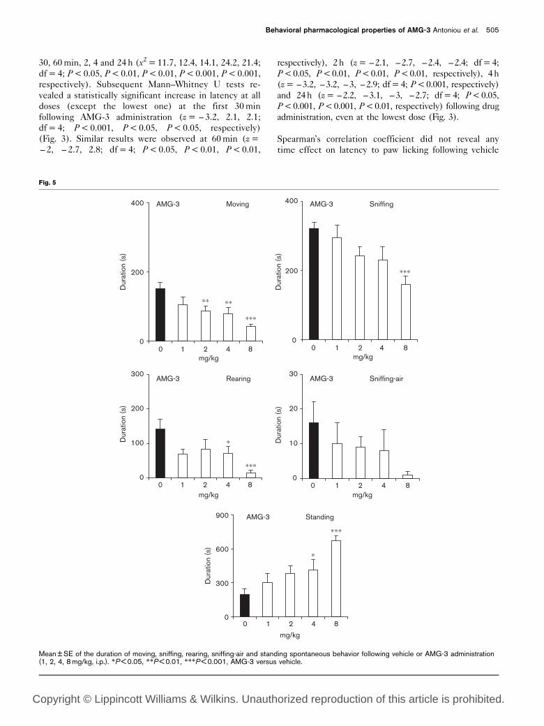
Fig. 5
AMG-3 Standing
AMG-3 Rearing AMG-3 Sniffing-air
AMG-3 SniffingAMG-3 Moving
∗∗∗
∗
0
300
600
900
mg/kg
∗∗ ∗∗
∗∗∗
0
200
400
Dur
atio
n (s
)D
urat
ion
(s)
Dur
atio
n (s
)
Dur
atio
n (s
)D
urat
ion
(s)
∗∗∗
0
200
400
∗
∗∗∗0
100
200
300
0
10
20
30
0 1 2 4 8
mg/kg0 1 2 4 8
mg/kg0 1 2 4 8
mg/kg0 1 2 4 8
mg/kg0 1 2 4 8
Mean ± SE of the duration of moving, sniffing, rearing, sniffing-air and standing spontaneous behavior following vehicle or AMG-3 administration(1, 2, 4, 8 mg/kg, i.p.). *P < 0.05, **P < 0.01, ***P < 0.001, AMG-3 versus vehicle.
Behavioral pharmacological properties of AMG-3 Antoniou et al. 505
Copyright © Lippincott Williams & Wilkins. Unauthorized reproduction of this article is prohibited.
administration. The same analysis revealed a time effect
on latency at all doses of AMG-3 (R = 0.482, 0.768, 0.870,
0.794; P < 0.01, P < 0.001, P < 0.001, P < 0.001 for 1, 2,
4, 8 mg/kg AMG-3, respectively]. In particular, latency to
paw licking is positively correlated to different time
points of reassessment, showing that the effect of the
drug increased with time up to 24 h after administration.
Bar test
Indices of catalepsy at different time intervals and at the
higher doses of AMG-3 administration are presented in
Fig. 4. Kruskall–Wallis analysis revealed a dose effect on
the latency to remove front paws from the bar, especially
at 4 and 24 h (x2 = 4.1, 15.1; df = 4; P < 0.05, P < 0.01)
following AMG-3 administration. In particular, Mann–
Whitney U tests did not reveal any statistically significant
effect on descent latency at 30 min or 60 min follow-
ing AMG-3 administration. There is an increase in the
descent latency at the highest dose (z = – 2.7, P < 0.01),
2 h following AMG-3 administration (Fig. 4). Moreover,
the descent latency was increased in a dose-dependent
manner (z = – 1.9, – 2.6, – 2.1, – 2.6; P < 0.05, P < 0.01,
respectively), 4 h after the AMG-3 administration (Fig. 4).
Finally the descent latency was increased 24 h after the
AMG-3 administration, except at the lowest dose (z =
– 2.3, – 2.7, – 2.6; P < 0.01, respectively) (Fig. 4).
Fig. 6
Moving Sniffing
∗∗∗
∗#
0
200
400
Vehicle AMG-3 AM-251 AM-251+AMG-3
Vehicle AMG-3 AM-251 AM-251+AMG-3
Vehicle AMG-3 AM-251 AM-251+AMG-3
Vehicle AMG-3 AM-251 AM-251+AMG-3
Vehicle AMG-3 AM-251 AM-251+AMG-3
Dur
atio
n (s
)D
urat
ion
(s)
Dur
atio
n (s
)D
urat
ion
(s)
∗∗∗
#
0
200
400
Rearing
∗∗∗∗∗
0
100
200
300Sniffing-air
∗0
10
20
30
Standing
#
∗∗∗
0
300
600
900
Dur
atio
n (s
)
mg/kg
mg/kg mg/kg
mg/kg
mg/kg
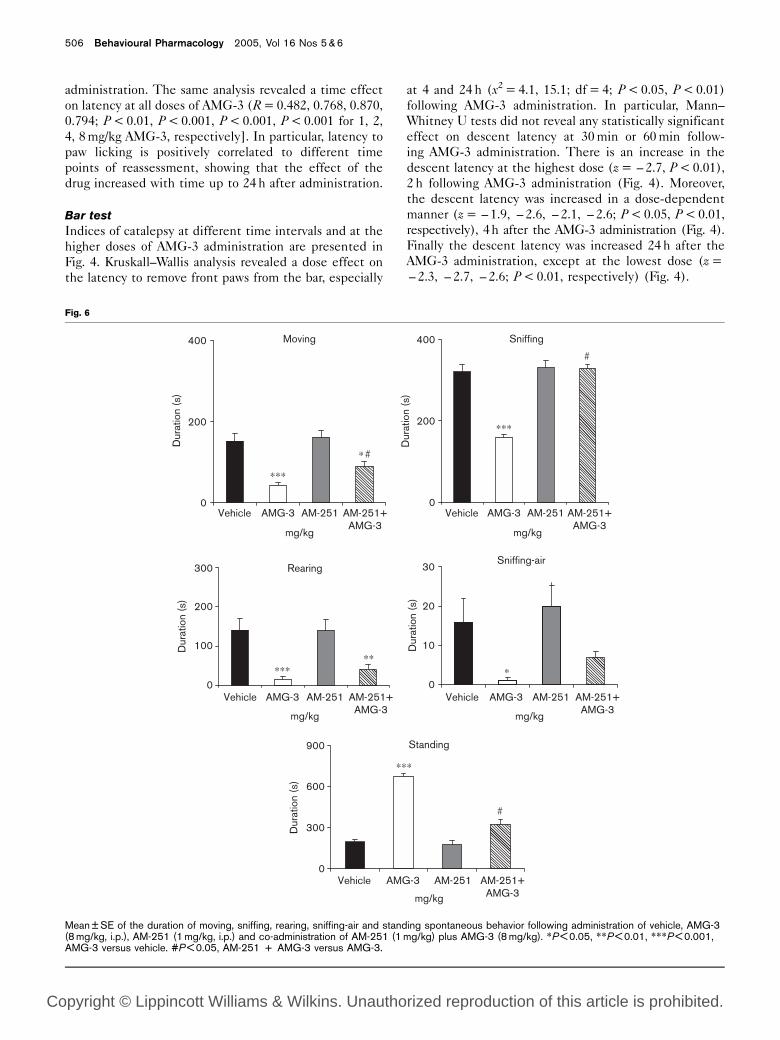
Mean ± SE of the duration of moving, sniffing, rearing, sniffing-air and standing spontaneous behavior following administration of vehicle, AMG-3(8 mg/kg, i.p.), AM-251 (1 mg/kg, i.p.) and co-administration of AM-251 (1 mg/kg) plus AMG-3 (8 mg/kg). *P < 0.05, **P < 0.01, ***P < 0.001,AMG-3 versus vehicle. #P < 0.05, AM-251 + AMG-3 versus AMG-3.
506 Behavioural Pharmacology 2005, Vol 16 Nos 5 & 6
Copyright © Lippincott Williams & Wilkins. Unauthorized reproduction of this article is prohibited.

Spearman’s correlation coefficients did not reveal any
time effect on descent latency in the bar test following
vehicle administration. The same analysis revealed a time
effect on latency at 1, 2, 4 and 8 mg/kg of AMG-3
administration (R = 0.472, 0.717, 0.842, 0.837; P < 0.01,
P < 0.001, P < 0.001, P < 0.001, respectively). In parti-
cular, descent latency in the bar test was mainly apparent
at the higher doses of AMG-3 2, 4 and 24 h after
administration.
Spontaneous activity
The effects of AMG-3 on various aspects of spontaneous
activity are presented in Fig. 5. One-way ANOVA
revealed that AMG-3 decreased the duration of moving,
sniffing, rearing behavior [F(4,31) = 6.2, 5.2, 4.2, 4.4,
P < 0.001, P < 0.01, P < 0.01, P < 0.01, respectively]. A
decrease in the sniffing-air duration was observed but did
not reach statistical significance. On the other hand,
standing duration was increased [F(4,31) = 6.2,
P < 0.001] following AMG-3 administration. Subsequent
LSD tests revealed that moving and rearing behavior
were decreased in a dose-dependent manner, while
sniffing behavior was mainly reduced at the highest dose
(Fig. 5). A dose-dependent increase of standing dura-
tion was also revealed following AMG-3 administration
(Fig. 5). Separate one-way ANOVAs revealed that pre-
treatment with AM-251 (cannabinoid antagonist) re-
versed the decrease in the afore-mentioned behavioral
aspects of spontaneous activity induced by AMG-3 at the
highest dose. In particular, AM-251 fully reversed the
decrease in sniffing duration as well as the increase of
standing duration [F(4,25) = 9.6, 27.5, P < 0.001, respec-
tively], partially reversed the decrease in moving behavior
[F(4,25) = 10.8, P < 0.001, respectively] but did not
reverse rearing or sniffing-air behavior induced by
AMG-3 at the highest dose (Fig. 6).
Self-stimulation procedure
The changes in self-stimulation threshold and asymptotic
rate of responding after systemic injection of the CB1
receptor agonist AMG-3 are presented in Figs 7A and B,
respectively. AMG-3 significantly increased self-stimula-
tion thresholds [F(4,34) = 5.06, P < 0.005] and de-
creased the asymptotic rate of responding [F(4,34) =
4.77, P < 0.005]. Post-hoc analysis with the LSD test
showed that these effects were significant in the group
receiving the highest dose of AMG-3 (8 mg/kg). There
was a significant increase of the self-stimulation thresh-
olds (P < 0.001) and the asymptotic rate of responding
(P < 0.001), compared with the vehicle group.
Fig. 7
0 1 2 4 80
50
100
150
200
∗
AMG-3Threshold
Threshold
Asymptote
Asymptote
AMG-3
0 1 2 4 8
∗
Vehicle AMG-8 AM1 AM1-AMG-80
50
100
150
200 AM-251 - AMG-3
∗
Doses of drugs (mg/kg, i.p.)
Doses of drugs (mg/kg, i.p.) Doses of drugs (mg/kg, i.p.)
Vehicle AMG-8 AM1 AM1-AMG-8Doses of drugs (mg/kg, i.p.)
% P
re-d
rug
% P
re-d
rug
% P
re-d
rug
% P
re-d
rug
0
50
100
150
200
0
50
100
150
200
AM-251 - AMG-3
(A) (B)
(C) (D)
Changes in self-stimulation threshold (A, C) and asymptotic rate (B, D) of responding (expressed as percentage of pre-drug values) following AMG-3(0, 1, 2, 4, 8 mg/kg, i.p.) and AM-251 (0, 1 mg/kg, i.p.) + AMG-3 (0, 8 mg/kg, i.p.) treatments. Vertical bars represent the standard errors of the mean.AMG8 represents AMG-3, 8 mg/kg; AM1 represents AM-251, 1 mg/kg. *Signifies an intracranial self-stimulation (ICSS) threshold and asymptotevalue significantly different from the control condition.
Behavioral pharmacological properties of AMG-3 Antoniou et al. 507
Copyright © Lippincott Williams & Wilkins. Unauthorized reproduction of this article is prohibited.

Figures 7C and D present the changes in self-stimula-
tion threshold and asymptotic rate of responding
after systemic injection of AM-251 or its vehicle and
AMG-3 or its vehicle. AMG-3 (8 mg/kg) produced an
increase in self-stimulation threshold. Administration
of AM-251 (1 mg/kg) significantly blocked this effect
[F(3,27) = 6.972, P = 0.014]. There were no significant
differences in the asymptotic rate of responding between
the different groups [F(3,27) = 0.81, NS].
DiscussionIn the present study the behavioral profile of a novel
cannabinoid (AMG-3) was examined, using different
experimental procedures. Binding studies confirmed that
AMG-3 displays an affinity for CB1 receptors which
appears to be superior to that of D8-THC (Papahatjis
et al., 1998). As was mentioned in the Introduction, the
high affinity of AMG-3 for CB1 receptors is attributed to a
hydrophobic subsite for CB1 receptors at the level of the
benzylic side-chain carbon. Therefore, the binding data
support the significance of the orientation and conforma-
tion of the side chain in determining cannabinomimetic
activity. This activity was assessed further at the
behavioral level.
The behavioral data demonstrated that AMG-3 induced
analgesic activity in a dose- and time-dependent manner.
Antinociception, as expressed by latency to paw licking in
the hot-plate test, was augmented by increasing the dose.
Additionally, this effect was more prominent at 4 and 24 h
after AMG-3 administration, showing that the drug has a
very long duration of action. Antinociceptive action for
cannabinoids has been widely described in experimental
animals, using a variety of analgesic tests (Fuentes et al.,1999). In particular, analgesic potency, as assessed by the
hot-plate test, is one of the most profound effects of
THC and several synthetic cannabinoids in most species
(Johnson et al., 1981; Dajani et al., 1999; Fuentes et al.,1999). Our data demonstrate a long-lasting analgesic
effect of AMG-3 and suggest that this compound might
prove therapeutically useful, with interesting pharmaco-
kinetic properties which need to be examined. Also,
reassessment of behavioral indices for analgesic effect at
additional time points, as well as comparisons with the
well-known cannabinomimetics, would add to the overall
estimation and clarification of this pharmacological
property.
It was also found that AMG-3 decreased spontaneous
motor activity, including forward locomotion and vertical
activity in a dose-dependent manner. Exploration as
indicated by sniffing behavior was also decreased follow-
ing AMG-3 administration, especially at the highest dose.
Interestingly, AMG-3-induced decreases in motor activ-
ity, including horizontal and vertical activity as reflected
by moving and rearing/sniffing-air, respectively, were
more prominent compared to the decrease in exploration
as reflected by sniffing behavior. This differentiation
between forward/vertical activity and sniffing behavior
possibly indicates that AMG-3-induced effects are
related to a discrete behavioral profile, related to
decreased motor activity rather than the inhibition of
some other aspects of behavior. These findings are in
agreement with the major effect of cannabinoid agonists
on motor activity (hypoactivity) and they suggest that
AMG-3 shares some of the pharmacological properties of
other CB1 agonists (Romero et al., 1996; Ferrari et al.,1999). Nevertheless, cannabinoid agonists induce bipha-
sic, or even triphasic (Sanudo-Pena et al., 2000), effects on
motor activity that are both time- and dose-dependent
(Davis et al., 1972; Dewey, 1986; Hollister, 1986;
McGregor et al., 1996; Poncelet et al., 1999). In particular,
an increase in motor activity has been associated with
relatively low doses of CB agonists, while higher doses
inhibit motor activity and produce catalepsy. The new
ligand AMG-3 did induce catalepsy, as shown by
increased descent latency in the bar test, but mainly at
the highest dose and at longer times after administration.
Catalepsy has been considered an active state that may or
may not be accompanied by inhibition of movement
(Klemm, 1989). Animals rendered cataleptic by CB
agonists display distress, mainly manifested by vocaliza-
tions and aggressiveness towards handling (Sanudo-Pena
et al., 2000). The data of this study showed that the novel
cannabinoid induced catalepsy-like behavior at a high
dose. Additionally, according to our observations, the
rats tested were also distressed during the handling
procedure.
It has been reported that cataleptic doses of cannabinoids
induce aversion in a conditioned place preference
procedure (Sanudo-Pena et al., 2000). In line with these
findings, the data of the present study demonstrated that
low doses of the CB1 receptor agonist AMG-3 did not
affect the rewarding efficacy of brain stimulation, whereas
the highest dose increased the brain stimulation reward
thresholds and decreased the response rates. This
decrease seems to be independent of cannabinoid
agonist-induced motor impairment, since changes in
threshold current may discriminate between reward and
performance (Liebman, 1983; Miliaressis et al., 1986;
Miliaressis and Rompre, 1987; Markou and Koob, 1992).
For example, Miliaressis and Rompre have shown that
motoric factors that produced profound quantitative
changes in the response rate of self-stimulation failed to
shift the rate–frequency function to any significant
degree (Miliaressis and Rompre, 1987). Moreover, the
self-stimulation threshold procedure applied in the
present study allowed determining threshold and re-
sponse rate separately and concurrently in the same
self-stimulation session. It is worth noting that a decrease
in performance is not always associated with an increase
in threshold frequency, and, more importantly, attenuated
performance has been observed in association with a
508 Behavioural Pharmacology 2005, Vol 16 Nos 5 & 6
Copyright © Lippincott Williams & Wilkins. Unauthorized reproduction of this article is prohibited.

decrease in threshold frequency (Panagis and Spyraki,
1996). More profound effects might also have been
observed if ICSS had been assessed at longer times after
AMG-3 administration.
The present results are consistent with previous studies
that showed that the synthetic cannabinoid receptor
agonists levonantradol, WIN 55,212-2, CP 55,940 and
HU-210 either did not affect, or increased, the brain
stimulation reward thresholds (Stark and Dews, 1980;
Kucharski et al., 1983; Arnold et al., 2001; Vlachou et al.,2003, 2005). However, the present results are not in
agreement with data from studies that show that THC
increases the rewarding efficacy of self-stimulation
(Gardner et al., 1988; Gardner and Vorel, 1998). These
discrepant results could be attributed to differences in
the pharmacological properties and the dose range of the
compounds tested, the methods followed and the strain
of the animals used. For example, Lepore et al. (1996)
reported the most pronounced action of THC in Lewis
rats, while they found that the effect was minimal in
Sprague–Dawley rats (such as those used in the present
study).
The effects of the highest dose of AMG-3 (8 mg/kg) were
completely reversed by pre-treatment with the CB1
receptor antagonist AM-251 at a dose that did not affect
the baseline of self-stimulation itself. In particular, the
AMG-3-induced decrease in moving and rearing behavior
was reversed to some extent, while the respective
induced decreases in sniffing, sniffing air and standing
behavior were fully reversed by AM-251. This indicates
that the inhibitory role of the cannabinoid ligand AMG-3
on spontaneous activity and brain stimulation reward is
probably mediated through CB1 receptor activation.
In summary, the present study clearly shows that the
cannabinoid ligand AMG-3 displays significant analgesic
properties, decreases motor activity at low doses and
induces catalepsy at high doses. AMG-3 does not
decrease reward threshold in the intracranial self-
stimulation paradigm, but rather has an inhibitory
influence on reward mechanisms at the highest dose
tested. These effects were reversed by AM-251, a
selective CB1 antagonist. This observation might indicate
that the behavioral effects examined are mediated
through the CB1 receptor.
In general, these findings show that AMG-3 produces a
characteristic behavioral profile in rats, which, although
reminiscent of the stimulation of cannabinoid receptors,
does not include a decreased reward threshold in the
intracranial self-stimulation paradigm. On the basis of
these data, one cannot argue that AMG-3 simply
possesses increased potency and efficacy as compared to
the classical cannabinomimetics, despite its long-lasting
effects on some behavioral tests and its high affinity for
CB receptors. Further studies, including comparisons
with additional new cannabinoid ligands of similar
structure are under way, with the aim to delineate and
establish structure–activity relationships that might
prove useful in developing drugs with promising ther-
apeutic value.
AcknowledgementsThe authors thank Ms Despina Papasava for technical
assistance and Dr Y. Alamanos, Assistant Professor of
Hygiene and Epidemiology, for his essential assistance in
the statistical procedures.
ReferencesAdams R, Harfenist M, Loewe S (1949). New analogs of tetrahydrocannabinol.
J Am Chem Soc 71:1624–1628.Antoniou K, Kafetzopoulos E (1996). The pattern of locomotor activity after
cocaine treatment in the rat. Behav Pharmacol 7:237–244.Antoniou K, Kafetzopoulos E, Papadopoulou-Daifoti Z, Hyphantis T, Marselos M
(1998). D-Amphetamine, cocaine and caffeine: a comparative study of cuteeffects on locomotor activity and behavioral patterns in rats. NeurosciBiobehav Rev 23:189–196.
Arnold JC, Hunt GE, McGregor IS (2001). Effects of the cannabinoid receptoragonist CP 55,940 and the cannabinoid receptor antagonist SR 141716 onintracranial self-stimulation in Lewis rats. Life Sci 70:97–108.
Chaperon F, Thiebot MH (1998). Behavioral effects of cannabinoid agents inanimals. Crit Rev Neurobiol 13:243–281.
Coulombe D, Miliaressis E (1987). Fitting intracranial self-stimulation data withgrowth models. Behav Neurosci 101:209–214.
Dajani EZ, Larsen KR, Taylor J, Dajani NE, Shahwan TG, Neeleman SD, et al.(1999). 10 ,10-Dimethylheptyl-delta-8-tetrahydrocannabinol-11-oic acid: anovel, orally effective cannabinoid with analgesic and anti-inflammatoryproperties. J Pharmacol Exp Ther 291:31–38.
Davis WM, Moreton JE, King WT, Pace HB (1972). Marihuana on locomotoractivity: biphasic effect and tolerance development. Res Commun ChemPathol Pharmacol 3:29–35.
Devane WA, Dysarz FA, Johnson RM, Melvin LS, Howlett AC (1988).Determination and characterization of a cannabinoid receptor in rat brain.Mol Pharmacol 34:605–613.
Dewey WL (1986). Cannabinoid pharmacology. Pharmacol Rev 38:151–178.Di Marzo V, Bifulco M, De Petrocellis L (2004). The endocannabinoid system and
its therapeutic exploitation. Nat Rev Drug Discov 3:771–784.Ferrari F, Ottani A, Giuliani D (1999). Cannabimimetic activity in rats and pigeons
of HU 210, a potent antiemetic drug. Pharmacol Biochem Behav 62:75–80.Fuentes JA, Ruiz-Gayo M, Manzanares J, Vela G, Reche I, Corchero J (1999).
Cannabinoids as potential new analgesics. Life Sci 65:675–685.Gardner EL (2002). Addictive potential of cannabinoids: the underlying
neurobiology. Chem Phys Lipids 121:267–290.Gardner EL, Vorel SR (1998). Cannabinoid transmission and reward-related
events. Neurobiol Dis 5:502–533.Gardner EL, Paredes W, Smith D, Donner A, Milling C, Cohen D, et al. (1988).
Facilitation of brain stimulation reward by D9-tetrahydrocannabinol. Psycho-pharmacology (Berl) 96:142–144.
Hall PW, Christie PM, Currow D (2005). Cannabinoids and cancer: causation,remediation, and palliation. Lancet Oncol 6:35–42.
Hollister LE (1986). Health aspects of cannabis. Pharmacol Rev 38:1–20.Howlett AC, Breivogel CS, Childers SR, Deadwyler SA, Hampson RE, Porrino LJ
(2004). Cannabinoid physiology and pharmacology: 30 years of progress.Neuropharmacology 47:345–358.
Johnson MR, Melvin LS, Althuis TH, Bindra JS, Harbert CA, Milne GM, et al.(1981). Selective and potent analgesics derived from cannabinoids. J ClinPharmacol 21:271–282.
Khanolkar AD, Abadji V, Lin S, Adam W, Hill G, Taha G, et al. (1996). Head groupanalogs of arachidonylethanolamide, the endogenous cannabinoid ligand.J Med Chem 39:4515–4519.
Khanolkar AD, Palmer SL, Makriyannis A (2000). Molecular probes for thecannabinoid receptors. Chem Phys Lipids 108:37–52.
Klemm WR (1989). Drug effects on active immobility responses: what they tell usabout neurotransmitter systems and motor functions. Prog Neurobiol32:403–422.
Behavioral pharmacological properties of AMG-3 Antoniou et al. 509
Copyright © Lippincott Williams & Wilkins. Unauthorized reproduction of this article is prohibited.

Kucharski LT, Williams JE, Kornetsky C (1983). The effects of levonantradol onrewarding brain stimulation thresholds in the rat. Pharmacol Biochem Behav19:149–151.
Lepore M, Liu X, Savage V, Matalon D, Gardner EL (1996). Genetic differences inD9-tetrahydrocannabinol-induced facilitation of brain stimulation reward asmeasured by a rate–frequency curve-shift electrical brain stimulationparadigm in three different rat strains. Life Sci 58:365–372.
Liebman JM (1983). Discriminating between reward and performance: a criticalreview of intracranial self-stimulation methodology. Neurosci Biobehav Rev7:45–72.
Makriyannis A, Rapaka RS (1990). The molecular basis of cannabinoid activity.Life Sci 47:2173–2184.
Maldonado-Irizarry CS, Stellar JR, Kelley AE (1994). Effects of cocaine and GBR-12909 on brain stimulation reward. Pharmacol Biochem Behav 48:915–920.
Markou A, Koob GF (1992). Construct validity of a self-stimulation thresholdparadigm: effects of reward and performance manipulations. Physiol Behav51:111–119.
McGregor IS, Issakidis CN, Prior G (1996). Aversive effects of the syntheticcannabinoid CP 55,940 in rats. Pharmacol Biochem Behav 53:657–664.
Miliaressis E, Rompre P-P (1987). Effects of concomitant motor reactions on themeasurement of rewarding efficacy of brain stimulation. Behav Neurosci101:827–831.
Miliaressis E, Rompre PP, Laviolette P, Philippe L, Coulombe D (1986). Thecurve-shift paradigm in self-stimulation. Physiol Behav 37:85–91.
Munro S, Thomas KL, Abu-Shaar M (1993). Molecular characterization of aperipheral receptor for cannabinoids. Nature 365:61–65.
Panagis G, Spyraki C (1996). Neuropharmacological evidence for the role ofdopamine in ventral pallidum self-stimulation. Psychopharmacology (Berl)123:280–288.
Papahatjis D, Kourouli T, Abadji V, Goutopoulos A, Makriyannis A (1998).Pharmacophoric requirements for cannabinoid side chains: multiple bond andC10-substituted D8-tetrahydrocannabinols. J Med Chem 41:1195–1200.
Papahatjis DP, Nikas SP, Andreou T, Makriyannis A (2002). Novel 10 ,10-chainsubstituted D8-tetrahydrocannabinols. Bioorg Med Chem Lett 12:3583–3586.
Paxinos G, Watson C (1998). The Rat Brain in Stereotaxic Coordinates. 4th ed.London: Academic Press.
Pertwee RG (1997). Pharmacology of cannabinoid CB1 and CB2 receptors.Pharmacol Ther 74:129–180.
Pertwee RG (2001). Cannabinoids and receptors and pain. Prog Neurobiol63:569–611.
Piomelli D (2003). The molecular logic of endocannabinoid signaling. NatureNeurosci 4:873–883.
Poncelet M, Barnouin MC, Breliere JC, Soubrie P (1999). Blockade ofcannabinoid (CB1) receptors by SR 141716 selectively antagonizes drug-induced reinstatement of exploratory behaviour in gerbils. Psychopharmacol-ogy (Berl) 144:144–150.
Ranaldi R, Beninger RJ (1994). The effects of systemic and intracerebralinjections of D1 and D2 agonists on brain stimulation reward. Brain Res651:283–292.
Razdan RK (1986). Structure–activity relationships in cannabinoids. PharmacolRev 38:75–149.
Romero J, Garcia-Palomero E, Lin SY, Ramos IA, Makriyannis A, Fernandez-RuizJJ (1996). Extrapyramidal effects of methanadamide, an analog of ananda-mide, the endogenous CB1 receptor ligand. Life Sci 15:1249–1257.
Romero J, Lastres-Becker I, deMiguel R, Berrendero F, Ramos JA, Fernandez-RuizJ (2002). The endogenous cannabinoid system and the basal ganglia:biochemical, pharmacological and therapeutic aspects. Pharmacol Ther95:137–152.
Sanudo-Pena MC, Tsou K, Delay ER, Hohman AG, Force M, Walker JM (1997).Endogenous cannabinoids as an aversive or counter-rewarding system in therat. Neurosci Lett 223:125–128.
Sanudo-Pena MC, Romero J, Seale GE, Fernandez-Ruiz JJ, Walker JM (2000).Activational role of cannabinoids on movement. Eur J Pharmacol 391:269–274.
Spruijt BM, Gispen WH (1984). Behavioral sequences as an easily quantifiableparameter in experimental studies. Physiol Behav 32:707–710.
Stark P, Dews PB (1980). Cannabinoids: behavioral effects. J Pharmacol ExpTher 214:124–130.
Stellar JR, Rice MB (1989). Pharmacological basis of intracranial self-stimulationreward. In: Liebman JM, Cooper SJ (editors): The NeuropharmacologicalBasis of Reward. Oxford: Oxford Science Publications; pp. 14–65.
Tanda G, Goldberg SR (2003). Cannabinoids: reward, dependence, andunderlying neurochemical mechanisms – a review of recent preclinical data.Psychopharmacology (Berl) 169:135–140.
Vlachou S, Nomikos GG, Panagis G (2003). WIN 55,212-2 decreases thereinforcing actions of cocaine through CB1 cannabinoid receptor stimulation.Behav Brain Res 141:215–222.
Vlachou S, Nomikos GG, Panagis G (2005). CB1 cannabinoid receptor agonistsincrease intracranial self-stimulation thresholds in the rat. Psychopharmacol-ogy (Berl) 179:498–508.
Wise RA (1980). Action of drugs of abuse on brain reward systems. PharmacolBiochem Behav 13:213–223.
Wise RA (1996). Addictive drugs and brain stimulation reward. Annu RevNeurosci 19:319–340.
Wise RA (1998). Drug-activation of brain reward pathways. Drug AlcoholDepend 51:13–22.
510 Behavioural Pharmacology 2005, Vol 16 Nos 5 & 6
Copyright © Lippincott Williams & Wilkins. Unauthorized reproduction of this article is prohibited.