Behavior of reinforced concrete segmental hollow core slabs ...
Upload
independentCategory
view
0download
0
Invariant Natural Killer T Cell Agonist ModulatesExperimental Focal and Segmental GlomerulosclerosisRafael L. Pereira1., Vanessa O. Reis1., Patricia Semedo1, Bruna N. Buscariollo1, Cassiano Donizetti-
Oliveira1, Marcos A. Cenedeze1, Maria Fernanda Soares1, Alvaro Pacheco-Silva1,2, Paul B. Savage3,
Niels O. S. Camara1,4, Alexandre C. Keller1,5*
1 Departamento de Medicina – Nefrologia, Universidade Federal de Sao Paulo, Sao Paulo, Brasil, 2 Unidade de Transplante Renal, Instituto Israelita de Ensino e Pesquisa
Albert Einstein, Sao Paulo, Brasil, 3 Department of Chemistry and Biochemistry Brigham Young University, Provo, Utah, United States of America, 4 Departamento de
Imunologia, Universidade de Sao Paulo, Sao Paulo, Brasil, 5 Departamento de Microbiologia, Imunologia e Parasitologia, Universidade Federal de Sao Paulo, Sao Paulo,
Brasil
Abstract
A growing body of evidence demonstrates a correlation between Th2 cytokines and the development of focal andsegmental glomerulosclerosis (FSGS). Therefore, we hypothesized that GSL-1, a monoglycosylceramide from Sphingomonasssp. with pro-Th1 activity on invariant Natural Killer T (iNKT) lymphocytes, could counterbalance the Th2 profile andmodulate glomerulosclerosis. Using an adriamycin(ADM)-based model of FSGS, we found that BALB/c mice presentedalbuminuria and glomerular degeneration in association with a Th2-like pro-fibrogenic profile; these mice also expressed acombination of inflammatory cytokines, such as IL-4, IL-1a, IL-1b, IL-17, TNF-a, and chemokines, such as RANTES and eotaxin.In addition, we observed a decrease in the mRNA levels of GD3 synthase, the enzyme responsible for GD3 metabolism, aglycolipid associated with podocyte physiology. GSL-1 treatment inhibited ADM-induced renal dysfunction and preservedkidney architecture, a phenomenon associated with the induction of a Th1-like response, increased levels of GD3 synthasetranscripts and inhibition of pro-fibrotic transcripts and inflammatory cytokines. TGF-b analysis revealed increased levels ofcirculating protein and tissue transcripts in both ADM- and GSL-1-treated mice, suggesting that TGF-b could be associatedwith both FSGS pathology and iNKT-mediated immunosuppression; therefore, we analyzed the kidney expression ofphosphorylated SMAD2/3 and SMAD7 proteins, molecules associated with the deleterious and protective effects of TGF-b,respectively. We found high levels of phosphoSMAD2/3 in ADM mice in contrast to the GSL-1 treated group in whichSMAD7 expression increased. These data suggest that GSL-1 treatment modulates the downstream signaling of TGF-bthrough a renoprotective pathway. Finally, GSL-1 treatment at day 4, a period when proteinuria was already established,was still able to improve renal function, preserve renal structure and inhibit fibrogenic transcripts. In conclusion, our workdemonstrates that the iNKT agonist GSL-1 modulates the pathogenesis of ADM-induced glomerulosclerosis and mayprovide an alternative approach to disease management.
Citation: Pereira RL, Reis VO, Semedo P, Buscariollo BN, Donizetti-Oliveira C, et al. (2012) Invariant Natural Killer T Cell Agonist Modulates Experimental Focal andSegmental Glomerulosclerosis. PLoS ONE 7(3): e32454. doi:10.1371/journal.pone.0032454
Editor: Maria Pia Rastaldi, Fondazione IRCCS Ospedale Maggiore Policlinico & Fondazione D’Amico per la Ricerca sulle Malattie Renali, Italy
Received May 27, 2011; Accepted January 30, 2012; Published March 12, 2012
Copyright: � 2012 Pereira et al. This is an open-access article distributed under the terms of the Creative Commons Attribution License, which permitsunrestricted use, distribution, and reproduction in any medium, provided the original author and source are credited.
Funding: This work was supported by Fundacao de Amparo a Pesquisa do Estado de Sao Paulo (nu 2007/07120-0)and Conselho Nacional de DesenvolvimentoCientıfico e Tecnologico (nu501848/2009-6 and 484445/2010-3), Brazil. The funders had no role in study design, data collection and analysis, decision to publish, orpreparation of the manuscript.
Competing Interests: Co-author NOSC is a PLoS ONE Editorial Board member. This does not alter the authors’ adherence to all the PLoS ONE policies on sharingdata and materials.
* E-mail: [email protected]
. These authors contributed equally to this work.
Introduction
Focal and segmental glomerulosclerosis (FSGS) is a growing
cause of adult nephrotic syndrome and chronic kidney disease.
Although FSGS presents diverse histological patterns and
etiological associations, podocyte injury is a common denominator
[1]. The immunological mechanisms involved in the pathogenesis
of FSGS are not fully understood, but various studies demonstrate
an association between a Th2-like profile and disease develop-
ment. Yap and colleagues were the first to demonstrate a
correlation between increased IL-13 mRNA expression and
idiopathic nephrotic syndrome (INS) during childhood; because
FSGS is one of the most common causes of INS, it was considered
an indication of the association between Th2 cytokines and FSGS
[2]. In the spontaneous FSGS Buffalo/Mna rat model, Le Berre
and colleagues found an early imbalance in Th1/Th2 cytokines
due to a T-cell infiltrate with a predominant Th2 profile, which in
turn down-regulated Th1 responses [3]. Consistent with these
results, Lai and colleagues demonstrated that IL-13 over-
expression induced minimum change-like nephropathy, a phe-
nomenon associated with podocyte structural changes and
increased expression of IL-4Ra and IL-13Ra2 in the glomeruli
[4].
Combined, these previous studies support a correlation between
Th2 cytokines and the development of FSGS. Because of the
antagonism between Th1 and Th2 cytokines, we hypothesized
that the polarization of immune responses toward a Th1 profile
could inhibit or even modulate the pathogenesis of FSGS. In this
PLoS ONE | www.plosone.org 1 March 2012 | Volume 7 | Issue 3 | e32454
sense, the activation of invariant natural killer T lymphocytes
(iNKT) by their agonist a-galactosylceramide (a-GaCer) or
analogs has been shown to increase Th1-mediated responses, a
property that has been used successfully to modulate Th2-
mediated diseases, such as asthma [5,6,7,8].
iNKT cells are non-conventional lymphocytes that can
modulate the outcome of different immune-mediated diseases
through the prompt secretion of different cytokines upon TCR
stimulation [9]. A characteristic feature of iNKT cells is their
selectivity for glycolipid antigens presented by the nonpolymorphic
MHC class I-like molecule CD1d, which has been used to
modulate different immune responses by exogenous agonists
[10,11,12]. We chose to study the effect of GSL-1, a mono-
glycosylceramide obtained from Sphingomonas ssp. with a pro-Th1
nature, on FSGS pathogenesis [13]. To this end, we used an
experimental model that is based on the susceptibility of podocytes
to the cytotoxic effects of doxorubicin hydrochloride, also known
as adriamycin (ADM) [14].
ADM-induced FSGS not only has an immune system-
dependent component but is also a reliable mimetic of the human
disease [15,16]. Although the immune component is not fully
understood, Th2-prone strains, such as BALB/c mice, are well
known to be more susceptible to ADM injury, corroborating the
idea that FSGS is a Th2-associated disease. Therefore, ADM-
FSGS is a useful model to both reproduce the disease and study
the effects of Th1 polarization on disease pathogenesis.
In this study, we demonstrate that GSL-1 treatment modulates
the development of ADM-induced FSGS in an iNKT-dependent
manner. A Th1-polarization, increased mRNA for enzymes
associated with ganglioside metabolism, and the modulation of
proteins involved with the regulation of TGF-b downstream
signaling were observed in this model.
Results
GSL-1 treatment inhibits ADM-induced proteinuria andalbuminuria
We first tested the effect of GSL-1 treatment on the renal failure
induced by ADM administration. BALB/c mice, aged 8 to 10
weeks, were injected at day 0 with ADM (10 mg/Kg) or ADM
plus GSL-1 (5 mg/animal). Figure 1A demonstrates that ADM
mice lost body weight in a time-dependent manner, likely as a
result of ADM-induced nephropathy. Consistent with this result,
the proteinuria/creatininuria ratio at day 7 was significantly
elevated in the ADM group compared with the control mice,
indicating impaired renal function (Figure 1B). In contrast, GSL-1-
treated mice gained body weight, as did the control animals,
suggesting that GSL-1 administration exerts a protective effect on
ADM-induced disease. In fact, the proteinuria levels found in
ADM+GSL-1 mice were comparable to that of the control group,
corroborating the renoprotective effect of GSL-1 treatment. The
total proteinuria/creatininuria ratio can be a marker for renal
function but is not a reliable indicator of glomerular alterations. In
contrast, albumin is a high molecular weight protein that is not
found in urine, and therefore, an increase in albumin levels reflects
a loss of the glomerular filtration barrier (GFB) due to podocyte
injury. Therefore, we measured the albuminuria/creatininuria
ratio to better characterize the extent of podocyte injury.
Consistent with data from Figure 1B, ADM mice showed
increased albuminuria compared with control and ADM+GSL-1
mice, supporting the idea that GSL-1 treatment protected
podocytes from ADM cytotoxicity and consequently preserved
the GFB (Figure 1C).
To better address the relationship between the renoprotective
effects of GSL-1 treatment and iNKT activation, we took
advantage of the BALB/c iNKT-deficient (Jalpha182/2) strain
[17]. As in BALB/c WT mice, ADM administration in
Jalpha182/2 animals resulted in a time-dependent loss of renal
function, illustrated by the increase in the proteinuria/creatini-
nuria ratio (Figure 1D). The glomerular damage was represented
by the elevated levels of albuminuria found at day 28 post-ADM
(Figure 1E). In contrast with the WT mice, the treatment of
Jalpha182/2 mice with GSL-1 was unable to inhibit FSGS
development; therefore, our data demonstrate that the protective
effect of GSL-1 is iNKT-dependent, corroborating the specificity
of GSL-1 as an iNKT agonist.
GSL-1 treatment inhibits ADM-induced renal injuryTo determine the extent of the renal tissue damage, we
performed histological analyses at day 7 post-ADM. Figure 2
demonstrates that ADM injection resulted in alterations of renal
tissue, inducing mesangial hypercellularity, signs of glomerular
sclerosis and tubular degeneration when compared with control
mice (Figures 2A to 2D, respectively). In contrast, GSL-1
treatment preserved renal architecture, corroborating the conser-
vation of renal function depicted in Figure 1.
GSL-1 treatment modulates the pro-Th2 milieu inducedby ADM and inhibits the expression of pro-fibrotictranscripts
The qPCR analyses of renal tissue revealed that ADM injection
resulted in an early increase in the mRNA levels of the pro-Th2
transcription factor GATA3 along with transcripts for IL-4 and
IL-13, two cytokines extensively associated with Th2 responses
(Figures 3A to 3C, respectively) [18,19,20]. In contrast, GSL-1
treatment inhibited GATA3 and IL-4 mRNA expression, a
phenomenon associated with increased mRNA expression of T-
bet (T box expressed in T-cells), a pro-Th1-related transcription
factor, TNF-a, a classical type 1 cytokine and CXCL16, a
chemokine associated with iNKT-mediated Th1 responses
(Figures 3D to 3F, respectively) [21,22]. These data are consistent
with the idea that FSGS is associated with Th2 responses and
support the hypothesis that GSL-1 treatment promotes a pro-Th1
polarization of the immune system and thereby protects the
kidneys.
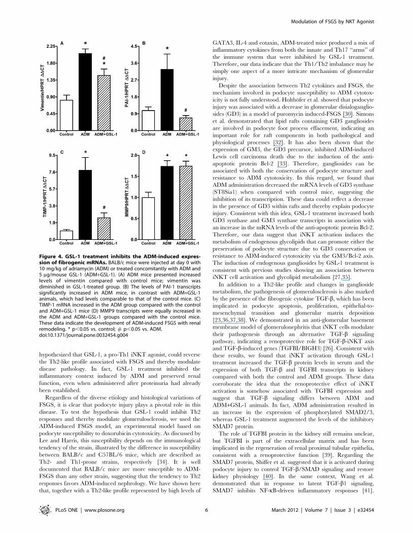
Because FSGS pathology can be associated with kidney fibrosis,
we decided to determine the mRNA expression of vimentin,
plasminogen activator inhibitor-1 (PAI-1) and TIMP-1 (tissue
inhibitor of metalloproteinase-1). Figure 4A demonstrates that
ADM mice showed increased levels of mRNA for vimentin, which
was diminished in the GSL-1 group. Consistent with these results,
we found that the induction of PAI-1 mRNA was inhibited in
GSL-1 treated mice compared with the ADM group (Figure 4B).
Finally, TIMP-1 transcript levels also increased in the ADM group
compared with control and ADM+GSL-1 mice (Figure 4C). The
mRNA levels of metalloproteinase 9 (MMP9) were slightly
increased in both ADM and ADM+GSL-1 animals when
compared with the control group (Figure 4D). The western blot
analysis of kidney tissue corroborated the MMP9 and PAI-1
transcript findings, also showing a significant increase in desmin in
ADM animals compared with both control and ADM+GSL-1
animals (Figure S1). This result corroborates previous findings
associating podocyte injury with desmin expression [23]. Taken
together, our data demonstrate that the renoprotective effect of
GSL-1 is associated with the inhibition of important fibrogenic-
Modulation of FSGS by NKT Agonist
PLoS ONE | www.plosone.org 2 March 2012 | Volume 7 | Issue 3 | e32454
associated factors that are increased in response to ADM-induced
injury.
GSL-1 treatment inhibits the inflammatory contextinduced by ADM
To better characterize the inflammatory environment associ-
ated with ADM-induced FSGS pathogenesis, we performed a
multiparameter analysis of inflammatory cytokines present in
kidney tissue. Figure 5 demonstrates increased levels of IL-4, IL-
1a, IL-1b, TNF-a, IL-12p40 and IL-17 in the ADM group
compared with the control animals; such increases were not
observed in GSL-1-treated mice (Figures 5A, 5B, 5C, 5D, 5E and
5F, respectively). In addition, we found high levels of RANTES
and eotaxin in ADM- but not in GSL-1-treated mice (Figures 5G
and H, respectively); eotaxin is a chemokine extensively
associated with Th2 inflammation [24]. These data demonstrate
that, together with a Th2-like profile, FSGS development is also
associated with cytokines from the innate and Th17 ‘‘arms’’ of
the immune response. Moreover, these results further demon-
strate the inhibitory effect of GSL-1 treatment on the inflamma-
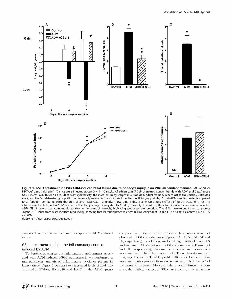
Figure 1. GSL-1 treatment inhibits ADM-induced renal failure due to podocyte injury in an iNKT-dependent manner. BALB/c WT oriNKT-deficient (Jalpha182/2) mice were injected at day 0 with 10 mg/kg of adriamycin (ADM) or treated concomitantly with ADM and 5 mg/mouseGSL-1 (ADM+GSL-1). (A) As a result of ADM cytotoxicity, the mice lost body weight in a time-dependent fashion, in contrast to the control, untreatedmice, and the GSL-1-treated group. (B) The increased proteinuria/creatininuria found in the ADM group at day 7 post-ADM injection reflects impairedrenal function compared with the control and ADM+GSL-1 animals. These data indicate a renoprotective effect of GSL-1 treatment. (C) Thealbuminuria levels found in ADM animals reflect the podocyte injury due to ADM cytotoxicity. In contrast, the albuminuria/creatininuria ratio in theADM+GSL-1 group was comparable to that in the control animals, indicating podocyte conservation. The GSL-1 treatment failed to protectJalpha182/2 mice from ADM-induced renal injury, showing that its renoprotective effect is iNKT-dependent (D and E). * p,0.05 vs. control; # p,0.05vs. ADM.doi:10.1371/journal.pone.0032454.g001
Modulation of FSGS by NKT Agonist
PLoS ONE | www.plosone.org 3 March 2012 | Volume 7 | Issue 3 | e32454

tory response induced by ADM, supporting its renoprotective
effect.
GSL-1 treatment induces an alternative TGF-b signalingpathway
Glomerulosclerosis pathogenesis has been extensively associated
with TGF-b synthesis and/or signaling; therefore, we decided to
determine the effect of GSL-1 on TGF-b production. Figure 6A
demonstrates that ADM injection increased the serum levels of
total TGF-b when compared with control mice, reinforcing the
idea that TGF-b overproduction is associated with glomeruloscle-
rosis [25]. Unexpectedly, we found that GSL-1 treatment further
elevated the serum levels of TGF-b. qPCR analysis of kidney tissue
revealed that the induction of TGF-b mRNA exactly reflected the
serum findings. The ADM group showed higher levels of TGF-btranscripts than the control animals but lower than those found in
GSL-1 mice (Figure 6B). These data suggest that, in our model,
TGF-b could be associated with either a renoprotective or
deleterious effect depending on the context in which it is produced.
We have previously shown that a renoprotective TGF-b signal
was associated with iNKT cells and the induction of TGF-b-
induced gene (TGFBI/BIGH3) transcripts [26]. Further PCR
analysis revealed that only treatment with GSL-1 was able to
induce the transcription of TGFBI mRNA (Figure 6C), suggesting
that the activation of iNKT cells by pro-Th1 agonists induced an
immunological milieu that favors the renoprotective effect of TGF-
b. To better understand this phenomenon, we used western
blotting to analyze the protein expression of SMAD2/3 and
SMAD7, which have been extensively associated with TGF-bsignaling. Figure 6D shows that ADM injection was associated
with increased levels of phosphorylated SMAD2/3 protein,
whereas GSL-1 treatment induced a slight increase in the levels
of SMAD7 protein. These data reinforce the idea that the effect of
TGF-b is dependent on the immune context induced by ADM or
ADM+GSL-1 treatment.
GSL-1 treatment increases the levels of GM3 and GD3ganglioside synthase transcripts
ADM-induced podocyte injury has been associated with
decreased levels of glomerular GD3 ganglioside, whereas iNKT
cell activation has been associated with the synthesis of glycolipids,
predominantly gangliosides [27,28]. Therefore, we determined the
mRNA expression of different classes of enzymes involved in
glycolipid metabolism. We found that ADM mice showed a
significant decrease in mRNA levels for GD3 synthase (ST8Sia1),
whereas GSL-1-treated mice showed increased expression of
Figure 2. GSL-1 treatment inhibits renal injury due to ADM cytotoxicity. BALB/c mice were injected at day 0 with 10 mg/kg of adriamycin(ADM) or treated concomitantly with ADM and 5 mg/mouse GSL-1 (ADM+GSL-1). (A) Representative image of the renal alterations induced by ADMadministration (black arrow). Figures B to D show graphic representation of the mesangial hypercellularity, signs of glomerular sclerosis and tubulardegeneration induced by ADM administration. Figure 2 corroborates the renoprotective effect of GSL-1 treatment depicted in Figure 1. * p,0.05 vs.control; # p,0.05 vs. ADM.doi:10.1371/journal.pone.0032454.g002
Modulation of FSGS by NKT Agonist
PLoS ONE | www.plosone.org 4 March 2012 | Volume 7 | Issue 3 | e32454

ST8Sia1 transcripts (Figure 7A). These results are consistent with
previous studies showing a correlation between GD3 gangliosides
and kidney physiology [29,30,31,32]. In addition, the expression
of GM3 gangliosides has been shown to be associated with
protection from ADM-cytotoxicity through the induction of the
anti-apoptotic protein Bcl-2 [33]. Although ADM administration
did not influence the levels of GM3 synthase (ST3Gal5)
transcripts, GSL-1 treatment induced a significant increase in
the mRNA levels of this enzyme (Figure 7B). Consistent with the
idea that GM3 could be associated with renoprotection through
the induction of Bcl-2, we detected increased levels of Bcl-2
transcripts in ADM+GSL-1 mice (Figure 7C). Taken together,
these data suggest that iNKT activation due to GSL-1 adminis-
tration induces endogenous glycolipids that can, in turn, protect
podocytes from ADM cytotoxicity.
GSL-1 treatment ameliorates ADM-induced FSGSFinally, we decided to determine whether the renoprotective
effect of GSL-1 could be extended to the treatment of an ongoing
disease. Therefore, we initiated GSL-1 treatment at day 4 post-
ADM injection, a time point when proteinuria was already
elevated when compared with control animals. Figure 8A
demonstrates that the time-dependent loss of body weight induced
by ADM administration was reversed by GSL-1 treatment at day
4. This phenomenon was associated with the reversion of time-
dependent renal failure, indicated by a continuous increase in
proteinuria and albuminuria levels in the ADM mice (Figures 8B
and 8C, respectively). Figures 8D and 8E demonstrate that the
recovery of renal function was associated with kidney preservation.
The transcripts for vimentin, PAI-1 and TIMP-1 were also
elevated in the ADM group when compared with the GSL-1
treated mice. Moreover, MMP9 mRNA was not detected in
ADM+GSL-1 mice, suggesting that MMP9 transcription was
inhibited (Figure 8F). Therefore, our data demonstrate that GSL-1
administration during the early stage of disease can reverse its
pathogenesis.
Discussion
The mechanisms involved in FSGS pathogenesis are still not
understood, but different studies have demonstrated, in both
humans and experimental models, that this disease is associated
with increased levels of Th2 cytokines [2,3,4]. Therefore, we first
Figure 3. GSL-1 treatment modulates the ADM-induced Th2-like cytokine profile and induces a pro-Th1 environment. BALB/c micewere injected at day 0 with 10 mg/kg of adriamycin (ADM) or treated concomitantly with ADM and 5 mg/mouse GSL-1 (ADM+GSL-1). The ADM micehad increased levels of mRNA for the Th2-related transcription factor GATA3 (A), reflecting increased transcript levels for IL-4 (B) and IL-13 (C). Incontrast, GSL-1 treatment inhibited these transcripts in association with an increase in the levels of mRNA for T-bet (D), a transcription factorassociated with Th1 responses, TNFa (E) and the chemokine ligand CXCL16 (F). Thus, these data corroborate the idea that ADM-induced FSGS isassociated with a Th2-like profile that can be inhibited by GSL-1 treatment and the generation of a pro-Th1 environment. * p,0.05 vs. control;# p,0.05 vs. ADM.doi:10.1371/journal.pone.0032454.g003
Modulation of FSGS by NKT Agonist
PLoS ONE | www.plosone.org 5 March 2012 | Volume 7 | Issue 3 | e32454
hypothesized that GSL-1, a pro-Th1 iNKT agonist, could reverse
the Th2-like profile associated with FSGS and thereby modulate
disease pathology. In fact, GSL-1 treatment inhibited the
inflammatory context induced by ADM and preserved renal
function, even when administered after proteinuria had already
been established.
Regardless of the diverse etiology and histological variations of
FSGS, it is clear that podocyte injury plays a pivotal role in this
disease. To test the hypothesis that GSL-1 could inhibit Th2
responses and thereby modulate glomerulosclerosis, we used the
ADM-induced FSGS model, an experimental model based on
podocyte susceptibility to doxorubicin cytotoxicity. As discussed by
Lee and Harris, this susceptibility depends on the immunological
tendency of the strain, illustrated by the difference in susceptibility
between BALB/c and C57BL/6 mice, which are described as
Th2- and Th1-prone strains, respectively [34]. It is well
documented that BALB/c mice are more susceptible to ADM-
FSGS than any other strain, suggesting that the tendency to Th2
responses favors ADM-induced nephrology. We have shown here
that, together with a Th2-like profile represented by high levels of
GATA3, IL-4 and eotaxin, ADM-treated mice produced a mix of
inflammatory cytokines from both the innate and Th17 ‘‘arms’’ of
the immune system that were inhibited by GSL-1 treatment.
Therefore, our data indicate that the Th1/Th2 imbalance may be
simply one aspect of a more intricate mechanism of glomerular
injury.
Despite the association between Th2 cytokines and FSGS, the
mechanism involved in podocyte susceptibility to ADM cytotox-
icity is not fully understood. Holthofer et al. showed that podocyte
injury was associated with a decrease in glomerular disialoganglio-
sides (GD3) in a model of puromycin induced-FSGS [30]. Simons
et al. demonstrated that lipid rafts containing GD3 gangliosides
are involved in podocyte foot process effacement, indicating an
important role for raft components in both pathological and
physiological processes [32]. It has also been shown that the
expression of GM3, the GD3 precursor, inhibited ADM-induced
Lewis cell carcinoma death due to the induction of the anti-
apoptotic protein Bcl-2 [33]. Therefore, gangliosides can be
associated with both the conservation of podocyte structure and
resistance to ADM cytotoxicity. In this regard, we found that
ADM administration decreased the mRNA levels of GD3 synthase
(ST8Sia1) when compared with control mice, suggesting the
inhibition of its transcription. These data could reflect a decrease
in the presence of GD3 within rafts and thereby explain podocyte
injury. Consistent with this idea, GSL-1 treatment increased both
GD3 synthase and GM3 synthase transcripts in association with
an increase in the mRNA levels of the anti-apoptotic protein Bcl-2.
Therefore, our data suggest that iNKT activation induces the
metabolism of endogenous glycolipids that can promote either the
preservation of podocyte structure due to GD3 conservation or
resistance to ADM-induced cytotoxicity via the GM3/Bcl-2 axis.
The induction of endogenous gangliosides by GSL-1 treatment is
consistent with previous studies showing an association between
iNKT cell activation and glycolipid metabolism [27,35].
In addition to a Th2-like profile and changes in ganglioside
metabolism, the pathogenesis of glomerulosclerosis is also marked
by the presence of the fibrogenic cytokine TGF-b, which has been
implicated in podocyte apoptosis, proliferation, epithelial-to-
mesenchymal transition and glomerular matrix deposition
[23,36,37,38]. We demonstrated in an anti-glomerular basement
membrane model of glomerulonephritis that iNKT cells modulate
their pathogenesis through an alternative TGF-b signaling
pathway, indicating a renoprotective role for TGF-b-iNKT axis
and TGF-b-induced genes (TGFBI/BIGH3) [26]. Consistent with
these results, we found that iNKT activation through GSL-1
treatment increased the TGF-b protein levels in serum and the
expression of both TGF-b and TGFBI transcripts in kidney
compared with both the control and ADM groups. These data
corroborate the idea that the renoprotective effect of iNKT
activation is somehow associated with TGFBI expression and
suggest that TGF-b signaling differs between ADM and
ADM+GSL-1 animals. In fact, ADM administration resulted in
an increase in the expression of phosphorylated SMAD2/3,
whereas GSL-1 treatment augmented the levels of the inhibitory
SMAD7 protein.
The role of TGFBI protein in the kidney still remains unclear,
but TGFBI is part of the extracellular matrix and has been
implicated in the regeneration of renal proximal tubular epithelia,
consistent with a renoprotective function [39]. Regarding the
SMAD7 protein, Shiffer et al. suggested that it is activated during
podocyte injury to control TGF-b/SMAD signaling and restore
kidney physiology [40]. In the same context, Wang et al.
demonstrated that in response to latent TGF-b1 signaling,
SMAD7 inhibits NF-kB-driven inflammatory responses [41].
Figure 4. GSL-1 treatment inhibits the ADM-induced expres-sion of fibrogenic mRNAs. BALB/c mice were injected at day 0 with10 mg/kg of adriamycin (ADM) or treated concomitantly with ADM and5 mg/mouse GSL-1 (ADM+GSL-1). (A) ADM mice presented increasedlevels of vimentin compared with control mice; vimentin wasdiminished in GSL-1-treated group. (B) The levels of PAI-1 transcriptssignificantly increased in ADM mice, in contrast with ADM+GSL-1animals, which had levels comparable to that of the control mice. (C)TIMP-1 mRNA increased in the ADM group compared with the controland ADM+GSL-1 mice (D) MMP9 transcripts were equally increased inthe ADM and ADM+GSL-1 groups compared with the control mice.These data indicate the development of ADM-induced FSGS with renalremodeling. * p,0.05 vs. control; # p,0.05 vs. ADM.doi:10.1371/journal.pone.0032454.g004
Modulation of FSGS by NKT Agonist
PLoS ONE | www.plosone.org 6 March 2012 | Volume 7 | Issue 3 | e32454

Thus, SMAD7 augmentation in response to GSL-1 treatment is
consistent with the ideas that this protein is associated with
podocyte and renal physiology and that GSL1 can modulate TGF-
b signaling.
In conclusion, our work demonstrates that FSGS pathogenesis is
regulated by an intricate network involving different inflammatory
cytokines and chemokines, glycolipid metabolism and TGF-bsignaling. We have also shown that pro-Th1 iNKT agonists can
modulate this network to maintain podocyte physiology, suggest-
ing a new approach to FSGS management.
Materials and Methods
Ethical StatementsThe animals used in this work were housed in individual
standard cages and maintained on a 12-h light/dark cycle in a
temperature-controlled room at 21–23uC with free access to water
and food. All procedures were approved by the internal ethics
committee of the Federal University of Sao Paulo (1874/07).
Animals and treatmentsIsogenic male Balb/c mice, aged 8–12 weeks (23–28 g), were
obtained from the Animal Care Facility at the Federal University
of Sao Paulo (UNIFESP). The BALB/c Jalpha182/2 mice were a
gift from Dr. Masaru Taniguchi at the RIKEN Research Center
for Allergy and Immunology (Japan) [17]. All animals were
housed in individual standard cages and had free access to food
and water. All procedures were previously reviewed and
approved by the internal ethics committee of the institution.
Focal segmental glomerulosclerosis was induced in mice using a
single tail-vein injection of 10 mg/kg adriamycin (Doxorubicin
hydrochloride, Pfizer, NY, USA); an equal volume of saline was
given to control mice [15,42,43]. GSL-1 (5 mg/animal i.v.) was
administered concomitantly with the ADM administration or
four days later. GSL-1 glycolipid was synthesized as previously
described [13].
Renal functionTo evaluate the renal function of the mice, urine samples were
collected at different time points to quantify proteinuria and the
albuminuia:creatininuria ratio. All samples were analyzed using
commercially available colorimetric assays: Labtest (Minas Gerais,
Brazil) for creatinine measurements and SensiprotH (Minas Gerais,
Brazil) for protein measurements. To estimate the urinary albumin
concentration, 10 mL of urine (1 mg/mL), corrected for the
Figure 5. GSL-1 treatment inhibits the inflammatory context induced by ADM administration. BALB/c mice were injected at day 0 with10 mg/kg of adriamycin (ADM) or treated concomitantly with ADM and 5 mg/mouse GSL-1 (ADM+GSL-1). ADM mice had increased levels of theinflammatory cytokines IL-4, IL-1a, IL-1b, TNF-a, IL-12p40 and IL-17 compared with both the control and GSL-1-treated groups (A to F, respectively).The administration of ADM also induced high levels of inflammatory chemokines, such as RANTES and eotaxin, which were inhibited by GSL-1treatment (G and H, respectively). These data indicate an inhibitory effect for GSL-1 treatment, supporting its renoprotective effect. * p,0.05 vs.control; # p,0.05 vs. ADM.doi:10.1371/journal.pone.0032454.g005
Modulation of FSGS by NKT Agonist
PLoS ONE | www.plosone.org 7 March 2012 | Volume 7 | Issue 3 | e32454

urinary creatinine levels, was run on a 10% SDS-PAGE gel and
Coomassie stained. The densities of the bands present in the gel
were analyzed using the GeneSnap and Gene Tools software
(Syngene, UK).
Real-time PCR analysisOn the day of sacrifice, kidney samples were quickly frozen in
liquid nitrogen. Total RNA was isolated using the TRIzol Reagent
(Invitrogen, USA). First-strand cDNAs were synthesized using the
MML-V reverse transcriptase (Promega, USA). Real-time PCR
was performed using the TaqMan PCR assay and the following
probes: TNF-a, Mm00443258; PAI-1, Mm 01204469; vimentin,
Mm00801666; BCL-2, Mm 02528810; MMP9, Mm01240560
(Applied Biosystems, USA). For the analyses of IL-4, IL-13,
GATA3, T-bet, glycosyltransferases, TGF-b and TGFBI (BiGH3),
real-time PCR was performed using SYBR Green (Table 1)
(Applied Biosystems, USA). The cycling conditions used with the
Taqman and SYBR Green primers were as follows: 10 min at
95uC, followed by 45 cycles of 30 s at 95uC, 30 s at 60uC and 30 s
at 72uC. The relative quantification of mRNA levels was
performed using the comparative threshold cycle method (Applied
Biosystems, USA). Briefly, the target gene amount was normalized
to the endogenous reference gene (HPRT, SYBR Green), and
then normalized to a calibrator (control animals) using the formula
22DDCt. Thus, all data were expressed as an N-fold difference
related to the expression in the matched controls. Analyses were
performed using the Sequence Detection Software 1.9 (Applied
Biosystems, USA).
Renal histology analysisOn the day of sacrifice, kidneys were fixed in 10% neutral
formalin for 24 h and then embedded in paraffin. Sections (3 mm)
were stained with hematoxylin/eosin and analyzed using a
trinocular optical microscope (Olympus Corporation, Japan).
Glomerulosclerosis was evaluated based on the percentage of
glomeruli damaged [44]. Tubulointerstitial injury was defined as
tubular dilation and/or atrophy or characterized by interstitial
fibrosis, as previously described [45,46]. Tubular injury was scored
as follows: 0 = changes in ,10% of the cortex; 1+ = changes in up
to 25% of the cortex; 2+ = changes in up to 50% of the cortex; and
3+ = changes in .50% of the cortex sections.
TGFb-1 protein levels were measured by enzyme-linked
immunosorbent assay (ELISA).
TGFb-1 protein was assessed in kidney tissues lysed with RIPA
Buffer (25 mM TrisNHCl pH 7.6, 150 mM NaCl, 1% NP-40, 1%
sodium deoxycholate, 0.1% SDS) supplemented with a protease
inhibitor (Sigma Aldrich, USA). Total TGFb-1 protein was
measured using a TGFb-1 Emax immunoassay system (Promega,
USA) according to the manufacturer’s instructions. The results
were presented as pg of TGFb-1/mg of total protein measured
using the Bradford assay (Bio-Rad, USA).
Western blot analysisBriefly, 100 mg of total protein from renal tissue were collected
and then diluted in sample buffer (Bio-Rad, USA) containing
20 mg/ml of 2-b- mercaptoethanol (Sigma, United States). The
samples were denatured for 5 min at 95uC and then separated on
a 10% polyacrylamide electrophoresis gel. Next, the proteins were
transferred onto a nitrocellulose membrane, blocked for an hour
with 5% albumin diluted in TBS-T solution and then incubated
with the primary antibody diluted in TBS. Finally, the membrane
was washed with TBS for an hour and incubated with the
secondary biotinylated antibody. The molecular masses of the
proteins were determined by comparison with the migration of
rainbow markers (Bio-Rad, USA). The following antibodies were
used for western blotting: phosphorylated SMAD2/3, SMAD7
and GAPDH (Santa Cruz Biotechnology Inc., USA) and MMP9,
PAI-1, Desmin and HPRT (Abcam, USA).
Statistical analysisAll data are presented as the mean 6 SEM. Different results
among the groups were compared by ANOVA. The threshold for
significance was established as p,0.05. All statistical analyses were
performed with the aid of GraphPad PRISM (Graphpad, USA).
Figure 6. GSL-1 treatment modulates TGF-b signaling. BALB/cmice were injected at day 0 with 10 mg/kg of adriamycin (ADM) ortreated concomitantly with ADM and 5 mg/mouse GSL-1 (ADM+GSL-1).(A) Total TGF-b protein increased in the serum of both the ADM andADM+GSL-1 groups compared with the control animals. (B) Theincrease in TGF-b transcripts in kidney tissue reflected the serumfindings showing that the ADM and ADM+GSL-1 groups had higherlevels of this cytokine compared with the control mice. GSL-1 treatmentinduced a more significant increase in serum TGFb and mRNAtranscripts compared with the ADM group; however, only ADM+GSL-1 mice presented transcripts to the TGFBI/BIGH3 protein (C), indicatingthat TGF-b production had a different effect in this group. This idea issupported by the presence of phosphorylated SMAD2/3 in ADM mice,whereas in the ADM+GSL-1 group, the expression of SMAD7 proteinincreased (D). These data indicate that GSL-1 treatment modulated thedownstream cascade of TGF-b signaling. * p,0.05 vs. control; # p,0.05vs. ADM.doi:10.1371/journal.pone.0032454.g006
Modulation of FSGS by NKT Agonist
PLoS ONE | www.plosone.org 8 March 2012 | Volume 7 | Issue 3 | e32454
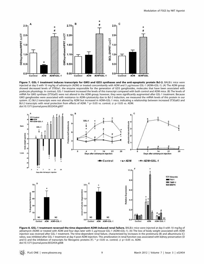
Figure 7. GSL-1 treatment induces transcripts for GM3 and GD3 synthases and the anti-apoptotic protein Bcl-2. BALB/c mice wereinjected at day 0 with 10 mg/kg of adriamycin (ADM) or treated concomitantly with ADM and 5 mg/mouse GSL-1 (ADM+GSL-1). (A) The ADM groupshowed decreased levels of ST8Sia1, the enzyme responsible for the generation of GD3 gangliosides, molecules that have been associated withpodocyte physiology. In contrast, GSL-1 treatment increased the levels of this transcript compared with both control and ADM mice. (B) The levels ofmRNA for GM3 synthase (ST3Gal5) were not altered in the ADM group; however, they were significantly augmented after GSL-1 treatment. BecauseGM3 gangliosides were associated with resistance to ADM-cytotoxicity due to Bcl-2 induction, we measured the mRNA levels of this protein in oursystem. (C) Bcl-2 transcripts were not altered by ADM but increased in ADM+GSL-1 mice, indicating a relationship between increased ST3Galt5 andBcl-2 transcripts with renal protection from effects of ADM. * p,0.05 vs. control; # p,0.05 vs. ADM.doi:10.1371/journal.pone.0032454.g007
Figure 8. GSL-1 treatment reversed the time-dependent ADM-induced renal failure. BALB/c mice were injected at day 0 with 10 mg/kg ofadriamycin (ADM) or treated with ADM and four days later with 5 mg/mouse GSL-1 (ADM+GSL-1). (A) The loss of body weight associated with ADMinjection was reversed after GSL-1 treatment. The time-dependent renal failure, characterized by increases in the proteinuria (B) and albuminuria (C)ratios, was inhibited after GSL-1 treatment at day 4 post-ADM injection. This amelioration in renal function was associated with kidney preservation (Dand E) and the inhibition of transcripts for fibrogenic proteins (F). * p,0.05 vs. control; # p,0.05 vs. ADM.doi:10.1371/journal.pone.0032454.g008
Modulation of FSGS by NKT Agonist
PLoS ONE | www.plosone.org 9 March 2012 | Volume 7 | Issue 3 | e32454

Supporting Information
Figure S1 GSL-1 treatment inhibits the expression offibrogenic proteins. BALB/c mice were injected at day 0 with
10 mg/kg of adriamycin (ADM) or treated concomitantly with
ADM and 5 mg/mouse GSL-1 (ADM+GSL-1). Consistent with
the transcript analysis of kidney tissue, ADM mice showed a slight
increase in the expression of PAI-1 protein (B), without significant
alteration in MMP9 protein levels (A), when compared with the
control and ADM+GSL-1 animals. In contrast, Desmin expression
significantly increased in ADM mice when compared with both
the control and GSL-1-treated groups (C). These data corroborate
our previous mRNA analysis and further demonstrate the
protective effect of GSL-1 treatment.
(TIF)
Acknowledgments
The authors would like to thank Bernardo P. Albe for expert technical
assistance with the histological preparations and Drs. Alexandre S. Basso
and J.D. Lopes for helpful discussion.
Author Contributions
Conceived and designed the experiments: ACK. Performed the experi-
ments: RLP VOR BB. Analyzed the data: ACK RLP VOR NOSC.
Contributed reagents/materials/analysis tools: ACK PBS APS NOSC
MFS MAC PS CDO. Wrote the paper: ACK.
References
1. Barisoni L, Schnaper HW, Kopp JB (2009) Advances in the biology and genetics
of the podocytopathies: implications for diagnosis and therapy. Arch Pathol Lab
Med 133: 201–216.
2. Yap HK, Cheung W, Murugasu B, Sim SK, Seah CC, et al. (1999) Th1 and
Th2 cytokine mRNA profiles in childhood nephrotic syndrome: evidence for
increased IL-13 mRNA expression in relapse. J Am Soc Nephrol 10: 529–
537.
3. Le Berre L, Herve C, Buzelin F, Usal C, Soulillou JP, et al. (2005) Renal
macrophage activation and Th2 polarization precedes the development of
nephrotic syndrome in Buffalo/Mna rats. Kidney Int 68: 2079–2090.
4. Lai KW, Wei CL, Tan LK, Tan PH, Chiang GS, et al. (2007) Overexpression of
interleukin-13 induces minimal-change-like nephropathy in rats. J Am Soc
Nephrol 18: 1476–1485.
5. Fujii S, Shimizu K, Smith C, Bonifaz L, Steinman RM (2003) Activation of
natural killer T cells by alpha-galactosylceramide rapidly induces the full
maturation of dendritic cells in vivo and thereby acts as an adjuvant for
combined CD4 and CD8 T cell immunity to a coadministered protein. J Exp
Med 198: 267–279.
6. Coppieters K, Van Beneden K, Jacques P, Dewint P, Vervloet A, et al. (2007) A
single early activation of invariant NK T cells confers long-term protection
against collagen-induced arthritis in a ligand-specific manner. J Immunol 179:
2300–2309.
7. Hachem P, Lisbonne M, Michel ML, Diem S, Roongapinun S, et al. (2005)
Alpha-galactosylceramide-induced iNKT cells suppress experimental allergic
asthma in sensitized mice: role of IFN-gamma. Eur J Immunol 35: 2793–2802.
8. Matsuda H, Suda T, Sato J, Nagata T, Koide Y, et al. (2005) alpha-
Galactosylceramide, a ligand of natural killer T cells, inhibits allergic airway
inflammation. Am J Respir Cell Mol Biol 33: 22–31.
9. Matsuda JL, Mallevaey T, Scott-Browne J, Gapin L (2008) CD1d-restricted
iNKT cells, the ‘Swiss-Army knife’ of the immune system. Curr Opin Immunol
20: 358–368.
10. Kawano T, Cui J, Koezuka Y, Toura I, Kaneko Y, et al. (1997) CD1d-restricted
and TCR-mediated activation of valpha14 NKT cells by glycosylceramides.
Science 278: 1626–1629.
11. Cerundolo V, Silk JD, Masri SH, Salio M (2009) Harnessing invariant NKT
cells in vaccination strategies. Nat Rev Immunol 9: 28–38.
12. Cerundolo V, Salio M (2007) Harnessing NKT cells for therapeutic applications.
Curr Top Microbiol Immunol 314: 325–340.
13. Long X, Deng S, Mattner J, Zang Z, Zhou D, et al. (2007) Synthesis and
evaluation of stimulatory properties of Sphingomonadaceae glycolipids. Nat
Chem Biol 3: 559–564.
14. Pippin JW, Brinkkoetter PT, Cormack-Aboud FC, Durvasula RV, Hauser PV,
et al. (2009) Inducible rodent models of acquired podocyte diseases. Am J Physiol
Renal Physiol 296: F213–229.
15. Wang Y, Wang YP, Tay YC, Harris DC (2000) Progressive adriamycin
nephropathy in mice: sequence of histologic and immunohistochemical events.
Kidney Int 58: 1797–1804.
16. Amore A, Mazzucco G, Cavallo F, Forni G, Gianoglio B, et al. (1996)
Adriamycin-induced proteinuria in nude mice: an immune-system-mediated
toxic effect. Nephrol Dial Transplant 11: 1012–1018.
17. Cui J, Shin T, Kawano T, Sato H, Kondo E, et al. (1997) Requirement for
Valpha14 NKT cells in IL-12-mediated rejection of tumors. Science 278:
1623–1626.
18. Zheng W, Flavell RA (1997) The transcription factor GATA-3 is necessary and
sufficient for Th2 cytokine gene expression in CD4 T cells. Cell 89: 587–596.
19. Lee GR, Fields PE, Flavell RA (2001) Regulation of IL-4 gene expression by
distal regulatory elements and GATA-3 at the chromatin level. Immunity 14:
447–459.
20. Ansel KM, Djuretic I, Tanasa B, Rao A (2006) Regulation of Th2 differentiation
and Il4 locus accessibility. Annu Rev Immunol 24: 607–656.
21. Szabo SJ, Kim ST, Costa GL, Zhang X, Fathman CG, et al. (2000) A novel
transcription factor, T-bet, directs Th1 lineage commitment. Cell 100: 655–669.
22. Shimaoka T, Seino K, Kume N, Minami M, Nishime C, et al. (2007) Critical
role for CXC chemokine ligand 16 (SR-PSOX) in Th1 response mediated by
NKT cells. J Immunol 179: 8172–8179.
23. Li Y, Kang YS, Dai C, Kiss LP, Wen X, et al. (2008) Epithelial-to-mesenchymal
transition is a potential pathway leading to podocyte dysfunction and
proteinuria. Am J Pathol 172: 299–308.
Table 1. Primer sequences for mRNA quantification.
Gene Sense Antisense
HPRT 59CTCATGGACTGATTATGGACAGGAC39 59GCAGGTCAGCAAAGAACTTATAGCC39
St3Gal5 (GM3synthase) 59GCGAAGACGGCTATGGCTCT39 59 TCCGGAATCCAAAAGGCG 39
ST8Sia1 (GD3synthase) 59CCTTCCAGCTGCCATTGAAG39 59GAATCCCACCGTTTCCCAC39
Gata-3 59AGAACCGGCCCCTTATCAA39 59 AGTTCGCGCAGGATGTCC 39
T-bet 59CAACAACCCCTTTGCCAAAG39 59 TCCCCCAAGCAGTTGACAGT 39
TIMP-1 59ACAGGAGAAGGGACGCCATG39 59 GCAGCTTATCGATGAATCCA 39
IL-4 59ACAGGAGAAGGGACGCCATG39 59GCAGCTTATCGATGAATCCCA39
IL-13 59GCTTATTGAGGAGCTGAGCAACA39 59 GGCCAGGTCCACACTCCATA 39
CXCL16 59TGAACTAGTGGACTGCTTTGAGC39 59GCAAATGTTTTTGGTGGTGA39
TGF-b 59TGGAGCAACATGTGGAACTC39 59GTCAGCAGCCGGTTACCA39
TGFBI 59TCCTTGCCTGCGGAAGTG39 59GGAGAGCATTGAGCAGTTCGA39
doi:10.1371/journal.pone.0032454.t001
Modulation of FSGS by NKT Agonist
PLoS ONE | www.plosone.org 10 March 2012 | Volume 7 | Issue 3 | e32454

24. Pease JE (2006) Asthma, allergy and chemokines. Curr Drug Targets 7: 3–12.
25. Lee HS, Song CY. Effects of TGF-beta on podocyte growth and diseaseprogression in proliferative podocytopathies. Kidney Blood Press Res 33: 24–29.
26. Mesnard L, Keller AC, Michel ML, Vandermeersch S, Rafat C, et al. (2009)
Invariant natural killer T cells and TGF-beta attenuate anti-GBM glomerulo-nephritis. J Am Soc Nephrol 20: 1282–1292.
27. Mattner J, Debord KL, Ismail N, Goff RD, Cantu C, 3rd, et al. (2005)Exogenous and endogenous glycolipid antigens activate NKT cells during
microbial infections. Nature 434: 525–529.
28. Paget C, Bialecki E, Fontaine J, Vendeville C, Mallevaey T, et al. (2009) Role ofinvariant NK T lymphocytes in immune responses to CpG oligodeoxynucleo-
tides. J Immunol 182: 1846–1853.29. Holthofer H, Reivinen J, Miettinen A (1994) Nephron segment and cell-type
specific expression of gangliosides in the developing and adult kidney. KidneyInt 45: 123–130.
30. Holthofer H, Reivinen J, Solin ML, Haltia A, Miettinen A (1996) Decrease of
glomerular disialogangliosides in puromycin nephrosis of the rat. Am J Pathol149: 1009–1015.
31. Reivinen J, Holthofer H, Miettinen A (1992) A cell-type specific ganglioside ofglomerular podocytes in rat kidney: an O-acetylated GD3. Kidney Int 42:
624–631.
32. Simons M, Schwarz K, Kriz W, Miettinen A, Reiser J, et al. (2001) Involvementof lipid rafts in nephrin phosphorylation and organization of the glomerular slit
diaphragm. Am J Pathol 159: 1069–1077.33. Noguchi M, Kabayama K, Uemura S, Kang BW, Saito M, et al. (2006)
Endogenously produced ganglioside GM3 endows etoposide and doxorubicinresistance by up-regulating Bcl-2 expression in 3LL Lewis lung carcinoma cells.
Glycobiology 16: 641–650.
34. Lee VW, Harris DC () Adriamycin nephropathy: a model of focal segmentalglomerulosclerosis. Nephrology (Carlton) 16: 30–38.
35. Paget C, Mallevaey T, Speak AO, Torres D, Fontaine J, et al. (2007) Activationof invariant NKT cells by toll-like receptor 9-stimulated dendritic cells requires
type I interferon and charged glycosphingolipids. Immunity 27: 597–609.
36. Yoshida S, Nagase M, Shibata S, Fujita T (2008) Podocyte injury induced by
albumin overload in vivo and in vitro: involvement of TGF-beta and p38
MAPK. Nephron Exp Nephrol 108: e57–68.
37. Wu DT, Bitzer M, Ju W, Mundel P, Bottinger EP (2005) TGF-beta
concentration specifies differential signaling profiles of growth arrest/differen-
tiation and apoptosis in podocytes. J Am Soc Nephrol 16: 3211–3221.
38. Mozes MM, Bottinger EP, Jacot TA, Kopp JB (1999) Renal expression of
fibrotic matrix proteins and of transforming growth factor-beta (TGF-beta)
isoforms in TGF-beta transgenic mice. J Am Soc Nephrol 10: 271–280.
39. Park SW, Bae JS, Kim KS, Park SH, Lee BH, et al. (2004) Beta ig-h3 promotes
renal proximal tubular epithelial cell adhesion, migration and proliferation
through the interaction with alpha3beta1 integrin. Exp Mol Med 36: 211–219.
40. Schiffer M, Schiffer LE, Gupta A, Shaw AS, Roberts IS, et al. (2002) Inhibitory
smads and tgf-Beta signaling in glomerular cells. J Am Soc Nephrol 13:
2657–2666.
41. Wang W, Huang XR, Li AG, Liu F, Li JH, et al. (2005) Signaling mechanism of
TGF-beta1 in prevention of renal inflammation: role of Smad7. J Am Soc
Nephrol 16: 1371–1383.
42. Zheng Z, Pavlidis P, Chua S, D’Agati VD, Gharavi AG (2006) An ancestral
haplotype defines susceptibility to doxorubicin nephropathy in the laboratory
mouse. J Am Soc Nephrol 17: 1796–1800.
43. Zheng Z, Schmidt-Ott KM, Chua S, Foster KA, Frankel RZ, et al. (2005) A
Mendelian locus on chromosome 16 determines susceptibility to doxorubicin
nephropathy in the mouse. Proc Natl Acad Sci U S A 102: 2502–2507.
44. Mu W, Ouyang X, Agarwal A, Zhang L, Long DA, et al. (2005) IL-10
suppresses chemokines, inflammation, and fibrosis in a model of chronic renal
disease. J Am Soc Nephrol 16: 3651–3660.
45. Harris RC, Neilson EG (2006) Toward a unified theory of renal progression.
Annu Rev Med 57: 365–380.
46. Zeisberg M, Kalluri R (2004) Experimental strategies to reverse chronic renal
disease. Blood Purif 22: 440–445.
Modulation of FSGS by NKT Agonist
PLoS ONE | www.plosone.org 11 March 2012 | Volume 7 | Issue 3 | e32454
Copyright © 2022 FDOKUMEN