Flash Suppressor Comparisons and Analysis for the F89 and ...
Upload
independentCategory
view
1download
0
Cell, Vol. 95, 29–39, October 2, 1998, Copyright 1998 by Cell Press
Negative Regulation of PKB/Akt-DependentCell Survival by the Tumor Suppressor PTEN
The PTEN gene encodes a 403-amino-acid polypep-tide with a high degree of homology to protein tyrosinephosphatases and tensin (Li et al., 1997; Steck et al.,
Vuk Stambolic,*†§ Akira Suzuki,*†§
Jose Lois de la Pompa,*† Greg M. Brothers,*†
Christine Mirtsos,*† Takehiko Sasaki,*†
1997). PTEN is capable of dephosphorylating both phos-Jurgen Ruland,*† Josef M. Penninger,*†
pho-tyrosine and phospho-serine/threonine-containingDavid P. Siderovski,*† and Tak W. Mak*†‡
synthetic substrates in vitro (Myers et al., 1997). Re-*Amgen Institutecent evidence also demonstrates the ability of PTEN620 University Avenueto dephosphorylate position D3 of phosphatidylinositolToronto, Ontario(3,4,5) trisphosphate, a direct product of phosphatidyl-†Ontario Cancer Institute and Departmentsinositol 39 kinase (PI39K) activity (Maehama and Dixon,of Medical Biophysics and Immunology1998). The importance of an intact PTEN phosphataseUniversity of Torontodomain for its tumor suppressor function is underscoredToronto, Ontarioby findings that a large proportion of tumor-associatedCanada M5G 2C1PTEN mutations map to the region encoding the phos-phatase domain (Rasheed et al., 1997; Marsh et al.,1998). Furthermore, unlike wild-type PTEN, catalytically
Summary inactive PTEN is unable to suppress growth and tumori-genicity of PTEN-deficient glioblastoma cells (Furnari et
PTEN is a tumor suppressor with sequence homology al., 1997).to protein tyrosine phosphatases and the cytoskeletal The N-terminal domain of PTEN shows extensive ho-
mology to tensin, a protein associated with the actinprotein tensin. mPTEN-mutant mouse embryos dis-cytoskeleton at focal adhesions (Lo et al., 1994). Recentplay regions of increased proliferation. In contrast,evidence demonstrates that overexpressed PTEN inter-mPTEN-deficient immortalized mouse embryonic fi-acts directly with focal adhesion kinase (FAK) and re-broblasts exhibit decreased sensitivity to cell death induces its tyrosine phospholylation. In addition, PTENresponse to a number of apoptotic stimuli, accompa-was shown to inhibit cell migration, integrin-mediatednied by constitutively elevated activity and phosphory-cell spreading, and formation of focal adhesions (Ta-lation of protein kinase B/Akt, a crucial regulator ofmura et al., 1998). Thus, both genetic and biochemicalcell survival. Expression of exogenous PTEN in mutantevidence has implicated PTEN in the regulation of sev-cells restores both their sensitivity to agonist-inducederal different cellular processes: cell growth, extracellu-apoptosis and normal pattern of PKB/Akt phosphory-lar matrix interactions, and/or cell migration.lation. Furthermore, PTEN negatively regulates intra-
To determine the role of the PTEN in normal develop-cellular levels of phosphatidylinositol (3,4,5) trisphos-ment and tumorigenesis, we have generated mPTEN-phate in cells and dephosphorylates it in vitro. Ourdeficient mice by targeted disruption of exon 3 to 5.results show that PTEN may exert its role as a tumorHomozygous mPTEN3-5 mutant mice die by day 9.5 ofsuppressor by negatively regulating the PI39K/PKB/development and show abnormal patterning and over-Akt signaling pathway.growth of the cephalic and caudal regions (A. S. et al.,submitted). Analysis of mPTEN3-5 embryos reveals re-gions of increased proliferation. In contrast, immortalizedIntroductionmouse embryonic fibroblast cell lines from mPTEN3-5
homozygous mutant embryos show decreased sensitiv-Loss of heterozygosity on human chromosome 10 is the
ity to various apoptotic stimuli, including UV irradiation,most common genetic alteration in the development of heat shock, osmotic stress, and tumor necrosis factormalignant gliomas (Bigner et al., 1998). Recent identifi-
a stimulation. mPTEN mutant cells display constitutivelycation of PTEN (also known as MMAC1 and TEP1) (Li elevated activity of PKB/Akt, which can be restored toand Sun, 1997; Li et al., 1997; Steck et al., 1997) as the normal levels by reexpression of mPTEN. Consistentcandidate tumor suppressor gene located on chromo- with these findings, an active mutant of PKB/Akt sup-some 10 initiated a number of studies that have demon- presses apoptosis induced by mPTEN overexpression.strated a significant rate of mutations of the PTEN gene Our results demonstrate that PTEN negatively regulatesnot only in gliomas (Rasheed et al, 1997) but also in intracellular levels of PI(3,4,5)P3, most likely via directendometrial (Tashiro et al., 1997), prostate (Cairns et al., dephosphorylation, providing a mechanism for PTEN1997), and breast cancers (Rhei et al., 1997). In addition action. We propose that PTEN exerts its role as a tumorto frequent mutations in sporadic tumors, germline mu- suppressor by negatively regulating the PI93K/PKB/Akttations of PTEN are believed to cause Cowden disease signaling pathway.(Liaw et al., 1997), an autosomal-dominant hamartomasyndrome, which results in increased risk for the devel- Resultsopment of breast, thyroid, and brain tumors (Eng, 1998).
Enhanced Proliferation in E8.5 mPTEN3-5
Mutant Embryos‡ To whom correspondence should be addressed (e-mail: tmak@mPTEN-deficient mice were generated by targeted dis-oci.utoronto.ca)
§ These authors contributed equally to this work. ruption of exon 3 to 5 of the murine PTEN gene. Homozy-
Cell30
Figure 1. Extensive Proliferation of mPTEN3-5
Mutant Embryos In Vitro and In Vivo
(A) Wild-type blastocyst outgrowth after 10days of culture. The inner cell mass (ICM) hasgrown extensively and remains coherent.(B) mPTEN3-5 mutant blastocyst showing alarge ICM that spreads out in the dish.(C) E7.5 wild-type embryo. Strongly BrdU-positive cells can be seen throughout all em-bryonic regions. al, allantois; ch, chorion; ee,embryonic ectoderm; m, mesoderm.(D) E7.5 mPTEN3-5 mutant embryo showingstrong BrdU incorporation and a very thickectoderm region (arrow).(E) Cross section through the headfold regionof a E8.5 wild-type embryo, showing BrdUstaining in the neuroepithelium (arrowhead)and underlying mesenchyme (mes).(F) Cross section of the headfold of a mPTEN3-5
mutant embryo. Very strong staining can beseen in the neuroepithelium (arrowhead) andunderlying mesenchyme. Bar, 40 mm in (A)and (B); 140 mm in (C)–(F).
gous mPTEN3-5 mutant mice die by day 9.5 of develop- type embryos showed extensive BrdU labeling, particu-larly in the ectoderm region (Figure 1C). A total of threement (E9.5), and, at E8.5, a typical phenotypic feature
of the mutants is dramatic overgrowth in the cephalic wild-type embryos were analyzed, and they showed aproliferative index of 0.7 6 0.1. mPTEN3-5 mutant em-and caudal regions (A. S. et al., submitted), suggesting
a possible defect in programmed cell death or cellular bryos appear more primitive and show very strong BrdUstaining in all germ layers (Figure 1D). Three mutantproliferation. Apoptosis, as measured by TUNEL assay
(Gavrieli et al., 1992) or DAPI staining, showed no obvi- embryos analyzed gave an index of 0.8 6 0.1. At E8.5,the differences in proliferation between wild-type andous differences between wild-type and mutant embryos
(data not shown). mutant embryos were apparent. Figure 1E shows BrdUstaining in the cephalic region of a E8.5 wild-type em-Next, we analyzed the proliferative capacity of mPTEN3-5
mutant embryos in vitro and in vivo. E3.5 blastocysts bryo. BrdU-positive cells appear scattered throughoutthe neuroepithelium of the head folds and underlyingcultured in vitro organize into a compact inner cell mass
and a surrounding trophoblast (Figure 1A). mPTEN3-5 cephalic mesenchyme. Three wild-type embryos wereanalyzed at E8.5, and they gave a proliferative index ofmutant blastocysts grow similarly to wild-type litter-
mates for a period of up to 10 days, although they tend 0.7 6 0.05. In contrast, mPTEN3-5 mutant embryos showBrdU staining throughout the deformed head folds andto appear more expanded and disorganized relative to
normal littermates (Figure 1B). To measure directly the in the cephalic mesenchyme that is significantly strongerthan in wild-type embryos (Figure 1F). Three mutanteffects of the mPTEN3-5 mutation on cellular proliferation,
we examined the incorporation of 5-bromo-29-deoxyuri- embryos analyzed gave an index of 0.9 6 0.03. Thesevalues were statistically significantly different (p , 0.05,dine (BrdU) into DNA during the S phase of the cell cycle.
Analysis was performed at E7.5 and E8.5, at times when Student’s t test). Thus, BrdU incorporation is quantita-tively increased in mutant embryos, suggesting that en-the mitotic activity in the mouse embryo undergoing
organogenesis is high (Snow, 1977). A proliferative index hanced proliferation is involved in the abnormal devel-opment of the cephalic region of mPTEN3-5 mutantbased on the ratio of proliferating cells (BrdU-positive
nuclei) to total cell number was calculated. Wild-type embryos. Similarly, increased BrdU incorporation was
PTEN in Negative Regulation of PKB/Akt31
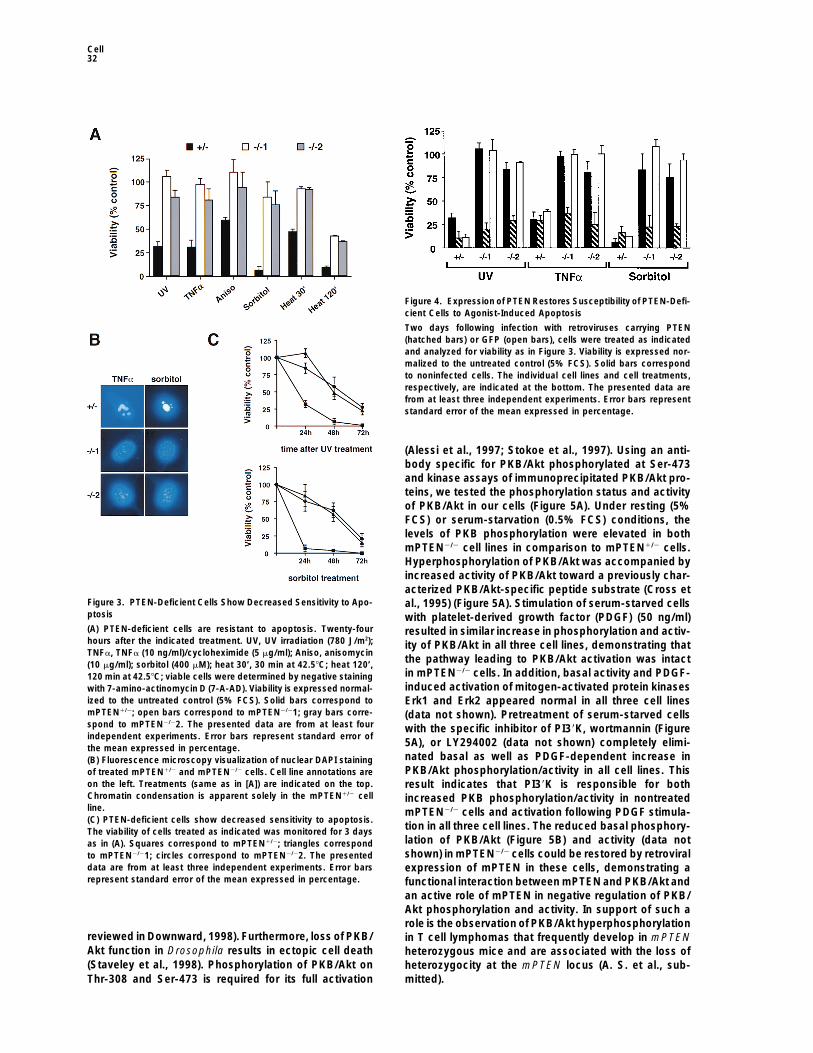
Surprisingly, we observed no significant difference inthe rate of proliferation between the mPTEN homo- andheterozygous cells (data not shown). We next examinedthe rate of agonist-induced cell death in heterozygousand mPTEN-deficient cells. As measured by viability dyestaining, more than 80% of mPTEN2/21 and mPTEN2/22cells were viable 24 hr following treatment with UV irradi-ation (780 J/m2), sorbitol (0.4 mM), TNFa (10 ng/ml)/cycloheximide (5 mg/ml), anisomycin (10 mg/ml), or 30min of heat shock at 42.58C (Figure 3A). In contrast,these same treatments resulted in extensive cell deathin mPTEN1/2 cells. Strikingly, even 1 day after a 2 hrheat shock, almost 40% of mPTEN2/2 cells were stillviable (Figure 3A). Similar results were obtained usingearly and late markers of programmed cell death,annexin V, and propidium iodide, respectively, sug-gesting that the mPTEN1/2 cells were dying from apo-ptosis (data not shown). To further test this possibility,8 hr after treatment, nuclear morphology of mPTEN1/2
and mPTEN2/2 cells was examined. Nuclei of mPTEN1/2
cells display chromatin condensation indicative of apo-ptosis, whereas nuclei of mPTEN2/2 appear normal(Figure 3B). Prolonged monitoring of mPTEN2/2 cellsindicates that they eventually die in response to UVirradiation and sorbitol treatment (Figure 3C), as well
Figure 2. Characterization of Immortalized Mouse Embryonic Fibro- as other apoptotic stimuli (data not shown), althoughblasts from mPTEN Mutant Mice at a significantly reduced rate in comparison to the(A) Western blot analysis of whole-cell lysates from brain, thymus, mPTEN1/2 cells. A similar decrease in sensitivity to apo-and mPTEN1/2 and mPTEN2/2 cell lines. The positions of molecular ptosis was observed in mPTEN-deficient ES cells (dataweight markers (kDa) are shown on the left. The position of PTEN
not shown). Thus, reduced sensitivity of mPTEN-nega-is indicated by an arrow.tive cells to apoptosis suggests that PTEN might exert(B) RT-PCR analysis of mPTEN1/2 and mPTEN2/2 cell lines. FSP1its function as a tumor suppressor by negatively regulat-indicates the reactions performed using fibroblast-specific protein
1–specific primers. b2M indicates the reactions performed using b2- ing cell survival.microglobulin-specific primers. H2O, water control (no RNA); spleen,total RNA from normal mouse splenocytes; mPTEN1/2, total RNA
Expression of Exogenous mPTEN infrom mPTEN1/2 cells; mPTEN2/21, total RNA from mPTEN2/21 cells;mPTEN2/22, total RNA from mPTEN2/22 cells. mPTEN-Deficient Cells Restores
Sensitivity to ApoptosisTo further investigate whether resistance to apoptosisobserved in the allantois at the posterior end of thein mPTEN2/2 cells was a result of the loss of PTENembryo (data not shown).function, mPTEN was expressed in all three cell linesusing recombinant retrovirus carrying an HA-taggedmPTEN-Deficient Mouse Embryonic Fibroblasts
Exhibit Decreased Sensitivity to Apoptosis copy of wild-type mPTEN, or green fluorescence proteinTo further investigate the role of PTEN in regulation of (GFP) as a control. As measured by fluorescence ofcellular processes, immortalized mouse embryonic fibro- control cells, the efficency of retroviral infection wasblast cell lines from two mPTEN3-5 homozygous mutant higher than 70% in all three cell lines (data not shown).embryos (mPTEN2/21 and mPTEN2/22, respectively) In contrast to GFP, expression of mPTEN in mPTEN-and one heterozygous mutant embryo (mPTEN1/2) were deficient cell lines restored normal apoptosis in re-generated. Reverse transcription–polymerase chain re- sponse to all stimuli tested (Figure 4). Retrovirus-medi-action (RT-PCR) using RNA from generated cells shows ated expression of mPTEN in mouse NIH 3T3 cells alsothe expression of fibroblast-specific protein 1 (FSP1) increased their sensitivity to cell death (data not shown),(Strutz et al., 1995) in all three cell lines, demonstrat- confirming the role of PTEN as a negative regulator ofing that the generated cells are fibroblasts (Figure 2). cell survival.Immunoblotting of cell lysates from the establishedcell lines confirmed the loss of mPTEN expression in
Increased Activity of Cell Survival Kinaseboth mPTEN2/21 and mPTEN2/22 cell lines, whereasPKB/Akt in PTEN Mutant CellsmPTEN1/2 expressed a protein of the molecular mass ofOne of the key regulatory molecules involved in theapproximately 55 kDa (Figure 2). The apparent molecularregulation of cell survival is protein kinase B/Akt (PKB/mass of mPTEN is consistent with a major immunoreac-Akt). PKB/Akt is implicated in protection from apoptosistive species identified in brain, thymus, and NIH3T3 cellsin response to several growth factors and cytokines, and(Figure 2) and of recombinant mPTEN (data not shown).it protects cells from apoptosis induced by withdrawal ofBoth mPTEN2/2 cell lines showed no morphological dif-survival signals, c-Myc overexpression (Kauffmann-Zehferences in comparison to their heterozygous counter-
part (data not shown). et al., 1997), or matrix detachment (Khwaja et al., 1997;
Cell32
Figure 4. Expression of PTEN Restores Susceptibility of PTEN-Defi-cient Cells to Agonist-Induced Apoptosis
Two days following infection with retroviruses carrying PTEN(hatched bars) or GFP (open bars), cells were treated as indicatedand analyzed for viability as in Figure 3. Viability is expressed nor-malized to the untreated control (5% FCS). Solid bars correspondto noninfected cells. The individual cell lines and cell treatments,respectively, are indicated at the bottom. The presented data arefrom at least three independent experiments. Error bars representstandard error of the mean expressed in percentage.
(Alessi et al., 1997; Stokoe et al., 1997). Using an anti-body specific for PKB/Akt phosphorylated at Ser-473and kinase assays of immunoprecipitated PKB/Akt pro-teins, we tested the phosphorylation status and activityof PKB/Akt in our cells (Figure 5A). Under resting (5%FCS) or serum-starvation (0.5% FCS) conditions, thelevels of PKB phosphorylation were elevated in bothmPTEN2/2 cell lines in comparison to mPTEN1/2 cells.Hyperphosphorylation of PKB/Akt was accompanied byincreased activity of PKB/Akt toward a previously char-acterized PKB/Akt-specific peptide substrate (Cross et
Figure 3. PTEN-Deficient Cells Show Decreased Sensitivity to Apo- al., 1995) (Figure 5A). Stimulation of serum-starved cellsptosis with platelet-derived growth factor (PDGF) (50 ng/ml)(A) PTEN-deficient cells are resistant to apoptosis. Twenty-four resulted in similar increase in phosphorylation and activ-hours after the indicated treatment. UV, UV irradiation (780 J/m2); ity of PKB/Akt in all three cell lines, demonstrating thatTNFa, TNFa (10 ng/ml)/cycloheximide (5 mg/ml); Aniso, anisomycin
the pathway leading to PKB/Akt activation was intact(10 mg/ml); sorbitol (400 mM); heat 30’, 30 min at 42.58C; heat 120’,in mPTEN2/2 cells. In addition, basal activity and PDGF-120 min at 42.58C; viable cells were determined by negative staininginduced activation of mitogen-activated protein kinaseswith 7-amino-actinomycin D (7-A-AD). Viability is expressed normal-
ized to the untreated control (5% FCS). Solid bars correspond to Erk1 and Erk2 appeared normal in all three cell linesmPTEN1/2; open bars correspond to mPTEN2/21; gray bars corre- (data not shown). Pretreatment of serum-starved cellsspond to mPTEN2/22. The presented data are from at least four with the specific inhibitor of PI39K, wortmannin (Figureindependent experiments. Error bars represent standard error of
5A), or LY294002 (data not shown) completely elimi-the mean expressed in percentage.nated basal as well as PDGF-dependent increase in(B) Fluorescence microscopy visualization of nuclear DAPI stainingPKB/Akt phosphorylation/activity in all cell lines. Thisof treated mPTEN1/2 and mPTEN2/2 cells. Cell line annotations are
on the left. Treatments (same as in [A]) are indicated on the top. result indicates that PI39K is responsible for bothChromatin condensation is apparent solely in the mPTEN1/2 cell increased PKB phosphorylation/activity in nontreatedline. mPTEN2/2 cells and activation following PDGF stimula-(C) PTEN-deficient cells show decreased sensitivity to apoptosis.
tion in all three cell lines. The reduced basal phosphory-The viability of cells treated as indicated was monitored for 3 dayslation of PKB/Akt (Figure 5B) and activity (data notas in (A). Squares correspond to mPTEN1/2; triangles correspondshown) in mPTEN2/2 cells could be restored by retroviralto mPTEN2/21; circles correspond to mPTEN2/22. The presented
data are from at least three independent experiments. Error bars expression of mPTEN in these cells, demonstrating arepresent standard error of the mean expressed in percentage. functional interaction between mPTEN and PKB/Akt and
an active role of mPTEN in negative regulation of PKB/Akt phosphorylation and activity. In support of such arole is the observation of PKB/Akt hyperphosphorylation
reviewed in Downward, 1998). Furthermore, loss of PKB/ in T cell lymphomas that frequently develop in mPTENAkt function in Drosophila results in ectopic cell death heterozygous mice and are associated with the loss of(Staveley et al., 1998). Phosphorylation of PKB/Akt on heterozygocity at the mPTEN locus (A. S. et al., sub-
mitted).Thr-308 and Ser-473 is required for its full activation

PTEN in Negative Regulation of PKB/Akt33
Figure 6. PTEN and PKB/Akt Act in the Same Signaling Pathway
NIH 3T3 cells were transfected with constructs expressing the indi-cated proteins (bottom) in the presence of tracer amounts of ab-gal construct. PKB/Akt alleles are as follows: wt, wild-type; AAA,alanine-substituted dominant-negative mutant; DD, aspartate-sub-stituted constitutively active mutant; gag, gag-PKB/Akt fusion pro-tein (see text for details). Forty-eight hours posttransfection cellswere fixed and stained with X-gal. The number of surviving bluecells was counted in ten randomly selected high-power fields undera microscope and expressed as a percentage relative to the controltransfection. The presented data are from at least three independentexperiments. Error bars represent standard error of the mean ex-pressed in percentage.
expression can result in cell death (Figure 6). Using acotransfection approach, we examined the ability ofwild-type or mutant PKB/Akt to alter mPTEN-inducedcell death in NIH 3T3 cells 48 hr after transfection. Ex-pression of wild-type PKB/Akt had no influence onmPTEN-mediated cell death (Figure 6). However, coex-pression of PKB-DD, a constitutively active PKB/Aktmutant in which Thr-308 and Ser-473 have beenchanged to aspartate to mimic phosphorylation (Welch
Figure 5. Negative Regulation of PI39K/PKB/Akt Signaling Pathway et al., 1998; Jing Jin and James R. Woodgett, personalby PTEN
communication), with mPTEN increases the number of(A) Increased phosphorylation and activity of PKB/Akt in PTEN-
surviving transfected cells to the levels comparable todeficient cells. (Top) Western blot analysis of cell lysates from cellsmock-transfected cells (Figure 6). Thus, expression oftreated as indicated. 0.5% FCS indicates incubation of cells in 0.5%constitutively active PKB/Akt rescues transfected cellsFCS for 24 hr prior to treatment. PDGF indicates treatment with
PDGF (50 ng/ml) for 15 min. W indicates pretreatment of cells with from mPTEN-induced apoptosis and indicates a poten-wortmannin (100 nM) for 20 min prior to addition of PDGF. Antibodies tial function of mPTEN upstream of PKB/Akt.used in Western blotting are indicated on the left. Comparable re- PKB-AAA is a dominant-negative PKB/Akt mutant insults were obtained from at least four independent experiments.
which both phosphorylation sites and the lysine in the(Bottom) PKB kinase activity in mPTEN1/2 and mPTEN2/2 cell lines.ATP-binding pocket have been mutated to alanine resi-PKB proteins were immunoprecipitated from cells treated as indi-dues, respectively, resulting in a complete loss of kinasecated (see above) and analyzed for incorporation of 32PO4 into a
previously characterized peptide substrate (see Experimental Pro- activity (Meier et al., 1997; Stephens et al., 1998; Jingcedures). The data are presented normalized to the activity of PKB/ Jin and James R. Woodgett, personal communication).Akt from untreated (5% FCS) mPTEN1/2 cells. The presented data Expression of PKB-AAA induced apoptosis in NIH 3T3are from at least three independent experiments. Error bars repre-
cells comparable to the levels achieved by mPTENsent standard error of the mean expressed in percentage.transfection alone (Figure 6). Significantly, coexpression(B) Expression of PTEN reduces basal phosphorylation of PKB/Aktof PKB-AAA and mPTEN did not result in an additivein mPTEN2/2 cells. Forty-eight hours after mock infection or infection
with the retrovirus expressing HA-PTEN, cells were treated as indi- effect on cell death (Figure 6), suggesting that PKB andcated on top (see [A]). Antibodies used in Western blotting are mPTEN act in the same signaling pathway.indicated on the left. Comparable results were obtained from at The role of mPTEN in PKB/Akt regulation is furtherleast three independent experiments.
supported by the effect of mPTEN on gag-PKB-medi-ated cell survival. gag-PKB is the oncogenic form ofPKB/Akt that was identified as an oncogene carried byA Constitutively Active Mutant of PKB/Akt Rescues
Cells from PTEN-Induced Apoptosis AKT8, the acute transforming virus from a rodent T celllymphoma (AKR) (Bellacosa et al., 1991). The myristylExpression of mPTEN using transient transfection of
NIH 3T3 cells resulted in a decrease in the number of group of viral gag protein serves to bring gag-PKB fusionprotein to the cell membrane, where it becomes acti-surviving transfected cells in comparison to the mock-
transfected controls, indicating that high levels of mPTEN vated by upstream kinases (Ahmed et al., 1993; Kennedy

Cell34
et al., 1997; Alessi and Cohen 1998). In our studies,transfection of gag-PKB into NIH 3T3 cells resulted inan increase in the number of surviving cells, possibly dueto the ability of gag-PKB to protect from transfection-induced death (Figure 6). However, cotransfection ofmPTEN with gag-PKB resulted in a decrease in the num-ber of transfected cells comparable to that induced bymPTEN alone (Figure 6). Considering that gag-PKB re-quires phosphorylation of Thr-308 and Ser-473 for ac-tivity (Kennedy et al., 1997), mPTEN is most likelynegatively regulating the phosphorylation of both PKBand gag-PKB, consistent with our previous findings (Fig-ure 6).
Phosphatidylinositol (3,4,5)P3 Is a DirectSubstrate of PTENThe activity of PI39K under normal or PDGF-stimulationconditions was identical in mPTEN1/2 and mPTEN2/2
cells (data not shown), suggesting that mPTEN was af-fecting the PKB/Akt phosphorylation and activity by act-ing downstream of PI39K. Treatment with wortmannin,a specific inhibitor of PI39K, sensitized mPTEN2/2 cellsto UV irradiation– and sorbitol treatment–induced celldeath (Figure 7A), demonstrating that an immediateproduct of PI39K activity was responsible for the alteredsensitivity of mPTEN2/2 cells to apoptosis. Furthermore,constitutively active PKB/Akt rescues transfected cellsfrom mPTEN-induced apoptosis, implying a function ofmPTEN upstream of PKB/Akt.
PKB/Akt phosphorylation on both Thr-308 and Ser-473 is directly dependent on the levels of intracellular
Figure 7. PTEN Dephosphorylates PI(3,4,5)P3phosphatidylinositol(3,4,5)P3, the key product of PI39K(A) Wortmannin restores the sensitivity of mPTEN2/2 cells to apopto-activity (Alessi et al., 1996, 1997; M. Delcommene et al.,sis. Twenty-four hours after the indicated treatment. W, wortmanninsubmitted). In mPTEN2/2 cells, PKB/Akt is hyperphos-(50 nM); UV, UV irradiation (780 J/m2); sorbitol (400 mM); viable cellsphorylated under resting conditions (5% FCS) (Figurewere determined by negative staining with 7-amino-actinomycin D
5A). Under those conditions, mPTEN2/2 cells exhibit ele- (7-A-AD). Viability is expressed normalized to the untreated controlvated levels of intracellular PI(3,4,5)P3 in comparison to (5% FCS). Solid bars correspond to mPTEN1/2; open bars corre-their heterozygous counterpart (Figure 7B), implying a spond to mPTEN2/21; hatched bars correspond to mPTEN2/22. The
presented data are from at least three independent experiments.direct role of mPTEN in dephosphorylation of PI(3,4,5)P3.Error bars represent standard error of the mean expressed in per-Furthermore, bacterially expressed mPTEN dephos-centage.phorylates the D3 position of PI(3,4,5)P3 in vitro (Figure(B) Lack of PTEN increases intracellular levels of PI(3,4,5)P3. Total
7C). Thus, PI(3,4,5)P3 is a physiological substrate of cellular lipids were extracted from cells metabolically labeled withPTEN. 32P-orthophosphate as described in Experimental Procedures and
resolved by ascending thin-layer chromatography together withPI(3,4,5)P3 prepared as described in Experimental Procedures.
Discussion Source of lipid preparations is indicated at the bottom. Quantifica-tion of relative levels of PI(3,4,5)P3 is shown on the right; PI(3,4,5)P3
Role of PTEN in Proliferation content relative to total cellular lipid was normalized to the relativeamount of PI(3,4,5)P3 in mPTEN1/2 cells. Positions of the origin andmPTEN3-5 mutant embryos die by E9.5 with abnormalPI(3,4,5)P3 (PIP3) are indicated. The presented data are from at leastpatterning and extensive overgrowth of the cephalic andthree independent experiments. Error bars represent standard errorcaudal regions. At E7.5 and more dramatically at E8.5,of the mean.
BrdU incorporation is increased in mPTEN3-5 mutants. (C) PTEN dephosphorylates PI(3,4,5)P3 in vitro. PI(3,4,5)P3 was incu-In the head, proliferation is enhanced in the neuroepi- bated with 0.5 mg of purified wild-type PTEN protein (Trx-PTEN) orthelium of the cephalic neural folds and in the underlying catalytically inactive mutant of PTEN (Trx-PTENC.S) as indicated,
extracted, and resolved by ascending thin-layer chromatography.mesenchyme. Increased proliferation in mPTEN3-5 em-Positions of the origin and PI(3,4,5)P3 (PIP3) are indicated.bryos is consistent with the ability of ectopic PTEN to
suppress cell growth (Furnari et al., 1997, Cheney etal., 1998). Nuclear integrity dye staining of mPTEN3-5
embryos does not reveal any alterations in apoptosis discrepancy between the phenotypes of mPTEN3-5 em-bryos and cells could be explained by the fact that pro-associated with the mPTEN loss. In contrast, mPTEN-
deficient fibroblasts display no increase in proliferation gramed cell death during early embryogenesis is a rareevent (Coucovanis and Martin, 1995), preventing, usingbut decreased sensitivity to apoptosis. This apparent
PTEN in Negative Regulation of PKB/Akt35
the available methods, the detection of a potential de- be interesting to utilize our mPTEN-deficient cells tofurther characterize the interaction between FAK andcrease in apoptosis in mPTEN3-5 embryos. Hence, hy-
perproliferation in mPTEN3-5 embryos could be a con- PTEN in vivo.sequence of a differentiation defect associated withdecreased apoptosis due to mPTEN deficiency. In PI(3,4,5)P3 Is a Physiological Substrate of PTENsupport of such a view, immunostaining of wild-type The intact PI39K activity in mPTEN2/2 cells and the abilitymouse embryos demonstrated that tissue distribution of wortmannin to sensitize mPTEN2/2 cells to agonist-of mPTEN coincides with regions where differentiation induced apoptosis suggested that the mechanism ofoccurs (data not shown). Alternatively, immortalized em- mPTEN action might be at the level of the products ofbryonic fibroblasts derived from mPTEN3-5 embryos PI39K activity. The evidence presented here shows thatmight not be analogous to the affected cells involved mPTEN negatively regulates the intracellular levels ofin the overgrowth in the mPTEN3-5 embryo and therefore PIP(3,4,5)P3, most likely via direct dephosphorylation,might not display an equivalent proliferation defect. resulting in decreased phosphorylation and activity of
PKB/Akt. Consistent with our findings, a recent report(Maehama and Dixon, 1998) has shown that PTEN isRole of PTEN in Apoptosiscapable of dephosphorylating PI(3,4,5)P3 in vitro andLoss of mPTEN in immortalized mouse embryonic fibro-down-regulating its levels in insulin-stimulated 293 cells.blasts results in decreased sensitivity to cell death inTaken together, our results establish PI(3,4,5)P3 dephos-response to diverse apoptotic stimuli. The effect ofphorylation as a major mechanism of PTEN action. Thus,mPTEN loss on apoptosis appears to be pleiotropicin the absence of mPTEN, elevated levels of PI(3,4,5)P3considering that mPTEN2/2 cells no longer die in re-result in increased PKB/Akt phosphorylation. Con-sponse to a number of treatments known to affect aversely, expression of mPTEN negatively regulates PKB/variety of different signaling pathways. Such a broadAkt phosphorylation/activity via dephosphorylation ofrole of mPTEN in regulation of cell survival suggestsPIP(3,4,5)P3.that mPTEN functions in a general pathway that regu-
lates cell death and survival. The evidence presentedhere identifies a crucial regulator of cell survival, PKB/ The Role of PTEN in Tumorigenesis
PTEN mutations have been identified in a large numberAkt, as a major target of mPTEN activity. mPTEN regu-lates phosphorylation and activity of PKB/Akt in cells. of different tumors, indicating that PTEN regulates fun-
damental cellular processes. Our results provide evi-Furthermore, expression of a constitutively active mu-tant of PKB/Akt rescues cells from PTEN-induced apo- dence for such a critical role of PTEN in control of normal
cell growth. In certain tumors, loss of PTEN may resultptosis. Activation of PKB/Akt has been implicated inprotection from apoptosis in response to several growth in decreased sensitivity of mutant cells to apoptotic
stimuli. In other tumors, mutations of PTEN may lead tofactors and cytokines, including IGF-1 (Kulik et al., 1997),IL-2 (Ahmed et al., 1997), and NGF (Ulrich et al., 1998). an increase in proliferative potential. Both survival and
proliferative advantages of PTEN-deficient cells wouldMoreover, elevated PKB/Akt activity protects cells fromapoptosis induced by withdrawal of survival signals promote cellular overgrowth and tumorigenesis.
PTEN mutations in humans have initially been identi-(reviewed in Downward, 1998), c-myc overexpression(Kauffmann-Zeh et al., 1997), UV irradiation (Kulik et fied in glioblastomas (Li et al., 1997). Frequent amplifica-
tion of platelet-derived growth factor receptor (PDGFR)al., 1997), CD95/Fas-induced cell killing (Hausler et al.,1998), and matrix detachment (Khwaja et al., 1997). De- and epidermal growth factor receptor (EGFR) genes
have also been demonstrated in glioblastoma (Chaffanetcreased sensitivity of mPTEN mutant cells to apoptosisin response to TNFa, sorbitol, and anisomycin suggests et al., 1992; Smits and Funa, 1998). Furthermore, the
production and cellular response to fibroblast growththat active PKB/Akt also protects cells from death in-duced by these agents. factor (FGF) are often elevated in astrocytomas and glio-
mas (Takahashi et al., 1992). PI39K is regulated by theActivation of SAPK/JNK, whose function in inductionand protection from apoptosis remains controversial activity of receptor-tyrosine kinases and is potentially
up-regulated in cells overexpressing PDGFR and EGFR,(Kyriakis and Avruch, 1996; Nishina et al., 1997), wasnormal in our cells in response to UV irradiation, aniso- as well as in cells exposed to higher than normal levels
of FGF. Hypothetically, this would result in increasedmycin, and sorbitol (data not shown), indicating that thispathway is not affected by the loss of mPTEN. Further- levels of intracellular PI(3,4,5)P3 and elevated activity of
PKB/Akt, giving the cells a survival advantage. The abil-more, TNFa stimulation of all three cell lines resulted ina comparable level of phosphorylation of IkB (data not ity of PKB/Akt to protect cerebral neurons from apopto-
sis supports such a scenario (Dudek et al., 1997). More-shown), suggesting that NF-kB activation in our mutantcells was intact and is therefore an unlikely cause of over, increased expression of PDGFR and EGFR, or
FGF overproduction in glioblastoma, could result in in-their altered cell death. A recent report (Tamura et al.,1998) has identified direct interaction between focal ad- creased proliferation. Thus, other genetic alterations in
glioblastoma leading to PI39K activation might have sim-hesion kinase (FAK) and PTEN and demonstrated a roleof PTEN in regulation of cell migration and spreading. ilar phenotypic consequences on tumor development
as the loss of PTEN function.Even though our preliminary evidence does not revealany differences in matrix attachment between mPTEN1/2 v-akt, an oncogenic form of PKB/Akt resulting in in-
creased kinase activity, causes mouse T cell lymphomaand 2/2 cells (V. S., unpublished observations), it will
Cell36
oligonucleotide primer pairs specific for mouse b2-microglobulin(AKR) (Staal and Hartley, 1988; Bellacosa et al., 1991).(sense primer 59-ATGGCTCGCTCGGTGACCCTAG-39; antisenseInterestingly, mice heterozygous for mPTEN frequentlyprimer 59-TCATGATGCTTGATCACATGTCTCG-39) or mouse fibro-develop T cell lymphomas that are associated with theblast-specific protein-1 (sense primer 59-CAGCGAAAGAGGGTGAC
loss of heterozygosity at the mPTEN locus (A. S. et al., AAGTTCA-39; antisense primer 59-ATGTGCGAAGAAGCCAGAGTAsubmitted). These tumors exhibit elevated phosphoryla- AGG-39) were used to amplify 1 mg of total RNA employing the
Titan one-tube RT-PCR system as per manufacturer’s instructionstion of PKB/Akt in comparison to normal tissue, consis-(Boehringer Mannheim/Roche). Reaction products were resolvedtent with our findings that PTEN negatively regulatesby 6% polyacrylamide gel electrophoresis and detected by ethidiumPKB/Akt signaling. In humans, overexpression of PKB/bromide staining.Akt has been demonstrated in 15% ovarian (Cheng et
al., 1992), 12% of pancreatic (Cheng et al., 1996), and 3% Cloning, Expression, and Purification of mPTENof breast cancers (Bellacosa et al., 1995). Significantly, in A cDNA encoding the entire mouse PTEN open reading frameaddition to mutations shown in other tumors, LOH in was amplified from mouse brain mRNA by RT-PCR as above
using sense primer 59-GGATCCGACATGACAGCCATCATCAAAG 239the human PTEN locus has been observed in ovarian,and antisense primer 59-CTCGAGTCAGACTTTTGTAATTTGTGAATGpancreatic, and breast tumor specimens (Teng et al.,CTG-39, sequenced, and subcloned in-frame with two N-terminal1997). Such correlation in the rate of LOH of PTEN andinfluenza virus hemagglutinin (HA) epitopes into the eukaryotic ex-
up-regulation of PKB/Akt in human cancer, together pression vector pcDNA3.1 (Invitrogen). An EcoRI-XhoI fragmentwith the evidence presented here, emphasizes the im- from the resultant pcDNA3.1HA2mPTEN vector, encoding N-termi-portance of PTEN/PKB/Akt interaction in tumor pro- nally HA-tagged mPTEN open reading frame, was ligated into the
EcoRI-SalI sites of the retroviral vector pBabeMN-z (a kind gift ofgression.Dr. Garry Nolan). Full-length mPTEN cDNA and a fragment encodingOur results suggest that PTEN belongs to a classa C-terminal portion of mPTEN (amino acids 151–403 of SwissProtof “gatekeeper” tumor suppressor genes together withO08586) were separately cloned into the thioredoxin-hexahistidine
p53, Rb, and APC. These genes participate in regulation tag (Trx) fusion vector pET32a (Novagen) and transfected into E.of normal cell growth, and their mutations directly con- coli strain BL21(DE3)pLysS or BL21(DE3), respectively (Stratagene,tribute to the neoplastic growth of the cell (Kinzler and La Jolla, CA).
Expression of Trx-mPTEN and Trx-mPTEN(151–403) fusion pro-Vogelstein, 1998). We propose that negative regulationteins was induced in bacterial cultures at an OD600nm of 0.8 by theof PI39K/PKB/Akt cell survival signaling pathway is aincubation in the presence of 0.8 mM isopropyl-b-D-thiogalactopy-likely mechanism by which PTEN exerts its tumor sup-ranoside (IPTG) for 4 hr at 378C or 6 hr at 288C, respectively. Cells
pressor function. Thus, loss of PTEN results in general were lysed by sonication in lysis buffer (20 mM Tris-HCl [pH 8.0],alteration of the normal cell homeostasis due to in- 100 mM NaCl, 20 mM imidazole, 10% glycerol, 5 mM b-mercaptoeth-creased survival potential, which could subsequently anol, 0.5% Nonidet P-40, and protease inhibitors), cell lysate was
clarified by centrifugation, and supernatents passed through nickel-lead to tumorigenesis. Further investigations, includingnitrilotriacetic acid resin (Qiagen, Valencia, CA). Resin was washedthose aimed at elucidating modes of PTEN regulation,with lysis buffer, and hexahistidine-tagged fusion protein was elutedwill provide valuable insight into the processes of cellusing an imidazole concentration gradient. Eluted Trx-mPTEN(151–
transformation and tumorigenesis. 403) protein was dialysed against storage buffer (50 mM Tris-HCl[pH 8.0], 2 mM EDTA, 2 mM DTT, 150 mM NaCl) and concentrated
Experimental Procedures by ultrafiltration.Eluted Trx-mPTEN fusion protein was dialysed against ion-
BrdU Labeling of Mouse Embryos exchange column equilibration buffer (25 mM Tris-HCl [pH 8.0], 2Generation of mPTEN3-5 mutant embryonic stem cells and mice will mM EDTA, and 2 mM DTT) and passed through a Q-Sepharose FFbe described elsewhere. BrdU labeling of cells in the S phase of ion exchange column (Pharmacia Biotech, Inc.) equilibrated in thethe cell cycle was performed according to the protocol described same buffer. The column was washed with 10 column bed volumeby Hayashi et al. (1988). BrdU (100 mg/g of body weight) was injected of equilibration buffer, and Trx-mPTEN protein was eluted using 20intraperitoneally into pregnant females at E7.5 and E8.5 of gestation. column bed volume of a 0–500 mM NaCl concentration gradient.Females were sacrificed 1 hr after injection, the uteri removed, and The Trx-mPTEN containing fractions were pooled, dialysed againstcomplete deciduas or embryos fixed in 4% paraformaldehyde at storage buffer (25 mM Tris-HCl [pH 7.4], 150 mM NaCl, 1 mM EDTA,48C overnight and processed for immunohistochemistry. The sec- 1 mM DTT), and concentrated by ultrafiltration.tions were incubated with an anti-BrdU monoclonal antibody (Boeh-ringer Mannheim) at a 1:10 dilution. Staining was performed ac- Generation of Anti-PTEN Antibodycording to the protocol described by Mishina et al. (1995). Rabbit antisera was raised against purified Trx-mPTEN(151–403)
protein (Antibodies, Inc., Davis, CA) and purified by preadsorptionEstablishment of Immortalized Embryonic Fibroblast on Trx-coupled CNBr-Sepharose 4B followed by affinity purificationCell Lines on GST-PTEN fusion protein coupled to CNBr-Sepharose 4B. Low-Day 8.5 heterozygous and homozygous mPTEN3-5 embryos (A. S. et affinity antibody was eluted in a pH 5.0 buffer, and high-affinity,al., submitted) were dissected, minced well by scissors, and cultured PTEN-specific antibody (used in this paper) was eluted off the affinityindividually in gelatinized 24-well plates containing Dulbecco modi- column with 0.1 M glycine (pH 2.5).fied Eagle’s medium (DMEM), 10% fetal calf serum (FCS), L-gluta-mine, and b-mercaptoethanol. After 7 days of culture, cells were Cell Lysistrypsinized, replated, incubated for 1 hr, and washed three times For the generation of cytoplasmic cell lysates, cells were washedwith PBS to remove nonadherent cells. Adherent cells were continu- in ice-cold PBS and lysed by incubation on ice in a buffer containingously cultured for 3 months using the 3T3 protocol (Goldman, 1998). 20 mM Tris-HCl (pH 8.0), 100 mM NaCl, 10% glycerol, 1% TritonEmbryo genotyping was performed by PCR with DNA from the yolk X-100, 50 mM NaF, 100 mM sodium vanadate, 1 mM benzamidine,sac, using specific primer sets to detect wild-type and the mutant and 5 mg/ml leupeptin. The lysates were centrifuged and superna-mPTEN allele, respectively. tants analyzed.
For the generation of whole-cell lysates, the tissues were homoge-nized in a buffer containing 10 mM Tris-HCl (pH 7.4), 1 mM MgCl2,RT-PCR
Total RNA was prepared from 5 3 106 cells using TRIZOL reagent as 1 mM EGTA, 0.5 mM CHAPS, 10% glycerol, 50 mM NaF, 100 mMsodium vanadate, 1 mM benzamidine, and 5 mg/ml leupeptin andper manufacturer’s protocols (Life Technologies). Intron-spanning
PTEN in Negative Regulation of PKB/Akt37
incubated on ice for 30 min. The lysates were centrifuged and super- critical reading of the manuscript; and Miki Sato for her support.J. R. is supported by a fellowship from the Deutsche Forschungs-natants analyzed.gemeinschaft.
Western Blot AnalysisProteins were resolved by 12.5% SDS-PAGE, electroblotted to PVDF
Received July 8, 1998; revised August 14, 1998.membrane (Boehringer Manheim), blocked in 4% skim milk, 13 PBS,0.05% Tween-20, and probed with primary antibodies. Anti-PTENis described above; anti-phosphoS473 PKB/Akt and anti-PKB/Akt
Referencesare from New England Biolabs. Following incubation with horserad-ish peroxidase-conjugated goat anti-rabbit antibody (Amersham),
Ahmed, N.N., Franke, T.F., Bellacosa, A., Datta, K., Gonzalez-Portal,bound immunoglobulins were detected using enhanced chemilumi-M.E., Taguchi, T., Testa, J.R., and Tsichlis, P.N. (1993). The proteinsnescence (Amersham).encoded by c-akt and v-akt differ in post-translational modification,subcellular localization and oncogenic potential. Oncogene 8, 1957–Analysis of Cell Death1963.For analysis of cell death 3 3 105 cells were plated and treated asAhmed, N.N., Grimes, H.L., Bellacosa, A., Chan, T.O., and Tsichlis,indicated on the following day. Twenty-four hours after treatment,P.N. (1997). Transduction of interleukin-2 antiapoptotic and prolifer-detached cells were collected, the remaining cells trypsinized, com-ative signals via Akt protein kinase. Proc. Natl. Acad. Sci. USA 94,bined, and cell viability determined by negative 7-amino-actinomy-3627–3632.cin D staining (Schmid et al., 1994).
For analysis of chromatin condensation, cells were grown in Alessi, D.R., and Cohen, P (1998). Mechanism of activation andchamber slides (Nunc), treated as indicated, fixed in freshly prepared function of protein kinase B. Curr. Opin. Gen. Dev. 8, 55–62.4% paraformaldehyde, and stained with DAPI (1 mg/ml). Mounted Alessi, D.R., Andjelkovic, M., Caudwell, B., Cron, P., Morrice, N.,cells were imaged using a fluorescent microscope. Cohen, P., and Hemmings, B.A. (1996). Mechanism of activation of
protein kinase B by insulin and IGF-1. EMBO J. 15, 6541–6551.Transient Transfection and Retroviral Infection
Alessi, D.R., Deak, M., Casamayor, A., Caudwell, F.B., Morrice, N.,Transient transfections of NIH 3T3 cells was performed using Lipo-
Norman, D.G., Gaffney, P., Reese, C.B., MacDougall, C.N., Harbison,fectamine (GIBCO-BRL) as per manufacturer’s instructions. Briefly,
D., Ashworth, A., and Bownes, M. (1997). 3-phosphoinositide-106 cells were plated on 10 cm dishes and transfected the following
dependent protein kinase-1 (PDK1): structural and functional homol-day with a total of 10 mg of DNA mixed with the liposomes with the
ogy with the Drosophila DSTPK61 kinase. Curr. Biol. 7, 776–789.constructs indicated in figure legends. Retroviruses expressing HA-
Bellacosa, A., Testa, J.R., Staar, S.P., and Tsichlis, P.N. (1991). Atagged PTEN were packaged within the φNX-Eco cell line andretroviral oncogene, akt, encoding a serine-threonine kinase con-mPTEN1/2 and mPTEN2/2 cells infected as previously describedtaining an SH2-like region. Science 254, 274–277.(Hitoshi et al., 1998).Bellacosa, A., de Feo, D., Godwin, A.K., Bell, D.W., Cheng, J.Q.,Altomare, D.A., Wan, M., Dubeau, L., Scambia, G., and Masciullo,Kinase and Phosphatase AssaysV. (1995). Molecular alterations of the AKT2 oncogene in ovarianPKB/Akt proteins were immunoprecipitated using an anti-PKB/Aktand breast carcinomas. Int. J. Cancer 64, 280–285.antibody (New England Biolabs) and analyzed for activity as de-
scribed previously (Cross et al., 1995), except that the peptides Bigner, S.H., Mark, J., Burger, P.C., Mahaley, M.S. Jr., Bullard, D.E.,were separated by tricine-SDS gel electrophoresis. Phosphorylated Muhlbaier, L.H., and Bigner, D.D. (1998). Specific chromosomal ab-peptides were visualized by the PhosphorImager (Molecular Dynam- normalities in malignant human gliomas. Cancer Res. 48, 405–411.ics, Inc.) and incorporation of 32P quantified using ImageQuant (MDI) Cairns, P., Okami, K., Halachmi, S., Harachmi, N., Esteller, M., Her-software. man, J.G., Jen, J., Isaacs, W.B., Bova, G.S., and Sidransky, D. (1997).
PI(3,4,5)P3 labeled with 32P at position D3 was generated as de- Frequent inactivation of PTEN/MMAC1 in primary prostate cancer.scribed previously (Sasaki et al., 1996). Phosphatase assays were Cancer Res. 57, 4997–5000.performed by incubation of 32P-labeled PI(3,4,5)P3 with 0.5 mg of
Chaffanet, M., Chauvin, C., Laine, M., Berger, F., Chedin, M., Rost,purified Trx-PTEN or Trx-PTENC.S in a buffer containing 50 mMN., Nissou, M.F., and Benabid, A.L. (1992). EGF receptor amplifi-Tris (pH 7.5), 50 mM NaCl, 2 mM DTT for 30 min at 308C. The reactioncation and expression in human brain tumours. Eur. J. Cancer 28,was terminated by the addition of CH3OH/CHCl3/6% HClO4 (30/15/2,11–17.v/v/v). The lipids were extracted and separated as described pre-Cheney, I.W., Johnson, D.E., Vaillancourt, M.T., Avanzini, J., Mori-viously (Sasaki et al., 1996).moto, A., Demers, G.W., Wills, K.N., Shabram, P.W., Bolen, J.B.,Tavtigian, S.V., and Bookstein R. (1998). Suppression of tumorige-Metabolic Cell Labeling and Lipid Extractionnicity of glioblastoma cells by adenovirus-mediated MMAC1/PTENCells (106) were plated on 10 cm glass plates. The next day thegene transfer. Cancer Res. 58, 2331–2334.cells were incubated for 2 hr in phosphate-free RPMI 1640 with 5%Cheng, J.Q., Godwin, A.K., Bellacosa, A., Taguchi, T., Franke, T.F.,dialysed FCS, followed by a 2 hr incubation with phosphate-freeHamilton, T.C., Tsichlis, P.N., and Testa, J.R. (1992). AKT2, a putativeRPMI 1640 with 5% dialysed FCS, 0.25 mCi/ml [32P] orthophosphateoncogene encoding a member of a subfamily of protein-serine/(NEN/Dupont), 10 mM HEPES (pH 7.5). Cells were washed two timesthreonine kinases, is amplified in human ovarian carcinomas. Proc.with phosphate-buffered saline and quenched by addition of chloro-Natl. Acad. Sci. USA 89, 9267–9271.form/methanol/8% HClO4 (5:10:4). Following vigorous vortexing,
chloroform/HClO4 (5:5) was added to separate the organic phase, Cheng, J.Q., Ruggeri, B., Klein, W.M., Sonoda, G., Altomare, D.A.,which was then washed four times in chloroform-saturated 1% Watson, D.K., and Testa, J.R. (1996). Amplification of AKT2 in humanHClO4 before being dried. Dried lipids were resolved in chloroform/ pancreatic cells and inhibition of AKT2 expression and tumorigenic-methanol (95:5) and resolved as described previously (Sasaki et al., ity by antisense RNA. Proc. Natl. Acad. Sci. USA 93, 3636–3641.1996). Coucovanis, E., and Martin, G.R. (1995). Signals for death and sur-
vival: a two-step mechanism for cavitation in the vertebrate embryo.Acknowledgments Cell 83, 279–287.
Cross, D.A., Alessi, D.R., Cohen, P., Andjelkovich, M., and Hem-We would like to thank Bryan Snow, Stephen Chung, Andrew Elia,mings, B.A. (1995). Inhibition of glycogen synthase kinase-3 by insu-Denis Bouchard, Joan Mangion, and Shayna Delovitch for technicallin mediated by protein kinase B. Nature 378, 785–789.assistance; Jing Jin and James R. Woodgett for providing reagentsDownward, J. (1998). Mechanisms and consequences of activationand data prior to publication; Shoukat Dedhar for providing dataof protein kinase B/Akt. Curr. Opin. Cell. Biol. 10, 262–267.prior to publication; Garry Nolan for providing retroviral vectors and
φNX cells; Alex Grossman, Lea Harrington, and Christian Sirard for Dudek, H., Datta, S.R., Franke, T.F., Birnbaum, M.J., Yao, R., Cooper,

Cell38
G.M., Segal, R.A., Kaplan, D.R., and Greenberg, M.E. (1997). Regula- Meier, R., Alessi, D.R., Cron, P., Andjelkovic, M., and Hemmings,B.A. (1997). Mitogenic activation, phosphorylation, and nucleartion of neuronal survival by the serine-threonine protein kinase akt.
Science 275, 661–665. translocation of protein kinase B beta. J. Biol. Chem. 272, 30491–30497.Eng, C. (1998). Genetics of Cowden syndrome: through the looking
glass of oncology. Int. J. Oncol. 12, 701–710. Mishina, Y., Sukuki, A., Veno, N., and Behringer, R.R. (1995). BMPRencodes a type 1 bone morphogenetic protein-receptor that is es-Furnari, F.B., Lin, H., Huang, H.J.S., and Cavenee, W.K. (1997).sential for gastrulation during mouse embryogenesis. Genes Dev.Growth suppression of glioma cells by PTEN requires a functional9, 3027–3037.phosphatase catalytic domain. Proc. Natl. Acad. Sci. USA 94, 12479–
12484. Myers, M.P., Stolarov, J.P., Eng, C., Li, J., Wang, S.I., Wigler, M.H.,Parsons, R., and Tonks, N.K. (1997). PTEN, the tumor suppressorGavrieli, Y., Sherman, Y., and Ben-Sasson, S.A. (1992). Identificationfrom human chromosome 10q23, is a dual-specificity phosphatase.of programmed cell death in situ via specific labeling of nuclearProc. Natl. Acad. Sci. USA 94, 9052–9057.DNA fragmentation. J. Cell Biol. 119, 493–501.Nishina, H., Fischer, K.D., Radvanyi, L., Shahinian, A., Hakem, R.,Goldman, A. (1998). Culture and biochemical analysis of cells. InRubie, E.A., Bernstein, A., Mak, T.W., Woodgett, J.R., and Penninger,Cells: A Laboratory Manual, D.L. Spector, R.D. Goldman, and L.A.J. (1997). Stress-signaling kinase Sek1 protects thymocytes fromLeinwand, eds. (Plainview, New York: Cold Spring Harbor Labora-apoptosis mediated by CD95 and CD3. Nature 385, 350–353.tory Press), 4.1–4.7.Rasheed, B.K.A., Stenzel, T.T., McLendon, R.E., Parsons, R., Fried-Hausler, P., Papoff, G., Eramo, A., Reif, K., Cantrell, D.A., and Ruberti,man, A.H., Fridman, H.S., Bigner, D.D., and Bigner, S.H. (1997). PTENG. (1998). Protection of CD95-mediated apoptosis by activation ofgene mutations are seen in high-grade but not in low-grade gliomas.phosphatidylinositide 3-kinase and protein kinase B. Eur. J. Immu-Cancer Res. 57, 4187–4190.nol. 28, 57–69.Rhei, E., Kang, L., Bogomolniy, F., Federici, M.G., Borgen, P.I., andHayashi, Y., Koike, M., Matsunami, M., and Hoshino, T. (1988). Ef-Boyd, J. (1997). Mutation analysis of the putative tumor suppressorfects of fixation time and enzymatic digestion in immunohistochemi-gene PTEN/MMAC1 in primary breast carcinomas. Cancer Res. 57,cal demonstration of bromodeoxyuridine in formalin-fixed, paraffin-3657–3659.embedded tissue. J. Histochem. Cytochem. 36, 511–514.Sasaki, T., Hazeki, K., Hazeki, O., Ui, M., and Katada, T. (1996).Hitoshi, Y., Lorens, J., Kitada, S.I., Fisher, J., LaBarge, M., Ring,Focal adhesion kinase (p125FAK) and paxillin are substrates forH.Z., Francke, U., Reed, J.C., Kinoshita, S., and Nolan, G.P. (1998).sphingomyelinase-induced tyrosine phosphorylation in Swiss 3T3Toso, a cell surface, specific regulator of Fas-induced apoptosis infibroblasts. Biochem. J. 315, 1035–1040.T cells. Immunity 8, 461–471.Schmid, I., Uittenbogaart, C.H., and Giorgi, J.V. (1994). SensitiveKauffmann-Zeh, A., Rodriguez-Viciana, P., Ulrich, E., Gilbert, C. Cof-method for measuring apoptosis and cell surface phenotype in hu-fer, P., Downward, J., and Evan, G. (1997). Suppression of c-Myc-man thymocytes by flow cytometry. Cytometry 15, 12–20.induced apoptosis by Ras signaling through PI(3)K and PKB. Nature
385, 544–548. Smits, A., and Funa, K. (1998). Platelet-derived growth factor (PDGF)in primary brain tumours of neuroglial origin. Histol. Histopathol. 13,Kennedy, S.G., Wagner, A.J., Conzen, S.D., Jordan, J., Bellacosa,511–520.A., Tsichlis, P.N., and Hay, N. (1997). The PI 3-kinase/Akt signaling
pathway delivers an anti-apoptotic signal. Genes Dev. 11, 701–713. Snow, M.H.L. (1977). Gastrulation in the mouse: growth and region-alization of the epiblast. J. Embryol. Exp. Morph. 42, 293–303.Khwaja, A., Rodriguez-Viciana, P., Wennstrom, S., Warne, P.H., and
Downward, J. (1997). Matrix adhesion and Ras transformation both Staal, S.P., and Hartley, J.W. (1988). Thymic lymphoma inductionby the AKT8 murine retrovirus. J. Exp. Med. 167, 1259–1264.activate a phosphoinositide 3-OH kinase and protein kinase B/Akt
cellular survival pathway. EMBO J. 16, 2783–2793. Staveley, B.E., Ruel, L., Jin, J., Stambolic, V., Mastronardi, F.G.,Heitzler, P., Woodgett, J.R., and Manoukian, A.S. (1998). GeneticKinzler, K.W., and Vogelstein, B. (1998). Landscaping the cancer
terrain. Science 280, 1036–1037. analysis of protein kinase B (AKT) in Drosophila. Curr. Biol. 8,599–602.Kulik, G., Klippel, A., and Weber, M.J. (1997). Antiapoptotic signaling
by the insulin-like growth factor I receptor, phosphatidylinositol Steck, P.A., Pershouse, M.A., Jasser, S.A., Yung, W.K.A., Lin, H.,3-kinase, and Akt. Mol. Cell. Biol. 17, 1595–1606. Ligon, A.H., Langford, L.A., Baumgard, M.L., Hattier, T., Davis, T.,
et al. (1997). Identification of a candidate tumour suppressor gene,Kyriakis, J.M., and Avruch, J. (1996). Sounding the alarm: proteinMMAC1, at chromosome 10q23.3 that is mutated in multiple ad-kinase cascades activated by stress and inflammation. J. Biol.vanced cancers. Nat. Genet. 15, 356–362.Chem. 271, 24313–24316.Stephens, L., Anderson, K., Stokoe, D., ErdjumentBromage, H.,Li, D.-M., and Sun, H. (1997). TEP1, encoded by a candidate tumorPainter, G.F., Holmes, A.B., Gaffney, P.R.J., Reese, C.B., McCor-suppressor locus, is a novel protein tyrosine phosphatase regulatedmick, F., Tempst, P., Coadwell, J., and Hawkins, P.T. (1998). Proteinby transforming growth factor b. Cancer Res. 57, 2124–2129.kinase B kinases that mediate phosphatidylinositol 3,4,5-trisphos-Li, J., Yen, C., Liaw, D., Podsypanina, K., Bose, S., Wang, S.I., Puc,phate-dependent activation of protein kinase B. Science 279,J., Miliaresis, C., Rodgers, L., McCombie, R., et al. (1997). PTEN, a710–714.putative protein tyrosine phosphatase gene mutated in human brain,Stokoe, D., Stephens, L.R., Copeland, T., Gaffney, P.R., Reese, C.B.,breast, and prostate cancer. Science 275, 1943–1947.Painter, G.F., Holmes, A.B., McCormick, F., and Hawkins, P.T. (1997).Liaw, D., Marsh, D.J., Li, J., Dahia, P.L.M., Wang, S.I., Zheng, Z.,Dual role of phosphatidylinositol-3,4,5-trisphosphate in the activa-Bose, S., Call, K.M., Tsou, H.C., Peacocke, M., Eng, C., and Parsons,tion of protein kinase B. Science 277, 567–570.R. (1997). Germline mutations of the PTEN gene in Cowden disease,Strutz, F., Okada, H., Lo, C.W., Danoff, T., Carone, R.L., Tomaszev-an inherited breast and thyroid cancer syndrome. Nat. Genet. 16,ski, J.E., and Neilson, E.G. (1995) Identification and characterization64–67.of a fibroblast marker: FSP1. J. Cell Biol. 130, 393–405.Lo, S.H., Weisberg, E., and Chen, L.B. (1994). Tensin: a potential
link between the cytoskeleton and signal transduction. Bioessays Takahashi, J.A., Fukumoto, M., Igarashi, K., Oda, Y., Kikuchi, H., andHatanaka, M. (1992). Correlation of basic fibroblast growth factor16, 817–823.expression levels with the degree of malignancy and vascularity inMaehama, T., and Dixon, J.E. (1998). The tumor suppressor, PTEN/human gliomas. J. Neurosurg. 76, 792–798.MMAC1, dephosphorylates the lipid second messenger, phosphati-
dylinositol 3,4,5-trisphosphate. J. Biol. Chem. 273, 13375–13378. Tamura, M., Gu, J., Matsumoto, K., Aota, S., Parsons, R., and Ya-mada, K.M. (1998). Inhibition of cell migration, spreading, and focalMarsh, D.J., Coulon, V., Lunetta, K.L., Rocca-Serra, P., Dahia,adhesions by tumor suppressor PTEN. Science 280, 1614–1617.P.L.M., Zheng, Z., Liaw, D., Caron, S., Duboue, B., Lin, A.Y., et
al. (1998). Mutation spectrum and genotype-phenotype analyses in Tashiro, H., Blazes, M.S., Wu, R., Cho, K.R., Bose, S., Wang, S.I.,Li, J., Parsons, R., and Ellenson, L.H. (1997). Mutations in PTENCowden disease and Bannayan-Zonana syndrome, two hamartoma
syndromes with germline PTEN mutation. Hum. Mol. Genet. 7, are frequent in endometrial carcinoma but rare in other commongynecological malignancies. Cancer Res. 57, 3935–3940.507–515.

PTEN in Negative Regulation of PKB/Akt39
Teng, D.H-F., Hu, R., Lin, H., Davis, T., Iliev, D., Frye, C., Swedlund,B., Hansen, K.L., Vinson, V.L., Gumpper, K.L., et al. (1997). MMAC1/PTEN mutations in primary tumor specimens and tumor cell lines.Cancer Res. 57, 5221–5225.
Ulrich, E., Duwel, A., Kauffmann-Zeh, A., Gilbert, C., Lyon, D., Rud-kin, B., Evan, G., and Martin-Zanca, D. (1998). Specific TrkA survivalsignals interfere with different apoptotic pathways. Oncogene 16,825–832.
Welch, H., Eguinoa, A., Stephens, L.A., and Hawkins, P.T. (1998).Protein kinase B and Rac are activated in parallel within a phos-phatidylinositide 3OH-kinase-controlled signaling pathway. J. Biol.Chem. 273, 11248–11256.
Copyright © 2022 FDOKUMEN