Compensation by tumor suppressor genes during retinal development in mice and humans
21
BioMed Central Page 1 of 21 (page number not for citation purposes) BMC Biology Open Access Research article Compensation by tumor suppressor genes during retinal development in mice and humans Stacy L Donovan †1 , Brett Schweers †1 , Rodrigo Martins 1 , Dianna Johnson 2 and Michael A Dyer* 1,2 Address: 1 Department of Developmental Neurobiology, St. Jude Children's Research Hospital, Memphis, TN 38105, USA and 2 Department of Ophthalmology, University of Tennessee Health Science Center, Memphis, TN 38163, USA Email: Stacy L Donovan - [email protected]; Brett Schweers - [email protected]; Rodrigo Martins - [email protected]; Dianna Johnson - [email protected]; Michael A Dyer* - [email protected] * Corresponding author †Equal contributors Abstract Background: The RB1 gene was the first tumor suppressor gene cloned from humans by studying genetic lesions in families with retinoblastoma. Children who inherit one defective copy of the RB1 gene have an increased susceptibility to retinoblastoma. Several years after the identification of the human RB1 gene, a targeted deletion of Rb was generated in mice. Mice with one defective copy of the Rb gene do not develop retinoblastoma. In this manuscript, we explore the different roles of the Rb family in human and mouse retinal development in order to better understand the species- specific difference in retinoblastoma susceptibility. Results: We found that the Rb family of proteins (Rb, p107 and p130) are expressed in a dynamic manner during mouse retinal development. The primary Rb family member expressed in proliferating embryonic retinal progenitor cells in mice is p107, which is required for appropriate cell cycle exit during retinogenesis. The primary Rb family member expressed in proliferating postnatal retinal progenitor cells is Rb. p130 protein is expressed redundantly with Rb in postmitotic cells of the inner nuclear layer and the ganglion cell layer of the mouse retina. When Rb is inactivated in an acute or chronic manner during mouse retinal development, p107 is upregulated in a compensatory manner. Similarly, when p107 is inactivated in the mouse retina, Rb is upregulated. No changes in p130 expression were seen when p107, Rb or both were inactivated in the developing mouse retina. In the human retina, RB1 was the primary family member expressed throughout development. There was very little if any p107 expressed in the developing human retina. In contrast to the developing mouse retina, when RB1 was acutely inactivated in the developing human fetal retina, p107 was not upregulated in a compensatory manner. Conclusion: We propose that intrinsic genetic compensation between Rb and p107 prevents retinoblastoma in Rb- or p107-deficient mice, but this compensation does not occur in humans. Together, these data suggest a model that explains why humans are susceptible to retinoblastoma following RB1 loss, but mice require both Rb and p107 gene inactivation. Published: 03 May 2006 BMC Biology 2006, 4:14 doi:10.1186/1741-7007-4-14 Received: 02 March 2006 Accepted: 03 May 2006 This article is available from: http://www.biomedcentral.com/1741-7007/4/14 © 2006 Donovan et al; licensee BioMed Central Ltd. This is an Open Access article distributed under the terms of the Creative Commons Attribution License (http://creativecommons.org/licenses/by/2.0 ), which permits unrestricted use, distribution, and reproduction in any medium, provided the original work is properly cited.
Transcript of Compensation by tumor suppressor genes during retinal development in mice and humans

BioMed CentralBMC Biology
ss
Open AcceResearch articleCompensation by tumor suppressor genes during retinal development in mice and humansStacy L Donovan†1, Brett Schweers†1, Rodrigo Martins1, Dianna Johnson2 and Michael A Dyer*1,2Address: 1Department of Developmental Neurobiology, St. Jude Children's Research Hospital, Memphis, TN 38105, USA and 2Department of Ophthalmology, University of Tennessee Health Science Center, Memphis, TN 38163, USA
Email: Stacy L Donovan - [email protected]; Brett Schweers - [email protected]; Rodrigo Martins - [email protected]; Dianna Johnson - [email protected]; Michael A Dyer* - [email protected]
* Corresponding author †Equal contributors
AbstractBackground: The RB1 gene was the first tumor suppressor gene cloned from humans by studyinggenetic lesions in families with retinoblastoma. Children who inherit one defective copy of the RB1gene have an increased susceptibility to retinoblastoma. Several years after the identification of thehuman RB1 gene, a targeted deletion of Rb was generated in mice. Mice with one defective copy ofthe Rb gene do not develop retinoblastoma. In this manuscript, we explore the different roles ofthe Rb family in human and mouse retinal development in order to better understand the species-specific difference in retinoblastoma susceptibility.
Results: We found that the Rb family of proteins (Rb, p107 and p130) are expressed in a dynamicmanner during mouse retinal development. The primary Rb family member expressed inproliferating embryonic retinal progenitor cells in mice is p107, which is required for appropriatecell cycle exit during retinogenesis. The primary Rb family member expressed in proliferatingpostnatal retinal progenitor cells is Rb. p130 protein is expressed redundantly with Rb inpostmitotic cells of the inner nuclear layer and the ganglion cell layer of the mouse retina. WhenRb is inactivated in an acute or chronic manner during mouse retinal development, p107 isupregulated in a compensatory manner. Similarly, when p107 is inactivated in the mouse retina, Rbis upregulated. No changes in p130 expression were seen when p107, Rb or both were inactivatedin the developing mouse retina. In the human retina, RB1 was the primary family member expressedthroughout development. There was very little if any p107 expressed in the developing humanretina. In contrast to the developing mouse retina, when RB1 was acutely inactivated in thedeveloping human fetal retina, p107 was not upregulated in a compensatory manner.
Conclusion: We propose that intrinsic genetic compensation between Rb and p107 preventsretinoblastoma in Rb- or p107-deficient mice, but this compensation does not occur in humans.Together, these data suggest a model that explains why humans are susceptible to retinoblastomafollowing RB1 loss, but mice require both Rb and p107 gene inactivation.
Published: 03 May 2006
BMC Biology 2006, 4:14 doi:10.1186/1741-7007-4-14
Received: 02 March 2006Accepted: 03 May 2006
This article is available from: http://www.biomedcentral.com/1741-7007/4/14
© 2006 Donovan et al; licensee BioMed Central Ltd. This is an Open Access article distributed under the terms of the Creative Commons Attribution License (http://creativecommons.org/licenses/by/2.0), which permits unrestricted use, distribution, and reproduction in any medium, provided the original work is properly cited.
Page 1 of 21(page number not for citation purposes)

BMC Biology 2006, 4:14 http://www.biomedcentral.com/1741-7007/4/14
BackgroundThe timing of cell cycle exit is coordinated with cell-fatespecification and differentiation in the developing centralnervous system (CNS) to ensure that the appropriatenumber of neurons and glia are generated in the correctproportion. If too many neural progenitor cells exit thecell cycle during the early stages of development, then theoverall number of cells is reduced, and the ratio of celltypes shifts [1,2].
Genetically engineered mice carrying targeted deletions oftumor suppressor genes that regulate cell cycle progres-sion have been used to study the coordination of prolifer-ation and cell-fate specification during development.Mice lacking the retinoblastoma susceptibility gene, Rb1[3-5], were among the first characterized. In embryonicday (E) 13.5 Rb-/- mouse embryos, ectopic mitosis wasobserved outside the ventricular zone of the developinghindbrain, and concurrent apoptosis was observed in theregion where postmitotic cells normally migrate and dif-ferentiate [4,5]. In contrast, the E13.5 Rb-/- retina wasindistinguishable from E13.5 wild-type or Rb-hetero-zygous retinas [3-6].
Lee and colleagues proposed that specific neuronal sub-lineages have unique requirements for Rb during develop-ment [5]. To explain the specific defects in neurogenesis inRb-deficient animals, Jacks and colleagues suggested thatother, closely related Rb proteins such as p107 play amore prominent role in certain lineages [4]. For example,tissues uniquely dependent on Rb (e.g., developing hind-brain) may exhibit defects in Rb-/- embryos, but lineagesthat are not (e.g., embryonic retina) appear normal in itsabsence.
Although the Rb-deficient embryonic mouse retina devel-ops normally up to E13.5, Rb may play an important roleat later stages of development [7]. Using Cre-expressingretroviruses and a retinal progenitor-specific Cre trans-genic mouse line (Chx10-Cre) mated to RbLox mice [8], wepreviously showed that the retinal defects first identifiedby Maandag and colleagues reflect a cell-autonomousrequirement for Rb at specific developmental stages in ret-inal progenitor cells and rod photoreceptors [6,9]. Twoother groups published similar findings using Cre trans-genic mouse lines with broader expression in the retina[10] and throughout the CNS [11]. Other phenotypescharacterized in Rb-/- embryos were recently found to besecondary non-cell-autonomous effects caused bychanges in extraembryonic tissues [12].
Analysis of mice carrying targeted deletions of p107 andRb (Rb+/-; p107-/-) indicated that p107 is important for ret-inal development [13]. Mild retinal dysplasia wasobserved in the Rb+/-; p107-/- mice at 4 to 6 months of age,
and histologic analysis of postnatal day (P) 4 retinas sug-gested that dysplasia originated during development [13].Subsequent studies using chimeric mice generated fromRb-/-; p107-/- embryonic stem cells [14] and those involv-ing conditional inactivation of Rb in the developing retina[10,15] or CNS [11] have not elucidated individual rolesof Rb or p107 in retinogenesis.
When progenitor cell proliferation is disrupted duringdevelopment, secondary effects on cell-fate specificationand differentiation are likely to occur [1,2]. Therefore, dis-tinguishing the roles of Rb and p107 in proliferation fromthose in cell-fate specification is a major challenge. Inaddition, elucidating the cell-autonomous and non-cell-autonomous [12] roles of the Rb family during develop-ment can be challenging [16]. Moreover, as several inves-tigators have proposed [3-5,13,17], individual Rbproteins are likely to have both unique and overlappingroles during development, thereby making it difficult toidentify their individual contributions. Compensationand redundancy can further complicate analyses andobscure the individual roles of Rb proteins during devel-opment [2,18,19]. Interestingly, the response to acuteinactivation of Rb in mouse embryonic fibroblasts (MEFs)differs from that seen when Rb is inactivated in a chronicmanner [20]. This finding increases the complexity of thegenetic analyses of Rb family function in vivo.
Relatively little is known about the expression or the roleof the Rb family during human fetal retinal development.Therefore, we do not know if Rb, p107 and p130 are play-ing different roles in the developing human and mouseretinas or if cell cycle regulation through the Rb family isevolutionarily conserved.
In this study, we used genetic tools (replication-incompe-tent retroviruses, in vivo electroporation, and SiRNA anal-yses) combined with cell cycle analysis to investigate theindividual cell-autonomous roles of Rb and p107 in thedeveloping retina. We also sought to compare Rb familyexpression and intrinsic genetic compensation during ret-inal development in mice and humans to explain whyhumans are susceptible to retinoblastoma following RB1inactivation, but mice are not.
ResultsExpression of the Rb family during mouse retinal developmentPrevious genetic studies have established that inactivationof Rb and p107 or Rb and p130 in the developing mouseretina can lead to retinoblastoma [6,10,11,14,15,21].However, it is not known if Rb, p107 and p130 areexpressed redundantly in the developing mouse retina orif intrinsic genetic compensation by p107, p130 or bothprevents retinoblastoma in Rb-deficient mice. As a first
Page 2 of 21(page number not for citation purposes)

BMC Biology 2006, 4:14 http://www.biomedcentral.com/1741-7007/4/14
step toward distinguishing between these two possibili-ties, we analyzed the expression of Rb, p107 and p130over seven stages of mouse retinal histogenesis. Real-timeRT-PCR and immunoblot analyses of E14.5 to P0 miceindicated that p107 is the primary Rb protein expressed inproliferating retinal progenitor cells (Fig. 1A, D). As the
levels of p107 mRNA and protein decreased at P0, thoseof Rb and p130 increased (Fig. 1B–D) [6]. However, a lowlevel of Rb was detected in a subset of embryonic retinalprogenitor cells and newly postmitotic (transition) cells atthe basal and apical surfaces of the developing inner neu-roblastic layer (INBL) (Fig. 1E–K) [6].
Dynamic expression of the Rb family during mouse retinal developmentFigure 1Dynamic expression of the Rb family during mouse retinal development. (A-C) Real-time PCR analysis of p107, Rb and p130 was done at seven stages of mouse retinal development. Data sets of three retinas per stage were analyzed twice, normalized to Gapdh and averaged. The standard deviations were within 5% of the mean. (D) Immunoblot analysis was done at five stages of mouse retinal development. For negative controls, MEF lysates prepared from knockout embryos were used. Positive controls were taken from 293T cells ectopically expressing the full-length cDNAs. Data were normalized to actin. (E-I) Immunofluorescent detection of Rb (green) in E14.5 and E17.5 retinas was overlaid on the nuclear counterstain (red). (J) Radioactive in situ hybridization analysis of Rb (blue silver grains) levels at P0 demonstrated broad expression in the onbl and inbl. This finding was consistent with immunofluorescent detection (green) of the protein (K). (L-O) Immunofluorescent detection of p107 (green) in E14.5 and E17.5 retinas was overlaid on the nuclear counterstain (red). (P) Radioactive in situ hybridization for p107 (blue silver grains) at E17.5 demonstrated broad expression in the onbl and little expression at P3 (Q). (R-T) Immunofluorescence of p130 at P12 (green) was overlaid on the nuclear counterstain (red). (U) Expression of p130 mRNA at P12 was consistent with the protein expression data. (V) Summary of the dynamic expression of the Rb family dur-ing retinal development. Abbreviations: GCL, ganglion cell layer; inbl, inner neuroblastic layer; INL, inner nuclear layer; MEF, mouse embryonic fibroblast; onbl, outer neuroblastic layer; ONL, outer nuclear layer. Scale bars: E, G, I, N, O and T, 10 µm; J, K, L, M, P, R and S, 25 µm.
Page 3 of 21(page number not for citation purposes)

BMC Biology 2006, 4:14 http://www.biomedcentral.com/1741-7007/4/14
The p107 mRNA and protein were restricted to proliferat-ing retinal progenitor cells in the outer neuroblastic layer(ONBL) (Fig. 1L–Q), and p130 mRNA and protein wererestricted to a subset of postnatal retinal progenitor cells,postmitotic neurons and glia in the inner nuclear layer
(INL) and ganglion cell layer (GCL) during the final stagesof retinal histogenesis (Fig. 1R–U and data not shown)[6]. These data indicated that the embryonic retinal pro-genitor cells primarily express p107 (Fig. 1V), which isconsistent with a lack of any retinal phenotype in Rb-/-
Expression of the Rb family during the cell cycleFigure 2Expression of the Rb family during the cell cycle. (A-D) The E14.5 retinal explants were maintain in culture in the pres-ence of [3H]thy for 1 h, washed and then maintained in culture for different periods. Dissociated retinas (dapi in A) were then immunostained for p107 (red in B) and overlaid with autoradiographic emulsion to detect the [3H]thy (C). The proportion of p107+ cells was then scored (250 cells in duplicate), and normalized data were plotted (D). The peak level of p107 was detected after 8 h of exposure, which coincided with the G2 and M phases of the cell cycle, and declined at 24 h, which coin-cided with the G1/G0 phase. (E-H) Similar experiments were done using P0 retinal explants and in P2 retinal explants (I-L). (H) The proportion of [3H]thy-labeled Rb+ cells from P0 retinal explants was high during 0 to 4 h of exposure (S, G2) and sub-sequently declined. (L) The level of [3H]thy-labeled p130+ cells in P2 retinal explants was low during the first 4 h (S, G2), peaked at 8 and 16 h (M phase) and then declined during G1/G0. Similar data for p27 are plotted as an internal reference for cell cycle phase estimates. Scale bars: A, E and I, 10 µm.
Page 4 of 21(page number not for citation purposes)
BMC Biology 2006, 4:14 http://www.biomedcentral.com/1741-7007/4/14
Page 5 of 21(page number not for citation purposes)
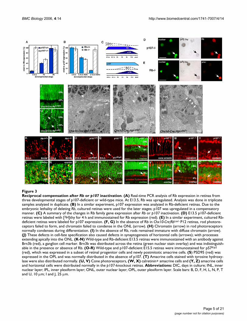
Reciprocal compensation after Rb or p107 inactivationFigure 3Reciprocal compensation after Rb or p107 inactivation. (A) Real-time PCR analysis of Rb expression in retinas from three developmental stages of p107-deficient or wild-type mice. At E13.5, Rb was upregulated. Analysis was done in triplicate samples analyzed in duplicate. (B) In a similar experiment, p107 expression was analyzed in Rb-deficient retinas. Due to the embryonic lethality of deleting Rb, cultured retinas were used for the later stages; p107 was upregulated in a compensatory manner. (C) A summary of the changes in Rb family gene expression after Rb or p107 inactivation. (D) E13.5 p107-deficient retinas were labeled with [3H]thy for 4 h and immunostained for Rb expression (red). (E) In a similar experiment, cultured Rb-deficient retinas were labeled for p107 expression. (F, G) In the absence of Rb in Chx10-Cre;RbLox/- P12 retinas, rod photore-ceptors failed to form, and chromatin failed to condense in the ONL (arrow). (H) Chromatin (arrow) in rod photoreceptors normally condenses during differentiation. (I) In the absence of Rb, rods remained immature with diffuse chromatin (arrow). (J) These defects in cell-fate specification also caused defects in synaptogenesis of horizontal cells (arrows); with processes extending apically into the ONL. (K-N) Wild-type and Rb-deficient E13.5 retinas were immunostained with an antibody against Brn3b (red), a ganglion cell marker. Brn3b was distributed across the retina (green nuclear stain overlay) and was indistinguish-able in the presence or absence of Rb. (O-R) Wild-type and p107-deficient E15.5 retinas were immunostained for p57Kip2
(red), which was expressed in a subset of retinal progenitor cells and newly postmitotic amacrine cells. (S) PSD95 (red) was expressed in the OPL and was normally distributed in the absence of p107. (T) Amacrine cells stained with tyrosine hydroxy-lase were also distributed normally. (U, V) Cone photoreceptors, (W, X) calretinin+ amacrine cells and (Y, Z) amacrine cells and horizontal cells were distributed normally in the p107-knockout retinas. Abbreviations: DIC, days in culture; INL, inner nuclear layer; IPL, inner plexiform layer; ONL, outer nuclear layer; OPL, outer plexiform layer. Scale bars: B, D, F, H, L, N, P, T and U, 10 µm; I and J, 25 µm.

BMC Biology 2006, 4:14 http://www.biomedcentral.com/1741-7007/4/14
embryonic retinas. In the postnatal retina and in differen-tiated neurons and glia of the INL and GCL, Rb and p130were expressed redundantly (Fig. 1V). Rb is the only fam-ily member expressed in rod photoreceptors, a findingthat is consistent with its unique role in the developmentof this neuronal cell type [6]. Together, these data suggestthat Rb and p107 are expressed in a largely nonoverlap-ping pattern during mouse retinal development, whichraises the possibility of compensation by p107 when Rb isinactivated. Moreover, Rb and p130 expression overlaps,which supports the idea that these proteins play redun-dant roles in a subset of retinal neurons.
Expression of the Rb family during the cell cycle in retinal progenitor cellsBeyond determining when and where each Rb familymember is expressed during retinal development, it isimportant to determine when during the cell cycle Rb,p107 and p130 are expressed. These data may help to elu-cidate their normal roles during development. For exam-ple, if a family member is only expressed in proliferatingretinal progenitor cells, then that protein is unlikely toplay a role in neuronal differentiation. It has been wellestablished in a variety of culture systems that Rb, p107and p130 are expressed during different phases of the cellcycle. To directly determine when during the cell cyclethese proteins are expressed throughout retinal develop-ment, we scored the colocalization of [3H]thymidine([3H]thy) with p107 (Fig. 2A–D), Rb (Fig. 2E–H) or p130(Fig. 2I–L) at five time points during four stages of devel-opment [2,22,23]. The expression of p27Kip1 has beencharacterized previously [22], and a parallel set of sampleswas analyzed for p27Kip1 expression to serve as an internalcontrol. The p27Kip1 protein is upregulated as cells exit thecell cycle throughout retinal development, and the kinet-ics are different at E14.5, P0 and P2, because the length ofthe retinal progenitor cell cycle increases during retino-genesis [24]. At E14.5, 62% to 77% of proliferating cellsexpressed p107, and 4% to 6% expressed Rb (see Addi-tional file 1). The 36% reduction in the proportion ofp107+ cells at the 24-h time point in E14.5 retinas wasconsistent with the previous finding that 20% to 25% ofrat retinal progenitor cells exit the cell cycle at a similarstage [22,24,25].
At P0, 76% to 87% of proliferating retinal progenitor cellsexpressed Rb (see Additional file 1; Fig. 2E–H), and theremaining cells expressed p107 (data not shown). Unlikep107+ cells, Rb+ P0 retinal progenitor cells continued toexpress Rb after they exited the cell cycle (compare Fig. 2Hand 2D). In the P2 retina, the level of p130 peaked in thelate G2/early M phase of the cell cycle and persisted intoG1/G0 (see Additional file 1; Fig. 2I–L). Many cells down-regulated p130 as they exited the cell cycle; this findingwas consistent with the birth of rod photoreceptors (80%
of the total cell population in the retina), which do notexpress p130 (Fig. 1R–U). Results from the [3H]thy pulse-labeling experiment combined with those from theimmunostaining, real-time RT-PCR, in situ hybridizationand immunoblot studies (Fig. 1) suggested that p107 isexpressed primarily in retinal progenitor cells duringembryonic retinal development; Rb is expressed in bothretinal progenitor cells and postmitotic neurons and gliain the postnatal retina, and p130 is expressed in cells exit-ing the cell cycle during the late stages of retinal develop-ment. These data help us to further refine the possiblecompensatory and redundant roles of Rb, p107 and p130during retinal development. Specifically, Rb and p130may play redundant roles in postnatal retinal progenitorcells and postmitotic differentiated neurons of the INLand GCL. Rb and p107 are both expressed in proliferatingretinal progenitor cells, albeit at different stages of devel-opment. Inactivation of either Rb or p107 in proliferatingretinal progenitor cells may lead to compensatory upreg-ulation of the other family member.
Reciprocal compensation between Rb and p107 in mouse retinal progenitor cellsTo directly determine whether Rb family members com-pensate for each other in the developing retina, we iso-lated RNA from p107-/-, p107+/-, p107+/+, Rb-/-, Rb+/- andRb+/+ retinas at E13.5, P0 and P6 for real-time PCR analy-sis. In E13.5 retinas lacking p107, Rb mRNA was upregu-lated in a compensatory manner (Fig. 3A). Similarly, inpostnatal retinas lacking Rb, p107 mRNA was upregulated(Fig. 3B, C). The expression of p130 did not change signif-icantly in any of the samples (data not shown).
Next, we tested whether the Rb and p107 proteins werealso upregulated in a compensatory manner in proliferat-ing retinal progenitor cells. Retinas were pulse-labeledwith [3H]thy for 1 h, dissociated and immunostained forRb and p107 (Fig. 3D, E). The mean proportion of Rb+cells in the E13.5 p107-/- retinas (54% ± 6%) was signifi-cantly higher than that in stage-matched wild-type retinas(4% ± 1.2%; p < 0.001). The mean proportion of p107+cells in the P2 Rb-/- retinas (7.3% ± 1.1%) was also signif-icantly higher than that in age-matched wild-type retina(0.3% ± 0.2%; p < 0.005). The proportion of p107+ cellswas lower than that of Rb+ cells, because there are fewerproliferating retinal progenitor cells at P2 than at E13.5[24].
We have shown previously that Rb is required cell auton-omously for the development of postmitotic rod photore-ceptors [6,9]. To test whether p107 compensates for Rb inregulating the development of these postmitotic neurons,we performed electron microscopic (EM) analysis ofChx10-Cre;RbLox/- retinas. One early morphologic featureof rod photoreceptor differentiation is chromatin conden-
Page 6 of 21(page number not for citation purposes)

BMC Biology 2006, 4:14 http://www.biomedcentral.com/1741-7007/4/14
Page 7 of 21(page number not for citation purposes)
Acute inactivation of RB1 in human fetal retinasFigure 4Acute inactivation of RB1 in human fetal retinas. (A-D) Representative in situ hybridization of fetal week 16 human ret-inas with DIG-labeled probes (A, C) and radioactively labeled probes (B, D) shows that RB1 is the major RB family member expressed in proliferating retinal progenitor cells during development. (E-G) Immunofluorescent detection of RB1 (green) in fetal week 16 human retinas confirmed that the protein is expressed in the same pattern as the mRNA. (H) Real-time RT-PCR analysis using TaqMan® probes for RB1 demonstrated that the expression level does not dramatically change over the course of retinal histogenesis. Data sets were analyzed twice, normalized to Gapdh expression and averaged. (I) The sclera and pig-mented epithelium were positive controls for p107 expression. (J) To inactivate RB1 in human fetal retinas, primary tissue was square-wave electroporated with a plasmid that encoded an siRNA to RB1 and a venus-YFP reporter gene. Retinas were then maintained in culture for several days and analyzed for compensation by p107. (K) COS cells transfected with the RB1 SiRNA vector shown in (J) showed a 21-fold reduction in the level of RB1 protein. Densitometry of normalized values for RB1 is shown in the lower portion of panel (K). (L, M) The venus-YFP+ retinal cells that were also [3H]thy+ downregulated RB1 but did not upregulate p107. The negative control SiRNA shown in (M) is the Gapdh SiRNA, but other nonspecific siRNAs gave similar results. The positive control samples are retinas that were square-wave electroporated with a plasmid expressing RB1, p107 or p130 and processed side-by-side with the SiRNA samples. Abbreviations: GCL, ganglion cell layer; inbl, inner neu-roblastic layer; onbl, outer neuroblastic layer; PE, pigmented epithelium. Scale bars: G and L, 10 µm; B, D and E, 25 µm.

BMC Biology 2006, 4:14 http://www.biomedcentral.com/1741-7007/4/14
sation (Fig. 3F–H). In the absence of Rb, p107 was upreg-ulated, but the cells fated to differentiate into rodphotoreceptors, failed to condense their chromatin (Fig.3I) and formed defective synapses in the outer plexiformlayer (OPL) (Fig. 3J). These data suggest that the p107compensation in the postnatal Rb-deficient mouse retina(Fig. 3B, C) helped to prevent deregulated proliferationbut did not rescue the rod differentiation phenotype [6,9].
Rb is also expressed in a small subset of embryonic retinalprogenitor cells (Fig. 1, 2; see Additional file 1), and sub-tle defects may occur in Rb-/- embryonic retinas if p107does not compensate at this stage of development. Weimmunostained Rb-/-, Rb+/- and Rb+/+ retinas at E13.5 withantibodies to several cell cycle proteins (p27Kip1, p57Kip2,cyclin D1, cyclin D3) and markers of differentiated celltypes (Brn3b, GAP43, syntaxin, calbindin, cone arrestin).There was no difference among the three genotypes, ineither the distribution or proportion of cells expressingany of these proliferation or differentiation markers (Fig.3K–R and data not shown).
To determine if the compensation by Rb in p107-deficientretinas (Fig. 3A, C) prevented any defects in retinal pro-genitor cell proliferation, differentiation or both, we ana-lyzed proliferation (BrdU labeling), apoptosis (TUNELassay), cell-fate specification and neuronal differentiation(35 antibodies, see Methods) in p107-/-, p107+/- and p107+/
+ retinas at E13.5, E15.5, P0, P3, P6 and P12. There was nosignificant difference among the genotypes at any stageexamined (Fig. 3E–H). Even in vivo lineage studies usinga replication-incompetent retrovirus in p107-/-, p107+/- andp107+/+ retinas revealed no defect in proliferation, sur-vival, cell-fate specification or differentiation (Schweersand Dyer, in preparation). Together, these genetic analy-ses suggest that reciprocal compensation between Rb andp107 in retinal progenitor cells prevents deregulated pro-liferation during development. The p107 protein cannotcompensate for Rb in postmitotic differentiating rods, andthere was no evidence for p130 compensation in eitherRb- or p107-deficient retinas at any stage of development.
p107 compensation does not occur in the human fetal retina after RB1 inactivationRB1 inactivation is sufficient for retinoblastoma initiationin humans, which suggests that p107 does not compen-sate or play a redundant role when RB1 is lost in humanfetal retinas. To begin to address this question, we charac-terized the expression of the Rb family during human fetalretinal development by performing immunostaining, insitu hybridization and real-time RT-PCR analyses (Fig.4A–H). Primary human fetal retinas from fetal weeks 9,10, 11, 12, 14, 16, 18, 20 and 23 were analyzed; thesestages of human fetal development correspond to theentire period of retinal progenitor cell proliferation (E14-
P10) in the mouse [26]. RB1 is the primary family mem-ber expressed in proliferating retinal progenitor cellsthroughout human retinal development. Little p107expression was detected by immunostaining, real-timeRT-PCR or in situ hybridization analyses (Fig. 4C, D). Aninternal positive control for these expression studies wasthe surrounding sclera and pigmented epithelium thatexpressed high levels of p107 (Fig. 4I). During the latestages of retinal development, p130 was expressed in theretina in a pattern similar to that seen in mouse retinaldevelopment (data not shown).
To test if p107 compensation occurs when RB1 is inacti-vated, we performed square-wave electroporation tointroduce the RB1 SiRNA and a venus-YFP reporter geneinto human fetal week 11 retinas (Fig. 4J). The SiRNAknocked down RB1 protein expression by approximately20 fold (Fig. 4K). An SiRNA to Gapdh and a scrambled RB1SiRNA were used as negative controls. Retinas were main-tained in culture for several days by using a procedure weoptimized for mouse and primate fetal retinas (see Meth-ods). Retinal progenitor cells proliferated normally withno appreciable cell death for at least 5 days while in thisculture system (see Additional file 2). At several timepoints after electroporation, we labeled proliferating reti-nal progenitor cells with [3H]thy, dissociated the retinas,plated the cells on glass slides and immunostained themfor RB1, p107 and p130 (Fig. 4L and data not shown). Byscoring the expression of the Rb family in venus-YFP+ cellsthat were also [3H]thy+, we found that RB1 was downreg-ulated by the SiRNA 48 h after electroporation, but therewas no compensation by p107 or p130 (Fig. 4M). Anidentical experiment carried out on fetal week 14 retinasgave similar results (data not shown). As positive controlsfor RB1, p107 and p130 immunostaining, we electropo-rated parallel cultures with a plasmid expressing theseproteins (Fig. 4M). The expression data combined withthat from the RB1 gene inactivation studies in primaryhuman fetal retinas suggested that p107 is not normallyexpressed in the developing human retina, and it cannotcompensate for RB1 loss in this tissue.
Acute inactivation of Rb in the developing mouse retinaCompensation of p107 in MEFs depends on whether Rb isinactivated in an acute or chronic manner [20]. Our stud-ies on Rb-knockout retinas including Chx10-Cre;RbLox/- ret-inas showed that p107 was upregulated in acompensatory manner after chronic Rb inactivation in theretina. To determine whether this result occurs after acuteRb gene inactivation in the developing retina in vivo, weintroduced a Cre-expressing plasmid (pCre-vYFP) into theeyes of newborn RbLox/- mice (Fig. 5A). Real-time RT-PCRanalysis of purified retinal cell populations (Fig. 5B–G) inwhich Rb was acutely inactivated showed that p107mRNA was upregulated (Fig. 5H), as was seen after
Page 8 of 21(page number not for citation purposes)

BMC Biology 2006, 4:14 http://www.biomedcentral.com/1741-7007/4/14
Page 9 of 21(page number not for citation purposes)
Acute inactivation of Rb in the developing mouse retinaFigure 5Acute inactivation of Rb in the developing mouse retina. (A-G) In vivo or in vitro square-wave electroporation and purification of cells with acute Rb inactivation. (C) A retina after electroporation is shown. (D-G) Dissociated cells before and after FACS purification are shown. (H) Real-time PCR analysis of YFP+ purified cells after acute Rb inactivation; each sample was analyzed in duplicate and normalized to Gapdh and Gpi1. (I, J) Immunostaining of purified cells after Rb inactivation. (K-N) To determine if retinal progenitor cells continue to divide after acute Rb inactivation, we scored the proportion of [3H]thy-labeled BrdU+ cells. An example of a double-positive cell is shown in (K). (L) A significant proportion of progenitor cells was sensitive to deregulated proliferation after acute Rb inactivation, as indicated by the increase in double-positive cells. Data are normalized to account for the fraction of cells labeled with BrdU during a 1-h pulse. (M) The proportion of retinal progenitor cells that continue to divide after acute Rb inactivation is shown in the venus-Cre, YFP+ column. (N) Real-time PCR analysis of cell samples used in (L) and (M) revealed a significant increase in the expression of p107 and retinal progenitor cell markers Sfrp1, Erdr1 and Chx10 after acute Rb inactivation. Scale bars: C, D, F, I and J, 10 µm.

BMC Biology 2006, 4:14 http://www.biomedcentral.com/1741-7007/4/14
Page 10 of 21(page number not for citation purposes)
Haploinsufficiency of p107 in the developing mouse retinaFigure 6Haploinsufficiency of p107 in the developing mouse retina. Immunostaining of P30 Rb+/-; p107-/- retinas in the region of the retinal dysplasia. (A, B) Antibodies against calbindin were used to identify horizontal cells; (C, D) those against Chx10, bipolar cells; (F, G) those against glutamine synthetase, Müller glia; and (H, I) those against bassoon, synapses in the OPL and IPL. (E) EM analysis of the P30 Rb+/-; p107-/- retinas showing the OPL with normal organization of synaptic connections in con-trast to the Rb-deficient retinas. (J-T) Immunostaining of P14 Chx10-Cre;RbLox/-; p107+/- retinas in the region of Cre-mediated Rb inactivation using antibodies to OPL synapses (PSD-95, J-M), horizontal cells (calbindin, O, P), amacrine/progenitor cells (Pax6, Q, R), and reactive Müller glia (GFAP, S, T). (N) EM analysis of the P14 Chx10-Cre;RbLox/-; p107+/- retinas showing the OPL with disrupted synaptic connections and an apical horizontal process (arrows). (U-Y) To determine if there are any ectopically dividing cells in Rb+/-; p107-/-, Rb-/-; p107+/- or Rb-/-; p107-/- retinas at E14.5, P0, P6, P12 and P30, we labeled retinas with [3H]thy and BrdU for 1 h and then dissociated and immunostained the cells with antibodies against 12 markers of different retinal cell types. BrdU and [3H]thy always colocalized. (W) The only markers that colocalized with [3H]thy were amacrine/progenitor cell markers Pax6 and syntaxin and bipolar/progenitor cell marker Chx10. There were also some reactive Müller glia, as indi-cated by GFAP, that incorporated [3H]thy. Ectopic proliferation was more prevalent in the double-knockout retinas than in the other genotypes. Each bar is the average of triplicate samples scored in duplicate. (X, Y) Real-time PCR analysis demonstrated that throughout development there were more progenitor cell markers such as Cdk2 and Sfrp1 expressed in the double-knockout and Rb-/-; p107+/- retinas. Abbreviations: GCL, ganglion cell layer; INL, inner nuclear layer; ONL, outer nuclear layer; OPL, outer plexiform layer. Scale bars: B, D, G, I, K, M, P, Q, T, U and V, 10 µm.

BMC Biology 2006, 4:14 http://www.biomedcentral.com/1741-7007/4/14
chronic Rb gene inactivation in the developing retina (Fig.3). There was little change in p130 expression (Fig. 5H).
Next, we determined whether acute inactivation of Rbaltered rod development. Real-time RT-PCR analysis ofRNA samples indicated that the early photoreceptor genesNrl and Nr2e3 were downregulated, as were several otherphotoreceptor-specific genes (Fig. 5H and data notshown). The cells that did not differentiate into photore-ceptors resembled progenitor cells, as indicated by theincreased expression of Eya2 and Fgf15 (Fig. 5H) [27]. Tofurther verify these developmental defects, we immunos-tained the purified cells that had undergone acute Rb inac-tivation and compared them to the control cells (Fig. 5I,J). Those data were consistent with real-time PCR datashowing that acute inactivation of Rb not only leads to acompensatory increase in p107 expression, but also dis-rupts rod development. Results of experiments in whichp107 was acutely inactivated in wild-type E14.5 retinas byusing an siRNA to p107 [20] were similar to those in thep107-knockout retinas, i.e., Rb was upregulated in a com-pensatory manner (data not shown).
Proliferating cells and nondividing or quiescent MEFsexhibit differences in the timing of their compensatoryupregulation of p107 after Rb inactivation [20]. To test fordifferences in compensation in proliferating retinal pro-genitor cells and transition cells [28] in the developing ret-ina, we acutely inactivated Rb at P3. Even though themajority (~ 95%) of cells in the P3 retina are postmitotic[24], a disproportionate number of proliferating retinalprogenitor cells (42% ± 5.5%) underwent ectopic roundsof cell division (Fig. 5K, L; see Additional file 3). Retinalprogenitor cells and transition cells were distinguished byBrdU labeling prior to electroporation (Fig. 5A). This find-ing suggests that retinal progenitor cells in which Rb hasbeen acutely inactivated are susceptible to deregulatedproliferation. A similar experiment in which all proliferat-ing retinal progenitor cells in the P3 retina were labeledwith [3H]thy for 24 h prior to acute Rb inactivationshowed the same result (Fig. 5M). Specifically, eventhough retinal progenitor cells comprise only 7% of thetotal cell population at P3, they comprised 74% ± 4% ofthe cells that continued to divide after acute Rb inactiva-tion (Fig. 5M). Real-time RT-PCR analysis of the samplesdemonstrated that p107 was upregulated in a compensa-tory manner and that retinal progenitor cell genes such asSfrp1, Erdr1 and Chx10 were also upregulated (Fig. 5N).
To test if p107 compensation after acute Rb inactivationprevents deregulated proliferation, we acutely inactivatedp107 by using a previously characterized SiRNA [20] inretinal cells in which Rb was chronically inactivated. Wefound that proliferation significantly increased from 18%± 1.9% to 34% ± 1.9% (p = 0.013) after acute p107 inacti-
vation (see Additional file 4). Moreover, the cells that con-tinued to divide expressed retinal progenitor cell markersChx10, Pax6 and syntaxin and were BrdU+ retinal progen-itor cells at the time of p107 inactivation (see Additionalfile 4). Together, these data support the idea that p107compensation is important for preventing deregulatedproliferation in retinal progenitor cells whether Rb is inac-tivated in an acute or chronic manner.
Haploinsufficiency of p107 in the absence of RbHaving shown that reciprocal compensation between Rband p107 in the developing retina prevents deregulatedproliferation of retinal progenitor cells, we tested whethera single copy of Rb was sufficient to compensate for p107deficiency and whether a single copy of p107 was suffi-cient to compensate for Rb deficiency. Immunostaining ofRb+/-; p107-/- retinas between P6 and P30 confirmed thepresence of minor focal retinal dysplasia, as reported pre-viously [13]. The laminar structure and synaptogenesisoutside the regions of focal dysplasia were not substan-tially disrupted (Fig. 6A–I and data not shown), as con-firmed by EM analysis of the OPL (Fig. 6E). In addition,EM analysis confirmed that rod photoreceptors [6] andhorizontal cells [9] form normally with just one copy ofRb and no p107 (Fig. 6E). Even in the regions of the mostsevere dysplasia (Fig. 6A–I), cells were in their appropriatecellular layers, and synaptic markers were present in thecorresponding plexiform layer sublaminae. EM analysissuggests that these focal dysplastic lesions are not formedby preneoplastic retinoblastoma cells. That is, there is noevidence for immature tumor cells localized to thelesions.
In contrast to the Rb+/-; p107-/- retinas, the Chx10-Cre;RbLox/
-; p107+/- retinas exhibited much more severe developmen-tal defects. Immunostaining revealed deregulated prolifer-ation and lamination in an apical-basal mosaic patternthat was consistent with the expression of Cre from theChx10 promoter of the Chx10-Cre transgene [9,29]. Rodphotoreceptors were not completely lost due to themosaic pattern of gene inactivation by the Chx10-Creallele [29]. Defects in OPL synaptogenesis (Fig. 6J–T) wereconfirmed by EM analysis of P43 Chx10-Cre;RbLox/-; p107+/
- retinas (Fig. 6N). Müller glia showed upregulated expres-sion of GFAP, which is a marker of reactive gliosis (Fig. 6S,T); however, GFAP expression was not detectable inMüller glia of the Rb+/-; p107-/- retinas.
To quantify the proportion and types of cells that wereproliferating in Rb+/-; p107-/-, Rb-/-; p107+/- and Rb-/-; p107-/
- retinas during development, we labeled P0, P6, P12 andP30 retinas with [3H]thy for 1 h, dissociated the retinasand immunostained the cells with antibodies that recog-nized each of the retinal cell types (see Additional files5,6,7,8). Few (if any) of the cells expressing differentiation
Page 11 of 21(page number not for citation purposes)

BMC Biology 2006, 4:14 http://www.biomedcentral.com/1741-7007/4/14
Page 12 of 21(page number not for citation purposes)
Retinal progenitor cells continue to proliferate throughout development in the absence of Rb and p107Figure 7Retinal progenitor cells continue to proliferate throughout development in the absence of Rb and p107. (A-T) Immunostaining of P30 Chx10-Cre;RbLox/-; p107-/- retinas by using antibodies against (A-D) BrdU to identify ectopically dividing cells; (E-H) calbindin, horizontal cells and Chx10, bipolar/progenitor cells; (M-P) GFAP, reactive Müller glia; (Q, R) cone arrestin, cone photoreceptors and (S, T) Pax6, amacrine/progenitor cells. (U, V) Electron micrographs showing that most retinal structures are disrupted in the absence of Rb and p107 and that continued proliferation eventually leads to retinoblast-oma. (W-Z) Newborn RbLox/Lox; p107-/- mice were injected with a retrovirus expressing Cre (LIA-Cre), and 3 weeks later, the retinas were sectioned and stained for alkaline phosphatase expression to identify the neurons that had lost Rb and p107. Ama-crine cells (W) and bipolar cells (X) formed normally in the absence of Rb and p107. Rod photoreceptors (Y) failed to form normally. There were also several clones that spanned the INL or INL and ONL, which was indicative of hyperproliferation (Z). Similar experiments were carried out in E17.5 retinal explants by using a Cre-expressing retrovirus that also expresses nuclear LacZ (AA). These clones can readily be scored for cell number to determine if loss of Rb and p107 leads to deregu-lated proliferation. Abbreviations: GCL, ganglion cell layer; INL, inner nuclear layer; olm, outer limiting membrane; ONL, outer nuclear layer. Scale bars: B, D, F, H, J, L, N, P, R, T and W-AA, 10 µm.

BMC Biology 2006, 4:14 http://www.biomedcentral.com/1741-7007/4/14
markers such as recoverin (photoreceptors), cone arrestin(cones) or PKCα (bipolar cells) incorporated [3H]thy (Fig.6U–W; see Additional files 5,6,7,8). Primarily, cellsexpressing progenitor cell markers (see Discussion) suchas syntaxin, Pax6 or Chx10 incorporated [3H]thy (Fig.6U–W; see Additional files 5,6,7,8). A small proportion ofMüller glia also incorporated [3H]thy, a finding that wasconsistent with reactive gliosis-associated proliferation(Fig. 6S, T) [30]. The overall distribution of cell types wasnot significantly altered in Rb+/-; p107-/- retinas (see Addi-tional files 5,6,7,8), but as expected, the number of rodswas reduced in Chx10-Cre;RbLox/-; p107+/- retinas (see Addi-tional files 5,6,7,8).
TUNEL assay results revealed no difference in the propor-tion of apoptotic cells in Rb+/-; p107-/- retinas and that oftheir wild-type littermates. The number of apoptotic cellsin Chx10-Cre;RbLox/-; p107+/- retinas was slightly higherat P12 and P30, which corresponded to the stages ofincreased proliferation (see Additional files 5,6,7,8). Real-time RT-PCR analysis revealed increased expression of theretinal progenitor cell genes Cdk2 (Fig. 6X) and Sfrp2 (Fig.6Y). Together, these data suggest that one copy of Rb inRb+/-; p107-/- retinas is sufficient for much of retinaldevelopment (with the exception of minor focal dyspla-sia), but one copy of p107 in Chx10-Cre;RbLox/-; p107+/- retinas is haploinsufficient to regulate proliferation. Spe-cifically, in retinal progenitor cells, two copies of the p107gene are required to compensate for loss of Rb, but onlyone copy of Rb is required to compensate for loss of p107.
Deregulated proliferation in Rb;p107-deficient retinal progenitor cellsIt has been shown previously that retinoblastoma formsin Rb;p107-deficient retinas [10,11,14,15]. Therefore, wepropose that reciprocal compensation between Rb andp107 in retinal progenitor cells prevents retinoblastomain mice. If this is true, then we predict that retinal progen-itor cells will continue to divide throughout development(E14-P30) in Rb;p107-deficient retinas. This is preciselywhat we found (Fig. 7A–D; see Additional files 5,6,7,8).The proliferating cells were detected in a mosaic pattern,which was consistent with Rb inactivation by the Chx10-Cre transgene (Fig. 7A–D) [9,29]. These immature prolif-erating cells disrupted synaptogenesis in the OPL (Fig.7E–H) and expressed the progenitor cell markers syntaxin,Pax6 and Chx10 (Figs. 6W, 7I–L; see Additional files5,6,7,8) [31-33]. With the exception of activated Müllerglia (Fig. 7M–P; see Additional files 5,6,7,8), few of theproliferating cells expressed markers of differentiated reti-nal cells. It is important to point out that syntaxin, Pax6and Chx10 are expressed in both retinal progenitor cellsand postmitotic differentiated cells. However, the combi-nation of BrdU, Pax6, Chx10 and syntaxin immunoreac-tivity combined with morphologic features of these cells
in electron micrographs lends support to their identifica-tion as retinal progenitor cells.
In contrast to findings from a recent study [10], we did notdetect any defect in cone photoreceptor production in ret-inal sections (Fig. 7Q, R) or in dissociated cells scored atP0, P6, P12 or P30 (see Additional files 5,6,7,8). EM anal-ysis of Chx10-Cre;RbLox/-; p107-/- retinas revealed immaturecells spanning all cellular layers of the retina (Fig. 7U, V).These electron micrographs support the hypothesis thatChx10+, Pax6+, BrdU+ cells in the ONL and INL are reti-nal progenitor cells rather than differentiated cells. Inaddition, real-time PCR analysis demonstrated anincrease in the expression of retinal progenitor cell mark-ers in Chx10-Cre;RbLox/-; p107-/- retinas at P6, P12 and P30(Fig. 6X, Y).
Our analysis of the Rb;p107-deficient retinas over thecourse of postnatal retinal development showed that at allstages of development studied, there are ectopically divid-ing cells with morphologic features of retinal progenitorcells that express markers of retinal progenitor cells. Ourdata support the hypothesis that the role of reciprocalcompensation between Rb and p107 is to prevent deregu-lated proliferation of retinal progenitor cells. If this is true,then we would expect that inactivation of Rb and p107 ina single proliferating retinal progenitor cell would lead toclonal expansion.
We infected proliferating retinal progenitor cells in new-born RbLox/-; p107-/- mice with the Cre-expressing retrovi-rus LIA-Cre (Fig. 7W–Z). We observed significantly largerclone sizes and changes in cell-fate specification (Fig. 7Y,Z; see Additional file 9). To quantify the deregulated pro-liferation, we used the NIN-Cre retrovirus, whichexpresses nuclear lacZ and Cre, in E17.5 RbLox/Lox; p107-/-
retinas; NIN virus was used as a control. Clone sizeincreased and was consistent with deregulated retinal pro-genitor cell proliferation. In control retinas infected withNIN, 248 of 313 (80%) clones were one cell, and 11 of313 (3.5%) contained more than two cells. In retinasinfected with NIN-Cre, 100 of 190 (52%) clones were onecell, and 42 of 190 (24%) contained more than two cells(Fig. 7AA). Similar results were seen in studies using theE1A 13S oncogene [6,15].
Proliferation of progenitor cells and postmitotic transition cells in Rb;p107-deficient retinasThe developmental studies and clonal analyses describedabove suggest that reciprocal compensation between Rband p107 prevents deregulated retinal progenitor cell pro-liferation and retinoblastoma. However, we have shownthat p107 compensation also occurs at P3 when themajority (93%) of cells are postmitotic (Fig. 5N). There-fore, it is possible that reciprocal compensation between
Page 13 of 21(page number not for citation purposes)

BMC Biology 2006, 4:14 http://www.biomedcentral.com/1741-7007/4/14
Rb and p107 prevents newly postmitotic cells from re-entering the cell cycle rather than preventing deregulatedproliferation of retinal progenitor cells. To test this possi-bility directly, we labeled all proliferating retinal progeni-tor cells in P3 RbLox/Lox; p107-/- retinas with [3H]thy (Fig.8A), inactivated Rb expression by electroporating a plas-mid that expressed Cre and vYFP and then incubated theretinas with BrdU. Cells that were BrdU+, vYFP+ and[3H]thy+ were retinal progenitor cells at the beginning ofthe experiment that continued to divide after acute Rbinactivation. In contrast, cells that were BrdU+, vYFP+ and[3H]thy- were postmitotic transition cells at the beginningof the experiment that re-entered the cell cycle after acute
Rb inactivation. The time point P3 was selected becausethe vast majority (~ 93%) of cells in the retina at that stageare postmitotic [24]. If a postmitotic cell re-enters the cellcycle after acute Rb inactivation in RbLox/Lox; p107-/- retinas,then the majority of BrdU+ cells would be [3H]thy-. If aretinal progenitor cell is more susceptible to deregulatedproliferation after acute Rb inactivation, then we wouldpredict that the majority of BrdU+ cells would be[3H]thy+. If both retinal progenitor cells and postmitoticcells divide after acute Rb inactivation, then the propor-tions of BrdU+, [3H]thy- cells and BrdU+, [3H]thy+ cellswill reflect the proportions of postmitotic cells andmitotic cells in the starting population, respectively.
Retinal progenitor cells are sensitive to deregulated proliferation after acute inactivation of Rb and p107Figure 8Retinal progenitor cells are sensitive to deregulated proliferation after acute inactivation of Rb and p107. (A) To determine if retinal progenitor cells are sensitive to deregulated proliferation after acute Rb inactivation in a p107-deficient genetic background, we labeled RbLox/Lox; p107-/- P0 or P3 retinas for 24 h with [3H]thy and then electroporated them with a Cre-venus-YFP plasmid. After 3 days in culture, the retinas were labeled for 1 h with BrdU and dissociated, and the YFP+ and YFP- cells were purified by FACS. (B-F) The cells were then analyzed by immunostaining, single-cell scoring and real-time PCR. (B, C) Colocalization of BrdU and [3H]thy demonstrated that most cells that continued to proliferate after Rb inactivation were retinal progenitor cells. (D) Real-time RT-PCR analysis confirmed that the proliferating cells expressed retinal progenitor cell markers. (E, F) Immunostaining and colocalization with [3H]thy further confirmed the retinal progenitor cell characteris-tics of these cells. Scale bars: B and E, 10 µm.
Page 14 of 21(page number not for citation purposes)

BMC Biology 2006, 4:14 http://www.biomedcentral.com/1741-7007/4/14
Most (79.8% ± 6%) ectopically dividing cells were BrdU+,[3H]thy+ cells (Fig. 8B, C). Consistent with this finding,results from real-time PCR and immunostaining analysesrevealed that ectopically dividing cells expressed severalretinal progenitor cell markers including Pax6, Chx10,Syntaxin, Erdr1 and Sfrp1 (Fig. 8D–F). However, there wasa small increase in the proportion of BrdU+, [3H]thy- cells(Fig. 8C), which leaves open the possibility that either aretinal progenitor cell or a transition cell is the cell of ori-gin for retinoblastoma (discussed in [28]).
DiscussionWe report here that in the developing mouse retina p107is expressed in embryonic retinal progenitor cells, and Rbis expressed in postnatal retinal progenitor cells. Whenp107 is inactivated, Rb is upregulated in a compensatorymanner, and when Rb is inactivated, p107 compensates.The p130 protein is expressed during the late stages ofretinogenesis and persists in the INL and GCL in the adultretina. Rb and p130 are expressed redundantly in thesecells. There was no evidence of compensatory changes inp130 expression following either Rb or p107 inactivation.Compensation by p107 occurred when Rb was inactivatedacutely or chronically in the developing mouse retina.Two copies of the p107 gene were required to preventderegulated proliferation of Rb-deficient retinal progeni-tor cells, making p107 haploinsufficient for compensationin the developing retina. In contrast, only one copy of Rbwas required to regulate proliferation of p107-deficientretinal progenitor cells. We propose that reciprocal com-pensation between Rb and p107 prevents deregulatedproliferation of retinal progenitor cells and retinoblast-oma in mice. Similarly, redundant expression of Rb andp130 may help to prevent retinoblastoma in mice.
In contrast to mice, human fetal retinal progenitor cellsprimarily express RB1 during development. When RB1was acutely inactivated in human fetal retinas, there waslittle if any compensatory upregulation of p107. This mayexplain why humans are susceptible to retinoblastomafollowing RB1 gene mutations, but mice require inactiva-tion of Rb and p107 or Rb and p130.
Overlapping and unique functions of the Rb family in the developing mouse retinaImmunostaining, in situ hybridization, real-time RT-PCRand immunoblot analyses revealed that p107 is expressedprimarily in proliferating retinal progenitor cells of theembryonic mouse retina. Pulse-labeling with [3H]thydemonstrated that p107 is downregulated as cells exit thecell cycle. There were no defects in cell-fate specification ordifferentiation in p107-deficient retinas; thus, we proposethat p107 primarily regulates retinal progenitor cell prolif-eration in the embryonic retina. Rb is expressed in postna-tal retinal progenitor cells and differentiating neurons and
glia. Consistent with a role in cell-fate specification or dif-ferentiation, Rb was expressed throughout the cell cycleand continued to be expressed as cells entered G0. In theabsence of Rb, immature cells persist in the ONL and rodphotoreceptors failed to form, a finding that indicates thatRb regulates the developmental processes in this specificsublineage.
Recent studies have extended these findings by focusingon the mechanism of Rb's role in rod development. Sunand colleagues generated an exon-specific knock-in alleleof Rb with a single amino acid substitution (R654W) thatreduces Rb binding to E2F1, E2F2 and E2F3 [34]. InChx10-Cre;Rb654/Lox retinas, rods still fail to form. Thisfinding led to a model in which Rb regulates a rod repres-sor gene through E2F1, E2F2 or E2F3, and in the absenceof Rb, the rod lineage is blocked. Moreover, we predictthat inactivation of the E2F that regulates this putative"rod repressor" would rescue the rod developmentaldefect. Preliminary data have shown that Rb;E2F1-defi-cient retinas have normal rod development, a finding thatsuggests that E2F1 is involved in this important develop-mental process.
Our data suggest that Rb and p107 share the same role inregulating mouse retinal progenitor cell proliferation butthe role of Rb is unique in rods. An extensive genetic anal-ysis of p130 is beyond the scope of this manuscript, butbased on our expression studies and [3H]thy-labelingexperiments, we predict that Rb and p130 are redundantin newly postmitotic cells of the INL and GCL. It is notknown if p130 is required for terminal cell cycle exit in thedeveloping retina, for maintaining cells in the postmitoticstate or both.
A recent study published by Spencer and colleagues char-acterized the expression of the Rb family in developingmouse retina and adult human retina [35]. Our data differfrom theirs, and we have presented the most relevant dif-ferences in Figure 1. Our analysis combined several inde-pendent methods (real-time RT-PCR, in situhybridization, immunostaining of tissue sections, immu-nostaining of [3H]thy-labeled dissociated cells and immu-noblotting); these approaches provided the same overallpicture of the dynamic changes in the expression of the Rbfamily during seven stages of mouse retinal development.Spencer and colleagues provided a more limited data setwith internal inconsistencies. For example, immunostain-ing was done on samples from just three stages of mouseretinal development (E14, P0 and P5), and in situ hybrid-ization was carried out for Rb but not for p107 or p130.Their real-time RT-PCR and immunoblotting data showhigh levels of p107 expression in the embryonic retinas,but no p107+ nuclei are detected in their immunostainedsections. Clearly, all expression data are limited by the rea-
Page 15 of 21(page number not for citation purposes)

BMC Biology 2006, 4:14 http://www.biomedcentral.com/1741-7007/4/14
gents being used. For example, it is possible that the Rb,p107 and p130 expression patterns in our study or that ofSpencer and colleagues are broader than reported due tothe limited sensitivity of the antibodies used. We haveattempted to minimize such limitations by combiningseveral independent approaches such as in situ hybridiza-tion, immunostaining, real time RT-PCR and dissociatedcell staining. Nontheless, we anticipate that the retinalexpression pattern of the Rb family will be further refinedas new reagents become available.
Rb family redundancy and compensation in the developing mouse retinaThe Rb family proteins are expressed in a dynamic mannerduring mouse retinal development. We show here thatp107 is expressed in proliferating retinal progenitor cellsin the embryonic retina and is downregulated as cells exitthe cell cycle and undergo terminal differentiation.Around birth, the expression of p107 tapers off, and thatof Rb takes it place in proliferating retinal progenitor cells.Rb expression persists in postmitotic differentiating neu-rons and glia of the mouse retina. The p130 protein isexpressed in the late postnatal period of mouse retinaldevelopment. As cells exit the cell cycle, they upregulatep130, and its expression persists in postmitotic differenti-ating cells of the INL and GCL. Based on the expressionpattern alone, one might predict that proliferation wouldbe deregulated in p107-deficient embryonic retinas andRb-deficient postnatal retinas. However, we report herethat when p107 is inactivated, Rb is upregulated in a com-pensatory manner in the embryonic retina. Similarly,when Rb is inactivated, p107 is upregulated in a compen-satory manner in the postnatal retina. There is no evi-dence for compensation by p130 in either Rb- or p107-deficient retinas.
One of the interesting features of the compensation thatwe uncovered in this study is the haploinsufficiency ofp107 in this process. We show here that two copies of p107are required for efficient compensation of Rb loss, butonly one copy of Rb is required to compensate for p107loss. This may indicate that there are different mecha-nisms for Rb and p107 gene compensation or that retinalprogenitor cells exhibit different sensitivities to Rb andp107 protein levels. One intriguing possibility is that p107haploinsufficiency for compensation may reflect the dif-ferential binding of Rb and p107 to different E2F familymembers.
It has been shown previously that p107 is an E2F-respon-sive gene; therefore, the mechanisms for p107 compensa-tion most likely involve a direct feedback loop [36].However, the mechanism for Rb compensation in theabsence of p107 has not been elucidated. We propose thatRb-deficient retinal progenitor cells fail to form retino-
blastoma because p107 is upregulated in a compensatorymanner. However, it is possible that there is some over-lapping, redundant expression around P0 in the develop-ing mouse retina. Interestingly, Rb and p130 are expressedredundantly in the INL and GCL. The recent observationthat retinoblastoma develops in Rb;p130-deficient retinassuggests that redundant expression of these two Rb familymembers prevents retinoblastoma in mice [11].
Previous studies using Rb-/- MEFs demonstrated that inac-tivation of Rb in proliferating cells leads to compensatoryupregulation of p107 and quiescence after serum starva-tion[20]. The p107 gene is persistently expressed in thesequiescent Rb-deficient MEFs and is required for maintain-ing them in G0. When Rb was acutely inactivated in qui-escent RbLox/Lox MEFs, p107 was not immediatelyupregulated in a compensatory manner, and the cells re-entered the cell cycle [20]. Therefore, chronic or acuteinactivation of Rb can lead to very different outcomes inMEFs. The molecular changes that occur in MEFs as theybecome quiescent have been compared with those thataccompany terminal cell cycle exit during development[20]. However, in the developing CNS, postmitotic neu-rons not only exit the cell cycle, but also migrate to theirappropriate layer, extend axons and dendrites and formsynaptic connections.
To test if there was any difference in p107 compensationfollowing acute versus chronic Rb inactivation in thedeveloping retina, we developed genetic tools [16] toacutely inactivate Rb in this context. Our data show thatp107 compensation occurs when Rb is inactivated acutelyor chronically in the developing retina. In addition, rodphotoreceptors failed to form when Rb was acutely inacti-vated. These data suggest that p107 compensates for Rb inproliferating retinal progenitor cells, but it cannot take theplace of Rb in differentiating rod photoreceptors.
We extended our acute inactivation studies to knock outRb in p107-deficient cells and to knock out p107 in Rb-deficient cells. In these experiments, we compared theeffects of acute gene inactivation on proliferating retinalprogenitor cells and newly postmitotic transition cells.The newly postmitotic Rb-deficient transition cells inwhich p107 was acutely inactivated did not show exten-sive ectopic cell division. These data are consistent withthe idea that p107 is important in regulating retinal pro-genitor cell proliferation but not in preventing newlypostmitotic cells from re-entering the cell cycle. However,a small subset of postmitotic cells still re-entered the cellcycle following acute Rb inactivation in p107-deficient ret-inas. Therefore, we cannot rule out the possibility that theretinoblastoma cell of origin is a retinal transition cell[28]. Definitive proof will rely on following the expansionof clones lacking p107 and Rb to determine whether any
Page 16 of 21(page number not for citation purposes)

BMC Biology 2006, 4:14 http://www.biomedcentral.com/1741-7007/4/14
rare transition cells re-enter the cell cycle and expand togenerate retinoblastoma or if only retinal progenitor cellscontinue to proliferate, as shown here [28].
Our data on the proliferation of Rb;p107-deficient retinalcells during development contrasts with previous studiesby Chen and colleagues [10]. We show that ectopicallydividing retinal cells are present at P0, P6, P12 and P30,whereas the previous study argued that proliferationceased at P30. This difference at P30 may be due to themore sensitive and reproducible [3H]thy labeling we usedin our study compared to the BrdU labeling used by Chenet al. Alternatively, it could reflect the non-cell-autono-mous effect of broad Rb inactivation in Pax6-Cre;RbLox/Lox;p107-/- retinas examined by Chen et al. compared with themore restricted mosaic Rb inactivation in Chx10-Cre;RbLox/
-; p107-/- retinas that we investigated [16].
Another difference between our data and the study previ-ously published by Chen and colleagues involves theinterpretation of the markers expressed in proliferatingcells of the Rb;p107-deficient retinas. We found that at P0,the ectopically dividing cells were largely Pax6+ (see Addi-tional file 5). By P6, the proliferating cells were Pax6+,Chx10+ (see Additional file 6) and by P12, they werePax6+, Chx10+ and syntaxin+ (see Additional file 7). Pax6[32] and syntaxin [31] are expressed in both retinal pro-genitor cells and differentiating amacrine cells. Chx10 isexpressed in retinal progenitor cells and in differentiatedbipolar cells and Müller glia [29]. Interestingly, at P12 wenoted that GFAP+ Müller glia were also [3H]thy+. Thisfinding is consistent with proliferation associated withreactive gliosis in Rb;p107-deficient retinas [30]. Chx10immunopositivity may mark the proliferating Müller glia,bipolar cells or progenitor cells at this stage. However, wedo not believe that the Chx10+ cells are bipolar cells,because there were no PKCα+ cells that incorporated[3H]thy at P12. At P30, most of the proliferating [3H]thy+cells were syntaxin+. A smaller proportion were Pax6+,recoverin+ and PKCα+. Recoverin is expressed in a subsetof bipolar cells as well as photoreceptors, so the PKCα+and recoverin+ cells that incorporated [3H]thy are mostlikely a small cohort of bipolar cells that re-entered thecell cycle.
Based on the timeline of marker colocalization with[3H]thy that we present here, we propose that a Pax6+,Chx10+ and syntaxin+ retinal progenitor cell populationthat is biased toward the amacrine/horizontal cell fatecontinues to proliferate through development in Chx10-Cre;RbLox/-; p107-/- retinas and eventually gives rise to retin-oblastoma. This model is consistent with the extensivedifferentiation of mouse retinoblastomas along amacrine/horizontal cell lineage (Johnson and Dyer, in prepara-tion). There is an occasional round of ectopic prolifera-
tion in reactive Müller glia and possibly bipolar cells atP30, but based on the absence of glial and bipolar markersin mouse retinoblastomas, we propose that these cells donot contribute significantly to retinoblastoma in mice.
Chen and colleagues [10] showed that Pax6+, Chx10+,Prox1+, Math3+, Crx+ and Hes5+ cells incorporatedBrdU. With the possible exception of Crx, each of thesemarkers is expressed in both progenitor cells and differen-tiated cells [18,32,33,37-42]. We have found that severaladditional retinal progenitor cell markers (syntaxin 1,Fgf15, Sfrp1, Erdr1 and Eya2) are expressed in proliferat-ing cells of Rb;p107-deficient retinas. Overall, 10 of the 11genes that are expressed in proliferating Rb;p107-deficientretinal cells have one feature in common; they are allexpressed normally in retinal progenitor cells. The mor-phological features of the Rb;p107-deficient retinal cells inelectron micrographs are also consistent with retinal pro-genitor cells. While these data support the hypothesis thatretinal progenitor cells continue to divide during develop-ment of Rb;p107-deficient retinas and eventually give riseto retinoblastoma in mice, marker expression alone can-not distinguish between a progenitor cell and a transitioncell as the cell of origin for retinoblastoma (discussed in[28]). Definitive proof of the cell of origin for retinoblas-toma will require clonal analysis of tumor formation froma single cell in vivo.
Retinoblastoma susceptibility in mice and humansWhen the Rb-knockout mouse was generated over 13years ago, it was expected that retinoblastoma woulddevelop in Rb+/- mice, as it does in RB1+/- humans. How-ever, that was not the case. We show here that the normalexpression of Rb family members is dynamic duringmouse retinal development and that reciprocal compen-sation between Rb and p107 may prevent retinoblastomain mice.
In the human fetal retina, RB1 is the major family mem-ber expressed during much of retinal development. Thereis little or no expression of p107. We acutely inactivatedRB1 in primary human fetal retinas by electroporating anSiRNA to RB1. We demonstrated that although the RB1protein was downregulated 20-fold, there was no com-pensation by p107 or p130. These data suggest thathumans are susceptible to retinoblastoma formation afterRB1 inactivation, because they cannot upregulate p107 ina compensatory manner. It is possible that the 20-foldreduction in RB1 protein induced by the SiRNA used forthese experiments is not sufficient to induce p107 com-pensation. However, we do not believe this is the casebecause another RB1 target gene (p14ARF) is induced by10-fold following acute RB1 inactivation using the sameSiRNA (Laurie et al., submitted). One possible explana-tion for the lack of compensation in the human retina is
Page 17 of 21(page number not for citation purposes)

BMC Biology 2006, 4:14 http://www.biomedcentral.com/1741-7007/4/14
that the p107 promoter is sequestered in an inactive chro-matin conformation in human fetal retina, but it is in anactive chromatin conformation in mouse retinal progeni-tor cells and is thus poised to respond to a loss of Rb.Indeed, it is believed that promoters and genes that havebeen recently transcribed remain in an active chromatinconfiguration. Therefore, this model is consistent with theexpression data for mouse and human p107. That is, theembryonic expression of p107 in the mouse retina resultsin an open chromatin conformation, thereby making itreadily accessible for compensatory upregulation if Rb isinactivated. Detailed promoter and transcription analyseswill be required to test this hypothesis.
ConclusionWe have found that Rb and p107 are expressed in a largelynon-overlapping pattern during mouse retinal develop-ment and that reciprocal compensation between thesetwo Rb family members prevents retinoblastoma in mice.p107 compensation occurs whether Rb is inactivatedchronically or acutely. In human retinal development,there is very little p107 expressed and compensation doesnot occur when RB1 is inactivated using an siRNA. Wepropose that this difference in compensation betweenmice and humans may explain why humans are suscepti-ble to retinoblastoma following RB1 gene inactivation butmice require simultaneous inactivation of the Rb andp107 genes.
Our data also shed light on the retinoblastoma cell of ori-gin. Proliferating retinal progenitor cells are dispropor-tionately more susceptible to deregulated proliferationfollowing Rb;p107 gene inactivation in comparison tonewly postmitotic transition cells [28]. Consistent withthis observation, 10 of 11 genes expressed in proliferatingRb;p107-deficient retinal cells are normally expressed inretinal progenitor cells. Electron micrographs of Rb;p107-deficient retinal cells provides further support for animmature cell with features of progenitor cells as the retin-oblastoma cell of origin. However, based on our data, itremains a formal possibility that a postmitotic amacrineor horizontal cell is the retinoblastoma cell of origin.Ongoing lineage studies should help to determine if aprogenitor cell, a postmitotic neuron or both can give riseto retinoblastoma in mice.
MethodsMouse strainsRb+/- mice were obtained from The Jackson Laboratory(Bar Harbor, ME), and RbLox/Lox mice were obtained fromthe National Cancer Institute. The p107-knockout micewere obtained from Dr. Tyler Jacks (MIT). Chx10-Cre micewere obtained from Dr. Connie Cepko (Harvard MedicalSchool). All mice were crossed to C57Bl/6 mice purchasedfrom Charles River Laboratories (Wilmington, MA). The
St. Jude Children's Research Hospital Institutional AnimalCare and Use Committee approved all of the animalexperiments.
Antibodies, immunostaining, BrdU, [3H]thymidine and TUNEL studiesWe immunolabeled retinal cryosections from mice of var-ious genotypes (p107-/-, p107+/-, p107+/+) at various post-natal stages (P3, P6, P12 and adult) and dissociatedretinas (500 cells per sample in triplicate) as previouslydescribed [22,43]. The list of antibodies used is provided(Supplementary Table 9). To label S-phase retinal progen-itor cells, we incubated freshly dissected retinas in 1 mlexplant culture medium containing [3H]thy (5 µCi/ml; 89µCi/mmol) or 10 µM BrdU for 1 h at 37°C. For thesepulse-labeling experiments, retinas were dissected at fourstages of development (E14.5, E17.5, P0, and P2) andexposed to the label for various durations (0, 4, 8, 16 and24 h) to allow retinal progenitor cells to progress throughthe cell cycle. The p27Kip1 protein, which is upregulated atG1/G0, was used as an internal control (Fig. 2D, H, L),because its expression during retinal development hasbeen characterized previously [2,22,23].
Autoradiography and BrdU detection were carried out asdescribed previously [22,43]. For apoptosis analysis, wesectioned (14-µm) retinas on a cryostat. We used thecolorimetric TUNEL apoptosis system (Promega, Madi-son, WI) per the manufacturer's instructions; however, fordetection, we used tyramide-Cy3 (Perkin-Elmer, Welles-ley, MA) rather than the colorimetric substrate.
Electroporation and FACSRetinas were electroporated in vivo at P0 by injecting 0.5µl CsCl preparation plasmid DNA (5 µg/µL) into the eye.Electroporation consisted of five 50-µsec pulses of 80 Veach separated by 950-µsec recovery periods. As a control,retinas were electroporated with a plasmid lacking Cre.For explant cultures, the DNA was purified and resus-pended in HBSS at 1 µg/µL.
Electroporation consisted of five pulses of 25 V for 50 µseceach with 950-µsec recovery periods. For FACS purifica-tion of electroporated cells, retinas were dissociated asdescribed previously [22,43], resuspended in explant cul-ture medium and sorted using vYFP fluorescence on a Bec-ton-Dickson FACS system (Rockville, MD).
Real-time RT-PCRReal-time RT-PCR experiments were performed using theABI 7900 HT Sequence Detection System (Applied Biosys-tems, Foster City, CA). Primers and probes were designedusing Primer Express® software (Applied Biosystems). Taq-Man® probes were synthesized with 5'-FAM and 3'-BHQ.RNA was prepared using Trizol, and cDNA was synthe-
Page 18 of 21(page number not for citation purposes)

BMC Biology 2006, 4:14 http://www.biomedcentral.com/1741-7007/4/14
sized using the Superscript system (Invitrogen, Carlsbad,CA). Samples were analyzed in duplicate and normalizedto Gapdh, Gpi1 and Mmt2 expression levels.
In situ hybridization and immunoblot analysesFor immunoblot analysis, retinas of C57Bl/6 mice weredissected at the following seven stages of development:E14.5, E17.5, P0, P3, P6, P12 and adult. Retinas were son-icated in RIPA with protease and phosphatase inhibitors(Sigma, St. Louis, MO) to clear the lysate. The debris waspelleted, and the concentration of each lysate was deter-mined using a Bradford assay (BioRad Laboratories, Her-cules, CA). Lysate (30 µg) was loaded onto each lane andtransferred to nitrocellulose. Each antibody was used at adilution of 1:1000, and HRP-conjugated secondary anti-bodies were used at 1:5000. The Amersham ECL system(Piscataway, NJ) was used to detect antibody binding perthe manufacturer's instructions. For in situ hybridization,retinas from each stage of development were rapidly dis-sected, fixed for 1 h in 4% paraformaldehyde, cryopro-tected in 30% sucrose/PBS and embedded in OCT beforefreezing on dry ice. Cryosections (14-µm thick) were cut,mounted on slides and dried. Tissue was rehydrated inand treated briefly with proteinase K. Slides were thenacetylated in acetic anhydride/0.1 M TEA and dehydratedusing an alcohol series. Prehybridization was carried outwith formamide/SDS hybridization solution, and theriboprobes were added (33P-labeled or DIG-labeledprobes at the same specific activity and total mass).Hybridization was carried out at 60°C overnight, and theslides were washed in a series of SSC washes. The sectionswere then counterstained with a nuclear dye (Sytox greenor PI) and dehydrated prior to immersion in autoradio-graphic emulsion. Exposure was carried out for 1 weekbefore the blots were developed.
Human fetal retinal culturesHuman fetal eyes were provided by ABR, Inc. (Alameda,CA). Eyes were transported in RPMI medium on wet ice.Retinas were isolated and divided into 8 radial sections.Each section was placed on a polycarbonate filter andmaintained in explant culture medium, as described pre-viously for mouse retinas [22,25,37]. BrdU labeling,[3H]thy labeling, dissociation and FACS were carried outas described above for mouse retinas. These experimentswere reviewed and approved by the St. Jude Children'sResearch Hospital Institutional Review Board.
MicroscopyBright-field and single-cell fluorescent images wereobtained using a Zeiss Axioplan-2 fluorescent microscopewith the Zeiss AxioCam digital camera. Fluorescentimages of tissue sections were obtained using a LeicaTCSNT confocal microscope. For EM, animals were anes-thetized with avertin until a loss of deep tendon reflexes.
Transcardial perfusion was performed with carboxygen-ated Ames Medium supplemented with 40 mM glucose toclear the vasculature, followed by perfusion with Soren-son's phosphate buffer (pH 7.2) containing 2% EM-gradeparaformaldehyde and 1% EM-grade glutaraldehyde. Eyeswere then harvested; a slit was made in the cornea to aidin diffusion; and the tissue was placed in 3% glutaralde-hyde in Sorenson's phosphate buffer overnight. Tissuewas washed with 0.2 M cacodylate buffer in 5% sucrose,post-fixed in 1% OsO4, embedded, sectioned and viewedby transmission EM.
Retroviruses and retinal culturesRetroviruses and retinal culture procedures have beenextensively described elsewhere [22,37,43].
Authors' contributionsSD carried out in situ hybridization for mouse and humanRb family genes and performed the square wave electro-poration and FACS experiments on human and mouseretinae. BS analyzed the mouse retinae from the differentgenetic combinations of Rb and p107 deficient mice andperformed the reciprocal compensation experiments. BSalso assisted with single cell immunostaining and scoringand performed the real time RT-PCR these samples. RMdeveloped and characterized the RB1 siRNA and per-formed the immunoblot experiments. DJ performed allEM analysis. MD performed the immunostaining of theRb family in human and mouse retinae and performedthe [3H]-thymidine pulse-labeling experiment. Allauthors contributed to the preparation of the manuscript.
Additional material
Additional file 1Expression of the Rb, p107 and p130 during the cell cycle in retinal pro-genitor cells.Click here for file[http://www.biomedcentral.com/content/supplementary/1741-7007-4-14-S1.DOC]
Additional file 2Human fetal retinal explant cultures.Click here for file[http://www.biomedcentral.com/content/supplementary/1741-7007-4-14-S2.TIFF]
Additional file 3Effects of acute inactivation of Rb in the developing retina.Click here for file[http://www.biomedcentral.com/content/supplementary/1741-7007-4-14-S3.DOC]
Page 19 of 21(page number not for citation purposes)

BMC Biology 2006, 4:14 http://www.biomedcentral.com/1741-7007/4/14
AcknowledgementsWe would like to thank L. Dabo for assistance with mouse genotyping, J. Gray for assistance with real-time RT-PCR, J. Sage and E. Levine for helpful discussions and A. McArthur for editing the manuscript. This work was sup-ported in part by grants (to M.A.D.) from the National Institutes of Health, Cancer Center Support from the National Cancer Institute, the American Cancer Society, the American Lebanese Syrian Associated Charities (ALSAC) and the Pearle Vision Foundation. This research was also sup-ported by a grant to M.A.D. from Research to Prevent Blindness. M.A.D. is a Pew Scholar.
References1. Donovan SL, Dyer MA: Regulation of proliferation during cen-
tral nervous system development. Semin Cell Dev Biol 2005,16(3):407-421.
2. Dyer MA, Cepko CL: Regulating proliferation during retinaldevelopment. Nat Rev Neurosci 2001, 2(5):333-342.
3. Clarke AR, Maandag ER, van Roon M, van der Lugt NM, van der ValkM, Hooper ML, Berns A, te Riele H: Requirement for a functionalRb-1 gene in murine development. Nature 1992,359(6393):328-330.
4. Jacks T, Fazeli A, Schmitt EM, Bronson RT, Goodell MA, WeinbergRA: Effects of an Rb mutation in the mouse. Nature 1992,359(6393):295-300.
5. Lee EY, Chang CY, Hu N, Wang YC, Lai CC, Herrup K, Lee WH, Bra-dley A: Mice deficient for Rb are nonviable and show defectsin neurogenesis and haematopoiesis. Nature 1992,359(6393):288-294.
6. Zhang J, Gray J, Wu L, Leone G, Rowan S, Cepko CL, Zhu X, CraftCM, Dyer MA: Rb regulates proliferation and rod photorecep-tor development in the mouse retina. Nat Genet 2004,36(4):351-360.
7. Maandag EC, van der Valk M, Vlaar M, Feltkamp C, O'Brien J, vanRoon M, van der Lugt N, Berns A, te Riele H: Developmental res-cue of an embryonic-lethal mutation in the retinoblastomagene in chimeric mice. Embo J 1994, 13(18):4260-4268.
8. Marino S, Vooijs M, van Der Gulden H, Jonkers J, Berns A: Inductionof medulloblastomas in p53-null mutant mice by somaticinactivation of Rb in the external granular layer cells of thecerebellum. Genes Dev 2000, 14(8):994-1004.
9. Donovan SL, Dyer MA: Developmental defects in Rb-deficientretinae. Vision Res 2004, 44(28):3323-3333.
10. Chen D, Livne-Bar I, Vanderluit JL, Slack RS, Agochiya M, Bremner R:Cell-specific effects of RB or RB/p107 loss on retinal develop-ment implicate an intrinsically death-resistant cell-of-originin retinoblastoma. Cancer Cell 2004, 5(6):539-551.
11. MacPherson D, Sage J, Kim T, Ho D, McLaughlin ME, Jacks T: Celltype-specific effects of Rb deletion in the murine retina.Genes Dev 2004, 18(14):1681-94.
12. Wu L, de Bruin A, Saavedra HI, Starovic M, Trimboli A, Yang Y,Opavska J, Wilson P, Thompson JC, Ostrowski MC, et al.: Extra-embryonic function of Rb is essential for embryonic develop-ment and viability. Nature 2003, 421(6926):942-947.
13. Lee MH, Williams BO, Mulligan G, Mukai S, Bronson RT, Dyson N,Harlow E, Jacks T: Targeted disruption of p107: functionaloverlap between p107 and Rb. Genes Dev 1996,10(13):1621-1632.
14. Robanus-Maandag E, Dekker M, van der Valk M, Carrozza ML, JeannyJC, Dannenberg JH, Berns A, te Riele H: p107 is a suppressor ofretinoblastoma development in pRb-deficient mice. GenesDev 1998, 12(11):1599-1609.
15. Zhang J, Schweers B, Dyer MA: The First Knockout MouseModel of Retinoblastoma. Cell Cycle 2004, 3(7):.
16. Schweers B, Dyer MA: New Genetic Tools for Studying RetinalDevelopment and Disease. Vis Neurosci 2005, 22:552-560.
17. Cobrinik D, Lee MH, Hannon G, Mulligan G, Bronson RT, Dyson N,Harlow E, Beach D, Weinberg RA, Jacks T: Shared role of the pRB-related p130 and p107 proteins in limb development. GenesDev 1996, 10(13):1633-1644.
18. Dyer MA: Regulation of proliferation, cell fate specificationand differentiation by the homeodomain proteins Prox1,Six3, and Chx10 in the developing retina. Cell Cycle 2003,2(4):350-357.
19. Dyer MA: Mouse models of childhood cancer of the nervoussystem. J Clin Pathol 2004, 57(6):561-576.
20. Sage J, Miller AL, Perez-Mancera PA, Wysocki JM, Jacks T: Acutemutation of retinoblastoma gene function is sufficient forcell cycle re-entry. Nature 2003, 424(6945):223-228.
21. Dannenberg JH, Schuijff L, Dekker M, van der Valk M, te Riele H: Tis-sue-specific tumor suppressor activity of retinoblastomagene homologs p107 and p130. Genes Dev 2004,18(23):2952-2962.
22. Dyer MA, Cepko CL: p27Kip1 and p57Kip2 regulate prolifera-tion in distinct retinal progenitor cell populations. J Neurosci2001, 21:4259-4271.
Additional file 4Acute inactivation of p107 in the Rb-deficient retinae during develop-ment.Click here for file[http://www.biomedcentral.com/content/supplementary/1741-7007-4-14-S4.DOC]
Additional file 5Expression of Proliferation and Differentiation Markers in P0 Retinae Lacking Rb and/or p107.Click here for file[http://www.biomedcentral.com/content/supplementary/1741-7007-4-14-S5.DOC]
Additional file 6Expression of Proliferation and Differentiation Markers in P6 Retinae Lacking Rb and/or p107.Click here for file[http://www.biomedcentral.com/content/supplementary/1741-7007-4-14-S6.DOC]
Additional file 7Expression of Proliferation and Differentiation Markers in P12 Retinae Lacking Rb and/or p107.Click here for file[http://www.biomedcentral.com/content/supplementary/1741-7007-4-14-S7.DOC]
Additional file 8Expression of Proliferation and Differentiation Markers in P30 Retinae Lacking Rb and/or p107.Click here for file[http://www.biomedcentral.com/content/supplementary/1741-7007-4-14-S8.DOC]
Additional file 9Clone composition and distribution from in vivo lineage analysis.Click here for file[http://www.biomedcentral.com/content/supplementary/1741-7007-4-14-S9.DOC]
Additional file 10Antibodies used for immunofluorescence on adult tissue.Click here for file[http://www.biomedcentral.com/content/supplementary/1741-7007-4-14-S10.DOC]
Page 20 of 21(page number not for citation purposes)
http://www.ncbi.nlm.nih.gov/entrez/query.fcgi?cmd=Retrieve&db=PubMed&dopt=Abstract&list_uids=1406937
http://www.ncbi.nlm.nih.gov/entrez/query.fcgi?cmd=Retrieve&db=PubMed&dopt=Abstract&list_uids=1406937
http://www.ncbi.nlm.nih.gov/entrez/query.fcgi?cmd=Retrieve&db=PubMed&dopt=Abstract&list_uids=1406933
http://www.ncbi.nlm.nih.gov/entrez/query.fcgi?cmd=Retrieve&db=PubMed&dopt=Abstract&list_uids=1406932
http://www.ncbi.nlm.nih.gov/entrez/query.fcgi?cmd=Retrieve&db=PubMed&dopt=Abstract&list_uids=1406932
http://www.ncbi.nlm.nih.gov/entrez/query.fcgi?cmd=Retrieve&db=PubMed&dopt=Abstract&list_uids=7925271
http://www.ncbi.nlm.nih.gov/entrez/query.fcgi?cmd=Retrieve&db=PubMed&dopt=Abstract&list_uids=7925271
http://www.ncbi.nlm.nih.gov/entrez/query.fcgi?cmd=Retrieve&db=PubMed&dopt=Abstract&list_uids=7925271
http://www.ncbi.nlm.nih.gov/entrez/query.fcgi?cmd=Retrieve&db=PubMed&dopt=Abstract&list_uids=8682293
http://www.ncbi.nlm.nih.gov/entrez/query.fcgi?cmd=Retrieve&db=PubMed&dopt=Abstract&list_uids=8682293
http://www.ncbi.nlm.nih.gov/entrez/query.fcgi?cmd=Retrieve&db=PubMed&dopt=Abstract&list_uids=9620848
http://www.ncbi.nlm.nih.gov/entrez/query.fcgi?cmd=Retrieve&db=PubMed&dopt=Abstract&list_uids=9620848
http://www.ncbi.nlm.nih.gov/entrez/query.fcgi?cmd=Retrieve&db=PubMed&dopt=Abstract&list_uids=8682294

BMC Biology 2006, 4:14 http://www.biomedcentral.com/1741-7007/4/14
Publish with BioMed Central and every scientist can read your work free of charge
"BioMed Central will be the most significant development for disseminating the results of biomedical research in our lifetime."
Sir Paul Nurse, Cancer Research UK
Your research papers will be:
available free of charge to the entire biomedical community
peer reviewed and published immediately upon acceptance
cited in PubMed and archived on PubMed Central
yours — you keep the copyright
Submit your manuscript here:http://www.biomedcentral.com/info/publishing_adv.asp
BioMedcentral
23. Levine EM, Close J, Fero M, Ostrovsky A, Reh TA: p27(Kip1) regu-lates cell cycle withdrawal of late multipotent progenitorcells in the mammalian retina. Dev Biol 2000, 219(2):299-314.
24. Alexiades MR, Cepko C: Quantitative analysis of proliferationand cell cycle length during development of the rat retina.Dev Dyn 1996, 205(3):293-307.
25. Dyer MA, Cepko CL: p57(Kip2) regulates progenitor cell pro-liferation and amacrine interneuron development in themouse retina. Development 2000, 127(16):3593-3605.
26. Clancy B, Darlington RB, Finlay BL: Translating developmentaltime across mammalian species. Neuroscience 2001,105(1):7-17.
27. Blackshaw S, Harpavat S, Trimarchi J, Cai L, Huang H, Kuo WP,Weber G, Lee K, Fraioli RE, Cho SH, Yung R, Asch E, Ohno-MachadoL, Wong WH, Cepko CL: Genomic Analysis of Mouse RetinalDevelopment. PLoS Biol 2004, 2(9):E247.
28. Dyer MA, Bremner R: The Search For the Retinoblastoma Cellof Origin. Nature Reviews Cancer 2005, 5(2):91-101.
29. Rowan S, Cepko CL: Genetic analysis of the homeodomaintranscription factor Chx10 in the retina using a novel multi-functional BAC transgenic mouse reporter. Dev Biol 2004,271(2):388-402.
30. Dyer MA, Cepko CL: Control of Muller glial cell proliferationand activation following retinal injury. Nat Neurosci 2000,3(9):873-880.
31. Alexiades MR, Cepko CL: Subsets of retinal progenitors displaytemporally regulated and distinct biases in the fates of theirprogeny. Development 1997, 124(6):1119-1131.
32. Marquardt T, Ashery-Padan R, Andrejewski N, Scardigli R, GuillemotF, Gruss P: Pax6 is required for the multipotent state of reti-nal progenitor cells. Cell 2001, 105(1):43-55.
33. Burmeister M, Novak J, Liang MY, Basu S, Ploder L, Hawes NL, VidgenD, Hoover F, Goldman D, Kalnins VI, Roderick TH, Taylor BA, Hank-ins MH, McInnes RR: Ocular retardation mouse caused byChx10 homeobox null allele: impaired retinal progenitorproliferation and bipolar cell differentiation. Nat Genet 1996,12(4):376-384.
34. Sun H, Chang Y, Schweers B, Dyer MA, Zhang X, Hayward SW,Goodrich DW: An E2F Binding-Deficient Rb1 Protein PartiallyRescues Developmental Defects Associated with Rb1 Nul-lizygosity. Mol Cell Biol 2006, 26(4):1527-1537.
35. Spencer C, Pajovic S, Devlin H, Dinh QD, Corson TW, Gallie BL: Dis-tinct patterns of expression of the RB gene family in mouseand human retina. Gene Expr Patterns 2005, 5(5):687-694.
36. Aslanian A, Iaquinta PJ, Verona R, Lees JA: Repression of the Arftumor suppressor by E2F3 is required for normal cell cyclekinetics. Genes Dev 2004, 18(12):1413-1422.
37. Dyer MA, Livesey FJ, Cepko CL, Oliver G: Prox1 function controlsprogenitor cell proliferation and horizontal cell genesis inthe mammalian retina. Nat Genet 2003, 34(1):53-58.
38. Furukawa T, Morrow EM, Cepko CL: Crx, a novel otx-like home-obox gene, shows photoreceptor-specific expression andregulates photoreceptor differentiation. Cell 1997,91(4):531-541.
39. Furukawa T, Morrow EM, Li T, Davis FC, Cepko CL: Retinopathyand attenuated circadian entrainment in Crx-deficient mice.Nat Genet 1999, 23(4):466-470.
40. Inoue T, Hojo M, Bessho Y, Tano Y, Lee JE, Kageyama R: Math3 andNeuroD regulate amacrine cell fate specification in the ret-ina. Development 2002, 129(4):831-842.
41. Hojo M, Ohtsuka T, Hashimoto N, Gradwohl G, Guillemot F,Kageyama R: Glial cell fate specification modulated by thebHLH gene Hes5 in mouse retina. Development 2000,127(12):2515-2522.
42. Ohtsuka T, Ishibashi M, Gradwohl G, Nakanishi S, Guillemot F,Kageyama R: Hes1 and Hes5 as notch effectors in mammalianneuronal differentiation. Embo J 1999, 18(8):2196-2207.
43. Dyer MA, Cepko CL: The p57(Kip2) cyclin kinase inhibitor isexpressed by a restricted set of amacrine cells in the rodentretina [In Process Citation]. J Comp Neurol 2001,429(4):601-614.
Page 21 of 21(page number not for citation purposes)
http://www.ncbi.nlm.nih.gov/entrez/query.fcgi?cmd=Retrieve&db=PubMed&dopt=Abstract&list_uids=8850565
http://www.ncbi.nlm.nih.gov/entrez/query.fcgi?cmd=Retrieve&db=PubMed&dopt=Abstract&list_uids=8850565
http://www.ncbi.nlm.nih.gov/entrez/query.fcgi?cmd=Retrieve&db=PubMed&dopt=Abstract&list_uids=9102299
http://www.ncbi.nlm.nih.gov/entrez/query.fcgi?cmd=Retrieve&db=PubMed&dopt=Abstract&list_uids=9102299
http://www.ncbi.nlm.nih.gov/entrez/query.fcgi?cmd=Retrieve&db=PubMed&dopt=Abstract&list_uids=9102299
http://www.ncbi.nlm.nih.gov/entrez/query.fcgi?cmd=Retrieve&db=PubMed&dopt=Abstract&list_uids=8630490
http://www.ncbi.nlm.nih.gov/entrez/query.fcgi?cmd=Retrieve&db=PubMed&dopt=Abstract&list_uids=8630490
http://www.ncbi.nlm.nih.gov/entrez/query.fcgi?cmd=Retrieve&db=PubMed&dopt=Abstract&list_uids=8630490
http://www.ncbi.nlm.nih.gov/entrez/query.fcgi?cmd=Retrieve&db=PubMed&dopt=Abstract&list_uids=9390562
http://www.ncbi.nlm.nih.gov/entrez/query.fcgi?cmd=Retrieve&db=PubMed&dopt=Abstract&list_uids=9390562