Differential Thermogenic Response in Juvenile-Onset Type ...
Upload
independentCategory
view
3download
0
OPEN
MYCN gene expression is required for the onset of thedifferentiation programme in neuroblastoma cells
L Guglielmi1,3, C Cinnella1,3, M Nardella1, G Maresca1, A Valentini2, D Mercanti1, A Felsani1 and I D’Agnano*,1
Neuroblastoma is an embryonic tumour of the sympathetic nervous system and is one of the most common cancers inchildhood. A high differentiation stage has been associated with a favourable outcome; however, the mechanisms governingneuroblastoma cell differentiation are not completely understood. The MYCN gene is considered the hallmark of neuroblastoma.Even though it has been reported that MYCN has a role during embryonic development, it is needed its decrease so thatdifferentiation can be completed. We aimed to better define the role of MYCN in the differentiation processes, particularly duringthe early stages. Considering the ability of MYCN to regulate non-coding RNAs, our hypothesis was that N-Myc protein might benecessary to activate differentiation (mimicking embryonic development events) by regulating miRNAs critical for this process.We show that MYCN expression increased in embryonic cortical neural precursor cells at an early stage after differentiationinduction. To investigate our hypothesis, we used human neuroblastoma cell lines. In LAN-5 neuroblastoma cells, MYCN wasupregulated after 2 days of differentiation induction before its expected downregulation. Positive modulation of variousdifferentiation markers was associated with the increased MYCN expression. Similarly, MYCN silencing inhibited suchdifferentiation, leading to negative modulation of various differentiation markers. Furthermore, MYCN gene overexpression inthe poorly differentiating neuroblastoma cell line SK-N-AS restored the ability of such cells to differentiate. We identified threekey miRNAs, which could regulate the onset of differentiation programme in the neuroblastoma cells in which we modulatedMYCN. Interestingly, these effects were accompanied by changes in the apoptotic compartment evaluated both as expression ofapoptosis-related genes and as fraction of apoptotic cells. Therefore, our idea is that MYCN is necessary during the activation ofneuroblastoma differentiation to induce apoptosis in cells that are not committed to differentiate.Cell Death and Disease (2014) 5, e1081; doi:10.1038/cddis.2014.42; published online 20 February 2014Subject Category: Cancer
Neuroblastoma is an embryonic tumour of the sympatheticnervous system and is one of the most common childhoodcancers. The clinical signs and symptoms of neuroblastomaare extremely variable.1 A high differentiation stage has beenassociated with a favourable outcome;1 however, themechanisms governing neuroblastoma cell differentiationare not completely understood. Nevertheless, the ability ofmany neuroblastoma cell lines to maintain their differentiationcapability in vitro has made them suitable models for studyinghuman neuronal differentiation.2
It is well known that the MYC family member N-Myc,encoded by MYCN, has a key role during neuroblastomadifferentiation. N-Myc has been found to be overexpressed inapproximately 25% of primary neuroblastoma tumours.3 TheMYC gene family is composed of three members, MYC,MYCN and MYCL. The Myc proteins act as transcriptionfactors: they recognise a consensus sequence (CACGTG)known as the E-box sequence and form a heterodimericcomplex with their functional partner, Max. The Myc-Maxheterodimer recruits other transcriptional co-factors andactivates or represses gene expression.4,5 Similar to theother Myc proteins, N-Myc is a transcription factor that
controls the expression of many target genes involved infundamental cellular processes.6,7 c-Myc was found to beoverexpressed in Burkitt lymphomas; however, during mouseembryogenesis and in highly proliferative adult tissues it isusually expressed, functioning as an inhibitor of differentiationprocesses and a promoter of cell proliferation.8 Interestingly,MYCN shows a more targeted expression pattern, withtemporal and tissue specificity. It is first detected during theseventh day of pregnancy, is observed at high levels duringthe ninth and eleventh days and rapidly decreases after thetwelfth day.9,10 The importance of MYCN expression duringthe first steps of developmental processes is demonstrated bymutations in the human MYCN gene being associated withbirth defects. Mouse embryos defective for MYCN are unableto survive past embryonic stage E11.5 and exhibit hypoplasiain diverse organs and tissues: strongly reduced thickness ofthe encephalic walls, reduction of mature neurons in theganglia of the trunk region, hearts underdeveloped oftenretaining the S-shape typical of 9-day-old embryos, markedunderdevelopment in the lung airway epithelium, failure in theorganisation of the mesonephros of the genitourinary system,absence of a bulging stomach structures.11–16 Moreover, in
1CNR, Institute of Cell Biology and Neurobiology, Rome, Italy and 2PTV, Laboratory Medicine and Internal Medicine Departments, University of Rome ‘Tor Vergata’,Rome, Italy*Corresponding author: I D’Agnano, CNR, Institute of Cell Biology and Neurobiology, Via Ramarini 32, 00015 Monterotondo, Roma, Italy. Tel: +39 6 90091341;Fax: þ 390 6 90091260; E-mail: [email protected] authors contributed equally to this work.
Received 23.7.13; revised 09.1.14; accepted 13.1.14; Edited by G Melino
Keywords: MYCN; neuroblastoma; differentiation; miRNAAbbreviations: RA, retinoic acid; NF200, neurofilament 200; BrdU, bromodeoxyuridine; PI, propidium iodide; qRT-PCR, quantitative PCR
Citation: Cell Death and Disease (2014) 5, e1081; doi:10.1038/cddis.2014.42& 2014 Macmillan Publishers Limited All rights reserved 2041-4889/14
www.nature.com/cddis

adult tissues, MYCN is expressed at early stages indeveloping B cells and at low levels in the brain, testis andheart.17,18 Nevertheless, it is widely accepted that MYCNexpression undergoes a necessary decrease during differ-entiation processes; otherwise, high MYCN levels lead to aneoplastic phenotype.19
The aim of this work was to study the role of the N-Mycprotein in neuroblastoma differentiation processes, particu-larly during the early stages. Our hypothesis was that N-Mycmight be necessary to activate neuroblastoma differentiation(mimicking embryonic development events) by regulatingcertain non-coding RNAs critical for differentiation.
Indeed, our data demonstrate that MYCN gene expressionis required for neuroblastoma cells to activate the differentia-tion programme in the early stages. We found that N-Mycexpression increased during the early differentiation phases,and its downregulation prevented differentiation in humanneuroblastoma LAN-5 cells. Moreover, MYCN gene over-expression in the poorly differentiating neuroblastoma cell lineSK-N-AS predisposed the cells to complete the differentiationprocess. These effects were accompanied by modulation ofthe apoptotic programme and were mediated by non-codingRNAs, which in turn regulated the expression of variousapoptosis-related genes.
Results
Retinoic acid (RA) triggers differentiation in the humanneuroblastoma LAN-5 cell line. First, we performed acomparative western blot analysis of the main proteinsstudied in the three cell models discussed in the paper(Supplementary Figure S1).
Figure 1a shows neurite outgrowth in LAN-5 cells culturedin a medium supplemented with 10 mM RA. Untreated cellscontinued expansion without changing morphology or shape;however, in RA-treated cells, neurite appeared after the firstday of treatment and increased in number and length after 5days. Moreover, neurite interconnections were stimulated byRA: the exposure caused the cells to form new networks. Theexpression of the differentiation marker neurofilament 200(NF200) was induced by RA (Figure 1b).
Analysis of a group of typical molecular neuronal markersconfirmed that RA treatment activated the differentiationprogramme in LAN-5 cells. Quantitative PCR (qRT-PCR)revealed increasing levels of GAP43, ChAT, MAPT, SLC18A,ENO2 and CDK5 (Figure 1c); western blotting analysis of GAP43and ChAT proteins confirmed the qRT-PCR data (Figure 1d).
Differentiation induction was offset by a reduction in thegrowth of RA-treated cells; the mean doubling time wasapproximately 27 h in control cells and 54 h in RA-treated ones(Supplementary Figure S2a). RA prevented cell growth byarresting cells in the G0/G1 phase of the cell cycle (thepercentage of cells in G0/G1 was 50.40% in control cells and71.48% in RA-treated cells after 3 days of growth;Supplementary Figure S2b). Moreover, a bromodeoxyuridine(BrdU) incorporation assay revealed reduced DNA synthesisafter RA exposure. The percentage of cells incorporatingBrdU was 53% for untreated control cells and 21% for RA-treated cells (Supplementary Figure S2c). Consistent with theincrease in the percentage of cells in the G0/G1 cell cycle
phase, cyclin A was downregulated, and the kinase inhibitorp27kip1 was upregulated (Supplementary Figure S2d).
N-Myc expression increases during the early phases ofRA-induced differentiation in cells of neural origin. LAN-5 cells showed an increase in N-Myc protein when exposedto RA. The maximal N-Myc expression was evident betweenthe first and the third day of treatment (Figure 2a). After-wards, N-Myc expression levels started to decrease.Analysis of MYCN levels using qRT-PCR confirmed theresults observed by western blot analysis, revealing amaximum of an approximately threefold enrichment of itsmRNA during the early induction of differentiation(Figure 2b). We also examined the mRNA levels of a well-known downregulated downstream target of N-Myc, N-Mycdownstream-regulated gene 1 (NDRG1),20 to verify thefunctional implications of MYCN upregulation. As expected,qRT-PCR analysis revealed that the modulation of the N-Myctarget NDRG1 inversely paralleled MYCN modulation duringdifferentiation (Figure 2b). The analysis of the sub-G1fraction, indicative of apoptosis, during the RA-induceddifferentiation in LAN-5 cells revealed a peak in the sub-G1region between the third and the fourth day of differentiation,when N-Myc expression was maximal (Figure 2c).
The MYCN expression analysis conducted in mousecortical embryonic neural progenitor cells induced to differ-entiate revealed that the MYCN levels in these cells trackedthose observed in LAN-5 cells during differentiation(Figure 2d). Evidently, MYCN expression increased duringthe early phases of differentiation and then decreased asexpected. The observed increase in CDK5 expression wasconsistent with the activation of differentiation. Interestingly,a significant decrease in MYC expression was observed fromthe early phases of differentiation, further supporting the ideathat the MYC and MYCN genes might have different roles inthe differentiation programme.
N-Myc is necessary to activate the differentiationprogramme in LAN-5 neuroblastoma cells. To determinewhether and to what extent MYCN has a determining role inearly differentiation, we silenced its expression in MYCN-amplified LAN-5 cells. We introduced an artificial miRNA tointerfere with N-Myc expression. We evaluated N-Myc levelsafter downregulation, comparing Mock cells and MYCN-KDones during RA treatment for 4 days (Figure 3a). We obtaineda decrease in protein levels of approximately 70%, maintainedat similar level during RA-induced differentiation. The down-regulation of the MYCN transcripts was approximately 40%,and the levels of the N-Myc downstream target NDRG1 weresignificantly upregulated (Figure 3b).
Phase contrast microscopy revealed a strong reduction ofneurite formation in MYCN-KD cells even after RA treatment.In contrast, Mock cells continued to present neurite exten-sions (Figure 3c). In the same differentiation conditions inMYCN-KD cells, we did not observe any significant expres-sion of the neurite marker NF200 (Figure 3d).
We studied plasma membrane polarisation using flowcytometry and fluorescent dyes responsive to acute changesin the plasma membrane potential to determine functionaleffects of MYCN downregulation in LAN-5 cells. As expected,
MYCN increase precedes neuronal differentiationL Guglielmi et al
2
Cell Death and Disease

MYCN-KD cells were characterised by a reduced ability topolarise the plasma membrane compared with Mock cells.(Figure 4a). In addition, the membrane hyperpolarisationusually observed in RA-differentiated cells was preventedwhen the MYCN gene was downregulated (Figure 4b). Weevaluated cell death in Mock and MYCN-KD cells byassessing the mitochondrial membrane potential. We foundthat the MYCN-silenced cells did not show any cell death afterRA treatment, whereas the control Mock cells exposed to RAexhibited apoptosis (Figure 4c), suggesting that MYCN might
be necessary in the early phases of differentiation to induceapoptosis in cells not committed to differentiation.
The expression of the molecular neuronal markers pre-viously analysed in the differentiating LAN-5 cells was alsostudied after MYCN silencing. We observed inhibited expres-sion levels of all four markers analysed at day 3 after RAtreatment (Figure 4d).
N-Myc overexpression induces differentiation in poorlydifferentiating neuroblastoma cells. The SK-N-AS
Figure 1 LAN-5 cells differentiate upon RA stimulus. (a) Inverted light microscope images of LAN-5 cells untreated (CTR) or RA treated for 1, 3 and 5 days. Scale bar,50mm. (b) Representative fluorescent images of LAN-5 cells untreated (CTR) or RA treated for 5 days. Red, NF200 immunostaining; blue, DAPI. Scale bar, 50 mm. (c) Thelevels of the indicated mRNA in LAN-5 cells untreated (0) or RA treated for 1, 2, 3, 4 and 5 days. The data are reported as the level of mRNA relative to the respective untreatedcells and are the meanþS.D. (n¼ 3). Statistical significance, *Pr0.05; **Pr0.01; ***Pr0.001. (d) Representative blots of the indicated proteins in LAN-5 cells untreated(0) or RA treated for 1, 2, 3, 4 and 5 days. GAPDH expression was used to normalise protein loading. The experiment was repeated three times with similar results.M, molecular weight markers; MW, molecular weight
MYCN increase precedes neuronal differentiationL Guglielmi et al
3
Cell Death and Disease

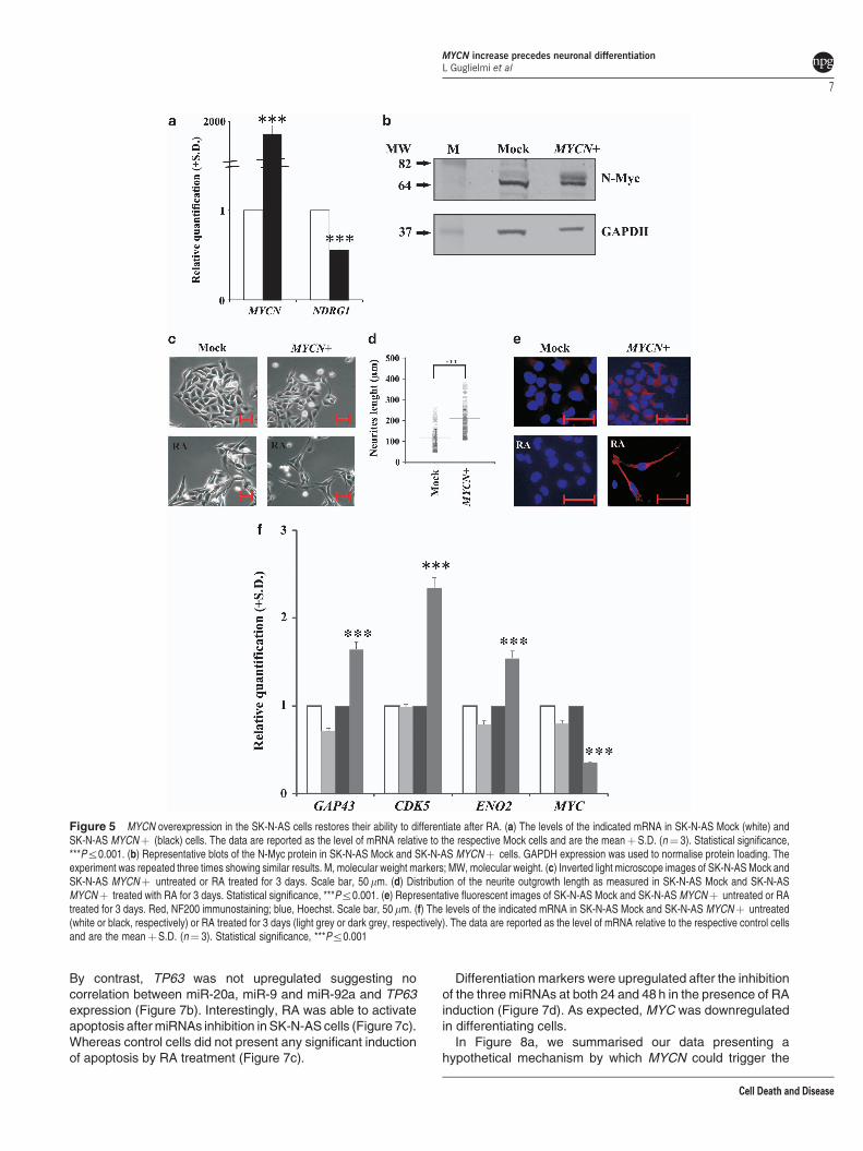
neuroblastoma cell line shows a very poor capacity todifferentiate after stimulation with RA, and these cells havea single copy of the MYCN gene, thus showing nooverexpression of this gene.21 We overexpressed the MYCNgene in these cells (Figure 5a) and studied differentiationactivation after RA induction. The overexpression of theMYCN gene was also confirmed by the protein level(Figure 5b). Figure 5c shows the micrographs of SK-N-ASMock and SK-N-AS MYCNþ cells, untreated or treated withRA for 3 days. In the presence of the MYCN gene, RAactivated differentiation in SK-N-AS cells, as indicated by theneurite outgrowth, with most of them showing a bipolarshape. The number of cells with neurites increased byapproximately 90% in MYCNþ cells after RA treatment. Theneurite length was significantly increased in MYCNþ cells
(Figure 5d; Mock cells, mean¼ 121mm, median¼ 120 mm,range¼ 60–254mm; MYCNþ cells, mean¼ 213 mm, median¼ 208mm, range¼ 115–375 mm). NF200 immunostainingconfirmed the ability of SK-N-AS MYCNþ cells to differ-entiate; NF200 was clearly expressed in these cells after RAexposure (Figure 5e). The analysis of GAP43, CDK5 andENO2 differentiation marker genes corroborated that MYCNoverexpression in SK-N-AS cells rendered these cells proneto differentiation after RA exposure (Figure 5f).
MYCN modulation modifies the expression of miRNAsinvolved in apoptosis preceding neuronal differentia-tion. The analysis of specific miRNAs selected by in silicoanalysis (miRWalk22) and reported to be regulated by MYCNand at the same time involved in apoptosis and neuron
Figure 2 MYCN increases during the first days of differentiation in LAN-5 cells and in mouse cortical embryonic neural progenitors. (a) Representative blots of the N-Mycprotein in LAN-5 cells untreated (0) or RA treated for 1, 2, 3, 4 and 5 days. GAPDH expression was used to normalise protein loading. The experiment was repeated threetimes showing similar results. M, molecular weight markers; MW, molecular weight. (b) Levels of the indicated mRNA in LAN-5 cells untreated (0) or RA treated for 1, 2, 3, 4and 5 days. The data are reported as the level of mRNA relative to the respective untreated cells and are the meanþS.D. (n¼ 3). Statistical significance, **Pr0.01;***Pr0.001. (c) Sub-G1 fractions estimated on the DNA content histograms in LAN-5 cells treated with RA for 1, 2, 3, 4 and 5 days. Data are reported as percent of control.Statistical significance, **Pr0.01; ***Pr0.001. (d) The levels of the indicated mRNA in mouse cortical embryonic neural progenitor cells undifferentiated (white) or induced todifferentiate for 3 and 6 days (black). The data are reported as the level of mRNA relative to the respective untreated cells and are the meanþ S.D. (n¼ 3). Statisticalsignificance, **Pr0.01; ***Pr0.001
MYCN increase precedes neuronal differentiationL Guglielmi et al
4
Cell Death and Disease

development revealed changes in the expression of miR-20a, miR-9 and miR-92a. We found that these miRNAs wereupregulated in MYCN-silenced LAN-5 cells (Figure 6a).In contrast, when MYCN was overexpressed in the SK-N-AScells, the same miRNAs were downregulated (Figure 6b).In particular, the p53-family members have been reported tobe regulated by these miRNAs. For this reason, weperformed PCR array analysis of the p53 signalling pathway(Figure 6c). As expected, and consistent with the expressionof miR-20a, miR-9 and miR-92a, the expression patternobserved in LAN-5 MYCN-KD cells was opposite to that in
SK-N-AS MYCNþ cells with regard to genes known to beinvolved in cell death regulation. We found that pro-apoptoticCASP9 and BAI1 genes were upregulated in the MYCNoverexpressing cells; whereas, the anti-apoptotic BCL2 genewas downregulated in the same MYCN condition. Bycontrast, CASP9 and BAI1 were downregulated and BCL2was upregulated when MYCN was silenced. As well as,genes known to be involved in the activation of apoptoticprogramme such as E2F1, E2F3, GADD45A and FOXO3were upregulated in MYCN-amplified and downregulated inMYCN-silenced cells, respectively. In addition, despite an
Figure 3 MYCN gene silencing in LAN-5 cells inhibits neurite outgrowth and NF200 expression after RA stimulus. (a) Upper panel, representative blots of the N-Mycprotein in LAN-5 cells silenced for MYCN gene (MYCN-KD) or in control cells (Mock) untreated (0) or RA treated for 1, 2, 3, 4 and 5 days. M, molecular weight markers; MW,molecular weight. Lower panel, blots densitometry as analysed by ImageJ software. Values are averagesþ S.D. of three independent experiments with similar results.GAPDH expression was used to normalise protein loading. (b) The levels of the indicated mRNA in MYCN-KD LAN-5 cells (black) or in control cells (white). The data arereported as the level of mRNA relative to the respective control cells and are the meanþ S.D. (n¼ 3). Statistical significance, **Pr0.01; ***Pr0.001. (c) Inverted lightmicroscope images in MYCN-KD LAN-5 cells or in Mock cells, untreated (CTR) or RA treated for 3 days. Scale bar, 50 mm. (d) Representative fluorescent images of MYCN-KD LAN-5 cells untreated (CTR) or RA treated for 3 days. Red, NF200 immunostaining; blue, Hoechst. Scale bar, 50 mm
MYCN increase precedes neuronal differentiationL Guglielmi et al
5
Cell Death and Disease

analysis of validated miRNA targets using miRWalk data-base reported miR-20a, miR-9 and miR-92a target TP53,TP73 and TP63 mRNAs, the PCR array analysis did notreveal any modulation of TP53 mRNA levels (data notshown); whereas an upregulation of both TP73 (aboutsevenfold) and TP63 (more than twofold) was observed inMYCN-overexpressing cells (Figure 6c).
Inhibition of miR-20a, miR-9 and miR-92a in the wild-typeSK-N-AS cells restores apoptosis and their differentia-tion ability. We inhibited miR-20a, miR-9 and miR-92a inMYCN-non-amplified SK-N-AS cells avoiding any possibleinterference by MYCN (Figure 7a).
The pro-apoptotic genes (CASP9, FOXO3, E2F1, E2F3, TP73)were upregulated by miRNAs inhibition after both 24 and 48 h.
Figure 4 MYCN gene silencing produces a depolarisation of the plasma membrane and inhibits the RA-induced upregulation of differentiation markers. (a) Left, FACSanalysis of the DiOC5 fluorescence distribution in MYCN-KD LAN-5 cells (full histogram) or in Mock control cells (empty histogram). Right, median values of the DiOC5
fluorescence distributions in MYCN-KD LAN-5 cells (black) or in Mock control cells (white). The experiment was repeated three times showing similar results. (b) The percentvariation of the median values of the DiOC5 fluorescence distributions in MYCN-KD LAN-5 cells (black) or in Mock control cells (white) after RA treatment.The experiment was repeated three times showing similar results. (c) Mitochondrial membrane potential analysed by JC-1 staining in Mock control LAN-5 untreated (Mock)and RA-treated (Mock-RA) cells compared with MYCN-KD LAN-5 untreated (MYCN-KD) and RA-treated (MYCN-KD-RA) cells (left panel). In the right panel are shown theDFvalues calculated on the FACS cytograms following the formula described in Materials and Methods section. Data are reported as percent of control and are average of at leastthree separate experiments. Bars represent standard deviation. (d) The levels of the indicated mRNA in MYCN-KD LAN-5 cells untreated (light grey) or RA treated for 3 days(black) and in Mock control cells untreated (white) or RA treated for 3 days (dark grey). The data are reported as the level of mRNA relative to the respective untreated cells andare the meanþ S.D. (n¼ 3). Statistical significance, ***Pr0.001
MYCN increase precedes neuronal differentiationL Guglielmi et al
6
Cell Death and Disease
By contrast, TP63 was not upregulated suggesting nocorrelation between miR-20a, miR-9 and miR-92a and TP63expression (Figure 7b). Interestingly, RA was able to activateapoptosis after miRNAs inhibition in SK-N-AS cells (Figure 7c).Whereas control cells did not present any significant inductionof apoptosis by RA treatment (Figure 7c).
Differentiation markers were upregulated after the inhibitionof the three miRNAs at both 24 and 48 h in the presence of RAinduction (Figure 7d). As expected, MYC was downregulatedin differentiating cells.
In Figure 8a, we summarised our data presenting ahypothetical mechanism by which MYCN could trigger the
Figure 5 MYCN overexpression in the SK-N-AS cells restores their ability to differentiate after RA. (a) The levels of the indicated mRNA in SK-N-AS Mock (white) andSK-N-AS MYCNþ (black) cells. The data are reported as the level of mRNA relative to the respective Mock cells and are the meanþS.D. (n¼ 3). Statistical significance,***Pr0.001. (b) Representative blots of the N-Myc protein in SK-N-AS Mock and SK-N-AS MYCNþ cells. GAPDH expression was used to normalise protein loading. Theexperiment was repeated three times showing similar results. M, molecular weight markers; MW, molecular weight. (c) Inverted light microscope images of SK-N-AS Mock andSK-N-AS MYCNþ untreated or RA treated for 3 days. Scale bar, 50 mm. (d) Distribution of the neurite outgrowth length as measured in SK-N-AS Mock and SK-N-ASMYCNþ treated with RA for 3 days. Statistical significance, ***Pr0.001. (e) Representative fluorescent images of SK-N-AS Mock and SK-N-AS MYCNþ untreated or RAtreated for 3 days. Red, NF200 immunostaining; blue, Hoechst. Scale bar, 50 mm. (f) The levels of the indicated mRNA in SK-N-AS Mock and SK-N-AS MYCNþ untreated(white or black, respectively) or RA treated for 3 days (light grey or dark grey, respectively). The data are reported as the level of mRNA relative to the respective control cellsand are the meanþS.D. (n¼ 3). Statistical significance, ***Pr0.001
MYCN increase precedes neuronal differentiationL Guglielmi et al
7
Cell Death and Disease

onset of differentiation programme in neuroblastoma cells.To further validate this hypothesis, we analysed the expressionkinetics of miRNAs and apoptotic genes presented in themodel (Figure 8b, middle and lower panels). To make datareading easier, we reported the kinetics of MYCN gene(Figure 8a, upper panel; see also Figure 2b). As MYCNgene is upregulated during RA treatment, the three miRNAs
are downregulated and consequently their targets areupregulated.
Discussion
Several studies of MYC- and MYCN-knockout mice haverevealed that embryos of these mice could not survive until
Figure 6 miRNAs are inversely regulated in MYCN-silenced and MYCN-upregulated neuroblastoma cells, and its expression is associated to a different modulation ofapoptosis-related genes. (a) The levels of the indicated hsa-miR in MYCN-KD LAN-5 cells (black) or in Mock control cells (white). The data are reported as the level of miRNArelative to the respective untreated cells and are the meanþ S.D. (n¼ 3). Statistical significance, **Pr0.01, ***Pr0.001. (b) The levels of the indicated hsa-miR in SK-N-ASMYCNþ cells (black) or in Mock control cells (white). The data are reported as the level of miRNA relative to the respective untreated cells and are the meanþS.D. (n¼ 3).Statistical significance, **Pr0.01; ***Pr0.001. (c) The levels of the indicated mRNA in SK-N-AS Mock (white), SK-N-AS MYCNþ (black), LAN-5 Mock (light grey) andLAN-5 MYCN-KD (dark grey). The data are reported as the level of mRNA relative to the respective control cells and are the meanþ S.D. (n¼ 3)
MYCN increase precedes neuronal differentiationL Guglielmi et al
8
Cell Death and Disease

Figure 7 miR-20a, miR-9 and miR-92a inhibitions in wild-type SK-N-AS cells lead to apoptotic death induction and expression of differentiation-related genes. (a) Thelevels of the indicated miRNAs in inhibitor negative control (white) and miRNA inhibitor (black) cells at 24 and 48 h after transfection. The data are reported as the level ofmiRNAs relative to the respective miRNAs negative control cells and are the meanþ S.D. (n¼ 3). Statistical significance, ***Pr0.001. (b) The levels of the indicated mRNAin inhibitor negative control (white), miRNA inhibitors at 24 h (black) and miRNA inhibitors at 48 h (grey). The data are reported as the level of mRNA relative to the respectivemiRNA negative control cells and are the meanþS.D. (n¼ 3). Statistical significance, *Pr0.05; **Pr0.01; ***Pr0.001. (c) FACS analysis of the Annexin V fluorescencedistribution in inhibitor negative control untreated (CTR) and RA-treated (CTR-RA) cells and miRNA inhibitors untreated (miRNA-INH) and RA-treated (miRNA-INH-RA) at 48 hafter transfection. The experiment was repeated three times showing similar results. (d) The levels of the indicated mRNA in inhibitor negative control (white), miRNA inhibitorsat 24 h (black) and at 48 h (grey) after transfection. The data are reported in RA-treated condition. The data are the meanþ S.D. (n¼ 3). Statistical significance, **Pr0.01;***Pr0.001
MYCN increase precedes neuronal differentiationL Guglielmi et al
9
Cell Death and Disease

gestation,11,14,16,23,24 suggesting that the MYC family genesare required for normal development at the beginning oforganogenesis. In particular, Knoepfler et al.24 showed thatN-Myc has an important role in complete nervous systemdevelopment. In fact, loss of N-Myc function during embry-ogenesis interrupts the ability of neural progenitors toexpand, differentiate and populate the brain, causingneurological dysfunction after birth. Unlike c-Myc, which isexpressed in all proliferating cells, N-Myc expression is morerestricted. Appropriate spatial and temporal expression ofN-Myc are important for normal embryonic development.25
Accordingly, we show that upregulation of the MYCN gene isrestricted to the early stages of differentiation induction inmouse cortical embryonic neural progenitor cells. In addition,as expected, MYC gene expression decreased duringdifferentiation progress. These data suggest that theMYCN gene might have a critical role in the activation ofneuronal differentiation. Nevertheless, as also reportedby others,19,26,27 differentiation completion was laterassociated with a decrease in MYCN expression. It is likelythat the N-Myc protein, as a transcription factor, is necessary
at the onset of neuronal differentiation to establishthe expression of a set of genes essential for subsequentphases.
To examine the role of N-Myc in the neuronal differentiationprogramme, we used the human neuroblastoma cell lineLAN-5, which show MYCN gene amplification.28 The LAN-5cell line, similar to many others neuroblastoma cell lines,maintains the ability to differentiate in vitro in the presence ofspecific stimuli, such as RA.28,29 In fact, our results confirmthat LAN-5 cells treated with RA form neurites; express theneuronal marker NF200, an intermediate filament thatprovides structural stability to the axon; and show theinduction of a series of neuronal molecular markers. Inagreement with previous papers,27,30 GAP43 and MAPTwere upregulated in differentiated LAN-5 cells; in addition,ChAT and SLC18A appeared to have increased expressionafter differentiation induction in the LAN-5 cell line, which isnormally committed to cholinergic differentiation.2 Asexpected, successful development is associated with cellproliferation inhibition, demonstrated by cell cycle analysisperformed using various approaches.
Figure 8 Model of the hypothetical role of MYCN during the early phases of neuroblastoma differentiation. (a) MYCN overexpression inhibits miR-9, miR-20a and miR-92a. We hypothesise that this inhibition allows targets of these miRNAs (E2F genes, TP73 and FOXO3) to be upregulated and in turn to activate apoptosis in those cells notcommitted to differentiate but still proliferating. In contrast, MYCN downregulation allows the overexpression of miR-9, miR-20a and miR-92a, preventing the upregulation ofapoptosis-related genes, thus inhibiting apoptosis and subsequent differentiation. (b) Upper panel, the levels of MYCN gene in LAN-5 cells untreated (0) or RA-treated for 1, 2,3, 4 and 5 days. Middle panel, the levels of miR-20a (diamond), miR-9 (open square) and miR-92a (star). Lower panel, the levels of E2F1 (closed circle), E2F3 (open square),FOXO3 (triangle), TP73 (star). The data are reported as the level of mRNA relative to the respective untreated cells and are the meanþ S.D. (n¼ 3). Statistical significance,*Pr0.05; **Pr0.01; ***Pr0.001
MYCN increase precedes neuronal differentiationL Guglielmi et al
10
Cell Death and Disease

In addition to what has been observed previously,19,26,27
we found peak MYCN expression during the first days ofRA-induced differentiation in LAN-5 cells. We demonstratedthe functionality of the increased N-Myc protein throughexpression analysis of the N-Myc target NDRG1, whichdecreased as expected.20
Furthermore, differentiation was inhibited by MYCN silen-cing in the same model; this was indicated by the failure ofneurite outgrowth, the decrease in known neuronal molecularmarkers and the loss of NF200 expression during differentia-tion. In addition, the plasma membrane of the MYCN-KD LAN-5 cells depolarised and was no longer able to hyperpolarise inresponse to differentiation stimuli. Plasma membrane polar-isation is a parameter known to be essential for differentiatedneuronal cells.31
In addition, when MYCN is overexpressed in cells with apoor differentiation capacity, developmental processes arerestored. This pattern was observed in SK-N-AS MYCNþcells, in which neurite outgrowth occurred, and the expressionof NF200 and known molecular neuronal markers increasedafter RA treatment.
These data, taken together, demonstrate that N-Mycprotein expression is required to activate the differentiationprocesses in neuroblastoma cells.
To explore the mechanisms by which N-Myc could triggerdifferentiation in neuroblastoma cells, we studied the expres-sion levels of miR20a, miR-9 and miR-92a, which have beenreported to be involved in differentiation processes and to bemodulated by MYCN.32,33 Through the up- or downregulationof the MYCN gene, we observed that all the three miRNAswere decreased or increased, respectively. Consistently, theinverse relationship between these miRNAs and the celldifferentiation status suggests that their downregulation isneeded for differentiation triggering. In fact, according to themiRWalk database, two of the validated targets of thesemiRNAs are the RA receptor genes RARA and RARG.34,35
The upregulation of miR-92a following MYCN silencing and itsdownregulation by RA treatment are in agreement with Chenand Stallings,36 who showed that the expression of differentmiRNAs correlates with neuroblastoma prognosis, differentia-tion and apoptosis, also in response to RA. Moreover,consistently with Haug et al.,37 who found that miR-92 inhibitsthe secretion of the tumour suppressor gene DKK3 inneuroblastoma, our data demonstrated the downregulationof miR-92a in cells with a restored ability to differentiate,characteristic of a less malignant phenotype. Our findings arealso supported by the data of Jee et al.,38 which showed thatinhibition of miR-20a expression induces definitive motorneuron survival and neurogenesis. Consistent with results ofYoo et al.,39 showing that miR-9 is repressed in neuralproliferating progenitors and is sequentially re-expressedin post-mitotic neurons, is the modulation of miR-9we observed in LAN-5 cells during the RA kinetics(see Figure 8b, middle panel).
In agreement with O’ Donnell et al.40 and Coller et al.,41 wedemonstrated that miR-20a negatively regulates the E2Ffactor genes. The transactivating p73 isoforms are transcrip-tionally induced by E2F and contribute to E2F-mediatedapoptosis.42–44 Consistent with these data, upregulation ofTP73 and E2F was observed in MYCNþ cells and after
miRNAs inhibition in SK-N-AS cells. Moreover, as expected,when MYCN was downregulated, and miR-20a and miR-92awere consequently upregulated, E2F and TP73 were inhibited.The role of p73 in the induction of neuronal differentiation hasbeen previously highlighted by De Laurenzi et al.,45 whoshowed an increase in p73 protein levels after RA stimuli inmurine neuroblastoma cells. In apparently disagreement withour data, they also showed reduced N-Myc protein levels afterRA treatment as a marker of neuronal differentiation.45
Indeed, they analysed N-Myc expression after 6 days of RAtreatment, whereas, although our data demonstrated a peakin N-Myc protein expression between the second and the thirdday of differentiation induction, we also observed a subse-quent decrease at day 5 of differentiation. It is likely thatN-Myc, by downregulating miR-20a and miR-92a, producesthe increase in E2Fs gene expression, which in turn inducesTP73 transcription. Interestingly, RA treatment in LAN-5cells confirmed the presence of a hierarchy between E2Fsand TP73, which are overexpressed at day 1 and day 4 of RAtreatment, respectively.
In addition, we found FOXO3 upregulated in differentiatingMYCNþ cells and in RA-treated LAN-5 cells. The humanFOXOs transcription factor family regulates the expression ofgenes associated with multiple biological processes such ascell cycle arrest and apoptosis.46 As demonstrated by Senyuket al.,47 miR-9 is able to bind directly to the 30 UTR of FOXO3.In fact, when we silenced the MYCN gene in LAN-5 cells, anincrease in miR-9 and a consequent decrease in FOXO3mRNA levels were obtained. These data are further demon-strated by the increase in FOXO3 expression achieved afterinhibiting miRNAs in SK-N-AS cells.
We reasoned that the miR-20a, miR-9 and miR-92ainhibition in MYCNþ , as well as, in RA-treated LAN-5 cellsand the subsequent pro-apoptotic genes increase could be anecessary step before differentiation programme. In fact, theinhibition of miR-20a, miR-9 and miR-92a in the MYCN-non-amplified SK-N-AS cells restored the ability of these cells torespond to RA treatment, as demonstrated by Annexin Vassay and differentiation-related gene expression analysis.
Even though the overexpression of MYCN in SK-N-AS cellsresulted in a downregulation of miR-92a and an upregulationof TP63, we did not observe an increase of TP63 afterinhibiting miR-92a. It is likely that other pathways downstreamMYCN are involved in TP63 regulation. More studies areneeded to establish if TP63 is directly or indirectly activated byN-Myc.
Therefore, we hypothesise that MYCN is necessary duringthe activation of neuroblastoma differentiation to induceapoptosis in cells that are not committed to differentiationbut are still proliferating. Programmed cell death is anecessary physiological process and involves a large numberof developing neurons during nervous system development48
and in adult neurons.49
Materials and MethodsCell line maintenance, differentiation and treatments. LAN-5 andSK-N-AS human neuroblastoma cell lines (both gifts of Dr. Doriana Fruci) weregrown in RPMI-1640 medium (Gibco, Paisley, UK) supplemented with 10% FBS(Hyclone, South Logan, UT, USA), 2 mM L-glutamine, 10 U/l penicillin and 10 U/lstreptomycin in a humidified incubator containing 5% CO2 at 37 1C. LAN-5 cellscontain amplified MYCN,28 whereas SK-N-AS cells are MYCN single copy.21
MYCN increase precedes neuronal differentiationL Guglielmi et al
11
Cell Death and Disease

Embryonic neural precursor cells (gift of Professor Stefano Biagioni andDr. Emanuele Cacci) were cultured and differentiated as previously described.50
For differentiation experiments, LAN-5 cells were seeded at a density of 5�103 cells/cm2. The following day, the cells were induced to differentiate with 10 mMretinoic acid (ATRA, designated RA throughout the paper; Sigma-Aldrich, St. Louis,MO, USA) in ‘differentiation medium’ composed of 50% fresh and 50% conditionedculture medium. The cells were fed after 3 days with differentiation mediumcontaining fresh RA. RA was dissolved in dimethyl sulphoxide (Sigma-Aldrich) andstored as a stock solution at � 80 1C. SK-N-AS cells were seeded at a density of3.5� 103 cells/cm2. The following day, the cells were induced to differentiate byadding 1mM RA in a ‘differentiation medium’ composed of RPMIþ 1% FBS.Because of the light sensitivity of RA, all incubations were performed under subduedlighting. The dimethyl sulphoxide concentration in each experiment was alwaysr0.01%, which was not toxic and did not induce differentiation.
A comparative western blot analysis has been conducted in the three cell linesemployed in this work to evidence basal expression level of the main mentionedproteins (Supplementary Figure S1).
Neurite quantification. After RA treatment, the cells were viewed with aLeica DM IRB inverted phase contrast microscope at � 200 (Leica, Wetzlar,Germany). The cells were scored as differentiated if the length of the neuriteextensions was at least twice the diameter of the cell body. The neurites wereevaluated based on the percentage of cells bearing neurites as determined fromthe cell culture images, which facilitated tracing the individual neurites andeventual branch points. A total of 4300 cells was examined in five randomlychosen fields in each treated and untreated sample. The projection images weresemiautomatically traced with NIH ImageJ using the NeuronJ plugin. The totaldendritic length of each individual neuron was analysed.
Western blot analyses. Cultured cells were washed twice with 1� PBSand then incubated for 1 min in urea buffer (8 M urea, 100 mM NaH2PO4 and10 mM Tris pH 8), scraped, harvested and briefly sonicated. The proteins weresubjected to SDS-polyacrylamide gel electrophoresis. The resolved proteins wereblotted overnight onto nitrocellulose membranes, which then were blocked in1� PBS containing 5% non-fat milk for at least 1 h. The blots were incubatedwith the following primary anti-human antibodies: rabbit polyclonal anti-N-Myc(C-19; Santa Cruz Biotechnology, Dallas, TX, USA); mouse monoclonal anti-GAP43 (GAP-7B10; Sigma-Aldrich); rabbit polyclonal anti-CDK5 (Cell SignalingTechnology, Danvers, MA, USA); mouse monoclonal anti-cyclin A (C-19; SantaCruz Biotechnology); rabbit polyclonal anti-p27kip1 (C-19; Santa Cruz Biotechnol-ogy); goat polyclonal anti-ChAT (Millipore, Billerica, MA, USA); mouse monoclonalanti-GAPDH (6C5; Millipore) and mouse monoclonal anti-HSP 72/73 (Ab1-W27;Oncogene Science Inc., Cambridge, MA, USA). The membranes were thenincubated for 45 min with the appropriate secondary antibody: donkey anti-rabbitIRdye800 (LI-COR Biosciences, Lincoln, NE, USA); donkey anti-mouse IRdye800(LI-COR) or donkey anti-goat IRdye800 (LI-COR). The membranes were thenanalysed with a Licor Odyssey Infrared Image System in the 800 nm channel. Blotscan resolution was 150 d.p.i.
Immunofluorescence. The cells were seeded on coverglass supports incomplete medium and treated with RA for the indicated times. The cells were fixedwith 4% (w/v) paraformaldehyde and permeabilised in PBS containing 0.1%Triton-X 100. NF200 was detected using an anti-NF200 monoclonal antibody(N52; Sigma-Aldrich). Alexa Fluor 594 goat anti-mouse antibody (MolecularProbes, Paisley, UK) was used as a secondary antibody. The antibodies werediluted in PBS. Cell nuclei were stained with 1 mg/ml DAPI or Hoechst 33342 inPBS for 5 min. Finally, the cells were washed in PBS and briefly rinsed in ddH2O,and the coverglass slips were mounted in ProLong Gold anti-Fade Reagent(Molecular Probes). Images were acquired with an Olympus BX51 fluorescencemicroscope and analysed with I.A.S. software (Delta Sistemi, Legnano (VR), Italy).The brightness and contrast of the acquired images were adjusted, and the figureswere generated using Adobe Photoshop 7.0.
Total RNA preparation. The cells were seeded in complete medium andharvested after the different treatments; total RNA was isolated using a Total RNApurification kit (Norgen Biotek, Thorold, ON, Canada). RNA quantity wasdetermined by absorbance at 260 nm using a NanoDrop UV-VIS spectro-photometer (Thermo Fisher Scientific, Wilmington, DE, USA). The quality andintegrity of each sample were confirmed using a BioAnalyzer 2100 (RNA 6000
Nanokit; Agilent, Santa Clara, CA, USA); samples with an RNA Integrity Numberindex lower than 8.0 were discarded.
Real-time RT-PCR analysis. RNA was reverse-transcribed with aHigh-Capacity cDNA Reverse Transcription Kit (Applied Biosystem, Paisley, UK)according to the manufacturer’s instructions. Equal amounts of cDNA were thensubjected to real-time PCR analysis with an Applied Biosystems 7900HT thermalcycler, using the SensiMix SYBR Kit (Bioline, London, UK) and specific primers,each at a concentration of 200 nM.: GAPDH (unigene Hs.544577) F: 50-AGCCACATCGCTCAGACA-30 and R: 50-GCCCAATACGACCAAATCC-30; TBP(Hs.590872) F: 50-GAACATCATGGATCAGAACAACA and R: 50-ATAGGGATTCCGGGAGTCAT-30; MAPT (Hs.101174) F: 50-ACCACAGCCACCTTCTCCTand R: 50-CAGCCATCCTGGTTCAAAGT-30; MYCN (Hs.25960) F: 50-CCACAAGGCCCTCAGTACC-30 and R: 50-TCCTCTTCATCATCTTCATCATCT-30; GAP43(Hs.134974) F: 50-GAGGATGCTGCTGCCAAG-30 and R: 50-GGCACTTTCCTTAGGTTTGGT-30; ChAT (Hs.302002) F: 50-CAGCCCTGATGCCTTCAT-30 and R:50-CAGTCTTCGATGGAGCCTGT-30; SLC18A3 (Hs.654374) F: 50-CCAGCCACTCCTCAACCTT-30 and R: 50-GATATGGAACGGGTCACAGG-30 and R: 50-CCTTGAACACAGTTCCGTAGG-30; ENO2 (Hs.511915) F: 50-ACTTTGTCAGGGACTATCCTGTG-30 and R: 50-TCCCTACATTGGCTGTGAACT-30; CDK5 (Hs.647078)F: 50-GCGATGCAGAAATACGAGAA-30 and R: 50-CCTTGAACACAGTTCCGTAGG-30; Mycn (Mm.16469) F: 50-CCTCCGGAGAGGATACCTTG-30 and R: 50-TCTCTACGGTGACCACATCG-30; Myc (Mm. 2444) F: 50-CCTAGTGCTGCATGAGGAGA-30 and R: 50-TCTTCCTCATCTTCTTGCTCTTC-30; Cdk5 (Mm.298798)F: 50-TGGACCCTGAGATTGTGAAGT-30 and R: 50-GACAGAATCCCAGGCCTTTC-30. Each experiment was performed in triplicate. The expression data werenormalised using the Ct values of GAPDH and TBP.
To validate mRNA levels of genes identified by PCR array, we reverse-transcribedRNA with a High-Capacity cDNA Reverse Transcription Kit (Applied Biosystem)according to the manufacturer’s instructions. Equal amounts of cDNA were thensubjected to real-time PCR analysis with an Applied Biosystems 7900HT thermalcycler, using the 2� RT2 SYBR Green/ROX qPCR Master Mix (Qiagen, Hilden,Germany) and specific primers, each at a concentration of 400 nM: E2F1(PPH00136G; Hs.654393); E2F3 (PPH00917F; Hs.269408); CASP9 (PPH00353B;Hs.329502); FOXO3 (PPH00807A; Hs.220950); TP63 (PPH01032F; Hs.137569);TP73 (PPH00725A; Hs.697294).
miRNA assays. Equal amounts of RNA were reverse transcribed with theTaqMan MicroRNA Reverse Transcription Kit (Applied Biosystem), according tothe manufacturer’s instructions, with a custom 1� RT primer pool (hsa-miR-20aID 000580; hsa-miR-9 ID 000583; hsa-miR-92a ID 000431). Real-time PCRanalysis was performed with an Applied Biosystem 7900HT thermal cycler using20� Individual TaqMan MicroRNA Assays.
miRNA inhibition. SK-N-AS cells were seeded at a density of 5� 104 cells/cm2. After 24 h, cells were transfected overnight in the presence of 10% FBS, bothwith mirVana miRNA inhibitors and mirVana miRNA inhibitor Negative Control.Lipofectamine RNAiMAX Reagent (Life Technologies, Paisley, UK) was used astransfection reagent according to manufacturer’s instructions. We transfectedhsa-miR-92a (MH10916, Ambion, Paisley, UK), hsa-miR20a (MH10057, Ambion)and hsa-miR9 (MH10022, Ambion) all together setting a concentration ofapproximately 10 nM each, in order to obtain the final concentration of 30 nM. ThemirVana miRNA inhibitor Negative Control was used to the final concentration of30 nM. After transfection cells were treated with RA (see Cell line maintenance,differentiation and treatments). Samples were harvested at 24 and 48 h to performmRNA expression analysis and at 48 h to detect apoptosis.
PCR array. RNA was reverse transcribed with a High-Capacity cDNA ReverseTranscription Kit (Applied Biosystem) according to the manufacturer’s instructions.Equal amounts of cDNA were then subjected to real-time PCR analysis with aBio-Rad iQ5 thermal cycler. Each sample was mixed with 2� RT2 SYBRGreen/ROX qPCR Master Mix (Qiagen) and added to each 96-well plate of Humanp53 Signaling Pathway PCR Array (PAHS-027A; Qiagen), according to themanufacturer’s instructions.
Cell cycle analysis. Cell cycle analysis was performed by pulse-chase BrdU(Sigma-Aldrich) incorporation and propidium iodide (PI) DNA staining. For BrdU
MYCN increase precedes neuronal differentiationL Guglielmi et al
12
Cell Death and Disease

incorporation, a pulse of 10mM BrdU was added to the cell culture during the last30 min before harvesting. Cells were fixed in ethanol 70% atþ 4 1C for at least1 h, and then processed as previously described. Primary anti-BrdU (BDBiosciences Italia, Buccinasco (MI), Italy) and secondary FITC-conjugated F(ab0)2rabbit anti-mouse IgG (DAKO, Glostrup, Denmark) antibodies were used to detectthe BrdU. Finally, after washing in PBS/BSA 0.5%, cells were stained with asolution containing 5 mg/ml PI and 75 KU/ml RNase overnight in dark. For PIstaining, cells fixed in ethanol 70% for at least 1 h were stained in a solutioncontaining 50mg/ml PI and 75 KU/ml RNase for at least 30 min in the dark. In eachcase, 20 000 events per sample were acquired by using a FACScancytofluorimeter and the CellQuest BD software. BrdU-positive cells representthe fraction of the cell population synthesising DNA. The percentages of the cellsin the different cell cycle compartments were estimated on linear PI histograms byusing the MODFIT software (Verity, Topsham, ME, USA).
Apoptosis detection. Apoptosis was detected in the specific conditions byusing Annexin V assay, mitochondrial membrane potential measurement andestimation of sub-G1 fraction. Description of these methods have been detailed inprevious papers.51,52
MYCN silencing. Proliferating LAN-5 cells were transfected with a plasmid(pcDNA6.2-GW/EmGFP-miR-MYCN) targeting MYCN mRNA and a non-silencingplasmid (pcDNA6.2-GW/EmGFP-miR-Neg) with no homology to mammaliangenes, using Lipofectamine2000 (Life Technologies) according to the manufac-turer’s instructions. We used 2 mg of plasmid. After transfection, the cells wereselected with blasticidin for 2 weeks before performing experiments.
MYCN overexpression. Proliferating SK-N-AS cells were transfected with aplasmid carrying the MYCN construct driven by the CMV promoter or a BlueScriptcontrol vector using Lipofectamine2000 (Life Technologies) according to themanufacturer’s instructions. We used 2 mg of DNA per sample. At 24 h after thetransfection, the cells were induced to differentiate by exposure to RA for 3 daysusing a specific differentiation protocol (see above).
Plasma membrane potential analysis. LAN-5 plasma membranepotential was measured by flow cytometry using the voltage-sensitive fluorescentdye DiOC5 (Molecular probes). The cells were harvested and washed once inPBSþ 0.1 M glucose, and 1� 106 cells were incubated in a solution containing50 nM DiOC5 at 37 1C for 15 min. The samples were then washed once inPBSþ 0.1 M glucose and immediately assessed by flow cytometry. As adepolarised control, we used a sample post-incubated with the oxidativephosphorylation inhibitor 4-trifluoromethoxyphenylhydrazone; as a hyperpolarisedcontrol, we used a sample incubated with the potassium-specific transportervalinomycin. PI at a final concentration of 10 mg/ml was added to each sampleimmediately before the measurements to exclude non-viable cells from theanalysis. A total of 10 000 events was acquired for each sample using a FACScancytofluorimeter.
Statistical analysis. Statistical significance of differences between groupswas tested by paired Student’s t-test or, if there were more than two groups, byone-way ANOVA.
Conflict of InterestThe authors declare no conflict of interest.
Acknowledgements. We thank Dr. Carla Musa for her contribution to ourLaboratory. Partially supported by a joint Grant CNR-EBRI ‘Molecular and cellularmechanisms of brain plasticity’ and by PNR-CNR Aging Program 2012-2014.
1. Brodeur GM. Neuroblastoma: biological insights into a clinical enigma. Nat Rev Cancer2003; 3: 203–216.
2. Hill DP, Robertson KA. Characterization of the cholinergic neuronal differentiation of thehuman neuroblastoma cell line LA-N-5 after treatment with retinoic acid. Brain Res DevBrain Res 1997; 102: 53–67.
3. Munoz J, Vendrell E, Aiza G, Nistal M, Pestana A, Peinado MA et al. Determination ofgenomic damage in neuroblastic tumors by arbitrarily primed PCR: MYCN amplification asa marker for genomic instability in neuroblastomas. Neuropathology 2006; 26: 165–169.
4. Eilers M, Eisenman RN. Myc’s broad reach. Genes Dev 2008; 22: 2755–2766.5. Westermark UK, Wilhelm M, Frenzel A, Henriksson MA. The MYCN
oncogene and differentiation in neuroblastoma. Semin Cancer Biol 2011; 21:256–266.
6. Malynn BA, de Alboran IM, O’Hagan RC, Bronson R, Davidson L, DePinho RA et al.N-myc can functionally replace c-myc in murine development, cellular growth, anddifferentiation. Genes Dev 2000; 14: 1390–1399.
7. Murphy DM, Buckley PG, Bryan K, Watters KM, Koster J, van SP et al. Dissection of theoncogenic MYCN transcriptional network reveals a large set of clinically relevantcell cycle genes as drivers of neuroblastoma tumorigenesis. Mol Carcinog 2011; 50:403–411.
8. Pelengaris S, Khan M, Evan G. c-MYC: more than just a matter of life and death. Nat RevCancer 2002; 2: 764–776.
9. Hui AB, Lo KW, Yin XL, Poon WS, Ng HK. Detection of multiple gene amplifications inglioblastoma multiforme using array-based comparative genomic hybridization. Lab Invest2001; 81: 717–723.
10. Nau MM, Brooks Jr BJ, Carney DN, Gazdar AF, Battey JF, Sausville EA et al. Humansmall-cell lung cancers show amplification and expression of the N-myc gene. Proc NatlAcad Sci USA 1986; 83: 1092–1096.
11. Charron J, Malynn BA, Fisher P, Stewart V, Jeannotte L, Goff SP et al. Embryonic lethalityin mice homozygous for a targeted disruption of the N-myc gene. Genes Dev 1992; 6:2248–2257.
12. Moens CB, Auerbach AB, Conlon RA, Joyner AL, Rossant J. A targeted mutation reveals arole for N-myc in branching morphogenesis in the embryonic mouse lung. Genes Dev1992; 6: 691–704.
13. Moens CB, Stanton BR, Parada LF, Rossant J. Defects in heart and lung development incompound heterozygotes for two different targeted mutations at the N-myc locus.Development 1993; 119: 485–499.
14. Sawai S, Shimono A, Hanaoka K, Kondoh H. Embryonic lethality resulting from disruptionof both N-myc alleles in mouse zygotes. New Biol 1991; 3: 861–869.
15. Sawai S, Shimono A, Wakamatsu Y, Palmes C, Hanaoka K, Kondoh H. Defects ofembryonic organogenesis resulting from targeted disruption of the N-myc gene in themouse. Development 1993; 117: 1445–1455.
16. Stanton BR, Perkins AS, Tessarollo L, Sassoon DA, Parada LF. Loss of N-myc functionresults in embryonic lethality and failure of the epithelial component of the embryo todevelop. Genes Dev 1992; 6: 2235–2247.
17. Squire J, Goddard AD, Canton M, Becker A, Phillips RA, Gallie BL. Tumour inductionby the retinoblastoma mutation is independent of N-myc expression. Nature 1986; 322:555–557.
18. Zimmerman KA, Yancopoulos GD, Collum RG, Smith RK, Kohl NE, Denis KA et al.Differential expression of myc family genes during murine development. Nature 1986; 319:780–783.
19. Thiele CJ, Deutsch LA, Israel MA. The expression of multiple proto-oncogenes isdifferentially regulated during retinoic acid induced maturation of human neuroblastomacell lines. Oncogene 1988; 3: 281–288.
20. Melotte V, Qu X, Ongenaert M, van CW, de Bruine AP, Baldwin HS et al. The N-mycdownstream regulated gene (NDRG) family: diverse functions, multiple applications.FASEB J 2010; 24: 4153–4166.
21. Suenaga Y, Kaneko Y, Matsumoto D, Hossain MS, Ozaki T, Nakagawara A. Positiveauto-regulation of MYCN in human neuroblastoma. Biochem Biophys Res Commun 2009;390: 21–26.
22. Dweep H, Sticht C, Pandey P, Gretz N. miRWalk–database: prediction of possiblemiRNA binding sites by "walking" the genes of three genomes. J Biomed Inform 2011; 44:839–847.
23. Davis A, Bradley A. Mutation of N-myc in mice: what does the phenotype tell us? Bioessays1993; 15: 273–275.
24. Knoepfler PS, Cheng PF, Eisenman RN. N-myc is essential during neurogenesis for therapid expansion of progenitor cell populations and the inhibition of neuronal differentiation.Genes Dev 2002; 16: 2699–2712.
25. Strieder V, Lutz W. E2F proteins regulate MYCN expression in neuroblastomas. J BiolChem 2003; 278: 2983–2989.
26. Amatruda III TT, Sidell N, Ranyard J, Koeffler HP. Retinoic acid treatment of humanneuroblastoma cells is associated with decreased N-myc expression. Biochem BiophysRes Commun 1985; 126: 1189–1195.
27. Sidell N. Retinoic acid-induced growth inhibition and morphologic differentiation of humanneuroblastoma cells in vitro. J Natl Cancer Inst 1982; 68: 589–596.
28. Ribatti D, Raffaghello L, Pastorino F, Nico B, Brignole C, Vacca A et al. In vivo angiogenicactivity of neuroblastoma correlates with MYCN oncogene overexpression. Int J Cancer2002; 102: 351–354.
29. Edsjo A, Nilsson H, Vandesompele J, Karlsson J, Pattyn F, Culp LA et al. Neuroblastoma cellswith overexpressed MYCN retain their capacity to undergo neuronal differentiation. Lab Invest2004; 84: 406–417.
30. Pahlman S, Ruusala AI, Abrahamsson L, Mattsson ME, Esscher T. Retinoic acid-induceddifferentiation of cultured human neuroblastoma cells: a comparison with phorbolester-induced differentiation. Cell Differ 1984; 14: 135–144.
31. Sundelacruz S, Levin M, Kaplan DL. Role of membrane potential in the regulation of cellproliferation and differentiation. Stem Cell Rev 2009; 5: 231–246.
MYCN increase precedes neuronal differentiationL Guglielmi et al
13
Cell Death and Disease

32. Schulte JH, Horn S, Otto T, Samans B, Heukamp LC, Eilers UC et al. MYCN regulates
oncogenic MicroRNAs in neuroblastoma. Int J Cancer 2008; 122: 699–704.33. Stallings RL. MicroRNA involvement in the pathogenesis of neuroblastoma: potential for
microRNA mediated therapeutics. Curr Pharm Des 2009; 15: 456–462.34. Li G, Luna C, Qiu J, Epstein DL, Gonzalez P. Alterations in microRNA expression in
stress-induced cellular senescence. Mech Ageing Dev 2009; 130: 731–741.35. Liu DZ, Ander BP, Tian Y, Stamova B, Jickling GC, Davis RR et al. Integrated analysis
of mRNA and microRNA expression in mature neurons, neural progenitor cells and
neuroblastoma cells. Gene 2012; 495: 120–127.36. Chen Y, Stallings RL. Differential patterns of microRNA expression in neuroblastoma
are correlated with prognosis, differentiation, and apoptosis. Cancer Res 2007; 67:
976–983.37. Haug BH, Henriksen JR, Buechner J, Geerts D, Tomte E, Kogner P et al. MYCN-regulated
miRNA-92 inhibits secretion of the tumor suppressor DICKKOPF-3 (DKK3) in neuroblastoma.
Carcinogenesis 2011; 32: 1005–1012.38. Jee MK, Jung JS, Im YB, Jung SJ, Kang SK. Silencing of miR20a is crucial
for Ngn1-mediated neuroprotection in injured spinal cord. Hum Gene Ther 2012; 23:
508–520.39. Yoo AS, Staahl BT, Chen L, Crabtree GR. MicroRNA-mediated switching of chromatin-
remodelling complexes in neural development. Nature 2009; 460: 642–646.40. O’Donnell KA, Wentzel EA, Zeller KI, Dang CV, Mendell JT. c-Myc-regulated microRNAs
modulate E2F1 expression. Nature 2005; 435: 839–843.41. Coller HA, Forman JJ, Legesse-Miller A"Myc’ed messages": myc induces transcription of
E2F1 while inhibiting its translation via a microRNA polycistron. PLoS Genet 2007; 3: e146.42. Irwin M, Marin MC, Phillips AC, Seelan RS, Smith DI, Liu W et al. Role for the p53
homologue p73 in E2F-1-induced apoptosis. Nature 2000; 407: 645–648.43. Stiewe T, Putzer BM. Role of the p53-homologue p73 in E2F1-induced apoptosis.
Nat. Genet 2000; 26: 464–469.44. Zaika A, Irwin M, Sansome C, Moll UM. Oncogenes induce and activate endogenous p73
protein. J Biol Chem 2001; 276: 11310–11316.
45. De L V, Raschella G, Barcaroli D, Annicchiarico-Petruzzelli M, Ranalli M, Catani MV et al.Induction of neuronal differentiation by p73 in a neuroblastoma cell line. J Biol Chem 2000;275: 15226–15231.
46. Eijkelenboom A, Burgering BM. FOXOs: signalling integrators for homeostasismaintenance. Nat Rev Mol Cell Biol 2013; 14: 83–97.
47. Senyuk V, Zhang Y, Liu Y, Ming M, Premanand K, Zhou L et al. Critical role of miR-9 inmyelopoiesis and EVI1-induced leukemogenesis. Proc Natl Acad Sci USA 2013; 110:5594–5599.
48. Oppenheim RW. Cell death during development of the nervous system. Annu RevNeurosci 1991; 14: 453–501.
49. Kim WR, Sun W. Programmed cell death during postnatal development of the rodentnervous system. Dev Growth Differ 2011; 53: 225–235.
50. Cacci E, Ajmone-Cat MA, Anelli T, Biagioni S, Minghetti L. In vitro neuronal and glialdifferentiation from embryonic or adult neural precursor cells are differently affected bychronic or acute activation of microglia. Glia 2008; 56: 412–425.
51. Gatti G, Maresca G, Natoli M, Florenzano F, Nicolin A, Felsani A et al. MYC preventsapoptosis and enhances endoreduplication induced by paclitaxel. PLoS One 2009; 4:e5442.
52. Maresca G, Natoli M, Nardella M, Arisi I, Trisciuoglio D, Desideri M et al. LMNAknock-down affects differentiation and progression of human neuroblastoma cells.PLoS One 2012; 7: e45513.
Cell Death and Disease is an open-access journalpublished by Nature Publishing Group. This work is
licensed under a Creative Commons Attribution-NonCommercial-NoDerivs 3.0 Unported License. To view a copy of this license, visithttp://creativecommons.org/licenses/by-nc-nd/3.0/
Supplementary Information accompanies this paper on Cell Death and Disease website (http://www.nature.com/cddis)
MYCN increase precedes neuronal differentiationL Guglielmi et al
14
Cell Death and Disease
Copyright © 2022 FDOKUMEN