mTORC1 in the Paneth cell niche couples intestinal stem-cell function to calorie intake
16
mTORC1 in the Paneth cell niche couples intestinal stem cell function to calorie intake Ömer H. Yilmaz 1,2,# , Pekka Katajisto 2,# , Dudley W. Lamming 2 , Yetis Gültekin 1,2 , Khristian E. Bauer-Rowe 2 , Shomit Sengupta 2 , Kivanc Birsoy 2 , Abdulmetin Dursun 1 , V. Onur Yilmaz 2 , Martin Selig 1 , G. Petur Nielson 1 , Mari Mino-Kenudson 1 , Lawrence Zukerberg 1 , Atul Bhan 1 , Vikram Deshpande 1 , and David M. Sabatini 2 1 Department of Pathology, Massachusetts General Hospital and Harvard Medical School, Boston, MA 02114 USA 2 Whitehead Institute for Biomedical Research, Boston, MA 02142 USA; Department of Biology, MIT, Cambridge, MA 02139; Howard Hughes Medical Institute, MIT, Cambridge, MA 02139; Broad Institute of Harvard and MIT, Seven Cambridge Center, Cambridge, MA 02142; The David H. Koch Institute for Integrative Cancer Research at MIT, Cambridge, MA 02139 SUMMARY How adult tissue stem and niche cells respond to the nutritional state of an organism is not well understood. Here, we find that Paneth cells, a key constituent of the mammalian intestinal stem cell (ISC) niche, augment stem cell function in response to calorie restriction (CR). CR acts by reducing mTOR complex 1 (mTORC1) signaling in Paneth cells, and the ISC-enhancing effects of CR can be mimicked by rapamycin. Calorie intake regulates mTORC1 in Paneth cells, but not ISCs, and forced mTORC1 activation in Paneth cells during CR abolishes their effects on ISCs. Finally, increased expression in Paneth cells of bone stromal antigen 1 (Bst-1), an ectoenzyme that produces the paracrine factor cyclic ADP ribose (cADPR), mediates the effects of CR and rapamycin on ISC function. Our findings establish that mTORC1 non-cell autonomously regulates stem cell self-renewal, and highlight a significant role of the mammalian intestinal niche in coupling stem cell function to organismal physiology. Mammalian tissue-specific stem cells maintain tissue homeostasis by undergoing either self- renewing or differentiation divisions that generate more stem cells or restricted progenitors, respectively 1 . Stem cells often require cues from their microenvironment or “niche” to regulate their fates. Caloric restriction (CR), an intervention in which caloric intake is reduced while maintaining adequate nutrition, promotes longevity in diverse organisms, possibly by preserving stem and progenitor cell function 2,3 . In mice, CR promotes the generation of new neurons from neural progenitors and prevents the decline of hematopoietic stem cell numbers and function in certain strains of mice with age 2,4–7 . These findings raise the question of how CR mediates these effects on stem cells, and whether the mammalian stem cell niche is involved. Address correspondence to [email protected]. # These authors contributed equally to this work. Author Contributions ÖHY and PK designed and performed all experiments and data analyses with input from D.M.S. S.S. generated the Rheb2 transgenic mice. DWL, KB, VD, AB, MM, and LZ participated in design and interpretation of experiments. YG, OY, AD, KBR performed and interpreted all of the immunohistochemistry and in situ hybridization under the guidance of ÖHY. MS and PN performed electron microscopy and helped with its interpretation. ÖHY wrote the paper with help from PK and DMS. NIH Public Access Author Manuscript Nature. Author manuscript; available in PMC 2012 December 28. Published in final edited form as: Nature. 2012 June 28; 486(7404): 490–495. doi:10.1038/nature11163. NIH-PA Author Manuscript NIH-PA Author Manuscript NIH-PA Author Manuscript
Transcript of mTORC1 in the Paneth cell niche couples intestinal stem-cell function to calorie intake

mTORC1 in the Paneth cell niche couples intestinal stem cellfunction to calorie intake
Ömer H. Yilmaz1,2,#, Pekka Katajisto2,#, Dudley W. Lamming2, Yetis Gültekin1,2, Khristian E.Bauer-Rowe2, Shomit Sengupta2, Kivanc Birsoy2, Abdulmetin Dursun1, V. Onur Yilmaz2,Martin Selig1, G. Petur Nielson1, Mari Mino-Kenudson1, Lawrence Zukerberg1, Atul Bhan1,Vikram Deshpande1, and David M. Sabatini21Department of Pathology, Massachusetts General Hospital and Harvard Medical School, Boston,MA 02114 USA2Whitehead Institute for Biomedical Research, Boston, MA 02142 USA; Department of Biology,MIT, Cambridge, MA 02139; Howard Hughes Medical Institute, MIT, Cambridge, MA 02139;Broad Institute of Harvard and MIT, Seven Cambridge Center, Cambridge, MA 02142; The DavidH. Koch Institute for Integrative Cancer Research at MIT, Cambridge, MA 02139
SUMMARYHow adult tissue stem and niche cells respond to the nutritional state of an organism is not wellunderstood. Here, we find that Paneth cells, a key constituent of the mammalian intestinal stemcell (ISC) niche, augment stem cell function in response to calorie restriction (CR). CR acts byreducing mTOR complex 1 (mTORC1) signaling in Paneth cells, and the ISC-enhancing effects ofCR can be mimicked by rapamycin. Calorie intake regulates mTORC1 in Paneth cells, but notISCs, and forced mTORC1 activation in Paneth cells during CR abolishes their effects on ISCs.Finally, increased expression in Paneth cells of bone stromal antigen 1 (Bst-1), an ectoenzyme thatproduces the paracrine factor cyclic ADP ribose (cADPR), mediates the effects of CR andrapamycin on ISC function. Our findings establish that mTORC1 non-cell autonomously regulatesstem cell self-renewal, and highlight a significant role of the mammalian intestinal niche incoupling stem cell function to organismal physiology.
Mammalian tissue-specific stem cells maintain tissue homeostasis by undergoing either self-renewing or differentiation divisions that generate more stem cells or restricted progenitors,respectively1. Stem cells often require cues from their microenvironment or “niche” toregulate their fates. Caloric restriction (CR), an intervention in which caloric intake isreduced while maintaining adequate nutrition, promotes longevity in diverse organisms,possibly by preserving stem and progenitor cell function2,3. In mice, CR promotes thegeneration of new neurons from neural progenitors and prevents the decline ofhematopoietic stem cell numbers and function in certain strains of mice with age2,4–7. Thesefindings raise the question of how CR mediates these effects on stem cells, and whether themammalian stem cell niche is involved.
Address correspondence to [email protected].#These authors contributed equally to this work.
Author Contributions ÖHY and PK designed and performed all experiments and data analyses with input from D.M.S. S.S.generated the Rheb2 transgenic mice. DWL, KB, VD, AB, MM, and LZ participated in design and interpretation of experiments. YG,OY, AD, KBR performed and interpreted all of the immunohistochemistry and in situ hybridization under the guidance of ÖHY. MSand PN performed electron microscopy and helped with its interpretation. ÖHY wrote the paper with help from PK and DMS.
NIH Public AccessAuthor ManuscriptNature. Author manuscript; available in PMC 2012 December 28.
Published in final edited form as:Nature. 2012 June 28; 486(7404): 490–495. doi:10.1038/nature11163.
NIH
-PA Author Manuscript
NIH
-PA Author Manuscript
NIH
-PA Author Manuscript

We have interrogated this question in the rapidly renewing mammalian small intestine. Theintestine is organized into crypts that contain the stem cells and the rapidly dividing transientamplifying cells (TA-cells), and villi composed primarily of post-mitotic absorptiveenterocytes. In response to fasting and refeeding the intestine undergoes structuralalterations such as changes in villi length, crypt depth, and cell turnover, suggesting thatorganismal physiology may modify intestinal progenitor function8,9. Recent studies havebegun to define the identity of intestinal stem cells as well as their interaction with theirPaneth cell niche10–15. Although no single marker identifies the entire ISC pool, Lgr5 isexpressed by a majority of ISCs throughout the intestinal tract13,15. Lgr5+ ISCs (aka cryptbase columnar cells or CBCs) can self-renew and differentiate for the life of the organism,and they reside at the base of crypts sandwiched between Paneth cells13,16. Loss of Panethcells in vivo leads to reduced numbers of Lgr5+ ISCs, while the addition of Paneth cells toin vitro cultures dramatically increases the potential of Lgr5+ ISCs to form multipotent, self-renewing organoid bodies reminiscent of “mini-intestines”14. Thus, Paneth cells constitute acritical component of the stem cell niche both in vivo and in vitro13,14.
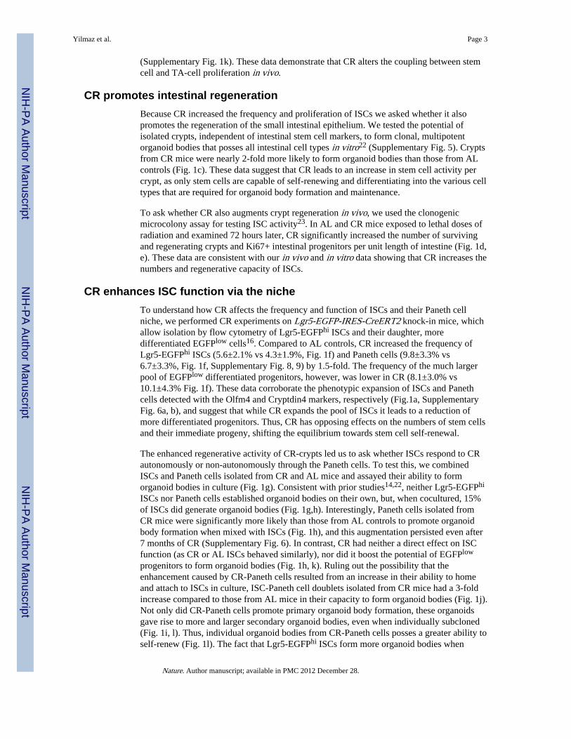
CR increases stem and niche cell numbersTo assess the effects of CR on intestinal homeostasis, we calorically restricted mice for 4 to28 weeks, which is sufficient to observe many of the metabolic phenotypes of CR17–19.Consistent with prior reports, CR mice had a 19.7± 5.8% loss in body mass compared to adlibitum (AL) fed counterparts (Supplementary Fig. 1a)20. In CR mice the small intestine wasmorphologically normal (Supplementary Fig. 1f), with no change in crypt density(Supplementary Fig. 1d), intestinal length (Supplementary Fig. 1c), or apoptotic cellfrequency (Supplementary Fig. 3). However, it did have reduced mass (1.8±0.4 vs 1.4±0.2g,Supplementary Fig. 1b) with villi that were 15% shorter and possessed fewer enterocytes(Supplementary Fig. 1e, f). CR did not affect the frequency of chromogranin A+enteroendocrine cells, but mildly reduced that of alcian blue+ secretory goblet cells(Supplementary Fig 2a, b). To address how CR influenced the frequency of ISCs, weperformed in situ hybridization for Olfactomedin-4 (Olfm4), a recently described markerthat is co-expressed by Lgr5+ ISCs21. CR led to a 35% increase in Olfm4+ primitiveintestinal progenitors compared to those in AL mice (Fig. 1a, Supplementary Fig. 6a).Interestingly, CR also caused a commensurate increase in Cryptdin4+ Paneth cells (Fig. 1a),which we confirmed by morphological examination of one-micron tissue sections(Supplementary Fig. 4a) and by electron microscopy (Supplementary Fig. 4b). Thesefindings lead to two intriguing conclusions: First, CR promotes the preservation and self-renewal of ISCs (increased Olfm4+ ISCs) at the expense of differentiation (shorter villi withfewer mature enterocytes). Second, ISCs and their Paneth cells increase in tandem, raisingthe possibility that the Paneth cell niche may coordinate ISC adaptation to CR.
The fact that CR augmented ISC numbers while reducing the total number of differentiatedenterocytes suggested that CR enhances the proliferation of ISCs while reducing theproliferation of more differentiated progenitors (TA-cells). To test this possibility, weassessed incorporation of BrdU into ISCs and TA-cells. After a 4 hour pulse of BrdU, CR-crypts had nearly 2-fold as many BrdU+ ISCs compared to AL-crypts (4.3±0.3 vs 2.4±0.2,Fig. 1b; Supplementary Fig. 1g, h). However, CR decreased the number of BrdU+ cells inthe larger pool of TA-cells (11.0±0.9 vs 9.4±0.5; Fig. 1b), suggesting that output andmigration into the villi from this compartment may also be reduced. Indeed, CR mice 24hours after a single dose of BrdU had fewer absolute numbers of BrdU labeled cells in thevilli compared to AL controls (14.5±1.5 vs 19.0±1.7, Supplementary Fig. 1i, j). However,there was no significant difference in the percentage of BrdU+ villous enterocytes,indicating that in CR mice TA-cells generate fewer progeny for shorter, less cellular villi
Yilmaz et al. Page 2
Nature. Author manuscript; available in PMC 2012 December 28.
NIH
-PA Author Manuscript
NIH
-PA Author Manuscript
NIH
-PA Author Manuscript
(Supplementary Fig. 1k). These data demonstrate that CR alters the coupling between stemcell and TA-cell proliferation in vivo.
CR promotes intestinal regenerationBecause CR increased the frequency and proliferation of ISCs we asked whether it alsopromotes the regeneration of the small intestinal epithelium. We tested the potential ofisolated crypts, independent of intestinal stem cell markers, to form clonal, multipotentorganoid bodies that posses all intestinal cell types in vitro22 (Supplementary Fig. 5). Cryptsfrom CR mice were nearly 2-fold more likely to form organoid bodies than those from ALcontrols (Fig. 1c). These data suggest that CR leads to an increase in stem cell activity percrypt, as only stem cells are capable of self-renewing and differentiating into the various celltypes that are required for organoid body formation and maintenance.
To ask whether CR also augments crypt regeneration in vivo, we used the clonogenicmicrocolony assay for testing ISC activity23. In AL and CR mice exposed to lethal doses ofradiation and examined 72 hours later, CR significantly increased the number of survivingand regenerating crypts and Ki67+ intestinal progenitors per unit length of intestine (Fig. 1d,e). These data are consistent with our in vivo and in vitro data showing that CR increases thenumbers and regenerative capacity of ISCs.
CR enhances ISC function via the nicheTo understand how CR affects the frequency and function of ISCs and their Paneth cellniche, we performed CR experiments on Lgr5-EGFP-IRES-CreERT2 knock-in mice, whichallow isolation by flow cytometry of Lgr5-EGFPhi ISCs and their daughter, moredifferentiated EGFPlow cells16. Compared to AL controls, CR increased the frequency ofLgr5-EGFPhi ISCs (5.6±2.1% vs 4.3±1.9%, Fig. 1f) and Paneth cells (9.8±3.3% vs6.7±3.3%, Fig. 1f, Supplementary Fig. 8, 9) by 1.5-fold. The frequency of the much largerpool of EGFPlow differentiated progenitors, however, was lower in CR (8.1±3.0% vs10.1±4.3% Fig. 1f). These data corroborate the phenotypic expansion of ISCs and Panethcells detected with the Olfm4 and Cryptdin4 markers, respectively (Fig.1a, SupplementaryFig. 6a, b), and suggest that while CR expands the pool of ISCs it leads to a reduction ofmore differentiated progenitors. Thus, CR has opposing effects on the numbers of stem cellsand their immediate progeny, shifting the equilibrium towards stem cell self-renewal.
The enhanced regenerative activity of CR-crypts led us to ask whether ISCs respond to CRautonomously or non-autonomously through the Paneth cells. To test this, we combinedISCs and Paneth cells isolated from CR and AL mice and assayed their ability to formorganoid bodies in culture (Fig. 1g). Consistent with prior studies14,22, neither Lgr5-EGFPhi
ISCs nor Paneth cells established organoid bodies on their own, but, when cocultured, 15%of ISCs did generate organoid bodies (Fig. 1g,h). Interestingly, Paneth cells isolated fromCR mice were significantly more likely than those from AL controls to promote organoidbody formation when mixed with ISCs (Fig. 1h), and this augmentation persisted even after7 months of CR (Supplementary Fig. 6). In contrast, CR had neither a direct effect on ISCfunction (as CR or AL ISCs behaved similarly), nor did it boost the potential of EGFPlow
progenitors to form organoid bodies (Fig. 1h, k). Ruling out the possibility that theenhancement caused by CR-Paneth cells resulted from an increase in their ability to homeand attach to ISCs in culture, ISC-Paneth cell doublets isolated from CR mice had a 3-foldincrease compared to those from AL mice in their capacity to form organoid bodies (Fig. 1j).Not only did CR-Paneth cells promote primary organoid body formation, these organoidsgave rise to more and larger secondary organoid bodies, even when individually subcloned(Fig. 1i, l). Thus, individual organoid bodies from CR-Paneth cells posses a greater ability toself-renew (Fig. 1l). The fact that Lgr5-EGFPhi ISCs form more organoid bodies when
Yilmaz et al. Page 3
Nature. Author manuscript; available in PMC 2012 December 28.
NIH
-PA Author Manuscript
NIH
-PA Author Manuscript
NIH
-PA Author Manuscript

cocultured with CR-Paneth cells indicates that most Lgr5-EGFPhi cells harbor stem cellpotential when exposed to the appropriate niche signals (Fig. 1h, k).
Calorie intake activates mTORC1 in the nicheBecause the mTORC1 (mechanistic target of rapamycin complex 1) kinase is a major sensorof the organismal nutritional state, we asked whether CR mediates its effects on Paneth cellsthrough it24. Consistent with this, overnight fasting decreased phosphorylation of S6 (P-S6),a marker of mTORC1 activity in the intestine (Fig. 2a). Interestingly, feeding or insulinactivated mTORC1 in Paneth cells but not in ISCs in a rapamycin-sensitive fashion (Fig.2a). To confirm that we were observing Paneth cells, we cytospun isolated Paneth cells(>95% lysozyme+; Fig. 2b) and found that P-S6 expression was indeed induced in thePaneth cells of fasted mice by insulin administration. Similarly in immunoblots fromisolated CR-crypts, phosphorylation of S6 and S6K1, the latter being a direct substrate ofmTORC1, were diminished (Fig. 2c). These data suggest that Paneth cells may modulateISC function by sensing the organismal nutritional status through mTORC1.
Niche mTORC1 mediates the effects of CRTo address whether reduced mTORC1 signaling in Paneth cells enhances ISC function inresponse to CR, we generated mice in which the expression of Rheb2 (an mTORC1activator) can be induced by doxycycline from the ubiquitously expressed ColA1 promoter(Rheb-tg) (Fig. 3a, b). In mice fasted overnight, induction of Rheb2 was sufficient toreactivate mTORC1 signaling in Paneth cells (Fig. 3c, d). Interestingly, Rheb2 inductionduring CR blocked the increase in clonogenicity per crypt (Fig. 3e) normally caused by CR,prevented isolated Paneth cells from enhancing organoid formation when cultured with ISCs(Fig. 3f), and abolished the increase in Olfm4+ ISC and Cryptdin4+ Paneth cell numbersobserved in CR in vivo (Supplementary Fig. 7a, b). To confirm that persistent mTORC1activity negates the increase in Paneth cell frequency observed in CR, TSC1—a negativeregulator of mTORC1—was excised using TSC1loxp/loxp; Rosa26-CreERT2 mice. Theexcision of TSC1 during CR (similar to Rheb2 induction) prevented the increase in Panethcell frequency as assessed by flow cytometry (Supplementary Fig. 7c). Thus, persistentmTORC1 activity during CR is sufficient to prevent CR-Paneth cells from promoting ISCfunction (Fig. 3e, f; Supplementary Fig. 7a–c).
As constitutive activation of mTORC1 abrogated the effects of CR, we asked whetherinhibition of mTORC1 with rapamycin mimics the effects of CR. Indeed, administration ofrapamycin to mice increased the frequency of ISCs and Paneth cells by more than 1.5-fold(Fig. 3g, h), and crypts isolated from mice treated for just 1 week with rapamycin were ascapable of forming organoid bodies as those from mice on CR (Fig. 3i). Furthermore,rapamycin increased the clonogenicity of crypts irrespective of whether they were isolatedfrom adult intestines expressing or lacking Rictor, a necessary and specific component ofmTORC2 (Fig. 3k)25. These data indicate that rapamycin mediates this enhancement incrypt clonogenicity by inhibiting mTORC1 and does so independently of mTORC2.
Like CR, rapamycin acts non-autonomously because when Paneth cells isolated fromrapamycin-treated mice were mixed with ISCs from control or rapamycin-treated mice, theycaused a prominent increase in the formation of primary and subcloned secondary organoidbodies (Fig. 3j, l, m). Moreover, rapamycin treatment and CR did not have additive effectson either the ability of crypts (Fig. 3i) or Paneth cells (Fig. 3j, l) to form organoid bodies,suggesting that CR mediates many of its effects by reducing mTORC1. These data, togetherwith the finding that CR and rapamycin have non-additive effects (Fig. 3i, j), demonstratethat CR and rapamycin indirectly promote ISC function and do so by reducing mTORC1signaling in Paneth cells.
Yilmaz et al. Page 4
Nature. Author manuscript; available in PMC 2012 December 28.
NIH
-PA Author Manuscript
NIH
-PA Author Manuscript
NIH
-PA Author Manuscript

CR boosts Bst-1 levels in Paneth cellsSeveral observations suggest that CR and rapamycin induce a state in Paneth cells that isquite stable. For example, Paneth cells taken from CR- or rapamycin-treated mice maintaintheir augmented capacity to promote ISC self-renewal even when placed in nutrient richmedia that should activate mTORC1 (Fig. 1h, 3l). Similarly, Paneth cells isolated from micethat had been on CR, but were returned to an AL diet for 3 days, also retain an enhancedcapacity to promote ISC function in the organoid assay (Fig. 1m). To gain mechanisticinsight into how CR mediates its effects in Paneth cells, we undertook gene expressionprofiling of Paneth cells isolated by flow cytometry from AL and CR mice (n= 3 and 4,respectively). CR significantly changed the expression of 401 genes (p<0.01), including 57that encode cell surface or secreted proteins; however, there were no changes in pathwayspreviously implicated in mediating the Paneth cell and ISC interaction, such as Wnt orNotch (Fig. 4a, b, Supplementary Table 1)14.
Of the genes upregulated by CR, we focused on bone stromal antigen 1 (Bst-1) because itsexpression□ in bone marrow stromal cells promotes the proliferation of hematopoieticprogenitors26. Bst-1 is an ectoenzyme that converts NAD+ to cyclic ADP ribose (cADPR), aparacrine effector that enters □responder cells via nucleoside transporters to activatecalcium signaling and promote proliferation. CR increased Bst-1 mRNA and proteinexpression in Paneth cells and caused a shift in its SDS-PAGE mobility suggestive ofprotein processing (Fig. 4b–e). Interestingly, while the addition of cADPR to crypt cultureboosted the organoid-forming potential of AL-crypts, it did not augment the ability of CR-crypts to form organoids (Fig. 4e). Lastly, we asked whether Bst-1 was necessary to mediatethe effects of CR in the organoid formation assay (Fig. 4f, g). Indeed, knockdown of Bst-1mRNA with 2 independent functional siRNAs abrogated the enhanced capacity of CR-crypts to form organoids, and the addition of exogenous cADPR was sufficient to rescue theloss of Bst-1. These data demonstrate that CR, in an mTORC1-regulated manner, inducesBst-1 in Paneth cells, that Bst-1 is necessary to mediate many of the effects of CR, and thatcADPR substitutes for Bst-1 in the organoid assay.
DiscussionOur data favor a model in which the mammalian intestinal stem cell niche couplesorganismal nutritional status to stem cell function. Reduced calorie intake leads to more ISCself-renewal (expansion of phenotypic ISCs in vivo), an accompanying increase in the ISCniche, and to an increase in ISC function and regeneration in vivo and in vitro. Although it isunclear why CR increases ISC numbers and function, one possibility is that in low calorieconditions it may be advantageous for ISCs to slightly shift the balance towards self-renewalwhile reducing the pool and proliferation of more differentiated TA-cells. Preserving orincreasing the stem cell pool may better prepare the intestine for rapid regeneration oncenutrients become available and so enable organisms to adapt to periods of prolonged famineinterspersed with times of abundant food.
Non-cell autonomous mechanisms also orchestrate intestinal remodeling in the Drosophilamid-gut, highlighting the importance of the stem cell niche as a sensor of organismalphysiology in diverse species27. Fasting in Drosophila diminishes ISC numbers whilerefeeding dramatically expands them. In contrast, we find that CR in the murine intestinepreserves the numbers of ISCs, but reduces the numbers and output of TA cells. Flexibleintestinal adaptation in the fly gut occurs by direct modulation of ISCs, the only cellscapable of proliferation, while in the murine intestine such adaptation can be achieved bydifferentially regulating ISCs and the larger, more proliferative pool of TA cells. In bothsystems, however, ISCs respond to organismal demands in part by niche signals.
Yilmaz et al. Page 5
Nature. Author manuscript; available in PMC 2012 December 28.
NIH
-PA Author Manuscript
NIH
-PA Author Manuscript
NIH
-PA Author Manuscript

CR mediates many of its effects on ISCs by reducing mTORC1 signaling in the ISC niche,emphasizing the importance of non-cell autonomous mechanisms in intestinal adaptationand regeneration. Moreover, our data raise the intriguing possibility that the use mTORC1inhibitors or Bst-1 mimetics, such as FDA-approved drugs like rapamycin or cADPR,respectively, may have a use in improving intestinal regeneration and function in patients. Itremains to be determined whether persistent mTORC1 activity in the ISC niche accounts forintestinal atrophy in intestinal diseases, and whether CR or rapamycin treatment canimprove intestinal regeneration in these patients.
Methods SummaryThe Rheb2 transgenic mouse was produced as described28 using the human Rheb2(hsRheb2) coding sequence. Mice used in this study include Lgr5-EGFP-IRES-CreERT2,Rosa26-CreERT2, UbiquitinC-CreERT2 (all obtained from the Jackson Laboratory),Rictorloxp, and TSCloxp. All were maintained on a C57BL/6 background. CR was achievedby providing individual or paired mice a daily portion of a chow diet fortified with vitaminsand minerals amounting to 60% of the daily food intake of their ad libitum counterparts20.All mice used in CR experiments were between the ages of 10 to 24 weeks and weresacrificed prior to their daily feeding. Intestinal crypts and ISCs were isolated and culturedas adapted from14,22. Unless otherwise indicated, data are presented as means ± s.d and two-tailed Student's t-tests were used to assess statistical significance (*P<0.05, **P<0.01,***P<0.001).
Materials and MethodMice and Calorie restriction
Mice were housed in the Unit for Laboratory Animal Medicine at the Whitehead Institutefor Biomedical Research. Lgr5-EGFP-IRES-CreERT2 mice (Strain name: B6.129P2-Lgr5tm1(cre/ERT2)Cle/J, Stock Number: 008875) were purchased from JacksonLaboratories. UbiquitinC-CreERT2 mice were obtained from the Jackson Laboratory (StrainName: B6;129S-Tg(UBC-cre/ERT2)1Ejb/J, Stock Number: 007001). Rictor floxed micewere generated as described in29 and backcrossed to C57BL/6 for at least 6 generations.TSC1loxp/loxp mice were the generous gift of D. Kwiatkowski (Harvard Medical School) andbackcrossed to C57BL/6 for at least 6 generations. Rosa26-CreERT2 mice (Rosa26 or R26)were obtained from the Jackson Laboratory (Strain Name: B6.129-Gt(ROSA)26Sortm1(cre/ERT2)Tyj/J, Stock Number 008463). CR was achieved by providingindividual or paired mice a daily portion of a chow diet fortified with vitamins and mineralsamounting to 60% of the daily food intake of their ad libitum counterparts20. All adult miceused in CR experiments were between the ages of 10 to 24 weeks and were sacrificed priorto their daily feeding. Rictor or TSC1 was excised by treatment with tamoxifen suspended incorn or sunflower seed oil (Spectrum) at a concentration of 10 mg/mL, and 200 μl per 25 gof body weight was injected intraperitoneally into mice once daily for 5–7 days. Controlanimals received an equal volume of the tamoxifen suspension, but did not express theCreERT2 fusion protein. Mice were allowed to recover for at least 7 days after the lasttamoxifen injection prior to any experiments. In vivo fate mapping in Lgr5-EGFP-IRES-CreERT2;Rosa26loxpstoploxp-LacZ compound mice was done with a single injection oftamoxifen given at 200 μl per 25 g. As described previously30, rapamycin (LC Laboratories)was administered by intraperitoneal injection for 7 to 28 consecutive doses at 4 mg/kg. Itwas reconstituted in absolute ethanol at 10 mg/ml and diluted in 5% Tween-80 (Sigma) and5% PEG-400 (Hampton Research) before injection. The final volume of all injections was200 μl. Regular insulin (Lilly) was administered at 0.75 U/kg diluted in PBS 20 minutesprior to sacrificing fasted mice.
Yilmaz et al. Page 6
Nature. Author manuscript; available in PMC 2012 December 28.
NIH
-PA Author Manuscript
NIH
-PA Author Manuscript
NIH
-PA Author Manuscript

Generation of Rheb2 Transgenic mouseThe Rheb2 transgenic mouse was produced as described before in28. Briefly, mouseembryonic stem cells (KH2) were obtained containing a neomycin resistance gene as well asa hygromycin resistance gene lacking a promoter and an ATG start codon at the ColA1locus. The presence of frt sites flanking the neomycin and hygromycin resistant genesallows site-specific integration of the transgene at the ColA1 locus. These embryonic cellsalso contain an M2rtTA transactivator at the endogenous Rosa26 promoter, which, in thepresence of doxycycline, leads to the transactivation of TetO-promoter driven transgenes.The human Rheb2 (hsRheb2) coding sequence was cloned into a vector downstream of aTetO promoter that also contained a PKG-ATG-frt element necessary for frt-site integration.This vector, along with another vector contained the FLPe recombinase, were thenelectroporated into the KH2 cells. As a result, the coding sequence for hsRheb2 along with aPGK promoter and the ATG initiation codon necessary for the expression of the hygromycinresistance gene is integrated into the genomic DNA of the KH2 cells at the ColA1 locus.Cells with properly integrated hsRheb2 were then selected by hygromycin resistance andsubsequently injected into blastocysts. Chimeric mice were then mated with C57BL/6J miceuntil germline transmission of the transgene was achieved.
PCR genotyping of the hsRheb2 transgenic mouse was performed with the followingprimers: Rheb-tg_F: CCAATTTGTGGAAGGCGAGTT, Rheb2-TG_R:CCATGGCCTTCATGTAGCTT
Immunohistochemistry/fluorescenceTissues were fixed in 10% formalin, paraffin embedded, and sectioned. Antigen retrievalwas performed with Borg Decloaker RTU solution (Biocare Medical) in a pressurizedDecloaking Chamber (Biocare Medical) for 3 minutes. Antibodies: rat anti-BrdU (1:2000;Abcam 6326), rabbit phospho-S6 Ser235/236 (1:500, CST 4858), rabbit cleaved caspase 3(1:500; CST 9664), rabbit chromogranin A (1:3000, Abcam 15160), rabbit Lysozyme(1:2000; Thermo), rabbit Bst-1 (1:1000, Abcam 74301) and mouse Bst-1 (1:100 to 500, BDPharmingen). For mouse Bst-1, the M.O.M (mouse on mouse) kit was used according to themanufacturer's instructions (Vector labs PK-2200). Biotin conjugated secondary donkeyanti-rabbit or rat antibodies were used from Jackson ImmunoResearch. The Vectastain EliteABC immunoperoxidase detection kit (Vector Labs PK-6101) followed by Dako LiquidDAB+ Substrate (Dako) was used for visualization. For immunofluorescence Alexa Fluor568 secondary antibodies (Invitrogen) were used. All antibody incubations were performedwith Common Antibody Diluent (Biogenex)
In situ hybridizationThe in situ probes used in this study correspond to expressed sequence tags or fullysequenced cDNAs obtained from Open Biosystems. The accession numbers (IMAGE mousecDNA clone in parenthesis) for these probes are as follow: mouse OLFM4 BC141127(9055739), mouse cryptdin4 BC134360 (40134597). To ensure the specificity of the probes,we generated both sense and antisense probes by in vitro transcription using DIG RNAlabelling mix (Roche) according to the manufacturer's instructions and to previouslypublished detailed methods21,31.
Radiation and clonogenic microcolony assayAL and CR adult mice were exposed to 15 Gy of ionizing irradiation from a 137-Cesiumsource (GammaCell) and sacrificed after 72 hours. The number of surviving crypts perlength of the intestine was enumerated from H&E-stained sections.
Yilmaz et al. Page 7
Nature. Author manuscript; available in PMC 2012 December 28.
NIH
-PA Author Manuscript
NIH
-PA Author Manuscript
NIH
-PA Author Manuscript
ImmunoblottingAntibodies: rabbit anti phospho-T389 S6K1, phospho-S240/244 S6, S6K1, and S6 fromCST; rabbit Bst-1 (ab74301) from Abcam; mouse anti □-actin (clone AC-15) from Sigma.Crypts or tissue were rinsed once with ice-cold PBS and lysed in ice-cold lysis buffer (50mM HEPES [pH 7.4], 40 mM NaCl, 2 mM EDTA, 1.5 mM sodium orthovanadate, 50 mMNaF, 10 mM pyrophosphate, 10 mM glycerophosphate, and 1% Triton X-100, and one tabletof EDTA-free protease inhibitors [Roche] per 25 ml). The soluble fractions of cell lysateswere isolated by centrifugation at 13,000 rpm for 10 min. Proteins extracts were denaturedby the addition of sample buffer, boiled for 5 min, resolved by SDS-PAGE, and analyzed byimmunoblotting.
Flow cytometry and isolation of ISCs and Paneth cellsSmall intestines were removed and the fat/mesentery was dissected away. The intestinallumen was washed with ice cold PBS (Mg-/Ca-) using a 18G feeding needle (RobozFN-7905) until the intestines appeared white/pink. They were then opened longitudinally.The mucous was removed by gently rubbing the intestine between fingers in cold PBS. Theintestines were cut into 3 to 5 mm fragments and placed into 50 ml conical tubes that werefill with ice cold 30 ml of PBS (Mg-/Ca-)/EDTA (10 mM). The samples were incubated andshook intermittently on ice for 30 minutes continuously discarding and replacing (at least 3times) the supernatant. The fragments were then continually resuspended with ice cold 30mlPBS (Mg-/Ca-)/EDTA (10 mM) and intermittently shook on ice for 10 minutes, discardingthe supernatant again for 3 times. The fragments were resuspended again with ice cold 30 mlPBS (Mg-/Ca-)/EDTA (10 mM) and incubated and intermittently shook while waiting on icefor 20 to 40 minutes. The samples were then triturated with a 10 ml pipette 1 to 2 times, andthe contents were filtered twice through a 70-μm mesh (BD Falcon) into a 50 ml conicaltube to remove villous material and tissue fragments. At this point the suspension wasmainly composed of crypts. Crypts were removed from this step for crypt cultureexperiments and embeded in matrigel with crypt culture media. For ISC isolation, the cryptsuspensions were centrifuged for 5 minutes at 250 g (4C or room temperature). The pelletswere gently resuspended in 1.0 ml of undiluted TrypLE Express (Invitrogen) + 120 μl ofDNase I (10U/μl, Roche) and transferred to 15 ml conical tubes. The samples wereincubated in a 32° C water bath for 1.25 to 2 minutes, were not titurated, and were thenplaced on ice. 12 ml of cold SMEM was added to each sample and were gently trituratedtwice. The samples were then centrifuged for 5 minutes at 250 g. The pellets wereresuspended and incubated for 15 minutes on ice in 0.5 to 1 ml SMEM that contained anantibody cocktail consisting of CD45-PE (eBioscience, 30-F11), CD31-PE (Biolegend,Mec13.3), Ter119-PE (Biolegend, Ter119), CD24-Pacific Blue (Biolegend, M1/69) andEPCAM-APC (eBioscience, G8.8). 12 ml of SMEM were added and the samples werecentrifuged for 5 minutes at 250 g. The pellets were resuspended with 0.5–2 ml (dependingon the size of the pellet) of SMEM/ 7-AAD solution (1:500 dilution). The samples werefiltered through a 40-μm mesh (BD Falcon) prior to cell sorting. ISCs were isolated as Lgr5-EGFPhiEpcam+CD24low/−CD31−Ter119−CD45−7-AAD−, EGFPlow progenitors wereisolated as EGFPlowEpcam+CD24low/−CD31−Ter119−CD45−7-AAD−, and Paneth cellswere isolated as CD24hiSideScatterhiLgr5-EGFP−Epcam+CD31−Ter119-CD45−7-AAD−
with a BD FACS Aria II SORP cell sorter into supplemented crypt culture medium. Deadcells were excluded from the analysis with the viability dye 7-AAD. When indicated,populations were cytospun (Thermo Cytospin 4) at 800 rpm for 2min, or allowed to settle at37°C in fully humidified chambers containing 6% CO2 onto poly-L-lysine coated slides(Polysciences). The cells were subsequently fixed in 4% paraformaldehyde (pH=7.4,Electron Microscopy Sciences) prior to staining.
Yilmaz et al. Page 8
Nature. Author manuscript; available in PMC 2012 December 28.
NIH
-PA Author Manuscript
NIH
-PA Author Manuscript
NIH
-PA Author Manuscript

Crypt culture mediaIsolated crypts were counted and embeded in matrigel (BD Bioscience 356231 growth factorreduced) that contains 1 μM Jagged (Ana-Spec) at 5–10 crypts/μl and cultured in a modifiedform of medium as described in22. Briefly, DMEM/F12 (Gibco) was supplemented by EGF40 ng/ml (R&D), Noggin 200 ng/ml (Peprotech), R-spondin 500 ng/ml (R&D or SinoBiological), N-Acetyl-L-cysteine 1 μM (Sigma-Aldrich) and Y-27632 dihydrochloridemonohydrate 20 ng/ml (Sigma-Aldrich). cADPR (Sigma), when indicated, was added toculture at 50 μM. 30–50 μl drops of matrigel with crypts were plated onto a flat bottom 48-well plate (Corning 3548) and allowed to solidify for 20 to 30 minutes in a 37° C incubator.350 μl of crypt culture medium was then overlaid onto the matrigel, changed every otherday, and maintained at 37°C in fully humidified chambers containing 6% CO2.Clonogenicity (colony-forming efficiency) was calculated by plating 50 to 400 crypts andassessing organoid formation 3 to 7 days after initiation of cultures.
Culture of isolated cells in supplemented crypt culture mediumIsolated ISCs or Paneth cells were centrifuged for 5 minutes at 250 g and then resuspendedin the appropriate volume of crypt culture medium (500–1000cells/μl) supplemented with1× N2 (Invitrogen), 1× B27 (Invitrogen), 100 ng/ml Wnt-3A (R&D), and with additional500 ng/ml R-Spondin to yield 1 μg/ml final concentration. ISCs were then seeded intomatrigel (BD Bioscience 356231 growth factor reduced) containing 1 μM Jagged (Ana-Spec) up to 5,000–10,000 cells/30–50 μl. 30 μl drops of 65% matrigel were plated onto aflat bottom 48-well plate (Corning 3548) and then Paneth cells were added at the same cellcount to the top of the matrigel drop. Alternatively, ISCs and Paneth cells were mixed aftersorting in a 1:1 ratio, centrifuged, and then seeded into matrigel. The matrigel drops withISCs and Paneth cells were allowed to solidify for 20 to 30 minutes in a 37° C incubator.350 μl of crypt culture medium was then overlayed onto the drops of matrigel andmaintained at 37°C in fully humidified chambers containing 6% CO2. The crypt media waschanged every second day. Organoid bodies were quantitated on days 3, 7, and 9 days ofculture, unless otherwise specified. In subcloning experiments, either individual or culturesof organoids were manually disrupted as indicated in the text on day 7–9 by rigoroustituration and replated into fresh matrigel; these secondary organoid bodies were quantitatedon day 18 after initiation of the primary cultures. When indicated, crypts were transfectedwith 100nM siRNAs targeting the Bst-1 (Thermo Scientific, J-044021-11 and J-044021-12)using the X-Tremegene siRNA transfection reagent, by incubating the crypts at +37°C for30 min with transfection mixture in crypt medium before mounting to matrigel.
Microarray analysis and validationApproximately 100,000 Paneth cells were harvested directly to the RLT buffer of theRNeasy plus extraction kit (Qiagen) by the flowcytometry isolation protocol. Total RNAextracts were subjected to microarray analysis by standard protocols of the WhiteheadInstitute Genome Technology Core(http://jura.wi.mit.edu/genomecorewiki/index.php/Main_Page) using GeneChip MouseGene 1.0 ST arrays (Affymetrix). Expression analysis was conducted with the help ofWhitehead Institute Bioinformatics and Research Computing. Briefly, CEL-files werepreprocessed with RMA using Bioconductor and the package oligo, and differentialexpression was assayed by moderated t-test, as implemented by limma. Expression changeswere validated by qRT-PCR using oligos:
Bst-1 ACCCCATTCCTAGGGACAAG, GCCTCCAATCTGTCTTCCAG,
Wnt3a GGGAGAAATGCCACTGTGTT, TCTCCGCCCTCAAGTAAGAA,
Myc TCTCCACTCACCAGCACAAC, TCGTCTGCTTGAATGGACAG,
Yilmaz et al. Page 9
Nature. Author manuscript; available in PMC 2012 December 28.
NIH
-PA Author Manuscript
NIH
-PA Author Manuscript
NIH
-PA Author Manuscript

Gapdh TGTTCCTACCCCCAATGTGT, TGTGAGGGAGATGCTCAGTG
Electron MicroscopyImmediately after removal from the animal, 1.0 mm sections of mouse intestine were placedinto Karnovsky's KII Solution (2.5% glutaraldehyde, 2.0% paraformaldehyde, 0.025%calcium chloride, in a 0.1 M sodium cacodylate buffer, pH 7.4), fixed overnight at 4° C, andstored in cold buffer. Subsequently, they were post-fixed in 2.0% osmium tetroxide, staineden bloc with uranyl acetate, dehydrated in graded ethanol solutions, infiltrated withpropylene oxide/Epon mixtures, flat embedded in pure Epon, and polymerized over night at60°C. One micron sections were cut, stained with toluidine blue, and examined by lightmicroscopy. Representative areas were chosen for electron microscopic study and the Eponblocks were trimmed accordingly. Thin sections were cut with an LKB 8801 ultramicrotomeand diamond knife, stained with Sato's lead, and examined in a FEI Morgagni transmissionelectron microscope. Images were captured with an AMT (Advanced MicroscopyTechiques) 2K digital CCD camera.
Supplementary MaterialRefer to Web version on PubMed Central for supplementary material.
AcknowledgmentsThis work was supported by the National Institutes of Health (CA103866 and CA129105 to D.M.S.), the KochInstitute (Initiator award to D.M.S.), Ellison Medical Foundation (D.M.S.), the Warshaw Institute from theMassachusetts General Hospital (Ö.H.Y.), and the Center for Inflammatory Diseases from the MassachusettsGeneral Hospital (Ö.H.Y. and D.M.S.), and fellowship support from the NCI (T32CA09216 to the Department ofPathology at the MGH to Ö.H.Y.), the Academy of Finland and the Foundations' Post Doc Pool (P.K.), the NIH(1F32AG032833-01A1 to D.W.L.) and Jane Coffin Childs Medical Fund (K.B.). We thank Patti Wisniewski andChad Araneo of the Whitehead flow cytometry core facility, the Whitehead Genome Technology Core andBioinformatics and Research Computing, Sven Holder for histology and help with special stains, Kathleen Ottinafor lab management, and Amanda Hutchins for animal husbandry and genotyping. D.M.S. is an investigator of theHoward Hughes Medical Institute.
Microarray data have been deposited in the GEO database under accession number GSE37209.
References1. Simons BD, Clevers H. Strategies for homeostatic stem cell self-renewal in adult tissues. Cell.
145:851–862. doi:S0092-8674(11)00594-0 [pii] 10.1016/j.cell.2011.05.033. [PubMed: 21663791]
2. Nakada D, Levi BP, Morrison SJ. Integrating physiological regulation with stem cell and tissuehomeostasis. Neuron. 70:703–718. doi:S0896-6273(11)00392-8 [pii] 10.1016/j.neuron.2011.05.011.[PubMed: 21609826]
3. McCay CM, Maynard LA, Sperling G, Barnes LL. The Journal of Nutrition. Volume 18 July--December, 1939. Pages 1--13. Retarded growth, life span, ultimate body size and age changes in thealbino rat after feeding diets restricted in calories. Nutr Rev. 1975; 33:241–243. [PubMed: 1095975]
4. Bondolfi L, Ermini F, Long JM, Ingram DK, Jucker M. Impact of age and caloric restriction onneurogenesis in the dentate gyrus of C57BL/6 mice. Neurobiol Aging. 2004; 25:333–340. doi:10.1016/S0197-4580(03)00083-6 S0197458003000836 [pii]. [PubMed: 15123339]
5. Ertl RP, Chen J, Astle CM, Duffy TM, Harrison DE. Effects of dietary restriction on hematopoieticstem-cell aging are genetically regulated. Blood. 2008; 111:1709–1716. doi:blood-2007-01-069807[pii] 10.1182/blood-2007-01-069807. [PubMed: 17947508]
6. Chen J, Astle CM, Harrison DE. Hematopoietic senescence is postponed and hematopoietic stemcell function is enhanced by dietary restriction. Exp Hematol. 2003; 31:1097–1103. [PubMed:14585375]
Yilmaz et al. Page 10
Nature. Author manuscript; available in PMC 2012 December 28.
NIH
-PA Author Manuscript
NIH
-PA Author Manuscript
NIH
-PA Author Manuscript

7. Yilmaz OH, Kiel MJ, Morrison SJ. SLAM family markers are conserved among hematopoietic stemcells from old and reconstituted mice and markedly increase their purity. Blood. 2006; 107:924–930. [PubMed: 16219798]
8. Dunel-Erb S, et al. Restoration of the jejunal mucosa in rats refed after prolonged fasting. CompBiochem Physiol A Mol Integr Physiol. 2001; 129:933–947. [PubMed: 11440878]
9. Altmann GG. Influence of starvation and refeeding on mucosal size and epithelial renewal in the ratsmall intestine. The American journal of anatomy. 1972; 133:391–400. doi:10.1002/aja.1001330403. [PubMed: 5016502]
10. Zhu L, et al. Prominin 1 marks intestinal stem cells that are susceptible to neoplastictransformation. Nature. 2009; 457:603–607. doi:nature07589 [pii] 10.1038/nature07589.[PubMed: 19092805]
11. Sangiorgi E, Capecchi MR. Bmi1 is expressed in vivo in intestinal stem cells. Nat Genet. 2008;40:915–920. doi:ng.165 [pii] 10.1038/ng.165. [PubMed: 18536716]
12. Breault DT, et al. Generation of mTert-GFP mice as a model to identify and study tissue progenitorcells. Proc Natl Acad Sci U S A. 2008; 105:10420–10425. doi:0804800105 [pii] 10.1073/pnas.0804800105. [PubMed: 18650388]
13. Barker N, et al. Identification of stem cells in small intestine and colon by marker gene Lgr5.Nature. 2007; 449:1003–1007. doi:nature06196 [pii] 10.1038/nature06196. [PubMed: 17934449]
14. Sato T, et al. Paneth cells constitute the niche for Lgr5 stem cells in intestinal crypts. Nature. 2011;469:415–418. doi:10.1038/nature09637. [PubMed: 21113151]
15. Takeda N, et al. Interconversion between intestinal stem cell populations in distinct niches.Science. 2011; 334:1420–1424. doi:10.1126/science.1213214. [PubMed: 22075725]
16. Snippert HJ, et al. Intestinal crypt homeostasis results from neutral competition betweensymmetrically dividing Lgr5 stem cells. Cell. 143:134–144. doi:S0092-8674(10)01064-0 [pii]10.1016/j.cell.2010.09.016. [PubMed: 20887898]
17. Hempenstall S, Picchio L, Mitchell SE, Speakman JR, Selman C. The impact of acute caloricrestriction on the metabolic phenotype in male C57BL/6 and DBA/2 mice. Mech Ageing Dev.2010; 131:111–118. doi:S0047-6374(10)00003-5 [pii] 10.1016/j.mad.2009.12.008. [PubMed:20064544]
18. Cohen DE, Supinski AM, Bonkowski MS, Donmez G, Guarente LP. Neuronal SIRT1 regulatesendocrine and behavioral responses to calorie restriction. Genes Dev. 2009; 23:2812–2817. doi:23/24/2812 [pii] 10.1101/gad.1839209. [PubMed: 20008932]
19. Spindler SR. Rapid and reversible induction of the longevity, anticancer and genomic effects ofcaloric restriction. Mechanisms of Ageing and Development. 2005; 126:960–966. doi:10.1016/j.mad.2005.03.016. [PubMed: 15927235]
20. Kalaany NY, Sabatini DM. Tumours with PI3K activation are resistant to dietary restriction.Nature. 2009; 458:725–731. doi:nature07782 [pii] 10.1038/nature07782. [PubMed: 19279572]
21. van der Flier LG, et al. Transcription factor achaete scute-like 2 controls intestinal stem cell fate.Cell. 2009; 136:903–912. doi:S0092-8674(09)00079-8 [pii] 10.1016/j.cell.2009.01.031. [PubMed:19269367]
22. Sato T, et al. Single Lgr5 stem cells build crypt-villus structures in vitro without a mesenchymalniche. Nature. 2009 doi:nature07935 [pii] 10.1038/nature07935.
23. Marsh V, et al. Epithelial Pten is dispensable for intestinal homeostasis but suppresses adenomadevelopment and progression after Apc mutation. Nat Genet. 2008; 40:1436–1444. doi:ng.256[pii] 10.1038/ng.256. [PubMed: 19011632]
24. Sengupta S, Peterson TR, Laplante M, Oh S, Sabatini DM. mTORC1 controls fasting-inducedketogenesis and its modulation by ageing. Nature. 468:1100–1104. doi:nature09584 [pii] 10.1038/nature09584. [PubMed: 21179166]
25. Sarbassov DD, et al. Prolonged rapamycin treatment inhibits mTORC2 assembly and Akt/PKB.Mol Cell. 2006; 22:159–168. doi:S1097-2765(06)00218-8 [pii] 10.1016/j.molcel.2006.03.029.[PubMed: 16603397]
26. Podesta M, et al. Concentrative uptake of cyclic ADP-ribose generated by BST-1 + stromastimulates proliferation of human hematopoietic progenitors. The Journal of biological chemistry.2005; 280:5343–5349. doi:10.1074/jbc.M408085200. [PubMed: 15574424]
Yilmaz et al. Page 11
Nature. Author manuscript; available in PMC 2012 December 28.
NIH
-PA Author Manuscript
NIH
-PA Author Manuscript
NIH
-PA Author Manuscript

27. O'Brien LE, Soliman SS, Li X, Bilder D. Altered modes of stem cell division drive adaptiveintestinal growth. Cell. 2011; 147:603–614. doi:10.1016/j.cell.2011.08.048. [PubMed: 22036568]
28. Beard C, Hochedlinger K, Plath K, Wutz A, Jaenisch R. Efficient method to generate single-copytransgenic mice by site-specific integration in embryonic stem cells. Genesis. 2006; 44:23–28. doi:10.1002/gene.20180. [PubMed: 16400644]
29. Guertin DA, et al. mTOR complex 2 is required for the development of prostate cancer induced byPten loss in mice. Cancer Cell. 2009; 15:148–159. doi:S1535-6108(08)00436-4 [pii] 10.1016/j.ccr.2008.12.017. [PubMed: 19185849]
30. Yilmaz OH, et al. Pten dependence distinguishes haematopoietic stem cells from leukaemia-initiating cells. Nature. 2006; 441:475–482. [PubMed: 16598206]
31. Gregorieff A, Clevers H. In situ hybridization to identify gut stem cells. Curr Protoc Stem CellBiol. Chapter 2 Unit 2F 1, doi:10.1002/9780470151808.sc02f01s12.
Yilmaz et al. Page 12
Nature. Author manuscript; available in PMC 2012 December 28.
NIH
-PA Author Manuscript
NIH
-PA Author Manuscript
NIH
-PA Author Manuscript

Figure 1. Calorie restriction augments the capacity of Paneth cells to boost ISC functiona. Olfm4+ ISCs and Cryptdin4+ Paneth cells were increased in CR mice (in situhybridization, proximal jejunum, n=3). b. Crypt base columnar cells (CB-cells) showed a 2-fold increase in BrdU incorporation and TA-cells revealed a reduction after a 4-hour pulse inCR mice (n=3). c. CR-crypts were 2-fold more capable of forming organoids (n=8).Representative AL and CR-organoids are shown at 5 days (red arrowhead marks organoidsand yellow asterisk indicates aborted crypts; scale bar = 50 μm). d–e. CR increased thenumber of surviving (d) and proliferating (e, Ki67+) crypts after irradiation induced damage(n=3 for d and e). f. Schematic demonstrating dark green Lgr5hi ISCs, red Paneth cells, andlight green EGFPlow progenitors. CR increased ISCs (dark green) and Paneth cells (red) by1.5-fold, and reduced EGFPlow (light green) progenitors by 20% (CR, n=27; AL, n=26). g.A schematic illustrating the mixing of ISCs with Paneth cells in matrigel. h. Organoidformation per Lgr5hi ISCs cocultured with Paneth cells from CR mice was significantlyincreased (n=5). Representative image of primary organoids at day 7. i. Dissociatedorganoids derived from CR-Paneth cells gave rise to larger secondary organoids at day 18(n=5). j. Sorted ISC-Paneth cell doublets plated at clonal density (50–100 doublets per 30 uldroplet of matrigel) demonstrated that CR-doublets had nearly 3-fold more organoidpotential (n=3). k. EGFPlow progenitors harbored little organoid potential (n=4). l.Subcloning of individual CR-Paneth derived organoids gave rise to 3-fold more secondaryorganoids (27 organoids from 3 independent mice per condition were analyzed, shades ofgrey or blue denote separate mice). m. Paneth cells isolated from mice that had been on CR,but were returned to an AL diet for 3 days, also retained an augmented capacity to promoteorganoid formation (n=3). (Unless other wise indicated, in all panels: values = mean; errorbars = s.d.; scale bars= 50 μm; * indicates P<0.05; ** P<0.01; and *** P<0.001).
Yilmaz et al. Page 13
Nature. Author manuscript; available in PMC 2012 December 28.
NIH
-PA Author Manuscript
NIH
-PA Author Manuscript
NIH
-PA Author Manuscript
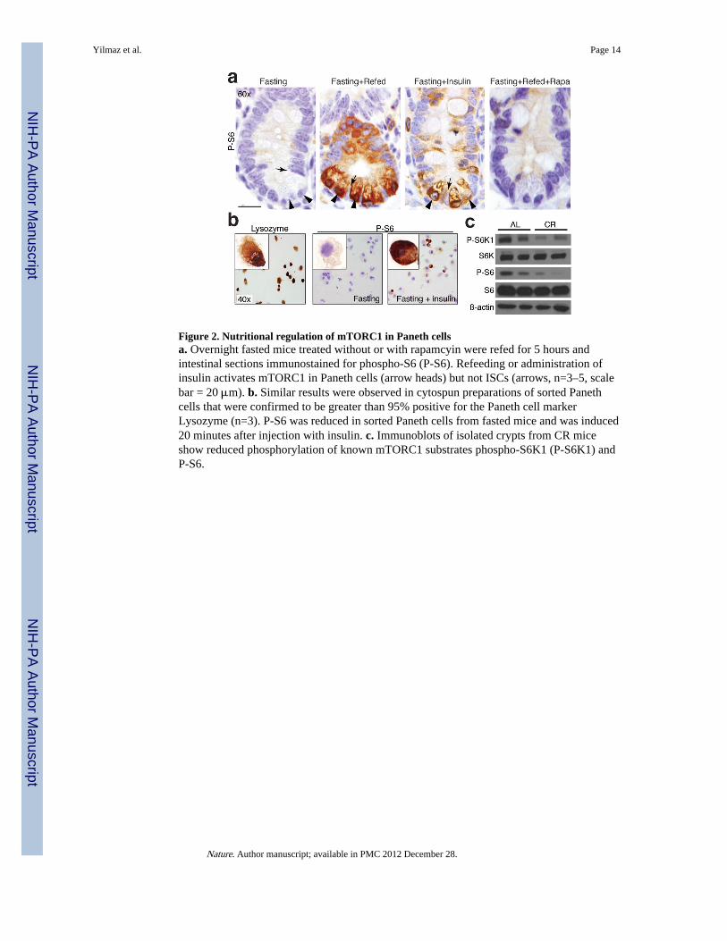
Figure 2. Nutritional regulation of mTORC1 in Paneth cellsa. Overnight fasted mice treated without or with rapamcyin were refed for 5 hours andintestinal sections immunostained for phospho-S6 (P-S6). Refeeding or administration ofinsulin activates mTORC1 in Paneth cells (arrow heads) but not ISCs (arrows, n=3–5, scalebar = 20 μm). b. Similar results were observed in cytospun preparations of sorted Panethcells that were confirmed to be greater than 95% positive for the Paneth cell markerLysozyme (n=3). P-S6 was reduced in sorted Paneth cells from fasted mice and was induced20 minutes after injection with insulin. c. Immunoblots of isolated crypts from CR miceshow reduced phosphorylation of known mTORC1 substrates phospho-S6K1 (P-S6K1) andP-S6.
Yilmaz et al. Page 14
Nature. Author manuscript; available in PMC 2012 December 28.
NIH
-PA Author Manuscript
NIH
-PA Author Manuscript
NIH
-PA Author Manuscript
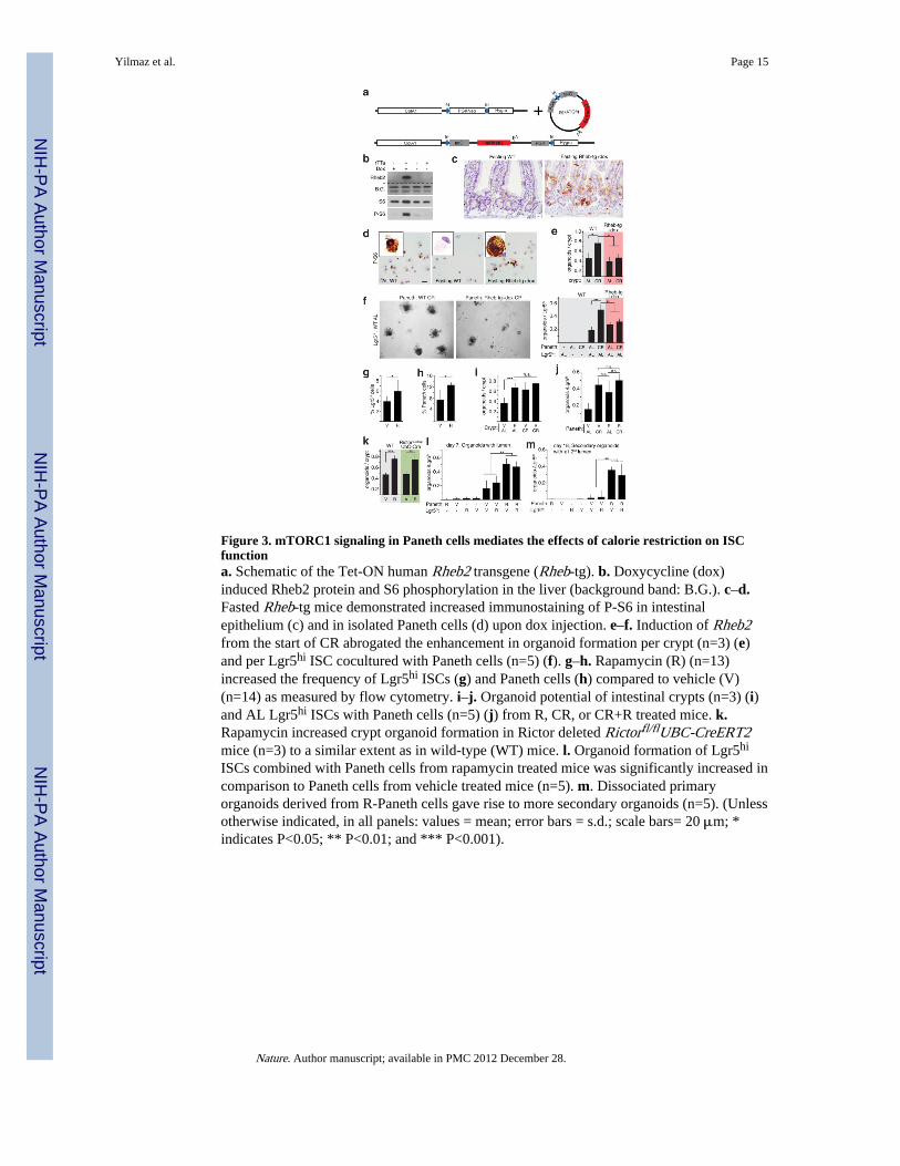
Figure 3. mTORC1 signaling in Paneth cells mediates the effects of calorie restriction on ISCfunctiona. Schematic of the Tet-ON human Rheb2 transgene (Rheb-tg). b. Doxycycline (dox)induced Rheb2 protein and S6 phosphorylation in the liver (background band: B.G.). c–d.Fasted Rheb-tg mice demonstrated increased immunostaining of P-S6 in intestinalepithelium (c) and in isolated Paneth cells (d) upon dox injection. e–f. Induction of Rheb2from the start of CR abrogated the enhancement in organoid formation per crypt (n=3) (e)and per Lgr5hi ISC cocultured with Paneth cells (n=5) (f). g–h. Rapamycin (R) (n=13)increased the frequency of Lgr5hi ISCs (g) and Paneth cells (h) compared to vehicle (V)(n=14) as measured by flow cytometry. i–j. Organoid potential of intestinal crypts (n=3) (i)and AL Lgr5hi ISCs with Paneth cells (n=5) (j) from R, CR, or CR+R treated mice. k.Rapamycin increased crypt organoid formation in Rictor deleted Rictorfl/flUBC-CreERT2mice (n=3) to a similar extent as in wild-type (WT) mice. l. Organoid formation of Lgr5hi
ISCs combined with Paneth cells from rapamycin treated mice was significantly increased incomparison to Paneth cells from vehicle treated mice (n=5). m. Dissociated primaryorganoids derived from R-Paneth cells gave rise to more secondary organoids (n=5). (Unlessotherwise indicated, in all panels: values = mean; error bars = s.d.; scale bars= 20 μm; *indicates P<0.05; ** P<0.01; and *** P<0.001).
Yilmaz et al. Page 15
Nature. Author manuscript; available in PMC 2012 December 28.
NIH
-PA Author Manuscript
NIH
-PA Author Manuscript
NIH
-PA Author Manuscript

Figure 4. Calorie restriction enhances expression of bone stromal antigen 1 (Bst-1) in Panethcells, whose product cyclic ADP ribose (cADPR) enhances ISC functiona. Transcriptional profiling of Paneth cells from CR and AL mice (n=4 and 3, respectively)identified significant expression changes in 401 genes, 57 of which encode plasmamembrane associated or secreted proteins. b. Validation of the increased transcription ofBst-1 by qRT-PCR (n=3). c–d. Increased Bst-1 protein in crypts from CR mice andrapamycin treated mice detected via immunoblotting (c) and in CR-Paneth cells byimmunostaining (d). e. Exogenous cADPR increased the organoid potential of AL-crypts toan extent similar as those of CR-crypts (n=3). f–g Inhibition of Bst-1 by siRNA-mediatedknock-down (n=3) (f) abrogated the enhanced potential of CR-crypts to form organoids(n=3) (g). i. A model of intestinal adaptation to calorie restriction by non-cell autonomousregulation of ISC self-renewal. CR attenuates mTORC1 activity in the Paneth cells,resulting in increased expression of Bst-1, whose paracrine product cADPR promotes ISCself-renewal. * indicates P<0.05; ** P<0.01, *** P<0.001. Values = means and error bars =s.d.
Yilmaz et al. Page 16
Nature. Author manuscript; available in PMC 2012 December 28.
NIH
-PA Author Manuscript
NIH
-PA Author Manuscript
NIH
-PA Author Manuscript