Morphological changes induced by heavy metals in dandelion (Taraxacum officinale Web.) growing on...
13
POTENTIALLY HARMFUL ELEMENTS IN SOIL-PLANT INTERACTIONS Morphological changes induced by heavy metals in dandelion (Taraxacum officinale Web.) growing on mine soils Laura Maleci & Gabriella Buffa & Mohammad Wahsha & Claudio Bini Received: 24 August 2013 /Accepted: 26 November 2013 /Published online: 11 December 2013 # Springer-Verlag Berlin Heidelberg 2013 Abstract Purpose Heavy metal accumulation produces significant physiological and biochemical responses in vascular plants. Plants growing on abandoned mine sites are of particular interest, since they are genetically tolerant to high metal concentrations. In this work, we examined the effect of heavy metals (HMs) on the morphology of T. officinale growing in pots with mine soils, with the following objectives: (1) to determine the evolution of HM concentration in leaves and roots over 3 years of cultivation; (2) to highlight possible damage at anatomical and cytological level. Materials and methods Wild specimens of Taraxacum officinale Web., with their soil clod, were gathered from three sites with different contamination levels by heavy metals (Cd, Cr, Cu, Fe, Pb, Zn) in the abandoned Imperina Valley mine (Northeast Italy). A control plant was also gathered from a non-contaminated site nearby. Plants were cultivated in pots at the botanical garden of the University of Florence (HBF), and appeared macroscopically not affected by toxic signals (re- duced growth, leaf necrosis) possibly induced by soil HM concentration. Leaves and roots taken at the same growing season were observed by light microscopy and transmission electron microscopy. Results and discussion Light microscopy observations show a clear difference in the cellular organisation of non- contaminated and contaminated samples. The unpolluted samples present a well-organised palisade tissue and spongy photosynthetic parenchyma. Samples from contaminated sites, instead, present a palisade parenchyma less organised, and a reduction of leaf thickness proportional to HM concen- tration. The poor structural organisations, and the reduced foliar thickness of the contaminated plants, are related to soil contamination. Differences in root micromorphology concern the cortical parenchyma. Moreover, all the samples examined present mycorrhiza. Ultrastructure observations of the paren- chyma cells show mitochondrial structure alteration, with lacking or reduced cristae of the internal membrane at in- creasing metal content. Instead, chloroplast organisation does not present significant differences, particularly in number and compartmentalization of thylakoids. Conclusions Although macromorphology does not present evidence of phytotoxicity, the recorded observations of the micromorphological characteristics of leaves and roots, show a suffering state of the plants, strictly related to HM content. Leaching reduced partly the HM content of the soil, therefore decreasing their phytotoxic effect. A gradual restoration of leaf organisation suggests that somewhat resilience occurred in plants. Moreover, the presence of stress-tolerant mycorrhi- zal fungi could contribute to reduce metal toxicity. Keywords Heavy metals . Mine soils . Plant morphology . Taraxacum officinale . Ultrastructure 1 Introduction Trace elements are ordinarily present in rocks, sediments and soils, but locally may be concentrated in rocks as ore bodies and generally dispersed in the environment through pollution Responsible editor: Jaume Bech L. Maleci (*) Department of Biology, University of Florence, Via P.A. Micheli, 3, 50121 Florence, Italy e-mail: [email protected] G. Buffa : C. Bini Department of Environmental Sciences, Informatics and Statistics, Ca’Foscari University of Venice, Dorsoduro 2137 30123 Venice, Italy M. Wahsha Marine Science Station, The University of Jordan—Aqaba Branch, Aqaba, Jordan J Soils Sediments (2014) 14:731–743 DOI 10.1007/s11368-013-0823-y
Transcript of Morphological changes induced by heavy metals in dandelion (Taraxacum officinale Web.) growing on...
POTENTIALLY HARMFUL ELEMENTS IN SOIL-PLANT INTERACTIONS
Morphological changes induced by heavy metalsin dandelion (Taraxacum officinale Web.) growingon mine soils
Laura Maleci & Gabriella Buffa & Mohammad Wahsha &
Claudio Bini
Received: 24 August 2013 /Accepted: 26 November 2013 /Published online: 11 December 2013# Springer-Verlag Berlin Heidelberg 2013
AbstractPurpose Heavy metal accumulation produces significantphysiological and biochemical responses in vascular plants.Plants growing on abandoned mine sites are of particularinterest, since they are genetically tolerant to high metalconcentrations. In this work, we examined the effect of heavymetals (HMs) on the morphology of T. officinale growing inpots with mine soils, with the following objectives: (1) todetermine the evolution of HM concentration in leaves androots over 3 years of cultivation; (2) to highlight possibledamage at anatomical and cytological level.Materials and methods Wild specimens of Taraxacumofficinale Web., with their soil clod, were gathered from threesites with different contamination levels by heavy metals (Cd,Cr, Cu, Fe, Pb, Zn) in the abandoned Imperina Valley mine(Northeast Italy). A control plant was also gathered from anon-contaminated site nearby. Plants were cultivated in pots atthe botanical garden of the University of Florence (HBF), andappeared macroscopically not affected by toxic signals (re-duced growth, leaf necrosis) possibly induced by soil HMconcentration. Leaves and roots taken at the same growingseason were observed by light microscopy and transmissionelectron microscopy.
Results and discussion Light microscopy observations show aclear difference in the cellular organisation of non-contaminated and contaminated samples. The unpollutedsamples present a well-organised palisade tissue and spongyphotosynthetic parenchyma. Samples from contaminatedsites, instead, present a palisade parenchyma less organised,and a reduction of leaf thickness proportional to HM concen-tration. The poor structural organisations, and the reducedfoliar thickness of the contaminated plants, are related to soilcontamination. Differences in root micromorphology concernthe cortical parenchyma. Moreover, all the samples examinedpresent mycorrhiza. Ultrastructure observations of the paren-chyma cells show mitochondrial structure alteration, withlacking or reduced cristae of the internal membrane at in-creasing metal content. Instead, chloroplast organisation doesnot present significant differences, particularly in number andcompartmentalization of thylakoids.Conclusions Although macromorphology does not presentevidence of phytotoxicity, the recorded observations of themicromorphological characteristics of leaves and roots, showa suffering state of the plants, strictly related to HM content.Leaching reduced partly the HM content of the soil, thereforedecreasing their phytotoxic effect. A gradual restoration ofleaf organisation suggests that somewhat resilience occurredin plants. Moreover, the presence of stress-tolerant mycorrhi-zal fungi could contribute to reduce metal toxicity.
Keywords Heavymetals .Mine soils . Plantmorphology .
Taraxacum officinale . Ultrastructure
1 Introduction
Trace elements are ordinarily present in rocks, sediments andsoils, but locally may be concentrated in rocks as ore bodiesand generally dispersed in the environment through pollution
Responsible editor: Jaume Bech
L. Maleci (*)Department of Biology, University of Florence, Via P.A. Micheli, 3,50121 Florence, Italye-mail: [email protected]
G. Buffa : C. BiniDepartment of Environmental Sciences, Informatics and Statistics,Ca’Foscari University of Venice, Dorsoduro 213730123 Venice, Italy
M. WahshaMarine Science Station, The University of Jordan—Aqaba Branch,Aqaba, Jordan
J Soils Sediments (2014) 14:731–743DOI 10.1007/s11368-013-0823-y
as a consequence of mining the ores (Davies 1987; Alloway1995). Environmental threats arise when ores are mined,milled and smelted, and a certain amount of potentially harm-ful elements (PHEs) is released in the surrounding areas and towaterways. Depending on the nature of the waste rock andtailings, a wide dispersion of these PHEs both in solution andin particulate form is possible (Sivri et al. 2010).
Industrial extraction of metals from ore minerals usuallyresults in large amounts of waste materials which often containelevated concentrations of PHEs such as As, Cd, Cr, Cu, Pb, Zn(Helios-Rybicka 1996; Lee et al. 2001; Navarro et al. 2008).These elements can be transported, dispersed in the environ-ment and accumulated in plants, and then may pass through thefood chain to human people as the final consumer, causingserious health problems as intoxication, lead poisoning,mercurialism and also cancer (Bernard 1995; Steinnes 2009).
The metal-enriched areas, therefore, represent an ideal nat-ural laboratory where to study the processes in order to providedescriptive models of the interactions between PHEs, thepedosphere, the biosphere and the hydrosphere. As stated byPreeti and Tripathi (2011), there is a direct relationship betweenchemical characteristics of soil, heavy metals concentration andmorphological and biochemical responses of plants. Indeed, itis well known that PHEs may have toxic effects on livingorganisms (microbes, plants and animals, including humans):decline of soil fertility and yield depression (Zhao et al. 2011),decrease inmicrobial activity (Kucera et al. 2008), limited plantgrowth and root elongation (Kidd et al. 2009), reduction of themeristematic zone (Giuliani et al. 2008; Lösch 2004), damagedepidermal cells, plasmolysis, reduced chlorophyll and caroteneproduction (Lopareva-Pohu et al. 2011).
In the last decades, the assessment of soil contaminationhas been extensively carried out through plant analysis (Ernst1996; Zupan et al. 2003; Bini 2010 and references therein).There are plants that can survive in a metal-enriched environ-ment, and other plants that can accumulate metals in theiraerial parts; both are genetically tolerant to heavy metals. Thefirst ones are diffused in abandoned mine sites or in naturallymetal-enriched soils (e.g. serpentine soils); they are good(passive accumulative) bioindicators for large scale and localsoil contamination (Baker 1981; Baker and Brooks 1989;Bargagli 1993; Zupan et al. 1995, 2003; Poschenrieder et al.2001). The second ones are bioaccumulator plants, and havebeen proposed recently as tools to clean up contaminated soilsby the environmental friendly technique of phytoremediation(Adriano et al. 1995; Baker et al. 2000; Bini 2009). A thirdgroup of plants do not tolerate metals, and are referred to asexcluder (Baker 1981).
Although plants are metal-tolerant or bioaccumulators,heavy metals interfere with their metabolism (Lopareva-Pohu et al. 2011), and several morphological modificationswere observed in their structure and ultrastructure(Mangabeira et al. 2001; Sarret et al. 2001;Maleci et al. 2001).
A large part of the phytotoxic effects of heavy metals havebeen assessed in laboratory studies on seeds germination or onplantlets (Mangabeira et al. 2001; Li et al. 2005; Preeti andTripathi 2011). On the contrary, few contributions have beenpublished on full-scale experiments and field observationscarried out on wild plants (Madejon et al. 2002; Yoon et al.2006; Unterbrunner et al. 2007; Llugany et al. 2009; Fontanaet al. 2010; Bini et al. 2010; Wahsha et al. 2012b).
Previous studies of our research group (Bini et al. 2000,2011; Fontana et al. 2010, 2011a, b; Bini 2012; Wahsha et al.2012a, b) investigated the heavy metal concentration of soilsdeveloped from mine waste material. Attention was focusedon the toxicity and influence of heavy metals on wild plantsgrowing on those contaminated soils, and on the metal uptakeby both known and unreported metal-tolerant plant species.
The plant selected for this study was dandelion (Taraxacumofficinale Web.), a species well known for his tolerance toheavy metals (Królak 2003; Zupan et al. 2003; Rosselli et al.2006). T. officinale is a very common plant, easy to identifyand greatly adaptable to different geo-morpho-pedologicaland climatic conditions (Keane et al. 2001; Malawska andWilkomirski 2001). Moreover, it is commonly collected to beeaten as a fresh salad or boiled vegetable; it is reported inEuropean Pharmacopoeia (2006), and used in traditional med-icine as hepatoprotector and diuretic. Therefore, when grownon heavily contaminated soils, it may be potentially harmful ifintroduced in dietary food or used as medicine.
Our preliminary results (Bini et al. 2012) confirmed thatT. officinale is a species tolerant to high metal concentrations,and suggested to use it as a bioindicator plant. Metals accumu-lated preferentially in roots, but leaves also proved to be accu-mulator organs, showing only little morphological damage.
In this work, we report the results of the chemical contentand its effect on the morphology of T. officinale plants grow-ing on soils with different contamination levels from the minearea of Imperina Valley (North East Italy), with the followingobjectives:
– To determine heavy metal (HM) concentration in leavesand roots
– To highlight possible damage at anatomical and cytolog-ical level
– To assess the possible plant resilience
2 Materials and methods
2.1 Site description
The Imperina Valley mining area is located in the Dolomitesmountain district (NE Italy, Fig. 1), with an altitude rangingbetween 543 and 990 m above sea level. The geological
732 J Soils Sediments (2014) 14:731–743
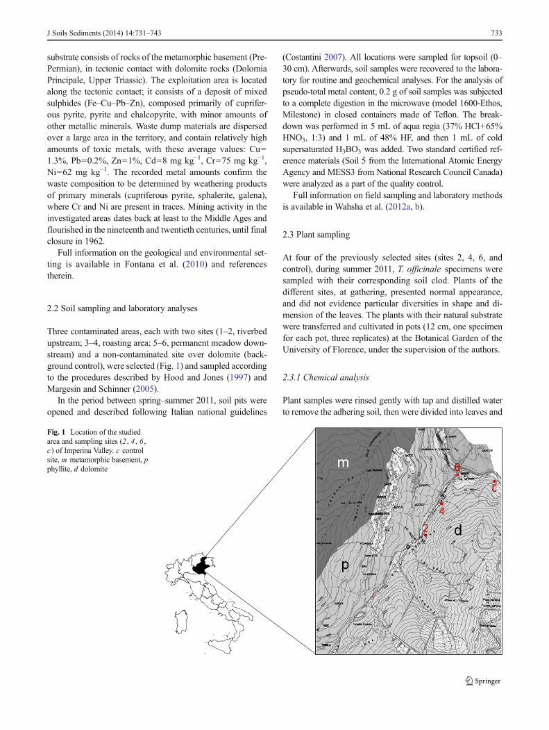
substrate consists of rocks of the metamorphic basement (Pre-Permian), in tectonic contact with dolomite rocks (DolomiaPrincipale, Upper Triassic). The exploitation area is locatedalong the tectonic contact; it consists of a deposit of mixedsulphides (Fe–Cu–Pb–Zn), composed primarily of cuprifer-ous pyrite, pyrite and chalcopyrite, with minor amounts ofother metallic minerals. Waste dump materials are dispersedover a large area in the territory, and contain relatively highamounts of toxic metals, with these average values: Cu=1.3%, Pb=0.2%, Zn=1%, Cd=8 mg kg−1, Cr=75 mg kg−1,Ni=62 mg kg−1. The recorded metal amounts confirm thewaste composition to be determined by weathering productsof primary minerals (cupriferous pyrite, sphalerite, galena),where Cr and Ni are present in traces. Mining activity in theinvestigated areas dates back at least to the Middle Ages andflourished in the nineteenth and twentieth centuries, until finalclosure in 1962.
Full information on the geological and environmental set-ting is available in Fontana et al. (2010) and referencestherein.
2.2 Soil sampling and laboratory analyses
Three contaminated areas, each with two sites (1–2, riverbedupstream; 3–4, roasting area; 5–6, permanent meadow down-stream) and a non-contaminated site over dolomite (back-ground control), were selected (Fig. 1) and sampled accordingto the procedures described by Hood and Jones (1997) andMargesin and Schinner (2005).
In the period between spring–summer 2011, soil pits wereopened and described following Italian national guidelines
(Costantini 2007). All locations were sampled for topsoil (0–30 cm). Afterwards, soil samples were recovered to the labora-tory for routine and geochemical analyses. For the analysis ofpseudo-total metal content, 0.2 g of soil samples was subjectedto a complete digestion in the microwave (model 1600-Ethos,Milestone) in closed containers made of Teflon. The break-down was performed in 5 mL of aqua regia (37% HCl+65%HNO3, 1:3) and 1 mL of 48% HF, and then 1 mL of coldsupersaturated H3BO3 was added. Two standard certified ref-erence materials (Soil 5 from the International Atomic EnergyAgency and MESS3 from National Research Council Canada)were analyzed as a part of the quality control.
Full information on field sampling and laboratory methodsis available in Wahsha et al. (2012a, b).
2.3 Plant sampling
At four of the previously selected sites (sites 2, 4, 6, andcontrol), during summer 2011, T. officinale specimens weresampled with their corresponding soil clod. Plants of thedifferent sites, at gathering, presented normal appearance,and did not evidence particular diversities in shape and di-mension of the leaves. The plants with their natural substratewere transferred and cultivated in pots (12 cm, one specimenfor each pot, three replicates) at the Botanical Garden of theUniversity of Florence, under the supervision of the authors.
2.3.1 Chemical analysis
Plant samples were rinsed gently with tap and distilled waterto remove the adhering soil, then were divided into leaves and
Fig. 1 Location of the studiedarea and sampling sites (2 , 4 , 6 ,c) of Imperina Valley. c controlsite, m metamorphic basement, pphyllite, d dolomite
J Soils Sediments (2014) 14:731–743 733
roots, dried in ventilated oven at 80 °C for 2 days. Driedtissues were grinded in an agate mill (<100 μm). Plant sam-ples (0.5 g) were digested in an acid mixture of 5 mL 65%HNO3 and 3 mL 30% H2O2 in open vessels on the hot plate,followed by filtration with filter cellulose Whatman n. 42.After digestion, plant samples were analyzed for PHEs. Theconcentration of metals (Cd, Cr, Cu, Pb, Zn, Fe) was deter-mined by inductively coupled plasma–optical emission spec-troscopy (ICP–OES, Perkin Elmer model 5300DV) accordingto the method reported by Margesin and Schinner (2005).
Analyses were carried out at gathering (June, 2011), andrepeated at the corresponding vegetative stage, during June,2013.
2.3.2 Microscope observations: Optical Microscopyand Transmission Electron Microscopy
Microscope observations were carried out in autumn, 2011,and summer, 2012 and 2013. Small pieces of leaves (taken inthe middle part of the leaf length), and thin roots, with prom-inent absorbing function, were withdrawn from plants of eachcontaminated site (Adriano et al. 1995; Ashraf et al. 2011;Baker and Brooks 1989) and from the control. Fresh materialwas pre-fixed in 2.5% glutaraldehyde in 0.1 M phosphatebuffer at a pH 6.8, post-fixed in 2% OsO4 in the same buffer,then dehydrated and embedded in Spurr’s epoxy resin. Semi-thin sections of embedded material were stained withToluidine Blu, and observed with a Leitz Light Microscopefor a general overview of the leaf and root morphology.Ultrathin sections were stained with uranyl acetate and subse-quently with lead citrate to observe leaf ultrastructure.Observations were performed with a Philips EM 300 trans-mission electron microscope.
2.4 Data analysis
Statistical analysis of metal content in soils and plants wasbased on ANOVA and is presented as means±S.D. The sta-tistical significance was declaredwhen p value was equal to orless than 0.05. Statistical analyses were performed usingSigma Stat statistical software version 3.5.
3 Results
3.1 Soil characters and heavy metals accumulation in soils
The soils of the study area, except control soil, are classified asSpolic Technosols (Wahsha et al. 2012b). Soils are shallow,sandy loam in texture and typically unsaturated with respect towater; they have low cation exchange capacity and relativelyhigh hydraulic conductivity. Organic carbon content is highlyvariable, ranging between 4 and 12 g kg−1 (33 g kg−1 at the
control site, Mollisol over dolomite), with the lowest values atthe most contaminated sites. The pH value is 7.5 at site 2,5.3 at site 4, and 7.6 at site 6, while the control pH is 7.8, therange depending on the lithology of parent material. Fulldescription of soil data is reported in Fontana et al. (2010).
The average concentrations of Cd, Cr, Cu, Pb, Zn, Fe in thesoils examined are reported in Table 1. All the studied sites,out of the non-contaminated one, are strongly enriched inmetals. The total concentrations of most of the investigatedmetals (Cd, Cu, Pb, Zn and Fe) in the soil samples weresignificantly higher than those of the non-contaminated site,and almost all above the toxicity threshold according to theItalian Legislative Decree (D.L. 152/2006). Chromium con-centration, instead, is below the regulatory limits, and consid-erably higher in non-contaminated soil than at contaminatedsites. Site 4 is the most contaminated, presenting very highmetal concentrations (Cd up to 4.35 mg kg−1, Cu up to4,100 mg kg−1, Pb up to 14,150 mg kg−1, Zn up to2,700 mg kg−1), with Fe up to 58% in the roasting area.Sites 2 and 6 too are highly contaminated by Cu, Pb, Zn, Fe,while Cd concentration is slightly above the non-contaminated site, and Cr is well below the control valueand under the detection limit at site 6.
Univariate statistics show that there is a positive linearcorrelation, significant at p <0.05, between Pb, Cu, Zn andFe (Cu/Pb 0.867; Pb/Zn 0.616; Cu/Zn 0.688; Cu/Fe 0.933).This is consistent with the composition of ore deposits foundin the Imperina Valley, as chalcopyrite (CuFeS2), sphalerite(ZnS) and galena (PbS). Instead, Cr is negatively correlatedwith Cu (−0.847), Pb (−0.816), Zn (−0.604) and Fe (−0.754).Iron presents a significant positive correlation with Pb (Fe/Pb0.734). Furthermore, Fe it is not significantly correlated withCd, as expected considering the counteracting geochemicalbehaviour of the two elements.
3.2 Heavy metals accumulation in T. officinale plants
The concentrations of heavy metals in roots and leaves ofdandelion plants collected in Imperina Valley are presented inTable 2. Data show that at contaminated sites this plant is ableto accumulate metals, out of Cd, at much higher concentra-tions than at the unpolluted site, and above the toxicity thresh-old indicated by Kabata-Pendias (2011); metal accumulationin shoots was proportional to soil metal content; therefore, thegreatest metal amount was found at the most contaminated site(sample 4).
Cadmium concentrations in both shoots and roots of plantsfrom sites 2, 4, 6, are below the control value (up to1.46 mg kg−1), and below the phytotoxicity threshold, withsite 2 presenting the highest value (1.05 mg kg−1).
Chromium concentrations in plants from contaminatedsites are slightly higher than the detection limit(1.00 mg kg−1), and very low in comparison to the
734 J Soils Sediments (2014) 14:731–743
phytotoxicity threshold reported by Kabata-Pendias (2011),consistently with concentration levels recorded in soil (seeTable 1).
Copper and lead concentrations in shoots present muchhigher levels (Cu up to 64 mg kg−1; Pb up to 193 mg kg−1)than at the non-contaminated site. However, Cu is nearlyequally accumulated in shoots and roots, while Pb is slightlymore abundant in shoots than in roots.
Zinc levels in T. officinale shoots from sites 2 and 6 fallwithin the normal range (27–150 mg kg−1) given by Kabata-Pendias (2011), while plants from site 4 present Zn concen-trations slightly above the normal values (189 mg kg−1).
Iron concentrations in leaves show a wide range of varia-tion, with the highest level up to 890 mg kg−1 at site 4.However, this metal is not considered toxic unless at veryhigh concentration: above 1,000 mg kg−1 according toKabata-Pendias (2011).
Heavy metals in roots of T. officinale present lower con-centrations with respect to leaves, with the exception of Cu atsite 4. This suggests the ability of this plant to translocate mostmetals from roots to shoots. This is particularly evident for Feand Zn (two- to threefold), which are essential micronutrients,
while Pb and especially Cu are less prone (0.20 and 0.0 fold,respectively) to translocate from roots.
Leaves of dandelion taken directly from the assisted pots atUniversity of Florence (HBF) during June, 2013 were ana-lyzed for heavy metals following the previously describedprocedure. Roots could not be sampled, to avoid death ofplants. The results obtained are shown in Table 3.
A comparison of HM concentrations in leaves between thetwo sampling periods indicates that metal uptake and transferto leaves decreased significantly (at p <0.05). Not all themetals, however, presented the same decreasing trend: Cd,Cr, Cu and Fe contents decreased by one- to fivefolds, whilePb content decreased up to 100-folds at sites 2 and 6, and up totenfolds at site 4; Zn content decreased by tenfolds at allcontaminated sites. It is likely that leaching with rain and tapwater decreased the metal content in soils (data not shown),and consequently in plants.
3.3 LM observations
Light microscope observations were primarily carried out inOctober 2011, few months after plants were gathered (i.e. the
Table 1 Heavy metals concentration in soils of Imperina Valley
Sampling site Cd Cr Cu Pb Zn Fe
2 0.85±0.5 i 14±3 i 2,822±40 i 11,280±37 i 1,096±11 i 320,437±178 i
4 4.35±1.1 i 31±2 i 4,098±36 i 14,147±95 i 2,717±13 i 578,632±229 i
6 0.98±0.6 i < DLa 1,894±35 i 12,124±56 i 2,513±20 i 47,571±287 i
Control 0.32±0.2 141±4 105±6 39±2 95±7 37,984±328
R. L.b – 150 120 200 150 –
C.S.T.Cc 3–8 75–100 60–125 100–400 70–400 1,000d
The letter i following ±S.D. indicates significant difference at p<0.05 according to ANOVA. Adapted from Wahsha et al. 2012a, b
Cd, Cr, Cu, Pb, Zn, Fe are expressed as milligrams per kilogram. All the values are mean of five replicates±S.D.a Less than the detection limitb Residential limits in the Italian legislation (D.L. 152/2006, Annex 5)c Critical soil total concentration (range of values above which toxicity is considered to be possible) (Source: Alloway 1995)d Source: Kabata-Pendias 2011
Table 2 Concentration of heavy metals in Taraxacum (milligrams per kilogram dry weight)
Sampling site Cd Cr Cu Pb Zn Fe
Leaves Roots Leaves Roots Leaves Roots Leaves Roots Leaves Roots Leaves Roots
2 1.00±0.1 1.05±0.1 3.22±0.7 1.00±0.0 64±4 58±4 120±8 99.8±1 101±28 67±8 620±4 187±22
4 0.69±0.1 0.17±0.1 3.67±0.4 1.71±0.2 49±3 50±2 193±7 142±7 189±17 71±6 890±14 320±24
6 0.39±0.2 0.34±0.3 2.58±0.1 0.95±0.3 45±2 40±4 147±2 118 ±2 115±8 59±2 490±19 167±14
Control 1.46±0.3 1.33±0.4 <DLa < DL a 9±1 7±1 3.32±1 1.41±2 44±9 33±4 470±24 108±27
Sampling period during June 2011. All the values are mean of five replicates±S.D.a Below detection limit
J Soils Sediments (2014) 14:731–743 735
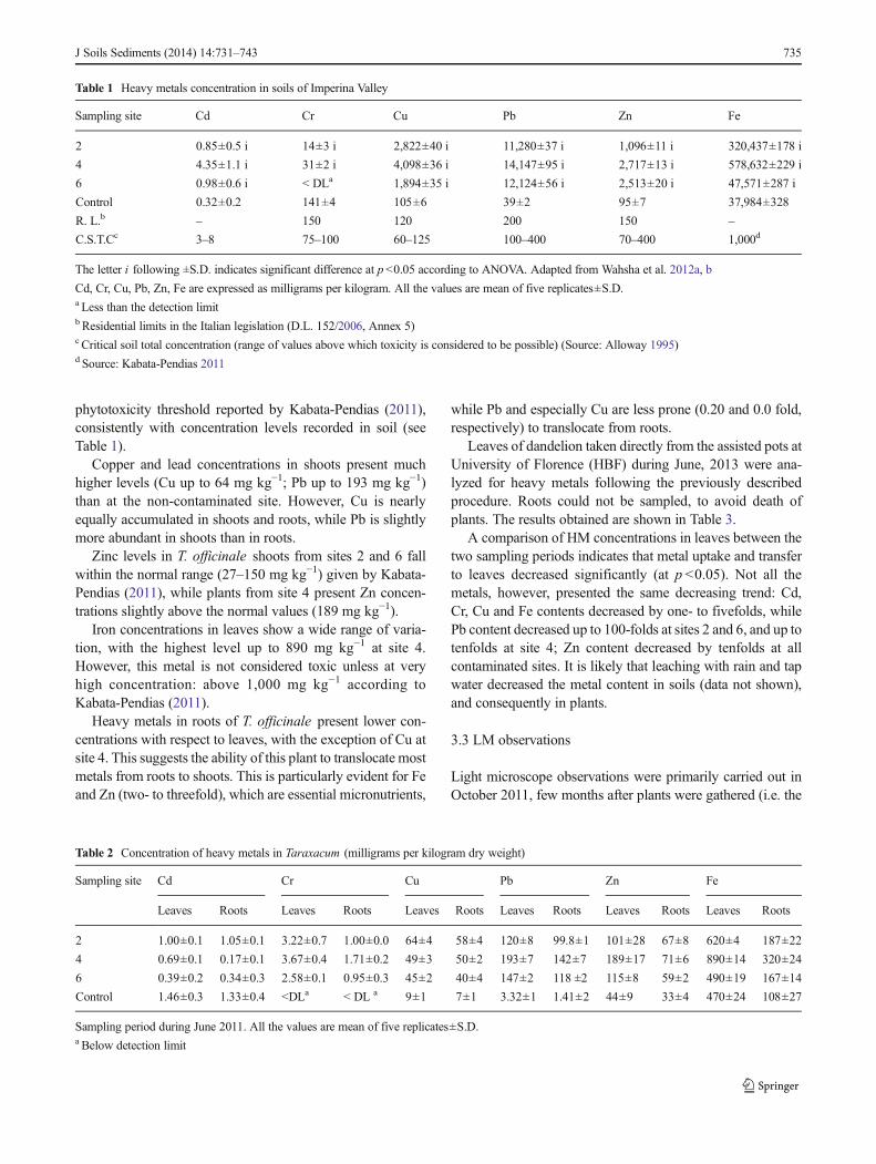
leaves are those grown during spring and summer in thedifferent sites). As clearly evidenced in Fig. 2, the thickness
and especially the organisation of the leaf blade is different inthe samples observed. Indeed, control samples present a well-
Table 3 Concentration of heavymetals in Taraxacum (milligramsper kilogram dry weight)
Sampling period during June2013. All the values are mean offive replicates±S.D.a Below detection limit
Sampling site Cd Cr Cu Pb Zn FeLeaves Leaves Leaves Leaves Leaves Leaves
2 0.64±0.1 1.36±0.5 11.13±3 1.26±0.7 27.18±18 189±14
4 0.66±0.1 1.81±0.5 12.25±3 10.80±7 40.41±14 636±18
6 0.32±0.4 1.11±0.4 14.89±2 3.14±0.9 40.42±8 312±14
Control 0.84±0.3 <DLa 8.29±1 1.99±1 22.95±9 147±24
Fig. 2 Transverse section (LM)of the leaf lamina (bar 100 μm).a Control plant in 2011; b controlplant in 2013; c plant from site 2in 2011; d plant from site 2 in2013; e plant from site 4 in 2011;f plant from site 4 in 2013; g plantfrom site 6 in 2011; h plant fromsite 6 in 2013
736 J Soils Sediments (2014) 14:731–743
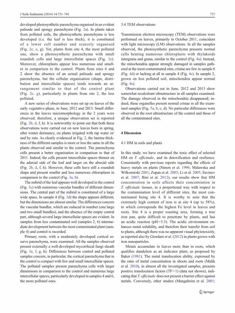
developed photosynthetic parenchyma organised in an evidentpalisade and spongy parenchyma (Fig. 2a). In plants takenfrom polluted soils, the photosynthetic parenchyma is lessdeveloped (i.e. the leaf is less thick), it is constitutedof a lower cell number and scarcely organised(Fig. 2c, e, g). Yet, plants from site 4, the most pollutedone, show a photosynthetic parenchyma with smallroundish cells and large intercellular spaces (Fig. 2e).Moreover, chloroplasts appear less numerous and small-er in comparison to the control. Plants from sites 6 and2 show the absence of an actual palisade and spongyparenchyma, but the cellular organisation (shape, distri-bution and intercellular spaces) tends towards an ar-rangement similar to that of the control plant(Fig. 2c, g), particularly in plants from site 2, the lesspolluted.
A new series of observations were set up on leaves of theearly vegetative phase, in June, 2012 and 2013. Small differ-ences in the leaves micromorphology in the 2 years wereobserved; therefore, a unique observation set is reported(Fig. 2b, d, f, h). It is noteworthy to point out that both theseobservations were carried out on new leaves born in spring,after winter dormancy, on plants irrigated with tap water or/and by rain. As clearly evidenced in Fig. 2, the lamina thick-ness of the different samples is more or less the same in all theplants observed and similar to the control. The parenchymacells present a better organisation in comparison to that of2011. Indeed, the cells present intercellular spaces thinner onthe adaxial side of the leaf and larger on the abaxial side(Fig. 2b, d, f, h). However, these cells have still a roundishshape and present smaller and less numerous chloroplasts incomparison to the control (Fig. 3a, b).
The mibrid of the leaf appears well developed in the control(Fig. 3c) with numerous vascular bundles of different dimen-sions. The central part of the mibrid is constituted of a largewide space. In sample 4 (Fig. 3d) the shape appears different,but the dimensions are almost similar. The differences concernthe vascular bundles, which are reduced in number (one largeand two small bundles), and the absence of the empty centralpart, although several large intercellular spaces are evident. Insamples from less contaminated soil (samples 2, 6) interme-diate development between themost contaminated plant (sam-ple 4) and control is recorded.
Primary roots, with a moderately developed cortical re-serve parenchyma, were examined. All the samples observedpresent externally a well-developed mycorrhizal fungi sheath(Fig. 3e, f, g, h). Differences between control and pollutedsamples concern, in particular, the cortical parenchyma that inthe control is compact with few and small intercellular spaces.The polluted samples present parenchyma cells with largerdimensions in comparison to the control and numerous largeintercellular spaces, particularly developed in samples 4 and 6,the more polluted ones.
3.4 TEM observations
Transmission electron microscopy (TEM) observations wereperformed on leaves, primarily in October 2011, coincidentwith light microscopy (LM) observations. In all the samplesobserved, the photosynthetic parenchyma presents normalcells bearing numerous chloroplasts with thylakoidsintergrana and grana, similar to the control (Fig. 4a). Instead,the mitochondria appear strongly damaged in samples gath-ered at the most contaminated sites, cristae are few in sample 6(Fig. 4d) or lacking at all in sample 4 (Fig. 4c). In sample 2,grown on less polluted soil, mitochondria appear normal(Fig. 4e).
Observations carried out in June, 2012 and 2013 showsomewhat recalcitrant ultrastructure in all samples examined.The damage observed in the mitochondria disappeared; in-deed, these organelles present normal cristae in all the exam-ined samples (Fig. 5a, b, c, d). No particular differences wereobserved in the root ultrastructure of the control and those ofall the contaminated sites.
4 Discussion
4.1 HM in soils and plants
In this study, we have examined the toxic effect of selectedHM on T. officinale , and its detoxification and resilience.Consistently with previous reports regarding the effects ofheavy metals on plants (Simon et al. 1996; Malawska andWilkomirski 2001; Zupan et al. 2003; Li et al. 2005; Savinovet al. 2007; Bini et al. 2012), our results show that HMconcentration in soils affects their concentration inT. officinale tissues, in a proportional way with respect tothe contamination level of different sites, the most con-taminated being site 4. It is worthy to note that theextremely high content of iron is at site 4 (up to 58%),to which corresponds the highest Fe level in leaves androots. Site 4 is a proper roasting area, forming a trueiron pan, quite difficult to penetrate by plants, and hasan acidic reaction (pH=5.3). The acidic environment en-hances metal solubility, and therefore their transfer from soilto plants, although there was no apparent visual phytotoxicity,as reported also by Giordani et al. (2012) in plants grown withiron nanoparticles.
Metals accumulate in leaves more than in roots, whichqualifies dandelion as an indicator plant, as proposed byBaker (1981). The metal translocation ability, expressed bythe ratio of metal concentration in shoots and roots (Maliket al. 2010), in almost all the investigated samples, presentspositive translocation factors (TF>1) (data not shown), indi-cating that T. officinale does not present a barrier effect againstmetals. Conversely, other studies (Mangabeira et al. 2001;
J Soils Sediments (2014) 14:731–743 737
Kabata-Pendias and Mukherjee 2007; Bini et al. 2008) reportsome root barrier for non-essential metals (e.g. Cd, Cr, Pb)uptake, suggesting some exclusion strategy by plants. Yet, asreported by Memon et al. (2001), plant species that have noexclusion mechanism in the roots absorb and translocatemetals and accumulate them in their shoots, especially inleaves, without showing any toxicity symptoms, via a sort ofinternal resistance or accumulation mechanism.
Concerning cadmium, it is interesting to note that its con-centration in both leaves and roots from the unpolluted site ishigher than that from contaminated sites. The different soilparent material is likely to regulate the uptake of Cd as Ca-substituted element in dolomite at the control site, as reportedby Dubois et al. (1998) in soils from Switzerland.
The differential HM content decrease in leaves collected in2013 in comparison to 2011 (up to 100-folds for Pb) suggests
Fig. 3 a , b Transverse section(LM) of the leaf lamina in 2013 atgreater magnification (bar50 μm). a Control plant; b plantfrom site 4 (ch chloroplasts). c , dtransverse section of the mibrid in2013 (bar 500 μm). c Controlplant; d plant from site 4. e , f , g ,h Transverse section of the roots(LM); arrows indicate themycorrhizal sheath, asterisk theintercellular spaces (bar 100 μm).e Control plant f plant from site 2;g plant from site 4; h plant fromsite 6
738 J Soils Sediments (2014) 14:731–743

Fig. 4 Ultrastructure ofparenchyma cells (TEM); arrowsindicate the mitochondrial cristae(bar 1 μm); a Control plant; bplant from site 2; c plant from site4; d plant from site 6
Fig. 5 Ultrastructure ofparenchyma cells (TEM) showingthe restored internal membrane ofmitochondria. Same samples as inFig. 4. a bar 1 μm; b , c , d bar0.5 μm
J Soils Sediments (2014) 14:731–743 739

that metals are leached with different pathways, according totheir available fraction in the soil. Metals with the highestconcentrations in 2011 leaves (Pb, Zn) were decreased to amajor extent in comparison to the less concentrated ones.Lead was the easiest to be removed, as indicated by its strongdecrease in 2013 leaves. Despite their less abundant content,Cd and Cr presented a minor decrease, which suggests someexclusion strategy for these non-essential elements. Copperand iron concentrations were relatively high (Cu up to15 mg kg−1, Fe up to 636 mg kg−1) in 2013 leaves, with aless than fivefold decrease in comparison to 2011, in agree-ment with their essential/critical role as micronutrients, and donot determine any visual symptoms of phytotoxicity, suggest-ing a Cu–Fe tolerance (Abreu et al. 2008).
4.2 Plant morphology
In this study, any evident variation of the macromorphologywas recorded in T. officinale by HM excess; indeed, all theplants examined (control and samples 2, 4, 6), even consider-ing the range of species variability, present similar morpholo-gy and dimensions. Consistently, any apparent toxicity symp-toms were visible in numerous accumulator plants, as reportedby Memon et al. (2001). However, we observed a little differ-ence in leaves colour (a less intense green colour in specimensof contaminated sites in comparison to control). These state-ments are consistent with findings by Kupper et al. (1998),who noted that in water plants transition metals such as Cd,Cu, Ni, Pb, Zn may substitute for Mg in the chlorophyllmolecule, thus reducing the photosynthetic function, whichresults in colour change (i.e. chloroplasts number decrease,and consequently the chlorophyll production). It is likely thatanalogous process would occur in our studied plants.
A modified growth of leaves and roots, instead, was re-cently observed byAshraf et al. (2011) in several plants grownon mine tailings. Although the leaf morphology ofT. officinale does not present particular differences amongthe specimens of various sites, at microscopic level morpho-logical and histological changes were observed. The actualthickness measured by microscopy (and which is not appre-ciable with the visual observation), is different in the observedsamples (Fig. 2), and decreases with increasing HM content ofplants. The different leaf thickness is consistent with thereduced, or even lacking, photosynthetic parenchyma normal-ly structured as palisade on the adaxial leaf surface, andspongy parenchyma on the abaxial surface, as observed incontrol plants.
In the mibrid, the reduced number of vascular bundles(proportional to the contamination level), in comparison tocontrol, have, as a consequence, less water availability forleaves, which certainly influences the plant metabolic activity.
Vacuoles constitute the cell compartment where HM, takenup by active transport systems, are deposited; in vacuoles,
plants, according to a metal-adaptive strategy, isolate HM,thus inhibiting any interference with the plant metabolic reac-tions ( Memon et al. 2001; Schutzendubel and Polle 2002).
At relatively low metal concentrations, metals isolated incell walls and vacuoles, are confined in roots, and therefore itis unlike to relate HM contents in soil with those in leaves, asreported by Rosselli et al. (2006). It is possible that plantsgrown at site 2 (the less contaminated) deposited all HM incell walls and in vacuoles, since no macroscopic neithercellular deformations were observed. With higher soil metalcontents, plants are able to translocate metals in the aerialparts, particularly in leaves (Keunen et al. 2011). In our study,plants from the most contaminated sites (samples 4, 6) pre-sented HM contents in leaves related to those of the corre-sponding soils (with the exception of Cu which probably has aless effective transporter). The metal amount transported toleaves provokes cell morphology modification, as shown infigures above. Yet, as observed in samples 4 and 6, the cellmetal content is responsible for serious deformations of mito-chondria, the organelles performing aerobic respiration.Keunen et al. (2011) reported that PHEs provoke an increasein reactive oxygen species (ROS) production, and a reductionof mitochondria respiration functions. Similarly,Karuppanapandian et al. (2011) found that ROS gener-ated under stress conditions provoke cell damage invarious cellular compartments, including chloroplasts,mitochondria, endoplasmic reticulum and plasma mem-branes. Our morphological study ascertained mitochon-dria cristae reduction in sample 6, and even lack in sample4, confirming recent physiological observations byKaruppanapandian et al. (2011), Keunen et al. (2011) andLopareva-Pohu et al. (2011).
Micromorphological studies on plant cells subjected to HMstress are nearly lacking. In a recent paper, Zhao et al. (2011)report the effects on the morphology and ultrastructure oftomato plants subjected to lead-induced stress. Besides areduction in size of different parts (fruits, leaves, stems androots), thinner cell walls, swollen and deformed chloroplastsare recorded at ultrastructural level. Reduction in thicknessand dimensions of leaf blade has been recorded also in soy-bean by Weryszko-Chmielewska and Chwil (2005). Morerecently, Giordani et al. (2012), in an experimental work onmetal nanoparticles (NP) application to tomato seed-lings, found clear effects at both morphological andgenetic level (e.g. root hair formation, epidermal cellsoutgrowth). In our study, no deformation was observed inchloroplasts; however, in sample 6, and in sample 4 in partic-ular, chloroplasts are smaller and numerically less than in thecontrol (Fig. 3a, b).
Considering the HM–plants relationships, and their adap-tion to contaminated environment, it is important to remindthat in geophytes like Taraxacum complete detoxificationoccurs at the end of the vegetative season, when plants loose
740 J Soils Sediments (2014) 14:731–743
leaves, which accumulate on the ground, and after winterdormancy, during the subsequent spring season, a new leafgeneration may start again with metal deposition in vacuoles.
It is worthy to note also that all the investigated samples,including the control plants, resulted to being mycorrhized.Several studies have dealt with a possible alleviation of metaltoxicity by mycorrhization, but only few presented evi-dence of such effects (Peterson et al. 2004 andreferences therein). In our samples, the PHEs effect iscertainly attenuated, since there is a hindered access ofmetals to the root surface, which suggests the metal-inducedstress response to be significantly reduced, as stated also bySchutzendubel and Polle 2002.
5 Conclusions
– Soil analysis shows that PHEs are released from minetailing because of low pH and low available water capac-ity; part of their bioavailable fraction is taken up byTaraxacum and transferred from roots to shoots, as it iscommon in indicator plants.
– The study shows that there is a relationship between highmetal content in Taraxacum plants and their modifiedmorphology. PHEs do not determine an evident modifi-cation of the macromorphology. However, the leaf thick-ness decreases, and the absence of a regularly structuredcellular organisation is likely related to the soil contami-nation degree.
– It is likely that a 2-year leaching reduced partly the HMcontent of the soil, therefore decreasing their phytotoxiceffect. A gradual restoration of leaf organisation suggeststhat, somewhat, resilience occurred in plants.
– Since all samples, including control, present mycorrhizedroots, it is suggested that stress-tolerant mycorrhizal fungicould contribute to reduce metal sorption. A comparisonwith non-mycorrhized plants could clarify the effectiverole of mycorrhizal fungi.
– Data which is generated through this study could behelpful in detecting the lethal levels of heavy metals forparticular plant species, their tolerance and remediationcapacity.
– The presence of heavy metals in dandelion has two con-sequences: a beneficial consequence, because the landaffected by anthropogenic pollution may be restoredby natural way; a fatal consequence, because bothhumans and animals feed dandelion, and, if plants arecontaminated, through this way toxic metals enter thefood chain.
Acknowledgments The authors wish to thank Corrado Tani, Pietro DiFalco and Flavia Visin for technical assistance.
References
Abreu MM, Tavares MT, Batista MJ (2008) Potential use of Ericaavandulensis and Erica australis in phytoremediation of sulphidemine environments: Sao Domingos, Portugal. J Geochem Explor96:210–222
Adriano DC, Chlopecka A, KaplandDI, Clijsters H, Vangrosvelt J (1995)Soil contamination and remediation philosophy, science and tech-nology. In: Prost R (ed) Contaminated soils. INRA, Paris, pp 466–504
Alloway BJ (1995) Heavy metals in soils. Blackie, London, p 368Ashraf M-L, Maah MJ, Yusoff I (2011) Heavy metals accumulation in
plants growing in ex tin mining catchment. Int J Environ Sci Tech8(2):401–416
Baker AJM (1981) Accumulators and excluders strategies in the responseof plants to heavy metals. J Plant Nutr 3:643–654
Baker AMJ, Brooks RR (1989) Terrestrial higher plants whichhyperaccumulate metallic elements—a review of their distribution,ecology and phytochemistry. Biorecovery 1:81–126
Baker A, Mc Grath S, Reeves R, Smith J (2000) Metal hyperaccumulatorplants: a review of the ecology and physiology of a biologicalresource for phytoremediation of metal-polluted soils. In: Terry N,Banuelos G (eds) Phytoremediation of contaminated soils. Lewis,London, pp 85–107
Bargagli R (1993) Plant leaves and lichens as biomonitors of naturals oranthropogenic emissions of mercury. In Markert B (ed) Plants asBiomonitors. Weinheim WCH, pp 468-484
Bernard AM (1995) Effects of heavy metals in the environment on thehuman health. In: Prost R (ed) Contaminated soils. INRA, Paris, pp21–34
Bini C (2009) Soil restoration: remediation and valorisation of contami-nated soils. In: Manual of Methods for Soil and Land Evaluation(E.A.C. Costantini edit), Science, Enfield, 137–160. (ISBN 978-1-57808-571-2)
Bini C (2010) From soil contamination to land restoration. In:Contaminated soils: environmental impact, disposal and treatment(R.V. Steinberg edit.), Nova, New York, 97-137 (ISBN 978-1-60741-791-0)
Bini C (2012) Environmental impact of abandoned mine waste: a review.Nova, New York, p 92
Bini C, Casaril S, Pavoni B (2000) Fertility gain and heavy metalaccumulation in plants and soils. Toxicol Environ Chem 77:131–142
Bini C, Maleci L, Romanin A (2008) The chromium issue in soilsof the leather tannery district in Italy. J Geochem Explor 96(2–3):194–202
Bini C, Fontana S, Wahsha M (2010) Land contamination by minedumps, plant toxicity and restoration perspectives byphytoremediation. Int J Environ Qual EQA 4:173–180, TipografiaFanti, Imola. ISBN 10: 88-901261-7-5
Bini C, Fontana S,WahshaM (2011) Environmental impact of PTEs (Cu,Fe, Pb, Zn) from mixed sulphides mines in Italy. Proc. XI ICOBTE,I, 505-506. Florence, July, 3–7, 2011
Bini C,WahshaM, Fontana S,Maleci L (2012) Effect of heavymetals onmorphological characteristics of Taraxacum officinale Web growingon mine soils in NE Italy. J Geochem Explor 123:101–108
Costantini EAC (2007) Soil survey methods and information of soil data.S.E.L.C.A, Firenze (in Italian)
D.L.—Legislation Act no 152/2006. Official Gazette n.88, 14/04/2006—Supplement no 96 (in Italian)
Davies BE (1987) Consequences of environmental contamination by leadmining in Wales. Hydrobiologia 149:213–220
Dubois JP, Okopnik F, Benitez N, Vedy JC (1998) Origin and spatialvariability of cadmium in some soils of the Swiss Jura. Proc. 16thIUSS Congress, Montpellier. Symposia 25:1–8
J Soils Sediments (2014) 14:731–743 741
Ernst WHO (1996) Bioavailability of heavy metals and decontaminationof soils by plants. Appl Geochem 11:163–167
European Pharmacopoeia (2006)—VI ed., Council of Europe, StrasburgFontana S, Wahsha M, Bini C (2010) Preliminary observations on heavy
metal contamination in soils and plants of an abandoned mine inImperina Valley (Italy). Agrochimica 4:218–231, LIV
Fontana S, Bini C, Wahsha M, Bullo M (2011a) Heavy metals contam-ination in soils and their transfer to common wheat (Triticumaestivum L.): a case study. Geophys Res Abstr 13:1751
Fontana S, Bini C, Wahsha M, Bullo M (2011b) PTEs in agroecosystemsand implications for the food chain. Proc. XI ICOBTE, I, 507–508.Florence, July, 3–7, 2011
Giordani T, Fabrizi A, Guidi L, Natali L, Giunti G, Ravasi F, Cavallini A,Pardossi A (2012) Response of tomato plants exposed to treatmentwith nano particles. Int J Environ Qual 8:27–38
Giuliani C, Pellegrino F, Tirillini B,Maleci L (2008)Micromorphologicaland chemical characterization of Stachys recta subsp serpentini(Fiori) Arrigoni in comparison to S. recta subsp. recta(Lamiaceae). Flora 203:376–385
Helios-Rybicka E (1996) Impact of mining and metallurgical industrieson the environment in Poland. Appl Geochem 11(1–2):3–11
Hood TM, Jones BJ Jr (1997) Soil and plant analysis in sustainableagriculture and environment. Marcel Dekker, New York, p 877
Kabata-Pendias A (2011) Trace elements in soils and plants, 3rd edn.CRC, Boca Raton, p 365
Kabata-Pendias A, Mukherjee AB (2007) Trace elements from soil tohuman. Springer, Berlin, p 550
Karuppanapandian T, Moon J, Kim C, Manoharan K, Kim W(2011) Reactive oxygen species in plants: their generation,signal transduction, and scavenging mechanisms. Aust J Crop Sci5(6):709–725
Keane B, Collier MH, Shann JR, Rogstad SH (2001) Metal content ofdandelion (Taraxacum officinale) leaves in relation to soil contam-ination and airborne particulate matter. Sci Total Environ 281:63–78
Keunen E, Remans T, Bohler S, Vangronsveld J, Cuypers A (2011)Metal-induced oxidative stress and plant mitochondria. Int J MolSci 12:6894–6918
Kidd P, Barcelo J, Bernal MP, Navari Izzo F, Poschenrieder C, Shilev S,Clemente R, Monterroso C (2009) Trace element behaviour at theroot-soil interface: implications in phytoremediation. Environ ExpBot 67:243–259
Królak E (2003) Accumulation of Zn, Cu, Pb and Cd by dandelion(Taraxacum officinale Web.) in environments with various degreesof metallic contamination. Pol J Environ Stud 12(6):713–721
Kucera T, Horáková H, Šonská A (2008) Toxic metal ions in photoauto-trophic organisms. Photosynthetica 46:481–489
Kupper H, Kupper F, Spiller M (1998) In situ detection of heavy metalsubstituted chlorophylls in water plants. Photosynth Res 58(2):123–133
Lee CG, Chon H, Jung MC (2001) Heavy metal contamination in thevicinity of the Daduk Au–Ag–Pb–Zn mine in Korea. ApplGeochem 16:1377–1386
Li WJ, Khan MA, Yamaguchi S, Kamiya Y (2005) Effects of heavymetals on seed germination and early seedling growth ofArabidopsis thaliana. Plant Growth Regul 46:45–50
Llugany M, Lombini A, Dinelli E, Poschenrieder C, Barcelo J (2009)Transfer of selected mineral nutrients and trace elements in the host–hemiparasite association, Cistus–Odontides lutea , growing on andoff metal-polluted sites. Plant Biol 11:170–178
Lopareva-PohuA,Verdin A, GarçonG, Sahraoui AL, Pourrut B, DebianeD, Waterlot C, Laruelle F, Bidar G, Douay F, Shirali P (2011)Influence of fly ash aided phytostabilisation of Pb, Cd and Zn highlycontaminated soils on Lolium perenne and Trifolium repens metaltransfer and physiological stress. Environ Pollut 159:1721–1729
Lösch R (2004) Plant mitochondrial respiration under the influence ofheavy metals. In: Prasad (ed) Heavy metal stress in plants. From
biomolecules to ecosystems, 2nd edn. Springer, Berlin, Germany, pp182–200
Madejon P, Murillo JM, Marañon T, Cabrera F, Lopez R (2002)Bioaccumulation of As, Cd, Cu, Fe and Pb in wild grasses affectedby the Aznalcollar mine spill (SW Spain). Sci Total Environ 290:105–120
Malawska M, Wilkomirski B (2001) An analysis of soil and plant(Taraxacum officinale ) contamination with heavy metals andpolycyclic aromatic hydrocarbons (PAHs) in the area of therailway junction Ilawa Glòwna, Poland. Water Air Soil Pollut127:339–349
Maleci L, Bini C, Paolillo A (2001) Chromium (III) uptake byCalendulaarvensis L. and related phytotoxicity. Proc. VI ICOBTE, Guelph,On., 384 (abstract)
Malik RN, Husain SZ, Nazir I (2010) Heavy metal contamination andaccumulation in soil and wild plant species from industrial area ofIslamabad, Pakistan. J Bot 42(1):291–301
Mangabeira P, Almeida AA,MielkeM, Gomes FP,Mushrifah I, Escaig F,Laffray D, Severo MI, Oliveira AH, Galle P (2001) Ultrastructuralinvestigations and electron probe X-ray microanalysis of chromium-treated plants. Proc. VI ICOBTE, Guelph, 555 (abstract)
Margesin R, Schinner F (2005)Manual for soil analysis—monitoring andassessing soil bioremediation, 1st edn. Springer, Berlin, Germany, p359
Memon AR, Aktoprakligul D, Zdemur A, Vertii A (2001) Heavy metalaccumulation and detoxification mechanisms in plants. Turk J Bot25:111–121
Navarro MC, Pérez-Sirvent C, Martínez-Sánchez MJ, Vidal J, Tovar PJ,Bech J (2008) Abandoned mine sites as a source of contaminationby heavy metals. A case study in a semi-arid zone. J GeochemExplor 96:183–193
Peterson RL, Massicotte HB, Melville LH (2004) Mycorrhizas: anatomyand cell biology. NRC Research, Ottawa
Poschenrieder C, Bech J, Llugany M, Pace A, Fenes E, Barcelo J (2001)Copper in plant species in a copper gradient in Catalonia (North EastSpain) and their potential for phytoremediation. Plant Soil 230:247–256
Preeti P, Tripathi AK (2011) Effect of heavy metals onmorphological andbiochemical characteristics of Albizia procera (Roxb.) Benth. seed-lings. Int J Environ Sci 1(5):1009
Rosselli W, Rossi M, Sasu I (2006) Cd, Cu and Zn contents in the leavesof Taraxacum officinale . Snow Landsc Res 80(3):361–366
Sarret G, Vangronsveld J, Roux M, Coves J, Manceau A (2001)Bioaccumulation of metal in plants and microorganisms studied byelectron microscopy and EXAFS spectroscopy. Proc. VI ICOBTE,Guelph, 131 (abstract)
Savinov AB, Kurganova LN, Shekunov YI (2007) Lipid peroxidationrates in Taraxacum officinale Wigg. and Vicia cracca L. frombiotopes with different levels of soil pollution with heavy metals.Russ J Ecol 38(3):174–180
Schutzendubel A, Polle A (2002) Plant responses to abiotic stresses:heavy metal-induced oxidative stress and protection bymycorrhization. J Exp Bot 53(372):1351–1365
Simon L, Martin HW, Adriano DC (1996) Chicory (Cichoriumintybus L.) and dandelion (Taraxacum officinale Web.) asphytoindicators of cadmium contamination. Water Air SoilPollut 91(3–4):351–362
Sivri Y, Munoz M, Sappin-Didier V, Riotte J, Denaix L, de Parceval P,Destrigneville C, Dupré B (2010) Multimetallic contamination fromZn-ore smelter: solid speciation and potential mobility in riverinefloodbank soils of the upper Lot River (SW France). Eur J Mineral22:679–691
Steinnes E (2009) Soils and geomedicine. Environ Geochem Health 31:523–535
Unterbrunner R, Puschenreiter M, Sommer P, Wieshammer G, Tlustos P,Zupan M, Wenzel WW (2007) Heavy metal accumulation in trees
742 J Soils Sediments (2014) 14:731–743
growing on contaminated sites in Central Europe. Environ Pollut148:107–114
Wahsha M, Bini C, Fontana S, Zilioli D, Wahsha A (2012a) Toxicityassessment of contaminated soils from a mining area in northeastItaly by using lipid peroxidation assay. J Geochem Explor 113:112–117
Wahsha M, Bini C, Argese E, Minello F, Fontana S, Wahsheh H (2012b)Heavy metals accumulation in willows growing on SpolicTechnosols from the abandoned Imperina Valley mine in Italy. JGeochem Explor 123:19–24
Weryszko-Chmielewska E, Chwil M (2005) Lead-induced histologicaland ultrastructural changes in the leaves of soybean (Glycine max(L.) Merr.). Soil Sci Plant Nutr 51(2):203–212
Yoon J, Cao X, Zhou Q, Ma LQ (2006) Accumulation of Pb, Cu, and Znin native plants growing on a contaminated Florida site. Sci TotalEnviron 368:456–464
Zhao S, Ye X, Zheng J (2011) Lead-induced changes in plant morphol-ogy, cell ultrastructure, growth and yields of tomato. Afr JBiotechnol 10(50):10116–10124
Zupan M, Hudnik V, Lobnik F, Kadunc V (1995) Accumulation of Pb,Cd, Zn from contaminated soil to various plants and evaluation ofsoil remediation with indicator plant (Plantago lanceolata). In:Prost R (ed) Contaminated soils. INRA, Paris, pp 325–335
Zupan M, Kralj T, Grcman H, Hudnik V, Lobnik F (2003) The accumu-lation of Cd, Zn, Pb in Taraxacum officinale and Plantagolanceolata from contaminated soils. Proc VII ICOBTE, Uppsala Sv.
J Soils Sediments (2014) 14:731–743 743