Zingiber officinale Roscoe and Bryonia dioica Jacq
95
Zingiber officinale Roscoe and Bryonia dioica Jacq: Promising anti-inflammatory tubers Hala Haddad Dissertation submitted to Escola Superior Agrária de Bragança to obtain the Degree of Master in Biotechnology Engineering under the scope of the double diploma with Higher Institute of Biotechnology of Monastir Supervised by: Doctor Ricardo Calhelha Doctor Lillian Barros Professor Elham Hassen Bragança 2021
-
Upload
khangminh22 -
Category
Documents
-
view
1 -
download
0
Transcript of Zingiber officinale Roscoe and Bryonia dioica Jacq

Zingiber officinale Roscoe and Bryonia dioica Jacq:
Promising anti-inflammatory tubers
Hala Haddad
Dissertation submitted to Escola Superior Agrária de Bragança to obtain the Degree
of Master in Biotechnology Engineering under the scope of the double diploma with
Higher Institute of Biotechnology of Monastir
Supervised by:
Doctor Ricardo Calhelha
Doctor Lillian Barros
Professor Elham Hassen
Bragança
2021

Dissertation made under the agreement of Double Diploma between the Escola
Superior Agrária de Bragança | IPB and the Higher Institute of Biotechnology of
Monastir |ISBM, to obtain the Degree of Master in Biotechnological Engineering

Acknowledgements
I would like to show my sincere appreciation to those who have contributed to
this dissertation and supported me in one way or the other during this amazing journey
for without the generous support of them, this work would not have been possible.
First and foremost, I want to express my heartfelt gratitude to my supervisor,
Doctor Ricardo Calhelha, for his unwavering support of my thesis research and
study, as well as his patience, encouragement, enthusiasm, and profound expertise. His
guidance was invaluable in the research and writing of this thesis.
Besides, special thanks go to my supervisors Doctor Lillian Barros and
Professor Elham Hassen for being such a wonderful, professional and perfectionist
persons as my thesis supervisors. I am deeply thankful for your continuous support
and patience in this learning path, for your persistent help, and for the constant
motivation to do more and better. You offered continuous advice and encouragement
and I benefitted from your knowledge and scientific experience. I appreciate your
effort and I want to thank you for your wise guidance, kindness, and continuous
availability.
My heartfelt gratitude to Doctor Sandrina A. Heleno, for her ultimate support
and mentor. I am so grateful for everything that I have learned from you during our lab
work time, your irreplaceable guidance and great help in laboratory procedures; you
are a kind person with a warm loving heart.
I am deeply grateful to all members of the jury who kindly honoured me by their
presence to participate in the defence of this thesis.
My big appreciation to my special and wonderful friends and family who helped
me in every possible way to achieve my dreams and successfully complete this
research work. I will never forget all their support and effort. You’re the best.
Keeping the best for last, I would like to thank my parents Mohsen Haddad and
Mongia Kraiem and my brothers Ghazi Haddad and Sofien Haddad whose help
made this journey possible from the beginning they believe in me and supported my
studies abroad both morally and financially. They have been my inspiration and my
unfailing source of passion and energy. Thank you for your irreplaceable support, for
all your love, patience, and kindness, for always encouraging me to pursuit my

education, for believing in me and for everything that you have done to make me the
person I am now. Without you none of this would have been possible. I love you!

i
TABLE OF CONTENTS
TABLE OF CONTENTS .............................................................................................. i
List of Figures .............................................................................................................. iii
List of Tables ............................................................................................................... vi
Abstract ......................................................................................................................... x
Resumo ........................................................................................................................ xii
1. Introduction ....................................................................................................... 1
1.1. Inflammatory mechanisms ........................................................................... 2
1.1.1. Initiation of the inflammatory response ..................................................... 2
1.1.1.1 Vasodilatation, fluid exudation and leukocyte migration ......................... 2
1.1.1.2 Activation of the coagulation cascade during inflammation ..................... 5
1.1.2. Complement system .......................................................................................... 6
1.1.3. Amplification of the inflammatory response ................................................. 8
1.1.3.1 Innate immune system ..................................................................................... 8
1.1.3.2 Acquired immune response............................................................................. 9
1.2. Relationship between inflammatory process and chronic diseases .......... 9
1.2.1. A generic model of chronic inflammatory disease ..................................... 9
1.2.2. Examples of chronic diseases related with inflammation ........................ 11
1.2.2.1 Relationship between inflammation and atherosclerosis ......................... 11
1.2.2.2 Relationship between inflammation and Alzheimer’s disease .................. 13
1.3. Treatment of inflammatory process .......................................................... 17
1.3.1. Corticosteroids ............................................................................................... 17
1.3.2. Non-steroidal Anti-Inflammatory Drugs ..................................................... 17
1.3.3. Natural anti-inflammatory agents................................................................ 23
1.4. Methods for assessing anti-inflammatory activity .................................... 30
1.4.1. In vivo assessment .......................................................................................... 30
1.4.2. In vitro assessment ......................................................................................... 31
1.5. Importance of new methods/compounds for the treatment of these
processes .................................................................................................................. 32
1.5.1. Targeting glycolysis with small molecules to elicit an anti-inflammatory
effect….……………………….………………………………………………………....32

ii
1.5.2. Modulating the tricarboxylic acid cycle (TCA cycle) therapeutically to
limit inflammation .......................................................................................................... 33
1.5.3. Molecular docking; in silico study ............................................................... 35
1.6. Cellular cycle and apoptosis ....................................................................... 36
2. Objectives ......................................................................................................... 37
3. Materials and methods ................................................................................... 38
3.1. Preparation of the samples ......................................................................... 38
3.2. Preparation of the hydroethanolic extracts .............................................. 39
3.3. Chemical characterization of the extracts ................................................. 39
3.3.1. Phenolic compounds ...................................................................................... 39
3.3.2. Organic acids ................................................................................................. 40
3.4. Bioactive properties ..................................................................................... 41
3.4.1. Anti-inflammatory activity ............................................................................ 41
3.4.2. Cytotoxicity ..................................................................................................... 42
3.4.3. iNOS activity ................................................................................................... 44
3.4.4. Cellular antioxidant activity (CAA) ............................................................. 45
3.4.5. Cell cycle analysis .......................................................................................... 46
3.4.6. Apoptosis ......................................................................................................... 47
3.5. Statistical analysis ........................................................................................ 47
4. Results and discussion ..................................................................................... 47
4.1. Chemical characterization of the extracts ................................................. 47
4.1.1. Phenolic compounds ...................................................................................... 47
4.1.2. Organic acids ................................................................................................. 51
4.2. Bioactivity ..................................................................................................... 52
4.2.1. Anti-inflammatory activity ............................................................................ 52
4.2.2. Cytotoxic activity ............................................................................................ 54
4.2.3. iNOS activity ................................................................................................... 58
4.2.4. Cellular antioxidant activity (CAA) ............................................................. 59
4.2.5. Cell cycle Analysis ......................................................................................... 60
4.2.6. Apoptosis…………………………..……..……………….………..….………62
5. Conclusions ...................................................................................................... 64
6. References ........................................................................................................ 66

iii
List of Figures
Figure 1: The production of vasodilatory prostaglandins through the actions of
phospholipase and cyclooxygenase. The major vasodilatory prostaglandins are
prostacyclin (PGI2) and the prostaglandins PGD2, PGE2 and PGF2a. COX, cyclo-
oxygenase; PG, prostaglandin. ....................................................................................... 3
Figure 2: Neutrophil rolling processes, adhesion system, diapedesis and chemotaxis.
The formation of loose connections between endothelial cells and neutrophils is
caused by inflammation, which produces neutrophil margination. Selectins and their
ligands moderate these connections, allowing neutrophils to roll more easily.
Interactions between beta-integrins and intercellular adhesion molecules (ICAM) then
enhance neutrophil adherence. After interacting with chemoattractant molecules such
chemokines and bacterial products, neutrophils move to the site of inflammation. . .... 4
Figure 3 : The coagulation cascade.The extrinsic and intrinsic coagulation pathways
combine to form a final common pathway. Tissue factor, which is released in response
to tissue damage and macrophage activity, activates the extrinsic pathway. The
intrinsic pathways are activated when subendothelial collagen and activated platelets
are accessible. The development of a fibrin clot is the final outcome. ......................... 6
Figure 4: The complement system Several mechanisms, including the classic,
alternative, and lectin pathways, activate the complement cascade. C3a and C3b,
which act as pro-inflammatory mediators, are the most important mediators of the
complement system. Membrane disruption is also caused by the development of the
membrane attack complex (C5-C9) .............................................................................. 7
Figure 5: Common inflammatory and immune processes on different cell types.
Inflammatory and immune activity alters the function of various types of epithelial
cells such as endothelial cells, synoviocytes, enterocytes, glomerular/tubular epithelial
cells, and bronchoalveolar cells, and each cell type characteristically participates in the
development of a different disease (atherosclerosis, arthritis, inflammatory bowel
disease, kidney disease, and lung disease, respectively). Inflammatory and immune
processes may also act on different types of mesenchymal cells (e.g., smooth muscle
cells, fibroblasts, myofibroblasts, mesangial cells, or pericytes), leading to the
development of chronic disease depending on the specific cells targeted. .................. 10

iv
Figure 6: Figure A shows a normal artery with normal blood flow. The inset image
shows a cross-section of a normal artery. Figure B shows an artery with plaque
buildup. The inset image shows a cross-section of an artery with plaque buildup. ..... 12
Figure 7: Simplified model illustrating the effects of inflammation on the integrity of
the plaque fibrous cap signaling between T cells, mononuclear phagocytes, and vessel
wall cells occurs when the arterial intima is inflamed. Through decreased collagen
synthesis or increased breakdown, this mechanism causes the fibrous plaque to
weaken. IFN- mediates decreased synthesis while increased breakdown is a result of
proteinases induced by inflammatory signaling. The production of tissue factor, a key
activator of thrombus formation, is enhanced by CD40 ligation, which increases the
thrombo-genecity of the lipid core . ............................................................................. 13
Figure 8: Comparison between healthy and Alzheimer affected brain; the neuron of
the affected brain contain neuro-fibrillary tangles and amyloid plaques . ................... 13
Figure 9 : Block of COX action by the non-steroidal anti-Inflammatory. The NSAIDs
can block COX action and thereby prevent the formation of the COX-derived
inflammatory mediators. 5-HPETE = 5-hydroperoxyeicosatetraenoic acid; LTC4 =
leukotriene C4; PGE2 = prostaglandin E2; PGF2 = prostaglandin F2; PGI2 =
prostacyclin; TXA2 = thromboxane............................................................................. 18
Figure 10: Targeting enzymes of glycolysis as an anti-inflammatory strategy. Glut-1
glucose transporter-1, 2-DG 2-deoxyglucose, SLE systemic lupus erythematosus, RA
rheumatoid arthritis, GAPDH Glyceraldehyde 3-phosphate dehydrogenase, EAE
experimental autoimmune encephalomyelitis, AKI acute kidney injury. .................... 33
Figure 11: Modifying intermediates of the TCA cycle to drive an anti-inflammatory
response. Enzymes include citrate synthase (CS), isocitrate dehydrogenase (IDH),
aconitase (ACO2), α-ketoglutarate dehydrogenase (OGDH), succinyl-CoA
synthestase, succinate dehydrogenase (SDH) which makes up Complex II of the
electron transport chain, fumarase (FH), malate dehydrogenase (MDH) and
Immuneresponsive gene-1 (IRG-1), also known as Aconitate Decarboxylase 1
(ACOD1), the enzyme responsible for the production of itaconate ............................ 35
Figure 12: Inner circle: Phases of generic eukaryotic cell cycle. Outer circle:
characteristic phenotype of budding yeast cells in distinct cell cycle phases. ............. 37
Figure 13: Lyophilized samples; a: Z. officinale., b: B. dioica; .................................. 38
Figure 14: Grounded samples; a: Z. officinale, b: B. dioica, ...................................... 38
Figure 15: Preparation of the hydroethanolic extracts. .............................................. 39

v
Figure 16: Equipment (HPLC-MS) used in the determination of the phenolic
compounds. .................................................................................................................. 40
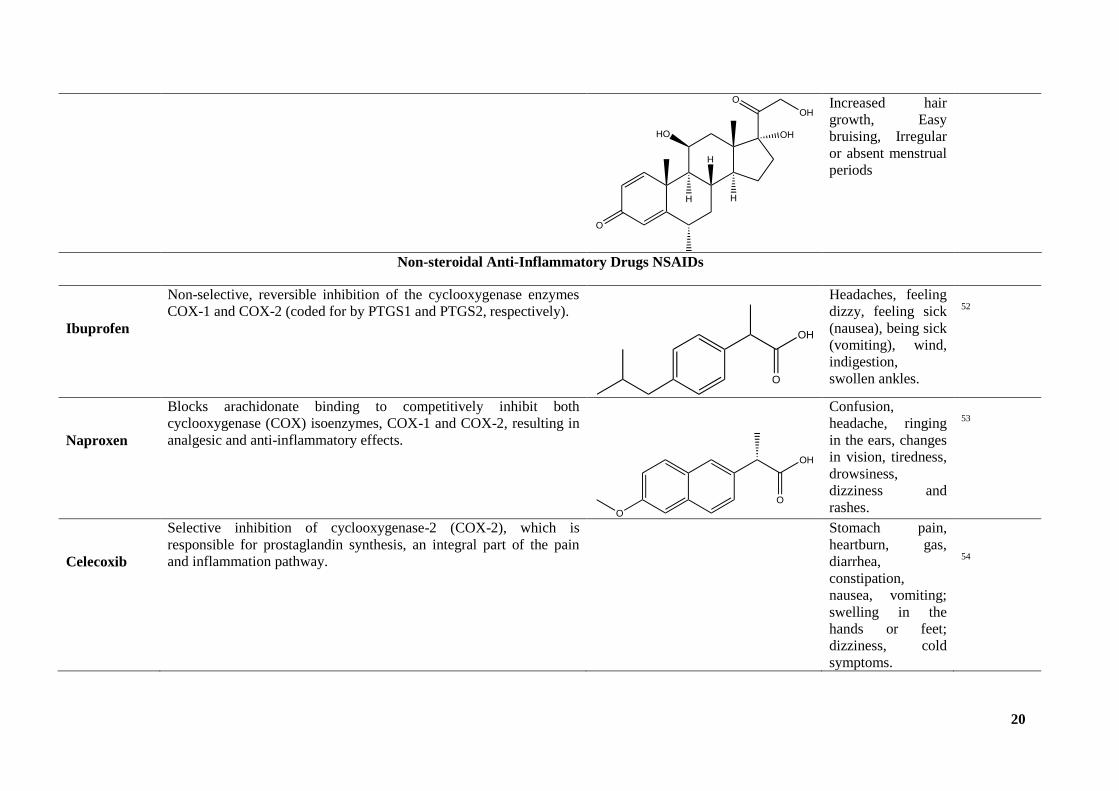
Figure 17: UFLC-PDA equipment. ............................................................................ 41
Figure 18: Microplate used in the anti-inflammatory activity assay. ......................... 42
Figure 19; Microplate reader for the cytotoxic activity evaluation. ........................... 44
Figure 20: Phenolic compound´s profile of Z. officinale ............................................ 49
Figure 21: Phenolic compound´s profile of B. dioica. ................................................ 50
Figure 22: Anti-inflammatory activities of the Z. officinale and B. dioica, and their
combination. ................................................................................................................. 54
Figure 23: Dose-response curve for Z. officinale (A) and B. dioica (B) hydroethanolic
extract. .......................................................................................................................... 58
Figure 24: Cell cycle profile of the different samples: (a) control test, (b) Z. officinale
and (c) B. dioica ........................................................................................................... 61
Figure 25: Apoptotic effect of (a) control test , (b) Z. officinale, (c) B. dioica........... 63

vi
List of Tables
Table 1: Examples of chronic diseases related with inflammation. ............................ 15
Table 2: Mechanism of action, Structure and Side effects of some steroidal and non-
steroidal anti-inflammatory drugs. ............................................................................... 19
Table 3: In vitro anti-inflammatory activity of natural matrices. ................................ 26
Table 4: Chromatographic responses, tentative identification, and quantification of
the phenolic compounds found in the hydroethanolic extracts of Z. officinal ............. 50
Table 5: Organic acid´s composition (g/100g dw) of Z. officinale and B. dioica. ...... 52
Table 6: Anti-inflammatory activities of the Z. officinale and B. dioica, and their
combination. ................................................................................................................. 53
Table 7: Cytotoxic activities (GI50; μg/mL) of Z. officinale and B. dioica. ............. 56
Table 8: iNOS activity (IC50; μg/mL) of Z. officinale and B. dioica. ....................... 59
Table 9: Cellular antioxidant activity of Z. officinale and B. dioica ......................... 60
Table 10: Cell cycle analysis of Z. officinale, B. dioica and control test ................. 61
Table 11: Apoptotic effect of Z. officinale , B. dioica,and control test ...................... 62

vii
List of Abbreviations
AAPH 2.2’-Azobis (2-methylpropionamidine) dihydrochloride
ACLY ATP citrate lyase
AGS Adenocarcinoma gastric cell line
AKI Acute kidney injury
ATF3 Activating transcription factor 3
BL Burkitt’s lymphoma
C Component
CD Crohn's disease
CE Cholesteryl ester
CIC CItrate carrier
CNS Central nervous system
COX Cyclooxygenase
CS Citrate synthase
DAD Diode array detector
DCF-DA Dichlorofluorescin diacetate
2-DG 2-Deoxyglucose
DHA Docosahexaenoic acids
DMEM Dulbecco's modified eagle medium
DMM Dimethyl malonate
DMSO Dimethyl sulfoxide
OD Optical density
DW Dry weight
EAE Experimental autoimmune encephalomyelitis
EC50 Concentration achieving 50% of the Inhibition
ECACC European collection of authenticated cell culture
EPA Eicosapentaenoic
F Fumarase
FITC Fluorescein isothiocyanate
GAPDH Glyceraldehyde 3-phosphate dehydrogenase
GI50 Growth inhibitory dose 50%
GMP Guanosine monophosphate

viii
GluT-1 Glucose transporter-1
HAT Histone acetyltransferase
HBSS Hanks balanced saline solution
HDAC Histone deacetylases
HPLC High performance liquid chromatography
HeLa Cervical carcinoma cell line
HepG2 Hepatocellular carcinoma cell line
Hif-1α Hypoxiainducible factor 1-alpha
ICAM Intercellular adhesion molecules
IDH Isocitrate dehydrogenase,
IFN Interferon
IL Interleukine
iNOS inducible nitric oxide synthase
IRG-1 Immune responsive gene-1
Ig Immunoglobulin
L Leukotriene
LOX Lipoxygenase
LPS Lipopolysaccharides
M-CSF Macrophage colony stimulating factor
MCF-7 Breast carcinoma cell line
MCP-1 Monocutyes chemoattractant protein-1
MDH Malate dehydrogenase
MS Mass spectrometer
NCI-H460 Lung carcinoma cell line
ND Not detected
NED N-(1-naphthyl)ethylenediamine
NF-kB Nuclear factor-kappa-B
NK Natural killer
NO Nitric oxide
NSAID Non steroidal anti-inflammatory drugs
PAF Platelet activating factor
PARP Poly(adp-ribose) polymerase
PBS Phosphate buffered saline

ix
PDB Protein data bank
PG Prostaglandin
PKM2 Pyruvate kinase M2
PLP2 Porcine liver primary cell line
PTGS Prostaglandin endoperoxide synthase
PUMA P53 up-regulated modulator of apoptosis
RA Rheumatoid arthritis
RMSD Root mean square deviation
ROS Reactive oxygen species
RPMI Roswell park memorial institute
SD Standard deviation
SDH Succinate dehydrogenase
SLE Systemic lupus erythematosus,
SRB Sulforhodamine B
SUCNR1 Succinate receptor 1
TCA Trichloroacetic acid
TEPP-46 Thieno pyrrole pyridazinones
TGF Transforming growth factor
TLR Toll like receptor
TNF-α Tumour necrosis factor-alpha
Tr Traces
Th T helper
UFC Unit forming colony
UFLC Ultra fast liquid chromatography
VCAM-1 Vascular cell adhesion molecule-1
Vero Fibroblast-like from African green monkey kidney cell line

x
Abstract
Inflammation is a defense mechanism designed to eliminate microorganisms,
among other agents, to protect living tissues from infections, injuries, and to enhance
tissue repair mechanisms. Non-steroidal anti-inflammatory drugs and synthetic
glucocorticoids are widely used, but unfortunately, they have been associated with
many side effects, namely the increased risk of upper gastrointestinal complications.
Therefore, the discovery of new, efficient, and safer anti-inflammatory agents is
crucial to prevent and treat inflammatory processes. Natural matrices, such as tubers,
have been explored and described as having a huge diversity of bioactive molecules
with anti-inflammatory capacity and with few undesirable effects. Thus, in the present
work, the hydroethanolic extracts of two tubers, Zingiber officinale Roscoe (Ginger)
and Bryonia dioica Jacq. Were studied as potential anti-inflammatory agents.
The main objective of this work was to validate the anti-inflammatory activity of
these tubers through two anti-inflammatory assays in vitro: i) inhibition of the
production of nitric oxide (NO) in a cell model, RAW 264.7 - stimulated rat
macrophage cell line by lipopolysaccharide (LPS); ii) inhibition of the activity of the
inducible nitric oxide synthase (iNOS) enzyme. To ensure the safe use of these
extracts, cytotoxic activity in tumor and non-tumor lines and cellular antioxidant
activity were evaluated. Cell cycle analysis and apoptosis were also assessed using
flow cytometry techniques. In addition, these extracts were characterized in terms of
organic acids and phenolic compounds through chromatographic techniques to
establish a structure-activity relationship.
According to the results obtained, the two tubers revealed the presence of
organic acids, well described bioactive molecules, and four phenolic compounds were
also identified in Z. officinale. Both samples are promising anti-inflammatory agents,
since they exhibited the ability to inhibit NO production with very low IC50 values. It
should be noted that B. dioica showed an anti-inflammatory and cytotoxic activity
more effective than the standards used. The results of antioxidant activity were less
promising, as both samples were less effective than quercetin. The results of the cell
cycle show that B. dioica suppresses the G0/G1 phase, while apoptotic analysis
indicates that Z. officinale has a greater capacity to induce cell death by the apoptotic
pathway. These results are very promising and highlight the anti-inflammatory

xi
potential of these tubers, however further tests are needed to evaluate other
inflammatory mediators and confirm in vivo their efficacy and safety.

xii
Resumo
A inflamação é um mecanismo de defesa concebido para eliminar
microrganismos,entreoutros agentes, para proteger os tecidos vivos de infeções,
lesões, e para potenciar os mecanismos de reparação tecidular. Os anti-inflamatórios
não esteroides e glucocorticoidessintéticos são muito utilizados, mas infelizmente têm
sido associados a muitos efeitos secundários, nomeadamenteao risco acrescido de
complicações gastrointestinais superiores. Portanto, a descoberta de agentes anti-
inflamatórios novos, eficientes e mais seguros é crucial para prevenir e tratar
processos inflamatórios. As matrizes naturais, como os tubérculos, têm sido
exploradas e descritas como detentores de uma enorme diversidade de moléculas
bioativas com capacidade anti-inflamatória e com poucos efeitos indesejáveis. Assim,
no presente trabalho, os extractoshidroetanólicos de dois tubérculos, Zingiber
officinale Roscoe (Ginger) e Bryonia dioica Jacq. foram estudadoscomo potenciais
agentes anti-inflamatórios. O principal objetivo deste trabalho foi a validação da
atividade anti-inflamatória destes tubérculos através de dois ensaios anti-inflamatórios
in vitro: i) inibição da produção de óxido nítrico (NO) num modelo celular,RAW
264.7–linhacelularde macrófagos deratoestimulado por lipopolissacarídeo (LPS); ii)
inibição da atividade da enzima óxido nítrico sintaseindutível (iNOS). Para garantir a
segurança da utilização dos referidos extratos foi avaliada a atividade citotóxica em
linha tumorais e não tumorais e a atividade antioxidante celular. A análise do ciclo
celular e a apoptose também foram avaliadas usando técnicas decitometria de fluxo.
Além disso, estes extratosforam também caracterizados em termos de ácidos orgânicos
e compostos fenólicos através de técnicas cromatográficas a fim de estabelecer uma
relação estrutura-atividade. De acordo com os resultados obtidos, ambas as amostras
revelaram a presença de ácidos orgânicos, moléculas de reconhecida bioatividade,
tendo sido ainda detetados 4 compostos fenólicos na amostra Z. officinale. Os dois
tubérculos são agentes anti-inflamatórios promissores, já que exibiram capacidade
para inibir a produção de NO com valores muito baixos de IC50. De salientar que a
B.dioica mostrou uma atividade anti-inflamatória e citotóxica mais eficaz do que os
padrões utilizados. Os resultados daatividade antioxidante foram menos promissores,
já que ambas as amostras foram menos eficazes do que a quercetina. Os resultados do
ciclo celular mostram que a B.dioica suprime a fase G0/G1, enquanto a análise
apoptótica indica que a Z. Officinale apresenta maior capacidade para induzir morte

xiii
celular pela via apoptótica. Estes resultados são muito promissores e realçam o
potencial anti-inflamatório destes tubérculos, no entanto é necessário fazer mais
ensaios para avaliar outros mediadores inflamatórios e confirmar in vivo a sua eficácia
esegurança

1
1. Introduction
Inflammation is a defense mechanism that is designed to eradicate microbes or
irritants to protect living tissues from infection, injuries, and to potentiate tissue repair.
This process leads to changes in blood flow, an increase in permeability of blood
vessels, and the migration of fluid, proteins, and white blood cell (leukocytes) from the
circulation system to the site of the damaged tissue. If the inflammatory response last
for few days it is called acute inflammation however if it last for longer time it is
referred as chronic inflammation and can cause physiological decompensation, organ
dysfunction and death1.
The inflammation process is characterized by five major stages: i) rubor
(redness, due to hyperemia); tumor (swelling caused by increased permeability of the
microvasculature and leakage of protein into the interstitial space); ii) calor (heat
associated with the increased blood flow and the metabolic activity of the cellular
mediators of inflammation); iii) dolor (pain, in part due to changes in the peri-
vasculature and associated nerve endings); and iv) Functiolaesa (dysfunction of the
organs involved)2.
The common anti-inflammatory drugs can calm the symptoms or limit the
deleterious effects of inflammation in the organism. Usually, there are two types of
anti-inflammatory drugs, the non-steroidal and glucocorticoids. These drugs can be
applied in different forms, namely by oral treatment, suppository, inhalation, infusion
or local by ointment, eye drops among others); however these anti-inflammatory drugs
have serious side effects3. The use of oral steroids, aspirin and acetaminophen at doses
of approximately 2 g each is associated with a double increase in the risk of upper
gastrointestinal complications; non-steroidal anti-inflammatory drugs are associated
with a nearly fourfold increase in risk 3.
Therefore, it is crucial to find new and efficient anti-inflammatory drugs that
can eliminate the infection without undesired side effects. Natural products have been
explored and described as possessing a huge diversity of bioactive molecules with
anti-inflammatory capacity 4. Thus, in this study we are going to evaluate the
bioactivities of B. dioica and Z. officinale as promising anti-inflammatory agent susing
the hydro-ethanolic extract.
Beside the anti-inflammatory activity, we are going to test the cytotoxicity, NOS
activity, CAA, the cell cycle and the apoptosis and determine the phenolic compound

2
and the organic composition to understand better the mechanisms of action of both
samples as anti-inflammatory agents.
1.1. Inflammatory mechanisms
1.1.1. Initiation of the inflammatory response
1.1.1.1 Vasodilatation, fluid exudation and leukocyte migration
Vasodilatation is a typical characteristic of acute inflammation, which is
clinically characterized at the injury site by redness which warmth. The vasodilatory
response aims at promoting the local delivery of soluble mediators and inflammatory
cells. Inflammation-induced vasodilatation is mainly mediated by vasodilating
prostaglandins and NO. Upon exposure to microbial products or pro-inflammatory
cytokines, the activated leukocytes develop inducible NOS (iNOS). The NO provided
by cyclic GMP-dependent mechanisms induces subsequent smooth muscle relaxation.
Prostacycline (PGI2), PGD2, PGE2 and PGF2a are the main vasodilatory
prostaglandins (Figure1) 1. These prostaglandins are formed by the phospholipase
and cyclooxygenase activities5.
Vasodilatation caused by inflammation first requires arterioles accompanied by
the formation of new micro-vascular beds. Widespread vasodilatation can induce
systemic hypotension and shock in cases of severe systemic inflammation such as
sepsis. Such physiological modifications are potentiated by myocardial depression
caused by the sepsis stage, a disease triggered by the activities of NO and pro-
inflammatory cytokines such as tumour necrosis factor- (TNF-) 6 (Figure1).

3
Figure 1: The production of vasodilatory prostaglandins through the actions of
phospholipase and cyclooxygenase. The major vasodilatory prostaglandins are
prostacyclin (PGI2) and the prostaglandins PGD2, PGE2 and PGF2a. COX, cyclo-
oxygenase; PG, prostaglandin.1
At the same time, capillary hydrostatic pressure at the injury site is increased
early during inflammation or damage triggered by local vasodilatation. Outpouring of
protein-rich fluid induces erythrocyte aggregation in small vessels and increase blood
viscosity. This flow of trans-vascular fluid gradually returns to normal intravascular
pressure at the inflammation site. At the same time, plasma protein loss reduces
oncotic pressure intravascularly1.
The increase in vascular permeability, the temporary rise in capillary hydrostatic
pressure and the decrease in plasma oncotic pressure work together to induce a trans-
vascular fluid and protein flow into the inflamed interstitium. The role of these
alterations is to facilitate the delivery to the injury site of soluble factors such as
antibodies and acute-phase proteins. However, extreme systemic inflammation can
cause excessive increases in vascular permeability, which can contribute to the
development of oedema in the lungs and extremities. In critically ill patients, the
accumulation of fluid in the lungs causes the acute respiratory distress syndrome, a
significant source of morbidity and death 7.
Leukocyte margination, adhesion, and migration are followed by vasodilatation
and fluid exudation. Neutrophils are the first and most numerous leukocytes to appear
in an infection or inflammatory site. The process of neutrophil movement from
intravascular space into the inflamed interstitium occurs mainly in the systemic
circulation of postcapillary venules and in the lung's pulmonary capillaries. The
process of transmigration is categorized into several distinct stages, namely

4
margination, spinning, adhesion, diapedesis and chemotaxis 8 (Figure 2).The process
of neutrophil migration from the main bloodstream to the vessel's periphery is
denomited marginalisation. Stasis after fluid exudation at the site of inflammation and
physical contact between erythrocytes and neutrophils facilitates this process. A poor
adhesive association occurs after margination between neutrophils and endothelial
vascular cells, allowing neutrophils to linger in close proximity to the vascular
endothelium. Neutrophil rolling is encouraged by the shear stress of moving
erythrocytes, the rolling speed being proportional to the velocity of red cells 9.
These interactions are mediated by selectins and their ligands, promoting rolling
of the neutrophils. The adherence to neutrophils is then potentiated by interactions
with beta-integrins and intercellular adhesion molecules (ICAM). Neutrophils then
move by contact with chemoattractant molecules such as chemokines and bacterial
products to the site of the inflammation 10
.
Figure 2: Neutrophil rolling processes, adhesion system, diapedesis and
chemotaxis.The formation of loose connections between endothelial cells and
neutrophils is caused by inflammation, which produces neutrophil margination.
Selectins and their ligands moderate these connections, allowing neutrophils to roll
more easily. Interactions between beta-integrins and ICAM then enhance neutrophil
adherence. After interacting with chemoattractant molecules such chemokines and
bacterial products, neutrophils move to the site of inflammation.1.
1 2

5
1.1.1.2 Activation of the coagulation cascade during inflammation
Inflammation is closely associated with coagulation. The cascade of coagulation
occurs after skin damage and during an infection process. It is divided into two
pathways which converge and ultimately cause thrombin activation with the
subsequent fibrinogen cleavage into fibrin (Figure 3). The intrinsic pathway is a
sequence of plasma proteins regulated by Hageman factor (factor XII), a synthesized
protein in the liver that is regulated by binding to collagen, basement membrane or
activated platelets 11
. Enabled Hageman factor activates a cascade of proteins to start
resulting in thrombin production. Guided tissue pain most commonly stimulates the
intrinsic pathway.
The extrinsic pathway, by comparison, is triggered by the development of factor
tissue. Recent studies suggest that the extrinsic pathway is the primary coagulation
pathway that is triggered during infection and systemic inflammation, particularly
during sepsis and the systemic inflammatory response syndrome 12
.Tissue factor is
exerted on surfaces of tissues that are not usually exposed to the vascular
compartment, such as subcutaneous tissues and blood vessel adventitial layers. In
addition, endothelial cells and activated monocytes develop tissue factor in response to
TNF-a, IL-1, IL-6 and C-reactive protein during cycles of inflammation 13
. The
presence of tissue factor triggers the activation of factor VII, which then forms a
complex with tissue factor and eventually induces the production of thrombin by
stimulating a variety of coagulation factors (Figure 3).

6
Figure 3 : The coagulation cascade. The extrinsic and intrinsic coagulation pathways
combine to form a final common pathway. Tissue factor, which is released in response
to tissue damage and macrophage activity, activates the extrinsic pathway. The intrinsic
pathways are activated when sub-endothelial collagen and activated platelets are
accessible. The development of a fibrin clot is the final outcome.1.
Coagulation cascade activation is not only important for the development of
fibrin clots but it also has major effects on the pro-inflammatory response. Pro-
inflammatory activity has been shown to be caused by factor Xa, thrombin, and tissue
factor – VIIa complex In particular, thrombin and the tissue factor – VIIa complex that
induce mononuclear and endothelial cells to produce pro-inflammatory cytokines such
as TNF-14
. This influence tends to be mediated by binding certain factors on the
surface of target cells to protease-activated receptors. Acute inflammation then allows
the coagulation cascade to start, which can then potentiate the inflammatory response
further.
1.1.2. Complement system
The complement system is a collection of microbial-activated proteins that help
to facilitate inflammation and microbial degradation. The nutrient cascade is also
likely to be activated during tissue injury, which plays a role in cell damage associated
with severe injuries which burns 15
. There are three methods that trigger the

7
complement cascade: the classical pathway, the alternative pathway, and the lectin
pathway, each pathway is activated through different mechanism, the classic pathway
is activated by IgM or IgG antibodies that attach to the surface of microbes, the
alternative pathway is activated directly by microbial surface molecules that bind the
C3 component supplement, the lectin pathway is activated by mannose-binding lectin
that interacts with microbial glycoproteins and glycolipids. (Figure 4). 1.
Figure 4: The complement system Several mechanisms, including the classic,
alternative, and lectin pathways, activate the complement cascade. C3a and C3b,
which act as pro-inflammatory mediators, are the most important mediators of the
complement system. Membrane disruption is also caused by the development of the
membrane attack complex (C5-C9)1.
Any of these paths will enable the cleavage into C3a and C3b of the complement
portion. The C3a acts as chemo-attractant for neutrophils and C3b binds to the surface
of the microbes to promote phagocyte detection and enable phagocytosis16
.
Furthermore, C3b forms a proteolytic complex with other complementary components
to allow C5 to be cleaved into C5a and C5b 17
. C5a is a chemotactic driver for
neutrophils and also affects vascular permeability at the inflammatory site. The C5b
attaches to the microbial surface and promotes the development of the membrane
attack complex consisting of C6, C7, C8 and C9 18
. Complex membrane attack
induces microbial cell membrane destruction and eventual death.

8
1.1.3. Amplification of the inflammatory response
1.1.3.1 Innate immune system
Immune response may be split into natural and adaptive responses to tissue
injury or infection. The primary response to tissue invasion is supported by the innate
immune system. The phases of vasodilatation described above, increased vascular
permeability and cellular infiltration form part of the innate immune response.
Macrophages, dendritic cells, natural killer cells (NK) and neutrophils are the main
cell components of the innate immune system. Besides these cellular elements,
circulating effectors proteins such as complement, acute-phase reactants and the
cascade of coagulation play important roles in innate immunity 1.
The extent of innate reaction is primarily determined by the production of
cytokines and non-cytokine inflammation mediators. Cytokines are polypeptides
formed by immune system cells in response to an infection or damage to the tissue.
They work to control inflammatory and immune reactions. Generally speaking,
cytokine production is self-limited although certain cytokines can remain in circulation
for long periods of time. What's more, cytokine reactions are pleiotropic and
repetitive. TNF- is prototypical pro-inflammatory cytokine 19
. TNF- is mainly
activated by macrophages within minutes of local or systemic damage, and modulates
a number of immunological and metabolic events. TNF- initiates an inflammatory
response at sites of local infection or inflammation, which stimulates anti-microbial
resistance mechanisms and replaces tissue until the infection has been eradicated 20
. It
is a potent neutrophil and mononuclear phagocyte activator that also acts as a growth
factor for fibroblasts and an angiogenesis factor.
These PAF, a phospholipid autocoid produced by endothelial cells that controls
the release of cytokines and amplifies the pro-inflammatory response, has been
implicated in many non-cytokine inflammatory processes21
. The adhesion of
neutrophils to endothelial cells seems to be an important factor. PAF's extended
involvement in serum of SIRS patients has associated with negative outcome.
Eicosanoids are metabolites of arachadonic acid which regulates many aspects of the
immune response. Leukotrienes (LTC4–LTE4) allow the endothelial cells to contract
and promote capillary leakage. Thromboxane A2, a catalyst originating from the
macrophage and platelets, causes platelet accumulation, vasoconstriction, and likely
tissue thrombosis 1.

9
1.1.3.2 Acquired immune response
Often, the innate immune response helps to activate and enhance the immunity
acquired. This influence is mainly mediated by IL-12, which induces T-cell activation
and facilitates the segregation of T-cells naive into the Th1 phenotype22
. However, the
adaptive immune response is mainly triggered by the introduction of foreign antigens
to T-cells CD4 + and CD8+ T. Activation of CD4+ T-cells triggers further
development of cytokines and amplifies the innate and acquired immune systems. At
the time of antigen presentation, the different cytokines generated by CD4+ cells are
dependent upon the immunological microenvironment.
CD4 T-cells are the best-defined subsets of Th1 and Th2 cells. Those subsets are
characterized primarily by the cytokines they generate. IFN- is the principal cytokine
formed by Th1 cells, In fact, IFN- amplifies the pro-inflammatory reaction by
inducing activation of the macrophage and activating the cytolytic functions of CD8
T-cells 23
. IFN- also activates B-cells to develop IgG1 and IgG3 antibodies that are
opsonizing and complement binding 24
. Helminths and the susceptibility to allergens
induce the differentiation of Th2. Such stimuli induce sustained stimulation of T-cells
without an significant innate immune response or activation of the macrophage.
Two more T-cell subsets are Th3 and T-regulatory 1 (Tr1) cells. Th3 cells
produce TGF-b and play an important role in the development of immune tolerance,
particularly after exposure to antigens supplied via the gastrointestinal tract 25
.
1.2. Relationship between inflammatory process and chronic
diseases
1.2.1. A generic model of chronic inflammatory disease
Several diseases such as type 2 diabetes, obesity, Alzheimer’s disease, heart
disease and allergies have few common factors; first and foremost, they are all
primarily lifestyle diseases and are usually related to chronic inflammation.
A generic model of chronic inflammatory disease is proposed to change the
traditional thoughts that have claimed the chronic diseases of various organ systems as
their own, and probed the pathophysiology in a reductionist manner, however these
chronic diseases have more mechanisms in common than usually recognized so this
model highlights these shared phathophysiologic mechanisms26
.

10
The epithelial and mesenchymal cells of the affected organs are two prototypic
cell types induced by signals resulting from interactions between the innate and
adaptative immune systems (Figure 5). These signals induce tissue responses such as
recruitment of leukocytes involved in chronic inflammation, extracellular matrix
remodeling, cellular proliferation or death, and angiogenesis (Figure 5)26
.
Figure 5: Common inflammatory and immune processes on different cell
types.Inflammatory and immune activity alters the function of various types of
epithelial cells such as endothelial cells, synoviocytes, enterocytes,
glomerular/tubular epithelial cells, and bronchoalveolar cells, and each cell type
characteristically participates in the development of a different disease
(atherosclerosis, arthritis, inflammatory bowel disease, kidney disease, and lung
disease, respectively). Inflammatory and immune processes may also act on different
types of mesenchymal cells (e.g., smooth muscle cells, fibroblasts, myofibroblasts,
mesangial cells, or pericytes), leading to the development of chronic disease
depending on the specific cells targeted.26
.
Atherosclerosis, interstitial lung disease, rheumatoid arthritis and cirrhosis share
the same fundamental mechanisms and mediators drive the disease process; however
they act in totally different ways 27,28
. In many organs, helper T-cell bound in the
lesions of chronic inflammation including atherosclerotic plaques, forms of chronic
hepatidites, rheumatoid synovium and in a number of pulmonary diseases.
Mononuclear phagocyte, histocyte, microglia, or alveolar macropages are also found
in such lesions. Every tissue involved has specific epithelial cells: the vascular
endothelial cells in atherosclerosis; the glomerular or tubular epithelial cell in renal
disease; and enterocytes in inflammatory bowel diseases. Similarly, depending on the
organ involved, inflammatory and immune mechanisms have different types of

11
mesenchymal cells, arterial smooth muscle cells, fibroblasts, myofibroblasts,
synoviocytes, pericytes or mesangial cells. The first step of inflammation involves
selective and sequential migration of blood cells into tissues and the second step is the
local activation and interaction of these blood-based cells with resident tissue cells.
Only some limited elements of this classic inflammatory process can be displayed by
some conditions. However, the key inflammatory mediators dominate but without the
context of the classic inflammatory mechanisms.
In osteoporosis, mediators such as IL-1, IL-6 and TNF- are primarily
furnished by resident stromal cells to regulate bone turnover unaccompanied by other
components of the classic innate immune response. A normal host defense mechanism
can coax into an injurious response due to a persistent stimulus. Infection due to a
pyogenic microbial pathogen engenders an acute leukocyte response to clear the
invading organism.
1.2.2. Examples of chronic diseases related with inflammation
There are numerous chronic diseases related with inflammation (Table1).
Nevertheless, some diseases are considered more relevant as they cause irreversible
damage to the human organisms.
1.2.2.1 Relationship between inflammation and atherosclerosis
Atherosclerosis is a disease in which the inside of an artery narrows due to the
buildup of plaque (Figure 6). In initial stages there are generally no symptoms, but in
advanced stages, depending on the type and place of the affected arteries, it can result
in different complication such as: coronary artery disease, stroke, peripheral artery
disease, or kidney problems28
.

12
Figure 6: Figure A shows a normal artery with normal blood flow. The inset image
shows a cross-section of a normal artery. Figure B shows an artery with plaque
buildup. The inset image shows a cross-section of an artery with plaque buildup28
.
From a deep point of view, vascular endothelial cells resist to prolonged contact
with leucocytes; however they express a palette of vascular cell adhesion molecule-1
(VCAM-1) and members of the selectin family, P- and E-selectin, when they are
exposed to an activating stimulus such as modified lipoproteins, microbial
constituents or pro-inflammatory cytokines29
; Monocytes migrate directly into artery
wall after adhesion to the endothelial surface; this directed migration is mediated by
chemokines such as monocutyes chemoattractant protein-1 (MCP-1)30
. Once resident
in the arterial intima, these monocytes differentiate into macrophages and proliferate
after exposition to such activating and co-mitogenic mediators as macrophage colony-
stimulating factor (M-CSF)31
,. These macrophages over-express scavenger receptors
and through endocytosis they engulf modified lipoprotein particles, then they
accumulate cholesteryl ester (CE) in cytoplasmic droplets creating foam cells,
considered a hallmark of the nascent atherosclerotic plaque32
.
The inflammatory and immune process contribute to acute thrombotic which is
one o the ultimate complications o atherosclerosis30
. Inflammation within the
arterialintima leads to signal exchange among the T-cell, mononuclear phagocyte, and
cells of the vessel wall. This pathway leads to a weakening of the fibrous plaque
through decreased collagen synthesis or increased degradation. IFN- mediates
decreased synthesis while increased breakdown is a result of proteinases induced by

13
inflammatory signaling. CD40 ligation augments the thrombogenecity of the lipid core
by expression of tissue factor, a major trigger of thrombus formation (Figure7)33
.
Figure 7: Simplified model illustrating the effects of inflammation on the integrity
of the plaque fibrous cap signaling between T cells, mononuclear phagocytes, and
vessel wall cells occurs when the arterial intima is inflamed. Through decreased
collagen synthesis or increased breakdown, this mechanism causes the fibrous
plaque to weaken. IFN-mediates decreased synthesis while increased breakdown is
a result of proteinases induced by inflammatory signaling. The production of tissue
factor, a key activator of thrombus formation, is enhanced by CD40 ligation, which
increases the thrombo-genecity of the lipid core26
.
1.2.2.2 Relationship between inflammation and Alzheimer’s disease
Alzheimer’s disease is a progressive chronic neurodegenerative disease that get
worsens over time. This chronic disease is characterized by cognitive decline and the
presence of two core pathologies, amyloid β plaques and neurofibrillary tangles 34
.
Figure 8: Comparison between healthy and Alzheimer affected brain; the neuron of
the affected brain contain neuro-fibrillary tangles and amyloid plaques 34
.

14
In pathologically vulnerable regions of Alzheimer’s disease, clearly local
peripheral inflammatory responses with full complexity were found. The inflammation
is stimulated by degenerating tissue and deposition of highly insoluble abnormal
materials, likewise damaged neurons, and neurites and highly insoluble
amyloid β peptide deposits and neurofibrillary tangles provide obvious stimuli for
inflammation. These stimuli are discrete, microlocalized, and present from early
preclinical to terminal stages of Alzheimer’s disease that’s why local upregulation of
complement, cytokines, acute phase reactants, and other inflammatory mediators are
also discrete, microlocalized, and chronic. Direct and bystander damage from
Alzheimer’s disease inflammatory mechanisms are cumulated over many years and
are likely to significantly give rise to pathogenic processes. Clinical studies and
animal models strongly suggest that Alzheimer’s disease inflammation significantly
contributes to Alzheimer’s disease pathogenesis 35
.

15
Table 1: Examples of chronic diseases related with inflammation.
Disease Description Inflammatory mechanism Treatment Reference
s
Chronic
obstructive
pulmonary
disease
(COPD)
A chronic
inflammatory lung
disease that causes
obstructed airflow
from the lungs.
Both innate (macrophages/neutrophils) and adaptive inflammatory
immune cells (CD4, CD8 and B lymphocytes) which developed
lymphoid follicles increased the tissue volume of the bronchial wall
characterized by infiltration of the wall.
Phosphatidylcholine
(PC)(naturel) 18:1 PC (cis), 1,
2-dioleoyl-sn-glycero-3-
phosphocholine (DOPC)
(synthetic)
36
Alcoholic
fatty liver
disease
(AFLD)
a build-up of
fats in the liver
caused by drinking a
large amount of
alcohol.
Expression of the following inflammatory molecules in the
liver: tumor necrosis factor α (TNF-α), monocyte chemotactic protein
1 (MCP-1), chemokine (C-X-C motif) ligand 1 (CXCL-1) and
interleukin 1 beta (IL-1β).
Phosphoesterase complex
(Pho)
37
Obesity a complex
disease involving an
excessive amount of
body fat.
Abnormal cytokine production, increased synthesis of acute-
phase reactants and activation of inflammatory signaling pathways
Weight lost physical activities 38
Chronic
kidney disease
a long-term
condition where the
kidneys don't work
as well as they
Activation of the prototypical proinflammatory signaling
pathway, the best characterized being NF-κB and AP-1, mainly based
on the stimulation of multiple mediators, including proinflammatory
cytokines such as interleukin-1 (IL-1) and tumour necrosis factor α
Ramipril, Enalapril, Lisinopril.
Atorvastatin ,Simvastatin,
VitaminD, furosemide,
cyclophosphamide.
39

16
should. (TNF-α).
Autoimmune
diseases
(SLE,
RA, Sjogren's
Syndrome)
A condition in which
the immune system
mistakenly attacks
the body.
Substitution of the AU-rich element (ARE) in the IFN- 3 'un-
translated region (called ARE-Del) with random nucleotides which
results in a weak but chronic expression of IFN-γ.
Nonsteroidal anti-
inflammatory drugs (NSAIDs),
such as ibuprofen (Motrin,
Advil) and naproxen
(Naprosyn).
40
Myocardial
infarction
(MI)
A heart attack,
occurs when blood
flow decreases or
stops to a part of the
heart, causing
damage to the heart
muscle.
inhibition of the signaling pathways of nuclear transcription
factor κB (NF-κB), p38, c-Jun NH2-terminal kinase (JNK) and
transforming growth factor β (TGF-β).
Thrombolytics, Aspirin,
Nitroglycerin, Beta-blockers,
ACE inhibitors.
41
Psoriasis A skin
condition that causes
red, flaky, crusty
patches of skin
covered with silvery
scales.
Heightened innate and adaptive immune activation.
T helper (Th)1 and Th17 cells drive pro-inflammatory
cytokines including TNFα, interferon-γ, IL17A, and IL23
Anti-TNFα therapy,Topical –
creams and Ointments,
Phototherapy.
42

17
1.3. Treatment of inflammatory process
1.3.1. Corticosteroids
Corticosteroids are a class of steroid hormones that are produced in the
adrenal cortex of vertebrate. Two main classes of corticosteroids, namelly
glucocorticoids and mineralo-corticoids, are involved in the regulation of
inflammatory response.
This class of drugs represents a natural starting point for empirical anti-
inflammatory therapy. The most broadly active anti-
inflammatory/immunosuppressive agents in clinical use are the glucocorticoids43
.
Suppression of neutrophil adherence, aggregation, phagocytosis and accumulation,
suppression of monocyte accumulation, inhibition of prostaglandin and leukotriene
production, lympholysis, inhibition of immunoglobulin production, and impairment
of delayed hypersensitivity are known as specific actions of corticosteroids.
Steroids have been widely used in the treatment of idiopathic inflammatory
diseases of the central nervous system (CNS), including lupus cerebritis and
temporal arteritis, as also to treat multiple sclerosis. Nevertheless, there are serious
drawbacks to clinical trials of this class of drugs in Alzheimer’s disease.
A high doses of corticosteroids can cause toxic effects such hypertension,
hyperglycemia, fluid retention and exacerbation of con- gestive heart failure,
psychiatric syndromes including psychosis, mania, and depression, gastrointestinal
ulceration or perforation, increased susceptibility to infection, osteoporosis with
vertebral compression fractures, aseptic necrosis of bone, myopathy, truncal
obesity, ac- celerated atherogenesis, glaucoma, adrenal suppression, and cataracts44
.
1.3.2. Non-steroidal Anti-Inflammatory Drugs
These drugs are members of a drug class that reduces pain, decreases
fever, prevents blood clots, and decreases inflammation in higher doses.
The first line drugs for inflammatory diseases are non-steroidal anti-
inflammatory drugs such as rheumatoid arthritis and gout. Their main role is the
inhibition of neutrophil function and prostaglandin synthesis45
. Non-steroidal anti-
inflammatory drugs interact primarily with pro-inflammatory cytokines interleukin
(IL)-1a, IL1b, IL-6 and tumor necrosis factor (TNF-α)46
. The cardinal signs of
inflammation occur because of the increase of TNF-α concentration; they also

18
stimulate white cell phagocytosis and the production of inflammatory lipid
prostaglandin E2 (PGE2). The major mechanism that leads to the success of these
medications is their ability to interfere with the production of prostaglandin during
the inflammatory cascade (Figure 9)47
.
Figure 9: Block of COX action by the non-steroidal anti-Inflammatory.The NSAIDs
can block COX action and thereby prevent the formation of the COX-derived
inflammatory mediators. 5-HPETE = 5-hydroperoxyeicosatetraenoic acid; LTC4 =
leukotriene C4; PGE2 = prostaglandin E2; PGF2 = prostaglandin F2; PGI2 =
prostacyclin; TXA2 = thromboxane.4
Steroidal and non-steroidal anti-inflammatory medications have a significant
side effect profiles; there is a greater interest in natural compounds, such as dietary
supplement and herbal remedies, which have been used for centuries to reduce
inflammatory response 48
.

19
Table 2: Mechanism of action, Structure and Side effects of some steroidal and non-steroidal anti-inflammatory drugs.
Drug type Mechanism of action Chemical structure Side effects Reference
Corticosteroid drugs
Prednisone
Decreases inflammation through suppression of the migration of
polymorphonuclear leukocytes and reversing increased capillary
permeability. It also suppresses the immune system by reducing the
function and the amount of the immune system.
Nausea,
vomiting,
loss of appetite,
heartburn,
trouble sleeping,
increased sweating,
acne.
49
Cortisone
Switching off multiple activated inflammatory genes through
inhibition of HAT and recruitment of HDAC2 activity to the
inflammatory gene transcriptional complex.
Confusion,
Excitement,
Restlessness,
Headache, Nausea,
Vomiting, Skin
problems including:
acne thin skin
heavy sweating
redness trouble
Sleeping.
50
Methylpredni
solone
The methylprednisolone-glucocorticoid receptor complex binds and
blocks promoter sites of pro-inflammatory genes, promotes
expression of anti-inflammatory gene products, and inhibits the
synthesis of inflammatory cytokines, mainly by blocking the function
of transcription factors, such as nuclear factor-kappa-B (NF-kB).
Upset stomach,
Stomach irritation,
Vomiting,
Headache,
Dizziness
Insomnia,
Restlessness,
Depression,
Anxiety, Acne,
51
O
O OH
H H
H
O
OH
O
O OH
OHO
H H
H
20
Increased hair
growth, Easy
bruising, Irregular
or absent menstrual
periods
Non-steroidal Anti-Inflammatory Drugs NSAIDs
Ibuprofen
Non-selective, reversible inhibition of the cyclooxygenase enzymes
COX-1 and COX-2 (coded for by PTGS1 and PTGS2, respectively).
Headaches, feeling
dizzy, feeling sick
(nausea), being sick
(vomiting), wind,
indigestion,
swollen ankles.
52
Naproxen
Blocks arachidonate binding to competitively inhibit both
cyclooxygenase (COX) isoenzymes, COX-1 and COX-2, resulting in
analgesic and anti-inflammatory effects.
Confusion,
headache, ringing
in the ears, changes
in vision, tiredness,
drowsiness,
dizziness and
rashes.
53
Celecoxib
Selective inhibition of cyclooxygenase-2 (COX-2), which is
responsible for prostaglandin synthesis, an integral part of the pain
and inflammation pathway.
Stomach pain,
heartburn, gas,
diarrhea,
constipation,
nausea, vomiting;
swelling in the
hands or feet;
dizziness, cold
symptoms.
54
O
HO OH
H H
H
O
OH
O
OH
O
OH
O

21
Diclofenac
Inhibition of prostaglandin synthesis by inhibiting cyclooxygenase-1
(COX-1) and cyclooxygenase-2 (COX-2) with relative equipotency.
Headaches,
dizziness, stomach
pain, feeling or
being sick,
diarrhoea and
rashes.
55
Indomethacin
Inhibition of prostaglandin synthesis by inhibiting cyclooxygenase-1
(COX-1) and cyclooxygenase-2 (COX-2)
Vomiting, upset
stomach, heartburn,
diarrhea, a feeling
of bowel fullness,
constipation,
bloating, gas, rectal
irritation, dizziness,
drowsiness,
nervousness.
56
Piroxicam
Inhibition of cyclooxygenase (COX-1 and COX-2). Piroxicam is a
potent inhibitor of prostaglandin (PG) synthesis in vitro.
Abnormal liver
function tests,
NN
F
F
F
S
H2NO
O
HN
Cl
Cl
O
HO

22
urination problems,
upset stomach,
heartburn, loss of
appetite, stomach
pain, nausea,
vomiting, gas,
diarrhea,
constipation;
dizziness,
headache; itching,
rash, ringing in the
ears.
57
S
N
NH
O
N
OH
O O

23
Table 2 shows that corticosteroid and non-steroidal anti-inflammatory drug
molecules contain at least one chemical group in their composition, such as hydroxyl,
methyl, and ketone. This property will assist us in finding a natural matrix that will
substitute these synthetic drugs with lower side effects.
1.3.3. Natural anti-inflammatory agents
Most anti-inflammatory medications were linked with an elevated risk of severe
upper gastrointestinal problems. The level of risk for anti-inflammatory drugs has been
estimated through epidemiological studies. Using oral steroids or low-dose aspirin, the
likelihood of upper gastrointestinal tract leakage or perforation rises by almost twice
as high as with the use of non-aspirin, non-steroidal, anti-inflammatory medicines.
Overall, with more than one anti-inflammatory drug administered concurrently, the
risk is dose dependent and is higher. Therefore, in order to minimize the risk of severe
upper gastrointestinal complications, anti-inflammatory drugs should be prescribed
during mono-therapy and at the lowest effective dose whenever possible 3.
Owing to the substantial side-effect profiles of steroidal and NSAID drugs, there
is a greater interest in natural compounds, such as nutritional supplements and herbal
medicines, which have been used for centuries to reduce pain and inflammation. Many
of these natural compounds often act in a similar fashion to NSAIDs by inhibiting
inflammatory pathways. Many natural compounds function to inhibit the inflammatory
pathways of nuclear factor-kB (NF-kB) in addition to the COX pathway 4.
One of the most effective natural anti-inflammatory agents available is the
omega-3 polyunsaturated fatty acids58
. The American Heart Association for the
prevention of this disease recommends the consumption of fish and fish oil
supplements after the discovery that vascular inflammation is the underlying cause of
coronary artery disease59
.
Eicosapentaenoic (EPA) and docosahexaenoic acids (DHA) are the active
ingredients in fish oil and can enhance the conversion of COX to prostaglandin E3 that
is a natural anti-inflammatory agent. Prostaglandin E3 competitively inhibits the
effects of the arachidonic acid conversion to prostaglandin E2, a highly inflammatory
substance. It also inhibits the synthesis of TNF-α and IL-1b, both of which are
inflammatory cytokines. The 5-LOX pathway can be inhibited by the EPA and DHA,
which converts arachidonic acid to inflammatory leukotrienes, by competitive
inhibition as well4. Omega-3 EFA, found in fish oil, can reduce the inflammation in

24
synovial cartilage by the direct reduce of the degenerative enzymes, aggrecanase and
matrix metalloproteinase, as well as IL-1, TNF-α, and COX260
.
One of the oldest herbal remedies for pain and inflammation is bark from the
white willow tree, dating back to ancient Egyptian, Roman, Greek, and Indian
civilizations, as an analgesic and antipyretic agent. White willow bark and aspirin have
a similar mechanism of action which is the block of inflammatory prostaglandins by
nonselective inhibitor of COX-1 and COX-261
. Various studies comparing white
willow bark with non-steroidal anti-inflammatory drugs have shown an interesting
activity, comparable to the synthetic agents. White willow bark can lead to few side
effects because of the conversion of salicin to salicylic acid by the liver; but these side
effects are much fewer than the ones caused by aspirin and other agents. However, it is
costlier than aspirin, and should not be used in children, or in patients with peptic ulcer
disease, poorly controlled diabetes, hepatic or renal disorders, or other conditions in
which aspirin would be contraindicated62
.
Besides its efficacy in the prevention form cardiovascular diseases and cancer,
green tea has been used in the treatment of arthritic disease as an anti-inflammatory
agent. Polyphenolic compounds called catechins, and epigallocatechin-3 galate are the
most abundant constituents of green tea. Epigallocatechin-3 galate leads to proteogly
can release and type 2 collagen degradation in cartilage explants by the inhibition of
IL-146
, it also suppresses IL-1b and attenuates activation of the transcription factor
NF-k In human in vitro human models. The aggrecanases which degrade cartilage
it’s also inhibited by green tea4 .
In ancient civilizations, herbal practitioners relied on herbs to strengthen the
body's immune systems. In several countries, ginger and its derivatives have improved
the immune system. Gingerol, shogaol, and other structurally related compounds in
ginger block biosynthesis of prostaglandin and leukotriene by blocking 5-lipoxygenase
or prostaglandin synthetase. In addition, they can also inhibit pro-inflammatory
cytokine synthesis such as IL-1, TNF-α, and IL-8.Studies have shown that ginger
extract in liver cancer-induced rats has been able to suppress elevated expression of
NFκB. Similarly, elevated expression of TNF-α was also inhibited by the treatment
with ginger extract in liver cancer rats. It is obvious that ginger can serve as an anti-
cancer and anti-inflammatory agent by blocking NFκ activation through the
inhibition of TNF-α, a pro-inflammatory cytokine 63,64
.

25
B. dioica a perennial growing herb with tuberous roots that occurs in temperate
Europe, North Africa, and West Asia. B. dioica aqueous extract can induce apoptosis
in Burkitt’s lymphoma cells line BL41 by triggering the mitochondria mediated
pathway (the disruption of mitochondria, the activation of caspase-9 and -3, the
cleavage of PARP and degradation of PUMA). The phytochemical screening indicated
the existence of bioactive compounds which may contribute to the B. dioica aqueous
extract's apoptogenic function, such as flavonoids, triterpens and sterols. B. dioica may
also be deemed a potential avenue for the production of new therapies against Burkitt's
lymphoma 65,66
.
A lot of other plants are known for their anti-inflammatory activity, table 3
shows the technique used, the active compound and its anti-inflammatory effect, and
the anti-inflammatory activity which is presented by the EC50(g/mL - corresponding
to the sample concentration giving 50% of NO production inhibition)for various
studied plants, a low EC50 concentration means a high anti-inflammatory activity.

26
Table3: In vitro anti-inflammatory activity of natural matrices.
Plant species Anti-inflammatory assay Active compounds Mechanism of action Anti-
inflammatory
activityEC50
)μg/mL(
References
Acacia tortilis (Forssk.) inhibition of nitric oxide (NO)
production in a cell-based model of
lipopolysaccharide (LPS)-stimulated
RAW 264.7 murine macrophage-like
cell line
(epi)-gallocatechin
derivatives
Inhibition of
cyclooxygenase-1 (COX-1)
and COX-2 enzymes that are
involved in the inflammatory
response.
88 ± 4*
67
Acanthus montanus (Nees) inhibition of nitric oxide (NO)
production in a cell-based model of
lipopolysaccharide (LPS)-stimulated
RAW 264.7 murine macrophage-like
cell line
verbascoside - 91.50±0.95 68
inhibition of COX-2 expression 92.55±0.64
Aloe Vera (Carl Linnaeus) inhibition of nitric oxide (NO)
production in a cell-based model of
lipopolysaccharide (LPS)-stimulated
RAW 264.7 murine macrophage-like
cell line
aloe-emodin active against the
human colon cancer cell lines
DLD-1 and HT2.
8.6 ± 0.1
69
Ammodaucus leucotrichus
(Coss. & Dur.)
inhibition of nitric oxide (NO)
production in a cell-based model of
lipopolysaccharide (LPS)-stimulated
RAW 264.7 murine macrophage-like
cell line
perilla Aldehyde and
limonene
active against skin
pathologies
11.70
70
Asterace aeannuaL.
(C.Winkl.)
inhibition of nitric oxide (NO)
production in a cell-based model of
lipopolysaccharide (LPS)-stimulated
Artemisininscopoleti
n,
chrysosplenetin,eupa
- 87.43 71

27
RAW 264.7 murine macrophage-like
cell line
tin,
sitosterol-3-O-β-d-
glucopyranoside
Asteraceaeherba-alba Asso
(Bercht. & J.Presl)
inhibition of nitric oxide (NO)
production in a cell-based model of
lipopolysaccharide (LPS)-stimulated
RAW 264.7 murine macrophage-like
cell line
- - 60 72
Bauhinia variegate L.
inhibition of nitric oxide (NO)
production in a cell-based model of
lipopolysaccharide (LPS)-stimulated
RAW 264.7 murine macrophage-like
cell line cultured in DMEM medium
phenolic acids and
flavonoid glycoside
-
255 ± 16
73
Biophytumum braculum
(Welw.)
inhibition of nitric oxide (NO)
production in a cell-based model of
lipopolysaccharide (LPS)-stimulated
RAW 264.7 murine macrophage-like
cell line
- - 39.6±6.8 74
Brillantaisiao wariensis P.
inhibition of nitric oxide (NO)
production in a cell-based model of
lipopolysaccharide (LPS)-stimulated
RAW 264.7 murine macrophage-like
cell line
-
- 71.01±0.65 68
inhibition of COX-2 expression 71.01±0.65
Buddleja salviifolia (Lam)
inhibition of nitric oxide (NO)
production in a cell-based model of
lipopolysaccharide (LPS)-stimulated
RAW 264.7 murine macrophage-like
cell line
Acteoside - 42 75
Cassia fistula L. (Collad.) inhibition of nitric oxide (NO)
production in a cell-based model of
- - 83 75

28
lipopolysaccharide (LPS)-stimulated
RAW 264.7 murine macrophage-like
cell line
Eucalyptus camaldulensis
(Dehnh)
lipoxygenase inhibition activity (LOX) Essential oils - 36.79 76
Jacaranda arborea
(Urban)
inhibition of nitric oxide (NO)
production in a cell-based model of
lipopolysaccharide (LPS)-stimulated
RAW 264.7 murine macrophage-like
cell line
methyl (1-hydroxy-
4-oxocyclohexa-2,5-
dien-1-yl)acetate
(jacaranone) and its
ethyl ester
Inhibition of the
production of TNF-a in LPS-
treated macrophages with
low toxicity
0.99 (uM) 77
Morinda citrifolia
(Carl Linnaeus)
inhibition of nitric oxide (NO)
production in a cell-based model of
lipopolysaccharide (LPS)-stimulated
RAW 264.7 murine macrophage-like
cell line
- - 67 75
Nigella sativa (Mill.) inhibition of nitric oxide (NO)
production in a cell-based model of
lipopolysaccharide (LPS)-stimulated
RAW 264.7 murine macrophage-like
cell line
Essential oils,Trans-
sabinene
hydratemethyl
ether,1,2-epoxy-
menth-4-ene
- 6.3 78
Rauvol fiavomitoria (Afzel) inhibition of nitric oxide (NO)
production in a cell-based model of
lipopolysaccharide (LPS)-stimulated
RAW 264.7 murine macrophage-like
cell line
peraksine derivatives -
17.52 to 20.99
79
Rubus rosifolius (Sm.) inhibition of nitric oxide (NO)
production in a cell-based model of
lipopolysaccharide (LPS)-stimulated
Essential oils - 56 75

29
RAW 264.7 murine macrophage-like
cell line

30
As stated in Table 3, aloe-emodin, perilla aldehyde, phenolic acids, flavonoid
glycoside, flavan-3-ols, peraksine derivatives, methyl (1-hydroxy-4-oxocyclohexa-2,5-
dien-1-yl), acetate (jacaranone), ethyl ester , verbascoside , artemisinin , scopoletin ,
chrysosplenetin, eupatin , sitosterol-3-O-β-d—glucopyranoside, essential oils, trans-
sabinene hydrate, methyl-ether, 1,2-epoxy-menth-4-ene21, acteoside terpenoids ,
alkaloids, and triterpenoids are the most important bioactive molecules found in the
natural matrices that have chemical features responsible for the anti-inflammatory
properties. These molecules present in their structure chemical groups such as hydroxyl,
methyl, and ketone, similar to the synthetic drugs and thus corroborating the efficacy of
these functional groups in the treatment of inflammation processes. Hydroxyl and ketone
groups are required for glucocorticoid activity in the immune system, glucocorticoids are
part of the feedback process that decreases certain facets of immune function, such as
inflammation. Therefore, they are used in medicine to combat conditions such as
allergies, asthma, inflammatory diseases, and sepsis that are caused by an overactive
immune system.
1.4. Methods for assessing anti-inflammatory activity
1.4.1. In vivo assessment
There are several in vivo anti-inflammatory methods commonly used by the
scientific community such as: i) “Cotton Pellet Method of Meier, Schuler and
Desaulles”, based in the fact that anti-inflammatory medication decreases the deposition
of granulation tissue. This assay basically consists of the insertion of pellets in mice that
are further injected with anti-inflammatory agents. Afterwards the mice are sacrificed, the
pellets extracted, the extraneous tissue is cut off and the pellets are dried overnight. The
pellets are measured again and the volume of granulation tissue is calculated 80
; ii)
“Inhibition of the Tuberculin Reaction in B.C.G. Sensitized Guinea Pigs” method, also in
use, is based on the findings that some fractions of liquor ice extract were as active as
cortisone. In this assay, white guinea pigs are sensitized to tuberculin, and are
subcutaneously injected with the anti-inflammatory agent with the aim at analyzing the
inflammation reducing capacity 81
, iii) “Rat Foot Test”, in which formaldehyde solution
is injected into the right rear foot plantar aponeurosis and the degree of swelling is
measured. The difference between the amount of the injected and the none injected foot
can be contrasted with the percentage change of the regulation as measured as a

31
percentage of the none injected foot82
, iv) “Granuloma Pouch Method”, in which a
volume of air is gradually inserted under the skin of the rat's back through a fine
hypodermic needle. Anti-inflammatory medicine reduces pouch wall thickening and fluid
exudation into the sac 83
, v) “Topical anti-inflammatory activity” was tested in mice as an
inhibition of the ear oedema caused by croton oil. Croton oil suspended in aqueous
ethanol is applied to the inner surface of the right ear. The anti-inflammatory activity was
demonstrated as a proportion of the decrease in oedema in treated mice. Indomethacin, a
non-steroidal anti-inflammatory drug (NSAID), was used as a guideline84
; vi)
“Carrageenan-induced rat paw edema assay”, uses carrageenan-induced rat paw edema.
By subplantar injection of carrageenan (1 percent w/v), edema was briefly caused on the
right hind paw. The amount of injected and contra lateral paws was measured after
inflammatory activation using a plethysmometer (Orchid Scientific Laboratory). The
value is calculated as a decrease in volume percentage compared to the control group at
various time intervals85
, vii) “Detection of ROS and NO production in an established
zebrafish model; ROS is often over-released as inflammatory response happens, in
addition to extreme NO as an indication of inflammation response. A zebrafish model is
commonly used as a convenient and economical animal screening model to test anti-
inflammatory effects in vivo, where the output of NO and ROS is calculated 86
.
1.4.2. In vitro assessment
Regarding the in vitro assessments, there are also a wide range of assays used to
perform a first screening to select the most promising anti-inflammatory agents.
“PhagoBurst Assay”, consists in kits that use flow cytometry to examine the
phagocytic activity of granulocytes and monocytes in the whole blood. Phagotest allows
for the quantitative evaluation of leukocyte phagocytosis (bacterial uptake). It specifies
the percentage and activity (number of bacteria per cell) of phagocytes that ingest
fluorescein-isothiocyanate (FITC) labeled opsonized bacteria87
.
In the “Anti-inflammatory bioassay”, a reaction mixture consisting of egg albumin,
phosphate buffered saline and varying concentrations of the anti-inflammatory agent is
incubated with egg albumin under controlled experimental conditions. Then, the
absorbance is measured and the percentage inhibition of protein denaturation is calculated
by using the following formula 88
.

32
% inhibition = 100 × [ Vt / Vc – 1]
Where, Vt = absorbance of test sample, Vc = absorbance of control.
The “Cyclooxygenase inhibitors”, is also anin vitro approach used for more than
two decades and is based in the incubation of arachidonic acid with COX-1-containing
sheep seminal vesicle microsomes. However, distilled COX-2 and COX-1 were used in
2000 and 2004, respectively, by the same community. To identify the COX inhibitory
effect of plant extracts, various parameters were used. To be considered active, some
researchers set a minimum COX inhibition of 50 percent for water extracts and 70
percent for organic solvent extracts. The standards where the inhibition was below 20
percent were considered negligible. COX inhibition activity is known to be marginal
between 20 and 40%, mild between 40 and 70% and strong anti-inflammatory activity
above 7089
.
About the “Lipoxygenase inhibitors”, it is interesting to point out that leukotrienes
are another class of inflammatory mediators formed by LOX enzymes from arachidonic
acid metabolism. It has been documented that leukotrienes are responsible for binding
white blood cells to the endothelium of weakened blood vessels and function as
phagocyte chemoattractant. In addition, leukotriene development has been linked to
clinical symptoms of pathological disorders such as asthma and anaphylaxis.
Leukotrienes, formed by 5-LOX in inflammatory cells such as poly-morphonuclear
neutrophils, basophils, mast cells, eosinophils and macrophages, are the most actively
examined 89
.
Finally, the “Inhibition of nitric oxide (NO)”, maybe the most used one, is based on
the ability of anti-inflammatory agents to suppress inflammatory processes through the
inhibition of nitric oxide (NO) production, in a cell-based model of lipopolysaccharide
(LPS)-stimulated RAW 264.7 murine macrophage-like cell line 67
.
1.5. Importance of new methods/compounds for the treatment of
these processes
1.5.1. Targeting glycolysis with small molecules to elicit an anti-
inflammatory effect
Several models of infection and inflammation have shown an anti-inflammatory
response after the inhibition of hexokinase by 2-DG and GAPDH by Heptelidic acid as
well as tetramerization and the activation of the pyruvate kinase activity of PKM2 by

33
TEPP-46 90
. Targeting the glucose transporter Glut1 is one of the novels promising
targeted therapy to limit inflammation (Figure 10).
Figure 10:Targeting enzymes of glycolysis as an anti-inflammatory strategy. Glut-1
glucose transporter-1, 2-DG 2-deoxyglucose, SLE systemic lupus erythematosus, RA
rheumatoid arthritis, GAPDH Glyceraldehyde 3-phosphate dehydrogenase, EAE
experimental autoimmune encephalomyelitis, AKI acute kidney injury.
1.5.2. Modulating the Tricarboxylic acid cycle (TCA cycle) therapeutically
to limit inflammation
The TCA cycle is located in the mitochondrial matrix; this cycle serves to break
down nutrients such as glycolysis-generated pyruvate. Succinate, itaconate and citrate are
metabolic intermediates that play an important role in inflammation (Figure 11).
Succinate has several inflammatory process; the accumulation of succinate in LPS-
activated macrophages resulted in activation of Hif-1α, which is a key mediator in both
innate and adaptive immune responses, subsequent upregulation of the pro-inflammatory
cytokine IL-1β 4, a synergistic effect on TLR signaling, as well as increasing the antigen
presenting ability of dendritic cells91
. For example, scientists observed a SUCNR1
(Succinate Receptor 1)-dependent increase in the expression of IL-1β in synovial fluid
from rheumatoid arthritis patients with high levels of succinate, providing more evidence
of inhibition of SUCNR1 as a potentially interesting anti-inflammatory target 92
.

34
Itaconate is recently described as immuno-modulatory derivative of the TCA cycle;
it inhibits pro-inflammatory markers in classically activated macrophagesc (Figure 11).
Mechanistically, itaconate has been shown to exert its anti-inflammatory actions through
a broad range of mechanisms. It inhibits succinate dehydrogenase (SDH) and thereby
block IL-1β production similar to DMM, and regulate IkBζ and IL-6 expression via the
transcription factor ATF393,94
. This ability to influence key immune-modulators makes
boosting cellular itaconate levels an attractive therapeutic. In vivo studies shows that
itaconate is an important mediator of the resolution of inflammation, potentially
providing a novel therapeutic opportunity and furthermore, as an endogenous metabolite
there is strong evidence to predict good clinical tolerability 95
.
Citrate is another TCA cycle metabolite that accumulates in activated immune cells
which is converted into isocitrate by aconitase enzyme (Figure 11). Activated
macrophages and dendritic cells have an increased ratio of isocitrate to αKG and
furthermore, reduced expression of Idh1 which, in combination with increased expression
of the citrate carrier (CIC) and ACLY, is thought to contribute to the accumulation of the
detected citrate96,97
. Multiple processes play an important role in activated macrophages
and may provide an attractive target for limiting excessive inflammation. Hence, the
citrate metabolic pathways play central roles in regulating immune responses95
.

35
Figure 11: Modifying intermediates of the TCA cycle to drive an anti-inflammatory
response. Enzymes include citrate synthase (CS), isocitrate dehydrogenase (IDH),
aconitase (ACO2), α-ketoglutarate dehydrogenase (OGDH), succinyl-CoA synthestase,
succinate dehydrogenase (SDH) which makes up Complex II of the electron transport
chain, fumarase (FH), malate dehydrogenase (MDH) and Immuneresponsive gene-1 (IRG-
1), also known as Aconitate Decarboxylase 1 (ACOD1), the enzyme responsible for the
production of itaconate 95
1.5.3. Molecular docking; in silico study
Molecular docking has been consistently used to highlight ligand molecules'
molecular interactions and is thus a valuable method in the direction of drug discovery
and development. It is one of several computational methodologies developed to help
researchers classify candidates for new drugs and investigate them. Molecular docking
analysis was then used to elucidate the structural criteria for interaction of compounds
with various anti-inflammatory drug targets.
The Research Collaboratory for Structural Bioinformatics Protein Data Bank
(RCSB PDB, http://www.rcsb.org/pdb/home/home.do) will obtain three-dimensional
coordinates of various inflammation-related drug targetssuch as cyclooxygenase-2,
tumour necrosis factor-α, inducible nitric oxide synthase and galectin-3. The graphical
user interface of the Biovia Exploration Studio Visualizer and MGLTools packages can
be used to prepare all input files for docking. It is possible to extract native co-
crystallized ligands, water molecules and cofactors from each PDB and Gasteiger file and
process and use them to assign partial atomic charges. After combining non-polar

36
hydrogens, each rotatable bond of a ligand can be allocated. The 2D-structure can be
transformed by the MM2 process implemented in ChemBio3D Ultra 12.0 into its 3D-
coordinate and energy reduced. For molecular docking simulation using the Lamarckian
genetic algorithm technique, AutoDock 4.2 and AutoDock Vina 1.1 are included. The
default docking protocol needs to be introduced with 100 individual runs for a rigid
protein and a fluid ligand 98
.
Compared to the co-crystallized ones, the observed root mean square deviation
(RMSD) of the docked ligands with minimal binding energy should be around 2.0 Å,
suggesting that the scoring parameters implemented in both AutoDock 4.2 and AutoDock
Vina 1.1 are accurate 98
.
1.6. Cellular cycle and apoptosis
The cell cycle is a strictly mediated mechanism responsible for sufficient cell
division, growth, and differentiation. To ensure equal distribution of duplicated genetic
material in the daughter cells, it combines DNA replication with chromosomal
segregation. The phases of the cell cycle are classified into four sequential phases, which
proceed from quiescence (phase G0) to proliferation (phases G1, S, G2, and M) and back
to phase G0 as seen in (Figure 12). A characteristic of tumor cell growth is known to be
the deregulation of this process. Therefore, the development of drugs targeting cell cycle
phases has been heavily focused on both academic and commercial drug research
projects. Researchers can easily and accurately identify various phases of the cell cycle
using sophisticated flow cytometry99
. A disregulation of the cell cycle components may
lead to tumor formation; when certain genes, such as cell cycle inhibitors, RB, and p53,
change, they might cause the cell to grow uncontrollably, resulting in the creation of a
tumor. Although tumor cells have a cell cycle that is equivalent to or longer than normal
cells, the proportion of cells involved in active cell division is substantially higher in
tumors than in normal tissue. As a result, the number of cells that die through apoptosis
or senescence remains constant, resulting in a net increase in cell population100
.

37
Figure 12: Inner circle: Phases of generic eukaryotic cell cycle. Outer circle: characteristic
phenotype of budding yeast cells in distinct cell cycle phases.102
Apoptosis is a type of programmed cell death seen in multi-cellular organisms; it's
a characteristic cell changes (morphology) and death that are the result of biochemical
processes. Cell shrinkage, nuclear fragmentation, chromatin condensation, chromosomal
DNA fragmentation, and global mRNA degradation are manifestations of these
alterations. It’s important to note that the apoptosis kills between 50 and 70 billion cells a
day in the ordinary adult person101
.
2. Objectives
The aim of this study was to characterize the bioactive compounds and bioactive
activities of Z. officinale and B. dioica as potential natural matrice. It was also planned to
research the health-promoting properties of its hydroethanolic extracts in order to see
whether they could be used as functional ingredients.
The specific objectives were:
- Obtaining hydroethanolic extracts and assessing bioactive properties such as anti-
inflammatory activity, NOS activity, cytotoxicity in tumor and non-tumor cells,
antioxidant activity, cell cycle analysis and apoptosis.
- Phenolic compound and organic acid characterization of both samples.

38
3. Materials and methods
3.1. Preparation of the samples
The tuberculoses Z. officinale was bought at a local supermarket and B. dioca were
collected in Bragança in late summer of 2020, Both samples used in this research were
frozen and lyophilized. The samples after the lyophilization process are shown in Figure
13.
Figure 13: Lyophilized samples; a: Z. officinale., b: B. dioica
After ensuring that both samples were fully dehydrated, they were grounded (20
mesh) and stored at 4°C in a light-protected environment as shown in Figure 14.
Figure 14: Grounded samples; a: Z. officinale, b: B. dioica
a b
a b
a b

39
3.2. Preparation of the hydroethanolic extracts
1 g of each sample was mixed with 30 ml of ethanol: water (80:20 v/v) in a 30 ml flask.
After that, the samples were macerated for 1 hour at room temperature in a magnetic
stirrer plate (LBX H20D Labbox, 550W, Barcelona, Spain). The samples were then
filtered through a Whatman paper No 4 filter and the ethanol was evaporated under
reduced pressure Büchi rotary evaporator R-210, Flawil, Switzerland). The samples were
lyophilized (47 ºC, 0.045 bar; Freezone 4.5, Labconco, Kansas City, MO, USA)once the
ethanol was removed (Figure 15).
Figure 15: Preparation of the hydroethanolic extracts.
3.3. Chemical characterization of the extracts
3.3.1. Phenolic compounds
A Dionex Ultimate 3000 UPLC instrument (Thermo Scientific, San Jose, CA,
USA) fitted with a diode-array detector and coupled to a mass detector (LC-DAD-
ESI/MSn) was used to analyze the extracts (Figure 16). A quaternary compressor, an
autosampler held at 5°C, a degasser, a photodiode-array detector, and an automated
thermostatic column compartment made up the chromatographic device. Waters
Spherisorb S3 ODS-2 C18 (3 m, 4.6 mm 150 mm, Waters, Milford, MA, USA) column
thermostated at 35 °C was used for the chromatographic separation. (A) 0.1 percent
formic acid in water, (B) acetonitrile were used as solvents. The elution gradient was
isocratic: 15% for 5 minutes, 15% B to 20% B for 5 minutes, 20-25 percent B over 10
minutes, 25-35 percent B over 10 minutes, 35-50 percent for 10 minutes, and re-
equilibration of the column at 0.5 mL/min.

40
Double online detection was performed in the DAD using chosen wavelengths of
280, 330, and 370 nm, as well as in a mass spectrometer (MS) connected to the HPLC
device through the DAD cell outlet. The mass spectrometer, a Linear Ion Trap LTQ XL
mass spectrometer (Thermo Finnigan, San Jose, CA, USA) fitted with an ESI source, was
used in negative ion mode. The nitrogen sheath gas pressure was 50 psi, the spray voltage
was 5 kV, the source temperature was 325 °C, the capillary voltage was -20 V, and the
tube lens offset was -66 V. The entire mass spectrum was scanned, from m/z 100 to 1500.
35 kilojoules of collision energy is included (arbitrary units). The Xcalibur® computer
framework was used to collect data (Thermo Finnigan, San Jose, CA, USA).
When available, the phenolic compounds were classified by comparing their
retention times, UV-vis, and mass spectra to those of standard compounds. Otherwise,
compounds were tentatively described by comparing the collected data to publicly
available records. For quantitative analysis, the UV-Vis signal was used to create a
calibration curve for each available phenolic standard (apigenin-6-C-glucoside (y =
107,025x + 61,531, R2= 0.9989, LOD = 0.19 µg/mL; LOQ = 0.63 µg/mL, peak 4) and
quercetin-3-O-glucoside (y = 34,843x − 160,173, R2= 0.9998, LOD = 0.21 µg/mL; LOQ
= 0.71 µg/mL, peaks 1, 2, and 3).Quantification of the identified phenolic compounds for
which there was no commercial norm was done using the calibration curve of another
compound from the same phenolic community. The results were given in milligrams per
gram of extract102
.
Figure 16: Equipment (HPLC-MS) used in the determination of the phenolic compounds.
3.3.2. Organic acids
The analysis of organic acids was performed according to Silva AR et al103
.1 g of
each sample was combined with 25 mL of metaphosphoric acid in a simple procedure
(4.5 percent). The mixture was kept out of direct sunlight and macerated for 20 minutes at
room temperature. Following this, the sample was purified using a 0.2 m nylon filter
41
(Whatman). The organic acids were calculated using a photodiode array detector and
ultra-fast liquid chromatography (UFLC) Shimadzu 20A sequence (Shimadzu
Corporation, Kyoto, Japan) (PDA) (Figure 17).
The organic acids were separated using a SphereClone reverse phase C18 column
(5 m, 250 mm, 4.6 mm i.d- internal diameter.) thermostated at 35°C (Phenomenex,
Torrance, CA, USA). Using sulphuric acid (3.6 mM) and a flow rate of 0.8 mL/min, the
elution was carried out. The chosen wavelengths for detection were 215 and 245 nm (for
ascorbic acid). Calibration curves with established concentrations of industrial
requirements were generated for the quantitative study, and the organic acids found in the
two samples were calculated by comparing peak areas at 215 nm and 245 nm (for
ascorbic acid).
The calibration curve equations and R2 of the found organic acids were:
y = 1E+07x + 231891 R² = 0.9999 for oxalicacid
y = 950041x + 6255.6 R² = 0.9999 for malicacid
y = 1E+06x - 10277 R² = 0.9997 for citricacid
y = 1E+08x + 614399 R² = 0.9986 for fumaricacid
3.4. Bioactive properties
3.4.1. Anti-inflammatory activity
Following the procedure described by Taofiq et al104
, RAW 264.7 mouse macrophages
were used to assess anti-inflammatory activity. Cell cultures was collected in European
Figure 17: UFLC-PDA equipment.

42
Collection of Authenticated Cell Culture(ECACC) and grown in DMEM medium
supplemented with 10% heat-inactivated bovine serum and L-glutamine and held at 37°C
with humidified air and 5% CO2. According to the Trypan Blue exclusion method, cells
with successful growth were scraped and balanced to an experimental density of 5x10⁵
cells/mL, with a dead cell ratio of less than 5%.Cells were spread (300 L/well) into 96-
well microplates and incubated at 37° C and 5% CO2 for 24 hours to adhere and multiply.
After that, they were incubated for 1 hour with various extract solutions at final
concentrations ranging from 400 to 1,56 g/mL. Afterwards, they were provided
with lipopolysaccharides (LPS, 1 g/mL, 30 L) for 18 hours. Negative controls were
made without the addition of LPS to see whether they could induce changes in nitric
oxide levels at baseline (NO). Dexamethasone (50 mM) was used as a positive control. A
Griess Reagent Kit (Promega) containing sulfanilamide, N- (1-naphthyl) ethylenediamine
hydrochloride (NED), and nitrated solutions are used to assess the presence of nitric
oxide. The supernatant cell solution (100 μL) was transferred to a microplate, along with
sulfanilamide and NED solution, and mixed at room temperature for 5 to 10 minutes
each. A reference curve for NaNO2 (100 μM at 1.6 μM, y = 0.0066x + 0.1349; R2 =
0.9986) was prepared in a 96-well microplate (figure 18). The amount of nitric oxide
released was calculated by comparing the absorbance at 540 nm in an ELX800 Biotek
microplate reader to the calibration curve. Finally, the extract concentration needed to
inhibit NO output by 50% (EC50, g/ml) was calculated.
Figure 18: Microplate used in the anti-inflammatory activity assay.
3.4.2. Cytotoxicity
The Sulforhodamine B (SRB) assay was used to assess the cytotoxic effect of the
various extracts on various human tumor cell lines as well as a non-tumor cell line. As

43
tumoral cell lines; NCI-H460 (non-small cell lung cancer), MCF-7 (breast carcinoma),
Caco(colon carcinoma) and AGS(adenocarcinoma gastric cell)were obtained from DSMZ
- Leibniz - Institut DSMZ - Deutsche Sammlung von Mikroorganismen und Zellkulturen.
As non-tumoral cell lines; vero (fibroblast-like from African green monkey kidney) and
A cell culture was prepared from a freshly harvested porcine liver obtained from a local
slaughterhouse, according to an existing protocol, for hepatotoxicity testing, and it was
planned as PLP2 (porcine liver primary culture)105
.The cells were incubated at 37 ° C
with humidified air and 5% CO2 in RPMI-1640 containing heat-inactivated FBS (10%),
glutamine (2 mM), penicillin (100 U/mL), and streptomycin (100 μg/L). Prior to the
assays, trypsin (a proteolytic enzyme) was applied to adherent cell cultures to ensure cell
detachment and deagglomeration. The cells were pelleted after shedding by adding RPMI
and centrifuging (5 min, 1200 rpm), then collected and resuspended in RPMI medium.
Each cell line was prepared in 96-well plates at the required density (1.0 x 10⁴ cells/well)
and incubated for 24 hours to achieve cell attachment. The amounts of the extracts to be
examined were applied and incubated for another 48 hours. After that, the cells were
fixed with cold trichloroacetic acid (10%, 100 μL) and left to stand for 1 hour at 4 °C.
After that, the plates were washed three times in deionized water and air dried. SRB
solution (0.1 percent in 1% acetic acid, 100 μL) was added and incubated for 30 minutes
at room temperature. To extract excess SRB, the plates were washed with acetic acid
(1%) and allowed to air dry. Finally, adhered SRB was solubilized by adding tris-HCl (10
mM, 200 μL) to a microplate reader (BiotekElx800) and reading the absorbance at 540
nm (Figure 19).Dose-response curves were obtained for each cell line studied, and GI50
values, corresponding to the concentration of extract that inhibited 50% of cell growth,
were determined. Two separate tests were carried out for each compound, each in
duplicate, with the effects expressed as mean values and standard deviation (SD).
Ellipticine was used as a positive control 106
.

44
Figure 19; Microplate reader for the cytotoxic activity evaluation.
3.4.3. iNOS activity
During the work of this master dissertation, a new method was optimized to assess
the anti-inflammatory activity. This method is based in the inhibition of the enzyme
responsible for the production of nitric oxide using ab211083 NOS Activity Assay Kit
bought from Abcam.
NOS Reaction: 40 µL of Reaction Mix was prepared by mixing 10µL of diluted
NOS Cofactor 1, 20µL of NOS Cofactor 2 (1X), 5µL of NOS Substrateand 5 µL of
Nitrate Reductase, this mixture was added into standard, positive control, and samples
well, after pipetting up and down they were Incubated at 37°C for 1 hour. Then 95 µL of
NOS Assay Buffer and 5 µL of Enhancer were added to standard, positive control, and
sample well without adding them to the background control wells, all this mixture was
pipette up and down then incubated at room temperature for 10 minutes.
For the measurement,50 µL of Griess Reagent 1 and 50 µL of Griess Reagent 2
were added to the standard, positive control, and sample microplate wells, without adding
reagents 1 and 2 to the background control well. After well homogenised, the mixture
was incubated at room temperature for 10 minutes and the absorbance was measured
immediately (for all wells) on a microplate reader at OD 540 nm.
Nitrite activity (pmol/min/μg or mU/mg) in the test samples was calculated
according to the following formula:
i =

45
Where: B = Nitrite amount in sample calculated from the Standard Curve (pmol); T
= Reaction time (minutes) – 60 minutes. C = amount of protein (μg) 107
.
The calibration curve equation of the NOS was:
Y = 0.0002 X– 0.0031and R² = 0.9977
3.4.4. Cellular antioxidant activity (CAA)
RAW264.7 cells were incubated with antioxidant compounds and AAPH, a
compound that induces oxidative stress and promotes the production of free radicals, to
determine intracellular ROS. As a fluorescent marker, DCFH-DA was used108
. (DCF-DA
is a compound that is readily oxidized by peroxide radicals in the cell medium to produce
DCFH-DA, a fluorescent compound. When DCF-DA enters the cell, it is recognized by
esterase enzymes, which cleave the diacetate present in the molecule. The molecule is
oxidized by ROS, resulting in DCFH-DA, which has fluorescence. The activity of
antioxidant compounds will prevent oxidation by lowering the antioxidant ability of free
radicals (ROS). The antioxidant activity of the compounds studied is shown by the
decrease in fluorescence emission as compared to control cells109
.
The method of Wolfe and Liu (2007) was modified, and cellular antioxidant
analyses were performed. RAW cells were washed twice with sterile HBSS (pH 7.4) after
reaching confluence in the culture flasks, and then isolated from the surface with 0.05
percent trypsin-EDTA. In 96-well black background culture plates, cells were plated (3.0
x 104) in 100 μL of cell/well culture medium and incubated until confluency (24-48 h).
To minimize any difference caused by the plate's position, the perimeter wells were left
empty. After confluence, the growth medium was separated, and the cells were washed
with HBSS. Then, in triplicate, 200 μL of various extract concentrations were added to
each well with 50 μM DCFH-DA prepared in ethanol and diluted in HBSS. A negative
control of 200 μL of DCFH-DA was introduced in triplicate as a negative control. At
37°C, the cells were incubated for 1 hour. As a positive control, quercetin was used. The
cells were then washed three times with HBSS to ensure that any antioxidant results seen
later in the experiment were solely attributable to the compounds incorporated by the
cells. After that, 100μL of AAPH was added. Fluorescence was measured at an
excitation wavelength of 485 nm and an emission wavelength of 538 nm.
In terms of CAA quantification, the effectiveness of antioxidant therapies for both
cell lines was found by observing the percentage of reduction in fluorescence. The 14

46
fluorescence reaction readings were used to establish curves over the course of a 40-
minute assay. Excel and Integral Calculator (https://www.integral-calculator.com/) were
used to quantify the area under each curve. Since the DCFH-DA reaction was not
inhibited, the highest fluorescence was seen in the control wells.
The percent reduction (or the CAA unit) was calculated from the formula described
by Wolfe and Liu110
,shown below:
×100
Three separate assays listed with mean were used to calculate the percent reduction
in quercetin.
3.4.5. Cell cycle analysis
NCI-H460 with GI50 concentration determined from the cytotoxicity assay cells
were seeded in 6-well plates (4x10⁵ cells/well) and incubated with the GI50 concentration
for each sample, for 72 h. Cell cycle analysis was performed using propidium iodide (PI)
staining and flow cytometry. This assay is established on the measurement of the DNA
content of nuclei labeled with PI.
Following, the cells were staining according to the protocol PI/RNASE Solution
(Immunostep, Spain, Salamanca). Were harvested cells corresponding to 2 × 10⁵ to 1 ×
106, and centrifuged for 5 minutes at 300 g, and removed the supernatant. The cells were
fixed and added 200 µl of 70% ethanol and left in ethanol at 4 ºC for 30 minutes.
Following, the cells were washed once in 2 ml phosphate buffered saline (PBS) and
centrifuged for 5 minutes at 300 g and resuspended in 500 μL of PI solution (PI/RNASE)
and incubated in the dark at room temperature for 15 minutes. Cell cycle phase
distribution was evaluated using Accuri C6 flow cytometer (BD Biosciences, San Jose,
CA, USA).
The DNA content of at least 20,000 cells was counted per sample and the
percentage ofcells in different phases (G0/G1, S and G2/M phases) of cell cycle was
evaluated using BD Accuri C6 software (BD Biosciences, San Jose, CA, USA).

47
3.4.6. Apoptosis
The Fluorescein Isothiocyanate (FITC) Annexin V Apoptosis Kit (BD Biosciences,
San Jose, CA, USA) and flow cytometry were used to detect apoptosis. The percentage of
cells in a population that are successfully undergoing apoptosis are determined using
FITC Annexin V staining. The NCI-H460with GI50 concentration determined from the
cytotoxicity assay cells were washed twice in PBS and resuspended at 1 106 cells/ml in 1
x binding buffer (10 mM Hepes/NaOH, pH 7.4, 140 mM NaCl, 2.5 mM
CaCl2).Following that, 100 μL of the solution (1 105 cells) was transferred to a 5 ml
culture tube, 5 μL of FITC Annexin V (BD Biosciences, San Jose, CA, USA) and 5
μL of PI (BD Biosciences, San Jose, CA, USA) were introduced to each tube, and
incubated for 15 minutes in the dark at room temperature. Finally, each tube received 400
μL of 1x binding buffer. The Accuri C6 flow cytometer (BD Biosciences, San Jose, CA,
USA) was used to collect 30,000 cells, and the percentage of cell distribution was
calculated using the Accuri C6 software (BD Biosciences, San Jose, CA, USA)111
.
3.5. Statistical analysis
Throughout this work, results are expressed as mean ± standard deviation. An analysis os
variance (ANOVA) was used to determine the statistical difference among the samples.
When two independent samples were analyzed, a Student's T test was used to classify
them, placing an asterisk to discriminate between two statically different samples. When
three independent samples were analyzed, a Tukey's test was used for classification,
using different letters to classify statistically different samples. The equality of variance
of the samples was confirmed using a Levene’s test. Throughout the whole work, the p-
value for all statistical classifications was set at 0.05.
4. Results and discussion
4.1. Chemical characterization of the extracts
4.1.1. Phenolic compounds
Phenolic compounds are a kind of small molecules with at least one phenol unit in
its structure. They are classified into many subgroups based on their chemical
compositions, including phenolic acids, flavonoids, tannins, coumarins, and others.
Phenolic compounds are important in defensive responses in humans, such as anti-aging,

48
anti-inflammatory, antioxidant, and anti-proliferative activities. As a result, it is
advantageous to consume plant foods that are rich in antioxidant compounds, as these can
reduce the prevalence of some chronic diseases, such as diabetes, cancer, and
cardiovascular disease, by reducing oxidative stress for these reasons we determined the
phenolic compounds profile of our both samples; Z. officinale and B. dioica112
.
The phenolic compounds composition for Z. officinale is presented in Table 4,
Figure 20 and Figure 21, where it can be seen the chromatographic characteristics of
retention time, UV-vis maximum absorption, pseudo-molecular ion, and fragmentation
pattern, tentative identification, and quantification (mg/g extract) of the phenolic
compounds present in the hydroethanolic extracts of Z. officinale.
Four compounds were tentatively identified in the sample of Z. officinale. Three O-
glycosylated isorhamnetin derivatives and one C-glycosylated derivative (aglycone
unknown).
Peaks 1 and 2 presented a pseudomolecular ion [M-H]- at m/z 769 with a unique
MS2 fragment at m/z 315 (isorhamnetin aglycone), corresponding to the loss of one
hexosyl moiety (162 u) and two deoxyhexosyl moieties (146 u + 146 u), being tentatively
identified as isorhamnetin-O-hexosyl-di-deoxyhexoside. Peak 3 presented a
pseudomolecular ion [M-H]- at m/z 623 with a unique MS
2 fragment at m/z 315),
corresponding to the loss of one hexosyl (162 u) and one deoxyhexosyl moieties (146 u),
being tentatively identified as isorhamnetin-O-deoxyhexoside-hexoside.
Finally, peak 4 (M-H]- at m/z 723) presented the same chromatographic
characteristics as previously described by Prakash, Baskaran &Kudachikar113
,which led
to the tentative identification of this peak as an unknown C-glycosil derivative. This
compound was the major one found, with 1.33±0.04 mg/g of extract.
The chromatogram obtained for this sample revealed the presence of more peaks.
However, the tune method used to break the compounds, for later identification, is not the
most appropriate and therefore did not allow the tentative identification of what we think
are gingeriol-related compounds. These type of compounds have been extensively studied
in this samples by other authors114-115
, and will be properly studied when a more
appropriate tune method is developed.
Regarding the B. dioica sample, it was not possible to identify phenolic compounds
in the sample. It is possible to enumerate a series of reasons for which this happened,
among which is highlighted: i) the type of extraction used for phenolic compounds was
not the most adequate; ii) the sensitivity of the HPLC apparatus did not allow a response

49
to the type of compounds present in the sample; iii) the MS method was not the most
suitable for breaking the compounds present in the sample and, consequently, their
tentative identification; and, finally, iv) the hydroethanolic extract would need an extra
purification step to remove proteins, sugars, and other types of cellular constituents that
may be co-eluting with the phenolic compounds.
Since it was not possible to repeat the extractions and/or to improve the
chromatographic method, these steps are foreseen as future work for samples of B.
dioica, but also for Z. officinale as its crucial to reveal the phenolic compound´s
composition to better understand the mechanism of action of both samples and to
correlate these compounds with the exhibited bioactivity, especially for B. dioica
considering its important activities.
Figure 20: Phenolic compound´s profile of Z. officinale
C:\Users\Inês\Desktop\Ginger_1 23/01/2021 12:46:13
RT: 0,00 - 60,00
0
50000
100000
150000
200000
250000
300000
350000
400000
mAU
3,00
41,79
2,85
2,60
6,87
4,14 31,38 41,19
43,39 40,15 22,96 28,75 44,93 15,76 16,98 24,32 13,90 12,46 22,37 33,97 39,04 8,64
52,94 54,27
3,01
NL:
4,33E5
UV_VIS_1
UV
Ginger_1

50
Table 4: Chromatographic responses, tentative identification, and quantification (mg/g extract) of
the phenolic compounds found in the hydroethanolic extracts of Z. officinale (Mean±SD).
Peak Rt
(min)
λmáx
(nm)
[M-H]-
(m/z) MS
2 (m/z) Tentative identification mg/g extract
1 15.82 345 769 315(100) Isorhamnetin-O-hexosyl-di-
deoxyhexoside 0.485±0.001
2 16.14 345 769 315(100) Isorhamnetin-O-hexosyl-di-
deoxyhexoside 0.479±0.002
3 20.72 337 623 315(100) Isorhamnetin-O-deoxyhexoside-
hexoside 0.476±0.001
4 31.38 281/32
3 723
677(100),451(2
0) Unknown C-glycosil derivative 1.33±0.04
Total identified phenolic compounds 2.772±0.035
Standard calibrations curves used: apigenin-6-C-glucoside (y = 107,025x + 61,531, R2= 0.9989, LOD = 0.19
µg/mL; LOQ = 0.63 µg/mL, peak 4) and quercetin-3-O-glucoside (y = 34,843x − 160,173, R2= 0.9998, LOD =
0.21 µg/mL; LOQ = 0.71 µg/mL, peaks 1, 2, and 3).
Figure 21: Phenolic compound´s profile of B. dioica.
C:\Users\Inês\Desktop\BD_2_210123114322 23/01/2021 11:43:22
RT: 0,00 - 59,90
0 5 10 15 20 25 30 35 40 45 50 55 Time (min)
0
20000
40000
60000
80000
100000
120000
140000
160000
180000
200000
220000
240000
260000
280000
300000
320000
340000
360000
380000
mAU
3,00
6,70
28,09 36,49
38,68 4,11
2,62
29,69 31,55 35,60 23,29 43,80 33,66 15,43 42,97 24,41 46,00 13,66 20,45 15,96 49,35 9,01
55,46 59,65 0,60
NL: 3,89E5 UV_VIS_1 UV BD_2_210 123114322

51
4.1.2. Organic acids
Organic acids play a role in a variety of essential cellular processes, such as cellular
pH regulation and redox state regulation. As a result, it's not surprising that these
compounds are involved in the regulation of different biochemical and physiological in
vivo processes 116
. For these reasons, in this work, organic acid´s composition of Z.
officinale and B. dioica were assessed.
Oxalic, malic, citric, and fumaric acids were detected in Z. officinale, with a total
organic acids content of 6.67±0.03g/100g dw. Oxalic, citric, and fumaric acids were
detected in B. dioica, with a total organic acids content of 7.4±0.2g/100g dw. as seen in
Table 5. The major organic acid found in Z. officinale was oxalic acid, which had a
concentration of 4.77±0.04g/100g dw. Malic acid was also present with a significant
concentration of 1.74±0.07g/100g dw, while fumaric and citric acids had low
concentrations of 0.10g/100g w and 0.05g/100g dw, respectively. Citric acid was the
most abundant one in B. dioica extract, with a concentration of 4.9±0.2g/100g dw,
followed by oxalic acid with a quite important concentration equal to 2.5±0.02g/100g dw
and fumaric acid which was quantified only in trace amount equal
to 0.0015±0.0001g/100g dw..
When comparing these findings with the literature, Aicha Jelled et al117
determined
the organic acid profile of Z. officinale and discovered the same composition, but with
different quantifications; the total organic acids was equal to 3.03±0.01g/100g dw and the
oxalic acid was the most abundant one with a concentration equal to 1.63±0.01g/100g.It's
possible that the concentration differences are attributable to the fact that different
extraction methods were used.
For B. dioica there is no available data in the literature reporting the organic acids
concentration of this sample, highlighting the importance of these characterizations of B.
dioica.

52
Table 5: Organic acid´s composition (g/100g dw) of Z. officinale and B. dioica.
Z. officinale B. dioica
Oxalic Acid 4.77±0.04* 2.5±0.02
Malic Acid 1.74±0.07 nd
Citric Acid 0.05±0.001* 4.9±0.2
Fumaric Acid 0.01±0.001* 0.0015±0.0001
Total Organic Acids 6.67±0.03* 7.4±0.2
nd - not detected. In each row, an asterisk means statistically significant differences between the
two samples with a p-value<0.05
4.2. Bioactivity
4.2.1. Anti-inflammatory activity
In terms of anti-inflammatory properties, NO is a signaling molecule that plays a
key role in the inflammation response118
. Its physiological levels are essential for a
variety of processes, including vasodilatation and neurotransmission. As a result,
excessive NO output may be a toxic and pro-inflammatory mediator, triggering
inflammation 119
. As a result, NO inhibition is related to the production of anti-
inflammatory drugs.
The capacity of hydroethanolic extracts of Z. officinale and B. dioica their
combination to modulate the synthesis of inflammatory mediator NO in RAW 264.7
macrophages was tested in this study, and the EC50 values (μg/mL) were determined.
According to recent studies, Z. officinale contain gingerol, shogaol, and other structurally
related compounds that block biosynthesis of prostaglandin and leukotriene by blocking
5-lipoxygenase or prostaglandin synthetase, In addition, they can also inhibit pro-
inflammatory cytokine synthesis such as IL-1, TNF-α, and IL-863,64
. B. dioica activate the
mitochondria-mediated cascade in Burkitt's lymphoma cells line BL41. The presence of
bioactive compounds such as flavonoids, triterpenes, and sterols may be the reason of the
apoptogenic role of the B. dioica aqueous extract65
.As anti-inflammatory responses are
usually associated with cell proliferation, in the present dissertation, the effects of Z.
officinale and B. dioica, extracts and their combination on the proliferation of RAW
264.7 cells were achieved.
As shown in the Table 6 and Figure 22, the different extracts suppressed the
growth of RAW 264.7 cells in a dose-dependent manner with different values of EC50.

53
B. dioica extracts presented the lowest EC50 value equal to 1.187±0.18μg/mL, which
prove the high anti-inflammatory potential comparing to Z. officinale and their
combination, it also shows an EC50 value even lower than the positive control
(dexamethasone: EC50 equal to 1.6 μg/mL). Z. officinale showed a value equal to
2.806±0.28μg/mL, while the combination of the 2 samples showed a value equal to
2.556±0.33μg/mL. The mixture of the two extracts did not increase the anti-inflammatory
activity, thus for the further assays the samples were analyzed separately, since no
synergism was verified, and no additional benefit was obtained by mixing the two
samples.
A recent research used various in vitro and in vivo approaches to test the anti-
inflammatory capacity of Z. officinale and found that the extract has important anti-
inflammatory properties through inhibiting macrophage and neutrophil activation, as well
as monocyte and leukocyte migration. The decline in pro-inflammatory cytokines and
chemokines, as well as the replenishment of total antioxidant ability, corroborates that120
.
Once again, to the best of the authors knowledge, there are no reports on this
subjected about B. dioica.
Table 6: Anti-inflammatory activities of the Z. officinale and B. dioica, and their
combination.
Anti-inflammatory activity(EC50; μg/mL)
Z. officinale B. dioica B. dioica + Z. officinale
RAW264.7 2.806±0.28b 1.187±0.18
a 2.556±0.33
b
EC50 values correspond to the extract concentration achieving 50% of the inhibition of NO-
production. EC50 values for dexamethasone (positive control): 1.6 μg/mL.In each row, different letters
mean statistical differences among samples.

54
Figure 22:Anti-inflammatory activities of the Z. officinale and B. dioica, and their
combination.
4.2.2. Cytotoxic activity
The cytotoxic activity of the hydroethanolic extracts was tested against four human
cancer cell lines: NCI-H460 (non-small cell lung cancer), MCF-7 (breast carcinoma),
Caco2 (colon carcinoma), AGS (adenocarcinoma gastric cell); and two non-tumor cell
lines: PLP2 (porcine liver primary cell line) and Vero (fibroblast-like from African green
monkey kidney). Ellipticine was used a positive control.
The hydroethanolic extracts of Z. officinale and B. dioica were active against the
different cell lines, as seen in Table 7. It is possible to note that Z. officinale has a much
higher GI50 than B. dioica and ellipticine for all cell types, meaning that it has a lower
cytotoxicity effect. Z. officinale has the highest cytotoxic activity against NCI H460 with
the lowest GI50 value of 29.28±1.78 μg/mL, followed by Caco2 with GI50 equal
to55.23±4.43 μg/mL, MCF-7 with GI50 of 60.71±3.26 μg/mL, and AGS with GI50 of
90.26±7.81μg/mL. The cytotoxic activity of Z. officinalis PLP2 is the lowest, with a GI50
value of 133.4±3.9 μg/mL. Regarding the activity exhibited for the non-tumor cells PLP2
and Vero, it was observed that besides the samples presents some cytotoxic effects on
these cells, the concentration needed to cause toxicity is much higher than the one needed
to inhibit the tumor cells.

55
A similar research was carried out by Kottarapat et al121
, but this time with Ginger
essential oil (GEO). The authors found that both Lymphoma Ascites (DLA) and Ehrlich
Ascites Carcinoma (EAC) cell lines demonstrated were strongly inhibited when exposed
toGEO. The concentration of ginger essential oil needed to kill 50% of DLA cell lines
(IC 50) was found to be 11 μg/mL for DLA cell line and 18 μg/mL for EAC cell line
respectively. The differences in these findings are due to the different types of extract
used and the different types of cells used.
The analysis of B. dioica data revealed that MCF-7 and NCI H460 have a very
similar GI50 values equal to 0.121±0.004 and 0.135±0.003 μg/mL, respectively,
consecutively followed by Caco2 which has a GI50 value of 0.43±0.05 μg/mL. The GI50
of the positive control ellipticine was higher than that of B. dioica for these three cancer
cell lines, indicating that B. dioica is more active against these cell lines. The GI50 for
AGS was 1.35±0.04 g/mL for B. dioica, which is slightly higher than it was for
ellipticine, indicating that ellipticine is more effective against the AGS cell line. PLP2
findings indicate that B. dioica was more active against cancer cell lines than normal cell
line, with a GI50 value 4.3±0.2 μg/mL, which is a positive signal since it is possible to
inhibit cancer cell lines without harming normal cell line simply by lowering the
concentration below 4.3±0.2 μg/mL. Ellipticine was more active against the PLP2 cell
line than B. dioica, making it more difficult to regulate their cytotoxicity against normal
cell lines, being more dangerous.
No similar study was performed to evaluate the cytotoxic activity of B. dioica, once
again highlighting the importance of analyzing the bioactive properties of B. dioica.

56
Table 7: Cytotoxic activities (GI50; μg/mL) of Z. officinale and B. dioica.
Z. officinale B. dioica Ellipticine
NCI H460 (non-small cell lung cancer
cell line)
29.28±1.78 0.135±0.003* 1.01 ± 0.01
MCF-7 (breast carcinoma cell line) 60.71±3.26 0.121±0.004* 1.02 ± 0.02
Caco2 (colon carcinomacell line) 55.23±4.43 0.43±0.05* 1.21± 0.02
AGS (adeno-carcinoma gastric cell
line)
90.26±7.81 1.35±0.04* 1.23 ± 0.03
PLP2 (Porcine liver primary cell line) 133.4±3.9 4.3±0.2* 1.4 ± 0.1
Vero (fibroblast-like from African
green monkey kidney cell line)
196.3±11.8 2.1±0.2* 1.41±0.06
GI50 (µg/mL) values, corresponding to the concentration of extract that inhibited 50% of cell growth. nd -
not detected. In each row, an asterisk means statistically significant differences between the two samples
with a p-value<0.05
The analysis of (Figure 23)(A) shows that for Vero, the percentage of inhibition
remains at 100% even though the concentration of Z. officinale is increased, then drops
suddenly to 0% when the concentration reaches 10-4.5
log (concentration, µg/ml), and for
all the other cell lines the percentage of inhibition decreases as the concentration of
Z. officinale increases. For B .dioica (B) all the cell line shows the same graph in which
the percentage of inhibition decreases as the concentration of the extract increases with
the exception of PLP2 cell line in which the percentage of inhibition remains at 100%
then drops to 0% when the concentration of the extract reaches 10-4
log (concentration,
µg/ml), this indicates that the dose response varies depending on the cell type.

57
(A)

58
Figure 23: Dose-response curve for Z. officinale (A)and B. dioica (B) hydroethanolic
extract.
4.2.3. iNOS activity
NO is a signalling molecule that plays an important role in inflammatory
pathogenesis. Under normal physiological conditions, it has an anti-inflammatory effect.
NO, on the other hand, is a pro-inflammatory mediator that causes inflammation when
produced in excess in irregular circumstances. Both Z. officinale and B. dioica have anti-
inflammatory properties, but we need to know at what concentrations they prevent the
synthesis of oxide nitric enzyme to further understand how these two samples function.
a
(B)

59
Table 8 indicates that Z. officinale has a higher IC50 value (equal to
101.68±7.59μg/mL)than B. dioica (IC50 equal to 42.17±0.55μg/mL), assuming that
B.dioica has a higher NOS activity and inhibits the synthesis of nitric oxide more
efficiently.
A study made by Nobuko Imanishiat al122
showed that Z. offifinale has the ability to
cause the expression of macNOS mRNA and NO development by itself. According to the
findings of the dose-dependent assay, 100 µg/ml of Z. offifinale was necessary for
induction (the inducible effects of 10 μg/ml and 1 μg/ml were small and negligible,
respectively). Once again, no studies were found in the literature to evaluate the NOS
activity of B. dioica.
Table 8: iNOS activity (IC50; μg/mL) of Z. officinale and B. dioica.
Z. officinale B. dioica
iNOS activity 101.68±7.59 42.17±0.55*
IC50 (µg/mL) values, corresponding to the concentration of extract that inhibited 50% of iNOS activity.In
each row, an asterisk means statistically significant differences between the two samples with a p-
value<0.05
4.2.4. Cellular antioxidant activity (CAA)
The antioxidant activity of Z. officinale and B. dioica was measured using a cellular
antioxidant activity (CAA) assay since this test is biologically relevant when compared to
the common chemical antioxidant activity analysis, as it covers some aspects of cell
consumption, metabolism, and location of antioxidant compounds.
Table 9 indicates that Z. officinale has a low percentage of oxidation inhibition at
the maximum concentration of 1000 µg/mL equal to 27±3% indicating that we can’t
calculate the IC50 values since this percentage did not rich 50%. B. dioica has a high
percentage of oxidation inhibition at the maximum concentration of 1000 µg/mL equal to
85±4 % so we can say that it has a good antioxidant activity .When compared to quercetin
(positive control), both samples have a lower efficiency, which is attributed to the fact
that quercetin is a synthetic and pure compound, whereas these samples are mixtures of
molecules that may not always have a synergistic effect or a high bioactivity. B. dioica
has an IC50 equal to 65418 µg/mL and quercetin (IC50 equal to = 0.08 µg/mL), these

60
findings show that Quercetin has the highest antioxidant activity, followed by B. dioica
and Z. officinale.
Other in vivo study performed by Qian-Qian Mao et al123evaluated the antioxidant
activity of Z. officinale using C57BL6/J mice with50 mg/mL dose. The authors found that
the extract of Z. officinale has a strong antioxidant activity and induce the inhibition of
TNF-α production, Akt activation, and NF-κB activation.
Until now no study has been performed to evaluate the antioxidant activity of B.
dioica.
Table 9: Cellular antioxidant activity of Z. officinale and B. dioica
Z. officinale B. dioica
% oxidation inhibition at the
maximum concentration of 1000
µg/Ml
27±3 85±4*
IC50 value (µg/mL) >1000 654±18*1
Quercetin (positive control): % oxidation inhibition at 0,3 ug/ml inhibits 95 %; GI50 = 0.08µg/mL.nd - not
detected. In each row, an asterisk means statistically significant differences between the two samples with a
p-value<0.05
4.2.5. Cell cycle Analysis
Cell cycle analysis is a very common flow cytometry application. A DNA profile
can be determined using a DNA-specific stain, such as finding the percentage of the
population in G0/G1, S, and G2/M. This data may be used to monitor the effectiveness of
an anticancer drug, for example.
The comparison of the results of Z. officinale and the control test (cells without
treatment) as seen in table 10 shows that there is a slight difference in the G0/G1 phase
(60.3% for Z. officinale vs 53.1% for control test ) and in S phase (7.7% for Z. officinale
vs 8.7% for control test).This difference is not significant, the G2 phase is the same for
both (20.1%), thus, Z. officinale don’t have a significant effect on all the phases of the
cell cycle. The analysis of B. dioica data shows that there is no significant difference
between the S and G2 phases as compared to the control test 9.6 % vs 8.7 % for S and
21.9 % vs 20.1 % for G2 for B. dioica and control test consecutively. The percentage of
the cells in G0/G1 phase for B. dioica was lower than it was in thecontrol test 22.4% vs
53.1%, indicating that B. dioica has an effect on the cell cycle by suppressing the G0/G1
phase.

61
According to the published literature, no research has been done to examine if Z.
officinale or B. dioica interfere with the cell cycle, hence this approach is important and
usefull.
Table 10: Cell cycle analysis of Z. officinale, B. dioica and control test
Sample control test Z. officinale B. dioica
G0/G1 (%) 53.1 60.3 22.4
S (%) 8.7 7.7 9.6
G2 (%) 20.1 20.1 21.9
Figure 24 confirms that the cell cycle profiles of Z. officinale (b) and control test
(a) are likely the same, but there is a significant difference between them and B. dioica
(c) in the G0/G1 phase, demonstrating that B. dioica has a G0/G1 phase suppressing
effect as discussed previously.
Figure 24: Cell cycle profile of the different samples: (a) control test, (b) Z. officinale
and (c) B. dioica
a b c

62
4.2.6 Apoptosis
Flow cytometry is an important technique for detecting and quantifying the level of
apoptosis in a population of cells at static points or over time. The apoptotic effect of
Z. officinale and B. dioica was studied as seen in Table 11 to better understand the
mechanism of action of both samples.
The analysis of Z. officinale results shows that it has a lower percentage of living
cells than the control test, 46.1% vs 63.6% but the percentage of apoptotic and dead cells
was higher in Z. officinale than it was in living cells; 7.1% vs 6.6% for apoptotic cells and
39.3% vs 26.2% for dead cells respectively, suggesting that Z. officinale causes the death
of the cells by apoptosis.
These results are conform with the study of Jennifer M. Rhode et al. 124
. These
authors studied the effect of Z. officinale on the ovarian cancer cells and they found that it
causes cell death in ovarian cancer cells via apoptosis properties through p53 pathway as
well as autophagy induction.
The comparison of B. dioica to the control test shows that there is no significant
difference; for live cells, B. dioica has 66.4 %, while the Blanc has 63.6 %.For apoptotic
cells, B. dioica has 6.8 %, while the control test has 6.6 %and for dead cells, B. dioica has
22.8 % vs 26.2 % for B. dioica and the control test, respectively.
The apoptotic effect of B. dioica not evaluated yet by other published studies.
Table 11: Apoptotic effect of Z. officinale , B. dioica, and control test
Samples control test Z. officinale B. dioica
Live cells (%) 63.6 46.1 66.4
Apoptoticcells (%) 6.6 7.1 6.8
Deadcells (%) 26.2 39.3 22.8
Figure 25 shows that the apoptotic profiles of B. dioica (c) and control test (a) are
most definitely the same, although there is a substantial variation between them and (b),
showing that Z. officinale has an apoptotic impact as previously mentioned.

63
Figure 25: Apoptotic effect of (a) control test, (b) Z. officinale, (c) B. dioica.
b a c

64
5. Conclusions
Inflammation is a protective mechanism intended to remove bacteria or irritants to
protect living tissues from infection and damage, and to potentiate tissue repair.
Inflammatory processes have a direct relationship with serious diseases such as
atherosclerosis and Alzheimer’s disease.
Different types of synthetic substances are used in the treatment of inflammation
but unfortunately, they have side effects; that’s why there is a crucial need to find natural
alternatives with higher efficiency and lower side effects. Some natural substances have
already been used, such as Omega-3 EFAs (fish oil), white willow bark, green tea among
others, thus, there is a crucial need to develop new strategies to evaluate the activity of
these natural substances.
The research study presented focused on evaluating the chemical characterization
and bioactive properties of B. dioica and Z. officinale, two plants noted for their potential
anti-inflammatory properties.
Both samples have revealed the presence of organic acids; however, B. dioica has a
higher overall organic acids quantity than Z. officinale. B. dioica contained oxalic, citric,
and fumaric acids, with citric acid being the most abundant. Z. officinale contained
oxalic, malic, citric, and fumaric acids, with oxalic acid being the most abundant. All
these organic acids are known for their potential importance in a lot of physiological
process.
Four phenolic compounds were found in the analysis of Z. officinale composition,
nevertheless it was not possible to identify the compounds present in B. dioica until this
moment, being this analysis ongoing.
The anti-inflammatory results indicate that both samples and their
combination have interesting anti-inflammatory activity, yet B. dioica has the most
impressive findings because its EC50 value is lower than that of dexamethasone, which is
used to treat a variety of anti-inflammatory diseases, so this discovery may have a major
impact on the pharmacology field.
The cytotoxic activity assay revealed that both samples were less active against
normal cell lines than cancer cell lines; this property is critical because it allows targeting
cancer cell lines while avoiding harming normal cell lines simply by lowering the
samples concentration. Z. officinale had a strong cytotoxic effect, and it was particularly
effective against NCI-H460. B. dioica exhibited an intriguing cytotoxic activity that was

65
much higher than that of ellipticine (a drug used to treat a variety of tumors) and it was
more potent against MCF-7 and NCI-H460.
The iNOS activity results show that B. dioica has a higher iNos activity than
Z. officinale, indicating that it reduces nitric oxide synthesis more effectively.
The CAA data revealed that B. dioica has a higher percentage of oxidation
inhibition and antioxidant activity (lowest GI50) than Z. officinale. However, their activity
was lower than quercetin, a positive control and known for its potential antioxidant
activity.
Only B. dioica interferes with the cell cycle by suppressing the G0/G1 process,
according to cell cycle experiment.
The apoptosis analysis reveals that only Z. officinale causes cell death by apoptosis.
The findings are fascinating and have the potential to benefit a variety of fields,
including pharmacology, industry, chemistry, food, and so on.
Z. officinale has been used as a potential plant in multiple fields, but B. dioica is a
new finding in this study that needs to be more exploited.
After this work's examination, four fundamental perspectives will be established:
i) complete the chemical characterization of both samples, ii) examinate the anti-
inflammatory activity using other methods and optimize them, iii) Repeat all these tests
in vivo to corroborate the in vitro data and to ensure their safety,iv)manufacturing of a
topical cream containing B. dioica and Z. officinale extracts that may be applied directly
to the site of inflammation.
Overall, both samples revealed their promising anti-inflammatory potential. Is also
relevant to state that these studies were the first reports on the chemical characterization
and bioactive properties of B. dioica, since there are no reports on the literature
describing these data.

66
6. References
1. ER Sherwood and T Toliver-Kinsky. Mechanisms of the inflammatory
response.2004. Best Practice and Research: Clinical Anaesthesiology.18(3): 385-
405.
2. L Ferrero-Miliani, OH Nielsen, PS Andersen and SE Girardin. Chronic
inflammation : importance of NOD 2 and NALP 3 in interleukin- 1b generation.
2007. Clinical and Experimental Immunology.147(2):227-35.
3. L Alberto, G Rodríguez and S Hernández-díaz. The risk of upper gastrointestinal
complications associated with nonsteroidal anti-inflammatory drugs ,
glucocorticoids , acetaminophen , and combinations of these agents.2001. Arthritis
Research Therapy.3(2):98-101.
4. JC Maroon, JW Bost and A Maroon. Natural anti-inflammatory agents for pain
relief.2010. Surgical Neurology International.13:1-80.
5. T Antonia, F Schneider, D Mitolo-chieppa, JC Stoclet and P Wolf.
Cyclooxygenase-2 and inducible nitric oxide synthase in omental arteries
harvested from patients with severe liver diseases .2003. Intensive Care
Medicine.29(2):262-70.
6. C Olivier, A Kumar, JE Parrillo, and AN Kumar. Clinical review : Myocardial
depression in sepsis and septic shock.2002. Critical Care Medicine.6(6):500-8.
7. RL Carlo, JN Adler, JT Rabban, RK Sethi, L Arkoff, JA Blair and R Sheridan.
Early predictors of myoglobinuria and acute renal failure following electrical
injury.1999. Journal of Emergency Medicine.17(5):783-9.
8. PD Gregory, GS Worthen, PM Henson, and DM Hyde. Neutrophil Sequestration
and Migration in Localized Pulmonary Inflammation Capillary Localization and
Migration across the Interalveolar Septum.1993. American Journal of Respiratory
and Critical Care Medicine.147(1):168-76.
9. MA Perry and DN Granger. Role of CD11 / CD18 in shear rate-dependent
leukocyte-endothelial cell interactions in cat mesenteric venules.1991. Journal of
Clinical Investigation.87(5):1798-804.
10. A Ronen, RC Fuhlbrigge, EB Finger and TA Springer. Interactions through L-
selectin between Leukocytes and Adherent Leukocytes Nucleate Rolling
Adhesions on Selectins and VCAM-1 in Shear Flow.1996. Journal of Cell

67
Biology.135(3):849-65.
11. CA Weston, BMJ Rana and DJ Cousins. Factor XII promotes blood coagulation
independent of factor XI in the presence of long chain polyphosphate.2013.
Journal of Thrombosis and Haemostasis.11(7):1341-52.
12. R Matthias and W Ruf. Role of coagulation protease cascades in sepsis.2003.
Journal of Critical Care.7(2):123-9.
13. CM Sue, KE Welty-wolf, DL Miller, TL Ortel, S Idell, AJ Ghio, LC Petersen
and CA Piantadosi. Blockade of Tissue Factor Treatment for Organ Injury in
Established Sepsis.2003. American Journal of Respiratory and Critical Care
Medicine.167(9):1200-9.
14. P Rafal, B Pedersen, B Kehrle, WC Aird, RD Frank, M Guha and N Mackman.
Regulation of tissue factor and inflammatory mediators by Egr-1 in a mouse
endotoxemia model.2003. Blood.101(10):3940-7.
15. L Clemenza, F Dieli, M Cicardi and A Salerno. Old issues revisited and a novel
sphere of influence.2003. Trends in Immunology.24(6):292-296.
16. Z Fishelson. Complement C3: A molecular mosaic of binding sites.1991.
Molecular Immunology.28( 4–5):545-552.
17. M K Pangburn and N Rawal. Structure and function of complement C5 convertase
enzymes.2002. Biochemical Society Transactions.30(6):1006-10.
18. CS Duncan and BP Morgan. Beyond lysis : how complement influences cell fate.
2003. Clinical Science.104 (5):455–466.
19. C Natanson, AF Suflfredini, RL Danner, R E Cunnion, and FP Ognibene. Septic
Shock in Humans Advances in the Understanding of Pathogenesis ,
Cardiovascular. 1990. Annals of Internal Medicine.113(3):227-42.
20. F Granucci, S Feau, I Zanoni, N Pavelka, C Vizzardelli, G Raimondi and P
Ricciardi-Castagnoli. The Immune Response Is Initiated by Dendritic Cells via
Interaction with Microorganisms and Interleukin-2 Production.2003. The Journal
of Infectious Diseases.15:187.
21. H Zen-ichiro, S Ishii and T Shimizu. Lipid Signaling Platelet-Activating Factor
Receptor.2000. Annual Review of Biochemistry.69:419-45.
22. A Göebel, E Kavanagh, A Lyons, IB Saporoschetz, C Soberg, JA Lederer, JA
Mannick and ML Rodrick. Major injury leads to predominance of the T helper-2
lymphocyte phenotype and diminished interleukin-12 production associated with
decreased resistance to infection.1995. Annals of Surgery.222(4): 482–492.

68
23. W Kerstin and W dietrich. Impaired antigen presentation by human monocytes
during endotoxin tolerance Impaired antigen presentation by human monocytes
during endotoxin tolerance.2000. Blood. 96(1):218-23.
24. D Thomas, M Müller, P Kovarik, S Stockinger and M Karaghiosoff. IFNs and
STATs in innate immunity to microorganisms Find the latest version : IFNs and
STATs in innate immunity to microorganisms.2002. Journal of Clinical
Investigation.109(10): 1271–1277.
25. HL Weiner. Oral tolerance: immune mechanisms and the generation of Th3-type
TGF-beta-secreting regulatory cells.2001. Microbes and Infection. 3(11):947-54.
26. P Libby. Inflammatory mechanisms: the molecular basis of inflammation and
disease.2007. Nutrition Reviews.65:12-2.
27. V Pasceri and ETH Yeh. A tale of two diseases: Atherosclerosis and rheumatoid
arthritis.1999. Circulation.100(21):2124-6.
28. P Libby and J Plutzky. Atherosclerosis: An inflammatory disease.1999.The New
England Journal of Medicine340(2):115-26.
29. P Libby, PM Ridker and A Maseri. New Frontiers Inflammation and
Atherosclerosis The Scientific Basis of Inflammation.2012. Circulation.
105:1135–1143.
30. P Libby. Inflammation and cardiovascular disease mechanisms.2006. The
American Journal of Clinical Nutrition.83(2):456-460.
31. J Smith, D Trogan, E Ginsberg, M Grigaux and CJ Tian. Decreased
atherosclerosis in mice deficient in both macrophage colony-stimulating factor
(op) and apolipoprotein E.1995. Proceedings of the National Academy of Sciences
of the United States of America.92(18): 8264–8268.
32. MS Brown and JL Goldstein. Lipoprotein metabolism in the macrophage:
implications for cholesterol deposition in atherosclerosis.1983. Annual Review of
Biochemistry.52:223-61.
33. JN Wilcox, KM Smith, SM Schwartzt and D Gordont. Localization of tissue
factor in the normal vessel wall and in the atherosclerotic plaque.1989.
Proceedings of the National Academy of Sciences of the United States of
America.86(8):2839-43.
34. JW Kinney, SM Bemiller, AS Murtishaw, AM Leisgang and BT Lamb.
Inflammation as a central mechanism in Alzheimer ’ s disease.2018. Alzheimer's
and Dementia (New York).4:575-590.

69
35. S Barger, S Barnum, B Bradt, J Bauer and GM Cole. Inflammation and
Alzheimer’s disease.2000. Neurobiology of Aging.21(3): 383–421.
36. K Varsha, V Sita, RR Allam, PCL Kwok, B Sheikholeslami, L Owen, A Jaffe
and SA Waters. A phospholipid-based formulation for the treatment of airway
inflammation in chronic respiratory diseases.2020. European Journal of
Pharmaceutics and Biopharmaceutics.157:47-58.
37. D Ma, J Hu, X Wenqi, Y Wang, J Wang, L Li, S Wang, H Zhou, Y Li and L Liu.
Phosphoesterase complex modulates microflora and chronic inflammation in rats
with alcoholic fatty liver disease.2020. Life Sciences.262:118-509.
38. T Herbert and AR Moschen. Insulin resistance , inflammation and non-alcoholic
fatty liver disease.2008. Trends Endocrinology Metabolism.19(10):371-9.
39. M Mazzaferro, SND Martini, S Rotondi and L Tartaglione. Bone , inflammation
and chronic kidney disease.2020. Clinica Chimica Acta. 506:236-240.
40. BR Heekyong, PSC Leung, DL Hodge, JM Fenimore, J Seon-min, V Thovarai and
A Dzutsev. Multi-omics : Differential expression of IFN-γ results in distinctive
mechanistic features linking chronic inflammation, gut dysbiosis , and autoimmune
diseases.2020. Journal of Autoimmunity.111:102-436.
41. W Yueheng, S Chen, P Wen, M Wu, Y Wu and M Mai. PGAM1 deficiency
ameliorates myocardial infarction remodeling by targeting TGF-β via the
suppression of inflammation, apoptosis and fibrosis.2021. Biochemical and
Biophysical Research Communications.534:933-940.
42. A Milena, SS Lateef, P Anzenberg, AK Dey and NN Mehta. Trends in
Cardiovascular Medicine Chronic inflammation , cardiometabolic diseases and
effects of treatment .2020. Trends in Cardiovascular Medicin.30(8):472-478.
43. PS Aisen and LD Kenneth. Inflammatory mechanisms in Alzheimer’s disease:
Implications for therapy.1994. American Journal of Psychiatry. 151(8):1105-13.
44. DS David, MH Grieco and PJ Cushman. Adrenal glucocorticoids after twenty
years. A review of their clinically relevant consequences.1970. Journal of Chronic
Disease.22(10):637-711.
45. S Gurkirpa, RR Dena, D Morfeld and JF Fries. Comparative toxicity of non-
steroidal anti-inflammatory agents.1994. Pharmacology and Therapeutics.62(1-
2):175-91.
46. S Ghosh, MJ May and EB Kopp. NF-kappa B and Rel proteins: evolutionarily
conserved mediators of immune responses.1998. Annual Review of Immunology.

70
16:225-60.
47. T Pamela and T Paul. The Importance of Using Scientific Principles in the
Development of Medicinal Agents from Plants.2001. Academic Medicine.
76(3):238-47.
48. JF Reynolds, TD Noakes, MP Schwellnus, A Windt and P Bowerbank.
inflammatory drugs fail to enhance healing of acute hamstring injuries treated with
physiotherapy.1995. South African Medical Journal.85(6):517-22.
49. L Yong, H Li, M Wei, J Lu, and L Jin. pH-Responsive composite based on
prednisone-block copolymer micelle intercalated inorganic layered matrix:
Structure and in vitro drug release.2009. Chemical Engineering Journal 151( 1–3).
359-366.
50. K Adree, JS Hub and MC Rheinstädter. Steroid–steroid interactions in biological
membranes: Cholesterol and cortisone.2019. Chemistry and Physics of
Lipids.221:193-197.
51. Z Luzhong, Y Li, C Wang, G Li, Y Zhao and Y Yang. Synthesis of
methylprednisolone loaded ibuprofen modified inulin based nanoparticles and their
application for drug delivery.2014. Oncotarget.8(59): 99666–99680.
52. Z Mao, Y Huang, D Hao, Y Ji and D Ouyang. Solvation structure and molecular
interactions of ibuprofen with ethanol and water: A theoretical study.2020. Fluid
Phase Equilibria.510: 112-454.
53. T Alketa, M Zampakou, S Perontsis, K Lafazanis and G Psomas. Inorganica
Chimica Acta Manganese ( II ) complexes of tolfenamic acid or naproxen in
polymeric structures or encapsulated in [ 15-MC-5 ] manganese ( III )
metallacrowns : Structure and biological activity.2018. Inorganica Chimica
Acta.483: 579-592.
54. Y Naoki, K Suzuki, Y Yamashita, T Katsu and K Hanaya. Bioorganic & Medicinal
Chemistry Structure – activity relationship of celecoxib and rofecoxib for the
membrane permeabilizing activity.2014. Bioorganic and Medicinal
Chemistry.22(8):2529-34.
55. T Xinyuan, Z Li, W Chen, J Wang, X Li, J Mu and Y Tang. Efficient catalytic
ozonation of diclofenac by three-dimensional iron ( Fe ) -doped SBA-16
mesoporous structures.2021. Journal of Colloid and Interface Science.11(4): 428.
56. G Ginikachukwu, S Salunke-gawali, AS Patil, RJ Butcher and J Ayoola. Synthesis
and structures of tetrahedral zinc ( II ) complexes bearing indomethacin and

71
nitrogen donor ligands.2020. Inorganica Chimica Acta.513: 119-941.
57. PP Upadhyay, CS Changquan and DB Andrew. Relating the tableting behavior of
piroxicam polytypes to their crystal structures using energy-vector models.2018.
International Journal of Pharmaceutics.543(1–2):46-51.
58. JE Bernstein, DR Bickers, MV Dahl and JY Roshal. Treatment of chronic
postherpetic neuralgia with topical capsaicin A preliminary study.1987. Journal of
the American Academy of Dermatology.17(1):93-6.
59. PY Marie , M Angioï, N Danchin, P Olivier, JM Virion, A Grentzinger, G
Karcher, Y Juillière, D Fagret, F Cherrier and A Bertrand. Assessment of
myocardial viability in patients with previous myocardial infarction by using
single-photon emission computed tomography with a new metabolic tracer: [123I]-
16-iodo-3-methylhexadecanoic acid (MIHA). Comparison with the rest-reinjection
thallium-201 technique.1997. Journal of the American College of Cardiology.
30(5):1241-8.
60. M Ralf, M Neumann, S Chuvpilo, C Escher, B Kneit, A Avots, A Schimplz, and E
Serfling. Cyclosporin A interferes with the inducible degradation of NF-xB
inhibitors , but not with the processing of plOYNF-xB1 in T cells.1997. European
Journal of Immunology.27(7):1601-9.
61. O P Gulati. The Nutraceutical Pycnogenol: its role in cardiovascular health and
blood glucose control.2008. Biomedical Reviews.16:49-57.
62. M Kerstin, S Daniels, DOP Kotey, BC Seidenberg and PJ Desjardins.
Comparison of Rofecoxib and Celecoxib , Two Cyclooxygenase-2 Inhibitors , in
Postoperative Dental Pain : A Randomized , Placebo- and Active-Comparator-
Controlled Clinical Trial.1999. Clinical Therapeutics.21(10):1653-63.
63. SC Penna, MV Medeiros, FSC Aimbire and JAA Sertié. Anti-inflammatory effect
of the hydralcoholic extract of Zingiber officinale rhizomes on rat paw and skin
edema.2003. Phytomedicine.10(5):381-5.
64. H Shafina, H Mohd, IS Makpol, IN Aini, A Hamid and I Srijit. Ginger extract (
Zingiber officinale ) has anti-cancer and anti-inflammatory effects on ethionine-
induced hepatoma rats.2008. Clinics.63(6): 807–813.
65. B Benarba, M Boumedienne and A Abdelkader. Bryonia dioica aqueous extract
induces apoptosis through mitochondrial intrinsic pathway in BL41 Burkitt ’ s
lymphoma cells.2012. Journal of Ethnopharmacology.141(1):510-6.
66. U Motohiko, T Akihisa, K Yasukawa, H Tokuda, M Toriumi, K Koike and Y

72
Kimura. Anti-Inflammatory and Anti-Tumor-Promoting Effects of Cucurbitane
Glycosides from the Roots of Bryonia dioica.2002. Journal of Natural
Products.65(2):179-83.
67. B Ziani, EC Marcio, RMV Abreu, K Bachari, MJ Alves, RC Calhelha, O Talhi, L
Barros and ICFR Ferreira. Phenolic profiling, biological activities and in silico
studies of Acacia tortilis (Forssk.) Hayne ssp. raddiana extracts.2020. Food
Bioscience.36:100-616.
68. M Añibarro-Ortega, J Pinela, L Barros, A Ćirić, SP Silva, E Coelho and A Mocan.
Compositional features and bioactive properties of aloe vera leaf (Fillet, mucilage,
and rind) and flower. 2019. Antioxidants (Basel).8(10): 444.
69. H Noureddine, SA Heleno, P Costa, IP Fernandes, RC Calhelha, K Boucherit, AE
Rodrigues, ICFR Ferreira and MF Barreiro. Chemical profile and bioactive
properties of the essential oil isolated from Ammodaucus leucotrichus fruits
growing in Sahara and its evaluation as a cosmeceutical ingredient.2018. Industrial
Crops and Products.119(1): 249-254.
70. ALCH Villavicencio, SA Heleno, RC Calhelha, C Santos-Buelga, L Barros and
ICFR Ferreira. The influence of electron beam radiation in the nutritional value,
chemical composition and bioactivities of edible flowers of Bauhinia variegata L.
var. candida alba Buch.-Ham from Brazil. 2018. Food Chemistry.241:163-170.
71. TC Finimundy, C Pereira, M In, C Caleja, AM Carvalho, RC Calhelha and M
Sokovic. Phenolic Compounds and Bioactive Properties.2020. Industrial Crops and
Products.122: 574-581.
72. F Souilem, MI Dias, L Barros, RC Calhelha, MJ Alves, F Harzallah-Skhiri, and
ICFR Ferreira. Phenolic profile and bioactive properties of carissa macrocarpa
(Eckl.) A.DC. An in vitro comparative study between leaves, stems, and
flowers.2019. Molecules.24(9): 16-96.
73. Z Guanqun, R Miao, F Zhang, Y Hao, Y Zhang, Y Zhang, M Khurm, X Zhang
and Z Guo. Peraksine derivatives with potential anti-inflammatory activities from
the stems of Rauvolfia vomitoria.2020. Fitoterapia.146:104-704.
74. H Minako, M Zhang, LM Echenique-Diaz, K Mizota, SD Ohdachi, G Begué-
Quiala and JL Delgado-Labañino. Isolation and structure–activity relationship
studies of jacaranones: Anti-inflammatory quinoids from the Cuban endemic plant
Jacaranda arborea (Bignoniaceae).2020. Tetrahedron Letters.61:152005.
75. P Luca, S Marzocco, S Adesso, M Monizi, S Schwaiger, C Neinhuis, H Stuppner

73
and T Lautenschläger. Medicinal plants of northern Angola and their anti-
inflammatory properties.2018. Journal of Ethnopharmacology.16:26-36.
76. D Khlifi, RM Sghaier, S Amouri, D Laouini, M Hamdi and J Bouajila.
Composition and anti-oxidant, anti-cancer and anti-inflammatory activities of
Artemisia herba-alba, Ruta chalpensis L. and Peganum harmala L. 2013. Food and
Chemical Toxicology.55:20-28.
77. RDK Chougouo, YMM Nguekeu, JP Dzoyem, MD Awouafack, J Kouamouo, P
Tane, LJ Mcgaw and JN Eloff. Anti-inflammatory and acetylcholinesterase
activity of extract, fractions and five compounds isolated from the leaves and twigs
of Artemisia annua growing in Cameroon.2016. Springerplus.5(1): 15-25.
78. MC Jonville, H Kodja, D Strasberg, A Pichette, E Ollivier, M Frédérich, L
Angenot and J Legault. Antiplasmodial, anti-inflammatory and cytotoxic
activities of various plant extracts from the Mascarene Archipelago.2011. Journal
of Ethnopharmacology.136(3):525-31.
79. B Bagora, I Henri, N Bassole, C Gnoula, R Nebie, A Yonli, L Morel, G Figueredo,
JB Nikiema, JMA Lobaccaro and J Simpore. Chemical composition, antioxidant,
anti-inflammatory and anti-proliferative activities of essential oils of plants from
Burkina Faso.2014. PLOS One.9(3): 92-122.
80. A Ingvild, AT Pham, C Nguyen, YF Zou, D Diallo, KE Malterud and H
Wangensteen. Antiplasmodial, anti-complement and anti-inflammatory in vitro
effects of Biophytum umbraculum Welw. traditionally used against cerebral
malaria in Mali.2016. Journal of Ethnopharmacology.190:159-64.
81. S Bourgou, A Pichette, B Marzouk and J Legault. Bioactivities of black cumin
essential oil and its main terpenes from Tunisia. 2010. South African Journal of
Botany.76 (2):210-216.
82. R Meier, W Schuler and P Desaulles. On the question of the mechanism of the
inhibition of connective tissue growth by cortisol.1950. Journal of Investigative
Dermatology.71 (1): 24-35.
83. N Rasmussen. Making the First Anti-Depressant: Amphetamine in American
Medicine. 2014. Journal of the History of Medicine and Allied Sciences.61(3):288-
323.
84. H Selye. Participation of Adrenal Cortex in Pathogenesis of Arthritis.1949.
British Medical Journal.2(4637): 1129–1135.
85. H Selye and C Montreal. on the mechanism through which hydrocortisone affects

74
the resistance of tissues to injury.2015. Journal of the American Medical
Association.152(13):1207-13.
86. C Filomena, S Sosa, M Marrelli, F Menichini, GA Statti, D Uzunov, A Tubaro,
F Menichini and Roberto Della. In vivo anti-inflammatory and in vitro
antioxidant activities of Mediterranean dietary plants.2008. Journal of
Ethnopharmacology.116(1):144-51.
87. A Fiaz, K Mohammad-Din, R Rukhba, A Sadiq, MS Jan, AM Minhas and A
Khan. Phytochemical investigation, anti-inflammatory, antipyretic and
antinociceptive activities of Zanthoxylum armatum DC extracts-in vivo and in
vitro experiments. 2020. Heliyon.6 (11) : 055-71.
88. W Peng, Z Song, Y Li, H Wang, H Zhang, JB and L Yuhao. Bioactive
triterpenoids from Lantana camara showing anti-inflammatory activities in vitro
and in vivo.2020. Bioorganic Chemistry.101:104-004.
89. N Vhutshilo and P Masoko. In Vitro Assessment of Cytotoxicity , Antioxidant ,
and Anti-Inflammatory Activities of Ricinus communis ( Euphorbiaceae ) Leaf
Extracts.2014. Evidence-based Complementary and Alternative Medicine.
2014:625-961.
90. C Sangita, P Dey, S Bhattacharya and W Bengal. Preliminary in vitro assessment
of anti-inflammatory property of Mikania scandens flower extract.2012. Journal
of Advanced Pharmacy Education and Research.2(1):25-31.
91. EE Elgorashi and LJ Mcgaw. African plants with in vitro anti-inflammatory
activities: A review.2019. South African Journal of Botany.126:142-169.
92. EM Palsson-Mcdermott, AM Curtis, G Goel, MAR Lauterbach, FJ Sheedy, LE
Gleeson, MWM Bosch, SR Quinn, R Domingo-Fernandez, DGW Johnston, J
Jian-Kang, WJ Israelsen, J Keane , C Thomas, C Clish , MV Heiden, RJ Xavier
and LAJ O'Neill. Pyruvate Kinase M2 Regulates Hif-1 a Activity and IL-1 b
Induction and Is a Critical Determinant of the Warburg Effect in LPS-Activated
Macrophages.2015. Cell Metabolism. 21(1):65-80.
93. T Rubic, G Lametschwandtner, S Jost, S Hinteregger, J Kund, N Carballido-
Perrig, C Schwärzler, T Junt, H Voshol, JG Meingassner, X Mao, G Werner, A
Rot and JM Carballido. Triggering the succinate receptor GPR91 on dendritic cells
enhances immunity.2008. Nature Immunology.9(11):1261-9.
94. A Littlewood-Evans, S Sarret, V Apfel , P Loesle, J Dawson , J Zhang , A Muller ,
B Tigani, R Kneuer, S Patel, S Valeaux, N Gommermann, T Rubic-Schneider, T

75
Junt and JM Carballido. GPR91 senses extracellular succinate released from
inflammatory macrophages and exacerbates rheumatoid arthritis.2016. Journal of
Experimental Medicine.213(9):1655-62.
95. M Bambouskova, L Gorvel, V Lampropoulou, A Sergushichev, E Loginicheva, K
Johnson, D Korenfeld, ME Mathyer, H Kim, H Li-Hao, D Duncan, H Bregman, A
Keskin, A Santeford, RS Apte, R Sehgal, B Johnson, GK Amarasinghe, MP
Soares, T Satoh, S Akira, T Hai, CG Strong, K Auclair, TP Roddy, SA Biller, M
Jovanovic, E Klechevsky, KM Stewart, GJ Randolph and MN Artyomov.
Electrophilic properties of itaconate and derivatives regulate the IκBζ-ATF3
inflammatory axis.2018. Nature. 556(7702):501-504.
96. EL Mills, GD Ryan, HA Prag, D Dikovskaya, D Menon, Z Zaslona, MP
Jedrychowski, ASH Costa, M Higgins, E Hams, J Szpyt, MC Runtsch, MS King,
JF McGouran, R Fischer, BM Kessler, AF McGettrick, MM Hughes, RG
Carroll, LM Booty, EV Knatko, PJ Meakin, MLJ Ashford, LK Modis, G
Brunori, DC Sévin, PG Fallon, ST Caldwell, ERS Kunji, ET Chouchani, C
Frezza, AT Dinkova-Kostova, RC Hartley, MP Murphy and LA O’Neill.
Itaconate is an anti-inflammatory metabolite that activates Nrf2 via alkylation of
KEAP1.2018. Nature.556:113–117.
97. EM Palsson-Mcdermott and L O'Neill. Targeting immunometabolism as an anti-
inflammatory strategy.2020. Cell Research.30(4):300-314.
98. B Everts, E Amiel, SC Huang, AM Smith, C Chih-Hao, WY Lam, V Redmann,
TC Freitas, J Blagih, GJW Windt, MN Artyomov, RG Jones, EL Pearce and EJ
Pearce. TLR-driven early glycolytic reprogramming via the kinases TBK1-IKK:
supports the anabolic demands of dendritic cell activation.2014. Nature
Immunology.(15):323–332.
99. V Infantino, V Iacobazzi, F Palmieri and A Menga. Biochemical and Biophysical
Research Communications ATP-citrate lyase is essential for macrophage
inflammatory response.2013. Biochemical and Biophysical Research
Communications.440(1):105-11.
100. H Sarfaraj, F Azam, H Ahmed, I Alkskas, J Abdurahman, J Mohammed, H Ismail,
M Ali, M Arif and A Haque. Anti-inflammatory , analgesic and molecular
docking studies of Lanostanoic acid 3- O - a -D-glycopyranoside isolated from
Helichrysum stoechas.2020. Journal of Food Science.76(6):398-403.
101. M Niwa. A cell cycle checkpoint for the endoplasmic reticulum.2020. Biochimica

76
et Biophysica Acta.1867(12):118-825.
102. T Hunt, K Nasmyth and B Novák. The cell cycle. 2011. Philos Trans R Soc Lond
B International Journal of Biological Sciences.366(1584): 3494–3497.
103. P Saikumar, Z Dong, V Mikhailov, M Denton, JM Weinberg and MA
Venkatachalam. Apoptosis: Definition, mechanisms, and relevance to
disease.1999. The American Journal of Medicine.107(5):489-506.
104. AV Oliveira, R Vilaça, CN Santos, V Costa and R Menezes. Exploring the power
of yeast to model aging and age-related neurodegenerative disorders.2017.
Biogerontology.18(1):3-34.
105. AR Silva, A Fernandes, AP García, L Barros and ICFR Ferreira. Cytinus
hypocistis (L.) L. subsp. macranthus Wettst.: Nutritional Characterization.2019.
Molecules.24(6):1111.
106. T Oludemi, RC Calhelha, S Heleno, L Barros, A Martins, C Santos-Buelga, M JRP
Queiroz, ICFR Ferreira. The contribution of phenolic acids to the anti-
inflammatory activity of mushrooms: Screening in phenolic extracts, individual
parent molecules and synthesized glucuronated and methylated derivatives.2015.
Food Research International. 76(3):821-827.
107. V Vichai and K Kirtikara. Sulforhodamine B colorimetric assay for cytotoxicity
screening.2006. Nature Protocols.1(3):1112-6.
108. KT Petrova, TM Potewar, P Correia-da-Silva, MT Barros, RC Calhelha, AĆiric, M
Soković, ICFR Ferreira. Antimicrobial and cytotoxic activities of 1,2,3-triazole-
sucrose derivatives. 2015. Carbohydrate Research.417:66-71.
109. M Elphick and Q Mary. Localization of nitric oxide synthase using NADPH
diaphorase histochemistry.2014. Methods in Molecular Biology.72:153-8.
110. W Song, CM Derito, MK Liu, X He, M Dong and RH Liu. Cellular antioxidant
activity of common vegetables.2010. Journal of Agricultural and Food
Chemistry.58(11):6621-9.
111. L Robinson, S Kellett and J Delgadillo. Dose-response patterns in low and high
intensity cognitive behavioral therapy for common mental health problems.2020.
Depression and Anxiety.37(3):285-294.
112. KL Wolfe and RH Liu. Cellular antioxidant activity (CAA) assay for assessing
antioxidants, foods, and dietary supplements.2007.Journal of Agricultural and
Food Chemistry.55(22):8896–8907.
113. R Hingorani, J Deng, E Jeanne , C McIntyre and D Mittar. Detection of

77
Apoptosis Using the BD Annexin V FITC Assay on the BD FACSVerse
System.2016. Pharmaceutical Biology.56:(1)422-432.
114. RL De and E Alvarez-parrilla. Phenolic Compounds Traditional Foods From
Tropical Root and Tuber Crops Phenolic Compounds : A Good Choice Against
Chronic Degenerative Diseases.2019. Journal of Functional Foods.18: 820-897.
115. O Prakash, R Baskaran and VB Kudachikar. Characterization, quantification of
free, esterified and bound phenolics in Kainth (Pyrus pashia Buch.-Ham. Ex
D.Don) fruit pulp by UPLC-ESI-HRMS/MS and evaluation of their antioxidant
activity.2019. Food Chemistry.299:125-114.
116. H Jiang, BN Timmermann and DR Gang. Characterization and identification of
diarylheptanoids in ginger (Zingiber officinale Rosc.) using high-performance
liquid chromatography/electrospray ionization mass spectrometry.2007. Rapid
Commun Mass Spectrom.21(4):509-18.
117. YI Tao, W Li, W Liang and RBV Breemen. Identification and quantification of
gingerols and related compounds in ginger dietary supplements using high-
performance liquid chromatography-tandem mass spectrometry.2009. Journal of
Agricultural and Food Chemistry.57(21):10014-21.
118. MF Drincovich, LM Voll and VG Maurino. Editorial : On the Diversity of Roles
of Organic Acids.2016. Frontiers in Plant Science.7:1592.
119. A Jelled, A Fernandes, L Barros, H Chahdour, L Achour, ICFR Ferreira, H Ben-
Cheikh. Chemical and antioxidant parameters of dried forms of ginger
rhizomes.2015. Industrial Crops and Products.77:30-35.
120. SK Sharma, A Datta, T Saud, M Saxena, TK Mandal, YN Ahammed, BC Arya.
Seasonal variability of ambient NH3, NO, NO2 and SO2 over Delhi.2010. Journal
of Environmental Sciences.22(7):1023-1028.
121. TJ Guzik, R Korbut and T Adamek-Guzik. Nitric oxide and superoxide in
inflammation and immune regulation.2003. Journal of Physiology and
Pharmacology.54(4):469-87.
122. SM Ezzat, MI Ezzat, MM Okba, ET Menze and AB Abdel-naim. The hidden
mechanism beyond ginger ( Zingiber o ffi cinale Rosc .) potent in vivo and in vitro
anti-in fl ammatory activity.2018. Journal of Ethnopharmacology.214:113-123.
123. J Kottarapat and VB Liju. Antitumor and cytotoxic activity of ginger essential oil (
Zingiber officinale roscoe ) Innovare.2018. International Journal of Pharmacy and
Pharmaceutical Sciences.7 (8):341-344.

78
124. MC Line. Inducible activity of ginger rhizome (Zingiber offifinale Rosc.) on the
mRNA expression of macrophage-inducible nitric oxide (NO) synthase and NO
production in a macrophage cell line, RAW264.7 cells.2004. The American
Journal of Chinese Medicine.32(5):727-35.
125. M Qian-Qian, X Xiao-Yu, C Shi-Yu, G Ren-You, H Corke, T Beta and L Hua-Bin
.Compounds B. Bioactive Compounds and Bioactivities of Ginger ( Zingiber
officinale Roscoe).2019. Foods.8(6):185.
126. R Pashaei-asl, F Pashaei-asl, PM Gharabaghi and K Khodadadi. The Inhibitory
Effect of Ginger Extract on Ovarian Cancer Cell Line ; Application of Systems
Biology The Inhibitory Effect of Ginger Extract on Ovarian Cancer Cell Line .
Application of Systems Biology.2017. Advanced pharmaceutical bulletin.7(2):241-
249.