Antioxidant and Cytotoxic Activities of Gmelina arborea ROXB. Leaves

www.wjpr.net Vol 4, Issue 01, 2015.
857
Krishna et al. World Journal of Pharmaceutical Research
ANTI-TUMOR ACTIVITY OF FRUIT EXTRACTS OF MOMORDICA
DIOICA ROXB.
Revathy Sivan, Bhavana V, Krishna KL*, Mahalakshmi AM,
Ramprasad KL, Tekuri Manoj Kumar.
Department of Pharmacology, JSS College of Pharmacy, JSS University, Sri
Shivarathreeshwara Nagar, Mysore-570015, Karnataka, India.
ABSTRACT
Various parts of Momordica dioica roxb. (MDR) have been
traditionally used as ethnomedicine for a number of disorders. The aim
of the present study was to examine the antitumor effect of various
extracts of MDR fruits on DLA induced tumour models in mice. The
crude chloroform and methanolic extract were prepared by soxhlet
extraction. The marc remained after methanolic extraction was
macerated with chloroform water (5:95) to yield the aqueous extract.
All the three extracts were evaluated for their in vitro antioxidant and
in vitro antitumor activity to select the promising ones. The promising
extracts were then subjected for in vivo anti tumour activity against
DLA induced tumour model using swiss albino mice.
Cyclophosphamide was used as the standard anticancer agent.
Methanolic and chloroform extract showed best antioxidant and free radical scavenging
activity owing to their high phenolic content. Chloroform extract has shown maximum
activity on in vitro anti cancer activity among all three extracts. The activity was found to be
significant and more for chloroform extract than methanolic extract. This may be due to
chloroform soluble phytochemical may possess good antitumor activity. These results
suggest that MDR fruit extracts possess strong antioxidant and anti tumour activity against
the transplantable tumour models. However, further studies are required to support the
assumption.
KEYWORDS: Momordica dioica, Anti-tumour, DLA, Brain shrimp, Solid tumor.
World Journal of Pharmaceutical Research
SJIF Impact Factor 5.045
Volume 4, Issue 01, 857-869. Research Article ISSN 2277– 7105
Article Received on
23 October 2014,
Revised on 14 Nov 2014,
Accepted on 03 Dce 2014
*Correspondence for
Author
Dr. K. L. Krishna,
Department of
Pharmacology, JSS
College of Pharmacy,
JSS University, Sri
Shivarathreeshwara
Nagar, Mysore-570015,
Karnataka, India.
www.wjpr.net Vol 4, Issue 01, 2015.
858
Krishna et al. World Journal of Pharmaceutical Research
INTRODUCTION
By definition, ‘traditional’ use of herbal medicines implies substantial historical use, and this
is certainly true for many products that are available as ‘traditional herbal medicines’. In
many developing countries, a large proportion of the population relies on traditional
practitioners and their armamentarium of medicinal plants in order to meet healthcare needs.
[1] The pharmacological treatment of disease began long ago with the use of herbs. Methods
of folk healing throughout the world commonly used herbs as part of their tradition.Cancer is
a generic term for a group of over hundred diseases that can affect any part of the body. An
important feature of cancer is the rapid creation of abnormal cells which grow beyond their
usual boundaries and can invade adjoining parts of the body and spread to other organs, a
process referred to as metastasis. Metastases are the major cause of death from cancer. [2]
Every year about 85, 0000 new cancer cases are diagnosed in India resulting in about 58,
0000 cancer related deaths every year. India has the highest number of the oral and throat
cancer cases in the world. Every third oral cancer patient in the world is from India. In males
- oral, lungs and stomach cancers are the three most common causes of cancer incidence and
death. In females - cervical, breast and oral cancers are the three main causes of cancer
related illnesses and death. Overall cervical cancer is the number one cause of cancer death in
India. [2,3]
Cancer is caused by both external factors (tobacco, chemicals, radiation, and
infectious organisms) and internal factors (inherited mutations, hormones, immune
conditions, and mutations that occur from metabolism). These causal factors may act together
or in sequence to initiate or promote carcinogenesis. [4]
Plants have a long history of use in the
treatment of cancer. [5]
The search for anti-cancer agents from plant sources started in the late
1950’s, with the discovery and development of the vinca alkaloids, (vinblastine and
vincristine) and isolation of cytotoxic podophyllotoxins. As a result, the United States
National Cancer Institute (NCI) initiated an extensive plant collection program in 1960. This
led to the discovery of many other compounds such as taxanes, camptothecins and
combrestatins [6]
, paclitaxel (Taxol), vinorelbine (Navelbine), teniposide (Vumon) and
various water-soluble analogs of camptothecin (e.g., Hycamtin) which are being used in
cancer treatment with varied degrees of success. More over plant based drugs are cheap,
locally available, and free from severe side effects. Over 60% of currently used anti-cancer
agents are derived in one-way or another from natural sources, including plants, marine
organisms and microorganisms. [7]
www.wjpr.net Vol 4, Issue 01, 2015.
859
Krishna et al. World Journal of Pharmaceutical Research
Momordica dioica Roxb. (MDR) is a perennial climbing creeper belonging to the family
Curcurbitaceae and generally found in the forests of Southern India, Bengal, Maharashtra and
Madhya Pradesh and occurs naturally throughout India, Sri Lanka, Burma, China and
Malaya; cultivated in the Deccan. [8]
The plant is reported up to an altitude of 1500 m in
Assam and Garo hills of Meghalaya. Kakrol is a Cucurbitaceous crop originated in the Indo-
Malayan region. [9]
Alcoholic extract of MDR possess anti-allergic activity and found that it
is effective to inhibit passive cutaneous anaphylaxis in mouse and rat. [10]
Shreedhara et al.
[11] reported the anti-fertility activity of aqueous and ethanolic extract of root of MDR in
female rat. Ilango et al.[12]
observed the analgesic and anti-inflammatory activities in MDR
fruit pulp hexane and methanol extracts. The plant is reported for anti-feedent activity and
exhibited moderate and concentration dependent anti-feedent activity. [13]
The aqueous and
ethanolic extracts of MDR have antioxidant and hepatoprotective activity. [14]
The plant
exhibited hypoglycaemic and hypolipidemic activities on alloxan-induced diabetic rats. [15]
Local people routinely use this fruit as vegetables and also for the treatment of various
diseases. The hypolipidemic activity of MDR was reported earlier [16]
but no scientific
investigation was conducted on the particular phytochemical constituent which is responsible
for hypolipidemic effect. As on today there is no reference for an anti-cancer activity of
MDR. With this background the present study has been undertaken to evaluate the anti-
cancer activity of various extract of MDR. Recently hypolipidemic and cardioprotective
activities of extract of MDR were reported from our laboratory. [17,18,19]
METHODOLOGY
Chemicals:Ascorbic acid, Chloroform, Dimethyl sulfoxide, Disodium hydrogen phosphate,
Ethyl acetate, Ferric chloride, Ferrous sulphate, Formalin, Hydrogen peroxide, Ketamine,
Methanol, Potassium dihydrogen phosphate.
Animals: The experiments were carried out on 8-10 weeks old Swiss albino mice of either
sex weighing 25-35 gm. Animals used in the study were procured from JSS Medical College
central animal facility centre, Mysore. The animal expert protocol was approved by IAEC of
JSSCP, Mysore. (IAEC Proposal number-093/2011)
Total number of animals=48
www.wjpr.net Vol 4, Issue 01, 2015.
860
Krishna et al. World Journal of Pharmaceutical Research
Cell lines
DLA (Dalton’s ascites lymphoma) cells, obtained from JSS College of Pharmacy, Ooty,
Tamilnadu were used to induce solid tumor in Swiss albino mice. The cell lines were
maintained and propagated intraperitonially by serial transplantation in adult Swiss albino
mice.
Plant material (Momordica Dioica)
The fresh fruits of Momordica dioica, Roxb. (MDR) were collected from Karnataka,
identified by Dr.K.L Krishna Asst. Professor, Dept. of Pharmacology, JSS Collage of
Pharmacy, Mysore and authenticated by Dr.M.N.Naganandini, Asst. Professor, Dept. of
Pharmacognosy, JSS Collage of Pharmacy, Mysore. The fruits were cleaned to remove
impurities and cut into small pieces and shade dried. The coarsely powdered leaves were
weighed and stored in air tight containers. A specimen sample (SAMD032) is deposited in
the Dept. of Pharmacognosy of JSS Collage of Pharmacy, Mysore.
Preparation of the Extracts [20]
The dried powdered fruits of MDR were extracted with 100% methanol (MEMD) and
chloroform (CEMD) using soxhlet extractor. The marc remained after soxhlet extraction was
macerated with chloroform water (5:95) for 3 days to get aqueous extract (AEMD). All the
extracts were concentrated using flash rotary evaporator and then dried under vacuum.
Preliminary Phytochemical Screening [20,21]
All the extracts were subjected to preliminary phytochemical studies to know the presence of
various phytochemicals and their distribution in different fractions and extracts.
In vitro antioxidant activity
All the extracts of MDR i.e. MEMD, AEMD and CEMD were subjected to different in vitro
antioxidant and free radical scavenging activity like DPPH radical scavenging assay[22,23,24]
,
hydrogen peroxide scavenging activity[25,26]
and alkaline DMSO method. [26]
All the
experiments were performed in triplicate and EC50 values were calculated by linear
regression of the plot. Ascorbic acid was used as standard antioxidant in all the methods.
www.wjpr.net Vol 4, Issue 01, 2015.
861
Krishna et al. World Journal of Pharmaceutical Research
In vitro anti tumor screening
Brine Shrimp Lethality Bioassay[27,28]
Brine shrimp lethality assay is a preliminary cytotoxicity assay used for screening various
categories of compounds. The cytotoxicity of the compounds is investigated by the ability of
the compounds to cause death to the shrimps.
Brine shrimp (Artemia salina Leach) eggs were hatched in a beaker filled with sea water
under constant aeration with a drop of yeast suspension. After 48 h, nauplli were collected by
pipette against a lighted background. Ten such nauplli were transferred to each sample vial.
Prior to the experiment the extracts were dissolved in Dimethyl sulphoxide DMSO (0.2 %)
and diluted with sea water to obtain a stock concentration of 500 μM.
The nauplli were treated with different concentrations of extracts (1, 10, 50, 100 μM) in 5 ml
of sea water for 24h. A drop of yeast suspension was added to each vial. The vials were
maintained under illumination. After 24 h, number of surviving nauplli was counted using 3 x
magnifying glasses and the percentage cytotoxicity was determined. IC50 values were
calculated.
In vivo antitumor activity of extracts of MDR against DLA induced solid tumor model.
[29, 30]
Induction of Solid tumor
DLA cells were aspirated from the peritoneal cavity of DLA bearing mouse, after 15 days of
tumor transplantation. The ascitic fluid was drawn using an 18-gauge needle into a sterile
syringe and a small amount was tested for microbial contamination. Tumor cells viability was
determined by Tryphan blue exclusion test and total number of viable cells were counted
using haemocytometer. The ascitic fluid was suitably diluted in saline to get a concentration
of 107cells/ml of tumor cell suspension. Around 0.1 ml of this solution was injected
subcutaneously to the right hind limb of mice to obtain a solid tumor. Treatment was started
after 24 h of tumor inoculation and continued for 10 consecutive days as shown in Table 1.
Cyclophosphamide (25 mg/kg) was used as standard drug to compare the activity of extract.
www.wjpr.net Vol 4, Issue 01, 2015.
862
Krishna et al. World Journal of Pharmaceutical Research
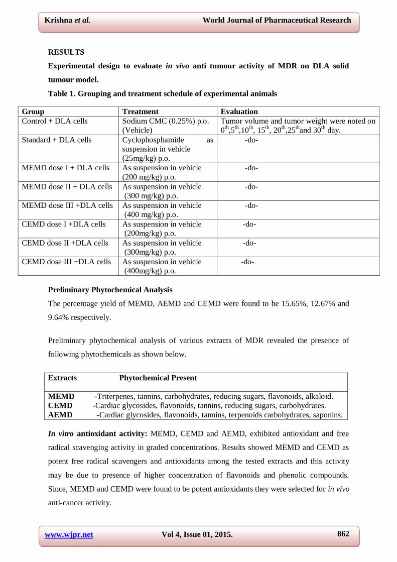
RESULTS
Experimental design to evaluate in vivo anti tumour activity of MDR on DLA solid
tumour model.
Table 1. Grouping and treatment schedule of experimental animals
Preliminary Phytochemical Analysis
The percentage yield of MEMD, AEMD and CEMD were found to be 15.65%, 12.67% and
9.64% respectively.
Preliminary phytochemical analysis of various extracts of MDR revealed the presence of
following phytochemicals as shown below.
Extracts Phytochemical Present
MEMD -Triterpenes, tannins, carbohydrates, reducing sugars, flavonoids, alkaloid.
CEMD -Cardiac glycosides, flavonoids, tannins, reducing sugars, carbohydrates.
AEMD -Cardiac glycosides, flavonoids, tannins, terpenoids carbohydrates, saponins.
In vitro antioxidant activity: MEMD, CEMD and AEMD, exhibited antioxidant and free
radical scavenging activity in graded concentrations. Results showed MEMD and CEMD as
potent free radical scavengers and antioxidants among the tested extracts and this activity
may be due to presence of higher concentration of flavonoids and phenolic compounds.
Since, MEMD and CEMD were found to be potent antioxidants they were selected for in vivo
anti-cancer activity.
Group Treatment Evaluation
Control + DLA cells Sodium CMC (0.25%) p.o.
(Vehicle)
Tumor volume and tumor weight were noted on
0th,5
th,10
th, 15
th, 20
th,25
thand 30
th day.
Standard + DLA cells Cyclophosphamide as
suspension in vehicle
(25mg/kg) p.o.
-do-
MEMD dose I + DLA cells As suspension in vehicle
(200 mg/kg) p.o.
-do-
MEMD dose II + DLA cells As suspension in vehicle
(300 mg/kg) p.o.
-do-
MEMD dose III +DLA cells As suspension in vehicle
(400 mg/kg) p.o.
-do-
CEMD dose I +DLA cells As suspension in vehicle
(200mg/kg) p.o.
-do-
CEMD dose II +DLA cells As suspension in vehicle
(300mg/kg) p.o.
-do-
CEMD dose III +DLA cells As suspension in vehicle
(400mg/kg) p.o.
-do-
www.wjpr.net Vol 4, Issue 01, 2015.
863
Krishna et al. World Journal of Pharmaceutical Research
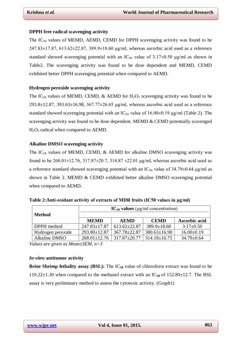
DPPH free radical scavenging activity
The IC50 values of MEMD, AEMD, CEMD for DPPH scavenging activity was found to be
247.83±17.87, 613.62±22.87, 389.9±18.60 μg/ml, whereas ascorbic acid used as a reference
standard showed scavenging potential with an IC50 value of 3.17±0.50 μg/ml as shown in
Table2. The scavenging activity was found to be dose dependent and MEMD, CEMD
exhibited better DPPH scavenging potential when compared to AEMD.
Hydrogen peroxide scavenging activity
The IC50 values of MEMD, CEMD, & AEMD for H2O2 scavenging activity was found to be
293.8±12.87, 393.63±16.98, 367.77±26.65 μg/ml, whereas ascorbic acid used as a reference
standard showed scavenging potential with an IC50 value of 16.00±0.19 μg/ml (Table 2). The
scavenging activity was found to be dose dependent. MEMD & CEMD potentially scavenged
H2O2 radical when compared to AEMD.
Alkaline DMSO scavenging activity
The IC50 values of MEMD, CEMD, & AEMD for alkaline DMSO scavenging activity was
found to be 268.01±12.76, 317.87±20.7, 314.87 ±22.01 μg/ml, whereas ascorbic acid used as
a reference standard showed scavenging potential with an IC50 value of 34.79±0.64 μg/ml as
shown in Table 2. MEMD & CEMD exhibited better alkaline DMSO scavenging potential
when compared to AEMD.
Table 2:Anti-oxidant activity of extracts of MDR fruits (IC50 values in μg/ml)
Method
IC50 values (μg/ml concentration)
MEMD AEMD CEMD Ascorbic acid
DPPH method 247.83±17.87 613.62±22.87 389.9±18.60 3.17±0.50
Hydrogen peroxide 293.80±12.87 367.78±22.87 380.63±16.98 16.00±0.19
Alkaline DMSO 268.01±12.76 317.87±20.77 314.18±16.75 34.79±0.64
Values are given as Mean±SEM, n=3
In-vitro antitumor activity
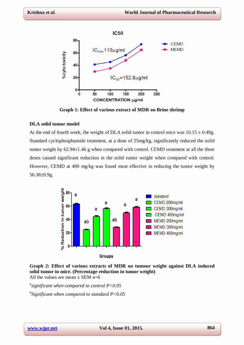
Brine Shrimp lethality assay (BSL): The IC50 value of chloroform extract was found to be
110.22±1.30 when compared to the methanol extract with an IC50 of 152.80±12.7. The BSL
assay is very preliminary method to assess the cytotoxic activity. (Graph1)
www.wjpr.net Vol 4, Issue 01, 2015.
864
Krishna et al. World Journal of Pharmaceutical Research
Graph 1: Effect of various extract of MDR on Brine shrimp
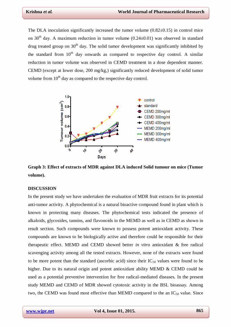
DLA solid tumor model
At the end of fourth week, the weight of DLA solid tumor in control mice was 10.15 ± 0.49g.
Standard cyclophosphamide treatment, at a dose of 25mg/kg, significantly reduced the solid
tumor weight by 62.94±1.46 g when compared with control. CEMD treatment at all the three
doses caused significant reduction in the solid tumor weight when compared with control.
However, CEMD at 400 mg/kg was found most effective in reducing the tumor weight by
56.38±0.9g.
Graph 2: Effect of various extracts of MDR on tumour weight against DLA induced
solid tumor in mice. (Percentage reduction in tumor weight)
All the values are mean ± SEM n=6
asignificant when compared to control P<0.05
bSignificant when compared to standard P<0.05
www.wjpr.net Vol 4, Issue 01, 2015.
865
Krishna et al. World Journal of Pharmaceutical Research
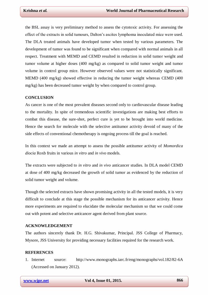
The DLA inoculation significantly increased the tumor volume (0.82±0.15) in control mice
on 30th day. A maximum reduction in tumor volume (0.24±0.01) was observed in standard
drug treated group on 30th day. The solid tumor development was significantly inhibited by
the standard from 10th
day onwards as compared to respective day control. A similar
reduction in tumor volume was observed in CEMD treatment in a dose dependent manner.
CEMD (except at lower dose, 200 mg/kg,) significantly reduced development of solid tumor
volume from 10th day as compared to the respective day control.
Graph 3: Effect of extracts of MDR against DLA induced Solid tumour on mice (Tumor
volume).
DISCUSSION
In the present study we have undertaken the evaluation of MDR fruit extracts for its potential
anti-tumor activity. A phytochemical is a natural bioactive compound found in plant which is
known in protecting many diseases. The phytochemical tests indicated the presence of
alkaloids, glycosides, tannins, and flavonoids in the MEMD as well as in CEMD as shown in
result section. Such compounds were known to possess potent antioxidant activity. These
compounds are known to be biologically active and therefore could be responsible for their
therapeutic effect. MEMD and CEMD showed better in vitro antioxidant & free radical
scavenging activity among all the tested extracts. However, none of the extracts were found
to be more potent than the standard (ascorbic acid) since their IC50 values were found to be
higher. Due to its natural origin and potent antioxidant ability MEMD & CEMD could be
used as a potential preventive intervention for free radical-mediated diseases. In the present
study MEMD and CEMD of MDR showed cytotoxic activity in the BSL bioassay. Among
two, the CEMD was found most effective than MEMD compared to the an IC50 value. Since
www.wjpr.net Vol 4, Issue 01, 2015.
866
Krishna et al. World Journal of Pharmaceutical Research
the BSL assay is very preliminary method to assess the cytotoxic activity. For assessing the
effect of the extracts in solid tumours, Dalton’s ascites lymphoma inoculated mice were used.
The DLA treated animals have developed tumor when tested by various parameters. The
development of tumor was found to be significant when compared with normal animals in all
respect. Treatment with MEMD and CEMD resulted in reduction in solid tumor weight and
tumor volume at higher doses (400 mg/kg) as compared to solid tumor weight and tumor
volume in control group mice. However observed values were not statistically significant.
MEMD (400 mg/kg) showed effective in reducing the tumor weight whereas CEMD (400
mg/kg) has been decreased tumor weight by when compared to control group.
CONCLUSION
As cancer is one of the most prevalent diseases second only to cardiovascular disease leading
to the mortality. In spite of tremendous scientific investigations are making best efforts to
combat this disease, the sure-shot, perfect cure is yet to be brought into world medicine.
Hence the search for molecule with the selective antitumor activity devoid of many of the
side effects of conventional chemotherapy is ongoing process till the goal is reached.
In this context we made an attempt to assess the possible antitumor activity of Momordica
diocia Roxb fruits in various in vitro and in vivo models.
The extracts were subjected to in vitro and in vivo anticancer studies. In DLA model CEMD
at dose of 400 mg/kg decreased the growth of solid tumor as evidenced by the reduction of
solid tumor weight and volume.
Though the selected extracts have shown promising activity in all the tested models, it is very
difficult to conclude at this stage the possible mechanism for its anticancer activity. Hence
more experiments are required to elucidate the molecular mechanism so that we could come
out with potent and selective anticancer agent derived from plant source.
ACKNOWLEDGEMENT
The authors sincerely thank Dr. H.G. Shivakumar, Principal. JSS College of Pharmacy,
Mysore, JSS University for providing necessary facilities required for the research work.
REFERENCES
1. Internet source: http://www.monographs.iarc.fr/eng/monographs/vol.182/82-6A
(Accessed on January 2012).
www.wjpr.net Vol 4, Issue 01, 2015.
867
Krishna et al. World Journal of Pharmaceutical Research
2. American Cancer Society, The History of Cancer,
http://www.cancer.org/docroot/CRI/content/CRI_2_6x_the_history_of_cancer_72.asp
(Accessed on September 2012).
3. Baquiran DC. Lippincott’s Cancer Chemotherapy Handbook. 2nd ed., Philadelphia;
Lippincott Williams and Wilkins: 2001.
4. Danaei G, Vender HS. Causes of cancer in the world: Comparative risk assessment of
nine behavioral and environmental risk factors. The Lancet, 2005; 366:1784-93.
5. Hartwell JC. Plants used against Cancer, a survey. Quarterman Publication, Lawrence,
MA: 1982.
6. Pinney KG, Jelinek C, Edvardsen K, Chaplin DJ, Petit GR. The discovery and
development of the combrestatins. In: Cragg GM, Kingston DG, Newman DJ (Eds.). Anti
cancer Agents from Natural Products. Brunner- Routledge Psychology press, Taylor &
Francis Group, Boca Raton, Florida; 2005.
7. National Plant Data Center. NRCS, USDA. Baton Rouge, LA 70874-4490 USA.
http://plants.usda.gov (Accessed on February 2012).
8. Bhavana B, Mukesh D, Chauhan NS, VK Dixit, and Saraf DK. Phyto-pharmacology of
Momordica dioica Roxb. ex. Willd. Int J Phytomed, 2010; 2: 1-9.
9. Gupta PP, Simal RC and Tandon JS. Antiallergic activity of some traditional Indian
medicinal plants”. Int J Pharmcog, 1993; 31(1): 15-8.
10. Shreedhar CS, Pai KS, Vaidya VP. Postcoital antifertility activity of the root of
Momordica dioica Roxb. Ind J PharmSci, 2001; 6: 528-31.
11. Ilango K, Maharajan G, Narasimhan S. Analgesic and Anti-inflammatory Activities of
Momordica dioica Fruit Pulp. Nat ProdSci, 2003; 9(4): 210-2.
12. Narsimhan S, Kannan S and Maharajan G. Antifeedent activity of Momordicadioicafruit
pulp extract on SpodopteraLitura. FITOTERAPIA, 2005; 76 (7-8): 715-7.
13. Jain A, Soni M, Deb L. Antioxidant and hepatoprotective activity of ethanolic and
aqueous extracts of Momordica dioica Roxb. Leaves. J ETHNOPHARMACOL, 2008; 4:
115-8.
14. Ilango K, Maharajan G, Narasimhan S. Hypoglycemic and Hypolipidemic Activities of
Momordica dioica Roxb Fruit Pulp Extracts on alloxan-induced diabetic rats. Int J Health
Res, 2009; 2(2): 195-9.
15. Firdous SM, Banerjee, Subhasis, Koneri R, Antihyperlipidemic activity of Momordica
dioica Roxb. Int J Drug Dev Res, 2010; 2(1): 16-24.
www.wjpr.net Vol 4, Issue 01, 2015.
868
Krishna et al. World Journal of Pharmaceutical Research
16. Finar IL. Stereochemistry and Chemistry of Natural Products. 5th ed., Longman Scientific
and Technical, London; 1975: 232.
17. Safia AK, Krishna KL. Evaluation of hypolipidemic and anti-obesity activites of
Momordica dioica Roxb. Fruit extracts on atherogenic diet induced hyperlipidemc rats.
PHARMACOPHORE, 2013; 4(6): 233-41.
18. Shamala S and, Krishna KL. Cardioprotective activity of fruit extract of Momordica
dioica Roxb. On Doxorubicin induced toxicity on rats. SCIINTL, 2013; 1(12): 392-400.
19. Ramyashree B, Krishna KL. A study to assess cardioprotective activity of biofraction of
Momordica diocia Roxb. on rats. Thesis submitted to JSS UNIVERSITY for award of
Mpharm degree, 2013.
20. Finar IL. Stereochemistry and chemistry of natural products. 5th ed., Longman Scientific
and Technical, London; 1975: 276-761.
21. Kokate CK, Purohit AP, Gokhale SB. Pharmacognosy. 4th
ed., Nirali Prakashan, Pune;
1996: 133-525.
22. Mruthunjaya K, Hukkeri V. Ph.D Thesis submitted to RAJIV GANDHI UNIVERSITY of
Health Sciences, Karnataka. Phytochemical and Pharmacological Screening of some
herbs used in folk medicine for their Hepatoprotective and antioxidant activity. 2008.
23. Sreejayan N, Rao MNA. Free radical scavenging activity by curcuminoids. DRUG RES,
1996; 46: 169-71.
24. Kaur G, Alam MS, Jabbar Z, Javed K. Evaluation of antioxidant activity of Cassia siamea
flowers. J. ETHNOPHARMACOL, 2006; 108: 340-8.
25. Elizabeth K, Rao MNA. Generation of superoxide anion and hydrogen peroxide by (+)-
cyanidanol-3. Int J Pharmceutics, 1990; 65(3): 261-3.
26. Oyaizu M. Studies on products of browning reaction: Antioxidative activities of products
of browning reaction prepared from glucosamine. JPN J NUTR, 1986; 44: 307-15.
27. Mayer BH, Ferrigni NR, McLaughlin JL, Putnam JE, Jocobaon L. Brine Shrimp. A
convenlent general bioassay for active plant constituent. PLANTA MEDICA, 1982;
4531-34.
28. Mossman T. Rapid colorimetric assay for cellular growth and survival: application to
proliferation and cytotoxicity assays. J Immunometods, 1983; 65: 55-63.
29. Gupta M, Mazumder UK, Sambath KR, Sivakumar T, Vamsi MLM. Antitumour activity
and antioxidant status of Caesapinia bonducella against Ehrlich ascites carcinoma in
Swiss albino mice. J Pharmacol Sci, 2004; 94: 177–184.
www.wjpr.net Vol 4, Issue 01, 2015.
869
Krishna et al. World Journal of Pharmaceutical Research
30. Senthilkumar N, Shrishailappa B, Santoshkumar HD, Ashok G. Antitumor Activity and
Antioxidant Status of the Methanol Extract of Careya arborea Bark Against Dalton’s
Lymphoma Ascites Induced Ascitic and Solid Tumor in Mice. J Pharmacol Sci, 2007;
103: 12–23.
Copyright © 2022 FDOKUMEN