Characterization of molten globule state of fetuin at low pH
Upload
cas-historydeptt-amuCategory
view
0download
0
www.bba-direct.com
Biochimica et Biophysica Acta 1699 (2004) 191–199
Molten globule-like folding intermediate of asialofetuin at acidic pH
Farah Naseem, Basir Ahmad, Mohd. Tashfeen Ashraf, Rizwan Hasan Khan*
Interdisciplinary Biotechnology Unit, Aligarh Muslim University, Aligarh 202002, India
Received 1 August 2003; received in revised form 24 February 2004; accepted 25 February 2004
Available online 19 March 2004
Abstract
In our earlier communication on acid-induced unfolding of bovine serum fetuin (BSF), we showed the existence of a molten globule (MG)-
like state of BSF at pH 1.8. The MG state was characterized by higher content of secondary structure than native and almost complete loss of
tertiary structure and more solvent exposed hydrophobic surface [Biochim. Biophys. Acta 1649 (2003) 164]. In this work we have shown the
presence of an MG-like partially folded intermediate of asialofetuin at around pH 1.8, which is much different from the MG state observed in
BSF in secondary structure contents. The results show that asialofetuin at pH 1.8 retains f 45% secondary structure, as evident from far-UVCD
spectra. The near-UV CD spectra showed almost complete loss of tertiary structure. The intrinsic fluorescence and acrylamide quenching of the
lone tryptophan residue showed that in acid-induced state, it is buried in the interior in a nonpolar environment. The temperature dependence of
far-UV CD signal of asialofetuin at pH 1.8 exhibits a weak cooperative thermal transition. A significant increase in ANS fluorescence showed
extensive solvent exposure of nonpolar cluster. Size exclusion chromatography (SEC) indicates a slight increase in the hydrodynamic size of
acid-induced protein. These results suggest that asialofetuin at pH 1.8 represents the MG-like folding intermediate. Moreover, our results
showed that glycosylation might play a role in stabilization of secondary structure during acid and/or thermal denaturation.
D 2004 Elsevier B.V. All rights reserved.
Keywords: Asialofetuin; MG state; Circular dichroism; pH; Acid-unfolding
1. Introduction
To understand fully the conformational behavior of a
protein, it is necessary to define not only the structure of its
native state but also that of various denatured states. The
molten globule (MG) state is the intermediate between the
native and unfolded states [1] in which the protein molecule
is almost as compact as in the native state [2,3] and has a
loosely packed nonpolar core [4]. MGs have now been
observed for a significant number of proteins, and it is
assumed that most if not all proteins can form such species.
In other words, MGs are general intermediates in protein
1570-9639/$ - see front matter D 2004 Elsevier B.V. All rights reserved.
doi:10.1016/j.bbapap.2004.02.011
Abbreviations: MG, molten globule; CD, circular dichroism; GnHCl,
guanidinium hydrochloride; ANS, 8-anilinonaphthalene-1-sulfonic acid;
NATA, N-acetyl-L-tryptophanamide; SEC, size exclusion chromatography
* Corresponding author. Tel.: +91-571-272-0388; fax: +91-571-272-
1776.
E-mail addresses: [email protected],
[email protected] (R.H. Khan).
folding. The development of broad range of techniques has
led to the identification and characterization of stable
intermediates in several proteins [5–7] Recently a number
of equilibrium intermediates have been detected and char-
acterized, like in our earlier communications we have
reported the MG states in proteins like a-chymotrypsino-
gen-A, stem bromelain, glucose-oxidase [8–10] and fetuin
[11]. The high cooperativity and complexity of the protein
folding process makes the characterization of conformation-
al transitions and equilibrium intermediate states hard to
achieve; different methods are employed for the character-
ization of the folding intermediates and for understanding
the different factors leading to the formation and stabiliza-
tion of these intermediates. One of the oldest known
methods of denaturing proteins is by the addition of acids.
Acid-induced unfolding of proteins varies from apparently
fully unfolded to substantial remaining structure [12].
In the present study, we have characterized MG-like
partially folded intermediate of asialofetuin. Asialofetuin
is the desyalylated form of fetuin.

F. Naseem et al. / Biochimica et Biophysica Acta 1699 (2004) 191–199192
Fetuin is a glycoprotein abundant in calf serum. It is a
protein found predominantly during fetal stage. Experi-
ments with serum from calves of different ages have
shown that the amount of this globulin has its highest
value in newly born calves and decreases with time. It is
associated with the period when the greatest building and
development of the animal takes place. The carbohydrate
portion of fetuin is made up of three branched hetero-
polysaccharide units. These units have been shown to have
a similar monosaccharide composition. The peptide portion
consists of a single chain with six intrachain disulfide
bonds. The carbohydrate chains comprise 24% of the total
molecular weight, Mr 48,000 Da [13]. Equivalent of fetuin
in other mammalian species is alpha 2-HS glycoprotein
(AHSG) [14]. Fetuin is thought to play an important role
in fetal brain development [15,16] and neonatal skeletal
development [17]. It is also important for postnatal bone
growth and remodeling [18] and contributes to insulin
resistance during normal pregnancy and gestational diabe-
tes [17]. It has been shown that fetuin and various
cytokines have a developmentally regulated appearance
and expression in the developing neocortex [15,16]. A
short 19-amino-acid sequence of fetuin shows a degree of
homology to an 18-amino-acid sequence of the transform-
ing growth factors—beta (TGF-beta) type-II receptor, and
in vitro fetuin binds to members of the TGF-beta family of
cytokines. It has been suggested that fetuin is the biolog-
ically significant antagonist of these cytokines [16]. Thus,
fetuin has a very important role in the overall development
of the fetus. Any misfolding in this protein can have
adverse effects on its functioning and its interaction with
cells and other molecules leading to general development
as well as brain development anomalies. Folding studies
on fetuin and knowledge of its different structural aspects
may prove useful in better understanding of its folding
pathway so that attempts can be made to correct the
misfolding.
In our previous studies on fetuin, it has been shown that
MG state exists at pH 1.8, which has substantial amount of
secondary structure and significantly disordered tertiary
structure; this was inferred on the basis of CD spectra taken
in the far as well as near-UV regions and fluorescence
studies. Thermal studies in the far-UV region showed that
the secondary structure is quite stable and is resistant to high
temperatures while thermal spectra in the near-UV region
were indicative of cooperative unfolding at pH 7 and loss of
cooperativity, i.e. non-cooperative unfolding at pH 1.8,
which again suggested the presence of MG state at this
pH. Based on our previous results on the characterization of
MG state of BSF at low pH, we report here in a systematic
way the conformational behavior of asialofetuin in the
acidic region and contribution of the sialic acid in main-
taining the structural integrity of the protein. For this
purpose, fluorescence and CD spectroscopic measurements
were performed depending on pH. The results are discussed
in the context of known properties of BSF.
2. Materials and methods
2.1. Materials
Asialofetuin from fetal calf serum type-I (lot-90K7650)
and GnHCl were purchased from Sigma Chemical Co. (St.
Louis, MO, USA). Acrylamide was purchased from Quali-
gens Fine Chemicals (India). All other reagents and buffer
compounds used were of analytical grade.
2.2. Methods
A stock solution of 10 mg ml� 1 protein was prepared in
distilled water and it was dialysed for 4 h at low temperature
(4 jC) and the respective solutions were made in different
pH buffers. GnHCl solution (6 M) was prepared in 10 mM
sodium phosphate buffer, pH 7.
2.2.1. Protein concentration determination
Protein concentrations were determined spectrophoto-
metrically using an extinction coefficient E2781% = 5.3 [19]
on a Hitachi U-1500 spectrophotometer or alternatively
by the method of Lowry et al. [20]. pH measurements
were carried out on an Elico digital pH meter (model
LI610).
2.2.2. CD measurements
CD measurements were carried out with a Jasco spec-
tropolarimeter, model J-720, equipped with a microcom-
puter. The instrument was calibrated with D-10-camphor-
sulfonic acid. All the CD measurements were carried out at
25 jC with a thermostatically controlled cell holder at-
tached to a Neslab RTE-110 water bath with an accuracy of
F 0.1 jC. Far-UV CD spectra were measured at a protein
concentration of 3.0 AM and near-UV CD spectra were
measured at protein concentration of 30 AM. The path-
lengths were 1 mm and 1 cm, respectively. The results are
expressed as mean residue ellipticity (MRE) in deg cm2
dmol� 1 defined as
MRE ¼ hobs ðmdegÞ=10� n� Cp� l
where hobs is the CD in millidegree; n = 309 is the number
of amino acid residues; l is the pathlength of the cell in cm
and Cp is the molar fraction. The a-helical content of fetuin
was calculated from the MRE value at 222 nm using the
following equation as described by Chen et al. [21].
% a-helix ¼ MRE222 � 2340=30300� 100:
For thermal-transition studies, a water-jacketed 1-mm
pathlength cell was used for far-UV CD and a water-
jacketed 10-mm pathlength cell was used for near-UV CD
attached to the RTE-110 water bath interfaced with a
microcomputer. A protein concentration of 30 AM was used
for near-UV CD measurements and 3 AM was used for far-
UV CD measurements.

Fig. 1. pH dependence of CD spectra of asialofetuin at 222 nm. Protein
concentration was 3 AM. Experiments were carried out at 25 jC in 10 mM
of the following buffers: pH 1–2.8, glycine-HCl buffer; pH 3–5, sodium-
acetate buffer; pH 6–7, sodium-phosphate buffer.
F. Naseem et al. / Biochimica et Biophysica Acta 1699 (2004) 191–199 193
2.2.3. Fluorescence measurements
Fluorescence measurements were performed on a Shi-
madzu spectrofluorometer, model RF-540. The fluorescence
spectra were measured at a protein concentration of 3 AM.
Solutions were prepared in buffers 20 mM each of different
pH values ranging from pH 0.6 to 10 [pH 1–2.8, glycine-
HCl buffer; pH 3–5, sodium-acetate buffer; pH 6–7,
sodium-phosphate buffer]. Before making measurements
the solutions were incubated overnight at 4 jC. Intrinsicspectra were recorded between 300 and 400 nm with
excitation wavelength of 280 and 295 nm. For ANS
fluorescence in the ANS binding experiments, the excitation
was set at 380 nm and the emission spectra were taken in the
range of 400–600 nm.
2.2.4. Quenching experiments
In the quenching experiments, aliquots of 5 M quencher
stock solution were added to protein solutions (3 AM) to
achieve the desired range of quencher concentration (0.1–
1M). Excitation was set at 295 nm in order to excite
tryptophan residues only and the emission spectrum was
recorded in the range 300–400 nm. The decrease in fluo-
rescence intensity at kmax was analyzed according to the
Stern–Volmer equation [22].
F0=F ¼ 1þ Ksv½Q�
where F0 and F are the fluorescence intensities at an
appropriate wavelength in the absence and presence of
quencher, respectively, Ksv is the Stern–Volmer constant,
and [Q] is concentration of the quencher.
2.2.5. Size exclusion chromatography (SEC)
SEC experiments were performed on a Sephacryl-S 100
HR (60� 0.8) column. The column was pre-equilibrated
with 25 mM sodium phosphate buffer pH 7 and glycine
HCl buffer of pH 1.8. Two milliliters of 5 mg/ml native
and pH-treated protein solution was applied to the column
and eluted at 20 ml/h. The eluted fractions were read at
280 nm.
3. Results and discussion
In our previous report, we have shown that an MG state
exists in bovine serum fetuin (BSF) at low pH [13]. In order
to monitor the role of charged sialic acid residues in
stabilization of native conformation of the protein, we have
performed similar studies on asialofetuin.
As shown in Fig. 1, the conformation of polypeptide
backbone of asialofetuin was monitored by ellipticity
measurements in the far-UV region at 222 nm. The
MRE at 222 nm showed no apparent change between
pH 7 and 4.0, but when pH was decreased below 4.0,
MRE222 decreased markedly to a minimum value at
around pH 2.4. Further, no apparent change was observed
in the pH range 2.4–1.0. Thus, the pH-induced transition
was found to follow a single step two state transition. The
observed pH denaturation curves as monitored by MRE
measurements at 222 nm of asialofetuin and its sialated
form fetuin were markedly different. The results of sec-
ondary structure resolved analysis are presented in Table 3
for fetuin and asialofetuin.
To detect and characterize the tertiary structure of asia-
lofetuin, near-UV CD, intrinsic tryptophan fluorescence
measurements and ANS binding experiments were used.
Fig. 2a and b shows the fluorescence spectra of asialo-
fetuin in the native state at pH 7, acid-unfolded state at pH
1.8 and completely denatured state in the presence of 6 M
GnHCl in the 300–400 nm range after exciting the protein
at 280 and 296 nm, respectively.When protein was excited
at 280 nm (Fig. 2a), a significant increase in fluorescence
intensity of acid-unfolded protein (curve 2) was observed as
compared to native (curve 1) and GnHCl-denatured protein
(curve 3) along with a red shift of approximately 12 nm as
compared to that of native state; this increase in fluores-
cence intensity may be ascribed to the disruption of hydro-
gen bonding between the hydroxyl group of tyrosyl residue
and carboxyl groups of peptide backbone emphasized by the
loss of secondary structure of asialofetuin in acid-induced
state [23,24]. In the presence of 6 M GnHCl, fluorescence
intensity was more and kmax shifted to longer wavelength
from native protein. On the other hand, when asialofetuin
was excited at 296 nm (Fig. 2b), fluorescence intensity of
completely denatured protein (curve 3) was slightly more
than that of the acid-induced state (curve 2) as well as native
state (curve 1) because at 296 nm there was no excitation of
the tyrosine residues, a slight red shift of 4 nm was
observed. Slight red shift of kmax when protein was excited
at 296 nm shows little change in the tryptophanyl environ-

Fig. 2. Fluorescence emission spectra of asialofetuin when the protein was excited at 280 nm (a) and 296 nm (b). Asialofetuin in the native state at pH 7 (—), in
acid-unfolded state at pH 1.8 (- - -), in 10 mM glycine-HCl buffer and in the presence of 6 M GnHCl (. . .). Protein (3 AM) was incubated with 10 mM buffers of
different pH for 3 h. Spectra were recorded in the range 300–400 nm. Experiments were carried out at 25 jC.
F. Naseem et al. / Biochimica et Biophysica Acta 1699 (2004) 191–199194
ment, which is also evident from Fig. 6 where MRE290 of
acid-unfolded protein is almost similar to that of native
protein. Red shift of kmax in both cases, i.e. when protein
was excited at 280 and 296 nm, is indicative of more polar
environment of the single tryptophan residue, which may be
either due to exposure of tryptophan to acidic environment
or addition of protons to the residues in the immediate
environment of tryptophan. Observed lower Ksv value at pH
1.8 indicated that tryptophan residue becomes more inac-
cessible to solvent in the acid-denatured state. Thus, slight
red shift could be ascribed to protonation of acidic group in
the vicinity of tryptophan residue.
Fig. 3 shows the acid-induced unfolding of asialofetuin
as monitored by ellipticity measurements in the near-UV
region at 256 nm. The CD signals at 256 nm are not
assigned to specific aromatic amino acid residues, but in
this spectral region disulfide bonds have possible contribu-
tions to the CD signals and, nevertheless, they are reflective
of changes in the overall tertiary conformation of the
protein. As can be seen from the figure, there was no
significant change in MRE 256 values from pH 7 to 4;
further decrease in pH leads to a loss in tertiary structure. At
pH 1.8 the loss in tertiary structure was found to be
maximum and MRE value approached to that of GnHCl-
denatured protein (Table 1).
Fig. 4 shows the acid-induced unfolding as monitored by
ANS fluorescence at 470 nm. From the figure, it can be
observed that there was a slight increase in fluorescence
with decrease in pH (upto pH 5.5); further, it remained
constant in the pH range 5.0–2.0. There was maximum

Fig. 3. pH dependence of CD spectra of asialofetuin at 256 nm. Protein
concentration was 30 AM. Experiments were carried out at 25 jC in 10 mM
of the following buffers: pH 1–2.8, glycine-HCl buffer; pH 3–5, sodium-
acetate buffer; pH 6–7, sodium-phosphate buffer.
Fig. 4. pH dependence of ANS fluorescence spectra of asialofetuin at 470
nm. Protein concentration was 3 AM and the experiment was carried out at
25 jC in 10 mM of the following buffers: pH 1–2.8, glycine-HCl buffer;
pH 3–5, sodium-acetate buffer; pH 6–7, sodium-phosphate buffer.
F. Naseem et al. / Biochimica et Biophysica Acta 1699 (2004) 191–199 195
ANS binding at pH 1.8, which is suggestive of exposure of
hydrophobic patches as reported in our earlier communica-
tions [9–11].
To ascertain whether the protein state observed at pH 1.8
represented the MG-like state, we compared the far-UV and
near-UV CD spectra, cooperativity of unfolding and ANS
binding of asialofetuin at pH 1.8 with those obtained at pH
7. Fig. 5 shows the far-UV CD spectra of asialofetuin in the
native state (curve 1), acid-unfolded state (curve 2) and
completely denatured state in the presence of 6 M GnHCl
(curve 3). The curve for the native state has two minima,
Table 1
Summary of different spectral properties of asialofetuin
Variable Native Acid-
denatured
state
GnHCl-
denatured
state
MRE at 222 nm � 14,800 � 10,300 � 2000
MRE at 256 nm � 45.5 � 10 � 5.7
% Helix 47 29 –
Relative fluorescence
intensity at 340 nm
(excitation at 280 nm)a
100 137 120.9
Relative fluorescence
intensity at 340 nm
(excitation at 296 nm)a
100 111.8 125
kmax (when protein was
excited at 280 nm)
328 340 356
kmax (when protein was
excited at 296 nm)
328 332 352
ANS fluorescence
intensity
38 92 –
Cooperativity
(thermal transition)
cooperative weakly
cooperative
–
a Fluorescence of native protein was taken as 100%.
one at 208 nm and the other at 222 nm, characteristic of a-
helical structures determined from MRE value at 222 nm by
the method of Chen et al. [21]. The spectra of the acid-
unfolded state retained all the features of secondary struc-
ture, although there was a decrease in the MRE values at
222 nm (Table 1), indicating a loss of about 32% a-helical
structure from native state. Asialofetuin in the presence of 6
M GnHCl lost all the features of secondary structure. Fig. 6
shows the near-UV CD spectra in the range 300–250 nm for
Fig. 5. Far-UV CD spectra of asialofetuin in the native state (—) at pH 7, in
acid-unfolded state (- - -) at pH 1.8 and completely denatured state (. . .) in6 M GnHCl. Spectra were recorded in the wavelength region 200–250 nm.
Experimental conditions were similar as stated in legend for Fig. 1.
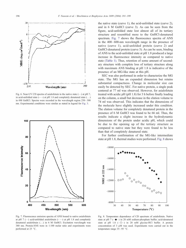
Fig. 6. Near-UV CD spectra of asialofetuin in the native state (—) at pH 7,
in acid-unfolded state (- - -) at pH 1.8 and completely denatured state (. . .)
in 6M GnHCl. Spectra were recorded in the wavelength region 250–300
nm. Experimental conditions were similar as stated in legend for Fig. 3.
Fig. 7. Fluorescence emission spectra of ANS bound to native asialofetuin
at pH 7 (—) acid-unfolded asialofetuin (- - -) at pH 1.8 and completely
denatured asialofetuin (. . .) in 6 M GnHCl. Excitation wavelength was
380 nm. Protein/ANS were in 1:100 molar ratio and experiments were
performed at 25 jC.
F. Naseem et al. / Biochimica et Biophysica Acta 1699 (2004) 191–199196
the native state (curve 1), the acid-unfolded state (curve 2),
and in 6 M GnHCl (curve 3). As can be seen from the
figure, acid-unfolded state lost almost all of its tertiary
structure and resembled more to the GnHCl-denatured
spectrum. Fig. 7 shows the fluorescence spectra of ANS
in the 400–600-nm wavelength range in the presence of
native (curve 1), acid-unfolded protein (curve 2) and
GnHCl-denatured protein (curve 3). As can be seen, binding
of ANS to the acid-unfolded state at pH 1.8 produced a large
increase in fluorescence intensity as compared to native
state (Table 1). Thus, retention of some amount of second-
ary structure with complete loss of tertiary structure along
with maximum ANS binding at pH 1.8 is indicative of the
presence of an MG-like state at this pH.
SEC was also performed in order to characterize the MG
state. The MG has an expanded dimension but retains
substantial compactness. Change in molecular size can
easily be detected by SEC. For native protein, a single peak
centered at 77 ml was observed. However, for asialofetuin
treated with acidic pH (pH 1.8) for 3 h before finally loading
on the column, a small but decrease in the elution volume to
74 ml was observed. This indicates that the dimensions of
the molecule have slightly increased under this condition.
The elution volume for completely denatured protein in the
presence of 6 M GnHCl was found to be 66 ml. Thus, the
results indicate a slight increase in the hydrodynamic
dimensions of the protein under acidic pH, which could
be due to the opening up of the tertiary structure as
compared to native state but they were found to be less
than that of completely denatured state.
For further confirmation of the MG-like intermediate
state at pH 1.8, thermal studies were performed. Fig. 8 shows
Fig. 8. Temperature dependence of CD spectrum of asialofetuin. Native
state at pH 7 (–.–) in 20 mM sodium-phosphate buffer; acid-denatured
state at pH 1.8 ( –o– ) in 20 mM glycine-HCl buffer. A protein
concentration of 3 AM was used. Experiments were carried out in the
temperature range 35–95 jC.

Table 2
Ksv values obtained from Stern–Volmer plot
Subject Ksv (M� 1)
Native asialofetuin 8.5
Acid-unfolded asialofetuin 5.2
GnHCl-denatured asialofetuin 14.2
NATA 19.3
F. Naseem et al. / Biochimica et Biophysica Acta 1699 (2004) 191–199 197
the temperature-induced unfolding of asialofetuin at pH 7.4
and 1.8 by MRE measurements at 222 nm. The thermal
unfolding of native asialofetuin is a cooperative process. As
can be seen from the figure, the transition started at 55 jC,decreased continuously up to 92 jC and then tailed off. On
the other hand, thermal transition of acid-induced state (pH
1.8) started at a lower temperature, 40 jC, i.e. thermal
transition started earlier in case of acid-denatured protein
and it is less stable than native. Thermal dependence of far-
UV CD signal indicates loosely ordered secondary structure
of acid-induced state as compared to secondary structure of
native asialofetuin. The results show that unfolding of MG
state of asaialofetuin is weakly cooperative. The weakly
cooperative thermal unfolding of the protein at pH 1.8 is
indicative of its MG-like nature. There are similar examples
in many other globular proteins including Equine, Canine,
Cytochrome c and SNase [25–28].
To confirm the microenvironment of the tryptophan
residue, we compared the exposure of the single tryptophan
residue in the native state with that in the acid-denatured
state and guanidine hydrochloride denatured state by fluo-
rescence quenching experiment, using uncharged molecules
of acrylamide as described by Eftink and Ghiron [22]. Fig. 9
shows the Stern–Volmer plot of quenching of fluorescence
by acrylamide in native state, acid-induced state (1.8) and
GnHCl-denatured state of asialofetuin. Results for the
tryptophan analogue NATA are also included as a standard
for complete accessibility to quencher. Table 2 shows the
Stern–Volmer constant fitted to the linear part of the curve.
Ksv for the native state (8.5) was found to be slightly higher
Fig. 9. Stern–Volmer plot of acrylamide quenching. Native asialofetuin at
pH 7 (– w – ); acid-denatured asialofetuin at pH 1.8 (–4–) and NATA
(– � – ) and GnHCl (–o–). Values shown are the ratios of fluorescence in
the absence of acrylamide ( F0) to the fluorescence at that concentration of
quencher ( F).
than that for the acid-induced state (5.2), a blue shift in kmax
from 340 to 337 nm also indicated that tryptophan residue in
acid-induced state was less accessible to quenching than in
native state. The Ksv value for the GnHCl-denatured state
and NATA were significantly higher than those for native
and acid-unfolded state, suggesting that tryptophan residue
even in the native state is internal. Taken together, these
results, i.e. the presence of significant amount of secondary
structure, higher magnitude of ANS binding, weak cooper-
ativity in the thermal transition and almost complete loss of
tertiary structure, suggest that the acid-unfolded state of
asialofetuin at pH 1.8 resembles the MG state as defined for
other proteins [10,11,25–31].
The results obtained from the experiments discussed
above and previous reports [11,32] lead us to conclude that
removal of sialic acid from the protein fetuin brings about a
considerable structural reorganization, suggesting the differ-
ent behavior of asialofetuin during acid denaturation. This
conclusion is supported by observations summarized in
Table 3. Far-UV CD spectrum of native asialofetuin has
higher content of a-helix as evident from the spectrum of
the native protein having minima at 208 and 222 nm typical
of a-helix. The essential absence of the 222-nm minimum in
Table 3
Comparison of the conformational properties of fetuin and asialofetuin
Variables Fetuina Asialofetuin
Native Acid-
unfolded
GnHCl-
denatured
Native Acid-
unfolded
GnHCl-
denatured
% Secondary
structure at
222 nmb
100 113 13 100 70 12.5
% Tertiary
structure at
256 nmb
100 38 38 100 22 12
% Tertiary
structure
at 290 nmb
100 55 5 100 100 46
Relative
fluorescence
intensity at
340 nm
(excitation
at 280 nm)c
100 62 51.5 100 137 120.9
ANS
fluorescence
intensity
3 58 – 38 92 –
a Taken from our earlier communication [13].b MRE of the native protein is taken as 100%.c Fluorescence intensity of native protein is taken as 100%.

F. Naseem et al. / Biochimica et Biophysica Acta 1699 (2004) 191–199198
fetuin suggests that the protein has low a-helix as compared
to that of asialofetuin. Further, it was observed that acid and
thermal denaturation does not lead to the destruction of
secondary structure of fetuin; on the other hand, there is
significant loss of secondary structure during acid and
thermal denaturation.
Near-UV CD spectrum of acid-unfolded fetuin at pH 1.8
[11] in the entire range from 250 to 300 nm resembled
more the spectrum of completely denatured fetuin in 6 M
GnHCl, but the near-UV CD spectrum of asialofetuin at pH
1.8 resembled more that of native protein in the region of
tryptophanyl residue (295 nm) while the rest of the
spectrum was similar to that of 6 M GnHCl-denatured
asialofetuin.
When protein was excited at 280 nm, intrinsic fluores-
cence of acid-unfolded fetuin was quenched along with a
red shift of 4 nm; while intrinsic fluorescence at 340 nm of
acid-unfolded asialofetuin was more than that of native
protein along with a red shift of approximately 12 nm.
Intrinsic fluorescence quenching of acid-denatured pro-
teins (fetuin and asialofetuin) by acrylamide is higher than
that of native, suggesting the internalization of the single
tryptophan residue in both cases.
The implications of the results presented in this paper
may prove useful in understanding the effect of sialic acid
on partially folded intermediate, which accumulates in the
folding pathway of a protein. Further, the results of thermal
transitions and acid denaturation monitored by far-UV CD,
presented in this paper, taken together with previous reports
[11,31], unambiguously demonstrate that sialic acid plays a
significant role in the stabilization of secondary structure of
fetuin during acid and thermal denaturation.
Acknowledgements
Facilities provided by A.M.U are gratefully acknowl-
edged. B.A. thanks the Council of Scientific and Industrial
Research, New Delhi for financial assistance. The authors
are also thankful to DST (FIST) for providing lab facilities.
References
[1] O.B. Ptitsyn, Molten globule and protein folding, Adv. Protein Chem.
47 (1995) 83–229.
[2] D.A. Dolgikh, R.I. Gilmanshin, E.V. Brazhnikov, V.E. Bychkova,
G.V. Semisotnov, S.Y. Venyaminiv, O.B. Ptitsyn, Alpha-Lactalbumin:
compact state with fluctuating tertiary structure? FEBS Lett. 136
(1981) 311–315.
[3] V.N. Uversky, Use of fast protein size-exclusion liquid chromatogra-
phy to study the unfolding of proteins which denature through the
molten globule, Biochemistry 32 (1993) 13288–13298.
[4] G. Damaschun, C. Gernat, H. Damaschun, V.E. Bychkova, O.B.
Ptitsyn, Int. J. Biol. Macromol. 8 (1986) 226–230.
[5] D. Barrick, F.M. Hughson, R.L. Baldwin, Molecular mechanisms of
acid denaturation. The role of histidine residues in the partial unfold-
ing of apomyoglobin, J. Mol. Biol. 237 (1994) 588–601.
[6] T.K.S. Kumar, V. Subbiah, T. Ramakrishna, M.W. Pandit, Trichloro-
acetic acid-induced unfolding of bovine pancreatic ribonuclease. Ex-
istence of molten globule-like state, J. Biol. Chem. 269 (1994)
12620–12625.
[7] A. Matouschek, L. Serrano, E.M. Meiering, M. Bycroft, A.R. Ferscht,
The folding of an enzyme: V. H/2H exchange-nuclear magnetic res-
onance studies on the folding pathway of barnase: complementarity to
and agreement with protein engineering studies, J. Mol. Biol. 224
(1992) 837–845.
[8] F. Khan, R.H. Khan, S. Muzammil, Alcohol-induced versus anionin-
duced states of alpha-chymotrypsinogen-A at low pH, Biochim. Bio-
phys. Acta 1481 (2000) 229–233.
[9] P. Gupta, R.H. Khan, M. Saleemuddin, Trifluoroethanol-induced
‘‘molten globule’’ state in stem bromelain, Arch. Biochem. Biophys.
413 (2003) 199–206.
[10] S.K. Haq, S. Rasheedi, R.H. Khan, Characterization of a partially
folded intermediate of stem bromelain at low pH, Eur. J. Biochem.
269 (2002) 47–52.
[11] F. Naseem, R.H. Khan, S.K. Haq, A. Naeem, Characterization of
molten globule state of fetuin at low pH, Biochim. Biophys. Acta
1649 (2003) 164–170.
[12] Y. Goto, L.J. Calciano, A.L. Fink, Acid induced folding of proteins,
Proc. Natl. Acad. Sci. U. S. A. 87 (1990) 573–577.
[13] E.D. Green, G. Adel, J.U. Baenziger, The asparagine-linked oligosac-
charides on bovine fetuin. Structural analysis of N-glycanase-released
oligosaccharides by 500-megahertz 1H NMR spectroscopy, J. Biol.
Chem. 263 (1988) 18253–18268.
[14] Y. Yoshioka, F. Gejyo, T. Marti, E.E. Rickli, W. Burgi, G.D. Offner,
R.F. Troxler, K. Schmid, The complete amino acid sequence of the A-
chain of human plasma alpha-2-HS glycoprotein, J. Biol. Chem. 261
(1986) 1665–1667.
[15] K.M. Dziegielewska, Y. Daikuhara, T. Ohnishi, P.M.E. Waite, J. Ek,
M.D. Habgood, M.A. Lane, A. Potter, N.R. Saunders, Fetuin in the
developing neocortex of the rat: distribution and origin, J. Comp.
Neurol. 423 (2000) 373–388.
[16] K.M. Dziegielewska, R.T. Williams, G.W. Knott, P.D. Kitchener, S.E.
Monk, A. Potter, N.R. Saunders, TGF-beta receptor type II and fetuin
in the developing sheep neocortex, Cell Tissue Res. 290 (1997) 515–
524.
[17] L. Kalabay, K. Cseh, A. Pajor, E. Baranyi, G.M. Melczer, G. Speen,
M. Kovacs, G. Siller, I. Karadi, G. Winkler, Correlation of maternal
serum fetuin/alpha2-HS-glycoprotein concentration with maternal in-
sulin resistance and anthropometric parameters of neonates in normal
pregnancy and gestational diabetes, Eur. J. Endocrinol. 147 (2002)
243–248.
[18] M. Sweras, D. Liu, E.A. Partridge, J. Pawling, B. Sukhu, C. Clokie, W.
Jahner Dechent, H.C. Tenenbaum, C.J. Swallow, M.D. Grynpas, J.W.
Dennis, Alpha-2-HS glycoprotein/fetuin, a transforming growth factor-
beta/bone morphogenetic protein antagonist, regulates postnatal bone
growth and remodeling, J. Biol. Chem. 277 (2002) 19991–19997.
[19] R.G. Spiro, Demonstration of a single peptide chain in the glycoprotein
fetuin: terminal amino acid analyses and studies of the oxidized and
reduced alkylated preparations, J. Biol. Chem. 238 (1963) 644–649.
[20] O.H. Lowry, N.J. Rosebrough, A.L. Farr, R.J. Randall, Protein mea-
surement with the folin phenol reagent, J. Biol. Chem. 193 (1951)
265–275.
[21] Y.H. Chen, J.T. Yang, H. Martinez, Determination of the secondary
structure of proteins by circular dichroism and optical rotatory disper-
sion, Biochemistry 11 (1972) 4120–4131.
[22] M.R. Eftink, C.A. Ghiron, Fluorescence quenching studies with pro-
teins, Anal. Biochem. 114 (1982) 199–227.
[23] R.W. Cowgill, Fluorescence and protein structure. In Relationship
between tyrosine fluorescence and various stages in denaturation of
Ribonuclease, Biochim. Biophys. Acta 120 (1966) 196–211.
[24] M. Dockal, D.C. Carter, F. Ruker, Conformational transitions of the
three recombinant domains of human serum albumin depending on
pH, J. Biol. Chem. 275 (2000) 3042–3050.

F. Naseem et al. / Biochimica et Biophysica Acta 1699 (2004) 191–199 199
[25] J.H. Carra, A.A. Elizabeth, P.L. Privalov, Three-state thermodynamic
analysis of the denaturation of staphylococcal nuclease mutants, Bio-
chemistry 33 (1994) 10842–10850.
[26] W. Colon, H. Roder, Kinetic intermediates in the formation of the
cytochrome cmolten globule, Nat. Struct. Biol. 12 (1996) 1019–1025.
[27] J.L. Marmorino, M. Lehti, G.J. Pielak, Native tertiary structure in an
A-state from resonance Raman spectroscopy, J. Mol. Biol. 275 (1998)
379–388.
[28] V.N. Uversky, A.S. Karnoup, D.J. Segal, S. Sheshadri, S. Doniach,
A.L. Fink, Anion-induced folding of Staphylococcal nuclease: char-
acterization of multiple equilibrium partially folded intermediates,
J. Mol. Biol. 278 (1998) 879–894.
[29] K. Kuwajima, The molten globule state as a clue for understanding
the folding and cooperativity of globular protein structure, Proteins 6
(1989) 87–103.
[30] O.B. Ptitsyn, in: T.E. Creighton (Ed.), 1992, pp. 243–300, Freeman,
New York.
[31] C.M. Dobson, Protein folding. Solid evidence for molten globules,
Curr. Biol. 4 (1994) 636–640.
[32] C. Wang, I. Lascu, A. Giartosio, Bovine serum fetuin is unfolded
through a molten globule state, Biochemistry 37 (1998) 8457–8464.
Copyright © 2022 FDOKUMEN


![A chemically modified [alpha]-amylase with a molten-globule state has entropically driven enhanced thermal stability](https://static.fdokumen.com/doc/165x107/631965ccbc8291e22e0f1555/a-chemically-modified-alpha-amylase-with-a-molten-globule-state-has-entropically.jpg)