Mobilization of Protein Kinase C in Macrophages Induced by Listeria monocytogenes Affects Its...
11
INFECTION AND IMMUNITY, Aug. 2002, p. 4650–4660 Vol. 70, No. 8 0019-9567/02/$04.000 DOI: 10.1128/IAI.70.8.4650–4660.2002 Copyright © 2002, American Society for Microbiology. All Rights Reserved. Mobilization of Protein Kinase C in Macrophages Induced by Listeria monocytogenes Affects Its Internalization and Escape from the Phagosome Sandra J. Wadsworth† and Howard Goldfine* Department of Microbiology, University of Pennsylvania School of Medicine, Philadelphia, Pennsylvania 19104-6076 Received 26 December 2001/Returned for modification 11 March 2002/Accepted 19 April 2002 Listeriolysin O (LLO) and a phosphatidylinositol-specific phospholipase C (PI-PLC) are known virulence factors of Listeria monocytogenes in both tissue cultures and the murine model of infection. LLO is a member of a family of pore-forming cholesterol-dependent cytotoxins and is known to play an essential role in escape from the primary phagocytic vacuole of macrophages. PI-PLC plays an accessory role, in that PI-PLC mutants are partially defective in escape. We have shown that both of these molecules are essential for initiating rapid increases in the calcium level in the J774 murine macrophage cell line (S. J. Wadsworth and H. Goldfine, Infect. Immun. 67:1770-1778, 1999). Here we show that both LLO and PI-PLC are required for translocation of protein kinase C (PKC ) to the periphery of J774 cells and for translocation of PKC II to early endosomes beginning within the first minute after addition of bacteria to the culture medium. Treatment with the calcium channel blocker SK&F 96365 inhibited translocation of PKC II but not PKC . Our findings lead us to propose a host signaling pathway requiring LLO and the formation of diacylglycerol by PI-PLC in which calcium-independent PKC is responsible for the initial calcium signal and the subsequent PKC II translocation. LLO-dependent translocation of PKC I to early endosomes also occurs between 1 and 4 min after infection, but this occurs in the absence of PI-PLC. All of these signals were observed in cells that had not internalized bacteria. Blocking PKC translocation with hispidin resulted in more rapid uptake of wild-type bacteria and greatly reduced escape from the primary phagocytic vacuoles of J774 cells. The capacity to survive and grow within macrophages is a hallmark of infections with Listeria monocytogenes. Our recent studies have focused on the earliest stages of the encounter between L. monocytogenes and a macrophage, as represented by the J774 murine macrophage cell line, a well-studied tissue culture model of infection (5, 30, 33). We have observed that the cytosolic calcium level is elevated at 1, 5, and 10 min after infection with wild-type L. monocytogenes but not after infec- tion with a strain with a listeriolysin O (LLO) mutation. Strains with deletions in the genes encoding two secreted phospho- lipases C did not produce some or all of these signals (35). Of specific interest to workers in our laboratory are signal transduction pathways activated by the two phospholipases that contribute significantly to virulence in the mouse model of infection (5, 30). One of these, a phosphatidylinositol (PI)- specific phospholipase C (PI-PLC), encoded by plcA, hydro- lyzes host PI, leading to production of the eukaryotic signaling molecule diacylglycerol (DAG) (4, 12, 23, 30). The other, a broad-range phospholipase C (BR-PLC), encoded by plcB, acts on many host polar lipids, including sphingomyelin, lead- ing to production of DAG and ceramide (10, 11). The elevated calcium levels produced by the combined actions of LLO and PI-PLC appear to be part of a signaling pathway that is inhib- itory to entry of L. monocytogenes, since strains with mutations in the genes encoding these two proteins enter J774 cells more rapidly than the wild type enters these cells (35). Calcium signals also appear to regulate the eventual fate of the bacte- rium since suppression of these signals results in more rapid entry and reduced escape of wild-type bacteria from the pri- mary phagocytic vacuole into the cytosol (35). Based on the rapid hydrolysis of PI and the formation of DAG as a result of listerial phospholipase activity on host lipids (12) and based on our observations of calcium signaling (35), we investigated the possibility that calcium signaling is coupled to protein kinase C (PKC) mobilization. In the present study we demonstrated that three of the four PKC isoforms (, I, and II) found in J774 cells are translocated before bacterial entry, that the presumed activation of PKC and PKC II is coupled to early calcium signals, and that both isoforms rapidly translocate to early endosomes. Pharmacolog- ical inhibition of PKC signaling resulted in more rapid entry and reduced escape of L. monocytogenes from the primary phagocytic vacuole. MATERIALS AND METHODS Bacterial strains and culture conditions. Bacterial strains used in this study are listed in Table 1. All strains were maintained on brain heart infusion agar plates, which were made at regular intervals from stock cultures stored at 80°C in Luria-Bertani medium with 40% glycerol. For all experiments an overnight culture was grown statically at 30°C, and a 1:10 dilution of this culture in brain heart infusion was prepared the morning of the experiment and grown at 37°C on a rotator until the optical density at 620 nm reached 0.490 to 0.510. Measurement of bacterial association with and entry into J774 cells. J774 cells, a murine macrophage cell line, were maintained in Dulbecco’s modified Eagle’s medium (DMEM) supplemented with 7.5% fetal calf serum, glutamine, and penicillin-streptomycin and incubated at 37°C under 5% CO 2 . Cells were plated on circular glass coverslips (diameter,12 mm; Fisher) in antibiotic-free * Corresponding author. Mailing address: Department of Microbi- ology, University of Pennsylvania School of Medicine, Philadelphia, PA 19104-6076. Phone: (215) 898-6384. Fax: (215) 898-9557. E-mail: goldfi[email protected]. † Present address: Department of Pharmacology, Temple University School of Medicine, Philadelphia, PA 19140-5104. 4650 on September 23, 2015 by guest http://iai.asm.org/ Downloaded from
-
Upload
independent -
Category
Documents
-
view
3 -
download
0
Transcript of Mobilization of Protein Kinase C in Macrophages Induced by Listeria monocytogenes Affects Its...
INFECTION AND IMMUNITY, Aug. 2002, p. 4650–4660 Vol. 70, No. 80019-9567/02/$04.00�0 DOI: 10.1128/IAI.70.8.4650–4660.2002Copyright © 2002, American Society for Microbiology. All Rights Reserved.
Mobilization of Protein Kinase C in Macrophages Induced byListeria monocytogenes Affects Its Internalization
and Escape from the PhagosomeSandra J. Wadsworth† and Howard Goldfine*
Department of Microbiology, University of Pennsylvania School of Medicine, Philadelphia, Pennsylvania 19104-6076
Received 26 December 2001/Returned for modification 11 March 2002/Accepted 19 April 2002
Listeriolysin O (LLO) and a phosphatidylinositol-specific phospholipase C (PI-PLC) are known virulencefactors of Listeria monocytogenes in both tissue cultures and the murine model of infection. LLO is a memberof a family of pore-forming cholesterol-dependent cytotoxins and is known to play an essential role in escapefrom the primary phagocytic vacuole of macrophages. PI-PLC plays an accessory role, in that PI-PLC mutantsare partially defective in escape. We have shown that both of these molecules are essential for initiating rapidincreases in the calcium level in the J774 murine macrophage cell line (S. J. Wadsworth and H. Goldfine,Infect. Immun. 67:1770-1778, 1999). Here we show that both LLO and PI-PLC are required for translocationof protein kinase C � (PKC �) to the periphery of J774 cells and for translocation of PKC � II to earlyendosomes beginning within the first minute after addition of bacteria to the culture medium. Treatment withthe calcium channel blocker SK&F 96365 inhibited translocation of PKC � II but not PKC �. Our findings leadus to propose a host signaling pathway requiring LLO and the formation of diacylglycerol by PI-PLC in whichcalcium-independent PKC � is responsible for the initial calcium signal and the subsequent PKC � IItranslocation. LLO-dependent translocation of PKC � I to early endosomes also occurs between 1 and 4 minafter infection, but this occurs in the absence of PI-PLC. All of these signals were observed in cells that hadnot internalized bacteria. Blocking PKC � translocation with hispidin resulted in more rapid uptake ofwild-type bacteria and greatly reduced escape from the primary phagocytic vacuoles of J774 cells.
The capacity to survive and grow within macrophages is ahallmark of infections with Listeria monocytogenes. Our recentstudies have focused on the earliest stages of the encounterbetween L. monocytogenes and a macrophage, as representedby the J774 murine macrophage cell line, a well-studied tissueculture model of infection (5, 30, 33). We have observed thatthe cytosolic calcium level is elevated at 1, 5, and 10 min afterinfection with wild-type L. monocytogenes but not after infec-tion with a strain with a listeriolysin O (LLO) mutation. Strainswith deletions in the genes encoding two secreted phospho-lipases C did not produce some or all of these signals (35).
Of specific interest to workers in our laboratory are signaltransduction pathways activated by the two phospholipasesthat contribute significantly to virulence in the mouse model ofinfection (5, 30). One of these, a phosphatidylinositol (PI)-specific phospholipase C (PI-PLC), encoded by plcA, hydro-lyzes host PI, leading to production of the eukaryotic signalingmolecule diacylglycerol (DAG) (4, 12, 23, 30). The other, abroad-range phospholipase C (BR-PLC), encoded by plcB,acts on many host polar lipids, including sphingomyelin, lead-ing to production of DAG and ceramide (10, 11). The elevatedcalcium levels produced by the combined actions of LLO andPI-PLC appear to be part of a signaling pathway that is inhib-itory to entry of L. monocytogenes, since strains with mutations
in the genes encoding these two proteins enter J774 cells morerapidly than the wild type enters these cells (35). Calciumsignals also appear to regulate the eventual fate of the bacte-rium since suppression of these signals results in more rapidentry and reduced escape of wild-type bacteria from the pri-mary phagocytic vacuole into the cytosol (35).
Based on the rapid hydrolysis of PI and the formation ofDAG as a result of listerial phospholipase activity on hostlipids (12) and based on our observations of calcium signaling(35), we investigated the possibility that calcium signaling iscoupled to protein kinase C (PKC) mobilization. In the presentstudy we demonstrated that three of the four PKC isoforms (�,� I, and � II) found in J774 cells are translocated beforebacterial entry, that the presumed activation of PKC � andPKC � II is coupled to early calcium signals, and that both �isoforms rapidly translocate to early endosomes. Pharmacolog-ical inhibition of PKC signaling resulted in more rapid entryand reduced escape of L. monocytogenes from the primaryphagocytic vacuole.
MATERIALS AND METHODS
Bacterial strains and culture conditions. Bacterial strains used in this studyare listed in Table 1. All strains were maintained on brain heart infusion agarplates, which were made at regular intervals from stock cultures stored at �80°Cin Luria-Bertani medium with 40% glycerol. For all experiments an overnightculture was grown statically at 30°C, and a 1:10 dilution of this culture in brainheart infusion was prepared the morning of the experiment and grown at 37°C ona rotator until the optical density at 620 nm reached 0.490 to 0.510.
Measurement of bacterial association with and entry into J774 cells. J774cells, a murine macrophage cell line, were maintained in Dulbecco’s modifiedEagle’s medium (DMEM) supplemented with 7.5% fetal calf serum, glutamine,and penicillin-streptomycin and incubated at 37°C under 5% CO2. Cells wereplated on circular glass coverslips (diameter,12 mm; Fisher) in antibiotic-free
* Corresponding author. Mailing address: Department of Microbi-ology, University of Pennsylvania School of Medicine, Philadelphia,PA 19104-6076. Phone: (215) 898-6384. Fax: (215) 898-9557. E-mail:[email protected].
† Present address: Department of Pharmacology, Temple UniversitySchool of Medicine, Philadelphia, PA 19140-5104.
4650
on Septem
ber 23, 2015 by guesthttp://iai.asm
.org/D
ownloaded from

DMEM 1 day prior to infection. To measure entry, we used the method ofDrevets and Campbell (8). Briefly, cells were infected with fluorescein isothio-cyanate (FITC)-labeled log-phase wild-type or mutant bacteria (25 to 30 bacteriaper cell) for 20 min. Bacteria were labeled with FITC as described previously(35). At various times during the 20-min infection period cells were washed fiveor six times with phosphate-buffered saline (PBS), stained with ethidium bromide(25 �g/ml), which stains only extracellular bacteria, and fixed with 3.3% form-aldehyde. Cells and bacteria were counted by using an 60� oil objective and bothfluorescein and rhodamine filters. Total bacteria, stained with FITC, were green,and extracellular bacteria, stained with ethidium bromide, were red. Thus, thepercentage of intracellular bacteria was obtained by subtracting the number ofextracellular bacteria (red) from the number of total bacteria (green), dividingthe difference by the total number of bacteria (green), and multiplying thisfraction by 100 (35).
Visualization of PKC translocation by immunofluorescence. Immunofluores-cence techniques were used to study PKC � and PKC � translocation andcolocalization of PKC � with early endosomes of J774 cells. For these studies,cells were plated on coverslips as described above. Cells were infected withFITC-labeled wild-type or mutant bacteria, and at various times during a 5-mininfection cells were washed five or six times with PBS, fixed, permeabilized withPBS containing 0.1% Triton X-100, and blocked with 1% bovine serum albumin.In some experiments unlabeled bacteria were used to determine if FITC labelingaltered the signals obtained with labeled bacteria. No difference in signaling wasseen when labeled and unlabeled bacteria were compared. The permeabilizedcells were then incubated at 37°C for 1 h with rabbit polyclonal antibodies (1:100)to murine PKC �, PKC � I, or PKC � II (Calbiochem, San Diego, Calif.). Thenthe cells were washed 10 times with PBS, the secondary antibody, anti-rabbitimmunoglobulin G conjugated to FITC, was added at 1:100, and the cells wereincubated for 30 min at 25°C. After this incubation the cells were washed 10times with PBS and mounted for fluorescence microscopy. Cells were viewedwith the 60� oil objective by using the fluorescein filter.
In experiments in which PKC � colocalization to early endosomes was studied,J774 cells were also labeled with rhodamine-conjugated transferrin (MolecularProbes), a specific marker for early endosomes. J774 cells were labeled for 45min at 37°C with transferrin according to the manufacturer’s directions, washed10 times with PBS, and then infected with wild-type or mutant bacteria. The cellswere then processed for immunofluorescence studies as described above. Thesecells were viewed with both the fluorescein and rhodamine filters.
Cytosolic Ca2� measurements. Cytosolic calcium measurements were ob-tained as previously described (35). Briefly, cells were loaded with a 5 �Msolution of the fluorescent calcium indicator fluo-3 AM and a 5 �M solution ofthe fluorescent indicator SNARF-1 AM for 30 min. SNARF-1 AM was used asthe reference since fluo-3 is a nonratiometric calcium indicator. The cells wereviewed with a 40� objective by using both fluorescein and rhodamine filters, anddual images were captured with the Phase 3 Imaging system (Phase 3 Imaging,Glen Mills, Pa.).
Early-endosome preparation and characterization: isolation of early endo-somes. Early endosomes were isolated and characterized by the methods of Diazet al. (7), with modifications. J774 cells, grown to confluence in 150-cm2 flasks,were infected with L. monocytogenes and washed three times with ice-cold PBS.Bacterial cultures were not labeled with FITC but otherwise were prepared asdescribed above. The volume of inoculum required varied according to thenumber of flasks of J774 cells used but was calculated based on a multiplicity ofinfection of 25 to 30. Immediately after the last wash liquid was removed, 3 mlof ice-cold lysis buffer (10 mM Tris, 1 mM diisopropyl fluorophosphate, 0.5 �g ofleupeptin per ml, 2 �g of aprotinin per ml, 1 mM phenylmethylsulfonyl fluoride,1 mM dithiothreitol, 1 mM EGTA, 1 mM EDTA; pH 7.4) was added to eachflask, and the cells were immediately scraped, transferred into a 15-ml conicalpolypropylene tube (Costar), and kept on ice. The cell suspension was incubatedfor approximately 5 min on ice or until more than 85% of the cells were lysed,as determined by microscopy. The suspension was centrifuged for 5 min at 1,500
� g at 4°C. The postnuclear supernatant (PNS) fraction was collected, trans-ferred to an SW41 ultracentrifuge tube (Beckman), and centrifuged for 15 minat 40,000 � g at 4°C. The supernatant (first supernatant fraction) was collectedand saved for marker assays. The pellet was resuspended in 0.25 ml of 0.25 Msucrose in lysis buffer; for the best results, we applied the 40,000-�-g pelletobtained from four 150-cm2 flasks to one Percoll gradient. This suspension wasthen layered onto 1.5 ml of a 1.05-g/ml Percoll solution. It was centrifuged for 45min at 40,000 � g in the SW41 rotor, and the early-endosome fraction wascollected from the top of the tube and used for marker assays or Westernblotting. The thin lipid layer, located below the early-endosome fraction, wasdiscarded, and the aqueous layer below this was referred to as late endosomes/lysosomes. This layer was also saved and was used for marker assays.
Transferrin receptor detection. Early endosomes were prepared as describedabove, the five fractions obtained from the preparation were loaded onto asodium dodecyl sulfate (SDS)–10% polyacrylamide gel, and the proteins wereseparated by electrophoresis. Proteins were then transferred to polyvinylidenedifluoride membranes (Bio-Rad), and Western immunoblotting was performedby using a monoclonal anti-transferrin receptor antibody. The transferrin recep-tor was found in the PNS fraction which was the starting material for theisolation, the 40,000-�-g pellet obtained after 15 min of ultracentrifugation, andthe purified early-endosome fraction. No bands were detected in the first super-natant or the late-endosome/lysosome fraction (data not shown).
HRP assay. Five separate preparations of early endosomes isolated from J774cells were characterized with respect to horseradish peroxidase (HRP) activity(1). One confluent 150-cm2 flask of J774 cells (20 � 108 to 30 � 108 cells) wasincubated for 1 h at 4°C with 200 �g of HRP-avidin conjugate (Sigma, St. Louis,Mo.) per ml. Uptake of the conjugated probe was initiated by addition ofprewarmed (37°C) tissue culture medium (without marker), and the pulse-chasereaction was stopped after 10 min by addition of ice-cold PBS. After incubationfor 10 min at 37°C, the HRP-avidin marker is located in the early endosomes(18). Cells were washed three times with ice-cold PBS, and then the earlyendosomes were isolated as described above. The five fractions described abovewere analyzed for HRP activity by the method of Alvarez-Dominguez et al. (1);o-dianisidine was used as the substrate. A calibration curve (0 to 50 ng ofHRP-avidin) was used to quantify the concentration of the marker in eachfraction.
In a second set of three preparations J774 cells were loaded with HRP-avidinas described above, except that the reaction was stopped with ice-cold PBS after45 min rather than after 10 min. The additional incubation time allowed the HRPmarker to pass through the early endosomes and enter the late endosomes andlysosomes (6). The fractions described above were assayed for HRP-avidin ac-tivity.
N-Acetyl-�-D-glucosaminidase assay. N-Acetyl-�-D-glucosaminidase, a lysoso-mal enzyme, was assayed by the method of Findlay et al. (9). This enzymecatalyzes hydrolysis of the substrate p-nitrophenyl-N-acetyl-�-D-glucosaminide,yielding free p-nitrophenol. Briefly, 0.5 ml of sample was added to 1 ml of 0.2 Ncitric acid buffer (pH 4.4) and 2 ml of 10 mM p-nitrophenyl-N-�-D-glucosami-nide. After incubation for 1 h at 38°C, the reaction was stopped by addition of 4ml of 0.4 M glycine–NaOH buffer (pH 10.5). The reaction mixture was centri-fuged at 3,000 � g, and the amount of p-nitrophenol in the supernatant wasdetermined by measuring absorbance at 430 nm. Specific activity was expressedin absorbance units per milligram of protein. Total activity was expressed inabsorbance units per milligram of protein in the whole preparation.
Western blotting for PKC � I and PKC � II colocalization with isolated earlyendosomes. J774 cells were maintained in DMEM with 7.5% fetal calf serum andantibiotics in 150-cm2 flasks at 37°C under 5% CO2. One day prior to infectionthe cells were switched to antibiotic-free DMEM with 7.5% fetal calf serum.Log-phase bacterial cultures were grown as described above. For PKC � I andearly-endosome colocalization, 12 � 109 to 15 � 109 cells were used for eachinfection; the same number of cells was used for the uninfected control. PKC �II colocalization with early endosomes required 16 � 109 to 18 � 109 cells perinfection or for the uninfected control. To isolate early endosomes colocalizedwith PKC � I, cells were infected with wild-type or mutant L. monocytogenes at37°C at a multiplicity of infection of 25 for 4 min (the time of maximal colocal-ization, as determined by immunofluorescence studies), washed three times withice-cold PBS, and processed as described above for isolation of early endosomes.For isolation of early endosomes colocalized with PKC � II, cells were infectedwith wild-type or mutant L. monocytogenes at 37°C for 45 s (the time of maximalcolocalization, as determined by immunofluorescence) and processed for early-endosome isolation.
The purified fractions (100 �g of protein) were applied to SDS–10% poly-acrylamide gels. After electrophoresis and transfer to polyvinylidene difluoridemembranes, Western immunoblotting was performed by using primary PKC
TABLE 1. L. monocytogenes strains used in this study
Straina Genotype Phenotype Reference
10403S Wild type 25DP-L1552 �plcA PI-PLC� 5DP-L1935 �plcB BR-PLC� 30DP-L1936 �plcA �plcB PI-PLC� BR-PLC� 30DP-L2161 �hly LLO� 15
a All mutants are in-frame deletion mutants of strain 10403S.
VOL. 70, 2002 PKC AND L. MONOCYTOGENES 4651
on Septem
ber 23, 2015 by guesthttp://iai.asm
.org/D
ownloaded from

antibodies at a dilution of 1:500 for 1 h and rabbit anti-immunoglobulin Gsecondary antibody conjugated to HRP at a dilution of 1:10,000 for 1 h. Blotswere developed with the Super Signal chemiluminescence system (Pierce, Rock-ford, Ill.).
RESULTS
Characterization of the early-endosome preparation. Theearly-endosome preparation was characterized by measuringthe activities of three subcellular markers in different fractionsof the preparation, as described in Materials and Methods andsummarized in Table 2. In five preparations in which J774 cellshad been given a 10-min pulse of the endocytic marker HRP,the HRP activity was completely recovered in the top fractionof the Percoll gradient, indicating that this fraction containedthe purified early-endosome fraction. A Western blot showedthat the PNS fraction, the first pellet, and the top fraction ofthe Percoll gradient also contained the early endosomal trans-ferrin receptor marker (data not shown). The Western blotthus confirmed that the top fraction of the gradient containedthe purified early-endosome fraction. HRP activity was alsomeasured in a second set of three preparations after a 45-minHRP pulse, during which HRP was expected to progress to lateendosomes or lysosomes (6). There was no significant HRPactivity remaining in the early-endosome fraction, indicatingthat this fraction was not contaminated with late endosomes orlysosomes. The activity of the lysosomal marker, N-acetyl-D-glucosaminidase, was measured, and no significant glucosamin-idase activity was detected in the early-endosome fraction,indicating that this fraction was free of lysosomal contamina-tion.
Translocation of PKC after infection with wild-type andmutant L. monocytogenes. (i) PKC � translocation. Addition ofwild-type L. monocytogenes or the strain lacking the BR-PLC(data not shown) to J774 cells caused rapid redistribution ofPKC � from the diffuse cytoplasmic signal seen in uninfectedcells (Fig. 1A, panel a), which resulted in peripheral localiza-tion (Fig. 1A, panel c) or clusters near the periphery (Fig. 1A,
panel d). Similar translocation was observed after treatmentwith phorbol myristate acetate (PMA), an activator of PKC(Fig. 1A, panel b). The observed redistribution began 30 s afteraddition of either strain to cell cultures and reached a maxi-mum by 45 s. By 5 min PKC � had redistributed to a cytosoliclocation (data not shown). Infection with the PI-PLC mutant(Fig. 1A, panel f), the LLO mutant (Fig. 1A, panel e), ordouble phospholipase mutants (�plcA �plcB) (data not shown)did not result in redistribution of PKC �, indicating that bothLLO and PI-PLC are required for PKC � translocation.
(ii) Effects of inhibitors on PKC � translocation. Although ithas been reported that PKC � activation is calcium indepen-dent, the effects of the calcium channel blocker SK&F 96365on PKC � translocation have not been examined. We testedSK&F 96365 (BioMol) at a concentration of 25 �M, the con-centration previously shown to completely inhibit calcium sig-naling in J774 cells (35), and we found that it did not affecttranslocation of PKC � after infection with the wild type (Fig.1B, panels c and d). Hispidin (Calbiochem), a potent inhibitorof PKC � isoforms (50% inhibitory concentration, 2 �M) (13),was also tested to determine its effects on PKC � activity. PKC� translocation was not affected by 5 �M hispidin, the concen-tration that resulted in complete inhibition of PKC � I andPKC II translocation to early endosomes (see below) (Fig. 1B,panels e and f). Rottlerin (Calbiochem), a relatively specificinhibitor of PKC � (14, 17), completely inhibited translocationat a concentration of 25 �M (Fig. 1B, panels g and h).
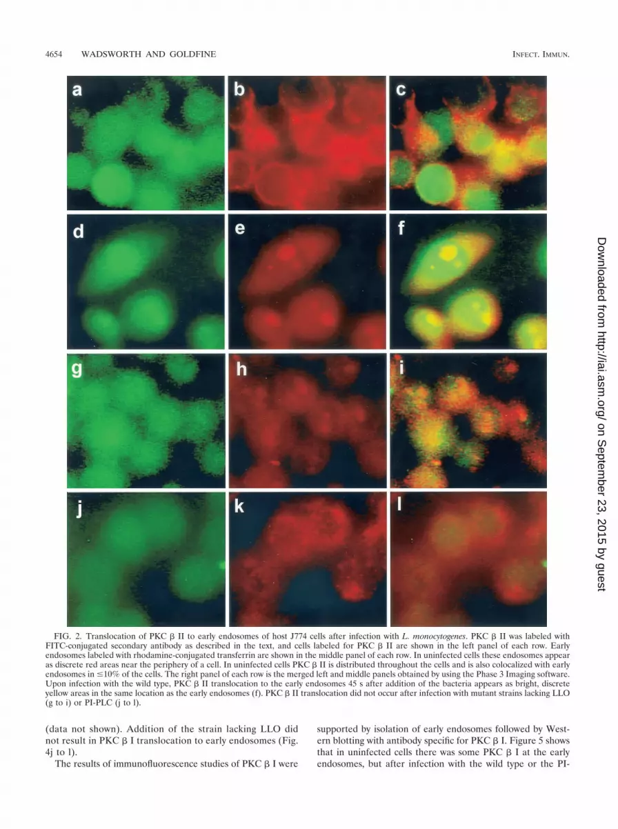
(iii) PKC � II translocation. Using epifluorescence andWestern immunoblotting, we observed that PKC � II translo-cated to early endosomes after infection with the wild type. Inuninfected cells PKC � II was distributed throughout the celland also colocalized with early endosomes (Fig. 2a to c). Thecells in Fig. 2a to c are representative of the population ofuninfected cells in which �10% colocalization of PKC � IIwith early endosomes was routinely observed. Colocalizationof PKC � II with an early endosome is visualized as a discrete,bright yellow area in one cell (Fig. 2c). PKC � II redistribution
TABLE 2. Characterization of early endosomes
Fraction Sp act Total activity % Activity
HRP assaya
PNS 2,080 � 372 6,250 � 1,120 (100)First supernatant 280 � 52 830 � 156 14 � 3.0Pellet 9,180 � 2,633 4,590 � 1,316 70 � 16Early endosomes (10-min pulse) 11,090 � 1,832 5,800 � 955 96 � 12Late endosomes/lysosomes (10-min pulse) 230 � 48 400 � 93 6.7 � 1.6Early endosomes (45-min pulse) 150 � 86 80 � 43 0.90 � 0.40Late endosomes/lysosomes (45-min pulse) 3,850 � 1360 5,770 � 2,040 80 � 11
N-Acetyl-�-D-glucosaminidase assayb
PNS 2.20 � 0.25 6.70 � 0.74 (100)First supernatant 0.067 � 0.04 0.20 � 0.12 3.3 � 2Pellet 13.0 � 7.2 6.7 � 3.6 94 � 40Early endosomes 0.09 � 0.11 0.05 � 0.05 0.7 � 0.9Late endosomes/lysosomes 2.90 � 0.72 4.30 � 1.1 60 � 15
a The HRP assay for five fractions was performed as described in text. The specific activity of the marker was calculated for each preparation and is expressed innanograms of HRP-avidin per milligram of protein, and the average total activity is expressed in nanograms of HRP-avidin per milligram of protein in the total volume.The PNS fraction activity was considered 100%, and the percentage of activity for each of the remaining fractions was determined as follows: total activity infraction/total activity in PNS fraction. The data are means � standard errors for five separate preparations.
b The N-acetyl-�-D-glucosaminidase assay for five fractions was performed as described in text. The specific activity is expressed in absorbance per milligram ofprotein. The PNS fraction activity was considered 100%, and the percentage of activity for each of the remaining fractions was determined as follows: total activity infraction/total activity in PNS fraction. The data are means � the standard errors for five separate preparations.
4652 WADSWORTH AND GOLDFINE INFECT. IMMUN.
on Septem
ber 23, 2015 by guesthttp://iai.asm
.org/D
ownloaded from

to early endosomes occurred within 30 s after addition of thewild-type strain, and the results after 45 s are shown in Fig. 2dto f. Similar redistribution was seen with the BR-PLC mutantstrain (data not shown). Redistribution was not observed after1 min. Figure 2e shows that transferrin-labeled early endo-somes are larger (1 to 6 �m) than the bodies in uninfected cellsand that their distribution is not mainly peripheral. In Fig. 2dseveral of these bodies are labeled with antibody to PKC � II,and in Fig. 2f they are colocalized as discrete yellow bodies.Infection with an LLO-deficient strain (Fig. 2g to i) or a PI-PLC-deficient strain (Fig. 2 j to l) did not result in transloca-tion of the PKC � II isoform, which suggests that PKC � IItranslocation depends on both LLO and PI-PLC. Some changein the distribution of transferrin-labeled early endosomes wasobserved upon infection with the LLO mutant (Fig. 2h), butthese endosomes did not colocalize with PKC � II. An increasein the number of larger endosomes was also detected in cellsinfected with the BR-PLC and double phospholipase mutant
strains (data not shown). Increased size of early endosomesdoes not appear to be coupled to PKC � II translocation.
The results of immunofluorescence studies were confirmedby isolation of early endosomes, followed by Western blottingwith antibody specific for PKC � II. Figure 3A shows that inuninfected cells there was some PKC � II in the early endo-somes, but after 45 s of infection with the wild type or theBR-PLC mutant the amount of PKC � II was greatly in-creased. As was the case for immunofluorescence, this increasewas not seen upon infection with the LLO, PI-PLC, or doublephospholipase mutants.
(iv) PKC � I translocation. In uninfected cells some colo-calization of PKC � I with transferrin-labeled early endosomeswas observed; this occurred in �10% of the cells (Fig. 4a to c).After infection with the wild type (Fig. 4d to f) or the PI-PLCmutant strain (Fig. 4g to i) noticeably more colocalization ofPKC � I with early endosomes was observed within 4 minafter addition of bacteria. As shown in Fig. 4, redistribution oftransferrin-labeled early endosomes occurred after infection,resulting in larger, more centrally located bodies. Colocaliza-tion of PKC � I with early endosomes was also seen afterinfection with the BR-PLC or double phospholipase mutantstrains (data not shown). By 5 min redistribution of PKC �I was no longer observed after infection with the wild type
FIG. 1. Translocation of PKC � after infection with L. monocyto-genes or pretreatment with PMA. J774 cells were infected with differ-ent strains of L. monocytogenes or the same volume of PBS for 45 s andthen processed for immunofluorescence analysis as described in thetext. (A) Panel a, uninfected cells; panel b, uninfected cells treatedwith PMA (100 nM) for 10 min; panels c and d, cells infected with thewild type (PKC � translocation is indicated by a bright green ringaround the periphery of a cell [panel c] or by discrete bright roundareas which appear to be clusters near the periphery of a cell [paneld]); panel e, cells infected with the LLO mutant strain; panel f, cellsinfected with the PI-PLC mutant strain. (B) Effects of inhibitors ontranslocation of PKC �. PBS, SK&F 96365 (25 �M), hispidin (5 �M),or rottlerin (25 �M) was added to J774 cells 30 min prior to infectionwith the wild type. Uninfected cells to which PBS or inhibitor wasadded 30 min prior to infection are shown in the left panel of each row.Control or inhibitor-treated cells infected with the wild type for 45 sare shown in the right panel of each row. Panels a and b, control;panels c and d, cells pretreated with SK&F 96365; panels e and f, cellspretreated with hispidin; panels g and h, cells pretreated with rottlerin.
VOL. 70, 2002 PKC AND L. MONOCYTOGENES 4653
on Septem
ber 23, 2015 by guesthttp://iai.asm
.org/D
ownloaded from
(data not shown). Addition of the strain lacking LLO didnot result in PKC � I translocation to early endosomes (Fig.4j to l).
The results of immunofluorescence studies of PKC � I were
supported by isolation of early endosomes followed by West-ern blotting with antibody specific for PKC � I. Figure 5 showsthat in uninfected cells there was some PKC � I at the earlyendosomes, but after infection with the wild type or the PI-
FIG. 2. Translocation of PKC � II to early endosomes of host J774 cells after infection with L. monocytogenes. PKC � II was labeled withFITC-conjugated secondary antibody as described in the text, and cells labeled for PKC � II are shown in the left panel of each row. Earlyendosomes labeled with rhodamine-conjugated transferrin are shown in the middle panel of each row. In uninfected cells these endosomes appearas discrete red areas near the periphery of a cell. In uninfected cells PKC � II is distributed throughout the cells and is also colocalized with earlyendosomes in �10% of the cells. The right panel of each row is the merged left and middle panels obtained by using the Phase 3 Imaging software.Upon infection with the wild type, PKC � II translocation to the early endosomes 45 s after addition of the bacteria appears as bright, discreteyellow areas in the same location as the early endosomes (f). PKC � II translocation did not occur after infection with mutant strains lacking LLO(g to i) or PI-PLC (j to l).
4654 WADSWORTH AND GOLDFINE INFECT. IMMUN.
on Septem
ber 23, 2015 by guesthttp://iai.asm
.org/D
ownloaded from
PLC mutant the amount of PKC � I was significantly greater.As determined by immunofluorescence, there was no translo-cation of PKC � I upon infection with the LLO mutant. Thetime course of translocation of PKC � I is shown in Fig. 6; itreached a maximum at 4 min and was essentially gone at 5 min.
(v) Effects of inhibitors on PKC � I and PKC � II translo-cation. The effects of the calcium channel blocker SK&F 96365on translocation of both PKC � I and PKC � II were studiedin order to determine calcium requirements for translocationof these molecules. The so-called classical isoforms of PKC,including PKC �, are activated by calcium and/or DAG (26). InJ774 cells, 25 �M SK&F 96365 did not prevent the transloca-tion of PKC � I to early endosomes after infection with thewild type, which indicates that translocation of this isoform wasindependent of calcium influx (Fig. 5). However, PKC � IItranslocation after infection with the wild type or the BR-PLCmutant strain was inhibited in cells treated with SK&F 96365,indicating that translocation of this isoform requires the ini-tial calcium influx observed at 1 min postinfection (35) (Fig.3B).
In contrast, hispidin (5 �M), the PKC � inhibitor, preventedtranslocation of both PKC � I (Fig. 5) and PKC � II (Fig. 3B)to early endosomes after infection with the wild type. At aconcentration of 25 �M, rottlerin, an inhibitor of PKC � atconcentrations lower than those required for inhibition ofother PKC isoforms, did not affect translocation of PKC � I to
early endosomes (data not shown), but it did inhibit translo-cation of PKC � II to early endosomes, which indicates thatPKC � II activation is downstream of PKC � mobilization (Fig.3C).
Effects of inhibitors on calcium signaling. Since wild-type L.monocytogenes and the strain lacking BR-PLC induce an influxof calcium within 1 min after addition of bacteria to cells (35)and rapid translocation of the calcium-independent PKC �isoform and the classical PKC � isoforms, we tested the effectsof the PKC � inhibitor rottlerin on calcium signaling. In un-treated cells the intracellular calcium concentration ([Ca2�]i)was elevated from 184 � 34 to 501 � 66 nM 1 min afterinfection with the wild type, as previously reported (35). Incells pretreated with 25 �M rottlerin for 30 min, the initialcalcium concentration after infection with the wild type wasreduced to 273 � 25 nM (P 0.05, compared to the signal inuntreated cells). Treatment of J774 cells with rottlerin prior toinfection with the BR-PLC (plcB) mutant, which also producesthe first increase in the calcium level (35), gave identical results(data not shown). Pretreatment with 5 �M hispidin for 30 minresulted in no significant inhibition of the calcium signal afterinfection with the wild type (initial calcium concentration, 428� 34 nM).
Effects of hispidin on entry of L. monocytogenes into J774cells. The results described above suggest that PKC � translo-cation precedes the first increase in the calcium level and thatboth of these signals are required for PKC � II translocation.PKC � activation requires DAG, which is provided by theaction of L. monocytogenes PI-PLC on host PI in the presenceof LLO (12). Accordingly, we propose the following signalingpathway: PI-PLC3 DAG3 PKC � translocation3 elevated[Ca2�]i 3 PKC � II translocation. As was the case when thefirst increase in calcium level was prevented by treatment withSK&F 96365 (35), we found that the total number of wild-typebacteria associated with hispidin-treated J774 cells at 1 minwas significantly higher than the number associated with un-treated J774 cells (Fig. 7A).
Internalization of the wild type was also affected by hispidintreatment. The percentage of internalized wild-type L. mono-cytogenes cells was significantly higher in treated J774 cells thanin untreated J774 cells (Fig. 7C). The percentage of internal-ized wild-type bacteria remained significantly elevated in his-pidin-treated J774 cells until 20 min. There was a decline from67% � 2.9% at 10 min to 38% � 2.3% at 20 min. This declinein part reflected the increased total number of bacteria asso-ciated with hispidin-treated cells at 20 min (Fig. 7A). Similarincreases in internalization of the BR-PLC mutant were alsoseen after treatment with hispidin (data not shown). Whilethese results suggest a role for PKC � activation in controllingentry of the wild type at very early time points, they do notdistinguish between the PKC � I and PKC � II isoforms.
Since hispidin affects both PKC � I and PKC � II translo-cation and since the PI-PLC mutant activates only PKC � I, wetested the effects of hispidin on the entry kinetics of this strainto determine if PKC � I translocation has a role in controllingentry of L. monocytogenes. We found that hispidin did not havea significant effect on the total number of cells of this strainassociated with J774 cells (Fig. 7B). Entry of the PI-PLC mu-tant strain into hispidin-treated cells was also not significantlydifferent from entry into untreated cells during the 20-min
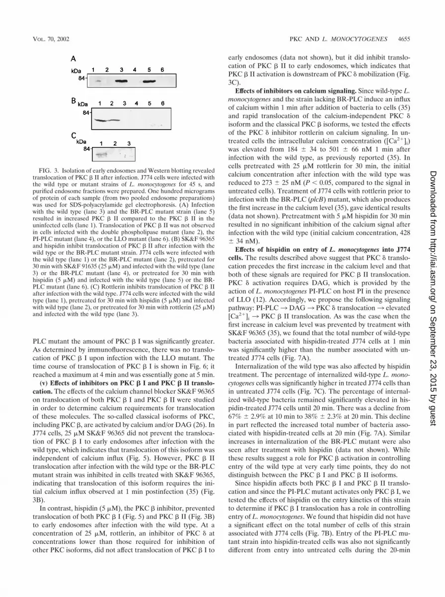
FIG. 3. Isolation of early endosomes and Western blotting revealedtranslocation of PKC � II after infection. J774 cells were infected withthe wild type or mutant strains of L. monocytogenes for 45 s, andpurified endosome fractions were prepared. One hundred microgramsof protein of each sample (from two pooled endosome preparations)was used for SDS-polyacrylamide gel electrophoresis. (A) Infectionwith the wild type (lane 3) and the BR-PLC mutant strain (lane 5)resulted in increased PKC � II compared to the PKC � II in theuninfected cells (lane 1). Translocation of PKC � II was not observedin cells infected with the double phospholipase mutant (lane 2), thePI-PLC mutant (lane 4), or the LLO mutant (lane 6). (B) SK&F 96365and hispidin inhibit translocation of PKC � II after infection with thewild type or the BR-PLC mutant strain. J774 cells were infected withthe wild type (lane 1) or the BR-PLC mutant (lane 2), pretreated for30 min with SK&F 91635 (25 �M) and infected with the wild type (lane3) or the BR-PLC mutant (lane 4), or pretreated for 30 min withhispidin (5 �M) and infected with the wild type (lane 5) or the BR-PLC mutant (lane 6). (C) Rottlerin inhibits translocation of PKC � IIafter infection with the wild type. J774 cells were infected with the wildtype (lane 1), pretreated for 30 min with hispidin (5 �M) and infectedwith wild type (lane 2), or pretreated for 30 min with rottlerin (25 �M)and infected with the wild type (lane 3).
VOL. 70, 2002 PKC AND L. MONOCYTOGENES 4655
on Septem
ber 23, 2015 by guesthttp://iai.asm
.org/D
ownloaded from

infection period (Fig. 7D). These results, which suggest thatboth PKC � I and PKC � II control the entry of L. monocy-togenes into J774 cells, are discussed below.
Effects of hispidin on escape from the primary vacuole.
When J774 cells were treated with the calcium signal inhibitorsSK&F 96365 and thapsigargin and infected with wild-type L.monocytogenes, the percentage of internalized bacteria thatescaped from the primary vacuole decreased from 44% to 14
FIG. 4. PKC � I translocation occurred after infection with the wild type or the PI-PLC mutant strain but not after infection with the LLOmutant strain. PKC � I was labeled with FITC-conjugated secondary antibody as described in the text, and cells labeled for PKC � I are shown in theleft panel of each row. Early endosomes were labeled with rhodamine-conjugated transferrin and appear in the middle panel of each row as discrete redareas. In uninfected cells PKC � I was located throughout the cytosol, could also appear as a discrete green area around the periphery of a cell, and couldbe colocalized with early endosomes. Cells were infected with the strains for 4 min. PKC � I translocation after infection with the wild-type strain (d tof) or the PI-PLC mutant strain (g to i) appears as discrete bright yellow areas corresponding to PKC � I colocalization with transferrin-labeled earlyendosomes. The images in the right panels were obtained as described in the legend to Fig. 2. Cells infected with the strain lacking LLO did notshow translocation of PKC � I to early endosomes (j to l). Bright green objects seen especially in panels j and l are FITC-labeled bacteria.
4656 WADSWORTH AND GOLDFINE INFECT. IMMUN.
on Septem
ber 23, 2015 by guesthttp://iai.asm
.org/D
ownloaded from

and 21%, respectively (35). These results suggested that cal-cium signaling affected the ability of L. monocytogenes to leavethe primary phagosome. Since calcium signaling is required fortranslocation of PKC � II to early endosomes, we examinedthe effects of inhibition of PKC � translocation by hispidin onescape. Like SK&F 96365, hispidin reduced the percentage ofwild-type bacteria that escaped at 1.5 h after infection from 43to 15% (Fig. 8A). The ability of the PI-PLC mutant to escapefrom the primary vacuole is significantly impaired compared tothe ability of the wild type (5, 30). When J774 cells weretreated with hispidin and infected with the PI-PLC mutant, inwhich PKC � I but not PKC � II was translocated to earlyendosomes, the percentage of bacteria that escaped was mark-edly reduced (from 23 to 4.5%) (Fig. 8B), suggesting an im-portant role for PKC � I in determining eventual escape fromthe primary phagocytic vacuole.
DISCUSSION
The observation that elevations in cytosolic calcium levelsoccurred 1 and 5 min after infection of J774 cells with wild-typeL. monocytogenes (35) and the finding that PKC � and PKC �isoforms were translocated within the first 5 min after infectionformed the basis for the present study, in which we examinedthe relationships among cytosolic calcium, bacterial phospho-lipase activities, and host PKC activation. We present evidencethat there is coupling of PKC �, calcium signaling, and PKC� II translocation in J774 cells after infection with L. monocy-togenes and demonstrate that pharmacological inhibition ofcalcium signals disrupts the signaling pathway. We also dem-
onstrate that an apparently calcium-independent but LLO-dependent translocation of PKC � I occurs.
The observation that PKC � translocates to the cell periph-ery by 30 s after infection is consistent with our previous hy-pothesis that it plays a role in the influx of calcium from theextracellular fluid that was observed to peak at 1 min afterinfection (35). Treatment with rottlerin, a relatively specificPKC � inhibitor, resulted in significant inhibition of calciumsignaling. Although recent work indicates that rottlerin may actas a mitochondrial uncoupler which reduces ATP levels (31),the specific effects of this compound on PKC � and PKC � II,but not on PKC � I translocation, are consistent with a path-way from PKC � to PKC � II. Inhibition of calcium entry withthe channel blocker SK&F 96365 did not prevent PKC � trans-location, whereas presumed inhibition of PKC activation by 3 h
FIG. 5. Isolation of early endosomes and Western blotting revealedtranslocation of PKC � I after infection. J774 cells were infected withwild-type or mutant L. monocytogenes for 4 min, and purified endo-some fractions were prepared. One hundred micrograms of protein ofeach sample (from two pooled endosome preparations) was used forSDS-polyacrylamide gel electrophoresis followed by Western blotting,as described in the text. PKC � I translocation was observed in cellsinfected with the wild type (lane 3) and the PI-PLC mutant (lane 6)compared to uninfected cells (lane 1). Translocation was not seen afterinfection with the LLO mutant (lane 2) or after 30 min of pretreatmentwith hispidin and infection with the wild type (lane 5). Cells pretreatedwith SK&F 96365 for 30 min showed translocation of PKC � I (lane 4).
FIG. 6. Time course of translocation of PKC � I to early endo-somes. Cells were infected with the wild type and harvested at intervalsafter infection. Early endosomes were isolated, and proteins wereprepared for SDS-polyacrylamide gel electrophoresis and Westernblotting as described in Materials and Methods. The proteins were alsoblotted with antibody to the transferrin receptor (upper bands). Infec-tions were carried out for 1 min (lane 1), 3 min (lane 2), 4 min (lane3), and 5 min (lane 4).
FIG. 7. Effects of hispidin on association of L. monocytogenes withJ774 cells and entry of L. monocytogenes into J774 cells. (A) Associa-tion of the wild type with untreated J774 cells (E) or cells pretreatedwith hispidin (5 �M) (�). (B) Association of the PI-PLC mutant withuntreated J774 cells (�) or cells pretreated with hispidin (5 �M) (�).(C) Percentage of associated wild-type bacteria that were internalizedin untreated J774 cells (E) or cells pretreated with hispidin (5 �M)(�). (D) Percentage of associated PI-PLC mutant bacteria that wereinternalized in untreated J774 cells (�) or cells pretreated for 30 minwith hispidin (5 �M) (�). The total number of bacteria associatedwith the J774 cells was calculated as follows: total number of FITC-labeled bacteria (green)/total number of cells per image. The resultsare expressed as averages � standard errors of the means for three setsof experiments in which a total of 200 to 250 J774 cells were counted.For association of the wild type in cells treated with hispidin P 0.05at 1, 5, and 10 min compared to the association for untreated cells. Thepercentage of internalized bacteria was calculated as described in thetext. The results are expressed as averages � standard errors of themeans for three sets of experiments in which a total of 200 to 250 J774cells were counted. For the percentage of internalized wild-type bac-teria in cells treated with hispidin P 0.05 compared to the percentagefor untreated cells at 1, 5, and 10 min.
VOL. 70, 2002 PKC AND L. MONOCYTOGENES 4657
on Septem
ber 23, 2015 by guesthttp://iai.asm
.org/D
ownloaded from

of pretreatment with PMA prevented the initial increase in[Ca2�]i (data not shown). PKC � has been shown to be in-volved in opening plasma membrane calcium channels (20, 34).
Based on these findings, we propose the following model forthe coupling of intracellular calcium and PKC activation.Translocation of PKC � to the periphery of the cell occursupon addition of wild-type L. monocytogenes or the BR-PLCmutant strain but not upon addition of mutants with mutationsin PI-PLC or LLO genes, which indicates that there is a re-quirement for at least two bacterial proteins, LLO and PI-PLC.Presumably, the combined activity of these proteins leads tohydrolysis of host PI and the formation of DAG (12, 27). Wepropose that this is responsible for the translocation of PKC �,a calcium-independent isoform, and the subsequent initial el-evation of intracellular calcium levels (Fig. 9), although a di-rect connection between PKC � translocation and opening ofthe calcium channel has not been formally demonstrated.
Translocation of PKC � II to early endosomes of J774 cellsalso began within 30 s after addition of L. monocytogenes. Thisrequired both LLO and PI-PLC and was also inhibited byrottlerin and the calcium channel blocker SK&F 96365. Thus,translocation of this isoform appears to be coupled to PKC �translocation and calcium signaling. In contrast to PKC � IItranslocation, PKC � I translocation to the early endosomes
occurred after infection with all of the strains tested except thestrain lacking LLO. Since no calcium signaling was observedwith the double phospholipase mutant (35), it appears thatLLO is responsible for initiating a pulse of DAG, which in thiscase appears to be sufficient for PKC � I translocation (26).Consistent with this is our finding that the double phospho-lipase mutant, but not the LLO mutant strain, activates hostpolyphosphoinositide-specific PLC (12). PKC � I translocationwas not inhibited by SK&F 96365, also indicating that eleva-tion of the calcium level is not required for PKC � I translo-cation to early endosomes. Translocation of PKC � I and PKC� II to early endosomes has not been reported previously.
The PI-PLC and the double phospholipase deletion strains,which mobilize PKC � I but not PKC � II or PKC �, demon-strated higher early association with J774 cells and more rapidentry than strains that mobilize all three PKC isoforms (Fig. 7).We suggest, therefore, that PKC � I activation permits earlierand more rapid bacterial entry and that PKC � II activationappears to modify this effect by temporarily slowing bacterialentry. When neither PKC � I nor PKC � II is translocated, asis the case with a mutant with a mutation in the LLO gene orwith heat-killed bacteria, uptake is also rapid (35), suggestingthat this rate is the rate for particles that bind to surfacereceptors and are unable to modify the host response. In arecent study, translocation of two nonclassical PKC isoforms,PKC � and PKC ε, was associated with uptake of opsonizedparticles into RAW 264.7 cells via the Fc receptor (19). Thesestudies and those reported here suggest that PKC � transloca-tion and downstream events may play a role in both Fc re-ceptor-mediated and Fc receptor-independent uptake.
As shown in Fig. 8, inhibition of PKC � translocation byhispidin had dramatic effects on the ability of both wild-typeand PI-PLC mutant bacteria to escape from the primary vac-uole. During infection of J774 cells with the wild type, 43% of
FIG. 8. Hispidin pretreatment inhibited escape of the wild type andthe PI-PLC mutant from the phagosome. Bacteria that escape fromthe phagosome and enter the cytosol become decorated with polymer-ized actin and can be stained with fluorescent phalloidin. After re-moval of uninternalized bacteria with gentamicin at 30 min, the per-centage of cells that escaped at 1.5 h after infection was calculated asdescribed in Materials and Methods. (A) Cells were either not pre-treated (control) or pretreated with hispidin (5 �M) for 30 min andinfected with the wild type. The data are the averages � standarderrors of the means for three experiments, and the difference is highlysignificant (P 0.001). The data for cells pretreated with SK&F 96365are taken from a previous study, in which the control gave an identicalvalue (35). (B) Cells were pretreated with hispidin (5 �M) for 30 minand infected with the PI-PLC mutant. The data are averages � stan-dard errors of the means for three experiments, and the difference ishighly significant (P 0.01).
FIG. 9. Suggested model for coupling of PKC activation and cal-cium signaling in host J774 cells after infection with L. monocytogenes.As described in the text, we suggest that PKC �, calcium influx, andPKC � II activation are coupled and are initiated as a result of activityof two listerial proteins, LLO and PI-PLC. Activation of PKC � I didnot require PI-PLC or elevated [Ca2�]i, and we suggest that it occursas a result of the formation of DAG as a result of the action of hostPLC on phosphatidylinositol 4,5-bisphosphate (PIP2) (12), yieldinginositol 1,4,5-triphosphate (IP3).
4658 WADSWORTH AND GOLDFINE INFECT. IMMUN.
on Septem
ber 23, 2015 by guesthttp://iai.asm
.org/D
ownloaded from

the internalized bacteria had escaped from the primary vacuoleat 1.5 h after infection. When cells were pretreated with thecalcium channel blocker SK&F 96365, which blocked all cal-cium signaling, and with thapsigargin, which emptied cellularcalcium stores and blocked the second and third calcium sig-nals, the ability of the wild type to escape was reduced to 14and 21%, respectively (35). Since blockage of the calcium sig-nals inhibited PKC � II translocation, we tested the effects ofhispidin, which blocked PKC � I and PKC � II translocation,on escape from the primary vacuole and observed that escapewas reduced from 43 to 15%, paralleling the results obtainedwith inhibitors of calcium signaling. Infection of J774 cells withthe mutant with a deletion in the PI-PLC gene resulted intranslocation of PKC � I but not translocation of PKC � II.When cells were treated with hispidin and infected with thePI-PLC mutant, escape was reduced from 23 to 4.5% (Fig. 8B).These results strongly suggest that PKC signaling has a pro-found effect on later events during an infection. In order todetermine if PKC signals in addition to those observed duringthe initial stages of infection are required, we performed ex-periments in which hispidin was removed from the medium 5and 15 min after infection with the wild type. We observed thatwashing the cells at these times, which were earlier than thenormal wash time (30 min), resulted in escape of fewer bacte-ria into the cytosol than usually observed (27 versus 38% inthese experiments). When hispidin was removed at either 5 or15 min after infection, the percentage of bacteria that escapedinto the cytosol did not differ from the percentage in controlsto which hispidin had not been added (data not shown). Theseresults suggest that PKC � signaling after internalization playsa role in the ability of L. monocytogenes to exit the primaryvacuole.
The finding that early endosomes enlarge upon infectionwith the wild type (Fig. 2 and 4) is intriguing and deservesfurther study. Rab5 is thought to play an important role inregulating early-endosome fusion, and large endosomesthought to arise from fusion have been associated with over-expression of Rab5 or expression of the GTPase mutant Rab5:Q79L (3, 21, 32). In an in vitro system, phagosomes containinglive LLO�L. monocytogenes recruited Rab5 faster than phago-somes containing dead bacteria recruited Rab5 (1).
Generation of the host signals appears to be coupled tospecific bacterial proteins, and thus L. monocytogenes is able tomanipulate host signaling mechanisms to ensure its own sur-vival. These studies, along with our previous work on calciumsignaling in J774 cells (35) and studies of endothelial cells (27,28) and neutrophils (29), draw additional attention to the en-gagement of secreted proteins, such as LLO and the two phos-pholipases, with host cells prior to uptake of the bacteria.Recent work with the extracellular pathogen Streptococcus pyo-genes demonstrated a role for streptolysin O, a member of thesame family of cholesterol-dependent cytolysins as LLO, inpermitting the entry of a bacterial protein into the host cell,analogous to the type III secretion system of gram-negativebacteria (22). A similar model was proposed for LLO andPI-PLC (27), and the present results and previous results (35)support such a model (Fig. 9). Expression of both LLO andPI-PLC is increased upon entry of L. monocytogenes into thephagocytic vacuole (2, 16, 24). Distinguishing between the ef-fects of these proteins before and after entry into the host cell
on the fate of L. monocytogenes in a macrophage will undoubt-edly be the focus of considerable effort in the future.
ACKNOWLEDGMENTS
This work was supported by Public Health Service grants GM 52797and AI 45153 (to H.G.) and by the University of Pennsylvania Re-search Foundation.
REFERENCES
1. Alvarez-Dominguez, C., A. M. Barbieri, W. Beron, A. Wandinger-Ness, andP. D. Stahl. 1996. Phagocytosed live Listeria monocytogenes influences rab5-regulated in vitro phagosome-endosome fusion. J. Biol. Chem. 271:13834–13843.
2. Bubert, A., Z. Sokolovic, S. K. Chun, L. Papatheodorou, A. Simm, and W.Goebel. 1999. Differential expression of Listeria monocytogenes virulencegenes in mammalian host cells. Mol. Gen. Genet. 261:323–336.
3. Bucci, C., R. G. Parton, I. H. Mather, H. Stunnenberg, K. Simons, B.Hoflack, and M. Zerial. 1992. The small GTPase rab5 functions as a regu-latory factor in the early endocytic pathway. Cell 70:715–728.
4. Camilli, A., H. Goldfine, and D. A. Portnoy. 1991. Listeria monocytogenesmutants lacking phosphatidylinositol-specific phospholipase C are avirulent.J. Exp. Med. 173:751–754.
5. Camilli, A., L. G. Tilney, and D. A. Portnoy. 1993. Dual roles of plcA inListeria monocytogenes pathogenesis. Mol. Microbiol. 8:143–157.
6. Claus, V., A. Jahraus, T. Tjelle, T. Berg, H. Kirschke, H. Faulstich, and G.Griffiths. 1998. Lysosomal enzyme trafficking between phagosomes, endo-somes, and lysosomes in J774 macrophages—enrichment of cathepsin H inearly endosomes. J. Biol. Chem. 273:9842–9851.
7. Diaz, R., L. Mayorga, and P. Stahl. 1988. In vitro fusion of endosomesfollowing receptor-mediated endocytosis. J. Biol. Chem. 263:6093–6100.
8. Drevets, D. A., and P. A. Campbell. 1991. Macrophage phagocytosis: use offluorescence microscopy to distinguish between extracellular and intracellu-lar bacteria. J. Immunol. Methods 142:31–38.
9. Findlay, J., G. A. Levvy, and C. A. Marsh. 1958. Inhibition of glycosidases byaldonolactones of corresponding configuration. Biochem. J. 69:467–476.
10. Geoffroy, C., J. Raveneau, J.-L. Beretti, A. Lecroisey, J.-A. Vazquez-Boland,J. E. Alouf, and P. Berche. 1991. Purification and characterization of anextracellular 29-kilodalton phospholipase C from Listeria monocytogenes.Infect. Immun. 59:2382–2388.
11. Goldfine, H., N. C. Johnston, and C. Knob. 1993. The nonspecific phospho-lipase C of Listeria monocytogenes: activity on phospholipids in Triton X-100mixed micelles and in biological membranes. J. Bacteriol. 175:4298–4306.
12. Goldfine, H., S. J. Wadsworth, and N. C. Johnston. 2000. Activation of hostphospholipases C and D in macrophages after infection with Listeria mono-cytogenes. Infect. Immun. 68:5735–5741.
13. Gonindard, C., C. Bergonzi, C. Denier, C. Sergheraert, A. Klaebe, L. Cha-vant, and E. Hollande. 1997. Synthetic hispidin, a PKC inhibitor, is morecytotoxic toward cancer cells than normal cells in vitro. Cell Biol. Toxicol.13:141–153.
14. Gschwendt, M., H. J. Muller, K. Kielbassa, R. Zang, W. Kittstein, G. Rincke,and E. Marks. 1994. Rottlerin, a novel protein kinase inhibitor. Biochem.Biophys. Res. Commun. 199:93–98.
15. Jones, S., and D. A. Portnoy. 1994. Characterization of Listeria monocyto-genes pathogenesis in a strain expressing perfringolysin O in place of list-eriolysin O. Infect. Immun. 62:5608–5613.
16. Klarsfeld, A. D., P. L. Goossens, and P. Cossart. 1994. Five Listeria mono-cytogenes genes preferentially expressed in infected mammalian cells: plcA,purH, purD, pyrE and an arginine ABC transporter gene, arpJ. Mol. Micro-biol. 13:585–597.
17. Kontny, E., M. Kurowska, K. Szczepanska, and W. Maslinski. 2000. Rot-tlerin, a PKC isozyme-selective inhibitor, affects signaling events and cyto-kine production in human monocytes. J. Leukoc. Biol. 67:249–258.
18. Lang, T., and C. De Chastellier. 1985. Fluid phase and mannose receptor-mediated uptake of horseradish peroxidase in mouse bone marrow-derivedmacrophages. Biochemical and ultrastructural study. Biol. Cell 53:149–154.
19. Larsen, E. C., J. A. DiGennaro, N. Saito, S. Mehta, D. J. Loegering, J. E.Mazurkiewicz, and M. R. Lennartz. 2000. Differential requirement for clas-sic and novel PKC isoforms in respiratory burst and phagocytosis in RAW264.7 cells. J. Immunol. 165:2809–2817.
20. Levin, R., A. Braiman, and Z. Priel. 1997. Protein kinase C induced calciuminflux and sustained enhancement of ciliary beating by extracellular ATP.Cell Calcium 21:103–113.
21. Li, G., M. A. Barbieri, M. I. Colombo, and P. D. Stahl. 1994. Structuralfeatures of the GTP-binding defective Rab5 mutants required for theirinhibitory activity on endocytosis. J. Biol. Chem. 269:14631–14635.
22. Madden, J. C., N. Ruiz, and M. Caparon. 2001. Cytolysin-mediated trans-location (CMT): a functional equivalent of type III secretion in Gram-positive bacteria. Cell 104:143–152.
23. Mengaud, J., C. Braun-Breton, and P. Cossart. 1991. Identification of phos-
VOL. 70, 2002 PKC AND L. MONOCYTOGENES 4659
on Septem
ber 23, 2015 by guesthttp://iai.asm
.org/D
ownloaded from

phatidylinositol-specific phospholipase C activity in Listeria monocytogenes: anovel type of virulence factor. Mol. Microbiol. 5:367–372.
24. Moors, M. A., B. Levitt, P. Youngman, and D. A. Portnoy. 1999. Expressionof listeriolysin O and ActA by intracellular and extracellular Listeria mono-cytogenes. Infect. Immun. 67:131–139.
25. Portnoy, D. A., P. S. Jacks, and D. J. Hinrichs. 1988. Role of hemolysin forthe intracellular growth of Listeria monocytogenes. J. Exp. Med. 167:1459–1471.
26. Ron, D., and M. G. Kazanietz. 1999. New insights into the regulation ofprotein kinase C and novel phorbol ester receptors. FASEB J. 13:1658–1676.
27. Sibelius, U., T. Chakraborty, B. Krogel, J. Wolf, F. Rose, R. Schmidt, J.Wehland, W. Seeger, and F. Grimminger. 1996. The listerial exotoxins list-eriolysin and phosphatidylinositol-specific phospholipase C synergize toelicit endothelial cell phosphoinositide metabolism. J. Immunol. 157:4055–4060.
28. Sibelius, U., F. Rose, T. Chakraborty, A. Darji, J. Wehland, S. Weiss, W.Seeger, and F. Grimminger. 1996. Listeriolysin is a potent inducer of thephosphatidylinositol response and lipid mediator generation in human en-dothelial cells. Infect. Immun. 64:674–676.
29. Sibelius, U., E. C. Schulz, F. Rose, K. Hattar, T. Jacobs, S. Weiss, T.Chakraborty, W. Seeger, and F. Grimminger. 1999. Role of Listeria mono-
cytogenes exotoxins listeriolysin and phosphatidylinositol-specific phospho-lipase C in activation of human neutrophils. Infect. Immun. 67:1125–1130.
30. Smith, G. A., H. Marquis, S. Jones, N. C. Johnston, D. A. Portnoy, and H.Goldfine. 1995. The two distinct phospholipases C of Listeria monocytogeneshave overlapping roles in escape from a vacuole and cell-to-cell spread.Infect. Immun. 63:4231–4237.
31. Soltoff, S. P. 2001. Rottlerin is a mitochondrial uncoupler that decreasescellular ATP levels and indirectly blocks protein kinase C� tyrosine phos-phorylation. J. Biol. Chem. 276:37986–37992.
32. Stenmark, H., R. G. Parton, O. Steele-Mortimer, A. Lutcke, J. Gruenberg,and M. Zerial. 1994. Inhibition of rab5 GTPase activity stimulates mem-brane fusion in endocytosis. EMBO J. 13:1287–1296.
33. Tilney, L. G., and D. A. Portnoy. 1989. Actin filaments and the growth,movement, and spread of the intracellular bacterial parasite Listeria mono-cytogenes. J. Cell Biol. 109:1597–1608.
34. Vazquez, G., and A. R. de Boland. 1996. Involvement of protein kinase C inthe modulation of 1�,25-dihydroxy-vitamin D3-induced 45Ca2� uptake in ratand chick cultured myoblasts. Biochim. Biophys. Acta 1310:157–162.
35. Wadsworth, S. J., and H. Goldfine. 1999. Listeria monocytogenes phospho-lipase C-dependent calcium signaling modulates bacterial entry into J774macrophage-like cells. Infect. Immun. 67:1770–1778.
Editor: E. I. Tuomanen
4660 WADSWORTH AND GOLDFINE INFECT. IMMUN.
on Septem
ber 23, 2015 by guesthttp://iai.asm
.org/D
ownloaded from