Analysis of carbon substrates used by Listeria monocytogenes during growth in J774A.1 macrophages...
14
ORIGINAL RESEARCH ARTICLE published: 03 November 2014 doi: 10.3389/fcimb.2014.00156 Analysis of carbon substrates used by Listeria monocytogenes during growth in J774A.1 macrophages suggests a bipartite intracellular metabolism Stephanie Grubmüller 1‡ , Kristina Schauer 2†‡ , Werner Goebel 3 , Thilo M. Fuchs 2 and Wolfgang Eisenreich 1 * 1 Lehrstuhl für Biochemie, Technische Universität München, Garching, Germany 2 Abteilung Mikrobiologie, Zentralinstitut für Ernährungs- und Lebensmittelforschung (ZIEL), Technische Universität München, Freising, Germany 3 Department for Bacteriology, Max von Pettenkofer Institute, Ludwig-Maximilians-Universität, München, Germany Edited by: Victor J. Torres, New York University School of Medicine, USA Reviewed by: Stephanie M. Seveau, The Ohio State University, USA John-Demian Sauer, University of Wisconsin-Madison, USA *Correspondence: Wolfgang Eisenreich, Lehrstuhl für Biochemie, Technische Universität München, Lichtenbergstr. 4, 85747 Garching, Germany e-mail: wolfgang.eisenreich@ ch.tum.de † Present address: Lehrstuhl für Hygiene und Technologie der Milch, Tiermedizinische Fakultät, Ludwig-Maximilians-Universität, München, Germany ‡ These authors have contributed equally to this work. Intracellular bacterial pathogens (IBPs) are dependent on various nutrients provided by the host cells. Different strategies may therefore be necessary to adapt the intracellular metabolism of IBPs to the host cells. The specific carbon sources, the catabolic pathways participating in their degradation, and the biosynthetic performances of IBPs are still poorly understood. In this report, we have exploited the technique of 13 C-isotopologue profiling to further study the carbon metabolism of Listeria monocytogenes by using the EGDe wild-type strain and mutants (defective in the uptake and/or catabolism of various carbon compounds) replicating in J774A.1 macrophages. For this goal, the infected macrophages were cultivated in the presence of [1,2- 13 C 2 ]glucose, [U- 13 C 3 ]glycerol, [U- 13 C 3 ]pyruvate, [U- 13 C 3 ]lactate, or a mix of [U- 13 C]amino acids. GC/MS-based isotopologue profiling showed efficient utilization of amino acids, glucose 6-phosphate, glycerol, and (at a low extent) also of lactate but not of pyruvate by the IBPs. Most amino acids imported from the host cells were directly used for bacterial protein biosynthesis and hardly catabolized. However, Asp was de novo synthesized by the IBPs and not imported from the host cell. As expected, glycerol was catabolized via the ATP-generating lower part of the glycolytic pathway, but apparently not used for gluconeogenesis. The intermediates generated from glucose 6-phosphate in the upper part of the glycolytic pathway and the pentose phosphate shunt likely serve primarily for anabolic purposes (probably for the biosynthesis of cell wall components and nucleotides). This bipartite bacterial metabolism which involves at least two major carbon substrates—glycerol mainly for energy supply and glucose 6-phosphate mainly for indispensible anabolic performances—may put less nutritional stress on the infected host cells, thereby extending the lifespan of the host cells to the benefit of the IBPs. Keywords: bacterial metabolism, bacterial pathogensis, intracellular bacteria, isotopic tracers, isotopologue profiling, Listeria monocytogenes INTRODUCTION Listeria monocytogenes is a Gram-positive, food-borne pathogen that can cause systemic infections in immune compromised, pregnant or elder persons (for recent reviews, see Velge and Roche, 2010; Camejo et al., 2011; Fuchs et al., 2012; Mostowy and Cossart, 2012; Cossart and Lebreton, 2014). Typical symptoms of listeriosis are septicaemia, (encephalo)-meningitis, placenti- tis, and stillbirth. The facultative intracellular pathogen is taken up by professional phagocytes, like macrophages and dendritic cells. It can also actively invade (with the help of the inter- nalins A and/or B) non-phagocytic cells, such as epithelial cells, fibroblasts or endothelial cells (Dussurget et al., 2004; Lecuit, 2005; Hamon et al., 2006). The subsequent escape of the bac- teria from the enclosing vacuole depends on listeriolysin and two phospholipases (PlcA and PlcB). Within the cytosol of the host cell, L. monocytogenes efficiently multiplies with a generation time of approximately 1 h and spreads into neighboring host cells (Hamon et al., 2012). The growth of intracellular bacterial pathogens (IBPs) depends on the efficient usage of carbon and nitrogen nutrients from the host. The metabolism of mammalian host cells involves hundreds if not thousands of metabolites that could be used by intracellu- lar bacteria as potential nutrients. The major catabolic reactions of the host cells occur in the cytosol (e.g., glycolysis, pentose- phosphate pathway) or in the mitochondria (e.g., citrate cycle, β-oxidation of fatty acids, glutaminolysis), but metabolites can also be exchanged between these compartments. The anabolic pathways (formation of glucose, amino acids, nucleotides, and fatty acids) mainly take place in the cytosol. Therefore, intracel- lular bacteria living in the cytosolic compartment of host cells Frontiers in Cellular and Infection Microbiology www.frontiersin.org November 2014 | Volume 4 | Article 156 | 1 CELLULAR AND INFECTION MICROBIOLOG Y
-
Upload
independent -
Category
Documents
-
view
3 -
download
0
Transcript of Analysis of carbon substrates used by Listeria monocytogenes during growth in J774A.1 macrophages...

ORIGINAL RESEARCH ARTICLEpublished: 03 November 2014
doi: 10.3389/fcimb.2014.00156
Analysis of carbon substrates used by Listeriamonocytogenes during growth in J774A.1 macrophagessuggests a bipartite intracellular metabolismStephanie Grubmüller1‡, Kristina Schauer2†‡, Werner Goebel3, Thilo M. Fuchs2 and
Wolfgang Eisenreich1*
1 Lehrstuhl für Biochemie, Technische Universität München, Garching, Germany2 Abteilung Mikrobiologie, Zentralinstitut für Ernährungs- und Lebensmittelforschung (ZIEL), Technische Universität München, Freising, Germany3 Department for Bacteriology, Max von Pettenkofer Institute, Ludwig-Maximilians-Universität, München, Germany
Edited by:
Victor J. Torres, New York UniversitySchool of Medicine, USA
Reviewed by:
Stephanie M. Seveau, The OhioState University, USAJohn-Demian Sauer, University ofWisconsin-Madison, USA
*Correspondence:
Wolfgang Eisenreich, Lehrstuhl fürBiochemie, Technische UniversitätMünchen, Lichtenbergstr. 4,85747 Garching, Germanye-mail: [email protected]†Present address:
Lehrstuhl für Hygiene undTechnologie der Milch,Tiermedizinische Fakultät,Ludwig-Maximilians-Universität,München, Germany
‡These authors have contributedequally to this work.
Intracellular bacterial pathogens (IBPs) are dependent on various nutrients provided bythe host cells. Different strategies may therefore be necessary to adapt the intracellularmetabolism of IBPs to the host cells. The specific carbon sources, the catabolic pathwaysparticipating in their degradation, and the biosynthetic performances of IBPs are still poorlyunderstood. In this report, we have exploited the technique of 13C-isotopologue profilingto further study the carbon metabolism of Listeria monocytogenes by using the EGDewild-type strain and mutants (defective in the uptake and/or catabolism of various carboncompounds) replicating in J774A.1 macrophages. For this goal, the infected macrophageswere cultivated in the presence of [1,2-13C2]glucose, [U-13C3]glycerol, [U-13C3]pyruvate,[U-13C3]lactate, or a mix of [U-13C]amino acids. GC/MS-based isotopologue profilingshowed efficient utilization of amino acids, glucose 6-phosphate, glycerol, and (at a lowextent) also of lactate but not of pyruvate by the IBPs. Most amino acids importedfrom the host cells were directly used for bacterial protein biosynthesis and hardlycatabolized. However, Asp was de novo synthesized by the IBPs and not imported fromthe host cell. As expected, glycerol was catabolized via the ATP-generating lower part ofthe glycolytic pathway, but apparently not used for gluconeogenesis. The intermediatesgenerated from glucose 6-phosphate in the upper part of the glycolytic pathway and thepentose phosphate shunt likely serve primarily for anabolic purposes (probably for thebiosynthesis of cell wall components and nucleotides). This bipartite bacterial metabolismwhich involves at least two major carbon substrates—glycerol mainly for energy supplyand glucose 6-phosphate mainly for indispensible anabolic performances—may put lessnutritional stress on the infected host cells, thereby extending the lifespan of the hostcells to the benefit of the IBPs.
Keywords: bacterial metabolism, bacterial pathogensis, intracellular bacteria, isotopic tracers, isotopologue
profiling, Listeria monocytogenes
INTRODUCTIONListeria monocytogenes is a Gram-positive, food-borne pathogenthat can cause systemic infections in immune compromised,pregnant or elder persons (for recent reviews, see Velge andRoche, 2010; Camejo et al., 2011; Fuchs et al., 2012; Mostowy andCossart, 2012; Cossart and Lebreton, 2014). Typical symptomsof listeriosis are septicaemia, (encephalo)-meningitis, placenti-tis, and stillbirth. The facultative intracellular pathogen is takenup by professional phagocytes, like macrophages and dendriticcells. It can also actively invade (with the help of the inter-nalins A and/or B) non-phagocytic cells, such as epithelial cells,fibroblasts or endothelial cells (Dussurget et al., 2004; Lecuit,2005; Hamon et al., 2006). The subsequent escape of the bac-teria from the enclosing vacuole depends on listeriolysin andtwo phospholipases (PlcA and PlcB). Within the cytosol of the
host cell, L. monocytogenes efficiently multiplies with a generationtime of approximately 1 h and spreads into neighboring host cells(Hamon et al., 2012).
The growth of intracellular bacterial pathogens (IBPs) dependson the efficient usage of carbon and nitrogen nutrients from thehost. The metabolism of mammalian host cells involves hundredsif not thousands of metabolites that could be used by intracellu-lar bacteria as potential nutrients. The major catabolic reactionsof the host cells occur in the cytosol (e.g., glycolysis, pentose-phosphate pathway) or in the mitochondria (e.g., citrate cycle,β-oxidation of fatty acids, glutaminolysis), but metabolites canalso be exchanged between these compartments. The anabolicpathways (formation of glucose, amino acids, nucleotides, andfatty acids) mainly take place in the cytosol. Therefore, intracel-lular bacteria living in the cytosolic compartment of host cells
Frontiers in Cellular and Infection Microbiology www.frontiersin.org November 2014 | Volume 4 | Article 156 | 1
CELLULAR AND INFECTION MICROBIOLOGY

Grubmüller et al. Intracellular metabolism of Listeria monocytogenes
could, in principle, efficiently recruit carbohydrates, amino acids,glycerol, lactate, fatty acids and many other metabolites for theirpurposes.
Nevertheless, the complex life style of IBPs requires specificmetabolic adaptations aimed to optimize survival and prolif-eration of the pathogen within the different compartments ofthe host cells. Most features of this complex metabolic interplaybetween the IBPs and the host cells are still unknown. Even thebasic nutrients and their pathways used by the IBPs have not yetbeen completely elucidated.
Based on the genome sequence, L. monocytogenes possessescomplete glycolytic and pentose-phosphate pathways (Glaseret al., 2001). Hence, glucose and glucose-6P can in principlebe easily catabolized to pyruvate by either of the two pathways.The citrate cycle lacks oxoglutarate dehydrogenase and malatedehydrogenase (Eisenreich et al., 2006). Therefore, and becauseexternal Asp can obviously not be imported by L. monocytogenes,the formation of oxaloacetate by intracellular L. monocytogenesdepends fully on the carboxylation of pyruvate catalyzed bypyruvate carboxylase (PycA) (Schär et al., 2010). C3- and C4-substrates, deriving from glycolytic and TCA cycle intermediatesof the host cell could also be taken up by L. monocytogenes andmay serve as energy source and could be used for gluconeogenesis.Not surprisingly, L. monocytogenes therefore multiplies in definedminimal media (Premaratne et al., 1991; Tsai and Hodgson, 2003;Stoll et al., 2008) either containing a PTS-carbohydrate (e.g.,glucose, mannose, cellobiose), or glycerol as sole carbon source(Schneebeli and Egli, 2013). Moreover, L. monocytogenes is able toutilize glucose 6-phosphate (glucose-6P) which is transported bythe phosphate antiporter UhpT. The encoding uhpT gene is underthe control of PrfA, the central transcriptional activator for mostlisterial virulence genes (Chico-Calero et al., 2002; de las Heraset al., 2011).
In defined media, L. monocytogenes requires Leu, Ile, Val, Met,Cys, and Arg for growth (Premaratne et al., 1991), althoughall genes encoding the enzymes for the biosynthesis of theseamino acids are present on the genome (Glaser et al., 2001)and expressed under suitable conditions (Joseph et al., 2006;Lobel et al., 2012). The requirement for these amino acids maytherefore reflect a severe shortage of necessary precursors, espe-cially pyruvate and sulfide, respectively, when growing in minimalmedia.
The current knowledge about the metabolism of L. monocy-togenes growing within host cells is still fragmentary and mainlybased on 13C-isotopologue, transcriptome, and mutant analy-ses using the established macrophage cell line J774A.1 as hostcell (Chatterjee et al., 2006; Joseph et al., 2006; Eylert et al.,2008; Schauer et al., 2010; Donaldson et al., 2011; Lobel et al.,2012). Recently, metabolic studies were also performed withprimary murine bone marrow-derived macrophages (BMM) ashost cells for infection with L. monocytogenes (Gillmaier et al.,2012). Intracellular L. monocytogenes replicating in both typesof host cells yielded—in the presence of [U-13C6]glucose—13C-isotopologue profiles in the protein-derived amino acids thatindicated the usage of glucose-6P and/or glycerol as preferred car-bon sources (Eylert et al., 2008; Gillmaier et al., 2012). Indeed,mutants defective in the uptake or catabolism of these carbon
substrates displayed reduced growth rates under intracellular con-ditions. Moreover, genes encoding the transport of glucose-6P(i.e., uhpT), as well as the uptake and catabolism of glycerol (i.e.,glpF, glpK, and glpD) were found to be up-regulated under intra-cellular conditions compared to L. monocytogenes growing in adefined medium (Joseph et al., 2006; Lobel et al., 2012).
However, uptake and the possible catabolism of amino acids,glycerol and other C3 compounds by intracellular L. monocyto-genes were not in the focus of the earlier studies. We thereforeapproached in the present study these questions in consid-erable detail using 13C-glycerol, 13C-lactate, 13C-pyruvate, or13C-amino acids as tracer substrates in infection assays withL. monocytogenes and J774.1 macrophages as host cells. Resultsfrom 13C-isotopologue profiling underline the important role ofglycerol and glucose-6P as major carbon substrates for energygeneration and anabolic performances, respectively, and showthat most amino acids provided by the host cell are directly usedfor protein biosynthesis and hardly catabolized.
MATERIALS AND METHODSMATERIALS[U-13C6]glucose, [1,2-13C2]glucose, [U-13C3]glycerol, [U-13C3]pyruvate, [U-13C3]lactate, and a mixture of [U-13C]amino acids(ISOGRO 13C-Powder Growth Medium) were purchased fromSigma-Aldrich (Steinheim, Germany).
BACTERIAL STRAINS AND GROWTH CONDITIONSStrains used in this study are listed in Table 1. Escherichia colistrain DH5α was used for cloning, and pLSV101 as constructionvector for mutagenesis. E. coli strains were cultivated in Luria-Bertani (LB) medium at 37◦C. L. monocytogenes wild type strainEGDe and mutant strains were grown under aerobic conditionsin brain heart infusion (BHI) broth or defined minimal medium(MM). If appropriate, MM was supplemented with 10 mM [1,2-13C2]glucose. When necessary, media were supplemented witherythromycin to final concentrations of 5 μg/ml for L. monocy-togenes or 300 μg/ml for E. coli. To determine growth curves,aliquots were retrieved at regular intervals, and the optical densityat 600 nm (OD600) was determined using a spectrophotome-ter. For infection of cells, L. monocytogenes strains were grownto the late exponential phase (OD600 of 1.0) at 37◦C in BHImedium, washed twice with sterile phosphate buffered saline(PBS), re-suspended in 20% (v/v) glycerol in PBS, and storedat −80◦C.
GENERAL TECHNIQUESPolymerase chain reaction (PCR) amplifications, cloning pro-cedures, isolation of chromosomal DNA and DNA manip-ulations were carried out according to standard protocols(Sambrook and Russell, 2001). The Listeria homepage of thePasteur Institute (http://genolist.pasteur.fr/ListiList/) and theNCBI database (http://www.ncbi.nlm.nih.gov/) were used forsequence comparison.
CONSTRUCTION OF DELETION MUTANTSDeletion mutants of L. monocytogenes EGDe were constructedas described previously (Joseph et al., 2006). Briefly, pLSV101
Frontiers in Cellular and Infection Microbiology www.frontiersin.org November 2014 | Volume 4 | Article 156 | 2

Grubmüller et al. Intracellular metabolism of Listeria monocytogenes
Table 1 | Strains and plasmids used in this study.
Name Characterization References
EGDe L. monocytogenes Sv 1/2a,wild type, derivative ofEGD
DH5α E. coli: deoR endA1gyrA96 hsdR17 (rk-mk+)recA1 relA1 supE44 λthi-1�(lacZYA-argFV169)
Hanahan, 1983
EGDe �C3 In-frame deletion of glpD(EGDe �lmo1293) anddhaK-1/dhaK-2 encodingDHA kinases (EGDe�lmo0347/lmo0348/�lmo2695/�lmo2696)
Mertins, unpublished;Eylert et al., 2008
EGDe �C3�uhpT EGDe�C3 with in-framedeletion of hpt
Mertins, unpublished;this study
EGDe �uhpT In-frame deletion of hpt Chico-Calero et al.,2002
GDe �ldhD EGDe with in-framedeletion of ldh (lmo0210)
This study
pLSV101 Temperature-sensitiveshuttle vector; EmR
Joseph et al., 2006
pLSV101-hptdel Deletion plasmid for uhpT Joseph et al., 2006
pLSV101-ldhdel Deletion plasmid for ldh This study
vector constructs were cloned which carried approximately0.8–1 kb L. monocytogenes EGDe DNA fragments, representingthe upstream and downstream sequences of the gene to bedeleted. These plasmids were transformed into L. monocytogenesEGDe by electroporation, and the bacteria were incubated onerythromycin containing BHI plates at 30◦C for 2 day. One sin-gle erythromycin-resistant colony was resuspended in 1 ml BHIand 20 μl of this suspension were plated on pre-warmed BHIagar plates containing 5 μg/ml erythromycin, and incubated at42◦C for 2 days. Erythromycin-resistant bacteria growing at 42◦Charboring the chromosomally integrated plasmid were selectedand subcultivated a few times in BHI without erythromycin at apermissive temperature of 30◦C. Erythromycin-sensitive bacteriawere screened by PCR for gene deletion from a double-crossoverrecombination event.
CELL INFECTION ASSAYSMouse monocyte macrophages (J774A.1; ACC 170) were receivedfrom the German Collection of Microorganisms and CellCultures (DSMZ, Braunschweig, Germany) and cultured at 37◦Cand 5% CO2 in RPMI 1640 medium with glutamine (BiochromKG, Berlin, Germany) and 10% heat-inactivated fetal calf serum(FCS; Perbio Science, Bonn, Germany). Cells were replated twicea week using a dilution rate of 1:3.
REPLICATION ASSAYSMouse monocyte macrophages (J774A.1 cells) were seeded at adensity of 1 × 10⁵ cells per well in 24-well tissue culture plates(Biochrom) 24 h prior to infection. The cells were washed twicewith 0.5 ml of pre-warmed PBS containing 100 mg/ml MgCl2 and100 mg/ml CaSO4(PBS-Mg2+Ca2+), respectively, and infectedat a multiplicity of infection (MOI) of 1 bacterium per cellfor 45 min. Then, cells were washed with pre-warmed PBS-Mg2+Ca2+ (t = 0 h) before they were overlaid with 0.5 ml ofRPMI 1640 containing 50 μg/ml gentamicin and incubated for1 h at 37◦C in the presence of 5% CO2. Subsequently, the mediumwas replaced with fresh medium containing 10 μg/ml gentam-icin. At intervals, cells were washed with cold PBS-Mg2+Ca2+,before the monolayer was lysed with 1 ml of cold Triton X-100(0.1%). Cell lysates were first vortexed for 30 s, and viable bac-terial counts of intracellular bacteria were determined by platingserial dilutions on BHI agar plates.
ISOTOPOLOGUE PROFILING OF INTRACELLULAR BACTERIAFeeding of living organisms with 13C-labeled glucose or othertracers, followed by the determination of the resulting isotopo-logue patterns in key metabolites (e.g., amino acids) from thebacteria and the host cell fraction, helps to identify substrates andmetabolic pathways of intracellular bacteria (Eylert et al., 2008;Gillmaier et al., 2012; Heuner and Eisenreich, 2013; Schunderet al., 2014). Briefly, using this method, 13C-labeled substrates(e.g., glucose) are supplied to host cells infected by intracellularbacteria. After uptake of the labeled supplement into the host cell,the tracer is further shuffled into the bacteria where it is utilizedfor catabolic or anabolic reactions. By these reactions, the labelis distributed through the bacterial metabolic network and givesrise to specific isotopologue mixtures in products. Notably, how-ever, with this experimental setting the original labeled precursorcould also by first converted into an intermediate or productby the host metabolism that is then incorporated and utilizedby the intracellular bacteria. For example, starting from a 13C-glucose supplement, labeled pyruvate, lactate, glycerol, alanineand more metabolic products could be generated by the hostcell finally serving as substrate(s) for the intracellular bacteria.Nevertheless, the careful comparison of the labeling patterns inmultiple bacterial and corresponding host metabolites typicallysuggests the nature of the preferred bacterial growth substrate andits pathways under intracellular conditions. Frequently, however,these hypotheses must be verified by e.g., corresponding labelingexperiments using bacterial mutants defective in the uptake andutilization of the potential substrate.
LABELING OF J774A.1 CELLS WITHOUT INFECTIONCells were seeded in 6 flasks (690 ml/150 cm2) and grown tosemiconfluence (2 × 10⁷ cells per flask) at 37◦C in the pres-ence of 5% CO2. Cells were then washed once with pre-warmed PBS-Mg2+Ca2+, before they were overlaid with RPMI1640 (Invitrogen, Darmstadt, Germany) without unlabeled glu-cose, but containing 10 mM [U-13C6]glucose or 20–40 mM[U-13C3]glycerol. After incubation with the labeled tracer for 6 h,the cells were washed with 10 ml cold PBS-Mg2+Ca2+, overlaidper flask with 10 ml PBS containing 50 μg/ml chloramphenicol,
Frontiers in Cellular and Infection Microbiology www.frontiersin.org November 2014 | Volume 4 | Article 156 | 3

Grubmüller et al. Intracellular metabolism of Listeria monocytogenes
5 μg/ml tetracycline and 20 mM NaN3, and shock-frozen for20 min at −80◦C. The frozen suspension was then thawed toroom temperature. Cells were harvested by centrifugation at1000 rpm for 10 min at 4◦C. Prior to protein hydrolysis, bothsupernatant and pelleted cell debris were stored at −80◦C.
LABELING OF J774A.1 INFECTED BY L. MONOCYTOGENES EGDeBacterial infection was performed in 6 flasks (690 ml/150 cm2)per cell line when the cells were semiconfluent (2 × 10⁷ cellsper flask). One day prior to the labeling experiment, the colony-forming units per milliliter (cfu/ml) of L. monocytogenes EGDestock solutions were determined in order to exactly adjust themultiplicity of infection. Before infection, the host cells werewashed with 10 ml pre-warmed PBS-Mg2+Ca2+ and then over-laid for 1 h with 20 ml of inoculum per flask composed ofFCS-free RPMI 1640 with unlabeled glucose and L. monocyto-genes (MOI = 25). In order to eliminate extracellular bacteria, theinfected cells were then washed with 10 ml of pre-warmed PBS-Mg2+Ca2+ and overlaid with 20 ml of FCS-free RPMI 1640 withunlabeled glucose containing 50 μg/ml gentamicin and incu-bated for 15 min at 37◦C in the presence of 5% CO2. After thistime, 20 ml of FCS-free RPMI 1640 containing the 13C-tracersource and 50 μg/ml gentamicin to kill non-invaded bacteria wereadded and incubated for 45 min. Then, the medium was replacedwith RPMI 1640 containing 10% FCS, 10 μg/ml gentamicin andone of the 13C-tracers specified below. The final concentra-tions were 10 mM [1,2-13C2]glucose, 20 mM [U-13C3]glycerol,20 mM [U-13C3]pyruvate, 20 mM [U-13C3]lactate, or 2g/l of [U-13C]amino acid mix (ISOGRO 13C-Powder Growth Medium).
After incubation for 5 h at 37◦C in the presence of 5% CO2,the cells were washed with 10 ml of cold PBS-Mg2+Ca2+, overlaidper flask with 10 ml of PBS containing 50 μg/ml chlorampheni-col, 5 μg/ml tetracycline and 20 mM NaN3, and shock-frozen for20 min at −80◦C. The frozen suspension was then thawed toroom temperature. In order to remove eukaryotic cell debris, thesuspension was centrifuged at 1000 rpm for 10 min at 4◦C. Forseparation of bacteria from soluble eukaryotic protein, the super-natant was centrifuged again at 6000 rpm for 10 min at 4◦C. Inorder to wash the bacterial cells, the pellet was resuspended in5 ml of RIPA-buffer containing 10 mM Tris (pH = 7.2), 5 mMMgCl2, 1% Nonidet P-40, 0.5% deoxycholic acid and 0.1% SDSand centrifuged at 6000 rpm for 10 min at 4◦C. Prior to pro-tein hydrolysis, the supernatant containing soluble eukaryoticproteins (in the following “J774A.1 protein fraction”), and thebacterial pellet were stored at −80◦C. Owing to the fact that cer-tain amino acids were only labeled in the bacterial fraction, butnot in the J774A.1 protein fraction, cross contamination appearedto be <10%.
PROTEIN HYDROLYSIS AND AMINO ACID DERIVATIZATIONBacterial cells (approximately 109 cells) or ca. 1 mg of thefreeze-dried host protein fraction were hydrolyzed in 0.5 mlof 6 M hydrochloric acid. The mixture was heated at 105◦Cfor 24 h under an inert atmosphere. Under these conditions,protein-derived Gln and Asn were converted into Glu and Asp,respectively. Trp and Cys were destroyed by this treatment. Thehydrolyzate was placed on a column of Dowex 50 W × 8 (H+
form, 200–400 mesh, 5 × 10 mm). The column was washedtwice with 500 μl of water and was developed with 1 ml of 4 Mammonium hydroxide. The eluate was dried under a streamof nitrogen, and the residue was dissolved in 50 μl of dryacetonitrile. A total of 50 μl of N-(tert-butyldimethyl-silyl)-N-methyl-trifluoroacetamide containing 1% tert-butyl-dimethyl-silylchlorid (Sigma) was added. The mixture was kept at 70◦C for30 min. The resulting mixture of tert-butyl-dimethylsilyl deriva-tives (TBDMS) of amino acids was used for GC/MS analysiswithout further work-up. The yields of TBDMS-Arg, -Met, -His,-Lys, and -Tyr were low. Therefore, isotopologue data of thesesamino acids are only listed when applicable.
GAS CHROMATOGRAPHY/MASS SPECTROMETRYGC/MS-analysis was performed on a QP2010 Plus GasChromatograph/Mass Spectrometer (Shimadzu, Duisburg,Germany) equipped with a fused silica capillary column (EquityTM-5; 30 m × 0.25 mm, 0.25 μm film thickness; SUPELCO,Bellafonte, PA) and a quadrupol detector working with electronimpact ionization at 70 eV. One μl of the solution containingTBDMS amino acids was injected in 1:10 split mode at aninterface temperature of 260◦C and a helium inlet pressure of70 kPa. The column was developed at 150◦C for 3 min and thenwith a temperature gradient of 10◦C min−1 to a final temperatureof 260◦C that was held for 3 min. With a sampling rate of 0.5 s,selected ion monitoring was used. Data were collected using theGC/MS solution software (Shimadzu). All samples were mea-sured three times. 13C-Excess and isotopologue abundances werecalculated as described before (Lee et al., 1991; Eylert et al., 2008)including: (i) determination of the TBDMS-derivate spectrum ofunlabeled amino acids, (ii) determination of mass isotopologuedistributions of labeled TBDMS-amino acids, and (iii) correctionof 13C-incorporation concerning the heavy isotope contributionsdue to the natural abundances in the TBDMS-moiety and theamino acid atoms.
RESULTSCHARACTERIZATION OF L. MONOCYTOGENES MUTANTS DEFECTIVEIN THE UPTAKE AND CATABOLISM OF GLYCEROL, GLUCOSE6-PHOSPHATE OR LACTATEPrevious studies (Eylert et al., 2008; Joseph et al., 2008) had shownthat glucose-6P and glycerol are important carbon sources forintracellularly replicating L. monocytogenes. However, these stud-ies did not rule out the use of amino acids or other C3-carbonsources, such as lactate or pyruvate, as additional catabolic car-bon sources, nor did these studies show whether glycerol is usedfor gluconeogenesis. To fill these important information gaps,we made use in the present study of the 13C-isotopologue tech-nique to determine the fate of externally added 13C-labeled aminoacids, 13C-glycerol,13C-lactate and 13C-pyruvate in L. monocyto-genes EGDe growing within J774A.1 macrophages. In additionto the L. monocytogenes EGDe wild-type strain, we included inthis study a previously constructed �C3 mutant, incapable ofutilizing glycerol and dihydroxyacetone due to the loss of glyc-erol 3-phosphate dehydrogenase and dihydroxyacetone kinases,the �uhpT mutant, deficient in glucose-6P uptake, and the newlyconstructed �ldh mutant, unable to convert lactate into pyruvate
Frontiers in Cellular and Infection Microbiology www.frontiersin.org November 2014 | Volume 4 | Article 156 | 4

Grubmüller et al. Intracellular metabolism of Listeria monocytogenes
due to the lack of lactate dehydrogenase (Table 1). The growthrates of these mutants were similar to that of the wild-type strainwhen cultured in brain heart infusion medium (BHI) indicat-ing that these mutations did not affect bacterial growth in richmedium (data not shown). These L. monocytogenes strains werethen infected (at a MOI of 10 bacteria per cell) in J774A.1cells, cultured in RPMI medium containing 2 mM glutamine and10 mM glucose or 20 mM glycerol. At the given time points,intracellular bacteria were isolated and counted.
In the presence of 10 mM glucose, growth of the �C3, �uhpT,and �C3�uhpT mutants was inhibited by 20–50% (Figure 1A)which is in line with previous results for the �uhpT and �C3mutants (Chico-Calero et al., 2002; Joseph et al., 2008). In con-trast, growth of the �ldh mutant was unaffected or even slightlyenhanced as compared to the wild-type strain. When glucose wasreplaced by 20 mM glycerol in the RPMI infection medium, theinhibition of intracellular growth of the �C3 mutant was signif-icantly higher (up to 60%) and even the �uhpT mutant showedreduced growth compared to the wild-type strain (Figure 1B).
These data confirmed the role of glycerol and glucose-6Pas major carbon sources for intracellularly growing L. monocy-togenes, but also showed that, in the absence of these carbonsubstrates, the listeriae are still able to replicate within the hostcells suggesting a switch to alternative carbon sources. Possiblecandidates are amino acids which can be efficiently taken upby intracellular listeriae from the host cells (Eylert et al., 2008),
glucose (or other glycolytic carbohydrates) and some of theircatabolic intermediates (e.g., pyruvate or lactate).
AMINO ACIDS IMPORTED FROM THE HOST CELLS ARE DIRECTLY USEDFOR LISTERIAL PROTEIN BIOSYNTHESIS BUT HARDLY CATABOLIZEDTo better understand the role of host amino acids in the intra-cellular metabolism of L. monocytogenes, we supplied the infectedJ774A.1 macrophages (in the presence of unlabeled glucose andGln) with a mixture of uniformly 13C-labeled amino acids (con-taining all amino acids, except for Asn, Gln, Cys, and Trp) (seeMaterials and Methods). After 6 h of growth, Ala, Asp, Glu,Gly, Ile, Leu, Phe, Pro, Ser, Thr, and Val were isolated from theacidic hydrolysates of the intracellular bacteria, silylated and ana-lyzed by GC/MS. With the exception of Asp and Glu, the aminoacids had acquired substantial amounts of 13C-label (about 10%13C-excess, boxes in red or orange) (Figure 2A). Asp and Glushowed only weak 13C-enrichments (about 1% 13C-excess, boxesin green). Some of the amino acids could in principle also bemetabolized by L. monocytogenes (e.g., Ala, Asp, Glu, Gly, Ile, Leu,Val, Ser, and Thr). However, all of these amino acids showed,again with the exception of Asp and Glu, the virtually same 13C-isotopologue profiles as the respective amino acids in the appliedtracer mix (Figure 2B). The unaltered 13C-isotopologue patternsof the labeled amino acids indicate that these amino acids weredirectly incorporated into listerial protein and not significantlycatabolized or de novo synthesized by the intracellular bacteria.
FIGURE 1 | Intracellular replication behavior of L. monocytogenes.
L. monocytogenes EGDe and its mutants were used to infectJ774A.1 macrophages in the presence of 10 mM glucose (A) or20 mM glycerol (B). 45 min after infection, extracellular bacteria were
removed by washing with PBS and adding gentamycin. At theindicated intervals, intracellular L. monocytogenes were counted bydisruption of the monolayer, cell lysis and plating of the supernatanton BHI.
Frontiers in Cellular and Infection Microbiology www.frontiersin.org November 2014 | Volume 4 | Article 156 | 5
Grubmüller et al. Intracellular metabolism of Listeria monocytogenes
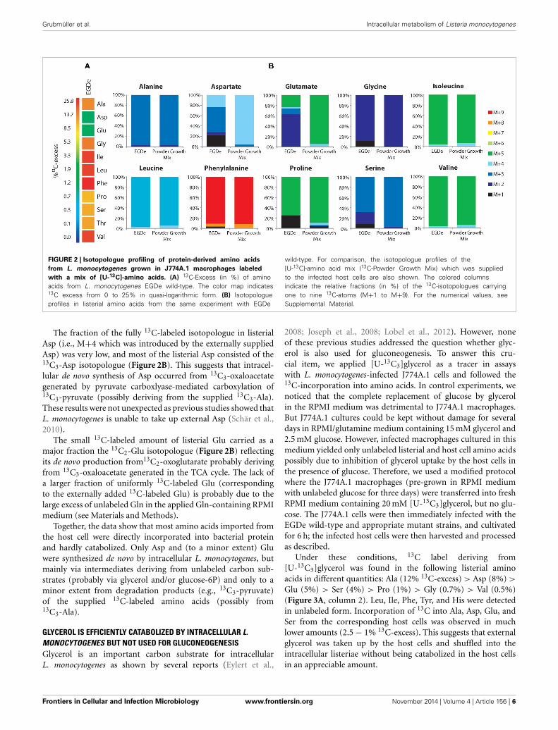
FIGURE 2 | Isotopologue profiling of protein-derived amino acids
from L. monocytogenes grown in J774A.1 macrophages labeled
with a mix of [U-13C]-amino acids. (A) 13C-Excess (in %) of aminoacids from L. monocytogenes EGDe wild-type. The color map indicates13C excess from 0 to 25% in quasi-logarithmic form. (B) Isotopologueprofiles in listerial amino acids from the same experiment with EGDe
wild-type. For comparison, the isotopologue profiles of the[U-13C]-amino acid mix (13C-Powder Growth Mix) which was suppliedto the infected host cells are also shown. The colored columnsindicate the relative fractions (in %) of the 13C-isotopologues carryingone to nine 13C-atoms (M+1 to M+9). For the numerical values, seeSupplemental Material.
The fraction of the fully 13C-labeled isotopologue in listerialAsp (i.e., M+4 which was introduced by the externally suppliedAsp) was very low, and most of the listerial Asp consisted of the13C3-Asp isotopologue (Figure 2B). This suggests that intracel-lular de novo synthesis of Asp occurred from 13C3-oxaloacetategenerated by pyruvate carboxlyase-mediated carboxylation of13C3-pyruvate (possibly deriving from the supplied 13C3-Ala).These results were not unexpected as previous studies showed thatL. monocytogenes is unable to take up external Asp (Schär et al.,2010).
The small 13C-labeled amount of listerial Glu carried as amajor fraction the 13C2-Glu isotopologue (Figure 2B) reflectingits de novo production from13C2-oxoglutarate probably derivingfrom 13C3-oxaloacetate generated in the TCA cycle. The lack ofa larger fraction of uniformly 13C-labeled Glu (correspondingto the externally added 13C-labeled Glu) is probably due to thelarge excess of unlabeled Gln in the applied Gln-containing RPMImedium (see Materials and Methods).
Together, the data show that most amino acids imported fromthe host cell were directly incorporated into bacterial proteinand hardly catabolized. Only Asp and (to a minor extent) Gluwere synthesized de novo by intracellular L. monocytogenes, butmainly via intermediates deriving from unlabeled carbon sub-strates (probably via glycerol and/or glucose-6P) and only to aminor extent from degradation products (e.g., 13C3-pyruvate)of the supplied 13C-labeled amino acids (possibly from13C3-Ala).
GLYCEROL IS EFFICIENTLY CATABOLIZED BY INTRACELLULAR L.MONOCYTOGENES BUT NOT USED FOR GLUCONEOGENESISGlycerol is an important carbon substrate for intracellularL. monocytogenes as shown by several reports (Eylert et al.,
2008; Joseph et al., 2008; Lobel et al., 2012). However, noneof these previous studies addressed the question whether glyc-erol is also used for gluconeogenesis. To answer this cru-cial item, we applied [U-13C3]glycerol as a tracer in assayswith L. monocytogenes-infected J774A.1 cells and followed the13C-incorporation into amino acids. In control experiments, wenoticed that the complete replacement of glucose by glycerolin the RPMI medium was detrimental to J774A.1 macrophages.But J774A.1 cultures could be kept without damage for severaldays in RPMI/glutamine medium containing 15 mM glycerol and2.5 mM glucose. However, infected macrophages cultured in thismedium yielded only unlabeled listerial and host cell amino acidspossibly due to inhibition of glycerol uptake by the host cells inthe presence of glucose. Therefore, we used a modified protocolwhere the J774A.1 macrophages (pre-grown in RPMI mediumwith unlabeled glucose for three days) were transferred into freshRPMI medium containing 20 mM [U-13C3]glycerol, but no glu-cose. The J774A.1 cells were then immediately infected with theEGDe wild-type and appropriate mutant strains, and cultivatedfor 6 h; the infected host cells were then harvested and processedas described.
Under these conditions, 13C label deriving from[U-13C3]glycerol was found in the following listerial aminoacids in different quantities: Ala (12% 13C-excess) > Asp (8%) >
Glu (5%) > Ser (4%) > Pro (1%) > Gly (0.7%) > Val (0.5%)(Figure 3A, column 2). Leu, Ile, Phe, Tyr, and His were detectedin unlabeled form. Incorporation of 13C into Ala, Asp, Glu, andSer from the corresponding host cells was observed in muchlower amounts (2.5 − 1% 13C-excess). This suggests that externalglycerol was taken up by the host cells and shuffled into theintracellular listeriae without being catabolized in the host cellsin an appreciable amount.
Frontiers in Cellular and Infection Microbiology www.frontiersin.org November 2014 | Volume 4 | Article 156 | 6

Grubmüller et al. Intracellular metabolism of Listeria monocytogenes
FIGURE 3 | Analysis of protein-derived amino acids from
L. monocytogenes grown in J774A.1 macrophages labeled with 10 mM
[U-13C6]glucose (Eylert et al., 2008) or 10 mM [U-13C3]glycerol. (A)13C-Excess (in % as a color map) of amino acids from L. monocytogenesEGDe. Column 1: from the labeling experiment with [U-13C6]glucose andEGDe wild-type (Eylert et al., 2008), column 2: from the labeling experimentwith [U-13C3]glycerol and EGDe wild-type (mean value of three replicates),column 3: from the labeling experiment with [U-13C3]glycerol and the EGDe
�C3 mutant, column 4: from the labeling experiment with [U-13C3]glyceroland the EGDe �C3�uhpT mutant, and column 5: from the labelingexperiment with [U-13C3]glycerol and the EGDe �ldH mutant. Boxes withwhite asterisks indicate high standard deviations in the measurement of theoverall 13C-enrichments. (B) Isotopologue profiles in listerial Ala, Asp, and Glufrom the same experiments. The colored columns indicate the relativefractions (in %) of the 13C-isotopologues (M+1 to M+5). For the numericalvalues, see Supplemental Material.
Notably, a similar set of bacterial amino acids acquired 13C-label in the infection experiments with [U-13C6]glucose (Eylertet al., 2008) (Figure 3A, column 1), and the 13C-isotopologuepatterns were similar in corresponding amino acids from theexperiments with [U-13C6]glucose (Eylert et al., 2008) or[U-13C3]glycerol, as shown in Figure 3B for Ala, Asp, andGlu. From [U-13C3]glycerol, Ala was again 13C3-labeled athigh abundance, suggesting that [U-13C3]glycerol was efficientlyconverted to [U-13C3]pyruvate that acted as precursor for 13C3-alanine. Listerial Asp consisted almost exclusively of the 13C3-isotopologue obviously via [1,2,3-13C3]oxaloacetate derived from[U-13C3]pyruvate and unlabeled CO2 and catalyzed by pyru-vate carboxylase (PycA). 13C-Labeled Glu showed high abun-dance for 13C2- and 13C4-isotopologues, the formation of whichcan be explained by the assembly of unlabeled or [1,2,3-13C3]-labeled oxaloacetate with unlabeled or [1,2-13C2]-labeled acetyl-CoA (obtained from [U-13C3]pyruvate) in the incomplete TCA
cycle resulting in the formation of [4,5-13C2]-, [2,3-13C2]-, and[2,3,4,5-13C4]α-oxoglutarate isotopologues, respectively, whichare finally transaminated to the corresponding Glu isotopologues.
In summary, external [U-13C6]glucose and [U-13C3]glycerolyielded in the intracellular L. monocytogenes a similar set of 13C-labeled amino acids with the same 13C-isotopologue patterns.In the case of the experiments with [U-13C3]glycerol, all of the13C-labeled amino acids derive from intermediates generated inthe lower part of the glycolytic pathway or the TCA cycle whileamino acids requiring intermediates from the pentose phos-phate shunt (e.g., His, Phe, and Tyr) were not 13C-labeled. Thesedata suggest that both external carbon substrates converge at thelevel of the same C3-glycolytic intermediate (most likely glyc-eraldehyde 3-phosphate/dihydroxyacetone 3-phosphate) feedingthe lower part of the glycolytic pathway and the TCA cycle. Thesecatabolic pathways include all reactions leading to the interme-diates required for the observed de novo synthesized amino acids
Frontiers in Cellular and Infection Microbiology www.frontiersin.org November 2014 | Volume 4 | Article 156 | 7

Grubmüller et al. Intracellular metabolism of Listeria monocytogenes
and to the generation of ATP by substrate phosphorylation, aswell as to NADH/H+ necessary for ATP production by oxidativephosphorylation via the electron transfer chain.
Infections of J774A.1 cells with the �C3 mutants under theseconditions further confirmed the usage of glycerol as majorcarbon source for intracellular L. monocytogenes as the rate of 13C-incorporation into Ala, Asp, and Glu dropped in these mutantsby a factor of more than 10 compared to the wild-type strain(Figure 3A, columns 3 and 4). This result also showed that13C-labeled glycerol was channeled into the listerial metabolismmainly by glycerol phosphate dehydrogenase which is defective inthe �C3 mutants.
There still remained, however, a low but reproducible 13C-incorporation into Ala, Gly, Pro, and Ser in the �C3 mutants(approximately 0.3% 13C-enrichments) in the presence of exter-nal [U-13C3]glycerol. These 13C-labeled amino acids could beproduced in the host cells and transported into the intracellu-lar bacteria. Alternatively, a fraction of [U-13C3]glycerol couldbe metabolized in the host cells to other C3-components (e.g.,[U-13C3]pyruvate, [U-13C3]lactate) which are then transportedinto the intracellular listeriae and subsequently converted intothese amino acids.
PYRUVATE AND LACTATE ARE INEFFICIENT SUBSTRATES FORINTRACELLULAR L. MONOCYTOGENESIn a defined minimal medium, L. monocytogenes EGDe is unableto grow in the RPMI medium supplemented with pyruvate orlactate as carbon and energy source (data not shown). To deter-mine whether these two carbon substrates can nevertheless beused by L. monocytogenes as supportive carbon substrates underintracellular conditions, L. monocytogenes EGDe-infected J774A.1cells were supplied with 20 mM [U-13C3]pyruvate or 20 mM[U-13C3]lactate in addition to equimolar amounts of glucosein the RPMI medium. In the presence of [U-13C3]pyruvate,Ala, Asp, and Glu from the L. monocytogenes-infected J774A.1macrophages, showed high 13C-enrichments ranging from 16%(mostly in form of 13C3-Ala) to 4% (mostly in form of 13C2-and 13C3-Asp, and 13C2-Glu) (Figures 4C,D). This demonstratedthat [U-13C3]pyruvate was taken up by the macrophages andconverted via 13C2-acetyl-CoA into [13C2]α-oxoglutarate and[13C2]oxaloacetate in the TCA cycle and by carboxylation of13C3-phosphoenol pyruvate into [13C3]oxaloacetate.
The amount of 13C-label in the same amino acids from thelisterial fraction was considerably lower, i.e., Ala (10.5%) >
Asp (2%) > Glu (1.5%) (Figures 4A,B). All other amino acidsremained unlabeled (<1%) which is different as compared tothe infection experiments with uniformly 13C-labeled glucoseor glycerol. The 13C-isotopologue patterns of listerial Asp andGlu were also different than those of the host cells, showingin particular a higher amount of [13C3]Asp (Figures 4C,D).These data are in line with the assumption that a small amountof [13C3]pyruvate was either directly taken up by the bac-teria or converted by the host cell to other C3-compounds,e.g., [13C3]glycerol, -lactate or -alanine, which were subse-quently transported into the intracellular listeriae giving riseto [13C3]pyruvate/Ala and by PycA-mediated carboxylation to[13C3]oxaloacetate/Asp.
Indeed, addition of [U-13C3]lactate to a similar infection assayshowed 13C-incorporation into bacterial Ala (8.6 − 1.4%), Asp(5.9 − 0.5%), and Glu (4.2 − 0.6%) (Figure 4A). As indicated bythe high deviations of the experimental values from several inde-pendent experiments (see error bars in Figure 4B), the presenceof lactate in the medium might lead to harmful effects for the hostcells which can hardly be controlled. We also noticed that the 13C-excess values of the bacterial amino acids were higher than thoseof the corresponding amino acids of the host cells (Figure 4D).Based on this observation, it is likely that lactate provided bythe host cells may serve as a direct, but rather inefficient C3-substrate for intracellular L. monocytogenes. This assumption wasfurther supported by the finding that the �ldh mutant showed13C-label only in Ala (9.5%), but not in any other amino acid(Figure 4A). This 13C3-Ala was probably generated in the hostcell and transported into the intracellular listeriae.
GLUCOSE 6-PHOSPHATE IS USED BY INTRACELLULAR L.MONOCYTOGENES MAINLY FOR ANABOLIC FUNCTIONSBesides glycerol (and, as outlined above, to a minor extent pos-sibly also other C3-compounds), glucose-6P was identified inprevious studies as an important carbon substrate for intracel-lularly replicating L. monocytogenes (Chico-Calero et al., 2002;Eylert et al., 2008). However, the question remained unansweredwhether (i) glucose-6P is catabolized via glycolysis and/or thepentose phosphate pathway, serves as source for energy and theproduction for intermediates in anabolic pathways or (ii) whetherit is mainly required for anabolic functions by its conversion viathe pentose phosphate pathway to the sugar components neces-sary for the biosynthesis of cell wall structures, aromatic aminoacids and nucleotides. For this goal, we compared the 13C-labeledamino acids of L. monocytogenes grown in minimal medium(in vitro) and in J774A.1 cells in the presence of [1,2-13C2]glucose.Although the 13C-enrichments in Ala, Asp, and Glu were approx-imately 3-fold higher from in vitro growing L. monocytogenes thanfrom intracellular bacteria, the 13C-isotopologue patterns of theseamino acids were highly similar in both cases (Figure 5A).
More specifically, the MS analysis of the silylated Ala (Ala-260)showed the presence of [13C2]Ala. To determine the positions ofthe 13C-atoms, a mass fragment (Ala-232) that had lost C-1 dur-ing ionization and therefore represents C-2 and C-3 of Ala wasanalyzed. Ala-232 still showed the presence of two 13C-atoms atthe same abundance as in Ala-260. It can therefore be concludedthat listerial Ala acquired 13C-label from [1,2-13C2]glucose atpositions C-2 and C-3. On this basis, pyruvate, the precur-sor of Ala, was characterized by the [2,3-13C2]-isotopologue(Figure 5B).
Moreover, the 13C-isotopologue pattern in Asp-418 (com-prising all carbon atoms of the original amino acids) andAsp-390 (after loss of a carboxylic atom) showed the for-mation of [2,3-13C2]oxaloacetate/Asp by pyruvate carboxylase-mediated carboxylation of [2,3-13C2]pyruvate (Figure 5B).The 13C-isotopologue pattern of Glu showing predominantly13C2- and 13C4-species can be explained by the incompletecitrate cycle: [2,3-13C2]oxaloacetate reacted with unlabeledacetyl-CoA finally yielding [2,3-13C2]α-ketoglutarate/Glu or with[13C2]acetyl-CoA (obtained from [2,3-13C2]pyruvate through
Frontiers in Cellular and Infection Microbiology www.frontiersin.org November 2014 | Volume 4 | Article 156 | 8

Grubmüller et al. Intracellular metabolism of Listeria monocytogenes
FIGURE 4 | Analysis of protein-derived amino acids from
L. monocytogenes grown in J774A.1 macrophages labeled with 20 mM
[U-13C3]pyruvate or 20 mM [U-13C3]lactate, respectively. (A) 13C-Excess (in%) of amino acids from L. monocytogenes wild-type (EGDe) and the �ldhmutant in the experiment with [U-13C3]lactate. The color map again indicates13C excess in quasi-logarithmic form in order to also visualize small values.Boxes with white asterisks indicate high standard deviations in themeasurement of the overall 13C-enrichments. (B) Isotopologue profiles in
listerial Ala, Asp, and Glu, respectively, from the same experiments. The coloredcolumns indicate the relative fractions (in %) of the 13C-isotopologues (M+1 toM+5) in the labeled amino acids (left scales). For comparison, the gray barsindicate the 13C-excess values with the standard deviations from three technicalreplicates (right scales). (C),13C-Excess (in %) of amino acids from J774A.1proteins in the infection experiments with [U-13C3]pyruvate or [U-13C3]lactate.(D), Isotopologue profiles of J774A.1 Ala, Asp, and Glu, respectively, from thesame experiments. For the numerical values, see Supplemental Material.
Frontiers in Cellular and Infection Microbiology www.frontiersin.org November 2014 | Volume 4 | Article 156 | 9

Grubmüller et al. Intracellular metabolism of Listeria monocytogenes
FIGURE 5 | Analysis of protein-derived amino acids from
L. monocytogenes EGDe in the experiments with [1,2-13C2]glucose.
(A) 13C-excess (in % as a color map) of L. monocytogenes EGDe grownin minimal medium (MM) in the presence of 10 mM [1,2-13C2]glucose(column 1) of from L. monocytogenes EGDe grown in J774A.1 cells in thepresence of 10 mM [1,2-13C2]glucose. Boxes with white asterisks indicatehigh standard deviations in the measurement of the overall
13C-enrichments. Isotopologue profiles in listerial Ser, Ala, Asp, and Glu,respectively, from the same experiments. The colored columns indicatethe relative fractions (in %) of the 13C-isotopologues (M+1 to M+5) inthe labeled amino acids. (B) Reaction scheme displaying the conversion of[1,2-13C2]glucose into the detected isotopologues of Ala, Asp, and Glu.13C-Label is indicated by blue bars. For the numerical values, seeSupplemental Material.
decarboxylation by pyruvate dehydrogenase) yielding [2,3,4,5-13C4]α-ketoglutarate/Glu (Figure 5B). These data are in accordwith assumption that the formation of [2,3-13C2]pyruvate occursby glycolytic degradation of [1,2-13C2]glucose, and not via thepentose phosphate pathway, which would result in the formationof [13C1]- or unlabeled pyruvate.
While the data obtained from L. monocytogenes grown in theminimal medium (MM) clearly indicate that the formation of[2,3-13C2]pyruvate arises from glycolytic degradation of [1,2-13C2]glucose by the listeriae, this is less obvious in case of the bac-teria grown in J774A.1 cells. Here, [2,3-13C2]pyruvate could begenerated by glycolytic degradation of [1,2-13C2]glucose withinthe host cell where it is further converted into [2,3-13C2]glycerol.This compound could be subsequently transported into the intra-cellular listeriae and further metabolized leading to the observedisotopologues in Ala, Ser, Asp, and Glu. Indeed, the latter expla-nation appears to be more likely when we compare the other13C-labeled, i.e., de novo synthesized amino acids, in MM-grownand in J774A.1 grown listeriae. Thus, MM-grown listeriae showed13C-label (in addition to the above described Ala, Ser, Asp, and
Glu) in Gly, Thr, Pro, His, and Phe (Figure 5A). The two latteramino acids require as precursors intermediates from the pentosephosphate pathway, i.e., erythrose-4P and ribose-5P, respectively.
In contrast, L. monocytogenes grown within J774A.1 did notshow 13C-label in these two amino acids. In this case, all 13C-labeled amino acids require as precursors for their biosynthesisintermediates from the lower part of glycolysis (Ser, Gly, Ala, Val)or the TCA cycle (Asp, Thr, Glu, Pro) which could be generatedfrom host cell-imported [2,3-13C2]glycerol.
These data indicate that MM-grown and J774A.1-grownL. monocytogenes use externally supplied glucose in different ways.While in the MM-grown listeriae glucose feeds the glycolyticpathway and the pentose phosphate shunt, the J774A.1-grownL. monocytogenes consume glucose in a dual manner with theparticipation of the host cell which converts glucose to glyc-erol and glucose-6P which are subsequently taken up as separatecarbon substrates by the intracellular listeriae. Glucose-derivedglycerol (generated in the host cell) is used as carbon substratefor the supply of energy and intermediates for the biosynthe-sis of some amino acids and (probably) fatty acids but not for
Frontiers in Cellular and Infection Microbiology www.frontiersin.org November 2014 | Volume 4 | Article 156 | 10

Grubmüller et al. Intracellular metabolism of Listeria monocytogenes
FIGURE 6 | Model for a bipartite metabolism for intracellular
L. monocytogenes. Gray-framed yellow boxes and yellow arrows mark(by color intensity) the major carbon compounds taken up by theintracellular listeriae from the host cell. Catabolic reactions fed byglucose-6P and glycerol are indicated by blue and black arrows,
respectively. Reactions leading to anabolic performances (in red-framedboxes) are indicated by red arrows. Abbreviations: PPP, pentosephosphate pathway; GL, glycolysis; GN, gluconeogenesis; TCA,tricarboxylic acid pathway; PYC, pyruvate carboxylase; ME, malic enzyme(decarboxylating malate dehydrogenase).
gluconeogenesis. Vice versa, glucose-6P (also generated in thehost cell) may not be catabolized to pyruvate to an appreciableextent, but rather converted in the pentose phosphate shunt intosugar components essential for the biosynthesis of the cell enve-lope and nucleotides. The aromatic amino acids which requirefor their biosynthesis erythrose-4P also generated in the pen-tose phosphate shunt seem to be mainly imported from thehost cell. A model for this bipartite intracellular metabolism ofL. monocytogenes is outlined in Figure 6.
THE CARBON METABOLISM OF J774A.1 HOST CELLS IS DIFFERENTLYAFFECTED BY THE WILD-TYPE AND THE �C3 AND �uhpT MUTANTSTRAINS OF L. MONOCYTOGENESThis bipartite carbon metabolism carried out by the intracellu-lar listeriae might put less nutrient stress on the host cell therebyextending the lifespan of the host cell—in favor of the intracellularbacteria. Some indirect experimental evidence for this assump-tion is provided by the different 13C-incorporation rates into
Ala, Asp, and Glu from the J774A.1 host cells infected with thewild-type, the �C3, or the �uhpT strains. The balanced bipartitemetabolism is expected to be disturbed in these two mutants dueto their inability of utilize glycerol and glucose-6P, respectively.
The J774A.1 host cell line derives from a mouse tumor andexpresses c-Myc constitutively (Fan et al., 1993). This leads toenhanced aerobic glycolysis and increased glutaminolysis (Fanet al., 1993; Wise et al., 2008; Dang et al., 2009). In the pres-ence of [U-13C6]glucose, the most efficiently 13C-labeled aminoacids of J774A.1 cells were therefore Ala, Glu, and Asp. However,the 13C-incorporation rates into these amino acids were slightlybut reproducibly increased when the same cells were infectedwith L. monocytogenes EGDe wild-type (WT) (Figure 7) whichis in line with earlier observations (Gillmaier et al., 2012).Interestingly, however, 13C-incorporation into these amino acidsdecreased in the J774A.1 cells upon infection with the �C3mutant strains, but increased upon infection with the �uhpTstrain in comparison to the reference experiment with the
Frontiers in Cellular and Infection Microbiology www.frontiersin.org November 2014 | Volume 4 | Article 156 | 11

Grubmüller et al. Intracellular metabolism of Listeria monocytogenes
FIGURE 7 | Analysis of protein-derived amino acids from J774A.1 cells
labeled with [U-13C6]glucose. The columns indicate 13C-excess (in %) ofAla, Asp, and Glu in experiments with uninfected cells, or cells infectedL. monocytogenes EGDe wild-type, �C3, �uhpT, and �C3�uhpTmutants, respectively.
wild-type strain (Eylert et al., 2008). This effect was less obviousin Glu which is probably caused by the abundant availability ofGlu in the J774A.1 host cells due to the enhanced glutaminolysis(Gillmaier et al., 2012). The relatively low 13C-enrichment in Gluderiving from of [U-13C6]glucose (an indication for Glu de novosynthesis) is in line with this assumption.
These changes of 13C-incorporation into host cell amino acidsupon infection by the mutant strains compared to the wildtype strain cannot be caused by a different efficiencies of intra-cellular replication since the number of intracellular wild-typebacteria was similar to that of the mutant strains. The datarather suggest that—as compensation for the defective glycerolor glucose-6P consumption—the resulting increased withdrawalof glucose-6P or glycerol, respectively, from the host cells obvi-ously causes severe changes in the central carbon fluxes of the hostcells.
DISCUSSIONIn order to further elucidate the intracellular metabolism ofL. monocytogenes and in particular for answering the questionwhich carbon substrates provided by the host cells are essentialfor driving the intracellular listerial metabolism, we performedinfection studies with the EGDe wild-type strain and isogenicmutants defective in the transport or catabolism of anticipatedcarbon substrates and J774A.1 macrophages as host cells. Forthis goal, we developed experimental protocols for the applica-tion of [U-13C]amino acids, [U-13C3]glycerol, [U-13C3]pyruvate,and [U-13C3]lactate as tracer carbon substrates to the RPMI cul-ture medium. The efficiency of utilization of these carbon sourcesand the resulting metabolic fluxes were determined from the 13C-isotopologue profiles of the 13C-labeled amino acids, a methodwhich we successfully applied before for studying the intracellu-lar metabolism of bacterial pathogens in mammalian cells (Eylertet al., 2008; Götz et al., 2010; Gillmaier et al., 2012).
The results confirm the essential role of glycerol and glucose-6P as carbon substrates for the intracellular listerial metabolismas already suggested by previous studies that were based on tran-scriptome analyses (Chatterjee et al., 2006; Joseph et al., 2006;Lobel et al., 2012) and on 13C-isotopologue profiling studies(Eylert et al., 2008; Gillmaier et al., 2012). The present data alsoshow that other C3-carbon substrates, like pyruvate or lactate pro-posed as possible alternative or supplemental carbon substrates,play only minor roles, if any. It should be noted, however, thateven a L. monocytogenes �C3�uhpT mutant which is appar-ently unable to utilize glycerol and glucose-6P can still replicatein the J774A.1 host cells albeit at a reduced rate. This suggeststhat there are alternative carbon sources which are able to at leastpartially replace the two major carbon substrates, glycerol andglucose-6P. Possible candidates are glucose, amino acids, glycerol3-phosphate, or intermediates of the TCA (especially succinateand malate) that could also be provided by the host cells.
The possible participation of glucose and/or mannose inintracellular listerial metabolism is difficult to demonstrate exper-imentally, since L. monocytogenes possess a large number ofPEP-dependent phosphotransferase systems (PTS) and even non-PTS transporters for glucose and mannose (Glaser et al., 2001;Stoll and Goebel, 2010; Ake et al., 2011). Deletions of the majorglucose/mannose PTS transporters do not seem to affect theintracellular listerial replication. However, in the pts deletionmutants, other transporters for these carbohydrates seem to beactivated (Stoll and Goebel, 2010).
There is still another reason which makes the participationof glucose or mannose as major carbon source for intracellularlisterial metabolism unlikely. The expression of the genes encod-ing the virulence factors essential for the intracellular listerial lifecycle and also the glucose-6P transporter UhpT depends on thecentral virulence gene activator PrfA (Chico-Calero et al., 2002).However, the activity of PrfA is strongly inhibited when glucoseor other glycolytic carbohydrates are used as the major carbonsource for listerial growth while PrfA activity is high with glyc-erol as carbon source (Joseph et al., 2008; Stoll et al., 2008; Götz,unpublished results).
The data presented here also rule out amino acids as importantcatabolic carbon substrates for the intracellular metabolism ofL. monocytogenes. An externally added mix of 13C-labeled aminoacids (containing all amino acids except Trp, Cys, Met, and Arg)is efficiently taken up by the bacteria (with the exception of Asp).However, according to the 13C-isotopologue profiles the majorglycogenic and ketogenic amino acids are mainly incorporatedinto listerial protein but hardly catabolized by the intracellularlisteriae. This is not surprising for the two most important glyco-genic amino acids, Asp and Glu, since Asp cannot be taken up byL. monocytogenes (Schär et al., 2010) and the missing oxoglutaratedehydrogenase prevents Asp and Glu degradation in the inter-rupted listerial TCA cycle (Eisenreich et al., 2006). These resultsare in line with previous studies (Eylert et al., 2008; Gillmaieret al., 2012) showing limited de novo synthesis of most aminoacids by intracellular listeriae and import of most amino acidsfrom the host cell in particular of the branched chain and aro-matic amino acids. The failure to biosynthesize these aminoacids during intracellular growth despite the presence of the
Frontiers in Cellular and Infection Microbiology www.frontiersin.org November 2014 | Volume 4 | Article 156 | 12

Grubmüller et al. Intracellular metabolism of Listeria monocytogenes
biosynthesis capacity on the basis of the genome sequence (Glaseret al., 2001) and transcriptome analyses (Chatterjee et al., 2006;Joseph et al., 2006; Lobel et al., 2012) suggests a shortage of essen-tial catabolic intermediates and/or co-factors required for thebiosynthesis of these amino acids under intracellular conditions.
Those amino acids that are de novo synthesized by the intra-cellular listeriae at significant rates derive from intermediatesgenerated in the lower part of the glycolytic pathway (Ser, Gly,Ala) or in the TCA pathway (Asp, Thr, Glu, Pro). These catabolicpathways can be fed by glycerol as major carbon source. Indeed,the nature of 13C-labeled amino acids as well as their 13C-isotopologue profiles are similar if not identical, irrespective ofwhether 13C-glucose or 13C-glycerol were added to the culturemedium of the infected host cells. This suggests that 13C-glucoseis converted in the host cell into 13C-glycerol that enters, afterbeing taken up by the intracellular listeriae, the glycolytic pathwayat the same position (most likely at the level of glyceraldehyde-3P/dihydroxyacetone-3P) as the externally added 13C-glycerol.These glycolytic intermediates are then further catabolized intopyruvate, oxaloacetate and α-ketoglutarate and their down-stream amino acids (see Figure 6), but not used anabolically ingluconeogenesis as indicated by the apparent absence of 13C-label in Phe, Tyr and His from exogenous 13C-glycerol. De novosynthesis of these amino acids would require sugar compo-nents, such as erythrose-4P and ribose-5P, respectively, whichare generated in the glucose-6P-driven pentose phosphate path-way. Indeed, these amino acids are synthesized and 13C-labeledby L. monocytogenes growing in an in vitro culture medium inthe presence of 13C-glucose as major carbon source (Eisenreichet al., 2006) as well as by intracellular (also cytosolically) repli-cating enteroinvasive E. coli (Götz et al., 2010). Notably, the latterbacterial pathogen uses mainly glucose as carbon substrate for itsintracellular metabolism.
This glycerol-driven metabolism of intracellular listeriae willlead to active PrfA (Stoll et al., 2008) and hence to the expres-sion of the glucose-6P transporter UhpT resulting in the uptakeof glucose-6P by the intracellular listeriae. We postulate thatthis additional carbon substrate may feed the pentose phos-phate shunt allowing the generation of the intermediates neededfor the biosynthesis of essential cell envelope components andnucleotides. Our presently applied analytical 13C-isotopologueapproach does not allow the direct detection of these compoundsand the experimental prove for this assumption therefore awaitsfurther studies with improved analytical tools.
Together, the data suggest that the intracellular metabolismof L. monocytogenes relies on two major carbon substrates: (i)glycerol (generated in the host cell from glucose or other precur-sors) which is used as carbon substrate for the supply of energy(ATP by substrate-level phosphorylation and aerobic respiration)and of intermediates for the biosynthesis of some amino acids(e.g., Ser, Gly, Ala, Asp, Thr, Glu, and Pro) and (probably) fattyacids, but not for gluconeogenesis, and (ii) glucose-6P (also gen-erated in the host cell from e.g., glucose) which may not becatabolized to pyruvate at significant rates, but rather convertedin the pentose phosphate shunt to sugar components essentialfor the biosynthesis of the cell envelope and nucleotides. Thearomatic amino acids which also require for their biosynthesis
erythrose-4P, also generated in the pentose phosphate shunt,seem to be mainly imported from the host cell. A model forthis “bipartite metabolism” of intracellular L. monocytogenes isoutlined in Figure 6.
This kind of bipartite metabolism of an IBP could in generalhave a selective advantage for its survival within the host cell, sinceit may impose less nutrient stress on the infected host cell than abacterial metabolism based mainly on the use of glucose. Indeed,L. monocytogenes �C3 and �uhpT mutants which are impairedin the balanced utilization of the two carbon substrates seem toimpose a greater metabolic burden on the host cells than the wild-type strain as shown by the changes of the isotopologue profilesof host amino acids in infection experiments with L. monocy-togenes �C3 or �uhpT in comparison to the wild-type strain.Furthermore, enteroinvasive E. coli strains which also replicate—similar to L. monocytogenes—in the host cells’ cytosol, but mainlyrely on glucose as a preferred carbon source for the intracellularmetabolism (Götz et al., 2010), kill the same host cells much fasterthan L. monocytogenes.
AUTHOR CONTRIBUTIONSWolfgang Eisenreich, Thilo M. Fuchs, and Werner Goebeldesigned the experiments; Kristina Schauer and Thilo M. Fuchscharacterized the mutants, Stephanie Grubmüller and KristinaSchauer performed the labeling experiments, SG performedthe GC/MS analysis; Stephanie Grubmüller, Kristina Schauer,Wolfgang Eisenreich, Thilo M. Fuchs, and Werner Goebel ana-lyzed and interpreted the data; and Werner Goebel, WolfgangEisenreich, and Thilo M. Fuchs wrote the paper.
ACKNOWLEDGMENTSThis work was supported by the priority program SPP1316 ofthe Deutsche Forschungsgemeinschaft (EI-384/6 and FU-375/5).We thank Birgit Lange and Christine Schwarz for their experthelp in sample preparation, and Erika Kutzner for help with thegraphics.
SUPPLEMENTARY MATERIALThe Supplementary Material for this article can be foundonline at: http://www.frontiersin.org/journal/10.3389/fcimb.2014.00156/abstract
REFERENCESAke, F. M., Joyet, P., Deutscher, J., and Milohanic, E. (2011). Mutational analysis of
glucose transport regulation and glucose-mediated virulence gene repressionin Listeria monocytogenes. Mol. Microbiol. 81, 274–293. doi: 10.1111/j.1365-2958.2011.07692.x
Camejo, A., Carvalho, F., Reis, O., Leitao, E., Sousa, S., and Cabanes, D. (2011). Thearsenal of virulence factors deployed by Listeria monocytogenes to promote itscell infection cycle. Virulence 2, 379–394. doi: 10.4161/viru.2.5.17703
Chatterjee, S. S., Hossain, H., Otten, S., Kuenne, C., Kuchmina, K., Machata, S.,et al. (2006). Intracellular gene expression profile of Listeria monocytogenes.Infect. Immun. 74, 1323–1338. doi: 10.1128/IAI.74.2.1323-1338.2006
Chico-Calero, I., Suarez, M., Gonzalez-Zorn, B., Scortti, M., Slaghuis, J., Goebel,W., et al. (2002). Hpt, a bacterial homolog of the microsomal glucose-6-phosphate translocase, mediates rapid intracellular proliferation in Listeria.Proc. Natl. Acad. Sci. U.S.A. 99, 431–436. doi: 10.1073/pnas.012363899
Cossart, P., and Lebreton, A. (2014). A trip in the “New Microbiology” withthe bacterial pathogen Listeria monocytogenes. FEBS Lett. 588, 2437–2445. doi:10.1016/j.febslet.2014.05.051
Frontiers in Cellular and Infection Microbiology www.frontiersin.org November 2014 | Volume 4 | Article 156 | 13

Grubmüller et al. Intracellular metabolism of Listeria monocytogenes
Dang, C. V., Le, A., and Gao, P. (2009). MYC-induced cancer cell energymetabolism and therapeutic opportunities. Clin. Cancer Res. 15, 6479–6483.doi: 10.1158/1078-0432.CCR-09-0889
de las Heras, A., Cain, R. J., Bielecka, M. K., and Vazquez-Boland, J. A. (2011).Regulation of Listeria virulence: PrfA master and commander. Curr. Opin.Microbiol. 14, 118–127. doi: 10.1016/j.mib.2011.01.005
Donaldson, J. R., Nanduri, B., Pittman, J. R., Givaruangsawat, S., Burgess, S. C., andLawrence, M. L. (2011). Proteomic expression profiles of virulent and avirulentstrains of Listeria monocytogenes isolated from macrophages. J. Proteomics 74,1906–1917. doi: 10.1016/j.jprot.2011.05.008
Dussurget, O., Pizarro-Cerda, J., and Cossart, P. (2004). Molecular determinantsof Listeria monocytogenes virulence. Annu. Rev. Microbiol. 58, 587–610. doi:10.1146/annurev.micro.57.030502.090934
Eisenreich, W., Slaghuis, J., Laupitz, R., Bussemer, J., Stritzker, J., Schwarz, C., et al.(2006). 13C isotopologue perturbation studies of Listeria monocytogenes carbonmetabolism and its modulation by the virulence regulator PrfA. Proc. Natl. Acad.Sci. U.S.A. 103, 2040–2045. doi: 10.1073/pnas.0507580103
Eylert, E., Schär, J., Mertins, S., Stoll, R., Bacher, A., Goebel, W., et al. (2008).Carbon metabolism of Listeria monocytogenes growing inside macrophages.Mol. Microbiol. 69, 1008–1017. doi: 10.1111/j.1365-2958.2008.06337.x
Fan, K., Barendsen, N., Sensenbrenner, L., and Chen, B. D. M. (1993). Deregulationof granulocyte-macrophage colony-stimulating factor (GM-CSF) receptor inmurine macrophage cell line J774A.1. J. Cell. Physiol. 154, 535–542. doi:10.1002/jcp.1041540312
Fuchs, T. M., Eisenreich, W., Kern, T., and Dandekar, T. (2012). Toward a systemicunderstanding of Listeria monocytogenes metabolism during infection. Front.Microbiol. 3:23. doi: 10.3389/fmicb.2012.00023
Gillmaier, N., Götz, A., Schulz, A., Eisenreich, W., and Goebel, W. (2012). Metabolicresponses of primary and transformed cells to intracellular Listeria monocyto-genes. PLoS ONE 7:e52378. doi: 10.1371/journal.pone.0052378
Glaser, P., Frangeul, L., Buchrieser, C., Rusniok, C., Amend, A., Baquero, F., et al.(2001). Comparative genomics of Listeria species. Science 294, 849–852. doi:10.1126/science.1063447
Götz, A., Eylert, E., Eisenreich, W., and Goebel, W. (2010). Carbon metabolism ofenterobacterial human pathogens growing in epithelial colorectal adenocarci-noma (Caco-2) cells. PLoS ONE. 5:e10586. doi: 10.1371/journal.pone.0010586
Hamon, M. A., Ribet, D., Stavru, F., and Cossart, P. (2012). ListeriolysinO: the Swiss army knife of Listeria. Trends Microbiol. 20, 360–368. doi:10.1016/j.tim.2012.04.006
Hamon, M., Bierne, H., and Cossart, P. (2006). Listeria monocytogenes: a multi-faceted model. Nat. Rev. Microbiol. 4, 423–434. doi: 10.1038/nrmicro1413
Hanahan, D. (1983). Studies on transformation of Escherichia coli with plasmids.J. Mol. Biol. 166, 557–580. doi: 10.1016/S0022-2836(83)80284-8
Heuner, K., and Eisenreich, W. (2013). The intracellular metabolism of Legionellaby isotopologue profiling. Methods Mol. Biol. 954, 163–181. doi: 10.1007/978-1-62703-161-5_8
Joseph, B., Mertins, S., Stoll, R., Schär, J., Umesha, K. R., and Luo, Q., et al. (2008).Glycerol metabolism and PrfA activity in Listeria monocytogenes. J. Bacteriol.190, 5412–5430. doi: 10.1128/JB.00259-08
Joseph, B., Przybilla, K., Stuhler, C., Schauer, K., Slaghuis, J., Fuchs, T. M., et al.(2006). Identification of Listeria monocytogenes genes contributing to intracel-lular replication by expression profiling and mutant screening. J. Bacteriol. 188,556–568. doi: 10.1128/JB.188.2.556-568.2006
Lecuit, M. (2005). Understanding how Listeria monocytogenes targets andcrosses host barriers. Clin. Microbiol. Infect. 11, 430–436. doi: 10.1111/j.1469-0691.2005.01146.x
Lee, W.-N. P., Byerley, L. O., Bergner, E. A., and Edmond, J. (1991). Mass iso-topomer analysis: theoretical and practical considerations. Biol. Mass Spect. 20,451–458. doi: 10.1002/bms.1200200804
Lobel, L., Sigal, N., Borovok, I., Ruppin, E., and Herskovits, A. A. (2012). Integrativegenomic analysis identifies isoleucine and CodY as regulators of Listeria
monocytogenes virulence. PLoS Genet. 8:e1002887. doi: 10.1371/journal.pgen.1002887
Mostowy, S., and Cossart, P. (2012). Virulence factors that modulate the cell biol-ogy of Listeria infection and the host response. Adv. Immunol. 113, 19–32. doi:10.1016/B978-0-12-394590-7.00007-5
Premaratne, R. J., Lin, W. J., and Johnson, E. A. (1991). Development of animproved chemically defined minimal medium for Listeria monocytogenes. Appl.Environ. Microbiol. 57, 3046–3048.
Sambrook, J., and Russell, D. W. (2001). Molecular Cloning. A Laboratory Manual,3rd Edn. New York, NY: Cold Spring Harbour Laboratory Press.
Schär, J., Stoll, R., Schauer, K., Loeffler, D. I., Eylert, E., Joseph, B., et al. (2010).Pyruvate carboxylase plays a crucial role in carbon metabolism of extra- andintracellularly replicating Listeria monocytogenes. J. Bacteriol. 192, 1774–1784.doi: 10.1128/JB.01132-09
Schauer, K., Geginat, G., Liang, C., Goebel, W., Dandekar, T., and Fuchs, T. M.(2010). Deciphering the intracellular metabolism of Listeria monocytogenes bymutant screening and modelling. BMC Genomics 11:573. doi: 10.1186/1471-2164-11-573
Schneebeli, R., and Egli, T. (2013). A defined, glucose-limited mineral mediumfor the cultivation of Listeria spp. Appl. Environ. Microbiol. 79, 2503–2511. doi:10.1128/AEM.03538-12
Schunder, E., Gillmaier, N., Kutzner, E., Herrmann, V., Lautner, M., Heuner, K.,et al. (2014). Amino acid uptake and metabolism of Legionella pneumophilahosted by Acanthamoeba castellanii. J. Biol. Chem. 289, 21040–21054. doi:10.1074/jbc.M114.570085
Stoll, R., and Goebel, W. (2010). The major PEP-phosphotransferase systems(PTSs) for glucose, mannose and cellobiose of Listeria monocytogenes, and theirsignificance for extra- and intracellular growth. Microbiol. 156, 1069–1083. doi:10.1099/mic.0.034934-0
Stoll, R., Mertins, S., Joseph, B., Müller-Altrock, S., and Goebel, W. (2008).Modulation of PrfA activity in Listeria monocytogenes upon growth in differentculture media. Microbiology 154, 3856–3876. doi: 10.1099/mic.0.2008/018283-0
Tsai, H. N., and Hodgson, D. A. (2003). Development of a synthetic minimalmedium for Listeria monocytogenes. Appl. Environ. Microbiol. 69, 6943–6945.doi: 10.1128/AEM.69.11.6943-6945.2003
Velge, P., and Roche, S. M. (2010). Variability of Listeria monocytogenes virulence:a result of the evolution between saprophytism and virulence? Future Microbiol.5, 1799–1821. doi: 10.2217/fmb.10.134
Wise, D. R., DeBerardinis, R. J., Mancuso, A., Sayed, N., Zhang, X. Y., Pfeiffer,H. K., et al. (2008). Myc regulates a transcriptional program that stimulatesmitochondrial glutaminolysis and leads to glutamine addiction. Proc. Natl.Acad. Sci. U.S.A. 105, 18782–18787. doi: 10.1073/pnas.0810199105
Conflict of Interest Statement: The authors declare that the research was con-ducted in the absence of any commercial or financial relationships that could beconstrued as a potential conflict of interest.
Received: 03 August 2014; accepted: 14 October 2014; published online: 03 November2014.Citation: Grubmüller S, Schauer K, Goebel W, Fuchs TM and Eisenreich W (2014)Analysis of carbon substrates used by Listeria monocytogenes during growth inJ774A.1 macrophages suggests a bipartite intracellular metabolism. Front. Cell. Infect.Microbiol. 4:156. doi: 10.3389/fcimb.2014.00156This article was submitted to the journal Frontiers in Cellular and InfectionMicrobiology.Copyright © 2014 Grubmüller, Schauer, Goebel, Fuchs and Eisenreich. This is anopen-access article distributed under the terms of the Creative Commons AttributionLicense (CC BY). The use, distribution or reproduction in other forums is permitted,provided the original author(s) or licensor are credited and that the original publica-tion in this journal is cited, in accordance with accepted academic practice. No use,distribution or reproduction is permitted which does not comply with these terms.
Frontiers in Cellular and Infection Microbiology www.frontiersin.org November 2014 | Volume 4 | Article 156 | 14