Ultra Deep Sequencing of Listeria monocytogenes sRNA Transcriptome Revealed New Antisense RNAs
10
Ultra Deep Sequencing of Listeria monocytogenes sRNA Transcriptome Revealed New Antisense RNAs Sebastian Behrens 1,5 , Stefanie Widder 1 , Gopala Krishna Mannala 5 , Xiaoxing Qing 5 , Ramakanth Madhugiri 2 , Nathalie Kefer 3,4 , Mobarak Abu Mraheil 5 , Thomas Rattei 1 , Torsten Hain 5 * 1 Department fu ¨ r Computational Systems Biology, Universita ¨t Wien, Wien, Austria, 2 Institute fu ¨ r Medizinische Virologie, Justus-Liebig Universita ¨t Giessen, Giessen, Germany, 3 febit biomed GmbH, Heidelberg, Germany, 4 Life Technologies GmbH, Darmstadt, Germany, 5 Institute fu ¨ r Medizinische Mikrobiologie, Justus-Liebig Universita ¨t Giessen, Giessen, Germany Abstract Listeria monocytogenes, a gram-positive pathogen, and causative agent of listeriosis, has become a widely used model organism for intracellular infections. Recent studies have identified small non-coding RNAs (sRNAs) as important factors for regulating gene expression and pathogenicity of L. monocytogenes. Increased speed and reduced costs of high throughput sequencing (HTS) techniques have made RNA sequencing (RNA-Seq) the state-of-the-art method to study bacterial transcriptomes. We created a large transcriptome dataset of L. monocytogenes containing a total of 21 million reads, using the SOLiD sequencing technology. The dataset contained cDNA sequences generated from L. monocytogenes RNA collected under intracellular and extracellular condition and additionally was size fractioned into three different size ranges from ,40 nt, 40–150 nt and .150 nt. We report here, the identification of nine new sRNAs candidates of L. monocytogenes and a reevaluation of known sRNAs of L. monocytogenes EGD-e. Automatic comparison to known sRNAs revealed a high recovery rate of 55%, which was increased to 90% by manual revision of the data. Moreover, thorough classification of known sRNAs shed further light on their possible biological functions. Interestingly among the newly identified sRNA candidates are antisense RNAs (asRNAs) associated to the housekeeping genes purA, fumC and pgi and potentially their regulation, emphasizing the significance of sRNAs for metabolic adaptation in L. monocytogenes. Citation: Behrens S, Widder S, Mannala GK, Qing X, Madhugiri R, et al. (2014) Ultra Deep Sequencing of Listeria monocytogenes sRNA Transcriptome Revealed New Antisense RNAs. PLoS ONE 9(2): e83979. doi:10.1371/journal.pone.0083979 Editor: Stefan Bereswill, Charite ´-University Medicine Berlin, Germany Received May 14, 2013; Accepted November 8, 2013; Published February 3, 2014 Copyright: ß 2014 Behrens et al. This is an open-access article distributed under the terms of the Creative Commons Attribution License, which permits unrestricted use, distribution, and reproduction in any medium, provided the original author and source are credited. Funding: This work was supported by grants from the German Federal Ministry of Education and Research (BMBF ERA-NET Pathogenomics Network to the sncRNAomics project (62080061) to T.H. as well as the German Centre for Infection Research, Justus-Liebig University Giessen. The funders had no role in study design, data collection and analysis, decision to publish, or preparation of the manuscript. Competing Interests: Biomarker Center GmbH) which declares (formerly known as febit biomed GmbH) that CBC, has no competing interest with the manuscript entitled ‘‘Ultra deep Sequencing of Listeria monocytogenes sRNA transcriptome revealed new antisense RNAs (see also the attached letter from CBC). N. Kefer present working address is now Life Technologies as stated in the manuscript. This does not alter the authors’ adherence to all the PLOS ONE policies on sharing data and materials. * E-mail: [email protected] Introduction Listeria monocytogenes is a Gram-positive, facultative intracellular pathogen, which is responsible for a foodborne infection, listeriosis, a rare but serious disease. It has become the prime model organism for intracellular pathogens [1]. Small non coding RNAs (sRNAs) have been proposed to play an important role in the pathogenicity of L. monocytogenes and some lead to attenuated infections when disabled [2,3]. These studies also showed that antisense transcrip- tion is common in L. monocytogenes [2,3]. Beside short antisense RNAs (asRNAs), a new type of long antisense RNAs (lasRNAs) functioning as an mRNA as well as antisense RNA that regulate adjacent genes at the level of transcription, was proposed [4]. Over the last decade reduced costs for high throughput sequencing (HTS) technologies facilitate the thorough and unbiased research of bacterial transcriptomes at an ever increasing rate [5–7]. As a result, identification of small non coding RNAs in all bacterial species have been reported [8–11]. Large numbers of small non coding RNAs have been found in both Gram-negative [12,13] and Gram-positive [14,15] bacteria. In particular L. monocytogenes has been subject to an extensive number of transcriptome studies using macro-/microarrays, Illumina GAIIx or Roche GS FLX sequenc- ing platforms [2–4,16–20]. The SOLiD sequencing platform used in this study, provides a very high throughput sequencing method with increased base calling accuracy due to its unique ‘color coded’ di-base sequencing technique [21]. Here we report the thorough reevaluation of the small RNA transcriptome of L. monocytogenes with increased coverage. A large HTS transcriptome dataset containing transcriptomic data of L. monocytogenes grown under intracellular and extracellular conditions was the basis of this study. The transcriptomic data was generated using the SOLiD HTS platform and consists of a total of 21 million reads. In this study a newly developed computational pipeline was used to identify and classify sRNAs. Furthermore, this computational pipeline leads to the discovery of nine yet unknown small non coding RNA candidates of L. monocytogenes. Materials and Methods Bacterial and cell culture and RNA extraction The strain of L. monocytogenes EGD-e [22] and the murine P388D1 macrophages were used for cell infection and RNA PLOS ONE | www.plosone.org 1 February 2014 | Volume 9 | Issue 2 | e83979
-
Upload
uni-giessen -
Category
Documents
-
view
3 -
download
0
Transcript of Ultra Deep Sequencing of Listeria monocytogenes sRNA Transcriptome Revealed New Antisense RNAs

Ultra Deep Sequencing of Listeria monocytogenes sRNATranscriptome Revealed New Antisense RNAsSebastian Behrens1,5, Stefanie Widder1, Gopala Krishna Mannala5, Xiaoxing Qing5,
Ramakanth Madhugiri2, Nathalie Kefer3,4, Mobarak Abu Mraheil5, Thomas Rattei1, Torsten Hain5*
1 Department fur Computational Systems Biology, Universitat Wien, Wien, Austria, 2 Institute fur Medizinische Virologie, Justus-Liebig Universitat Giessen, Giessen,
Germany, 3 febit biomed GmbH, Heidelberg, Germany, 4 Life Technologies GmbH, Darmstadt, Germany, 5 Institute fur Medizinische Mikrobiologie, Justus-Liebig
Universitat Giessen, Giessen, Germany
Abstract
Listeria monocytogenes, a gram-positive pathogen, and causative agent of listeriosis, has become a widely used modelorganism for intracellular infections. Recent studies have identified small non-coding RNAs (sRNAs) as important factors forregulating gene expression and pathogenicity of L. monocytogenes. Increased speed and reduced costs of high throughputsequencing (HTS) techniques have made RNA sequencing (RNA-Seq) the state-of-the-art method to study bacterialtranscriptomes. We created a large transcriptome dataset of L. monocytogenes containing a total of 21 million reads, usingthe SOLiD sequencing technology. The dataset contained cDNA sequences generated from L. monocytogenes RNA collectedunder intracellular and extracellular condition and additionally was size fractioned into three different size ranges from,40 nt, 40–150 nt and .150 nt. We report here, the identification of nine new sRNAs candidates of L. monocytogenes and areevaluation of known sRNAs of L. monocytogenes EGD-e. Automatic comparison to known sRNAs revealed a high recoveryrate of 55%, which was increased to 90% by manual revision of the data. Moreover, thorough classification of known sRNAsshed further light on their possible biological functions. Interestingly among the newly identified sRNA candidates areantisense RNAs (asRNAs) associated to the housekeeping genes purA, fumC and pgi and potentially their regulation,emphasizing the significance of sRNAs for metabolic adaptation in L. monocytogenes.
Citation: Behrens S, Widder S, Mannala GK, Qing X, Madhugiri R, et al. (2014) Ultra Deep Sequencing of Listeria monocytogenes sRNA Transcriptome RevealedNew Antisense RNAs. PLoS ONE 9(2): e83979. doi:10.1371/journal.pone.0083979
Editor: Stefan Bereswill, Charite-University Medicine Berlin, Germany
Received May 14, 2013; Accepted November 8, 2013; Published February 3, 2014
Copyright: � 2014 Behrens et al. This is an open-access article distributed under the terms of the Creative Commons Attribution License, which permitsunrestricted use, distribution, and reproduction in any medium, provided the original author and source are credited.
Funding: This work was supported by grants from the German Federal Ministry of Education and Research (BMBF ERA-NET Pathogenomics Network to thesncRNAomics project (62080061) to T.H. as well as the German Centre for Infection Research, Justus-Liebig University Giessen. The funders had no role in studydesign, data collection and analysis, decision to publish, or preparation of the manuscript.
Competing Interests: Biomarker Center GmbH) which declares (formerly known as febit biomed GmbH) that CBC, has no competing interest with themanuscript entitled ‘‘Ultra deep Sequencing of Listeria monocytogenes sRNA transcriptome revealed new antisense RNAs (see also the attached letter from CBC).N. Kefer present working address is now Life Technologies as stated in the manuscript. This does not alter the authors’ adherence to all the PLOS ONE policies onsharing data and materials.
* E-mail: [email protected]
Introduction
Listeria monocytogenes is a Gram-positive, facultative intracellular
pathogen, which is responsible for a foodborne infection, listeriosis,
a rare but serious disease. It has become the prime model organism
for intracellular pathogens [1]. Small non coding RNAs (sRNAs)
have been proposed to play an important role in the pathogenicity
of L. monocytogenes and some lead to attenuated infections when
disabled [2,3]. These studies also showed that antisense transcrip-
tion is common in L. monocytogenes [2,3]. Beside short antisense
RNAs (asRNAs), a new type of long antisense RNAs (lasRNAs)
functioning as an mRNA as well as antisense RNA that regulate
adjacent genes at the level of transcription, was proposed [4].
Over the last decade reduced costs for high throughput
sequencing (HTS) technologies facilitate the thorough and unbiased
research of bacterial transcriptomes at an ever increasing rate [5–7].
As a result, identification of small non coding RNAs in all bacterial
species have been reported [8–11]. Large numbers of small non
coding RNAs have been found in both Gram-negative [12,13] and
Gram-positive [14,15] bacteria. In particular L. monocytogenes has
been subject to an extensive number of transcriptome studies using
macro-/microarrays, Illumina GAIIx or Roche GS FLX sequenc-
ing platforms [2–4,16–20]. The SOLiD sequencing platform
used in this study, provides a very high throughput sequencing
method with increased base calling accuracy due to its unique
‘color coded’ di-base sequencing technique [21].
Here we report the thorough reevaluation of the small RNA
transcriptome of L. monocytogenes with increased coverage. A large
HTS transcriptome dataset containing transcriptomic data of L.
monocytogenes grown under intracellular and extracellular conditions
was the basis of this study. The transcriptomic data was generated
using the SOLiD HTS platform and consists of a total of 21
million reads. In this study a newly developed computational
pipeline was used to identify and classify sRNAs. Furthermore, this
computational pipeline leads to the discovery of nine yet unknown
small non coding RNA candidates of L. monocytogenes.
Materials and Methods
Bacterial and cell culture and RNA extractionThe strain of L. monocytogenes EGD-e [22] and the murine
P388D1 macrophages were used for cell infection and RNA
PLOS ONE | www.plosone.org 1 February 2014 | Volume 9 | Issue 2 | e83979

extraction as reported recently for this study [2]. The strain L.
monocytogenes EGD-e used in this study was grown in brain heart
infusion (BHI) broth (VWR) overnight at 37uC with shaking at
180 rpm (Unitron, Infors). Overnight cultures were diluted 1:50 in
20 ml fresh BHI broth using a 100 ml Erlenmeyer flask and were
incubated at the same conditions mentioned above until mid-
exponential phase (OD600 nm 1.0). Bacteria were added to P388D1
murine macrophage cells monolayer at a multiplicity of infection
(MOI) of ten bacteria per eukaryotic cell.
For RNA extraction from extracellularly grown L. monocytogenes,
we used aliquots of 0.5 ml from the same bacterial culture used to
infect P388D1 macrophages. The bacterial cells were treated with
1.0 ml RNA protect (Qiagen) for 5 min and were collected by
centrifugation for 10 min (80006g) and subsequently stored at
280uC until use. RNA extraction from intracellularly grown L.
monocytogenes in macrophages, 4 h post infection, was performed as
described previously [33][23]. Briefly, infected host cells were
lysed using cold mix of 0.1% (wt/vol) sodium dodecyl sulfate,
1.0% (vol/vol) acidic phenol and 19% (vol/vol) ethanol in water.
The bacterial pellets were collected by centrifugation for 3 min
(160006g).
Total RNA was extracted using miRNeasy kit (Qiagen) with
some modifications. The collected pellets were washed with SET
buffer [50 mM NaCl, 5 mM EDTA and 30 mM Tris-HCl
(pH 7.0)]. After centrifugation at 160006g for 3 min pellets were
resuspended in 0.1 ml Tris-HCl (pH 6.5) containing 50 mg/ml
lysozyme (Sigma), 25 U of mutanolysin (Sigma), 40 U of SUPER-
ase (Ambion), 0.2 mg of proteinase K (Ambion) and incubated at
37uC for 30 min at 350 rpm. QIAzol (Qiagen) was added, mixed
gently and incubated for 3 min at room temperature. An
additional incubation at room temperature was done after adding
0.2 volume chloroform followed by centrifugation at 160006g at
4uC for 15 min. The aqueous phase, containing RNA, was
transferred to a new collection tube and 1.5 volumes of 100%
ethanol was added and mixed thoroughly. The probes containing
RNA were transferred into columns supplied with the miRNeasy
Kit (Qiagen) and treated according to the manual including an on-
column DNase digestion (RNase-Free DNase, Qiagen). RNA was
eluted by RNase-free water and stored at 280uC until needed.
The quantity of the isolated total RNA was determined by
absorbance at 260 nm and 280 nm, and the quality was assessed
using Nano-chips for Agilent’s 2100 Bioanalyzer. For detection
and estimation of the small RNA fraction within the isolated total
RNA, a small RNA-chip (Agilent) was used, which visualizes
RNAs with sizes ranging from 20 to 150 nucleotides.
RNA sequencing6 mg of total RNA of the intracellular and the extracellular
sample was used as starting material. The quality was checked by
Nanodrop and Agilent Pico RNA Chip. Both samples were
prepared in parallel for all three different size ranges from ,40 nt,
40–150 nt and .150 nt.
.150 nt size fractionation library preparation. 2.5 mg of total
RNA of the sample was rRNA depleted using the Ribo Minus
Bacteria Module (Invitrogen Corporation) and purified with the
RiboMinus Concentration Module (Invitrogen Corporation) with
a modified protocol to keep all RNA transcripts ,200 nt. After the
rRNA depletion the samples were checked on the Pico RNA Chip
from Agilent showing remaining rRNA in the sample. However,
due to the small starting amount the rRNA depletion couldn’t be
repeated. Subsequently, the RNA was treated with Tobaco-Acid-
Pyrophosphatase (TAP) from epicenter H for 1.5 h at 37uC and
purified with the RiboMinus Concentration Module. Fragmenta-
tion of the RNA was done with RNaseIII (LifeTechnologies,
RNA-Seq Kit) (37uC, 10 min) and again purified with the
RiboMinus Concentration Module. The samples were dried with
a Speed Vacuum Pump, resuspended in 3 ml of nuclease-free
water and the SOLiD Adapters were ligated (65uC, 10 min; 16uC,
5 min). After ligation, mRNAs were reversely transcribed into
cDNA with Array Script TM Reverse Transcriptase (Life
Technologies, RNA-Seq Kit) and purification was done with
Qiagen’s MinElute PCR Purification Kit, eluting in 20 ml
nuclease-free water. cDNA fragments between 150 nt and
250 nt (fragmented transcripts + adaptor sequences) were isolated
from a 6% TBE Urea Gel (Novex-System, Invitrogen). cDNA
from gel slices was amplified with 16 PCR cycles using the same
59-Primer for each sample and two different 39-Primers including
the barcode sequences (SOLiD Multiplexing Barcoding Kit
01-16). Purification was done with the Micro PCR Purification
Kit (Invitrogen Corporation).
,40 nt and 40–150 nt size fractionation library preparation.
3.5 mg of total RNA of the sample was enriched with the
flashPAGE Fractionator (Ambion) with a modified protocol
(runtime 40 min) in order to enrich RNA molecules up to
150 nt. Purification was done with the flashPAGE Clean up Kit
(Ambion). The samples were dried with a Speed Vacuum Pump,
resuspended in 3 ml of nuclease-free water and the SOLiD
Adapters were ligated (65uC, 10 min; 16uC, 5 min). After ligation,
small RNAs were reverse transcribed into cDNA with Array
ScriptTM Reverse Transcriptase, (Life Technologies, RNA-Seq
Kit) and purification was done with Qiagen’s MinElute PCR
Purification Kit, eluting in 20 ml. Afterwards, the small RNAs
(cDNA) were size-selected on a 10% TBE Urea Gel (Novex-
System, Invitrogen). Different size ranges were collected from the
gel (60–80 nt, 80–120 nt, 120–150 nt) and amplified with 16 PCR
cycles using the same 59-Primer for each sample and four different
39-Primers including the barcode sequences (SOLiD Multiplexing
Barcoding Kit 01-16). PCR purification was done with the Micro
PCR Purification Kit (Invitrogen Corporation). A total of six
purified and barcoded DNA libraries were analyzed on a HS-
DNA Chip on the Agilent Bioanalyzer 2100 and subsequently
pooled in equimolar amounts.
Next generation sequencing. The pooled libraries were diluted
to a concentration of 60 pg/ml. DNA was amplified monoclonally
on magnetic beads in an emulsion PCR. Emulsions were broken
with butanol and the remaining oil was washed off the double-
stranded beads. DNA molecules on the bead surface were
denatured to allow hybridization to polystyrene enrichment beads.
Using a glycerol cushion null beads can be separated from the
templated beads. In an additional denaturation step, the templated
beads were separated from the enrichment beads. The 39-ends of
the DNA molecules on the bead’s surface were enzymatically
modified for deposition on the sequencing slide. The beads were
loaded onto a slide and sequenced on a SOLiD 3 Plus analyzer
producing reads of 50 nt length.
Data processingTo identify and characterize new candidates as well as to
compare known sRNAs to our transcriptome data set we
implemented a novel computational pipeline. See Fig. 1 for an
overview of all processing steps. We made use of the specific data
set properties including the SOLiD sequencing technique,
producing short and ‘‘color coded’’ sequencing data and data,
split into two growth conditions and three RNA size fractions. The
two growth conditions representing extracellular and intracellular
lifestyle of L. monocytogenes and the size fractions containing
extracted RNA of different lengths, namely ,40 nt, 40–150 nt
and .150 nt. The fragmentation will allow for a fine-grained
Listeria Monocytogenes sRNA Transcriptome
PLOS ONE | www.plosone.org 2 February 2014 | Volume 9 | Issue 2 | e83979

differentiation between degradation products of large RNA
molecules and independently expresses sRNAs.
Fig. 1 gives an overview over this pipeline, for a detailed
description of the pipeline and the used parameters see supple-
mentary file S1. In brief, the pipeline first maps reads onto a
reference genome using a short read mapper. We compared
different mapping programs for this purpose, including SHRiMP,
Bfast and BWA, and performed a parameter evaluation to achieve
an optimal mapping. Based on this evaluation we chose BWA as
mapper with a maximum mismatch rate per read of 2. Our
pipeline then utilizes annotation data as well as coverage
information from different size fractions to filter the dataset and
identify large RNA molecules expressed on the genome. The L.
monocytogenes genome annotation was obtained on 28/09/2011
from NCBI RefSeq: (ftp://ftp.ncbi.nih.gov/genomes/Bacteria/
Listeria_monocytogenes_EGD_e_uid61583/). Our pipeline con-
siders reads of smaller fractions that were aligned to a region in
which a larger fraction indicated a transcript as degradation
products originating from the larger transcript. After masking of all
known transcripts as well as degradation products, an expanding
window algorithm identified putative novel sRNA candidates
within the remaining transcriptome.
The pipeline also implements a number of downstream analysis
tools. These include an automatic comparison tool to identify
equivalent sRNAs between different size fractions, samples, or
studies, enabling us to quickly compare other studies of the same
organism or differential expression between experimental condi-
tions. An automated classification system is also part of the
pipeline to classify transcription start sites, asRNAs, and classical
sRNAs. A last tool enables a more fine-grained statistical analysis
of differential expression between two given datasets. It visualizes
the data in an MA-plot and lets the user select custom thresholds
depending on average expression, to fine-tune the significance of
the differential expression.
The pipeline as well as the corresponding java program
ncFinder are accessible at http://fileshare.csb.univie.ac.at/
ncFinder_associated_files/pipeline.tgz and http://fileshare.csb.
univie.ac.at/ncFinder_associated_files/ncFinder.zip respectively.
Differential expression analysisWe used NOIseq [37] to perform a differential expression
analysis. The method based on the assumption, that on average,
the expression is similar between case and control. We used
RPKM to normalize the data and required a p-value of ,0.1 for a
locus to be considered differentially expressed. We summarized
the results in supplemental table S2.
Figure 1. Schematic representation of the main computational pipeline used in this study and its input and output. The pipeline isoptimized to work with sequence data from fractionated RNA samples containing RNA fragments of different lengths. Data gathered under variousconditions can also be used for differential expression analysis. For this study we used data from the SOLiD High Throughput Sequencing (HTS)platform, but the pipeline will also process data from all major HTS platforms. The individual steps within the pipeline are colored either gray ororange representing steps for which existing software was used and newly implemented features respectively. The result of the pipeline will be listsof pre-classified sRNA candidates.doi:10.1371/journal.pone.0083979.g001
Listeria Monocytogenes sRNA Transcriptome
PLOS ONE | www.plosone.org 3 February 2014 | Volume 9 | Issue 2 | e83979

Conservation analysisMauve was used to check the conservation status of the nine
sRNAs. Multiple genome alignments were calculated using default
parameters for the following Listeria species: Listeria monocytogenes
serovar 1/2a EGD-e (NC_003210), Listeria innocua CLIP11262
(NC_003212.1), Listeria welshimeri serovar 6b str. SLCC5334
(NC_008555.1), Listeria seeligeri serovar 1/2b str. SLCC3954
(NC_013891.1), Listeria ivanovii subsp. ivanovii PAM 55
(NC_016011.1) and Listeria marthii FSL S4-120 (NZ_CM
001047.1).
OligonucleotidesOligonucleotides that were used for northern blot hybridization
and qRT-PCR are listed in supplementary table S3.
Northern blot analysisRNA samples (,30 mg were normalized to 5S rRNA hybrid-
ization signals) were denatured for five minutes at 65uC in loading
buffer containing 50% deionized formamide, separated on urea-
polyacrylamide (10%) gels, and transferred to nylon membrane by
electroblotting in a semi dry blotter according to the manufactur-
er’s recommendations. Membranes were hybridized with gene-
specific [c-32P]-end-labeled oligodeoxy-ribonucleotides [24].
59end labeling of primers with [c-32P]ATPDNA probes were generated by 59-end-labelling of RNA –
specific oligonucleotides with [c-32P] ATP which is described
elsewhere [24]. All probes were purified on G25 Microspin
columns (GE healthcare) and probes were further used for
hybridization.
Quantitative real-time PCR analysisTotal RNA was isolated from the L. monocytogenes EGD-e grown
in BHI medium and macrophages as described above. RNA
isolation was followed by production of strand-specific cDNA from
1 mg total RNA and SuperScript II Reverse Transcriptase
(Invitrogen) by using primers designated _a (see supplementary
table S3) which is complementary to the asRNA or the lmo2673.
The generated cDNA probes were subjected to quantitative real-
time PCR in a final volume of 25 ml using primers designated _b
(see supplementary table S3) and QuantiTect SYBR Green PCR
kit (Qiagen) according to the manufacturer’s instruction. A
standard curve was generated for the used primer pairs using
different copy numbers of genomic DNA from EGD-e (see
supplementary table S3). For each primer pair a negative control
(water), RNA sample without reverse transcriptase (to determine
genomic DNA contamination) and a sample with known amount
of copy numbers (to test the efficiency of the reaction) were
included as controls during cDNA quantification. All samples after
real-time PCR were run on a 1.5% agarose gel to verify that only a
single band was produced.
Statistical data analysisAll infection experiments for qRT-PCR and northern blots
analysis were performed in a minimum of three biological
experiments. Significant differences between two values were
compared with a paired Student’s t-test. Values were considered
significantly different when the p value was less than 0.05 (p,0.05).
Accession numberRNA sequencing data have been deposited to EBI (http://
www.ebi.ac.uk/), accession number PRJEB4644.
Results
To investigate the transcriptome of L. monocytogenes RNA was
extracted from bacteria grown either in BHI (extracellular growth)
or in murine macrophages (intracellular growth). The RNA was
then fractionated into 3 fractions with cutoffs ,40 nt, 40–150 nt
and .150 nt respectively to aid unambiguous differentiation
between sRNA and degradation products of larger RNA
molecules. Subsequently RNA extracts were sequenced using
SOLiD sequencing technology. A total of 21 million reads over six
sequencing runs were obtained. Reads from the fraction contain-
ing RNA ,40 nt were trimmed to 30 nt length since we expected
a high false sequencing error at the 39 end of these reads. We
applied quality filtering to the reads to ensure that reads which
very likely contain sequencing errors are not used in further
analysis. A total of 71% of reads were retained after filtering.
Detailed filtering counts are listed in supplementary table S4.
Application of our sRNA pipeline on the data yields a total of 711
sRNA candidates for further analysis.
Transcription start site detectionA specific pattern, creating a large pileup of reads with identical
starting positions, located shortly upstream of annotated genes and
operons, was a common structure seen in our data. Fig. 2 indicates
such a read pattern before the gene dnaA. Its location and well-
defined start was a hint, that these read patterns represent the
transcription start sites (TSS) of the corresponding downstream
gene or operon. An alignment of 20 randomly chosen samples of
putative TSS from our data with TSS data from Wurtzel and
colleagues [4] was performed to verify this assumption. Unfortu-
nately it is impossible to clearly identify TSS solely based on the
data at hand. However, we consistently found our putative TSS to
be within 1 nt from those described by Wurtzel and coworkers [4],
confirming that these patterns indicate TSS. Furthermore, we
cannot distinguish between independent sRNAs and processed
TSS’s. Hence we removed all sRNAs identified as possible TSS
from our later analysis.
Identification and validation of sRNAs in the sequencedata
The high coverage with a total of 21 million SOLiD reads of
50 nt length enabled us to compare all of the 263 known sRNAs in
L. monocytogenes, that were identified previously [2–4,18,20]. 142 of
the 711 automatically identified sRNA candidates from this study
were previously identified by three studies [2–4], as represented in
Fig. 3. While these 142 (55%) known sRNAs were recovered by
the automatic pipeline, a manual revision of known sRNAs
specifically aiming at sRNAs, which were missed due to either the
conservative coverage threshold applied or a filter discarding
candidates too close to, or overlapping with annotated genes,
increased the recovery rate to 90% of the previously described
small RNAs in at least one of the two conditions and at least one of
the 3 corresponding size fractions. When classifying the sRNAs
automatically and manually according to their location and read
patterns, we found 82 of the known sRNAs to represent UTRs of
downstream genes rather than independently transcribed sRNAs
in intergenic space. Furthermore, allowing for minor differences in
size we found that most known sRNA match our findings.
Notably, with all the differences between studies, there seemed to
be a general consensus on the 59 end of sRNAs, hence the
transcription start site, often varying only by 1 or 2 nt, while the 39
end and hence the transcription termination site of the same
sRNA identified by different studies often varied extensively. Both,
methodical limitation in the 39 accuracy as well as biological
Listeria Monocytogenes sRNA Transcriptome
PLOS ONE | www.plosone.org 4 February 2014 | Volume 9 | Issue 2 | e83979

variation due to unspecific termination of transcription may be a
possible explanation for this observation. We summarized our
findings in supplementary table S1, which contains a comprehen-
sive list of known sRNAs and their features as well as their class
indicated by our study.
The automated classification of sRNA candidates by our
pipeline revealed that 70% of our sRNA candidates resemble
TSS and long UTRs (.150 nt) instead of independent small
transcripts. We removed those candidates and all known sRNAs
from further analysis. The remaining 172 yet undescribed
candidates where manually analyzed for their potential to
resemble new sRNAs on the L. monocytogenes genome. Supplemen-
tary table S2 lists these 172 candidates and their individual
automated and manual classification. Most of the 172 candidates
identified by automated methods were dismissed after a manual
inspection for one of several reasons: (1) probable origin as TSS,
alternative TSS or 39 UTR of a regular gene or annotated ORF,
due to their location and read pattern, (2) expression below the
local noise level, and (3) expression peaks on lowly expressed
genes. The individual reasons to dismiss certain RNAs are also
given in supplementary table S2. However, we propose nine new
sRNAs candidates within the L. monocytogenes genome. These
candidates show sufficient expression above the noise level and
indications of independent expression.
Nine new asRNAsAnalysis of the SOLiD sequencing data lead to the discovery of
new small RNAs mostly transcribed anti-sense of annotated L.
monocytogenes genes. We have picked nine candidates for further
analysis. All nine candidates showed expression opposite of an
annotated gene and therefore were classified as antisense RNAs.
Fig. 4 and Fig. S1 show the read mappings of these nine asRNAs,
which are listed in table 1. For some of the corresponding genes, a
biological function is annotated, allowing us to infer a possible
function of asRNAs.
Conservation analysis was performed using the MAUVE
multiple genome alignment tool [25]. Of the nine candidates,
most were well conserved within other Listeria species. anti0055
however, was specific for L. monocytogenes and anti2330 was found
to be only conserved in L. innocua and L. welshimeri.
The asRNA anti0055 is located antisense of lmo0055 or purA, an
adenylosuccinate synthetase, important in the de novo synthesis of
purine nucleobases, which also plays roles in infection [26] and
intracellular growth [27]. Transcription of the antisense RNA
starts 365 nt downstream of the TSS of purA in the opposite
direction. The exact length of the transcript cannot be assessed,
but additional reads downstream of the sRNAs TSS suggest a
length of at least 289 nt. See Fig. 4(A) for read mappings in this
locus. Significant expression of both, the purA gene as well as its
asRNA can only be detected in the extracellular sample.
Expression in the intracellular sample is very low and not above
the expected noise level.
Another newly identified asRNA is transcribed opposite of
lmo2225, a putative fumarate hydratase according to the KEGG
database and based on orthology assumed to be active within the
Figure 2. Pileup of reads representing the TSS of the dnaA geneof L. monocytogenes. Reads are mapped onto the L. monocytogenesgenome and depicted as horizontal lines in the top half of the figure.Forward reads are mapped above, reverse reads below the base line.Blue reads are from the sample containing RNA fragments ,40 nt,green reads from the sample containing RNA between 40 nt and150 nt, red reads from the fraction containing RNA .150 nt. The lowerhalf of the figure shows the corresponding annotation at this genomelocation, with the beginning of the dnaA gene at position 318. Artemis[39] was used to illustrate the mapped reads and annotation of thegenome.doi:10.1371/journal.pone.0083979.g002
Figure 3. sRNAs identified by different studies [2–4] and this study and their overlap. sRNAs for this study were identified viaautomatic identification with our newly developed pipeline. 144 (55%) known sRNAs were recovered with the automated method. Of the711 sRNAs identified in total, 569 were yet undescribed. The majority of these, however, were later removed due to their likely origin as transcriptionstart site and 59 UTR of known genes. Most of sRNAs, which were not recalled by the automated method, were found by manual reevaluation,increasing the total recall rate to 90%.doi:10.1371/journal.pone.0083979.g003
Listeria Monocytogenes sRNA Transcriptome
PLOS ONE | www.plosone.org 5 February 2014 | Volume 9 | Issue 2 | e83979

citrate cycle. Its putative TSS is 110 nt upstream of the beginning
of the fumC gene, for which no independent TSS could be
identified. Again, the length of the transcript cannot be
determined with certainty, but additional reads suggest around
110 nt of length. Expression of anti_fumC can be found in intra-
and extracellular sample. However expression is roughly 10-fold
higher in the intracellular sample (see also Fig. 4(B)). Differential
expression analysis found this locus to be differentially expressed
with a p-value of 0.064. L. monocytogenes harbors a prophage locus
with genes from lmo2271 until lmo2332 [28], which at the very end
contains weak, but consistent expression of an antisense RNA. It
covers parts of the genes lmo2330 and lmo2331 and stretches from
near the 39 end of lmo2331 until the 39 end of lmo2330. Expression
can be detected in both extracellular and intracellular condition.
See Fig. 4(C) for a mapping of reads onto the corresponding locus.
Most notably among the nine new asRNAs is anti2367 opposite
of lmo2367 or pgi, coding for a glucose-6-phosphate-isomerase with
suggested function in the pentose-phosphate-pathway and glycol-
ysis (see KEGG-database). Expression starts 568 nt upstream and
on the opposite strand of the putative TSS for pgi. Its length can be
estimated between 325 and 700 nt and expression can only be
detected in the intracellular sample. Its differential expression
p-value is 0.026 with a normalized fold change of 10.
Experimental confirmation of novel asRNA candidatesTo confirm the transcriptional regulation of several new
asRNAs ($50 nt) in our study we selected anti0055, anti2106,
Figure 4. Pileup of reads representing four newly identified asRNAs of L. monocytogenes. Putative sRNAs are marked with red boxes. Eachcolored line represents a mapped read either on the forward strand (above the line) or the reverse strand (below the line). Blue reads are from thesample containing RNA fragments ,40 nt, green reads from the sample containing RNA between 40 and150 nt. Red reads from the sample of RNAs.150 nt. The lower half of each figure shows the corresponding annotation at this genome location. (A) anti0055 (purA). Shown is the extracellularcondition. (B) anti2225 (fumC). Shown is the extracellular condition. (C) anti2330 (lmo2331) in phage locus of L. monocytogenes. Shown is theextracellular condition. (D) anti2367 (pgi). Shown is the intracellular and extracellular condition respectively. Expression of the pgi gene and the boxedantisense RNA is mutual exclusive between the two conditions.doi:10.1371/journal.pone.0083979.g004
Listeria Monocytogenes sRNA Transcriptome
PLOS ONE | www.plosone.org 6 February 2014 | Volume 9 | Issue 2 | e83979
anti2225, anti2330 and anti2367 for performing qRT-PCR
analysis. The results showed that all selected asRNAs are
differentially expressed under intra- and extracellular growth
conditions (see Fig. 5). In addition we could confirm by using
northern blot analysis that anti0055 is up-regulated during
intracellular growth (see Fig. 5(B)).
In the case of anti2673 which is up-regulated during intracel-
lular growth, the corresponding gene lmo2673 (pgi) on the other
hand is down-regulated in the intracellular growth condition. See
Fig. 4 (D) for the alignment of intracellular and extracellular reads
to the L. monocytogenes genome, showing mutual exclusive
expression of pgi and the corresponding asRNA.
Long antisense RNAsWe were able to confirm the expression of five from six
proposed lasRNAs in our sequence data and were able to identify
asRNA candidates that have similar properties. These asRNAs
have been previously reported, but in this study we found these are
likely to resemble much longer lasRNAs. Specifically the asRNAs
anti2046, anti2259, anti2677 and anti2717 all stretch over several
genes and potentially form lasRNAs. Also see the comments of the
corresponding asRNAs in supplementary table S1 for additional
information on these lasRNAs. Supplementary Fig. S2 shows the
mapping for all of the aforementioned possible lasRNAs in the
artemis viewer.
Discussion
Small RNAs in L. monocytogenes have been subject to intensive
research over the last years. Improving technologies with increased
sensitivity lead to the identification of 257 sRNAs in total by
several studies using different techniques [2–4,18,20]. This study
re-evaluates these small RNAs with focus on their probable origin
and functional properties, and proposes nine new non-coding
sRNAs, making use of an extensive transcriptome dataset,
compiling a total of 21 million SOLiD sequencing reads. Five of
these nine new asRNA could be confirmed via qRT-PCR and one
candidate (anti0055) could also be validated in northern blot
experiments by performing three biological independent experi-
ments to show their biological relevance.
Computational prediction of sRNAs by a new pipelineWe implemented a specialized analysis pipeline for the
identification of sRNAs in SOLiD sequencing data. In contrast
to existing pipelines and analysis tools, this pipeline exploits the
specific properties of fractionated RNAseq data to identify sRNAs
with increased sensitivity and specificity. The pipeline makes use of
fractionated RNA data, to improve on the distinction between
degradation products of large RNA molecules and independent
small non-coding RNAs. Since distinction between long UTRs
and sRNAs located 59 of genes or polycistronic transcription and
intergenic sRNAs is often inaccurate based solely on annotational
data and read-pileup-shapes, a manual analysis of the data is still
advised where the complete context of gene expression in an area
can be assessed.
The pipeline was designed for use with SOLiD specific color-
coded sequencing data as an input, but is easily usable with other
next generation sequencing technologies as well, making it
universally applicable. While it is possible to analyze and identify
sRNAs based on a single RNA-Seq experiment with this pipeline,
particularly projects with a multitude of datasets with RNA of
different size fractions will strongly benefit from the pipelines
capabilities of integrating information from between different
datasets. Furthermore downstream analysis tools integrated into
the pipeline help in the fast interpretation of acquired data. They
include a clustering algorithm to identify the same sRNAs in
different samples or studies, an automated sRNA classification
system based on size, position, and read pattern of a candidate, as
well as differential expression analysis to compare data taken
under different conditions. The pipeline can be easily modified to
meet a wide range of requirement for the analysis of transcrip-
tomic data.
lasRNALong antisense RNAs are a type of non-coding RNAs that have
been described previously [3,4]. These lasRNAs are significantly
longer than typical, short asRNAs and typically stretch over whole
genes instead of just covering the UTR of a gene. Wurtzel and
colleagues proposed some of these lasRNAs have a double
function both as mRNA and asRNA and introduced a related
structure called excludon [4]. In this structure, two adjacent, yet
oppositely arranged genes overlap with the other gene with their
corresponding transcript and forms corresponding lasRNAs. This
structure has the potential to create an expression regulation by
mutual exclusion, where one gene cannot be expressed while the
other is, as the transcript for one gene will also act as asRNA for
the other.
We were able to identify four previously known asRNAs [3,4]
showing similar properties: anti2046, anti2259, anti2678 and
anti2717 were all found to be significantly longer than originally
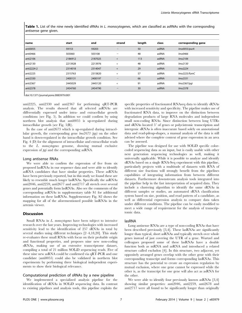
Table 1. List of the nine newly identified sRNAs in L. monocytogenes, which are classified as asRNAs with the correspondingantisense gene given.
name start end strand length class corresponding gene
anti0055 59153 59203 2 50 asRNA lmo0055/purA
anti0466 503060 503108 2 48 asRNA lmo0466
anti2106 2186912 2187025 + 113 asRNA lmo2106
anti2130 2213928 2213976 + 48 asRNA lmo2130
anti2224-2 2314018 2314047 + 29 asRNA lmo2224
anti2225 2315763 2315820 + 57 asRNA lmo2225/fumC
anti2330 2400131 2400197 2 66 asRNA lmo2331
anti2367 2445029 2445120 + 91 asRNA lmo2367/pgi
anti2378 2454760 2454790 2 30 asRNA lmo2378
doi:10.1371/journal.pone.0083979.t001
Listeria Monocytogenes sRNA Transcriptome
PLOS ONE | www.plosone.org 7 February 2014 | Volume 9 | Issue 2 | e83979

proposed. All four candidates have been originally described to
cover part of a single gene, but in our data were found to cover
four to six genes instead. See corresponding comments in
supplementary table S1 and the read pileups in supplemental
Fig. S2. Given the length of the lasRNAs, structures comparable to
the excludons described by Wurtzel and colleagues [4] are likely
for these lasRNAs. The most likely reason for us to identify those
sRNAs as significantly longer than before described, is the higher
sequencing coverage in our experiments. It enables us to identify
weekly but consistently transcribed areas better than before,
leading to the discovery of previously unidentified long transcripts
that were originally thought to be distinct or shorter.
Identification of nine new sRNA candidatesAutomated identification of asRNA in the data and manual
refinement of results revealed nine new sRNAs candidates in L.
monocytogenes. Most notably among these are four asRNAs opposite
of the genes lmo2225 (fumC), lmo2330, lmo0055 (purA) and lmo2367
(pgi).
The prophage A118 can be found in the L. monocytogenes EGD-e
genome inserted between the genes lmo2271 and lmo2332 [22]. At
the very end of this prophage region, covering the 39 end of
lmo2331 and the 59 end of lmo2330 we identified another down-
regulated asRNA (see Fig. 5 (C)). lmo2331 is predicted to encode a
cell wall lipoprotein, while lmo2330 is similar to the phage protein
gp33. Antisense transcription of the prophage genes has previously
been reported and this might be an additional case of such [2–4].
Figure 5. Validation of new asRNA transcripts from L. monocytogenes and their effect on gene regulation after transition to theintracellular growth conditions. A) The antisense RNA transcript anti0055 (purA) is validated by northern blot analysis and strand-specific qRT-PCR. The graph shows intracellular up-regulation of anti0055. B) Northern blot images of anti0055 and control 5S rRNA EC: Extracellular, IC:Intracellular. C) The presence of antisense transcripts anti2106 (lmo2106), anti2225 (fumC), and anti2330 (lmo2330) was determined by strand-specificqRT-PCR. anti2330 is down-regulated, anti2106 and anti2225 are up-regulated significantly. D) Strand-specific qRT-PCR analysis confirmed theexistence and up-regulation of antisense RNA transcript anti2367. pgi (lmo2367) was down-regulated, which indicates the possible role of anti2367 inpgi gene regulation. ‘*’ P#0.05; ‘**’ P#0.01; ‘***’ P#0.001.doi:10.1371/journal.pone.0083979.g005
Listeria Monocytogenes sRNA Transcriptome
PLOS ONE | www.plosone.org 8 February 2014 | Volume 9 | Issue 2 | e83979

Apart from this general antisense transcription it might represent
specific and active repression of phage gene expression, as phage
control by means of antisense transcription is a long known
phenomenon [29]. More recently Irnov and colleagues also
reported the expression of asRNA in prophages of Bacillus subtilis
and suggested a function in maintaining the phage host
equilibrium [14].
Antisense of the purA gene we were able to identify an asRNA at
the 59 end of the gene. The purA gene encodes a putative
adenylosuccinate synthetase with assumed function in the de novo
purine synthesis pathway, making it an essential enzyme in the
synthesis pathway of purine nucleobases. Purine synthesis seem-
ingly plays an important role for intracellular growth of L.
monocytogenes [27] and a L. monocytogenes serotype 4b strain with a
mutation of purA is known to be strongly attenuated in the
infection of mice [26]. This makes a lifestyle dependent regulation
of purA very likely, and asRNAs are known to play a major role in
the adaption to rapid environmental changes in general [30] as
well as the transition of L. monocytogenes from saprophytic to virulent
lifestyle in particular [31]. However, no classical or obvious
regulation pattern could be found when analyzing expression of
both the purA gene and its corresponding asRNA within the RNA-
Seq data which could be also observed by qRT-PCR (data not
shown). We observed increased expression of asRNA anti0055
under intracellular versus extracellular growth condition using
qRT-PCR analysis as well as northern blot analysis (see Fig. 5 (A
and B)). The biological relevance of this up-regulated asRNA has
to be characterized in future.
We identified a new asRNA anti2225 opposite of the fumC gene,
coding for a fumarate hydratase typically with central function in
the TCA-cycle. Interestingly, an antisense transcript of the
homologous gene has also been found in the Gram-negative
Helicobacter pylori and experimentally verified by northern blot and
RT-PCR [32]. In addition, many asRNAs of housekeeping genes
of Cyanobacterium synechocystis have been identified [33], demon-
strating that such asRNAs are a common mechanism of
transcriptional regulation. Furthermore L. monocytogenes is already
suspected to have an interrupted TCA-cycle [34]. Also it shown
that even an interrupted TCA-cycle may serve as an essential
generator for purine for which we already propose a regulation by
means of PurA [35]. Furthermore Schauer and coworkers have
shown the central role of purine biosynthesis for intracellular
growth [27]. Here we could show that expression of the fumC gene
(data not shown) as wells as anti2225 (see Fig. 5(C)) is up-regulated
after transition to the intracellular lifestyle. Biological interpreta-
tion of these finding is challenging at this point and needs further
experimental validation. Signs of classical asRNA regulation
patterns can be found expressed opposite of the gene lmo2367/
pgi for anti2367. Inspecting the sequencing data of the intracellular
and extracellular growth condition, the expression of either the
gene or the asRNA seems to be mutually exclusive, giving a hint
for a causal link and a possible regulation mechanism interfering
with the expression of pgi on the transcriptional level. This pattern
is clearly visible in Fig. 4(D) and Fig. 5(D) showing the mapped
reads for both the intracellular and the extracellular condition
which could be also confirmed by qRT-PCR analysis. Expression
of the pgi gene is low for the intracellular growth, and high for the
extracellular growth, while expression of the corresponding
asRNA on the opposite strand is high for the intracellular and
low for the extracellular condition (see Fig. 5(D)). lmo2367/pgi,
encodes a glucose-6-phosphate isomerase with central function in
the interface between glycolysis and the pentose phosphate
pathway. Previous reports link the transition from extracellular
to intracellular growth of L. monocytogenes to a reduced expression of
pgi [36] and a corresponding shift in metabolic pathways leading to
the degradation of glucose phosphate by the pentose phosphate
pathway [1]. Furthermore a proteomic study was able to identify
the pgi corresponding peptides under two different extracellular
conditions but not within intracellular conditions of L. monocytogenes
[37]. As a housekeeping gene, pgi is under the control of a
housekeeping promoter, and hence requires promoter indepen-
dent specific regulation of this gene. The identification of anti2367
sheds lights on the metabolic adaptation on transcriptional level by
antisense RNAs in L. monocytogenes.
ConclusionThe high coverage and strong strand specificity of our data
revealed a substantial amount of general antisense transcription
over the L. monocytogenes genome. Similar general antisense
transcription has been described previously [33,38]. The biological
relevance of this phenomenon is not yet fully understood, but the
finding of such in another bacterial organism underlines its
importance of further inquiry of the matter. Given the high
number of newly identified asRNAs as well as the identification of
exceptionally long non coding antisense RNAs, lasRNAs, it is
obvious that antisense transcription is an important factor in the
regulatory network of L. monocytogenes and it should be investigated
whether similar types of regulation are common in other bacterial
species.
Supporting Information
Table S1 List of previously identified sRNAs.
(XLS)
Table S2 List of newly identified sRNAs.
(XLS)
Table S3 Oligonucleotides used in this study.
(DOCX)
Table S4 Reads count summary of experimental tran-scriptome data.
(XLS)
File S1 Detailed description of data analysis pipeline.
(DOC)
Figure S1 Read mapping of asRNA anti0466, anti2106,anti2130, anti2224-2 and anti2378.
(PDF)
Figure S2 Read mappings of lasRNA like structures.
(PDF)
Acknowledgments
We would like to acknowledge Alexandra Amend for technical assistance.
Author Contributions
Conceived and designed the experiments: NK MAM TH . Performed the
experiments: NK MAM GKM XQ RM TH. Analyzed the data: SB SW
TR GKM XQ RM MAM TH. Wrote the paper: SB SW GKM RM NK
MAM TR TH.
Listeria Monocytogenes sRNA Transcriptome
PLOS ONE | www.plosone.org 9 February 2014 | Volume 9 | Issue 2 | e83979

References
1. Fuchs TM, Eisenreich W, Kern T, Dandekar T (2012) Toward a SystemicUnderstanding of Listeria monocytogenes Metabolism during Infection. Front
Microbiol 3: 23.
2. Mraheil MA, Billion A, Mohamed W, Mukherjee K, Kuenne C, et al. (2011)The intracellular sRNA transcriptome of Listeria monocytogenes during growth in
macrophages. Nucleic Acids Res 39: 4235–4248.
3. Toledo-Arana A, Dussurget O, Nikitas G, Sesto N, Guet-Revillet H, et al. (2009)The Listeria transcriptional landscape from saprophytism to virulence. Nature
459: 950–956.
4. Wurtzel O, Sesto N, Mellin JR, Karunker I, Edelheit S, et al. (2012)Comparative transcriptomics of pathogenic and non-pathogenic Listeria species.
Mol Syst Biol 8: 583.
5. Guell M, Yus E, Lluch-Senar M, Serrano L (2011) Bacterial transcriptomics:what is beyond the RNA horiz-ome? Nat Rev Microbiol 9: 658–669.
6. Siezen RJ, Wilson G, Todt T (2010) Prokaryotic whole-transcriptome analysis:
deep sequencing and tiling arrays. Microb Biotechnol 3: 125–130.
7. Sorek R, Cossart P (2010) Prokaryotic transcriptomics: a new view on regulation,
physiology and pathogenicity. Nat Rev Genet 11: 9–16.
8. Gottesman S (2005) Micros for microbes: non-coding regulatory RNAs inbacteria. Trends Genet 21: 399–404.
9. Livny J, Waldor MK (2010) Mining regulatory 59UTRs from cDNA deep
sequencing datasets. Nucleic Acids Res 38: 1504–1514.
10. Storz G, Altuvia S, Wassarman KM (2005) An abundance of RNA regulators.
Annu Rev Biochem 74: 199–217.
11. Waters LS, Storz G (2009) Regulatory RNAs in bacteria. Cell 136: 615–628.
12. Albrecht M, Sharma CM, Dittrich MT, Muller T, Reinhardt R, et al. (2011)
The transcriptional landscape of Chlamydia pneumoniae. Genome Biol 12: R98.
13. Vogel J, Luisi BF (2011) Hfq and its constellation of RNA. Nat Rev Microbiol 9:
578–589.
14. Irnov I, Sharma CM, Vogel J, Winkler WC (2010) Identification of regulatoryRNAs in Bacillus subtilis. Nucleic Acids Res 38: 6637–6651.
15. Pischimarov J, Kuenne C, Billion A, Hemberger J, Cemic F, et al. (2012)
sRNAdb: a small non-coding RNA database for gram-positive bacteria. BMC
Genomics 13: 384.
16. Camejo A, Buchrieser C, Couve E, Carvalho F, Reis O, et al. (2009) In vivo
transcriptional profiling of Listeria monocytogenes and mutagenesis identify new
virulence factors involved in infection. PLoS Pathog 5: e1000449.
17. Hain T, Ghai R, Billion A, Kuenne CT, Steinweg C, et al. (2012) Comparative
genomics and transcriptomics of lineages I, II, and III strains of Listeria
monocytogenes. BMC Genomics 13: 144.
18. Mandin P, Repoila F, Vergassola M, Geissmann T, Cossart P (2007)
Identification of new noncoding RNAs in Listeria monocytogenes and prediction
of mRNA targets. Nucleic Acids Res 35: 962–974.
19. Milohanic E, Glaser P, Coppee JY, Frangeul L, Vega Y, et al. (2003)
Transcriptome analysis of Listeria monocytogenes identifies three groups of genes
differently regulated by PrfA. Mol Microbiol 47: 1613–1625.
20. Oliver HF, Orsi RH, Ponnala L, Keich U, Wang W, et al. (2009) Deep RNA
sequencing of L. monocytogenes reveals overlapping and extensive stationary phase
and sigma B-dependent transcriptomes, including multiple highly transcribednoncoding RNAs. BMC Genomics 10: 641.
21. Metzker ML (2010) Sequencing technologies - the next generation. Nat Rev
Genet 11: 31–46.22. Glaser P, Frangeul L, Buchrieser C, Rusniok C, Amend A, et al. (2001)
Comparative genomics of Listeria species. Science 294: 849–852.23. Chatterjee SS, Hossain H, Otten S, Kuenne C, Kuchmina K, et al. (2006)
Intracellular gene expression profile of Listeria monocytogenes. Infect Immun 74:
1323–1338.24. Basineni SR, Madhugiri R, Kolmsee T, Hengge R, Klug G (2009) The influence
of Hfq and ribonucleases on the stability of the small non-coding RNA OxySand its target rpoS in E. coli is growth phase dependent. RNA Biol 6: 584–594.
25. Darling AE, Mau B, Perna NT (2010) progressiveMauve: multiple genome
alignment with gene gain, loss and rearrangement. PLoS One 5: e11147.26. Faith NG, Kim JW, Azizoglu R, Kathariou S, Czuprynski C (2012) Purine
biosynthesis mutants (purA and purB) of serotype 4b Listeria monocytogenes areseverely attenuated for systemic infection in intragastrically inoculated A/J Mice.
Foodborne Pathog Dis 9: 480–486.27. Schauer K, Geginat G, Liang C, Goebel W, Dandekar T, et al. (2010)
Deciphering the intracellular metabolism of Listeria monocytogenes by mutant
screening and modelling. BMC Genomics 11: 573.28. Loessner MJ, Inman RB, Lauer P, Calendar R (2000) Complete nucleotide
sequence, molecular analysis and genome structure of bacteriophage A118 ofListeria monocytogenes: implications for phage evolution. Mol Microbiol 35: 324–
340.
29. Wagner EG, Simons RW (1994) Antisense RNA control in bacteria, phages, andplasmids. Annu Rev Microbiol 48: 713–742.
30. Mehta P, Goyal S, Wingreen NS (2008) A quantitative comparison of sRNA-based and protein-based gene regulation. Mol Syst Biol 4: 221.
31. Storz G, Vogel J, Wassarman KM (2011) Regulation by small RNAs in bacteria:expanding frontiers. Mol Cell 43: 880–891.
32. Xiao B, Li W, Guo G, Li B, Liu Z, et al. (2009) Identification of small noncoding
RNAs in Helicobacter pylori by a bioinformatics-based approach. Curr Microbiol58: 258–263.
33. Georg J, Voss B, Scholz I, Mitschke J, Wilde A, et al. (2009) Evidence for amajor role of antisense RNAs in cyanobacterial gene regulation. Mol Syst Biol 5:
305.
34. Eisenreich W, Dandekar T, Heesemann J, Goebel W (2010) Carbon metabolismof intracellular bacterial pathogens and possible links to virulence. Nat Rev
Microbiol 8: 401–412.35. Huynen MA, Dandekar T, Bork P (1999) Variation and evolution of the citric-
acid cycle: a genomic perspective. Trends Microbiol 7: 281–291.36. Chatterjee SS, Hossain H, Otten S, Kuenne C, Kuchmina K, et al. (2006)
Intracellular gene expression profile of Listeria monocytogenes. Infect Immun 74:
1323–1338.37. Garcia-del Portillo F, Calvo E, D’Orazio V, Pucciarelli MG (2011) Association
of ActA to peptidoglycan revealed by cell wall proteomics of intracellular Listeria
monocytogenes. J Biol Chem 286: 34675–34689.
38. Sharma CM, Hoffmann S, Darfeuille F, Reignier J, Findeiss S, et al. (2010) The
primary transcriptome of the major human pathogen Helicobacter pylori. Nature464: 250–255.
39. Rutherford K, Parkhill J, Crook J, Horsnell T, Rice P, et al. (2000) Artemis:sequence visualization and annotation. Bioinformatics 16: 944–945.
Listeria Monocytogenes sRNA Transcriptome
PLOS ONE | www.plosone.org 10 February 2014 | Volume 9 | Issue 2 | e83979