Detection of Listeria monocytogenes using a commercial PCR kit and different DNA extraction methods
6
Detection of Listeria monocytogenes using a commercial PCR kit and different DNA extraction methods G. Amagliani a,b , C. Giammarini c , E. Omiccioli d , G. Brandi a,b , M. Magnani a,e, * a Centro di Biotecnologie, Universita ` di Urbino, via Campanella 1, 61032 Fano (PU), Italy b Istituto di Scienze Tossicologiche, Igienistiche e Ambientali, Universita ` di Urbino, via S. Chiara 23, 61029 Urbino (PU), Italy c Diatheva srl, viale Piceno 137/F, 61032 Fano (PU), Italy d Istituto Zooprofilattico Sperimentale Umbria e Marche, via Salvemini 1, 06126 Perugia, Italy e Istituto di Chimica Biologica ‘‘G. Fornaini’’, Universita ` di Urbino, via Saffi 2, 61029 Urbino (PU), Italy Received 10 April 2005; received in revised form 12 June 2006; accepted 13 June 2006 Abstract The aim of our work was to evaluate a new commercial test kit for the detection of Listeria monocytogenes by PCR, using different DNA extraction methods. Food samples (pork sausage and ‘‘mozzarella’’ cheese) were spiked with known concentrations of L. mono- cytogenes and culture-enriched for 24 h. DNA extracted using three commercial kits and two standard methods, was amplified in species- specific PCR employing a L. monocytogenes PCR Detection Kit (Diatheva). The PCR-based method proved to be a reliable means of detecting the pathogen in food samples independently from the extraction procedure used, even for a contamination cell number of 1 cfu/g before culture enrichment. The molecular assay, showing perfect agreement with standard microbiological tests and a considerably short- ened analysis time, provides a sensitive and rapid alternative for applications in the testing of foods for microbiological contamination, and highlights the potential of PCR technology in routine food control. Ó 2006 Elsevier Ltd. All rights reserved. Keywords: Listeria monocytogenes; PCR detection kit; Magnetic DNA extraction 1. Introduction Listeria monocytogenes is an important food pathogen when considering public health. In fact it causes severe illnesses associated with the contamination of various cat- egories of foods, particularly cheeses and dairy products (Rudolf & Scherer, 2001), beef and pork (Heredia, Garcia, Rojas, & Salazar, 2001). The mortality rate of listeriosis is very high, approximately 30% (Griffiths, 1989), and for this reason the FDA maintains a policy of zero-tolerance for L. monocytogenes (Anon, 2003). With this concern, the ability to detect this pathogen at low levels is considered essential because most of the foods susceptible to contam- ination are ready-to-eat products, which are not cooked or otherwise processed before consumption. These consider- ations underline the benefit of sensitive L. monocytogenes monitoring to ensure the microbiological quality of foods and to reduce risks for public health. Current microbiolog- ical culture methods rely on growth in culture media, fol- lowed by isolation, and biochemical and serological identification. However, the detection of this pathogen in food by these standard culture methods is made difficult by the sporadic or low levels of contamination (<100 cfu/ g), by the presence of a high level of background microflora and competitor organisms that could mask the presence of L. monocytogenes, and by interference due to food matrix components (Norton et al., 2001). Moreover, these methods are laborious and time consuming, requiring a minimum of five days to recognize Listeria spp. and about 10 days to identify L. monocytogenes by confirmatory tests (Anon, 1996), while immediate action should be taken in 0956-7135/$ - see front matter Ó 2006 Elsevier Ltd. All rights reserved. doi:10.1016/j.foodcont.2006.06.012 * Corresponding author. Tel.: +390721862832; fax: +390721862834. E-mail address: [email protected] (M. Magnani). www.elsevier.com/locate/foodcont Food Control 18 (2007) 1137–1142
-
Upload
independent -
Category
Documents
-
view
4 -
download
0
Transcript of Detection of Listeria monocytogenes using a commercial PCR kit and different DNA extraction methods

www.elsevier.com/locate/foodcont
Food Control 18 (2007) 1137–1142
Detection of Listeria monocytogenes using a commercial PCR kitand different DNA extraction methods
G. Amagliani a,b, C. Giammarini c, E. Omiccioli d, G. Brandi a,b, M. Magnani a,e,*
a Centro di Biotecnologie, Universita di Urbino, via Campanella 1, 61032 Fano (PU), Italyb Istituto di Scienze Tossicologiche, Igienistiche e Ambientali, Universita di Urbino, via S. Chiara 23, 61029 Urbino (PU), Italy
c Diatheva srl, viale Piceno 137/F, 61032 Fano (PU), Italyd Istituto Zooprofilattico Sperimentale Umbria e Marche, via Salvemini 1, 06126 Perugia, Italy
e Istituto di Chimica Biologica ‘‘G. Fornaini’’, Universita di Urbino, via Saffi 2, 61029 Urbino (PU), Italy
Received 10 April 2005; received in revised form 12 June 2006; accepted 13 June 2006
Abstract
The aim of our work was to evaluate a new commercial test kit for the detection of Listeria monocytogenes by PCR, using differentDNA extraction methods. Food samples (pork sausage and ‘‘mozzarella’’ cheese) were spiked with known concentrations of L. mono-
cytogenes and culture-enriched for 24 h. DNA extracted using three commercial kits and two standard methods, was amplified in species-specific PCR employing a L. monocytogenes PCR Detection Kit (Diatheva). The PCR-based method proved to be a reliable means ofdetecting the pathogen in food samples independently from the extraction procedure used, even for a contamination cell number of 1 cfu/gbefore culture enrichment. The molecular assay, showing perfect agreement with standard microbiological tests and a considerably short-ened analysis time, provides a sensitive and rapid alternative for applications in the testing of foods for microbiological contamination,and highlights the potential of PCR technology in routine food control.� 2006 Elsevier Ltd. All rights reserved.
Keywords: Listeria monocytogenes; PCR detection kit; Magnetic DNA extraction
1. Introduction
Listeria monocytogenes is an important food pathogenwhen considering public health. In fact it causes severeillnesses associated with the contamination of various cat-egories of foods, particularly cheeses and dairy products(Rudolf & Scherer, 2001), beef and pork (Heredia, Garcia,Rojas, & Salazar, 2001). The mortality rate of listeriosis isvery high, approximately 30% (Griffiths, 1989), and for thisreason the FDA maintains a policy of zero-tolerance forL. monocytogenes (Anon, 2003). With this concern, theability to detect this pathogen at low levels is consideredessential because most of the foods susceptible to contam-ination are ready-to-eat products, which are not cooked or
0956-7135/$ - see front matter � 2006 Elsevier Ltd. All rights reserved.
doi:10.1016/j.foodcont.2006.06.012
* Corresponding author. Tel.: +390721862832; fax: +390721862834.E-mail address: [email protected] (M. Magnani).
otherwise processed before consumption. These consider-ations underline the benefit of sensitive L. monocytogenes
monitoring to ensure the microbiological quality of foodsand to reduce risks for public health. Current microbiolog-ical culture methods rely on growth in culture media, fol-lowed by isolation, and biochemical and serologicalidentification. However, the detection of this pathogen infood by these standard culture methods is made difficultby the sporadic or low levels of contamination (<100 cfu/g), by the presence of a high level of background microfloraand competitor organisms that could mask the presence ofL. monocytogenes, and by interference due to food matrixcomponents (Norton et al., 2001). Moreover, thesemethods are laborious and time consuming, requiring aminimum of five days to recognize Listeria spp. and about10 days to identify L. monocytogenes by confirmatory tests(Anon, 1996), while immediate action should be taken in

1138 G. Amagliani et al. / Food Control 18 (2007) 1137–1142
case of contamination since it is of fundamental impor-tance to ensure the safety of food products, especially inthe case of those food matrices having short shelf-lives,such as meat or dairy products.
In the past years, advancements in biotechnology haveresulted in the development of rapid methods that reduceanalysis time and offer great sensitivity and specificity inthe detection of pathogens. Among these, PCR has beenincreasingly used for the rapid, sensitive and specific detec-tion of foodborne pathogens (Olsen et al., 1995). However,the successful application of PCR assays to food sampleshas been hindered by the lack of a rapid and efficientmethod for the preparation of PCR-amplifiable DNA(Lantz, Hahn Hagcrdal, & Radstrom, 1994). The presenceof PCR inhibitors in food samples (Bickley, Short, McDo-well, & Parkes, 1996; Rossen, Norskov, Holmstrom, &Rasmussen, 1992) represents the main limitation in thiskind of assays due to the production of false negativeresults. For this reason, the application of PCR-basedmethods is closely linked to the selection of suitable meth-ods for DNA extraction.
This study evaluates different procedures for the extrac-tion of L. monocytogenes DNA from enriched food sam-ples, followed by detection using the new commercialassay ‘‘Listeria monocytogenes PCR Detection Kit’’ (Diath-eva, Italy). Additionally, the equivalence of the molecularmethod and the reference culture method is investigated.
2. Materials and methods
2.1. Samples and media
Food samples of pork raw sausage and ‘‘mozzarella’’cheese were obtained from a local supermarket.
For bacterial growth and samples enrichment thefollowing media were used: Listeria Enrichment BrothBase supplemented with Listeria Selective EnrichmentSupplement (LEB); Fraser Broth Base supplemented withFraser Selective Supplement (FB); Listeria Selective AgarBase (Oxford) supplemented with Listeria Selective Supple-ment (Oxford), all reconstituted and supplemented accord-ing to producer’s recommendations. All culture media werepurchased from Oxoid (Basingstoke, UK).
Chemicals were obtained from Sigma-Aldrich (St.Louis, Missouri, USA).
2.2. Inoculum and samples preparation
The bacterial species used in this study is a collectionstrain of Listeria monocytogenes (ATCC 9525). L. monocyt-
ogenes was growth in LEB at 37 �C with shaking at200 rpm, to an optical density of 0.065 (at 600 nm), corre-sponding to about 108 cfu/ml. The bacterial concentrationof the contaminating culture was determined by plating onOxford medium and incubation at 37 �C for 24–48 h. Serial10-fold dilutions in 0.9% NaCl were made, and 1 ml ofeach was used for contamination of food samples prepared
as follows: twenty-five grams each of sausage and ‘‘mozza-rella’’ cheese were homogenized separately in 225 ml of FBor LEB, using a Stomacher 400 homogenizer (Seward,Worthington, UK). Subsequently, food homogenates wereinoculated with the described bacterial dilutions. Thecontamination rate of food samples ranged from 103 to1 cfu/g. A culture-negative control, consisting of an uncon-taminated food sample in culture medium, was alsoincluded for each food matrix.
Food homogenates were incubated at 30 �C for 24 h.Aliquots of 1 ml of the enriched samples were collectedand sterile gauze-filtered, then centrifuged at 5000g for10 min at 4 �C. Bacterial pellets were subjected to DNAextraction.
2.3. Nucleic acid isolation
In this section are outlined methods used for DNA iso-lation from enriched food samples. Brief descriptions arereported for DNA extraction methods carried out withcommercial kits, while full detail is provided for lab-basedprocedures.
DNeasy Tissue Kit (Qiagen, Hilden, Germany). Theextraction was performed following the manufacturer’sprotocol for Gram-positive bacteria. Cellular lysis was car-ried out by enzymatic treatment with lysozyme, RNase Aand proteinase K in detergents and guanidium salts-con-taining buffers. After buffering condition adjustment,DNA was selectively bound to a silica-gel membrane. Con-taminants were removed by two washing steps and finallyDNA was eluted in water.
Puregene Yeast and Gram Positive Bacteria Kit (Gentra
Systems, Minneapolis, Minnesota, USA). As described bythe manufacturer, bacteria were lysed with an anionicdetergent and a lytic enzyme solution in presence of aDNA stabilizer. Complete cell disruption was also ensuredby a heating step at 80 �C. RNA was removed by a 40 minRNase A digestion while proteins were salt precipitatedand then discarded. Bacterial DNA was recovered by pre-cipitation with isopropanol and dissolved in a bufferedsolution.
Listeria monocytogenes DNA Isolation Kit: Milk (Diath-
eva, Fano, Italy). The kit was designed for a magneticbased-DNA isolation using paramagnetic nanoparticles.Bacterial cells were lysed and enzymatically digested withlysozyme, RNase A and proteinase K, according to manu-facturer’s instructions. The lysates were then mixed withthe magnetic beads in presence of the specific binding buf-fer to allow DNA adsorbtion. DNA bound to magneticparticles was then washed, eluted in water and finally con-centrated by ethanol precipitation.
Phenol–chloroform extraction. The organic extractionwas performed according to the standard method by Sam-brook and Russel (2001). Briefly, pelletted cells were resus-pended in 500 ll of lysis solution (8 M urea, 0.3 M NaCl,10 mM Tris–HCl) with the addition of 500 ll 10% SDS.After 20 min at 37 �C, DNA was extracted with 2 volumes

G. Amagliani et al. / Food Control 18 (2007) 1137–1142 1139
of phenol, mixed 10 min and centrifuged at 3500g for10 min. An equal volume of chloroform:isoamyl alcoholwas added to the aqueous phase, samples were mixed asabove and centrifuged at 12000g for 5 min. Nucleic acidsin the aqueous phase were precipitated with 2.5 volumesof ethanol and 1/10 volume of 3 M sodium acetate pH5.2, incubating at �20 �C for 1 h. After centrifugation,DNA pellets were washed with 70% ethanol and dissolvedin 150 ll of water.
Boiling method (Bansal, McDonell, Smith, Arnold, &Ibrahim, 1996). Bacterial pellets were washed once with1 ml phosphate buffered saline (PBS), pH 7.4, resuspendedin a same volume of cold water and incubated in a boilingwater bath for 10 min. The clear supernatants obtainedafter a 5 min centrifugation at 12000g were used for PCRreaction.
2.4. Quality and yield of extracted nucleic acids
To investigate the quality of the extracted nucleic acids,sample absorbances at 260 and 280 nm were measured(Pharma Spec UV-1700 spectrophotometer; Shimadzu,Kyoto, Japan) and the A260/A280 ratios were calculated.The spectrophotometrical analysis also permitted to evalu-ate the yield of the differently extracted DNAs (Sambrook& Russel, 2001).
2.5. DNA amplification and electrophoretic analysis
L. monocytogenes DNA amplification was carried outaccording to the ‘‘Listeria monocytogenes PCR DetectionKit’’ (Diatheva) manual. Ten microliters of each extractedsample were used in the PCR reaction. Additionally, forevery amplification round, a reagent blank and a positivecontrol (supplied in the kit) were included. Besides reagentsand enzyme for detection of L. monocytogenes targetsequence, the PCR mix also contained an internal controlto identify reaction inhibition.
Twenty-five of 50 ll amplified mixture were electropho-resed through a 2% agarose (w/v) gel, stained with ethi-dium bromide in presence of a low molecular weightDNA standard (UX174 DNA/HaeIII, Roche, Basel, Swit-zerland). Amplicons were visualized and analyzed on a GelDoc 2000 apparatus using the Quantity One QuantitationSoftware (Bio-Rad, Hercules, California, USA).
An amplicon of 172 bp with or without the presence of a112 bp DNA fragment indicated a positive result. Negativesamples free of PCR inhibitors were identified by a 112 bpgel-band alone.
2.6. Detection limits and reproducibility of the
L. monocytogenes PCR Detection Kit
The sensitivity of the PCR kit was assessed by amplify-ing dilutions in NaCl 0.9% of a L. monocytogenes cultureprepared as described in Section 2.2. Bacterial concentra-tion ranged from 1 · 105 to 2 · 102 cfu/ml, corresponding
to 103 to 2 cfu per PCR reaction. Each dilution was testedin quadruplicate to assess the reproducibility of the results.Amplification and gel analysis were carried out asdescribed in previous paragraph.
2.7. Listeria monocytogenes detection in food samplesby reference method
Standard culture protocols, according to the official Ital-ian regulatory agency ‘‘Istituto Superiore di Sanita Nazio-nale’’ (Anon, 1996), were used as reference methods for L.
monocytogenes detection in artificially contaminated foodsamples. Briefly, samples were homogenized in enrichmentmedia (LEB for cheese samples and FB for sausages) andcontaminated as described in 2.2 section. Enrichment cul-tures were incubated at 30�C for 48 h. Subsequently, 0.1ml were streaked onto Oxford plates, and incubated at37 �C for 48 h. Presumptive Listeria colonies were sub-jected to biochemical (API Listeria, bioMerieux, Marcyl’Etoile, France) and serological (Listeria Rapid Test,Oxoid) confirmation tests.
3. Results
3.1. Quality and yield of extracted DNAs
Since both the purity and quality of extracted DNAsconstitute pre-requisites in PCR-based detection assays,we evaluated the A260/A280 ratios of purified samplesfrom both food enrichments used in this study contami-nated with a starting bacterial concentration of 103 cfu/g(Table 1).
The results indicated that the quality of DNAextracted from sausage enrichment cultures was invari-ably higher than that isolated from ‘‘mozzarella’’ cheese.The best A260/A280 ratio values were shown for extrac-tions using the Qiagen kit, phenol–chloroform and boil-ing methods, while the lowest ratio was obtained forGentra extracted samples. Nevertheless, the purity ofDNA recovered was still more than sufficient for subse-quent application and isolated DNA functioned satisfac-torily in PCR amplification. Finally, the data indicatedthat non commercial methods yielded the highest amountof DNA, followed by Diatheva and Qiagen, with similarresults (Table 1).
3.2. Detection limits and reproducibility of the
L. monocytogenes PCR Detection Kit
The PCR kit employed in this work was tested forsensitivity and reproducibility, to evaluate its capabilityto reveal low levels of contamination. As shown inFig. 1, the detection endpoint occurred at 2 cfu per PCRreaction, and this result was confirmed in all four replicateswith the same level of contamination, showing a highdegree of sensitivity and good reproducibility.
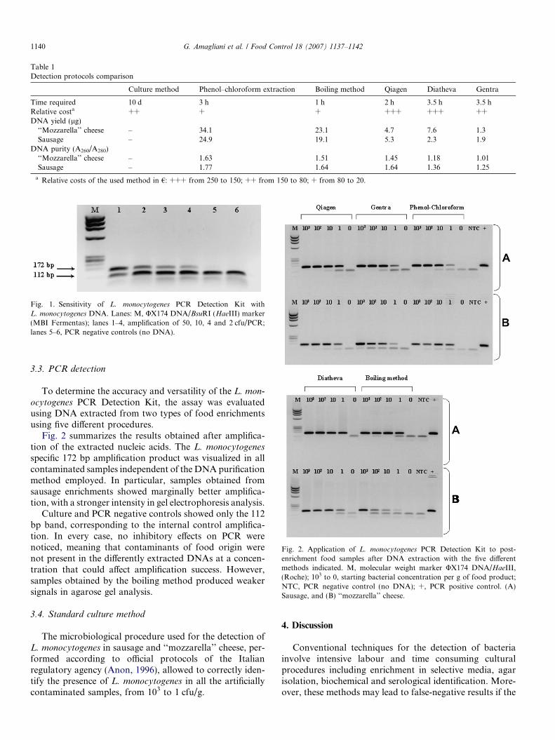
Table 1Detection protocols comparison
Culture method Phenol–chloroform extraction Boiling method Qiagen Diatheva Gentra
Time required 10 d 3 h 1 h 2 h 3.5 h 3.5 hRelative costa ++ + + +++ +++ ++DNA yield (lg)
‘‘Mozzarella’’ cheese – 34.1 23.1 4.7 7.6 1.3Sausage – 24.9 19.1 5.3 2.3 1.9
DNA purity (A260/A280)‘‘Mozzarella’’ cheese – 1.63 1.51 1.45 1.18 1.01Sausage – 1.77 1.64 1.64 1.36 1.25
a Relative costs of the used method in €: +++ from 250 to 150; ++ from 150 to 80; + from 80 to 20.
Fig. 1. Sensitivity of L. monocytogenes PCR Detection Kit withL. monocytogenes DNA. Lanes: M, UX174 DNA/BsuRI (HaeIII) marker(MBI Fermentas); lanes 1–4, amplification of 50, 10, 4 and 2 cfu/PCR;lanes 5–6, PCR negative controls (no DNA).
Fig. 2. Application of L. monocytogenes PCR Detection Kit to post-enrichment food samples after DNA extraction with the five differentmethods indicated. M, molecular weight marker UX174 DNA/HaeIII,(Roche); 103 to 0, starting bacterial concentration per g of food product;NTC, PCR negative control (no DNA); +, PCR positive control. (A)Sausage, and (B) ‘‘mozzarella’’ cheese.
1140 G. Amagliani et al. / Food Control 18 (2007) 1137–1142
3.3. PCR detection
To determine the accuracy and versatility of the L. mon-
ocytogenes PCR Detection Kit, the assay was evaluatedusing DNA extracted from two types of food enrichmentsusing five different procedures.
Fig. 2 summarizes the results obtained after amplifica-tion of the extracted nucleic acids. The L. monocytogenes
specific 172 bp amplification product was visualized in allcontaminated samples independent of the DNA purificationmethod employed. In particular, samples obtained fromsausage enrichments showed marginally better amplifica-tion, with a stronger intensity in gel electrophoresis analysis.
Culture and PCR negative controls showed only the 112bp band, corresponding to the internal control amplifica-tion. In every case, no inhibitory effects on PCR werenoticed, meaning that contaminants of food origin werenot present in the differently extracted DNAs at a concen-tration that could affect amplification success. However,samples obtained by the boiling method produced weakersignals in agarose gel analysis.
3.4. Standard culture method
The microbiological procedure used for the detection ofL. monocytogenes in sausage and ‘‘mozzarella’’ cheese, per-formed according to official protocols of the Italianregulatory agency (Anon, 1996), allowed to correctly iden-tify the presence of L. monocytogenes in all the artificiallycontaminated samples, from 103 to 1 cfu/g.
4. Discussion
Conventional techniques for the detection of bacteriainvolve intensive labour and time consuming culturalprocedures including enrichment in selective media, agarisolation, biochemical and serological identification. More-over, these methods may lead to false-negative results if the

G. Amagliani et al. / Food Control 18 (2007) 1137–1142 1141
sample contains the target species in a high backgroundof a mixed bacterial population. For example, severalresearchers have observed that L. innocua can outcompeteL. monocytogenes if the two species are cultivated togetherin commonly used enrichment media, including LEB(MacDonald & Sutherland, 1994; Petran & Swanson,1993). Consequently, DNA amplification technology hasproven to be beneficial when working with foodbornepathogens because of its high specificity, sensitivity andrapidity. The main limitation associated with PCR applica-tion to food-contaminating microorganisms concerns thepresence of inhibitory substances that are coextracted withDNA and may be present in the sample, causing a failure inthe amplification reaction which leads to false negativeresults. Therefore, quality and purity of extracted nucleicacids are primary requirements for a PCR-based detectionassay and the selection of a proper extraction method isdeterminant for a successful and valid PCR analysis.Mainly due to the high cost of labour and time constraintsin processing numerous samples, commercial kits havebeen developed and are becoming widely used for pathogenDNA detection. Some of these have been specifically devel-oped for food analysis, particularly for L. monocytogenes.
In this study we report the application of the commer-cial assay ‘‘Listeria monocytogenes PCR Detection Kit’’using DNA differently extracted by different methods fromtwo artificially contaminated foodstuffs: pork sausage, thatis a matrix having a high protein content, and ‘‘mozza-rella’’ cheese, containing fat and calcium ions, well knownto be PCR inhibitors (Bickley et al., 1996). DNA isolationwas carried out by either commercially available kits orconventional phenol–chloroform and lysis by boiling meth-ods. The three manufacturers did not supply completeinformation regarding the ingredients contained in theircommercial kits due to the proprietary nature of kit com-ponents. However, purity and yield of the extracted nucleicacids were compared, resulting in higher DNA contentfor conventional extraction methods and relatively lowA260/A280 ratios for the majority of DNA samples. Thisresult indicates contamination with other species, whichcould most probably be identified as proteins, having noinhibitory effects on DNA polymerase. Moreover, thePCR Kit correctly identified all contaminated samples,even at the lowest contamination ratio (1 cfu/g beforeculture enrichment). However, different substances of foodorigin, such as calcium ions (Bickley et al., 1996), polysac-charides or fats (Rossen et al., 1992) that commonly causeamplification inhibition are not detectable in the A280 read-ing. This consideration could explain results obtained forsamples extracted using the boiling method suggested byBansal et al. (1996), showing faint signals in gel analysis,particularly in the case of ‘‘mozzarella’’ samples, but withan A260/A280 ratio comparable to that of DNA obtainedby other procedures. Thus, although lysis by boilingappears the most convenient extraction protocol, particularcare should be taken when food contamination with verylow bacterial count is suspected.
The magnetically driven extraction (Diatheva), even ifspecifically recommended for DNA isolation directly frommilk, proved to be suitable for enriched food products aswell. Nevertheless, although these extraction proceduresworked well for raw pork sausage and ‘‘mozzarella’’cheese, they cannot extrapolate results when applied toother foods known to contain PCR inhibitors, such ascertain fruits (e.g., rasberries).
The magnetic isolation was slightly more time consum-ing and laborious than the column system (Qiagen), butthe limitation could be circumvented by automation ofthe entire procedure, offering several advantages to theend user, especially when large sample numbers are pro-cessed. Moreover, it is safer than using phenol–chloroform,avoiding the use and manipulation of harmful organicsolvents.
The molecular assay also revealed results which wereequivalent to those obtained with standard methods usedin microbiology, however the time required for analysiswas reduced from 7-10 to two working days. The applica-tion of the amplification assay after cultural enrichmentoffers some advantages, increasing the amount of targetsequences, diluting non-target DNA and compounds inter-fering with the PCR, and ensuring that a positive result isobtained from viable cells.
In conclusion, the PCR Kit tested showed very high sen-sitivity and robustness, requiring relatively little experiencein bio-molecular techniques. The presence of the internalcontrol, capable of revealing every possible PCR failurecaused by substances of food origin, provides completecertainty of negative results. It can be proposed in the rou-tine investigation of foods for a rapid preliminary screeningof negative samples, furnishing presumptive positive resultsthat should be confirmed by official reference methods.
Acknowledgement
We would like to thank Diatheva srl for providingreagents and equipment for this study.
References
Anon. (1996). Rapporti ISTISAN ISSN 1123-3117, 96/35.Anon. (2003). FDA/Center for Food Safety and Applied Nutrition,
USDA/Food Safety and Inspection Service, Centers for DiseaseControl and Prevention, Quantitative assessment of relative risk topublic health from foodborne Listeria monocytogenes among selectedcategories of ready-to-eat foods, September 2003.
Bansal, N. S., McDonell, F. H., Smith, A., Arnold, G., & Ibrahim, G. F.(1996). Multiplex PCR assay for the routine detection of Listeria infood. International Journal of Food Microbiology, 33, 293–300.
Bickley, J., Short, J. K., McDowell, D. G., & Parkes, H. C. (1996).Polymerase chain reaction (PCR) detection of Listeria monocytogenes
in diluted milk and reversal of PCR inhibition caused by calcium ions.Letters in Applied Microbiology, 22, 153–158.
Griffiths, M. W. (1989). Listeria monocytogenes: its importance in thedairy industry. Journal of the Science of Food and Agriculture, 47,133–158.

1142 G. Amagliani et al. / Food Control 18 (2007) 1137–1142
Heredia, N., Garcia, S., Rojas, G., & Salazar, L. (2001). Microbiologicalcondition of ground meat retailed in Monterrey, Mexico. Journal of
Food Protection, 64, 1249–1251.Lantz, P.-G., Hahn Hagcrdal, B., & Radstrom, P. (1994). Sample
preparation methods in PCR-based detection of food pathogens.Trends in Food Science and Technology, 5, 384–389.
MacDonald, F., & Sutherland, A. D. (1994). Important differencesbetween the generation times of Listeria monocytogenes and Listeria
innocua in two Listeria enrichment broths. Journal of Dairy Research,
61, 433–436.Norton, D.-M., McCamey, M. A., Gall, K. L., Scarlett, J. M., Boor, K. J.,
& Wiedmann, M. (2001). Molecular studies on the ecology of Listeria
monocytogenes in the smoked fish processing industry. Applied and
Environmental Microbiology, 67(1), 198–205.Olsen, J. E., Aabo, S., Hill, W., Notermans, S., Wernars, K., Granum, P.
E., et al. (1995). Probes and polymerase chain reaction for detection of
food-borne bacterial pathogens. International Journal of Food Micro-
biology, 28, 1–78.Petran, R. L., & Swanson, K. M. J. (1993). Simultaneous growth of
Listeria monocytogenes and L. innocua. Journal of Food Protection, 56,616–618.
Rossen, L., Norskov, P., Holmstrom, K., & Rasmussen, O. F. (1992).Inhibition of PCR by components of food samples, microbialdiagnostic assays and DNA extraction solutions. International Journal
of Food Microbiology, 17, 37–45.Rudolf, M., & Scherer, S. (2001). High incidence of Listeria monocytog-
enes in European red smear cheese. International Journal of Food
Microbiology, 63, 91–98.Sambrook, J., & Russel, D. W. (2001). Molecular cloning: A laboratory
manual (3rd ed.). Cold Spring Harbor, NY, USA: Cold Spring HarborPress.