Listeria monocytogenes-Infected Human Dendritic Cells: Uptake and Host Cell Response
Upload
independentCategory
view
0download
0
In Vivo Transcriptional Profiling of Listeriamonocytogenes and Mutagenesis Identify New VirulenceFactors Involved in InfectionAna Camejo1, Carmen Buchrieser2, Elisabeth Couve3, Filipe Carvalho1, Olga Reis1, Pierre Ferreira1,
Sandra Sousa1, Pascale Cossart4,5,6, Didier Cabanes1*
1 IBMC - Instituto de Biologia Molecular e Celular, Group of Molecular Microbiology, Universidade do Porto, Porto, Portugal, 2 Institut Pasteur, UP Biologie des Bacteries
Intracellulaires and CNRS URA 2171, Paris, France, 3 Institut Pasteur, Unite Genetique des Genomes Bacteriens CNRS URA 2171, Paris, France, 4 Institut Pasteur, Unite des
Interactions Bacteries-Cellules, Paris, France, 5 Inserm U604, Paris, France, 6 INRA USC2020, Paris, France
Abstract
Listeria monocytogenes is a human intracellular pathogen able to colonize host tissues after ingestion of contaminated food,causing severe invasive infections. In order to gain a better understanding of the nature of host–pathogen interactions, westudied the L. monocytogenes genome expression during mouse infection. In the spleen of infected mice, <20% of theListeria genome is differentially expressed, essentially through gene activation, as compared to exponential growth in richbroth medium. Data presented here show that, during infection, Listeria is in an active multiplication phase, as revealed bythe high expression of genes involved in replication, cell division and multiplication. In vivo bacterial growth requiresincreased expression of genes involved in adaptation of the bacterial metabolism and stress responses, in particular tooxidative stress. Listeria interaction with its host induces cell wall metabolism and surface expression of virulence factors.During infection, L. monocytogenes also activates subversion mechanisms of host defenses, including resistance to cationicpeptides, peptidoglycan modifications and release of muramyl peptides. We show that the in vivo differential expression ofthe Listeria genome is coordinated by a complex regulatory network, with a central role for the PrfA-SigB interplay. Inparticular, L. monocytogenes up regulates in vivo the two major virulence regulators, PrfA and VirR, and their downstreameffectors. Mutagenesis of in vivo induced genes allowed the identification of novel L. monocytogenes virulence factors,including an LPXTG surface protein, suggesting a role for S-layer glycoproteins and for cadmium efflux system in Listeriavirulence.
Citation: Camejo A, Buchrieser C, Couve E, Carvalho F, Reis O, et al. (2009) In Vivo Transcriptional Profiling of Listeria monocytogenes and Mutagenesis IdentifyNew Virulence Factors Involved in Infection. PLoS Pathog 5(5): e1000449. doi:10.1371/journal.ppat.1000449
Editor: Jorge E. Galan, Yale University School of Medicine, United States of America
Received November 21, 2008; Accepted April 27, 2009; Published May 29, 2009
Copyright: � 2009 Camejo et al. This is an open-access article distributed under the terms of the Creative Commons Attribution License, which permitsunrestricted use, distribution, and reproduction in any medium, provided the original author and source are credited.
Funding: This work was supported by grants from the Portuguese Fundacao para a Ciencia e a Tecnologia, FCT-POCI/SAU-MMO/60443/2004 and FCT-PTDC/SAU-MII/65406/2006, and by an ERANET Pathogenomics grant, FCT-ERA/PTG/0003-SPATELIS (http://alfa.fct.mctes.pt). AC and OR are recipients of FCT doctoralfellowships SFRH/BD/29314/2006 and SFRH/BD/28185/2006. SS is a recipient of an FCT postdoctoral fellowship, SFRH/BPD/21549/2005. PC is a Howard HughesMedical Institute international research scholar. The funders had no role in study design, data collection and analysis, decision to publish, or preparation of themanuscript.
Competing Interests: The authors have declared that no competing interests exist.
* E-mail: [email protected]
Introduction
Listeria monocytogenes is an intracellular food-borne pathogen that
causes listeriosis, an infection characterized by gastroenteritis,
meningitis, encephalitis, and maternofetal infections in humans. It
has one of the highest fatality rate among food-borne infections
(20%–30%) [1]. Our knowledge of the infectious process in vivo
mostly derives from infections in various animal models, in
particular the mouse model. It is considered that bacteria after
crossing the intestinal barrier reach, via the lymph and the blood,
the liver and the spleen where they replicate actively. Then,
bacteria via hematogenous dissemination, can reach the brain and
the placenta. The disease is thus due to the original property of L.
monocytogenes to be able to cross three host barriers: the intestinal
barrier, the blood brain barrier and the materno-fetal barrier. It is
also due to the capacity of Listeria to resist intracellular killing when
phagocytosed by macrophages and to invade many non-
phagocytic cell types. In the murine model, within minutes after
intravenous inoculation, most bacteria can be found in the spleen
and the liver [2].
L. monocytogenes ranks among the best-known intracellular
pathogens and, until now, 50 genes have been shown to be
involved in virulence in the mouse model (Table S1). However,
whereas the different steps of the cell infectious process and the
virulence factors specifically involved are well described [3], our
knowledge of the in vivo infectious process is still fragmentary.
Virulence is by definition expressed in a susceptible host, and
involves a dynamic cross talk between the host and the pathogen.
A detailed understanding of this interaction thus requires global
approaches in the context of an in vivo infection. Analysis of the
pathogen whole genome expression within the host should allow
the identification of new bacterial genes critical for the infectious
process, and lead to a better understanding of the molecular events
responsible for Listeria infection.
The technology of DNA arrays allows to both study the gene
content of different strains and measure gene expression levels on a
PLoS Pathogens | www.plospathogens.org 1 May 2009 | Volume 5 | Issue 5 | e1000449

genome-wide scale under different conditions. The genetic basis of
L. monocytogenes pathogenicity was addressed by comparative
genomics [4] and transcriptomics [5] using Listeria DNA arrays
and various L. monocytogenes strains. Listeria arrays were also used for
the analysis of the in vitro global gene expression of Listeria mutants
for PrfA, the central regulator of virulence genes [6], and for other
transcriptional regulators important for stress response and
virulence (sB, s54, HrcA, CtsR, VirR) [7–13]. Recently, this
approach was applied to the determination of the intracellular
gene expression profile of L. monocytogenes in epithelial and
macrophage cell lines [14,15]. In vivo genome profiles of other
pathogens (Streptococcus pneumoniae, S. pyogenes, Mycobacterium tubercu-
losis, Borrelia burgdoferi, Yersinia pestis) infecting different mouse
organs (dermis, soft tissue, lung, blood) were previously performed
[16]. However, to our knowledge, the genome expression of a
pathogen was never studied in infected mouse spleen.
Here, we present the first ‘‘in vivo’’ transcriptome of L.
monocytogenes. We compared expression profiles of L. monocytogenes
grown in standard culture medium in exponential phase vs.
bacteria recovered from mouse spleens 24, 48 and 72 hours after
intravenous infection. We determined the detailed expression
kinetics of the complete L. monocytogenes genome in the course of the
infection, and identified new Listeria virulence factors whose
expression was highly up regulated in vivo.
Results
The in vivo transcriptome approachWe used the DNA macroarray technology to profile the
transcriptome of Listeria during mouse infection. We used the
previously described L. monocytogenes whole-genome arrays containing
500-bp-long PCR products specific for each gene [6]. Ninety-nine
per cent of the 2853 predicted ORFs of the L. monocytogenes EGDe
genome are represented on the arrays. They were used to analyze
Listeria transcription profiles under in vitro growth in BHI in
exponential phase at 37uC under aerobic conditions with shaking
(pH 7) (Figure S1), and under in vivo growth conditions (mouse
spleen) at 1, 2 and 3 days post intravenous infection (p.i.). Listeria
present in spleen were analyzed because this organ is with the liver
one of the major sites of L. monocytogenes infection. For unknown
reasons, we never succeeded to prepare good quality bacterial RNAs
from infected mouse livers. The time points chosen reflect key steps
in the Listeria infectious process.
Culture in BHI in exponential phase at 37uC with shaking was
chosen as reference conditions because BHI is the Listeria reference
growth medium where bacteria divide in exponential growth
phase at rates that are comparable to those observed for
intracellular growth [17]. In addition, these are the in vitro
reference conditions used in all previous studies analyzing the
genome expression of L. monocytogenes in vitro or intracellularly [6–
15]. However, in order to analyze the potential impact of the in
vitro culture conditions used as reference on the relative gene
expression in vivo, we first analyzed the results obtained comparing
transcriptome from in vivo grown bacteria to transcriptomes from
bacteria grown in vitro in exponential or stationary phase (Table S2).
In addition, expression of known and potential virulence genes was
analyzed by quantitative real time-PCR (qPCR) on RNAs extracted
from bacteria cultured in BHI at 37uC in exponential or stationary
growth phase, or in defined minimal medium [18], and compared to
in vivo expression (Figure 1A). Results indicated that culture in
exponential growth phase are closer conditions to those met by
Listeria in vivo (Table S2). In addition, even if the expression of tested
genes behaved differently in function of the in vitro conditions,
expression of all the genes was always lower in vitro as compared to in
vivo, independently of the in vitro growth conditions (Figure 1A).
These experiments supported the choice of exponential growth
phase in BHI as reference conditions and minimized the impact of
the in vitro growth conditions on the identification of genes
differentially expressed in vivo.
The reliability of the macroarray expression data was further
assessed by qPCR. We selected a subset of 10 genes and performed
qPCR on cDNA from bacteria grown in either standard medium or
extracted from mouse spleens 48 h p.i.. qPCR results and array data
exhibited a high correlation coefficient (0.7) (Figure 1B). This strong
correlation was also observed for other infection time points (Figure
S2). However, the differences in gene expression, as measured by
qPCR, were generally higher, indicating that in vivo transcriptome
data rather underestimate changes in gene expression.
The procedure used for bacterial RNA extraction from infected
mouse spleens is an adaptation of the standard procedure
originally used for transcriptional analysis of RNA extracted from
pure culture. In order to test the effect of the RNA extraction
method on gene expression, RNAs from bacteria grown in pure
culture were extracted using the two methods. The relative
expression of known virulence genes, cold shock genes and
potential virulence genes was analyzed by qPCR in the two RNA
pools. The results showed that the relative expression of the genes
tested is not significantly affected by the RNA extraction
procedure (Figure 1C).
For bacteria cultured in BHI at 37uC in exponential phase or
extracted from infected mouse spleen at the different times p.i.,
two different RNA preparations from independent cultures (or
infections) were used for cDNA synthesis and subsequent
hybridization to two sets of arrays. To identify statistically
significant differences in gene expression, we used the Statistical
Analysis for Microarrays (SAM) program [19]. Subsequently, all
the genes showing statistically significant changes in the expression
level and an at least two-fold change in their level of expression
were considered in our analysis.
Important global changes in L. monocytogenes geneexpression occur during in vivo growth
Overall, a total of 568 genes representing <20% of the total
genome exhibited a differential expression during infection as
Author Summary
The facultative intracellular bacterial pathogen Listeriamonocytogenes is the etiological agent of a severefoodborne disease. In humans it causes a variety ofmanifestations ranging from asymptomatic intestinalcarriage and gastroenteritis to invasive and disseminatedsevere diseases. Septicemia, meningoencephalitis, andinfection of the foetus in pregnant women are the mostserious clinical features of listeriosis. Virulence is a trait thatonly manifests in a susceptible host, involving a highlycoordinated interaction between bacterial factors and hostcomponents. This article reports the use of in vivo genomeexpression profiling as a powerful approach to gain adetailed understanding of the Listeria responses and themolecular cross-talk taking place in infected mice. Weshowed that, during infection, L. monocytogenes shifts theexpression of its entire genome to promote virulence,subverting host defenses and adapting to host conditions.This first analysis of L. monocytogenes gene expression invivo significantly enhances our understanding of themeans by which intracellular pathogens promote infec-tion.
Listeria In Vivo Transcriptomics
PLoS Pathogens | www.plospathogens.org 2 May 2009 | Volume 5 | Issue 5 | e1000449

Figure 1. Macroarray validations. (A) Analysis of the impact of the in vitro culture conditions used as reference. The expression of known andpotential virulence factors was analyzed in BHI at 37uC in exponential (BHI log) or stationary (BHI stat) growth phase, or in minimal medium inexponential growth phase (MM log) by real-time RT-PCR, and normalized to expression in mouse spleen. (B) Validation of macroarray data by real-time RT-PCR. Fold changes in in vivo gene expression 48 h p.i. compared to that in BHI were measured by macroarray and real-time RT-PCR, logtransformed and compared for correlation analysis. (C) Analysis of the effect of the RNA extraction method on L. monocytogenes gene expression.RNAs from bacteria grown in BHI were prepared using the standard and adapted procedures for RNA extraction. The relative expression of potentialvirulence genes, cold shock genes and known virulence genes was determined by real-time RT-PCR.doi:10.1371/journal.ppat.1000449.g001
Listeria In Vivo Transcriptomics
PLoS Pathogens | www.plospathogens.org 3 May 2009 | Volume 5 | Issue 5 | e1000449

compared to growth in BHI at 37uC in exponential phase. Among
these 568 genes, 457 were up regulated (<80%) and 111 (<20%)
were down regulated during mouse infection as compared to
exponential growth in BHI medium (Table S3).
In order to identify genes potentially implicated in virulence, all
the genes differentially regulated in vivo were analyzed for the
presence of an ortholog in the nonpathogenic close relative Listeria
innocua strain CLIP11262 [20]. This analysis revealed that only 30
of the in vivo regulated genes (25 up and 5 down regulated) were
absent from L. innocua (Table 1). Of these 30 genes, 20 were L.
monocytogenes ‘‘specific’’ (i.e. also present in L. monocytogenes 1/2a
F6854, L. monocytogenes 4b F2365 and H7858 [21], and absent from
L. innocua). Interestingly, of these 20 genes, 16 were up regulated in
vivo. Among these 16 genes, 11 have been previously implicated in
Listeria virulence. The remaining 10 in vivo regulated genes, among
which 9 up- and 1 down-regulated in vivo, appeared lineage
specific, i.e. present only in the sequenced serovar 1/2a strains
(Table 1).
To identify genes regulated during different stages of listeriosis,
gene expression levels of spleen-recovered bacteria at different
time points p.i. were compared. This analysis revealed a core
regulon of 106 genes (68 up and 38 down regulated) whose
expression was significantly differentially regulated at all the time
points of the infection as compared to bacteria grown in pure
Table 1. L. monocytogenes EGDe genes absent from L. innocua and differentially regulated in the host.
Genedesignation Gene Annotation
Homolog in 1/2aF6854
Homolog in4b F2365
Homolog in 4bH7858
Foldchange24 h
Foldchange48 h
FoldChange72 h
prfA lmo0200 listeriolysin positive regulatory protein LMOf6854_0209 LMOf2365_0211 LMOh7858_0220 8,09 4,00
plcA lmo0201 phosphatidylinositol-specificphospholipase c
LMOf6854_0210 LMOf2365_0212 LMOh7858_0221 7,20 48,31 6,70
hly lmo0202 listeriolysin O precursor LMOf6854_0211 LMOf2365_0213 LMOh7858_0222 35,56 118,39 15,14
mpl lmo0203 zinc metalloproteinase precursor LMOf6854_0212 LMOf2365_0214 LMOh7858_0223 3,36 18,41 4,68
actA lmo0204 actin-assembly inducing proteinprecursor
LMOf6854_0213 LMOf2365_0215 LMOh7858_0224 6,02 15,58 4,49
plcB lmo0205 phospholipase C LMOf6854_0214 LMOf2365_0216 LMOh7858_0225 12,37 106,64 31,78
lmo0206 lmo0206 unknown protein LMOf6854_0214.1 LMOf2365_0217 LMOh7858_0225.1 3,11
lmo0257 lmo0257 unknown protein LMOf6854_0261.2 LMOf2365_0265 LMOh7858_0288 2,12
inlH lmo0263 internalin H LMOf6854_0275 LMOf2365_0281 LMOh7858_0295 3,08 6,60 2,41
inlA lmo0433 internalin A LMOf6854_0469 LMOf2365_0471 LMOh7858_0499 15,92 3,86
inlB lmo0434 internalin B LMOf6854_0470 LMOf2365_0472 LMOh7858_0501.2 3,82 2,55
uhpT lmo0838 hexose phosphate transport protein LMOf6854_0883 LMOf2365_0855 LMOh7858_0894 5,13 2,59
lmo0915 lmo0915 similar to phosphotransferase systemenzyme IIC
LMOf6854_0962 LMOf2365_0937 LMOh7858_0989 3,15 2,37
lmo1290 lmo1290 similar to internalin proteins, putativepeptidoglycan bound protein (LPXTG)
LMOf6854_1332 LMOf2365_1307 LMOh7858_1374.2 6,24
inlC lmo1786 internalin C LMOf6854_1844.2 LMOf2365_1812 LMOh7858_1916.1 8,56 3,57
lmo2157 lmo2157 unknown protein LMOf6854_2221 LMOf2365_2189 LMOh7858_2290.1 3,29 5,85 2,01
lmo2257 lmo2257 hypothetical CDS LMOf6854_2321.1 LMOf2365_2290 LMOh7858_2400 24,62 23,99
lmo2672 lmo2672 weakly similar to transcription regulatorLMOf6854_2788 LMOf2365_2652 LMOh7858_2935 22,31
lmo2733 lmo2733 similar to PTS system, fructose-specificIIABC component
LMOf6854_2852 LMOf2365_2720 LMOh7858_2997 22,35
lmo2736 lmo2736 unknown protein LMOf6854_2855 LMOf2365_2723 LMOh7858_3000 22,48
lmo1081 lmo1081 similar to glucose-1-phosphatethymidyl transferase
LMOf6854_1134 8,25 5,99 5,17
lmo1082 lmo1082 similar to dTDP-sugar epimerase LMOf6854_1135 47,21 24,06 21,01
lmo1083 lmo1083 similar to dTDP-D-glucose 4,6-dehydratase
LMOf6854_1136 3,49
lmo1084 lmo1084 similar to DTDP-L-rhamnose synthetase LMOf6854_1137 3,89
lmo2276 lmo2276 similar to an unknown bacteriophageprotein
LMOf6854_2338 22,72
lmo0150 lmo0150 unknown protein 16,53
lmo0471 lmo0471 unknown protein 3,18
lmo1099 lmo1099 similar to a protein encoded by Tn916 65,42 30,42 24,04
lmo1102 lmo1102 similar to cadmium efflux systemaccessory proteins
9,71 4,51 4,72
lmo2277 lmo2277 unknown protein 2,61
doi:10.1371/journal.ppat.1000449.t001
Listeria In Vivo Transcriptomics
PLoS Pathogens | www.plospathogens.org 4 May 2009 | Volume 5 | Issue 5 | e1000449

Figure 2. Venn diagrams showing the distribution of the up and down regulated genes at the three in vivo infection time points.doi:10.1371/journal.ppat.1000449.g002
Figure 3. Differentially regulated genes of L. monocytogenes EGDe obtained from temporal transcriptome profiling experiments,classified in functional categories.doi:10.1371/journal.ppat.1000449.g003
Listeria In Vivo Transcriptomics
PLoS Pathogens | www.plospathogens.org 5 May 2009 | Volume 5 | Issue 5 | e1000449

culture (Figure 2). No gene appeared specifically differentially
regulated at 24 h p.i. At two days p.i., a large proportion (245/
457) of genes was up regulated. The largest number of down
regulated genes was observed 72 h p.i..
Major virulence regulators and their downstream targetgenes are highly up regulated in vivo
As compared to Listeria grown in BHI at exponential phase,
bacteria extracted from mouse spleens showed a differential
expression of genes belonging to various functional categories
(Figure 3). In particular, analysis of the expression profile of the 50
genes previously implicated in Listeria virulence in the mouse
model revealed that 29 were up regulated during infection, and
two (stp and fbpA) down regulated in vivo (Figure 4 and Table S1).
We observed that the entire virulence gene cluster of L.
monocytogenes comprising the genes prfA, plcA, hly, mpl, actA and plcB
was highly activated during the 3 first days of infection (Table 2).
In addition to the virulence gene cluster, genes encoding the two
major L. monocytogenes factors implicated in entry into eukaryotic
cells (inlA and inlB) [22], and uhpT, a gene encoding a sugar
phosphate transporter that mediates rapid intracellular prolifera-
tion [23] were also activated during infection. PrfA is the principal
regulator of the expression of not only these key virulence genes,
but also of most other L. monocytogenes genes involved in
intracellular survival and virulence [6]. The 12 genes previously
reported to be preceded by a PrfA box and positively regulated by
PrfA in a transcriptional analysis of the PrfA regulon [6], were all
highly up regulated in mouse spleens (Table 2). From the 53 other
genes already shown as positively regulated by PrfA [6], 20 were
more expressed in vivo. As previously shown [8], 19 of these 20
genes are also controlled by SigB, including the LPXTG
internalin-like protein inlH known to be involved in Listeria
virulence [24]. Two genes, lmo0206 and lmo0207, recently shown
as regulated by PrfA and implicated in L. monocytogenes intracellular
survival [14] were also activated in infected mice. Importantly, no
gene previously shown under the PrfA positive regulation
appeared down regulated during mouse infection.
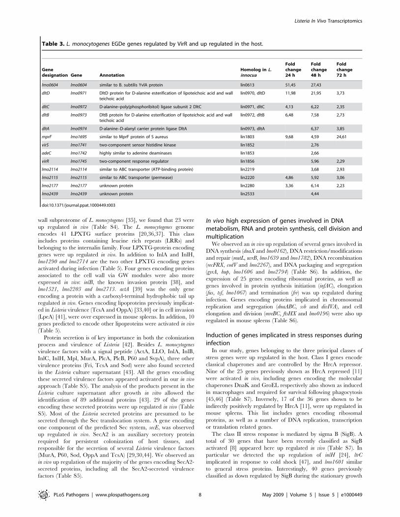
VirR, another key Listeria virulence regulator that mainly
controls genes involved in the modification of bacterial surface
components, is the response regulator of a two-component system
(TCS) implicated in cell invasion and virulence [13]. Using a
transcriptomic approach, 17 genes were previously identified as
regulated by VirR in vitro [13]. In our study, 13 of the 17 VirR
regulated genes, including the dlt operon and mprF, were up
regulated in vivo (Table 3). The dlt operon is necessary for D-
alanylation of lipoteichoic acid (LTA) and was reported to be
important for L. monocytogenes virulence [13]. The VirR regulated
mprF encodes a protein shown to be required for lysinylation of
phospholipids in the Listeria cytoplasmic membrane and to confer
Listeria resistance to cationic antimicrobial peptides (CAMPs) [25].
The virR and virS genes were themselves up regulated, constituting
the only TCS whose expression of both components was induced
in mouse spleens.
In addition to VirRS, the L. monocytogenes genome contains 15
additional predicted TCS systems [26]. Genes encoding one
component of three of these TCS (degU, resD and phoR) were also
up regulated in vivo. DegU is an orphan response regulator
(absence of the sensor kinase DegS in the L. monocytogenes genome)
and a pleiotropic regulatory system previously involved in Listeria
virulence [27,28]. In particular, DegU has been implicated in the
regulation of some Listeria secreted proteins (gap, tsf, sod, lmo0644)
[26]. Interestingly, the expression of these four genes was also
increased in mouse spleens.
Finally, OhrR a transcriptional regulator controlling OhrA, a
hydroxyperoxidase implicated in intracellular survival of Listeria
[14], as well as several predicted transcriptional regulators were up
regulated in infected mouse spleens.
Strong activation of genes encoding cell wall metabolismproteins during infection
In addition to genes already mentioned and involved in LTA
modification (dltABCD), we observed that several genes implicated
in peptidoglycan (PG) biosynthesis (lmo0516, lmo0540, lmo1438,
lmo1521, lmo1855, lmo2522, lmo2526 and pbpB), cell shape
determination (mreBC, lmo1713), cell wall peptide synthesis (murC)
were up regulated in bacteria growing in mouse spleens (Table 4).
The expression of 3 genes encoding virulence factors involved in
bacterial cell wall modifications (murA, iap, and pgdA) [29–31] was
also increased in vivo. MurA and P60, the iap gene product, are two
SecA2-secreted autolysins required for Listeria full virulence
[29,30]. pgdA encodes for the PG N-deacetylase of L. monocytogenes
that was demonstrated as playing an important role in virulence
and evasion from host defenses [31]. In addition, spl [32] and
lmo2203 are two other autolysins encoding genes up regulated in
vivo, but until now never implicated in virulence.
Figure 4. In vivo expression of virulence genes. Expression duringmouse spleen infection of the 31 known virulence genes differentiallyregulated in vivo. A peak of expression was observed for the majority ofthese virulence genes 48 h p.i. All measurements are relative to culturein exponential phase in BHI. Genes were selected for this analysis whentheir expression deviated from BHI by at least a factor of 2.0 in at leastone time point. The image was produced as described in Materials andMethods. Each gene is represented by a single row of colored boxes;each time point is represented by a single column.doi:10.1371/journal.ppat.1000449.g004
Listeria In Vivo Transcriptomics
PLoS Pathogens | www.plospathogens.org 6 May 2009 | Volume 5 | Issue 5 | e1000449

Moreover, prsA2, a gene encoding a surface protein involved in
protein folding and previously shown as implicated in Listeria
intracellular survival and virulence [14,33] was up regulated in vivo.
Interestingly, the gene encoding the sortase SrtB that covalently
links proteins to the Listeria peptidoglycan, and two genes encoding
SrtB substrates (svpA and lmo2186) [34], were also over expressed in
vivo (Table 4).
Differential expression of genes encoding specific surfaceand secreted proteins during infection
Whereas a total of 44 genes encoding potential surface proteins
were up regulated in vivo, only three were observed as down
regulated during infection (lspA, lmo1851 and lmo2642) (Table 5).
In addition, among the 55 proteins previously identified in the cell
Table 2. L. monocytogenes EGDe genes positively controlled by PrfA and up regulated in the host.
Genedesignation Gene Annotation
Homolog inL. innocua
Foldchange24 h
Foldchange48 h
Foldchange72 h
Group I PrfA regulated genes
prfA lmo0200 listeriolysin positive regulatory protein 8,09 4,00
plcA lmo0201 phosphatidylinositol-specific phospholipase C 7,20 48,31 6,70
hly lmo0202 listeriolysin O precursor 35,56 118,39 15,14
mpl lmo0203 zinc metalloproteinase precursor 3,36 18,41 4,68
actA lmo0204 actin-assembly inducing protein precursor 6,02 15,58 4,49
plcB lmo0205 phospholipase C 12,37 106,64 31,78
inlA lmo0433 internalin A 15,92 3,86
inlB lmo0434 internalin B 3,82 2,55
lmo0788 lmo0788 unknown protein lin0781 60,06 31,36 20,50
uhpT lmo0838 hexose phoshate transport protein 5,13 2,59
inlC lmo1786 internalin C 8,56 3,57
prsA2 lmo2219 similar to post-translocation molecular chaperone lin2322 12,02 24,00 8,95
Group III PrfA regulated genes co-controlled by SigB
lmo0169 lmo0169 similar to a glucose uptake protein lin0212 3,11
inlH lmo0263 internalin H 3,08 6,60 2,41
lmo0439 lmo0439 weakly similar to a module of peptide synthetase lin0460 2,76
lmo0539 lmo0539 similar to tagatose-1,6-diphosphate aldolase lin0543 18,71 24,03
lmo0596 lmo0596 unknown protein lin0605 23,31 68,15
lmo0781 lmo0781 similar to mannose-specific phosphotransferasesystem (PTS) component IID
lin0774 2,57
lmo0782 lmo0782 similar to mannose-specific phosphotransferasesystem (PTS) component IIC
lin0775 4,69
lmo0783 lmo0783 similar to mannose-specific phosphotransferasesystem (PTS) component IIB
lin0776 3,82
lmo0784 lmo0784 similar to mannose-specific phosphotransferasesystem (PTS) component IIA
lin0777 2,89
lmo0794 lmo0794 similar to B. subtilis YwnB protein lin0787 4,47
lmo0796 lmo0796 unknown protein lin0789 4,21
lmo0994 lmo0994 unknown protein lin0993 2,53
opuCD lmo1425 osmoprotectant transport system permease protein lin1464 2,69
lmo1601 lmo1601 similar to general stress protein lin1642 2,18
lmo1602 lmo1602 unknown protein lin1643 4,54 2,13
lmo2157 lmo2157 unknown protein 3,29 5,85 2,01
lmo2391 lmo2391 conserved hypothetical protein similar to B. subtilis YhfK protein lin2490 4,56
lmo2696 lmo2696 similar to hypothetical dihydroxyacetone kinase lin2844 2,49
lmo2697 lmo2697 unknown protein lin2845 3,14
Group III PrfA regulated gene not controlled by SigB
lmo0937 lmo0937 unknown protein lin0936 3,92
Other PrfA regulated genes
lmo0206 lmo0206 unknown protein 3,11
lmo0207 lmo0207 hypothetical lipoprotein lin0239 5,31 3,27
doi:10.1371/journal.ppat.1000449.t002
Listeria In Vivo Transcriptomics
PLoS Pathogens | www.plospathogens.org 7 May 2009 | Volume 5 | Issue 5 | e1000449
wall subproteome of L. monocytogenes [35], we found that 23 were
up regulated in vivo (Table S4). The L. monocytogenes genome
encodes 41 LPXTG surface proteins [20,36,37]. This class
includes proteins containing leucine rich repeats (LRRs) and
belonging to the internalin family. Four LPXTG-protein encoding
genes were up regulated in vivo. In addition to InlA and InlH,
lmo1290 and lmo2714 are the two other LPXTG encoding genes
activated during infection (Table 5). Four genes encoding proteins
associated to the cell wall via GW modules were also more
expressed in vivo: inlB, the known invasion protein [38], and
lmo1521, lmo2203 and lmo2713. actA [39] was the only gene
encoding a protein with a carboxyl-terminal hydrophobic tail up
regulated in vivo. Genes encoding lipoproteins previously implicat-
ed in Listeria virulence (TcsA and OppA) [33,40] or in cell invasion
(LpeA) [41], were over expressed in mouse spleens. In addition, 10
genes predicted to encode other lipoproteins were activated in vivo
(Table 5).
Protein secretion is of key importance in both the colonization
process and virulence of Listeria [42]. Besides L. monocytogenes
virulence factors with a signal peptide (ActA, LLO, InlA, InlB,
InlC, InlH, Mpl, MurA, PlcA, PlcB, P60 and SvpA), three other
virulence proteins (Fri, TcsA and Sod) were also found secreted
in the Listeria culture supernatant [43]. All the genes encoding
these secreted virulence factors appeared activated in our in vivo
approach (Table S5). The analysis of the products present in the
Listeria culture supernatant after growth in vitro allowed the
identification of 89 additional proteins [43]. 29 of the genes
encoding these secreted proteins were up regulated in vivo (Table
S5). Most of the Listeria secreted proteins are presumed to be
secreted through the Sec translocation system. A gene encoding
one component of the predicted Sec system, secE, was observed
up regulated in vivo. SecA2 is an auxiliary secretory protein
required for persistent colonization of host tissues, and
responsible for the secretion of several Listeria virulence factors
(MurA, P60, Sod, OppA and TcsA) [29,30,44]. We observed an
in vivo up regulation of the majority of the genes encoding SecA2-
secreted proteins, including all the SecA2-secreted virulence
factors (Table S5).
In vivo high expression of genes involved in DNAmetabolism, RNA and protein synthesis, cell division andmultiplication
We observed an in vivo up regulation of several genes involved in
DNA synthesis (dnaX and lmo0162), DNA restriction/modifications
and repair (mutL, uvrB, lmo1639 and lmo1782), DNA recombination
(recFRX, codV and lmo2267), and DNA packaging and segregation
(gyrA, hup, lmo1606 and lmo2794) (Table S6). In addition, the
expression of 25 genes encoding ribosomal proteins, as well as
genes involved in protein synthesis initiation (infAC), elongation
(fus, tsf, lmo1067) and termination (frr) was up regulated during
infection. Genes encoding proteins implicated in chromosomal
replication and segregation (dnaABC, ssb and divIVA), and cell
elongation and division (mreBC, ftsHX and lmo0196) were also up
regulated in mouse spleens (Table S6).
Induction of genes implicated in stress responses duringinfection
In our study, genes belonging to the three principal classes of
stress genes were up regulated in the host. Class I genes encode
classical chaperones and are controlled by the HrcA repressor.
Nine of the 25 genes previously shown as HrcA repressed [11]
were activated in vivo, including genes encoding the molecular
chaperones DnaK and GroEL respectively also shown as induced
in macrophages and required for survival following phagocytosis
[45,46] (Table S7). Inversely, 17 of the 36 genes shown to be
indirectly positively regulated by HrcA [11], were up regulated in
mouse spleens. This list includes genes encoding ribosomal
proteins, as well as a number of DNA replication, transcription
or translation related genes.
The class II stress response is mediated by sigma B (SigB). A
total of 30 genes that have been recently classified as SigB
activated [8] appeared here up regulated in vivo (Table S7). In
particular we detected the up regulation of inlH [24], ltrC
implicated in response to cold shock [47], and lmo1601 similar
to general stress proteins. Interestingly, 40 genes previously
classified as down regulated by SigB during the stationary growth
Table 3. L. monocytogenes EGDe genes regulated by VirR and up regulated in the host.
Genedesignation Gene Annotation
Homolog in L.innocua
Foldchange24 h
Foldchange48 h
Foldchange72 h
lmo0604 lmo0604 similar to B. subtilis YvlA protein lin0613 51,45 27,43
dltD lmo0971 DltD protein for D-alanine esterification of lipoteichoic acid and wallteichoic acid
lin0970, dltD 11,98 21,95 3,73
dltC lmo0972 D-alanine–poly(phosphoribitol) ligase subunit 2 DltC lin0971, dltC 4,13 6,22 2,35
dltB lmo0973 DltB protein for D-alanine esterification of lipoteichoic acid and wallteichoic acid
lin0972, dltB 6,48 7,58 2,73
dltA lmo0974 D-alanine–D-alanyl carrier protein ligase DltA lin0973, dltA 6,37 3,85
mprF lmo1695 similar to MprF protein of S aureus lin1803 9,68 4,59 24,61
virS lmo1741 two-component sensor histidine kinase lin1852 2,76
adeC lmo1742 highly similar to adenine deaminases lin1853 2,66
virR lmo1745 two-component response regulator lin1856 5,96 2,29
lmo2114 lmo2114 similar to ABC transporter (ATP-binding protein) lin2219 3,68 2,93
lmo2115 lmo2115 similar to ABC transporter (permease) lin2220 4,86 5,92 3,06
lmo2177 lmo2177 unknown protein lin2280 3,36 6,14 2,23
lmo2439 lmo2439 unknown protein lin2533 4,44
doi:10.1371/journal.ppat.1000449.t003
Listeria In Vivo Transcriptomics
PLoS Pathogens | www.plospathogens.org 8 May 2009 | Volume 5 | Issue 5 | e1000449

phase [8] were detected as activated in vivo (Table S7). These
include kat, a catalase involved in the oxidative stress response
[48], a large proportion of genes encoding ribosomal proteins or
implicated in translation, cell division and cell wall biogenesis.
Furthermore, iap, the P60 gene [29], is part of this group. Finally,
rsbU and rsbX, two components of the complex regulation system
of SigB [49] were also up regulated in mouse spleens (Table S7).
CtsR is a transcriptional repressor involved in the control of
class III stress proteins and previously shown to be responsible for
the repression of 42 genes [12], 15 of which appeared up regulated
in the host (Table S7). In particular, CtsR regulates the expression
of Clp proteases required for the degradation of abnormal proteins
and implicated in bacterial escape from macrophage vacuoles and
virulence in mice [50]. Expression of clpBCE was activated during
infection, as well as mcsA and mcsB the modulators of the CtsR
regulon.
In some host cells, bacteria are confronted with severe oxidative
stress due to the release of reactive oxygen intermediates. We
observed the in vivo activation of an important number of oxidative
stress resistance mechanisms. The qoxABCD operon that encodes a
quinol oxidase important for oxidative stress response, and two
major proteins implicated in protection against superoxides and
reactive oxygen species (ROS), Kat and Sod, were highly up
regulated in vivo (Table S7). Sod was previously shown as required
for Listeria full virulence and is a target of Stp, a serine-threonine
phosphatase also involved in L. monocytogenes virulence [44,51], and
Table 4. L. monocytogenes EGDe genes implicated in cell wall metabolism and differentially regulated in the host.
Genedesignation Gene Annotation
Homolog in L.innocua
Foldchange24 h
Foldchange48 h
Foldchange72 h
pgdA lmo0415 peptidoglycan N-acetylglucosamine deacetylase A lin0436 14,32 3,71
lmo0516 lmo0516 similar to Bacillus anthracis encapsulation protein CapA lin0516 14,22
lmo0540 lmo0540 similar to penicillin-binding protein lin0544 3,78 3,97 2,28
iap lmo0582 P60 extracellular protein, invasion associated protein Iap lin0591 34,54 28,64 2,04
dltD lmo0971 DltD protein for D-alanine esterification of lipoteichoic acid and wall teichoic acid lin0970, dltD 11,98 21,95 3,73
dltC lmo0972 D-alanine–poly(phosphoribitol) ligase subunit 2 DltC lin0971, dltC 4,13 6,22 2,35
dltB lmo0973 DltB protein for D-alanine esterification of lipoteichoic acid and wall teichoic acid lin0972, dltB 6,48 7,58 2,73
dltA lmo0974 D-alanine–D-alanyl carrier protein ligase DltA lin0973, dltA 6,37 3,85
lmo1075 lmo1075 similar to teichoic acid translocation ATP-binding protein TagH (ABC transporter) lin1063 9,25 5,46 4,20
lmo1081 lmo1081 similar to glucose-1-phosphate thymidyl transferase 8,22 5,98 5,17
lmo1082 lmo1082 similar to dTDP-sugar epimerase 47,18 24,08 20,97
lmo1083 lmo1083 similar to dTDP-D-glucose 4,6-dehydratase 3,51
lmo1084 lmo1084 similar to DTDP-L-rhamnose synthetase 3,89
lmo1291 lmo1291 similar to acyltransferase (to B. subtilis YrhL protein) lin1329 4,72
uppS lmo1315 similar to undecaprenyl diphosphate synthase lin1352 2,16
lmo1438 lmo1438 similar to penicillin-binding protein lin1477 3,56
lmo1521 lmo1521 similar to N-acetylmuramoyl-L-alanine amidase lin1556 2,73
mreC lmo1547 similar to cell-shape determining protein MreC lin1581 2,45
mreD lmo1548 similar to cell-shape determining protein MreB lin1582 4,08
murC lmo1605 similar to UDP-N-acetyl muramate-alanine ligase lin1646 2,39
mprF lmo1695 multiple peptide resistance factor lin1803 9,71 4,59 24,59
lmo1713 lmo1713 similar to cell-shape determining protein MreB lin1825 5,82
lspA lmo1844 signal peptidase II lin1958 22,01
lmo1851 lmo1851 similar to carboxy-terminal processing proteinase lin1965 22,12
lmo1855 lmo1855 similar to similar to D-alanyl-D-alanine carboxypeptidases lin1969 3,32
pbpB lmo2039 penicilin binding protein 2B lin2145 5,00
srtB lmo2181 sortase B lin2285 2,44
svpA lmo2185 unknown protein lin2289 6,61 11,11
lmo2186 lmo2186 unknown protein lin2290 2,72
lmo2203 lmo2203 similar to N-acetylmuramoyl-L-alanine amidase and to internalin B lin2306 4,50
prsA2 lmo2219 similar to post-translocation molecular chaperone lin2322 12,02 24,00 8,95
spl lmo2505 peptidoglycan lytic protein P45 lin2648, spl 12,21 2,68
lmo2522 lmo2522 similar to hypothetical cell wall binding protein from B. subtilis lin2666 5,74 3,07
lmo2526 lmo2526 UDP-N-acetylglucosamine 1-carboxyvinyltransferase lin2670 2,40
murA lmo2691 similar to autolysin, N-acetylmuramidase lin2838 4,06
doi:10.1371/journal.ppat.1000449.t004
Listeria In Vivo Transcriptomics
PLoS Pathogens | www.plospathogens.org 9 May 2009 | Volume 5 | Issue 5 | e1000449

Table 5. L. monocytogenes EGDe cell surface encoding genes differentially regulated in the host.
Genedesignation Gene Annotation
Homolog in L.innocua
Foldchange24 h
Foldchange48 h
Foldchange72 h
Sortase substrates: LPXTG and NXXTX proteins
inlH lmo0263 internalin H 3,07 6,59 2,41
inlA lmo0433 internalin A 15,89 3,86
lmo1290 lmo1290 similar to internalin proteins, putative peptidoglycan bound protein (LPXTGmotif)
6,23
svpA lmo2185 unknown protein lin2289 6,61 11,11
lmo2186 lmo2186 unknown protein lin2290 2,72
lmo2714 lmo2714 peptidoglycan anchored protein (LPXTG motif) lin2862 25,46 70,03 3,10
Proteins with noncovalent association to cell wall
inlB lmo0434 internalin B 3,84 2,55
iap lmo0582 P60 extracellular protein, invasion associated protein Iap lin0591, iap 34,64 28,54 2,05
lmo1521 lmo1521 similar to N-acetylmuramoyl-L-alanine amidase lin1556 2,73
murC lmo1605 UDP-N-acetylmuramate–L-alanine ligase lin1646 2,40
lmo1851 lmo1851 similar to carboxy-terminal processing proteinase lin1965 22,12
lmo2203 lmo2203 similar to N-acetylmuramoyl-L-alanine amidase lin1556 4,50
lmo2522 lmo2522 similar to hypothetical cell wall binding protein from B. subtilis lin2666 5,74 3,07
lmo2713 lmo2713 secreted protein with 1 GW repeat lin2861 17,03 2,79
Proteins with an hydrophobic tail
actA lmo0204 actin-assembly inducing protein precursor 6,02 15,56 4,50
Lipoproteins
qoxA lmo0013 AA3-600 quinol oxidase subunit II lin0013 2,95
lmo0153 lmo0153 similar to a probable high-affinity zinc ABC transporter (Zn(II)-bindinglipoprotein)
lin0191 5,31
lmo0207 lmo0207 hypothetical lipoprotein lin0239 5,31 3,27
lmo0303 lmo0303 putaive secreted, lysin rich protein lin0331 3,18
lmo0355 lmo0355 similar to Flavocytochrome C Fumarate Reductase chain A lin0374 2,97 6,68 2,51
lmo0366 lmo0366 putative lipoprotein lin0385 2,69 2,17
prs lmo0509 similar to phosphoribosyl pyrophosphate synthetase 2,87
lmo0541 lmo0541 similar to ABC transporter (binding protein) lin0545 6,19 10,27 2,07
tcsA lmo1388 CD4+ T cell-stimulating antigen, lipoprotein lin1425 2,19
lmo1649 lmo1649 unknown protein lin1689 3,39
lpeA lmo1847 similar to adhesion binding proteins and lipoproteins lin1961 6,54 31,12 10,63
lmo2184 lmo2184 similar to ferrichrome ABC transporter (binding protein) lin2288 5,10 2,57
oppA lmo2196 similar to pheromone ABC transporter (binding protein) lin2300 5,21 6,15 2,73
lmo2642 lmo2642 unknown protein lin2791 22,13
Surface proteins involved in cell wall metabolism
pgdA lmo0415 peptidoglycan N-acetylglucosamine deacetylase A lin0436 14,32 3,71
lmo0540 lmo0540 similar to penicillin-binding protein lin0544 3,78 3,97 2,28
iap lmo0582 P60 extracellular protein, invasion associated protein Iap lin0591 34,54 28,64 2,04
lmo1438 lmo1438 similar to penicillin-binding protein lin1477 3,56
lmo1521 lmo1521 similar to N-acetylmuramoyl-L-alanine amidase lin1556 2,73
lmo1855 lmo1855 similar to similar to D-alanyl-D-alanine carboxypeptidases lin1969 3,32
pbpB lmo2039 penicilin binding protein 2B lin2145 5,00
lmo2203 lmo2203 similar to N-acetylmuramoyl-L-alanine amidase and to internalin B lin2306 4,50
spl lmo2505 peptidoglycan lytic protein P45 lin2648, spl 12,21 2,68
lmo2522 lmo2522 similar to hypothetical cell wall binding protein from B. subtilis lin2666 5,74 3,07
murA lmo2691 similar to autolysin, N-acetylmuramidase lin2838 4,06
Surface proteins involved in protein processing, folding and cell surface anchoring
lspA lmo1844 signal peptidase II lin1958, lspA 22,01
Listeria In Vivo Transcriptomics
PLoS Pathogens | www.plospathogens.org 10 May 2009 | Volume 5 | Issue 5 | e1000449

detected down regulated in the host. A decrease in the level of Stp
was previously associated to an increase in phosphorylated Sod,
accompanied by the secretion of active non-phosphorylated Sod
by the SecA2 system [44,51]. Furthermore, genes encoding a
thioredoxin and two thioredoxin reductases involved in the
response to oxidative stress (lmo2152, trxB and lmo2390) were up
regulated in our study (Table S7). The ferritin protein Fri, that also
provides protection against reactive oxygen species, is essential for
virulence and is required for efficient bacterial growth at early
stages of the infection process [52,53]. Fri transcription is directly
regulated by Fur, the ferric uptake regulator. The expression of fri
and fur was activated during infection. In addition, ohrA and gap
were up regulated in vivo and encode two proteins respectively
involved in hydroperoxide resistance [54] and in resistance against
reactive oxygen species produced by host phagocytic cells in
Leishmania [55] (Table S7).
L. monocytogenes metabolism adaptation to in vivoconditions
Remarkably, 30% of the in vivo regulated genes are involved in
L. monocytogenes metabolism (99 metabolism-related genes were up
and 72 were down regulated) (Table S8). As described above, uhpT
is an in vivo highly up regulated virulence gene, regulated by PrfA
and that promotes the uptake of phosphorylated hexoses (glucose-
1-phosphate and glucose-6-phosphate) [23,56]. Phosphorylated
glucose is the product of glycogen hydrolysis in eukaryotic cells
and there is experimental evidence that the PrfA-dependent
utilization of this compound has a role in L. monocytogenes virulence
[23,56].
We observed an in vivo up regulation of several genes encoding
enzymes involved in the glycolysis, like gap, pgi, fbaA, and pgm.
Inversely, we found a down regulation of the expression of four
genes involved in the non-oxidative phase of the pentose
phosphate pathway (lmo2660, lmo2661, lmo2662 and lmo2674).
The final step of glycolysis leads to pyruvate, which is then
converted to acetyl-CoA by the pyruvate dehydrogenase complex.
We found this complex partly up regulated in vivo, as well as one of
its activator, the lipoate ligase protein LplA2 [57,58]. The citric
acid cycle is continuously supplied with acetyl-CoA during aerobic
respiration. We observed an up regulation of three citric acid cycle
genes (citBCG) (Table S8). The citric acid cycle is followed by
oxidative phosphorylation. In this study, we found the up
regulation of several genes implicated in biosynthesis and assembly
of components of the respiratory chain (menD, lmo1677, qoxABD,
ctaA, cydA, cydD, atpD). In addition, genes encoding resD, a regulator
of aerobic and anaerobic respiration [59] and rex, a redox-sensing
transcriptional repressor [60], were also up regulated in vivo. Genes
encoding the pyruvate-formate lyase (pfl) and pyruvate-formate
lyase activating enzymes (pflCB) are required for the anaerobic
metabolism of pyruvate and were activated in the host (Table S8).
Genes implicated in amino acid biosynthesis were also induced
in vivo, in particular aroA and pheA, two genes responsible for
aromatic amino acid biosynthesis. Mutations in aroA and pheA were
previously shown to induce an attenuation of virulence in the
mouse model [61,62]. Furthermore, the expression of genes
implicated in the biosynthetic pathways of branched amino acids
(alsS, ilvN and lmo0978), and amino acids of the aspartate and
glutamate families (ansB, lmo0594, lmo1006, lmo1011, lmo2413 and
glnA, lmo2770, respectively), was also increased in vivo (Table S8).
Significantly, mannose (lmo0781–lmo0784), maltose (lmo0278)
and cellobiose (lmo0301 and lmo0915) -specific PTS encoding genes
[63] were up regulated in vivo. Inversely, fructose (lmo2733),
galactitol (lmo2665) and mannitol (lmo2649) -specific PTS encoding
genes appeared down regulated.
Among the genes involved in bacterial ion uptake systems, a
potassium-transporting ATPase encoding gene (kdpB) was down
regulated in vivo. Cobalt (lmo1207), manganese (lmo1424) and
calcium (lmo0841) transporter systems were, inversely, up regulat-
ed. As indicated above, the ferritin and ferric uptake protein
encoding genes, fri and fur, shown to be activated under low iron
concentration [64,65], appeared highly up regulated in vivo (Table
S8).
Detection of potential virulence genes by in vivotranscriptomics
A major goal of this work was the identification of genes that
encode proteins that may play a role in the infectious process. To
identify such virulence genes and in order to establish a short list,
we arbitrarily used several criteria. The gene should be
preferentially 1) highly activated during infection; 2) absent in
the non pathogenic strain L. innocua and present in other L.
monocytogenes strains from diverse serotypes; 3) a member of a
specific protein family encoding gene (surface, secreted, stress)
possibly involved in virulence; 4) controlled by virulence regulators
(PrfA, VirR, CtsR, HrcA, SigB). Several candidates emerged,
matching, at least, some of the above criteria (Table 6).
lmo0206, lmo0257, lmo0915, lmo1290 and lmo2157 are genes that,
as eleven already known virulence factors, are L. monocytogenes
species-specific and induced in vivo. lmo0206 and lmo2157 are the
only two genes activated in vivo, controlled by PrfA, absent from L.
innocua and whose role in virulence was never investigated.
lmo0206, orfX [66], is in addition located at the end of the Listeria
virulence cluster and was recently implicated in intracellular
survival [14]. The expression of lmo2157 was shown to be
controlled by PrfA and SigB [6,8].
lmo1081, lmo1082, lmo1099 and lmo1102 are L. monocytogenes
EGDe species-specific genes highly up regulated in vivo over the
three time points of the infection (Table 6). Interestingly, these
genes encode proteins potentially involved in cell wall metabolism
and heavy metal detoxification.
Genedesignation Gene Annotation
Homolog in L.innocua
Foldchange24 h
Foldchange48 h
Foldchange72 h
lmo1851 lmo1851 similar to carboxy-terminal processing proteinase lin1965 22,12
srtB lmo2181 sortase B lin2285 2,44
prsA2 lmo2219 similar to post-translocation molecular chaperone lin2322 12,02 24,00 8,95
doi:10.1371/journal.ppat.1000449.t005
Table 5. Cont.
Listeria In Vivo Transcriptomics
PLoS Pathogens | www.plospathogens.org 11 May 2009 | Volume 5 | Issue 5 | e1000449

Ta
ble
6.
L.m
on
ocy
tog
enes
EGD
eg
en
es
dif
fere
nti
ally
reg
ula
ted
inth
eh
ost
and
po
ten
tial
viru
len
cefa
cto
rs.
Ge
ne
de
sig
na
tio
nG
en
eA
nn
ota
tio
nH
om
olo
gin
L.in
no
cua
Ho
mo
log
in1
/2a
F6
85
4H
om
olo
gin
4b
F2
36
5H
om
olo
gin
4b
H7
85
8
Fo
ldch
an
ge
24
h
Fo
ldch
an
ge
48
h
Fo
ldch
an
ge
72
hR
eg
ula
tio
nS
ecr
ete
d/S
urf
ace
pro
tein
lmo
0206
lmo
0206
un
kno
wn
pro
tein
LMO
f68
54
_0
21
4.1
LMO
f23
65
_0
21
7LM
Oh
78
58
_0
22
5.1
3,1
1P
rfA
Secr
ete
d
lmo
0257
lmo
0257
un
kno
wn
pro
tein
LMO
f68
54
_0
26
1.2
LMO
f23
65
_0
26
5LM
Oh
78
58
_0
28
82
,12
lmo
0540
lmo
0540
sim
ilar
top
en
icill
in-b
ind
ing
pro
tein
lin0
54
4LM
Of6
85
4_
05
81
LMO
f23
65
_0
56
9LM
Oh
78
58
_0
59
83
,77
3,9
62
,29
Secr
ete
d/S
urf
ace
lmo
0604
lmo
0604
sim
ilar
toB
sub
tilis
Yvl
Ap
rote
inlin
06
13
LMO
f68
54
_0
64
2.2
LMO
f23
65
_0
63
3LM
Oh
78
58
_0
66
3.2
51
,45
27
,43
Vir
R
lmo
0788
lmo
0788
un
kno
wn
pro
tein
lin0
78
1LM
Of6
85
4_
08
32
LMO
f23
65
_0
80
4LM
Oh
78
58
_0
84
26
0,0
63
1,3
62
0,5
0P
rfA
lmo
0915
lmo
0915
sim
ilar
top
ho
sph
otr
ansf
era
sesy
ste
me
nzy
me
IICLM
Of6
85
4_
09
62
LMO
f23
65
_0
93
7LM
Oh
78
58
_0
98
93
,15
2,3
7
lmo
1081
lmo
1081
sim
ilar
tog
luco
se-1
-ph
osp
hat
eth
ymid
yltr
ansf
era
seLM
Of6
85
4_
11
34
8,2
55
,99
5,1
7
lmo
1082
lmo
1082
sim
ilar
tod
TD
P-s
ug
are
pim
era
seLM
Of6
85
4_
11
35
47
,21
24
,06
21
,01
lmo
1099
lmo
1099
sim
ilar
toa
pro
tein
en
cod
ed
by
Tn
91
66
5,4
23
0,4
22
4,0
4
lmo
1102
lmo
1102
sim
ilar
toca
dm
ium
eff
lux
syst
em
acce
sso
ryp
rote
ins
9,7
14
,51
4,7
2
lmo
1290
lmo
1290
sim
ilar
toin
tern
alin
pro
tein
s(L
PX
TG
mo
tif)
LMO
f68
54
_1
33
2LM
Of2
36
5_
13
07
LMO
h7
85
8_
13
74
.26
,24
Surf
ace
lmo
1438
lmo
1438
sim
ilar
top
en
icill
in-b
ind
ing
pro
tein
lin1
47
7LM
Of6
85
4_
14
81
LMO
f23
65
_1
45
7LM
Oh
78
58
_1
53
33
,55
Secr
ete
d/S
urf
ace
lmo
1521
lmo
1521
sim
ilar
toN
-ace
tylm
ura
mo
yl-L
-ala
nin
eam
idas
elin
15
56
LMO
f68
54
_1
56
8LM
Of2
36
5_
15
40
2,7
3Si
g5
4Se
cre
ted
/Su
rfac
e
lmo
1601
lmo
1601
sim
ilar
tog
en
era
lst
ress
pro
tein
lin1
64
2LM
Of6
85
4_
16
53
.1LM
Of2
36
5_
16
22
LMO
h7
85
8_
17
07
.2
,18
Prf
A-S
igB
-Sig
L
lmo
1602
lmo
1602
un
kno
wn
pro
tein
lin1
64
3LM
Of6
85
4_
16
53
.2LM
Of2
36
5_
16
23
LMO
h7
85
8_
17
07
.44
,54
2,1
3P
rfA
-Sig
B-S
igL
Secr
ete
d
ad
eClm
o17
42h
igh
lysi
mila
rto
ade
nin
ed
eam
inas
es
lin1
85
3LM
Of6
85
4_
18
00
LMO
f23
65
_1
76
7LM
Oh
78
58
_1
86
72
,66
Vir
R
lmo
1855
lmo
1855
sim
ilar
tosi
mila
rto
D-a
lan
yl-D
-ala
nin
eca
rbo
xyp
ep
tid
ase
slin
19
69
LMO
f68
54
_1
91
5LM
Of2
36
5_
18
83
LMO
h7
85
8_
19
80
3,3
1Su
rfac
e
lmo
2048
lmo
2048
un
kno
wn
pro
tein
lin2
15
4LM
Of6
85
4_
21
09
.1LM
Of2
36
5_
20
79
LMO
h7
85
8_
21
76
.14
,93
2,5
1Si
gB
-Hcr
A
lmo
2114
lmo
2114
sim
ilar
toA
BC
tran
spo
rte
r(A
TP
-bin
din
gp
rote
in)
lin2
21
9LM
Of6
85
4_
21
78
LMO
f23
65
_2
14
7LM
Oh
78
58
_2
24
53
,68
2,9
3V
iR-C
tsR
-Sig
L
lmo
2115
lmo
2115
sim
ilar
toA
BC
tran
spo
rte
r(p
erm
eas
e)
lin2
22
0LM
Of6
85
4_
21
79
LMO
f23
65
_2
14
8LM
Oh
78
58
_2
24
64
,86
5,9
23
,06
ViR
-Sig
L
lmo
2157
lmo
2157
un
kno
wn
pro
tein
LMO
f68
54
_2
22
1LM
Of2
36
5_
21
89
LMO
h7
85
8_
22
90
.13
,29
5,8
52
,01
Prf
A-S
igB
lmo
2177
lmo
2177
un
kno
wn
pro
tein
lin2
28
0LM
Of6
85
4_
22
41
LMO
f23
65
_2
20
9LM
Oh
78
58
_2
31
03
,36
6,1
42
,23
Vir
R
fab
Flm
o22
01si
mila
rto
3-o
xoac
yl-a
cyl-
carr
ier
pro
tein
syn
thas
elin
23
04
LMO
f68
54
_2
26
5LM
Of2
36
5_
22
34
LMO
h7
85
8_
23
35
3,9
02
,03
Sig
B
lmo
2203
lmo
2203
sim
ilar
toN
-ace
tylm
ura
mo
yl-L
-ala
nin
eam
idas
elin
23
06
LMO
f68
54
_2
26
6.1
LMO
f23
65
_2
23
6LM
Oh
78
58
_2
33
74
,50
Secr
ete
d/S
urf
ace
lmo
2439
lmo
2439
un
kno
wn
pro
tein
lin2
53
3LM
Of6
85
4_
24
99
.3LM
Of2
36
5_
24
11
LMO
h7
85
8_
25
84
.44
,44
Vir
RSe
cre
ted
ga
plm
o24
59g
lyce
rald
eh
yde
-3-p
ho
sph
ate
de
hyd
rog
en
ase
lin2
55
3LM
Of6
85
4_
25
20
LMO
f23
65
_2
43
2LM
Oh
78
58
_2
60
84
,49
2,3
7H
crA
Surf
ace
lmo
2522
lmo
2522
sim
ilar
toh
ypo
the
tica
lce
llw
all
bin
din
gp
rote
inlin
26
66
LMO
f68
54
_2
58
4LM
Of2
36
5_
24
95
LMO
h7
85
8_
26
74
5,7
23
,07
Secr
ete
d/S
urf
ace
lmo
2713
lmo
2713
secr
ete
dp
rote
inw
ith
1G
Wre
pe
atlin
28
61
LMO
f68
54
_2
83
1.1
LMO
f23
65
_2
69
3LM
Oh
78
58
_2
97
6.1
8,8
61
7,0
82
,80
Secr
ete
d/S
urf
ace
lmo
2714
lmo
2714
pe
pti
do
gly
can
anch
ore
dp
rote
in(L
PX
TG
mo
tif)
lin2
86
2LM
Of6
85
4_
28
33
LMO
f23
65
_2
69
4LM
Oh
78
58
_2
97
82
5,4
57
0,2
73
,09
Surf
ace
do
i:10
.13
71
/jo
urn
al.p
pat
.10
00
44
9.t
00
6
Listeria In Vivo Transcriptomics
PLoS Pathogens | www.plospathogens.org 12 May 2009 | Volume 5 | Issue 5 | e1000449

Only two uncharacterized genes encoding LPXTG surface
proteins (lmo1290 and lmo2714) and three encoding GW surface
proteins (lmo1521, lmo2203 and lmo2713) were up regulated within
the host (Table 6). lmo1521 and lmo2203 are in addition predicted
autolysins. lmo2713 and lmo2714 seem to be part of a genomic
region over expressed at all time points of the infection and
Lmo2714 was found in the Listeria culture supernatant [43]. Four
genes (lm0540, lmo1438, lmo1855 and lmo2522) predicted to be
involved in cell wall metabolism were up regulated in vivo, and
similar to pgdA, iap, and murA [29–31], could participate in Listeria
infection.
Twenty-five uncharacterized genes activated in vivo encode
secreted proteins that may interact with the host cells, including
Lmo2201, a Tat-secreted protein [42], and GAPDH. GAPDH
was previously shown to be part of the Listeria cell wall
subproteome [35], and to impair Listeria phagosome maturation
[67]. GAPDH seems, in addition, to be implicated in the virulence
of several other pathogens [68–70].
lmo0788 is highly activated in mouse spleens during infection
and is the only gene of the group I PrfA-regulated genes (i.e.
preceded by a PrfA-box and positively regulated by PrfA) [6]
whose role during infection has never been addressed (Tables 2
and 6). lmo0788 encodes a protein similar to subunits (BadFG) of
the benzoyl-CoA reductase used by facultative aerobes in absence
of oxygen for reductive aromatic metabolism [71].
VirR appears as the second main regulator of virulence genes
and controls lmo0604, lmo1742, lmo2114, lmo2115, lmo2177 and
lmo2439, whose expression was activated in the host (Tables 3 and
6). lmo2114 and lmo2115 are in addition part of a transcriptional
unit co-regulated by CtsR and SigL [7].
Several stress protein encoding genes that are under the control
of different stress regulators were up regulated in vivo. In particular,
lmo2048 is a stress protein-encoding gene that is co-controlled by
CtsR and HrcA (Table 6). The 19 genes up regulated in vivo and
co-controlled by PrfA and SigB (Table 2) could also be important
for the infectious process. Among these, lmo1601 and lmo1602 are
furthermore regulated by SigL [7].
The use of such arbitrary criteria obviously not guaranteed that
a selected gene was a virulence factor, and conversely probably
excluded many virulence genes. In particular, it is worth
mentioning that 91 genes encoding proteins similar to unknown
proteins, and 31 encoding putative proteins with no similarity in
public databases were differentially expressed in vivo (Table S3),
representing a large reservoir of potential new virulence factors. Of
these genes, those highly regulated all over the infectious process
could be of special relevance for virulence.
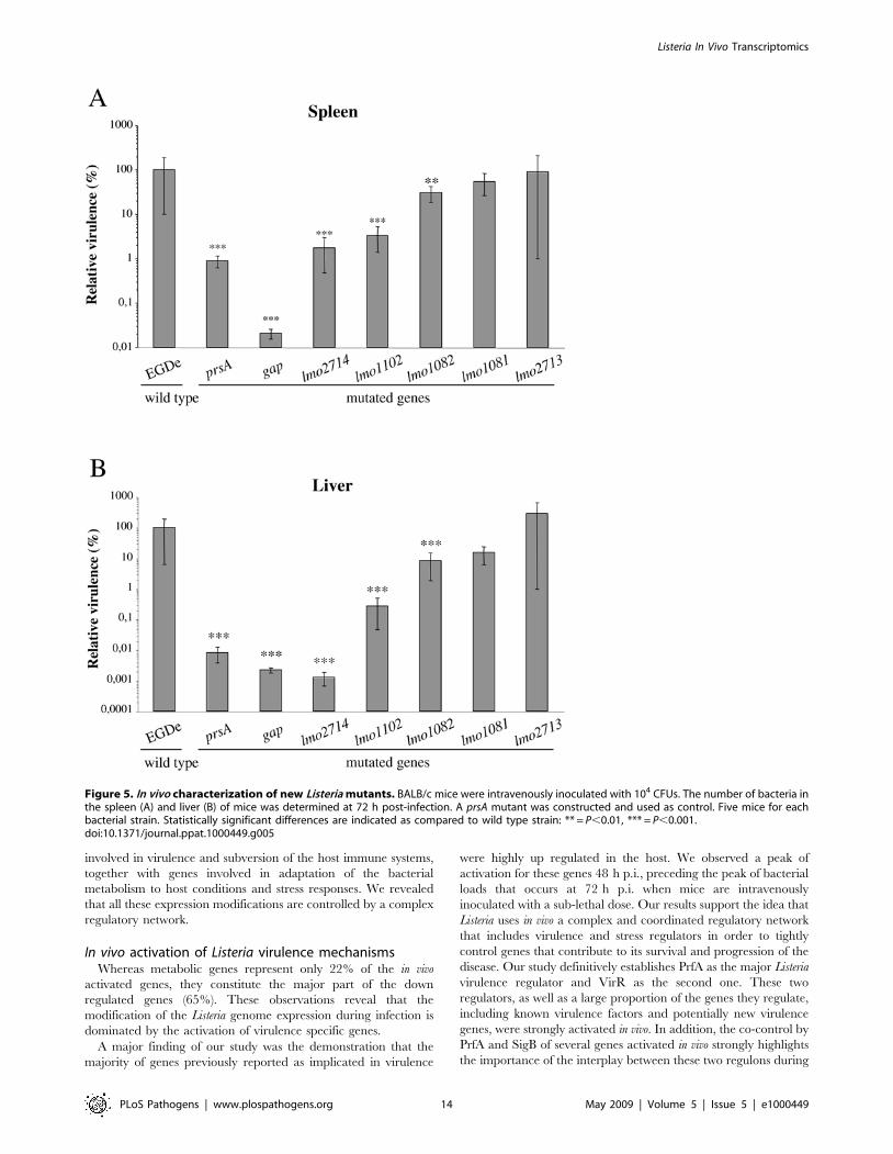
Identification of new L. monocytogenes virulence factorsIn order to validate our transcriptomics approach and identify
new L. monocytogenes virulence factors, 6 genes (lmo1081, lmo1082,
lmo1102, lmo2713, lmo2714 and gap) were selected for mutagenesis
using the criteria presented above. As we were unable to produce a
gap deletion mutant (probably because GAPDH is an essential
protein), we constructed a GAPDH secretion mutant.
To analyze the potential role of the selected genes in virulence,
we performed intravenous inoculations of BALB/c mice with wild
type (wt) and mutant strains, and the number of bacteria in the
mouse liver and spleen was determined 72 h after infection
(Figure 5). Mutants can be classified with respect to their virulence
potential. Bacterial counts for lmo1081 and lmo2713 mutants were
not significantly changed as compared to the wt strain, suggesting
the non-implication of these genes in Listeria virulence in mice. For
the lmo1082 mutant, bacterial counts were significantly affected
(<1 log) in mouse livers and at a lesser extent in the spleens.
Interestingly, for lmo1102, lmo2714 and gap mutants we observed a
remarkable decrease of bacterial counts in both mouse organs as
compared to the wt. In particular, the number of bacteria was
dramatically impaired in the liver 72 h after inoculation (<2,5 to
4,5 log). The gap mutant appeared as the most attenuated mutant
of our analysis with a considerable virulence decrease in both
organs reaching 3,5 log in the spleen and 4,5 log in the liver as
compared to the wild type (Figure 5).
In order to better characterize virulence attenuated strains,
mutants for lmo1082, lmo1102, lmo2714 and gap were comple-
mented. The corresponding wild-type gene was inserted as a single
copy under the control of its own promoter on the chromosome of
the mutant strain, at the PSA bacteriophage attachment site using
the pPL2 integration vector [72]. Wild type, mutant and
complemented strains were tested for growth in BHI at 37uCand for intracellular behavior after internalization in the murine
macrophage cell line J774 (Figure 6).
The growth rate observed in BHI at 37uC for the majority of the
strains tested was comparable to that of the wild type (Figure 6A).
However, the gap secretion mutant exhibited an important in vitro
reduced growth rate and reduced density at the stationary phase.
The growth defect observed for the gap mutant was even
accentuated in the complemented strain (Figure 6A). This is most
probably the result of an over expression of intracellular GAPDH,
expressed at the same time from the bacterial genome and from
the plasmid harbored by this strain. Surprisingly, the prsA2 mutant
presented also a notable growth delay. This growth defect was not
mentioned in previous studies implicating PrsA2 in intracellular
behavior and virulence [14,33].
Wild type, mutant and complemented strains were also tested
for intracellular behavior. As shown in Figure 6B, all the strains
grew with similar multiplication rates after internalization in J774
cells, indicating that the slight growth delay observed in BHI at
37uC for some strains has no consequences on intracellular
multiplication.
In addition, complemented strains were analyzed after intrave-
nous inoculations of BALB/c mice as compared to wt and mutant
strains, and the number of bacteria in the mouse liver and spleen
was determined 72 h after infection (Figure 7). The virulence
phenotype was restored, albeit partially in the case of lmo2714, in
complemented strains, except for the gap mutant. The virulence
defect of the gap complemented strain was even more severe in the
spleen as compare to the corresponding mutant (Figure 7A). This
was in correlation with the increased growth defect observed in
BHI at 37uC for the gap complemented strain.
These results revealed a role for lmo1082 and lmo1102, and at
less extent for lmo2714 and gap in Listeria virulence, validating our in
vivo transcriptomics approach.
Discussion
Identification of bacterial gene expression patterns during host-
pathogen interactions has long been a goal for understanding
infectious processes of intracellular pathogens [73]. Here, we
undertook the first time course study of the L. monocytogenes in vivo
transcriptome by comparing the genomic transcriptional patterns
of bacteria grown under laboratory conditions (BHI, 37uC,
exponential growth phase, pH 7) with that of in vivo-grown
bacteria over three days of infection (mouse spleen). This
constitutes also the first genome expression analysis of a pathogen
in infected mouse spleens. Our results indicate that a significant
part of the Listeria genome is differentially expressed for adaptation
to the host environment, essentially through gene activation. We
showed an in vivo over expression of an impressive number of genes
Listeria In Vivo Transcriptomics
PLoS Pathogens | www.plospathogens.org 13 May 2009 | Volume 5 | Issue 5 | e1000449
involved in virulence and subversion of the host immune systems,
together with genes involved in adaptation of the bacterial
metabolism to host conditions and stress responses. We revealed
that all these expression modifications are controlled by a complex
regulatory network.
In vivo activation of Listeria virulence mechanismsWhereas metabolic genes represent only 22% of the in vivo
activated genes, they constitute the major part of the down
regulated genes (65%). These observations reveal that the
modification of the Listeria genome expression during infection is
dominated by the activation of virulence specific genes.
A major finding of our study was the demonstration that the
majority of genes previously reported as implicated in virulence
were highly up regulated in the host. We observed a peak of
activation for these genes 48 h p.i., preceding the peak of bacterial
loads that occurs at 72 h p.i. when mice are intravenously
inoculated with a sub-lethal dose. Our results support the idea that
Listeria uses in vivo a complex and coordinated regulatory network
that includes virulence and stress regulators in order to tightly
control genes that contribute to its survival and progression of the
disease. Our study definitively establishes PrfA as the major Listeria
virulence regulator and VirR as the second one. These two
regulators, as well as a large proportion of the genes they regulate,
including known virulence factors and potentially new virulence
genes, were strongly activated in vivo. In addition, the co-control by
PrfA and SigB of several genes activated in vivo strongly highlights
the importance of the interplay between these two regulons during
Figure 5. In vivo characterization of new Listeria mutants. BALB/c mice were intravenously inoculated with 104 CFUs. The number of bacteria inthe spleen (A) and liver (B) of mice was determined at 72 h post-infection. A prsA mutant was constructed and used as control. Five mice for eachbacterial strain. Statistically significant differences are indicated as compared to wild type strain: ** = P,0.01, *** = P,0.001.doi:10.1371/journal.ppat.1000449.g005
Listeria In Vivo Transcriptomics
PLoS Pathogens | www.plospathogens.org 14 May 2009 | Volume 5 | Issue 5 | e1000449

the infectious process. The hypothesis on an in vivo intersecting
regulation of SigB and PrfA is in agreement with a very recent
study demonstrating the contribution of SigB and PrfA to a
regulatory network critical for appropriate regulation of virulence
gene expression [74]. The large number of additional regulons and
predicted transcriptional regulators differentially regulated inside
the host underlines the high degree of regulation required for
adaptation of Listeria to the host environment.
Another major aspect observed when Listeria interacts with its
host is the active remodeling of the bacterial envelope through
Figure 6. In vitro behavior of L. monocytogenes mutants. (A) Growth curves of L. monocytogenes EGDe strains in BHI at 37uC with shaking. (B)Intracellular behavior of L. monocytogenes EGDe strains in J774 cultured cells.doi:10.1371/journal.ppat.1000449.g006
Listeria In Vivo Transcriptomics
PLoS Pathogens | www.plospathogens.org 15 May 2009 | Volume 5 | Issue 5 | e1000449

activation of the cell wall metabolism and enhanced exposure of
virulence proteins at the bacterial surface.
Pathogens have evolved various systems for the secretion of
bacterial factors that contribute to the progression of the disease.
Listeria uses different secretion systems and a significant number of
their products were activated in vivo. It is particularly the case of
the SecA2 system, itself activated in vivo, and responsible for the
secretion of several proteins lacking a signal peptide and also up
regulated in the host, including known virulence factors.
Although competence genes have been found in L. monocytogenes
genome [20], Listeriae have never been shown to be naturally
competent. Interestingly, we observed several competence genes
(comEA, comEB, comGF, comGE, clpC, mecA and degU) up regulated in
vivo. This is the first report of a simultaneous activation of a great
number of competence genes in Listeria, suggesting that this
bacterium could be competent during infection and use this system
to incorporate DNA from the host environment in order to
acquire new potentialities.
Subversion of host defensesIn addition to the up regulation of a number of virulence
factors, L. monocytogenes activates mechanisms of subversion of the
host defenses. Lysinylation of phospholipids in Listeria membranes
by MprF, and D-alanylation of cell wall TAs and LTAs by the dlt
complex lead to a reduced negative charge of the bacterial surface.
One of the consequences of this process is the repulsion of cationic
antimicrobial peptides [13,25]. In vivo activation of both dlt and
mprF by VirR strongly suggests a regulation of L. monocytogenes
resistance to cationic peptides in the host, as previously proposed
[25]. Furthermore, surface components e.g. LTAs and PG play a
role in the innate immune response through receptors like Nods
and Toll-like receptors [75]. LTA modification by VirR regulated
factors could thus be used by Listeria to escape the host innate
immune response.
SecA2-dependent secretion has been proposed to coordinate
PG digestion by the activity of secreted autolysins [30]. The
muramyl glycopeptide predicted to be generated by the combined
activities of p60 and MurA is known to modify host inflammatory
responses [76,77]. Thus, the strong in vivo induction of SecA2 and
SecA2-secreted proteins may activate release of specific peptides
that interfere with host pattern recognition.
N-deacetylation by PgdA was shown to be a major modification
of Listeria PG, conferring the ability to survive in the gastrointes-
tinal tract, in professional phagocytes, evade the action of host
lysozyme, and modulate the inflammatory response [31]. The over
expression of pgdA during infection appears as an additional
strategy used by Listeria to subvert host pattern recognition and
control the host inflammatory responses to promote its own
survival.
In the same way, in accordance to the potent proinflammatory
activity of flagellin [78], Listeria down regulates flagella related
genes (lmo0681 and lmo0697) during the infection by the activation
of mogR, a repressor of motility and chemotaxis gene expression
[79].
Adaptation of the metabolism to the host environmentOnce inside the host, bacteria need to adapt to nutritional
changes, including carbon and nitrogen sources. This is illustrated
by the high number of metabolic genes differentially regulated in
vivo.
UhpT and several enzymes involved in glycolysis were up
regulated whereas enzymes implicated in the non-oxidative phase
of the pentose phosphate pathway were repressed. These data
suggest that phosphorylated glucose transported by UhpT is being
Figure 7. In vivo complementation of new Listeria mutants.BALB/c mice were intravenously inoculated with 104 CFUs. The numberof bacteria in the spleen (A) and liver (B) of mice was determined at72 h post-infection. Five mice for each bacterial strain. Statisticallysignificant differences are indicated as compared to the correspondingmutant for complemented strains: * = P,0.05, ** = P,0.01,*** = P,0.001.doi:10.1371/journal.ppat.1000449.g007
Listeria In Vivo Transcriptomics
PLoS Pathogens | www.plospathogens.org 16 May 2009 | Volume 5 | Issue 5 | e1000449

metabolized through glycolysis. Glucose or phosphorylated
glucose seems thus one of the major carbon sources in vivo, the
pentose phosphate cycle appearing not essential for the generation
of necessary intermediates and for gluconeogenesis. In addition to
genes involved in glycolysis, we observed the in vivo up regulation
of numerous genes implicated in the citric acid cycle and in
oxidative phosphorylation, indicating that L. monocytogenes is using
oxidative phosphorylation to generate energy. However, the
activation of genes involved in fermentation, suggests that, even
though L. monocytogenes is using oxidative phosphorylation to
generate energy, it might be experiencing some level of oxygen
starvation in spleen cells. The activation of the glycerol kinase and
glycerol-3-phosphate dehydrogenase, indicates that glycerol,
probably deriving from the activity of the phospholipases A and
B on cellular lipids, is an additional carbon source for intracellular
growth.
Metal ions are essential cofactors for functional expression of
many proteins in bacterial systems. Thus, alterations in Listeria ion
transport genes in vivo reflect the accessibility/inaccessibility of
those ions in the intracellular environment, i.e presence of
potassium and lack of cobalt, manganese, calcium and iron.
Due to defense mechanisms developed by the host to limit
bacterial multiplication, it could be expected a growth rate
decrease for invading pathogens in their host [16]. A very
interesting discovery resulting from our in vivo transcriptomics
analysis was the observation of an active growth status of Listeria in
infected mouse spleens. This was demonstrated by the increased
expression of numerous genes encoding proteins involved in
bacterial growth and multiplication, including genes implicated in
DNA replication and cell division. Listeria thus seems to have 24 h
p.i. overcome organism defenses and being engaged in an active
multiplication phase.
Stress responses to the host environmentBacterial responses to environmental changes are often
characterized by the induction of specific stress responses. The in
vivo induction of Listeria stress genes indicated that bacteria are
faced with stress within the host. The alternative sigma factor B is
the master regulator of stress. Even in the absence of a significant
regulation of sigB itself in vivo, an impressive number (70) of genes
previously shown to be under SigB-regulation were up regulated in
vivo, including numerous virulence factors. In addition, genes up
regulated in vivo and under the control of HrcA and CtsR seem
also to be particularly relevant for virulence. During the process of
host colonization, L. monocytogenes induces a host inflammatory
response [80]. This defense is accompanied by the generation of
ROS presented to the persistent pathogen. In addition to the
traditional ROS combating enzymes like catalase and superoxide
dismutase, this transcriptomic analysis revealed the in vivo
activation of panoply of genes implicated in the response to
oxidative stress, suggesting a special relevance of this response for
Listeria pathogenesis/persistence.
Strong difference between in vivo and ‘‘in cultured cells’’transcriptome data
Our study highlights that the analysis of a host-pathogen
interaction in its real context (i.e. the living host) is highly
informative. Indeed, it is not currently feasible to reconstruct in
vitro the exact environment faced by Listeria in the host. This is
illustrated by the strong difference observed between our in vivo
transcriptome and previous transcriptomes of Listeria growing
inside epithelial cells or macrophages [14,15], with only 15% and
29% overlap for the up regulated genes, and no more than 3% for
the down regulated genes. Furthermore, 25% and 44% of the
genes down regulated in epithelial cells and macrophages
respectively, were up regulated during mouse infection. One of
the main differences between in vitro intracellular growth and in vivo
infection was the much higher number of previously known
virulence genes activated in vivo (29 against 17), or down regulated
intracellularly (8 against 2). Genes identified in this study as up
regulated in vivo and implicated in virulence are not regulated in
epithelial cells, and not regulated (lmo1102) or even repressed
(lmo1082, gap) in macrophages. lmo2714 is the only of these genes
that appeared activated both in vivo and in macrophages. Other
significant differences between ‘‘in cultured cells’’ and in vivo
approaches were observed at the level of genes involved in cell
division and cell wall metabolism, repressed in macrophages but
activated in the host, suggesting a more active multiplication status
of Listeria in mouse organs. Listeria metabolism in the two
environments appeared also significantly different, in particular
concerning glycolysis and the pentose phosphate pathway that, in
contrast to what was observed in cultured cells [14,15], were
respectively activated and repressed in bacteria growing in mouse
spleens. Finally, we observed a strong in vivo down regulation of
flagella related genes, in accordance to the potent proinflamma-
tory activity of flagellin [78], and inversely to what observed
during intramacrophagic growth [14,15].
Identification of new L. monocytogenes virulence factorsby in vivo genome profiling and mutagenesis
In addition to the global analysis of the expression of the entire
Listeria genome during infection, a major goal of this study was the
identification of new Listeria virulence factors. The in vivo
differential expression of a remarkable number of genes previously
implicated in Listeria intracellular survival and virulence under-
scores the relevance of our approach. Our analysis allowed the
detection of several potential novel virulence genes. Mutagenesis of
6 of these genes demonstrated the implication of a majority of
them in virulence, thus definitively establishing the value of our
strategy.
lmo2714 is a gene up regulated during infection and that encode
a LPXTG surface protein [35,36]. The probable implication of
this LPXTG protein in L. monocytogenes virulence in mice confirms
the importance of this protein family for the Listeria-host
interaction and underlines the complexity of the mechanisms
developed by this pathogen to reach a maximal infectious
capacity. As other known surface virulence determinants [81,82],
Lmo2714 could interact with a specific cellular receptor or ligand
that remains to be identified. Lmo2714 was also shown as present
in the Listeria supernatant [43], and could thus also act as secreted
factor.
gap encodes GAPDH, a glycolytic enzyme involved in bacterial
energy generation that is essential for growth in the absence of
neoglucogenic substrates. In Listeria, GADPH was previously
described as a surface protein present in the cell wall, as well as a
secreted protein [35,43]. As a secreted product, GAPDH was
shown to impair Rab5a mediated phagosome–endosome fusion
[67]. Interestingly, GAPDH was also recently shown to be a key
virulence-associated protein of Streptococcus suis type 2 up regulated
in vivo [70]. The impossibility of constructing a gap deletion mutant
confirmed the essential role of this protein in the bacterial
metabolism as previously shown [70]. Using a gap secretion
mutant, we showed that, whereas required for full growth in BHI,
secreted GAPDH was not essential for intracellular multiplication.
In addition, mouse infection indicated a role for secreted GAPDH
in Listeria virulence, probably in part through its ability to retain
and inactivate phagosomal Rab5a as previously described [67].
However, the gap secretion mutant exhibited an important in vitro
Listeria In Vivo Transcriptomics
PLoS Pathogens | www.plospathogens.org 17 May 2009 | Volume 5 | Issue 5 | e1000449
growth defect. In addition, the complemented strain showed an
accentuated growth delay in vitro and a more pronounced virulence
decrease in vivo. These effects, even most probably due to an over
expression of intracellular GAPDH, should be corrected in order
to definitively prove the critical role of secreted GAPDH in
virulence.
lmo1082 is homolog to rmlC that encodes a dTDP-dehydror-
hamnose epimerase potentially implicated in the surface layer (S-
layer) glycoprotein synthesis [83]. lmo1082 is furthermore part of a
L. monocytogenes EGDe specific chromosomal region that contains
two other genes that are homologous to S-layer biosynthesis genes
in other organisms, several genes involved in TA biosynthesis, and
the autolysin Auto previously implicated in Listeria virulence [84].
S-layers are two-dimensional crystalline arrays that completely
cover bacterial cells. In addition to the impaired survival of the
lmo1082 mutant in mouse organs, the high in vivo activation of
lmo1082 could suggest a role for S-layer glycoproteins in Listeria
virulence. S-layers have been shown to be virulence factors of
several pathogens. In particular, the S-layer glycoproteins were
implicated in mechanisms evolved by pathogenic bacteria to evade
host immune systems. However, the examination of several strains
using different techniques has so far never demonstrated the
presence of S-layers in Listeria [85]. Preliminary experiments by
electron microscopy did not allow to confirm the presence of a
Listeria S-layer in vivo (data not shown). This aspect thus requires
further investigation.
Lmo1102 is similar to CadC, a protein required for cadmium
resistance. Cadmium is a heavy metal and its cation is toxic for
microbes in the environment. Cadmium resistance in Listeria is an
energy-dependent cadmium efflux system, involving two proteins,
CadA and CadC. Listeria cadA and cadC genes for cadmium
resistance were previously located, in the strains analyzed, on a
transposable element (Tn5422) closely related to Tn917 and
capable of intramolecular transposition [86,87]. In the L.
monocytogenes EGDe strain, the cadA and cadC genes are part of
an EGDe specific chromosomal region located downstream an
integrase encoding gene and containing 13 genes similar to Tn916
genes. This seems to indicate that L. monocytogenes EGDe has also
acquired resistance to cadmium by transposon insertion. The
strong in vivo activation of cadC and the significant impaired
virulence of the cadC mutant suggest that this heavy metal
resistance system constitutes an advantage for in vivo Listeria
survival. The real function of CadC in vivo reserves further
investigation.
Concluding remarksIn vivo, bacteria are challenged with unique cues that are difficult
to reproduce under in vitro conditions. The molecular analysis of in
vivo infectious processes by means of this approach provides the
first comprehensive view of how L. monocytogenes adapts to the host
environment in the course of the infection. We showed that the
remarkable shift of the Listeria genome expression during infection
is characterized by the activation of a number of genes involved in
virulence and subversion of the host immune systems, and is
associated with the adaptation of the bacterial metabolism to host
conditions. All these mechanisms are under the control of a
complex regulatory network. As confirmed here by the identifi-
cation of several new virulence genes, this analysis provides a
powerful tool for the detection of novel virulence determinants and
a better understanding of the complex strategies used by
pathogens to promote infections.
It would be now particularly interesting to perform the same in
vivo genome profile analysis on different infected organs (intestine,
liver, brain) and using different animal models in order to identify
organ- or host-specific virulence factors.
Materials and Methods
Bacterial strains and growth conditionsL. monocytogenes EGDe was grown in Brain Heart Infusion (BHI)
medium (BD-Difco) or in a defined minimal medium (modified
Welshimer’s broth [18]) at 37uC, under aerobic conditions with
shaking. Erythromycin was included at 5 mg/ml when the bacteria
carried pMAD and pAUL-A derivatives. Chloramphenicol was
included at 7 mg/ml when the bacteria carried pPL2 derivatives.
E. coli strains were grown in LB medium at 37uC, with shaking.
Ampicilin and erythromycin were added at 100 mg/ml and
300 mg/ml, respectively, when required.
Isolation of L. monocytogenes EGDe total RNAFrom pure culture. Cultures for preparing RNA samples
were grown overnight at 37uC under aerobic conditions in liquid
medium with shaking. Overnight pre-cultures were diluted in
liquid medium and incubated at 37uC under aerobic conditions
with shaking. Exponentially growing cells (OD600 = 0.6) or cells in
stationary growth phase (OD600 = 1.5) were harvested by
centrifugation for 15 min at 4700 rpm at 4uC. Total RNA was
extracted as previously described [6]. Quality of RNA was assessed
by determining the OD260/280 ratio and by visualization following
agarose gel electrophoresis and ethidium bromide stain.
From infected mice organs. Specific pathogen-free female
CD1 mice (Charles River) were intravenously infected with L.
monocytogenes EGDe. At 24, 48 and 72 h post-infection, the mice
were sacrificed and dissected. The livers and spleens were
harvested and immediately frozen in liquid nitrogen. The organs
were stored at 280uC. Prior to RNA isolation, the organs were
thawed on ice and homogenized in 20 ml of an ice-cold solution
composed of 0.2 M sucrose/0.01% SDS. The homogenate was
gently centrifuged for 20 min at 300 rpm and filtered to remove
large tissue debris. The tissue suspension was centrifuged for
20 min at 4000 rpm to pellet the bacteria. Centrifugations were
performed at 4uC. Bacterial RNA extraction was performed as
previously described [6]. Quality of RNA was assessed by
determining the OD260/280 ratio and by visualization following
agarose gel electrophoresis and ethidium bromide stain.
Construction of whole-genome macroarraysThe macroarrays used here are described in [6]. Briefly, specific
primer pairs were designed for each of the 2853 ORFs of the L.
monocytogenes EGDe genome, in order to amplify a fragment of
,500 bp specific for each ORF. For macroarray preparation,
nylon membranes were soaked in 10 mM TE, pH 7.6. Spot blots
of ORF-specific PCR products and controls were printed using a
Qpix robot. Immediately after spot deposition, membranes were
neutralized for 15 min in 0.5 M NaOH, 1.5 M NaCl, washed
three times with distilled water and stored wet at 220uC until use.
cDNA synthesis, labeling and hybridization tomacroarrays
cDNA synthesis, labeling and hybridization were performed as
previously described [6]. Briefly, cDNA was reversely transcribed
in the presence of [a-33P]-dCTP. Labeled cDNA was purified
using a QIAquick column (Qiagen). Hybridization and washing
steps were carried out using SSPE buffer. Macroarrays were pre-
wet in 26 SSC and pre-hybridized in hybridization solution (56SSPE, 2% SDS, 16 Denhardt’s reagent, 100 mg of sheared
salmon sperm DNA/ml) at 65uC. Hybridization was carried out
Listeria In Vivo Transcriptomics
PLoS Pathogens | www.plospathogens.org 18 May 2009 | Volume 5 | Issue 5 | e1000449

for 20 h at 65uC. After hybridization, membranes were washed
twice at room temperature and twice at 65uC in 0.56SSC, 0.2%
SDS. For each condition, two independent RNA preparations
were tested, and two cDNAs from each of the RNA preparations
were hybridized to two sets of arrays and analyzed.
Array results are available at the GEO database under the
accession number GPL7248 (http://www.ncbi.nlm.nih.gov/geo/
query/acc.cgi?token = jvqvnqwicgkoajy&acc = GSE13057).
Data analysisMembranes were scanned using a 445SI PhosphoImager. The
ARRAYVISION software was used for quantification of the
hybridization intensities. The intensity of each spot was normal-
ized according to the median value of the total intensities of all
spots on each array. The global background was calculated from
the median intensity of 610 ‘‘no-DNA’’ spots homogeneously
distributed throughout the membrane. For spots whose intensity
value was lower than the median background intensity, the
intensity value was replaced by the median background intensity,
for analysis purposes.
The significance analysis of microarrays (SAM) program was
used for identification of genes with statistically significant changes
in expression [19]. SAM was conducted with the following log2
ratios of gene expression values: 1) 24 h post-infection versus pure
culture, 2) 48 h post-infection versus pure culture, and 3) 72 h post-
infection versus pure culture. One-class responses were chosen to
test if the mean level of gene expression differed from a
hypothesized mean. A delta value corresponding to a false
discovery rate ,5% was chosen. Genes with at least a twofold
expression change that were significant according to this analysis
in at least one time point were taken into account.
For clustering analysis, data was log transformed, median
centered and an average-linkage clustering was carried out using
CLUSTER software and the results were visualized by TREE-
VIEW [88].
Quantitative real time RT-PCR analysisUp to 1 mg of total RNA was reverse-transcribed by using the
iScript kit (Bio-Rad). Forward and reverse primers (Table S6) were
designed using Primer3 software (http://frodo.wi.mit.edu/) to
produce an amplicon length of 70–200 bp. A standard curve was
generated for each primer pair by using four ten-fold dilutions of
cDNA from L. monocytogenes EGDe, to ensure that PCR efficiency
was 100%. Quantitative PCR was performed for 45 cycles with
2 ml of cDNA, 10 ml of 26 SYBR green PCR master mix (Bio-
Rad) and 0,25 pM (each) forward and reverse primers in a final
volume of 20 ml. For each primer pair, a negative control (water),
was included during cDNA quantification. After PCR amplifica-
tion, a melting curve was generated for every PCR product to
check the specificity of the PCR reaction. Data were analyzed by
the DDCt method which provides the target gene expression value
as unitless fold changes in the unknown sample compared with a
calibrator sample [89]. Both unknown and calibrator sample
target gene expression data were normalized by the relative
expression of 16S rRNA.
MutagenesisConstruction of deletion mutants (lmo1082,
lmo1102). Two ,1000 bp fragments flanking the target genes
were amplified by PCR from L. monocytogenes EGDe
chromosomal DNA. Primers used to generate the flanking
regions are shown in Table S9 in the supplemental material
(restriction sites are underlined). The purified PCR fragments were
digested as stated in Table S9 and coligated in the thermosensitive
plasmid pMAD [90]. Plasmid DNA of pMAD bearing the
fragments was used to electroporate L. monocytogenes EGDe to
generate the chromosomal deletion mutants, as described
previously [90]. The deletions were verified by PCR analysis of
chromosomal DNA using pairs of primers inside each gene (Table
S9).
Construction of insertion mutants (prsA2, lmo1081,
lmo2713, lmo2714). A 500–1000 bp fragment internal to the
target genes was amplified by PCR from L. monocytogenes EGDe
chromosomal DNA. Primers used to generate the internal regions
are shown in Table S9 (restriction sites are underlined). The
purified PCR fragments were digested as stated in Table S9 and
coligated in the thermosensitive plasmid pAUL-A [91]. Plasmid
DNA of pAUL-A bearing the fragments was used to electroporate
L. monocytogenes EGDe to generate the chromosomal intertion
mutants, as described previously [91]. The insertions were verified
by PCR analysis of chromosomal DNA using pairs of primers
described in Table S9.
Construction of gap secretion mutant. The prediction of
hydrophobicity of a putative hydrophobic tail was determined
based on the dense alignment surface (DAS) score as described at
the website http://www.sbc.su.se/,miklos/DAS/. The amino
acid sequence of a hydrophobic tail to be inserted at the C-
terminal end of GAPDH was based on the translated amino acid
sequence of the actA gene and was edited using the DAS method to
get an optimum DAS score ($3.0), one indicative of a typical
transmembrane location [92]. The gap gene and a ,1000 bp
DNA fragment corresponding to the downstream region of the gap
gene was amplified by PCR using primer pairs described in Table
S9 (restriction sites are underlined, hydrophobic tail is in boldface).
The purified PCR products were cloned in the multiple cloning
sites located upstream and downstream of the kanamycin
resistance gene in the plasmid pOD23 [93]. pOD23 bearing the
fragments was used to electroporate L. monocytogenes EGDe to
generate the chromosomal secretion mutant, as described
previously [93].
ComplementationFor complementation, the entire gene and flanking regions were
amplified using primers described in Table S9. PCR products
were digested as described in Table S9 and ligated to the site-
specific phage integration vector pPL2 [72]. Plasmid DNA of
pPL2 bearing the fragments was transformed into E. coli S17-1 and
the resulting strain was mated into each mutant strain.
Chloramphenicol-resistant transconjugants were tested by PCR
for pPL2 integration at the appropriate chromosomal site using
primers PL102 (59-TATCAGACCAACCCAAACCTTCC-39)
and PL95 (59-ACATAATCAGTCCAAAGTAGATGC-39). Prim-
ers described in Table S9 were used to confirm the presence of
each gene in the respective complemented strain.
Virulence studiesAnimal experiments were performed as previously described in
[94]. Bacterial growth in mice was studied by injecting 6-week-old
specific pathogen-free female BALB/c mice (Charles River)
intravenously with a sublethal bacterial inoculum, 104 CFUs, of
wild type or mutant strains. At 72 h after infection the liver and
spleen were sterilely dissected and the number of CFUs was
determined by plating serial dilutions of organ (liver and spleen)
homogenates on BHI agar medium (five animals for each strain).
Ethics statementAll animals were handled in strict accordance with good animal
practice as defined by the relevant national and local animal
Listeria In Vivo Transcriptomics
PLoS Pathogens | www.plospathogens.org 19 May 2009 | Volume 5 | Issue 5 | e1000449

welfare bodies, and all animal work was approved by the Direccao
Geral de Veterinaria (FCT-POCI/SAU-MMO/60443/2004,
FCT-PTDC/SAU-MII/65406/2006).
Intracellular multiplication assayListeria strains were grown to OD600 = 0.6, washed and diluted
in DMEM such that the MOI was about 10 bacteria per cell.
Bacterial suspensions were added to J774A.1 cells for 45 min.
Cells were then washed and non-phagocytosed bacteria were killed
by adding 20 mg/ml gentamicin for 1 h15 min. After washing,
cells were lysed in 0.2% Triton X-100, at 2 h, 5 h, 7 h and 20 h
post-infection and the number of viable bacteria released from the
cells was assessed after serial dilutions of the lysates on BHI agar
plates. Experiments were repeated two times in triplicate.
Supporting Information
Figure S1 Growth and pH curves of L. monocytogenes EGDe in
BHI at 37uC with shaking
Found at: doi:10.1371/journal.ppat.1000449.s001 (0.07 MB PDF)
Figure S2 Validation of macroarray data by real-time RT-PCR.
Fold changes in in vivo gene expression 24 h p.i. (A) or 72 h p.I. (B)
compared to that in BHI were measured by macroarray and real-
time RT-PCR, log transformed and compared for correlation
analysis.
Found at: doi:10.1371/journal.ppat.1000449.s002 (0.04 MB PDF)
Table S1 Known L. monocytogenes virulence factors
Found at: doi:10.1371/journal.ppat.1000449.s003 (0.03 MB PDF)
Table S2 L. monocytogenes genes regulated in the host as
compared to exponential or stationary growth phase in BHI
Found at: doi:10.1371/journal.ppat.1000449.s004 (0.05 MB PDF)
Table S3 L. monocytogenes genes differentially regulated in the host
as compared to exponential growth in BHI at 37uCFound at: doi:10.1371/journal.ppat.1000449.s005 (0.07 MB PDF)
Table S4 L. monocytogenes EGDe genes encoding proteins of the
cell wall subproteome and differentially regulated in the host
Found at: doi:10.1371/journal.ppat.1000449.s006 (0.03 MB PDF)
Table S5 L. monocytogenes genes encoding secreted proteins and
differentially regulated in the host
Found at: doi:10.1371/journal.ppat.1000449.s007 (0.03 MB PDF)
Table S6 L. monocytogenes genes involved in DNA metabolism,
RNA and protein synthesis, cell division and multiplication, and
up regulated in the host
Found at: doi:10.1371/journal.ppat.1000449.s008 (0.03 MB PDF)
Table S7 L. monocytogenes genes involved in stress responses and
differentially regulated in the host
Found at: doi:10.1371/journal.ppat.1000449.s009 (0.04 MB PDF)
Table S8 L. monocytogenes genes involved in metabolism and
differentially regulated in the host
Found at: doi:10.1371/journal.ppat.1000449.s010 (0.05 MB PDF)
Table S9 Primers
Found at: doi:10.1371/journal.ppat.1000449.s011 (0.04 MB PDF)
Author Contributions
Conceived and designed the experiments: AC DC. Performed the
experiments: AC EC FC OR PF SS DC. Analyzed the data: AC CB
DC. Contributed reagents/materials/analysis tools: AC CB EC FC OR PF
PC. Wrote the paper: AC CB SS PC DC.
References
1. Swaminathan B, Gerner-Smidt P (2007) The epidemiology of human listeriosis.
Microbes Infect 9: 1236–1243.
2. Berche P (1995) Bacteremia is required for invasion of the murine central
nervous system by Listeria monocytogenes. Microb Pathog 18: 323–336.
3. Cossart P, Toledo-Arana A (2008) Listeria monocytogenes, a unique model in
infection biology: An overview. Microbes Infect 10: 1041–1050.
4. Doumith M, Cazalet C, Simoes N, Frangeul L, Jacquet C, et al. (2004) New
aspects regarding evolution and virulence of Listeria monocytogenes revealed by
comparative genomics and DNA arrays. Infect Immun 72: 1072–1083.
5. Severino P, Dussurget O, Vencio RZ, Dumas E, Garrido P, et al. (2007)
Comparative transcriptome analysis of Listeria monocytogenes strains of the two
major lineages reveals differences in virulence, cell wall, and stress response.
Appl Environ Microbiol 73: 6078–6088.
6. Milohanic E, Glaser P, Coppee JY, Frangeul L, Vega Y, et al. (2003)
Transcriptome analysis of Listeria monocytogenes identifies three groups of genes
differently regulated by PrfA. Mol Microbiol 47: 1613–1625.
7. Arous S, Buchrieser C, Folio P, Glaser P, Namane A, et al. (2004) Global
analysis of gene expression in an rpoN mutant of Listeria monocytogenes.
Microbiology 150: 1581–1590.
8. Hain T, Hossain H, Chatterjee SS, Machata S, Volk U, et al. (2008) Temporal
transcriptomic analysis of the Listeria monocytogenes EGD-e sigmaB regulon. BMC
Microbiol 8: 20.
9. Kazmierczak MJ, Mithoe SC, Boor KJ, Wiedmann M (2003) Listeria monocytogenes
sigma B regulates stress response and virulence functions. J Bacteriol 185: 5722–5734.
10. Kazmierczak MJ, Wiedmann M, Boor KJ (2006) Contributions of Listeria
monocytogenes sigmaB and PrfA to expression of virulence and stress response genes
during extra- and intracellular growth. Microbiology 152: 1827–1838.
11. Hu Y, Oliver HF, Raengpradub S, Palmer ME, Orsi RH, et al. (2007)
Transcriptomic and phenotypic analyses suggest a network between the
transcriptional regulators HrcA and sigmaB in Listeria monocytogenes. Appl
Environ Microbiol 73: 7981–7991.
12. Hu Y, Raengpradub S, Schwab U, Loss C, Orsi RH, et al. (2007) Phenotypic
and transcriptomic analyses demonstrate interactions between the transcrip-
tional regulators CtsR and Sigma B in Listeria monocytogenes. Appl Environ
Microbiol 73: 7967–7980.
13. Mandin P, Fsihi H, Dussurget O, Vergassola M, Milohanic E, et al. (2005) VirR,
a response regulator critical for Listeria monocytogenes virulence. Mol Microbiol 57:
1367–1380.
14. Chatterjee SS, Hossain H, Otten S, Kuenne C, Kuchmina K, et al. (2006)
Intracellular gene expression profile of Listeria monocytogenes. Infect Immun 74:
1323–1338.
15. Joseph B, Przybilla K, Stuhler C, Schauer K, Slaghuis J, et al. (2006)
Identification of Listeria monocytogenes genes contributing to intracellular
replication by expression profiling and mutant screening. J Bacteriol 188:
556–568.
16. La MV, Raoult D, Renesto P (2008) Regulation of whole bacterial pathogen
transcription within infected hosts. FEMS Microbiol Rev 32: 440–460.
17. Slaghuis J, Goetz M, Engelbrecht F, Goebel W (2004) Inefficient replication of
Listeria innocua in the cytosol of mammalian cells. J Infect Dis 189: 393–401.
18. Premaratne RJ, Lin WJ, Johnson EA (1991) Development of an improved
chemically defined minimal medium for Listeria monocytogenes. App Environ
Microbiol 57: 3046–3048.
19. Tusher VG, Tibshirani R, Chu G (2001) Significance analysis of microarrays
applied to the ionizing radiation response. Proc Natl Acad Sci U S A 98:
5116–5121.
20. Glaser P, Frangeul L, Buchrieser C, Rusniok C, Amend A, et al. (2001)
Comparative genomics of Listeria species. Science 294: 849–852.
21. Nelson KE, Fouts DE, Mongodin EF, Ravel J, DeBoy RT, et al. (2004) Whole
genome comparisons of serotype 4b and 1/2a strains of the food-borne pathogen
Listeria monocytogenes reveal new insights into the core genome components of this
species. Nucleic Acids Res 32: 2386–2395.
22. Dussurget O, Pizarro-Cerda J, Cossart P (2004) Molecular Determinants of
Listeria monocytogenes Virulence. Annu Rev Microbiol 58: 587–610.
23. Chico-Calero I, Suarez M, Gonzalez-Zorn B, Scortti M, Slaghuis J, et al. (2002)
Hpt, a bacterial homolog of the microsomal glucose- 6-phosphate translocase,
mediates rapid intracellular proliferation in Listeria. Proc Natl Acad Sci U S A
99: 431–436.
24. Schubert WD, Urbanke C, Ziehm T, Beier V, Machner MP, et al. (2002)
Structure of internalin, a major invasion protein of Listeria monocytogenes, in
complex with its human receptor E-cadherin. Cell 111: 825–836.
25. Thedieck K, Hain T, Mohamed W, Tindall BJ, Nimtz M, et al. (2006) The
MprF protein is required for lysinylation of phospholipids in Listerial membranes
and confers resistance to cationic antimicrobial peptides (CAMPs) on Listeria
monocytogenes. Mol Microbiol 62: 1325–1339.
26. Williams T, Bauer S, Beier D, Kuhn M (2005) Construction and characteriza-
tion of Listeria monocytogenes mutants with in-frame deletions in the response
Listeria In Vivo Transcriptomics
PLoS Pathogens | www.plospathogens.org 20 May 2009 | Volume 5 | Issue 5 | e1000449

regulator genes identified in the genome sequence. Infect Immun 73:
3152–3159.
27. Knudsen GM, Olsen JE, Dons L (2004) Characterization of DegU, a response
regulator in Listeria monocytogenes, involved in regulation of motility and
contributes to virulence. FEMS Microbiol Lett 240: 171–179.
28. Gueriri I, Cyncynatus C, Dubrac S, Arana AT, Dussurget O, et al. (2008) The
DegU orphan response regulator of Listeria monocytogenes autorepresses its own
synthesis and is required for bacterial motility, virulence and biofilm formation.
Microbiology 154: 2251–2264.
29. Machata S, Hain T, Rohde M, Chakraborty T (2005) Simultaneous deficiency
of both MurA and p60 proteins generates a rough phenotype in Listeria
monocytogenes. J Bacteriol 187: 8385–8394.
30. Lenz LL, Mohammadi S, Geissler A, Portnoy DA (2003) SecA2-dependent
secretion of autolytic enzymes promotes Listeria monocytogenes pathogenesis. Proc
Natl Acad Sci U S A 100: 12432–12437.
31. Boneca IG, Dussurget O, Cabanes D, Nahori MA, Sousa S, et al. (2007) A
critical role for peptidoglycan N-deacetylation in Listeria evasion from the host
innate immune system. Proc Natl Acad Sci U S A 104: 997–1002.
32. Schubert K, Bichlmaier AM, Mager E, Wolff K, Ruhland G, et al. (2000) P45,
an extracellular 45 kDa protein of Listeria monocytogenes with similarity to protein
p60 and exhibiting peptidoglycan lytic activity. Arch Microbiol 173: 21–28.
33. Port GC, Freitag NE (2007) Identification of novel Listeria monocytogenes secreted
virulence factors following mutational activation of the central virulence
regulator, PrfA. Infect Immun 75: 5886–5897.
34. Bierne H, Garandeau C, Pucciarelli MG, Sabet C, Newton S, et al. (2004)
Sortase B, a new class of sortase in Listeria monocytogenes. J Bacteriol 186:
1972–1982.
35. Schaumburg J, Diekmann O, Hagendorff P, Bergmann S, Rohde M, et al.
(2004) The cell wall subproteome of Listeria monocytogenes. Proteomics 4:
2991–3006.
36. Cabanes D, Dehoux P, Dussurget O, Frangeul L, Cossart P (2002) Surface
proteins and the pathogenic potential of Listeria monocytogenes. Trends Microbiol
10: 238–245.
37. Bierne H, Cossart P (2007) Listeria monocytogenes surface proteins: from genome
predictions to function. Microbiol Mol Biol Rev 71: 377–397.
38. Dramsi S, Bourdichon F, Cabanes D, Lecuit M, Fsihi H, et al. (2004) FbpA, a
novel multifunctional Listeria monocytogenes virulence factor. Mol Microbiol 53:
639–649.
39. Lambrechts A, Gevaert K, Cossart P, Vandekerckhove J, Van Troys M (2008)
Listeria comet tails: the actin-based motility machinery at work. Trends Cell Biol
18: 220–227.
40. Borezee E, Pellegrini E, Berche P (2000) OppA of Listeria monocytogenes, an
oligopeptide-binding protein required for bacterial growth at low temperature
and involved in intracellular survival. Infect Immun 68: 7069–7077.
41. Reglier-Poupet H, Pellegrini E, Charbit A, Berche P (2003) Identification of
LpeA, a PsaA-like membrane protein that promotes cell entry by Listeria
monocytogenes. Infect Immun 71: 474–482.
42. Desvaux M, Hebraud M (2006) The protein secretion systems in Listeria: inside
out bacterial virulence. FEMS Microbiol Rev 30: 774–805.
43. Trost M, Wehmhoner D, Karst U, Dieterich G, Wehland J, et al. (2005)
Comparative proteome analysis of secretory proteins from pathogenic and
nonpathogenic Listeria species. Proteomics 5: 1544–1557.
44. Archambaud C, Nahori MA, Pizarro-Cerda J, Cossart P, Dussurget O (2006)
Control of Listeria superoxide dismutase by phosphorylation. J Biol Chem 281:
31812–31822.
45. Hanawa T, Fukuda M, Kawakami H, Hirano H, Kamiya S, et al. (1999) The
Listeria monocytogenes DnaK chaperone is required for stress tolerance and efficient
phagocytosis with macrophages. Cell Stress Chaperones 4: 118–128.
46. Gahan CG, O’Mahony J, Hill C (2001) Characterization of the groESL operon in
Listeria monocytogenes: utilization of two reporter systems (gfp and hly) for evaluating
in vivo expression. Infect Immun 69: 3924–3932.
47. Chan YC, Boor KJ, Wiedmann M (2007) SigmaB-dependent and sigmaB-
independent mechanisms contribute to transcription of Listeria monocytogenes cold
stress genes during cold shock and cold growth. Appl Environ Microbiol 73:
6019–6029.
48. Rea R, Hill C, Gahan CG (2005) Listeria monocytogenes PerR mutants display a
small-colony phenotype, increased sensitivity to hydrogen peroxide, and
significantly reduced murine virulence. Appl Environ Microbiol 71: 8314–8322.
49. Wiedmann M, Arvik TJ, Hurley RJ, Boor KJ (1998) General stress transcription
factor sigmaB and its role in acid tolerance and virulence of Listeria monocytogenes.
J Bacteriol 180: 3650–3656.
50. Rouquette C, de Chastellier C, Nair S, Berche P (1998) The ClpC ATPase of
Listeria monocytogenes is a general stress protein required for virulence and
promoting early bacterial escape from the phagosome of macrophages. Mol
Microbiol 27: 1235–1245.
51. Archambaud C, Gouin E, Pizarro-Cerda J, Cossart P, Dussurget O (2005)
Translation elongation factor EF-Tu is a target for Stp, a serine-threonine
phosphatase involved in virulence of Listeria monocytogenes. Mol Microbiol 56:
383–396.
52. Mohamed W, Darji A, Domann E, Chiancone E, Chakraborty T (2006) The
ferritin-like protein Frm is a target for the humoral immune response to Listeria
monocytogenes and is required for efficient bacterial survival. Mol Genet Genomics
275: 344–353.
53. Dussurget O, Dumas E, Archambaud C, Chafsey I, Chambon C, et al. (2005)Listeria monocytogenes ferritin protects against multiple stresses and is required for
virulence. FEMS Microbiol Lett 250: 253–261.
54. Fuangthong M, Atichartpongkul S, Mongkolsuk S, Helmann JD (2001) OhrR is
a repressor of ohrA, a key organic hydroperoxide resistance determinant in
Bacillus subtilis. J Bacteriol 183: 4134–4141.
55. Holzmuller P, Hide M, Sereno D, Lemesre JL (2006) Leishmania infantum
amastigotes resistant to nitric oxide cytotoxicity: Impact on in vitro parasitedevelopmental cycle and metabolic enzyme activities. Infect Genet Evol 6:
187–197.
56. Taylor CM, Beresford M, Epton HA, Sigee DC, Shama G, et al. (2002) Listeria
monocytogenes relA and hpt mutants are impaired in surface-attached growth and
virulence. J Bacteriol 184: 621–628.
57. Brookfield DE, Green J, Ali ST, Machado RS, Guest JR (1991) Evidence for two
protein-lipoylation activities in Escherichia coli. FEBS Lett 295: 13–16.
58. Keeney KM, Stuckey JA, O’Riordan MX (2007) LplA1-dependent utilization of
host lipoyl peptides enables Listeria cytosolic growth and virulence. Mol
Microbiol 66: 758–770.
59. Larsen MH, Kallipolitis BH, Christiansen JK, Olsen JE, Ingmer H (2006) The
response regulator ResD modulates virulence gene expression in response tocarbohydrates in Listeria monocytogenes. Mol Microbiol 61: 1622–1635.
60. Brekasis D, Paget MS (2003) A novel sensor of NADH/NAD+ redox poise inStreptomyces coelicolor A3(2). Embo J 22: 4856–4865.
61. Alexander JE, Andrew PW, Jones D, Roberts IS (1993) Characterization of an
aromatic amino acid-dependent Listeria monocytogenes mutant: attenuation,persistence, and ability to induce protective immunity in mice. Infect Immun
61: 2245–2248.
62. Stritzker J, Janda J, Schoen C, Taupp M, Pilgrim S, et al. (2004) Growth,
virulence, and immunogenicity of Listeria monocytogenes aro mutants. Infect Immun72: 5622–5629.
63. Barabote RD, Saier MH Jr (2005) Comparative genomic analyses of the
bacterial phosphotransferase system. Microbiol Mol Biol Rev 69: 608–634.
64. Polidoro M, De Biase D, Montagnini B, Guarrera L, Cavallo S, et al. (2002) The
expression of the dodecameric ferritin in Listeria spp. is induced by iron limitationand stationary growth phase. Gene 296: 121–128.
65. Fisher CW, Martin SE (1999) Effects of iron and selenium on the production ofcatalase, superoxide dismutase, and listeriolysin O in Listeria monocytogenes. J Food
Protection 62: 1206–1209.
66. Vazquez-Boland JA, Kocks C, Dramsi S, Ohayon H, Geoffroy C, et al. (1992)Nucleotide sequence of the lecithinase operon of Listeria monocytogenes and possible
role of lecithinase in cell-to-cell spread. Infect Immun 60: 219–230.
67. Alvarez-Dominguez C, Madrazo-Toca F, Fernandez-Prieto L,
Vandekerckhove J, Pareja E, et al. (2008) Characterization of a Listeria
monocytogenes protein interfering with Rab5a. Traffic 9: 325–337.
68. Madureira P, Baptista M, Vieira M, Magalhaes V, Camelo A, et al. (2007)
Streptococcus agalactiae GAPDH is a virulence-associated immunomodulatoryprotein. J Immunol 178: 1379–1387.
69. Terao Y, Yamaguchi M, Hamada S, Kawabata S (2006) Multifunctionalglyceraldehyde-3-phosphate dehydrogenase of Streptococcus pyogenes is essential for
evasion from neutrophils. J Biol Chem 281: 14215–14223.
70. Tan C, Liu M, Jin M, Liu J, Chen Y, et al. (2008) The key virulence-associated
genes of Streptococcus suis type 2 are upregulated and differentially expressed in
vivo. FEMS Microbiol Lett 278: 108–114.
71. Fuchs G (2008) Anaerobic metabolism of aromatic compounds. Ann N Y Acad
Sci 1125: 82–99.
72. Lauer P, Chow MY, Loessner MJ, Portnoy DA, Calendar R (2002)
Construction, characterization, and use of two Listeria monocytogenes site-specific
phage integration vectors. J Bacteriol 184: 4177–4186.
73. Bryant PA, Venter D, Robins-Browne R, Curtis N (2004) Chips with everything:
DNA microarrays in infectious diseases. Lancet Infect Dis 4: 100–111.
74. Ollinger J, Bowen B, Wiedmann M, Boor KJ, Bergholz TM (2009) Listeria
monocytogenes sigmaB modulates PrfA-mediated virulence factor expression. InfectImmun.
75. Takeda K, Akira S (2005) Toll-like receptors in innate immunity. Int Immunol
17: 1–14.
76. Girardin SE, Boneca IG, Carneiro LA, Antignac A, Jehanno M, et al. (2003)
Nod1 detects a unique muropeptide from gram-negative bacterial peptidogly-can. Science 300: 1584–1587.
77. Chamaillard M, Hashimoto M, Horie Y, Masumoto J, Qiu S, et al. (2003) Anessential role for NOD1 in host recognition of bacterial peptidoglycan
containing diaminopimelic acid. Nat Immunol 4: 702–707.
78. Way SS, Thompson LJ, Lopes JE, Hajjar AM, Kollmann TR, et al. (2004)Characterization of flagellin expression and its role in Listeria monocytogenes
infection and immunity. Cell Microbiol 6: 235–242.
79. Shen A, Higgins DE (2006) The MogR transcriptional repressor regulates
nonhierarchal expression of flagellar motility genes and virulence in Listeria
monocytogenes. PLoS Pathog 2: e30. doi:10.1371/journal.ppat.0020030.
80. Zenewicz LA, Shen H (2007) Innate and adaptive immune responses to Listeria
monocytogenes: a short overview. Microbes Infect 9: 1208–1215.
81. Seveau S, Pizarro-Cerda J, Cossart P (2007) Molecular mechanisms exploited by
Listeria monocytogenes during host cell invasion. Microbes Infect 9: 1167–1175.
82. Cabanes D, Sousa S, Cebria A, Lecuit M, Garcia-del Portillo F, et al. (2005)
Gp96 is a receptor for a novel Listeria monocytogenes virulence factor, Vip, a surfaceprotein. Embo J 24: 2827–2838.
Listeria In Vivo Transcriptomics
PLoS Pathogens | www.plospathogens.org 21 May 2009 | Volume 5 | Issue 5 | e1000449

83. Messner P, Steiner K, Zarschler K, Schaffer C (2008) S-layer nanoglycobiology
of bacteria. Carbohydr Res 343: 1934–1951.
84. Cabanes D, Dussurget O, Dehoux P, Cossart P (2004) Auto, a surface associated
autolysin of Listeria monocytogenes required for entry into eukaryotic cells and
virulence. Mol Microbiol 51: 1601–1614.
85. Davies EA, Falahee MB, Adams MR (1996) Involvement of the cell envelope of
Listeria monocytogenes in the acquisition of nisin resistance. J Appl Bacteriol 81:
139–146.
86. Lebrun M, Audurier A, Cossart P (1994) Plasmid-borne cadmium resistance
genes in Listeria monocytogenes are present on Tn5422, a novel transposon closely
related to Tn917. J Bacteriol 176: 3049–3061.
87. Lebrun M, Audurier A, Cossart P (1994) Plasmid-borne cadmium resistance
genes in Listeria monocytogenes are similar to cadA and cadC of Staphylococcus aureus
and are induced by cadmium. J Bacteriol 176: 3040–3048.
88. Eisen MB, Spellman PT, Brown PO, Botstein D (1998) Cluster analysis and
display of genome-wide expression patterns. Proc Natl Acad Sci U S A 95:
14863–14868.
89. Livak KJ, Schmittgen TD (2001) Analysis of relative gene expression data using
real-time quantitative PCR and the 2(-Delta Delta C(T)) Method. Methods 25:402–408.
90. Arnaud M, Chastanet A, Debarbouille M (2004) New vector for efficient allelic
replacement in naturally nontransformable, low-GC-content, gram-positivebacteria. Appl Environ Microbiol 70: 6887–6891.
91. Chakraborty T, Leimeister-Wachter M, Domann E, Hartl M, Goebel W, et al.(1992) Coordinate regulation of virulence genes in Listeria monocytogenes requires
the product of the prfA gene. J Bacteriol 174: 568–574.
92. Cserzo M, Wallin E, Simon I, von Heijne G, Elofsson A (1997) Prediction oftransmembrane alpha-helices in prokaryotic membrane proteins: the dense
alignment surface method. Protein Eng 10: 673–676.93. Dussurget O, Cabanes D, Dehoux P, Lecuit M, Buchrieser C, et al. (2002)
Listeria monocytogenes bile salt hydrolase is a PrfA-regulated virulence factorinvolved in the intestinal and hepatic phases of listeriosis. Mol Microbiol 45:
1095–1106.
94. Cabanes D, Lecuit M, Cossart P (2008) Animal models of Listeria infection. CurrProtoc Microbiol Chapter 9: Unit9B 1.
Listeria In Vivo Transcriptomics
PLoS Pathogens | www.plospathogens.org 22 May 2009 | Volume 5 | Issue 5 | e1000449
Copyright © 2022 FDOKUMEN