Distinction between ‘inflammatory’ and ‘immune’ macrophages killing Listeria monocytogenes...
10
Immunol. Cell Biol. (1990) 68. 289-297 Distinction between 'inflammatory' and 'immune' mac? killing Listeria monocytogenes in murine infection Huong Thi Thu Tran, Andrew D. Cook, Marina Ganas and Chrislina Cheers Department of Microbiology. University of Melbourne. Parkville. I'icioria. .Uistralia (Submitted 29 May 1990. Accepted for publication 20 1990.) Summary Two populations of efficiently phagocytic and baclcriolytic cells have been defined in the peritoneal cavity following infection of mice witli Listeria monocytof-cncs. One was the result of a transient inflammatory response 2 days after intraperiloneal (i.p.) infection. It consisted of a mixture of monocyte/macrophages and neutrophils which, when separated on Percoll gradients or hy adherence, were both highly bacleriolytic compared with normal resident peritoneal macrophages. Il was rich in recently divided eells as evidenced by in vivo labelling with tritiated thyniidine. Although having the enlarged, vacuolated appearance of 'activated' macrophages. three-quarters of the monocyte/macrophages stained positive for niycloperoxidase (MPO). characteristic of monocytes rat her than mature maerophages. In contrast. intravenous(i.v.}inrection. which localizes in spleen and liver, did not produce this early response in the peritoneal cavity. However. 8 days after either i.v. or i.p. infection there existed in the peritoneal caviiy a highly aetive population of celis comprising chictly macrophages of typical foamy appearance which did not stain for MPO + . They were actively phagocytic and bacteriolytic and. like the early inflammatory exudate. produced increased amounts of oxygen degradative products. They appear to typify the concept of macrophages activated by T cell mediated immunity. Two day peritoneal exudates induced in these previously infeeted mice by i.p. rechailenge with L. momnyiogcnes organisms comprised mostly MPO macrophages. INTRODUCTION The concept of the 'activated macrophage' was developed many years ago by Maekaness (1.2) in his studies on mice infected with Listeria mono- cytogene.s and Brucella ahorlus. Recovery from these, and other intracellular bacterial infections depends on expansion of a population of T cells which produce lymphokines to heighten the bac- tericidal efficiency of the macrophages (3). A complication to this picture emerged when Newborg and North (4) inferred from studies with athymic nude mice and their euthymic littermates that there were three phases of resist- ance to listeriosis: (i) an early (first 12 h) radio- resistant phase mediated by resident macro- Correspondenee: C. Cheers, Department of Micro- biology. University of Melbourne, Parkville, Vic. 3052, Australia. Abbreviations used in this paper: FCS, fetal calf serum; Hepes-DMEM, Hepes buffered Dulbecco's modified Eagles medium; i.p., intraperitoneal, i.v.. intravenous; MPBS, Mg-^-free phosphate buffered saline; MPO. myeloperoxidase; NBT, nitroblue tetrazolium; TdR. thymidine; TSB. trypticase soy broth. phagcs; (ii) followed by a T cell-independent radiosensitive phase lasting up to 48 h and {iii) finally, T cell mediated activation of niacto- phages with onset between 2 and 3 days. Studies have shown that the early (1-2 days) radio- sensitive mobilization of phagocytic cells is criti- cal to the genetic resistance of certain strains of mice to Lisieria infection (5-7). Thus the superior ability of C57BL and related strains of mice to control early bacterial growth in liver and spleen is extremely radiosensitive, while infection in susceptible strains is not exacer- bated by irradiation (7). When the mice are infected intraperitoneally (i.p.) there is an early increase in bactericidal activity of peritoneal cells in resistant mice not seen in susceptible strains (8). This rapid increase in killing capacity is also radiosensitive and appears to be related to the higher numbers of bone marrow preeursor cells in the resistant mice (9). Recently. Campbell (10) has suggested tbat aequired T eell mediated immunity to Listeria, and by extrapolation other intracellular bac- teria, rests not on macrophage activation, but on accelerated mobilization of inflammatory monocytes and polytnorphs of similar character to those in the early T cell independent intlam-
-
Upload
independent -
Category
Documents
-
view
0 -
download
0
Transcript of Distinction between ‘inflammatory’ and ‘immune’ macrophages killing Listeria monocytogenes...

Immunol. Cell Biol. (1990) 68. 289-297
Distinction between 'inflammatory' and 'immune' mac?killing Listeria monocytogenes in murine infection
Huong Thi Thu Tran, Andrew D. Cook, Marina Ganas and Chrislina Cheers
Department of Microbiology. University of Melbourne. Parkville. I'icioria. .Uistralia
(Submitted 29 May 1990. Accepted for publication 20 1990.)
Summary Two populations of efficiently phagocytic and baclcriolytic cells have been defined in theperitoneal cavity following infection of mice witli Listeria monocytof-cncs. One was the result of atransient inflammatory response 2 days after intraperiloneal (i.p.) infection. It consisted of a mixtureof monocyte/macrophages and neutrophils which, when separated on Percoll gradients or hyadherence, were both highly bacleriolytic compared with normal resident peritoneal macrophages. Ilwas rich in recently divided eells as evidenced by in vivo labelling with tritiated thyniidine. Althoughhaving the enlarged, vacuolated appearance of 'activated' macrophages. three-quarters of themonocyte/macrophages stained positive for niycloperoxidase (MPO). characteristic of monocytesrat her than mature maerophages. In contrast. intravenous(i.v.}inrection. which localizes in spleen andliver, did not produce this early response in the peritoneal cavity. However. 8 days after either i.v. ori.p. infection there existed in the peritoneal caviiy a highly aetive population of celis comprising chictlymacrophages of typical foamy appearance which did not stain for MPO + . They were activelyphagocytic and bacteriolytic and. like the early inflammatory exudate. produced increased amounts ofoxygen degradative products. They appear to typify the concept of macrophages activated by T cellmediated immunity. Two day peritoneal exudates induced in these previously infeeted mice by i.p.rechailenge with L. momnyiogcnes organisms comprised mostly MPO macrophages.
INTRODUCTION
The concept of the 'activated macrophage' wasdeveloped many years ago by Maekaness (1.2) inhis studies on mice infected with Listeria mono-cytogene.s and Brucella ahorlus. Recovery fromthese, and other intracellular bacterial infectionsdepends on expansion of a population of T cellswhich produce lymphokines to heighten the bac-tericidal efficiency of the macrophages (3). Acomplication to this picture emerged whenNewborg and North (4) inferred from studieswith athymic nude mice and their euthymiclittermates that there were three phases of resist-ance to listeriosis: (i) an early (first 12 h) radio-resistant phase mediated by resident macro-
Correspondenee: C. Cheers, Department of Micro-biology. University of Melbourne, Parkville, Vic.3052, Australia.
Abbreviations used in this paper: FCS, fetal calfserum; Hepes-DMEM, Hepes buffered Dulbecco'smodified Eagles medium; i.p., intraperitoneal, i.v..intravenous; MPBS, Mg-^-free phosphate bufferedsaline; MPO. myeloperoxidase; NBT, nitrobluetetrazolium; TdR. thymidine; TSB. trypticase soybroth.
phagcs; (ii) followed by a T cell-independentradiosensitive phase lasting up to 48 h and {iii)finally, T cell mediated activation of niacto-phages with onset between 2 and 3 days. Studieshave shown that the early (1-2 days) radio-sensitive mobilization of phagocytic cells is criti-cal to the genetic resistance of certain strains ofmice to Lisieria infection (5-7). Thus thesuperior ability of C57BL and related strains ofmice to control early bacterial growth in liverand spleen is extremely radiosensitive, whileinfection in susceptible strains is not exacer-bated by irradiation (7). When the mice areinfected intraperitoneally (i.p.) there is an earlyincrease in bactericidal activity of peritonealcells in resistant mice not seen in susceptiblestrains (8). This rapid increase in killing capacityis also radiosensitive and appears to be related tothe higher numbers of bone marrow preeursorcells in the resistant mice (9).
Recently. Campbell (10) has suggested tbataequired T eell mediated immunity to Listeria,and by extrapolation other intracellular bac-teria, rests not on macrophage activation, but onaccelerated mobilization of inflammatorymonocytes and polytnorphs of similar characterto those in the early T cell independent intlam-

290 H. T. T.
matory response. While not doubting the im-portance of attracting phagocylieeellsto the siteof infection, it is shown here that after the timeof onset of specitie T cell mediated immunitythere exists in the peritoneal cavity, remote fromthe site of intravenous (i.v.) infection, a popu-lation of macrophages with diftcrcnt character-istics from those participating in tbe earlyinflammatory response.
MATERIALS AND METHODS
Infcclinn and irradiation of miceC578L/I0 mice (Microbiology Animal Breeding
Unit, University of Melbourne) al least 6 weeks oldwere age and sex-matched within experiments.Twenty-four houractivety growing cultures of/,/.s7('™nionocyiogencs (LD50 for C57BL/10 mice: 2X10^ i.v.or 10 i.p.) on horse blood agar were washed from theplates with I % serum in distilled water. The inoeulumwas standardized turbidometrically. Mice were in-jected i.p. or i.v. with approximately 10'* Lislcriaorganisms in 0.2 mL. The dose was cheeked retro-spectively by viable counts (11). For some exper-iments, mice were subjected to graded doses of totalbody irradiation from a Cobalt 60 source 3 days beforeinfection.
In vivo labelling of proliferating cellsMice were given intravenous injections of 20 |4Ci
[3H]TdR in 0.5 mL saline at times speeified in Resultssection. To detect the local proliferation of cells in theperitoneal cavity, cells were harvested 30 min later.When dividing cells migrating to the peritoneal cavityfrom the bone marrow were to be included, 24 h wereallowed to elapse after the injection of [-'HJTdR andbefore harvesting (13). Autoradiography was carriedout as described by Rogers (14) using Kodak NuclearTrack Emulsion (NTBL). Slides fixed in methanolwere dipped in emulsion melted al 42--43°C for 30min, stored in boxes with silica gel as dessicant andexposed for 14 days at 4°C. The emulsion was devel-oped (Kodak developer D-19)at room temperature for2 min, dipped in O.2mol/L acetic acid for 30 s, tixed inIlford Hypam rapid fixer (Ilford Scientific Products)for 3 min. rinsed in running tap water for 15 min andfinally rinsed in distilled water. Percentages of labelledeells were determined by differentia! count under oilimmersion. Results are expressed as the mean and s.d.of three to five mice per group witb two or three slidesprepared from each mouse.
Phagocytosis and bacteriolytic assayThis assay was modified from Davies (15), Lisleria
organisms were grown at 26''C for 16 h in trypticasesoy brolh (TSB) containing 10 nCi/mL of [''HlTdR.The bacteria were washed free of unincorporatedradiolabel by centrifuging (7800 i-; 15 min, 4T) andresuspending four times. They were finally resus-
pended in Hepes buffered Dulbccco's modifiedLaglc's medium (Hepes-DMEM)withO.? mg/mL coldthymidine. Mice were given an overdose of fluothaneanaesthetic and their peritoneal cavity was Hushedwith 5 mL Hepes-DMEM with 10 units heparin/niL.Ihe cells were washed and resuspended in Hepes-DMEM. Bacteria equivalent lo 50 000et/min(-~4X 10' bacteria) in 0.5 mL were added lo 0.5 mLcontaining 2 X 10'' viable peritoneal cells in an Eppen-dorf lube and incubated in a 37°C walerbath.
Afler 30 min phagocytosis, the tubes were placed onice and an underlay of 0.5 niL ECS addeti- Following10 min centrifugation at 1000,1,'at 4°C. the supernatantwas removed and the cells resuspended in 0.5 niLHepes-DMEM with cold thymidine. This procedurewas earried out three times to remove bacteria notassociated with celts. Finally, ihe cells were resus-pended in 0.5 niL Hepes-DMEM and 0.25 mL wassampled into 4.5 mL scintillation fluid for radio-activity measurement. The results are expressed asct/min and s.d. from quadruplicate lubes and rep-resent the amount of cell-associalcd bacteria. Theremaining mixture of cells and cell associated bacteriawas further incubaicd ai 37°C for 2 h, after which50itL FCS was added and precipitation was carriedout in IO%trichloroaeeticacid(TCA). The tubes werekept at 4°C for at least 1 h before being centrifuged at15 600,?for 7 min. Radioactivity of 0.25 niLsuperna-lant fluid was measured and expressed as hacteriolyticactivity and s,d, deviation from triplicate tubes. Spon-taneous release of label by bacteria obtained fromtubes without cells was subtracted.
In other experiments, a total bacteriolytic assay wasperformed by omitting the washing step and allowingcontinuous phagocytosis and killing over ihe 2'/; hperiod, followed by TCA precipitation and measure-ment of soluble radioactivity.
Discontinuous PcrcoU density gradicm ccnlrifugationPercoll (density 1.130 ± 0.005 g/niL. osmolarity 15
mmol/kg waler. Pharmacia Fine Chemicals, Uppsala.Sweden) was diluted to 81. 65. 55 and 5^0% usingmouse osmolarity Ca-+ and Mg'"*-frec phosphatebuffered saline (MPBS)( 12), Gradients were preparedin straight sided polycarbonate tubes (tOOX 16 mm).using 2 mL of each solution. Between I and 8X10 'peritoneal cells in MPBS were layered onto the gradi-ent. The tubes were centrifuged at 1600 i'for 30 min at10°C. The cell bands were harvested from the gradientusing Pasteur pipettes, diluted in MPBS and the cellsrecovered by centrifuging at 1000;? for 10 min at rootiitemperature,
lodination by phagocytic cellsThis test was modified from Pincus and Klebanoff
(16) and carried out in Eppendorf tubes. The read ionmixture contained 50 nL of a 0,4 g/L D-glueose sol-ution in Dulbecco's phosphate buflered saline (PBS) towhich 50 |il of a heat-killed yeast suspension (absor-banee at 540 nm = l.fi) and 25 iL of one-quarterdiluted ECS were added followed by 2X 10" viablecells (50 | JL) . and approximately 250 000 ct/min of

MACROFHAGES KILLING LISTKRIA 29!
radioactive Na'-^l sotulion (50 nL). The tubes wereplaced in a 37°C water bath for 2 h. Control sampleswere kept al 4°C lo inhibil phagocytosis. Followingincubation and ihc addition of 25 nL FCS, precipita-tion in lO'i i TC.\ was carried out. The precipitateswere washed twice with 10% TCA and their radio-activity measured.
Myelopcroxidasc slainCytoeentrituge smears were prepared hy adding
200 pi- FCS and 4 X lO"* cells inio separate cytoccnlri-fuge chambers and spinning ai 70(1 ,i,' for 10 min.Smears were fixed in lormol-cthanol and stained withbenzidine dihydrochloridc mixture as described byKaplow (I 7). Cells with blue granules are positive formyeloperoxidase (MPO). Polymorphonuclear cellsand mononuclear cells were distinguished by size,nuclcarmorphology and ihc distribution of blue gran-ules in the cytoplasm,
Superoxidc union (Oj ) releaseThis test was modified from Rook el al. (18) for use
with cells in suspension. To each well of a flat-bottommicrotitre tray 2 X 10 viable cells (50 jiL) were added,followed by 100 (iL of a 1 mg/mL NBT solution con-taining lOOjjL/niL heal-killed yeasts (absorhance at540 nm = 1,6). Eight replicate wells were used for asingle cell sample, Absorbanceat 62Onm wasreeordedbefore and after I h incubation at 37°C when redueedNBT was visible as blue deposits. Results wereexpressed as absorbancc values and s,d. from eightreplicates. Background absorbancc recorded beforeincubation was subtracted.
Hydrogen peroxide (11:02) releaseThis test was modified from Pick and Kesari (19).
To 50 jiLofa cell suspension containing 2 X 10-* viablecells in a llat-bottom microtitre well. 100 ML of aHank's balance salt solution containing 0.56 mmol/Lphenol red. 20 units/mL horse radish peroxidase and100 jiL/mL heat-killed yeast (absorbance at540 nm = 1.6) was added. Trays were incubated at37°C for 90 min. Following incubation. 10(iL of 1 NNaOH was added to each of the remaining wells toterminate the reaction and absorbance at 620 nm re-corded. Results were expressed as absorbance valuesand s.d, of eight replicate wells. Background absor-
bance from control wells where the reaction was ter-minated by addition of NaOH before incubation wassubtracted.
RESULTS
Phagocylic and bacteriolytic activity of perito-neal cells 2 days after infection was measuredusing radiolabelled Listeria organisms. The pha-gocytic activity of the peritoneal cells, a.s meas-ured by the amount of radiolabelled bacteriaassociated with I(K' eells after 30 min at 37°C.showed a significant increase when donor micehad been injected i.p, with Lisieria 2 days earlier(Table 1). Radiolabel released after a further 2 hdue to the lysis of these bacteria was calculatedas a percentage of the atnount of label found incell-assoeiated bacteria at 30 min. This showed athree-fold increase in baeteriolytic efficiencyfollowing infection of the donors (Table 1). Noincreased activity in peritoneal cells wasobserved after i.v. injeetion. confirming that thisearly respon.se is a local inflammatory one.
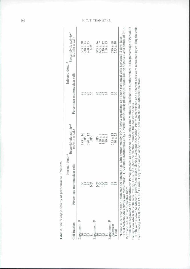
Because both macrophages and neutrophilsappear in peritoneal harvests from mice 2 daysafter i.p. infection, they were separated on dis-continuous Pereol! gradients in order to testwhich cell type(s) accounted for bacteriolyticactivity (results are shown in Table 2). Wherevirtually pure (95-100%) populations of mono-nuclear cells were obtained, those from miceinfeeted i.p. 2 days earlier were more efficientthan those from normal mice. Adherent mono-eytes from infeeted mice also showed strongerbacteriolytie activity than equivalent cells fromnormal mice {experiment 3). Interestingly, frae-tion 81 from infeeted mice, whieh contained86% neutrophils (experiment 2). was alsostrongly bacteriolytic indicating that neutro-pbils did not dilute out the efficiency of the acti-vated macrophages and presumably were alsocontributing to the bactericidal activity.
Table I. Effect of Listeria infection an phagocytosis and baeteriolytic aetivity of peritoneal cells.*
MiceCell-associated [ l
(ct/min ± s.d.)eells Release of soluble ['H]
(ct/min ±s.d.)Percentage lysis of
cell-associated bacteria
UninfeetedInfeeted i.p.Infected i.v.
2913 ±3536855 + 2773111 ±310
236 ± 401638 ±80259 ±21
8.123.9
8,3
•C57BI/1O mice were uninfeeted or infected i.p, with approximately lU"* Listeria organisms. Peritoneal eellswere obtained 2 days after infection. The amount of eell-assoeiated radioactivity was measured alier 30 minincubation of cells with radiolabelled bacteria. Release of TCA soluble radiolabel froni the washed cells wasmeasured after a further 2 h, allowing calculation of the pereentage lysis of cell-associated bacteria. Resultsrepresent the mean and s.d. for quadruplicate determination on lO''cells.
292 H. T. T. TRAN ETAL.
CQ
u+ l
o E
ffi
o+l
r-l — f l
+1 +1 +1 +1 +1 +1 +1 +1 +1iin u-i O O O "^
) Q + i a Q + i + i + i +1+1
UJ
s•c
w
Efe-g
•= • - 3
.= 00
Iso c
I 5 2.~
E3cco
tu
2 w
T3 CO
5 W
31-o
6
2Q&
ooUI
o
2>>oI / :
sOJ
c• ' c
uIi
"(3
c• "
"u."S5u[ / I
CO
C
•T3
J 3
u
Ccot it i :
un
CLI
.2
3uc
OO
CJ
5
5
ouU
c_o
> T:Io u
>
-z D o y
C m M J£ (ujfi
Of- ™U-U

MACROPHAGES KILLING USTERIA 293
The population of cells recovered from theperitoneal cavity of cither normal or infectedmice is a mixed one, analysed in Table 3. Intra-peritoneal infection leads to an increase in thenumbers of polymorphonuelear neutrophils byday 2 and the appearance of a new type of cellwith the morphology of large activated macro-phages but staining positive for MPO activity.an enzyme characteristic of monocytes, ratherthan of mature macrophages. The inference thatthere was a high proportion of newly formedcells in this population was confirmed by radio-labelling and autoradiography. When [- HJTdRwas injected 1 and 2 days before 41% of themacrophages and 13% of the neutrophils frominfected mice had incorporated [H-J], comparedwith 5 and O'i ) respectively for normal mice.Total iodinating activity, Oi" production andH2O2 production were dramatically increasedfollowing infection. The outcome of the combi-nation of increased phagocytic and killing activ-ity previously noted was measured by allowingcells and bacteria to interact eontinuously for2'/:h and counting the released TCA-solubleradiolabel. The cells from infected mice releasedfour to live times the amount of label per IO*"peritoneal cells compared with normal.
The 2 day inflammatory response in theperitoneal cavity was extremely radiosensitive(Fig. 1). Mice exposed to 300-700 R 3 daysbefore infection showed a dose dependentreduction in the bacteriolytic activity of their
peritoneal cells and in the percentage of MPO'*"monocyte/macrophages as well as, to a lesserextent, in the numbers of neutrophils. Irradi-ation had no significant efiect on the residentperitoneal population, either in bacteriolyticactivity or in composition (for simplicity the lat-ter results are not shown). The radiosensitivityof the inflammatory cells is consistent with theirrecent derivation tVom a rapidly dividing popu-lation.
The MPO+ cells, whether monocyte/macro-phage or neutrophil, did not persist long in theperitoneal cavity after i.p. infection. They hadalready declined by 4 days (Fig. 2b) and werevery rare at later time points (data not shown).A dip in bacteriolytic activity was often ob-served 3-4 days post-infection in differentexperiments (Fig. 2a). This dip may representthe decline of the inflammatory response beforethe induction of T cell medicated immunity.This is where 30 min labelling would help.
Finally, comparison was made between theacute inflammatory cells resulting from a 2 dayi.p. infection and the more "classically' activatedmacrophages appearing in the peritoneal cavity8 days after i.v. infection (Table 4). The twogroups showed similar increased bacteriotyticactivity and H2O2 production compared withnormal resident peritoneal cells. As notedearlier, the inflammatory response occurring2 days after i.p. infection included, togetherwitbpolymorphs, the appearance of MPO"*"
Table 3. Analysis of peritoneal cells from normal and 2 day infected mice.*
No. cells per mouse (per eenl of total)Normal Infected
Differential counts*Macrophages: MPO~Macrophages: MPO"*"Neutrophils: MPO+Others
Biochemical aetivityjlodinationOl" productionH3O2 production
RadiolabcllmgSMacrophagesNeutrophils
Bacleriolylic activity**
*Mtcc were uninfeeted or infected i.p. with approximately lO" Listeria organisms and Iheir peritoneal cellsharvested 2 days later.
"•"Counts were madeof triplieate eytocentrifuged preparations, stained for MPO and at least 200 eells per slidescanned.
JTotal MPO aetivity is expressed as et/min '-M incorporated in triplicate determinations. Oj- production andH2O2 productions are expressed as OD 620 nm. mean of eight replicates. All are shown + s'd.
§Mice were injected i.v. with 20 \iCi ['H]TdR I and 2 days before cell harvest. Autoradiographs were made asdescribed and counts were made of 200 cells on eaeh of triplicate slides from each group.
••Total bacteriolytic activity was measured as TGA soluble ct/min released hy 10'' eells eontinuouslyphagocytosing and killing Lisleria over a period of 2'/; h. Mean and s.d. of quadruplicate tubes.
190X 10" (72%)< l X 10-110 X IO-*(4%)65 X 10-'(24%)
160± 2400.02 ± 0.010.01 ±0.02
5%0%
400 ± 30
140X 10" (38%)HI X 10" (29%)SOX !O'>(I3%)70X IO''(I9%)
2400 ± 2400.74 ± 0.250.83 ±0.13
41%13%
1950± 100

294 H. T. T. TRANAT.-J/..
(a)500
1 0 0 •
0 300R 500R 700R
irtacJfation dose
(b)
BO
40
20
0 NORM 0 INF 300 INF 500 INF 700 INFIrradialion dose
Fig. I. Effect of whole body irradiation prior to i.p.infection on inflammatory response in the peritonealcavity. Mice were given graded doses of irradiation3 days before half were infected i.p. with 10-* Lisicriuorganisms. Peritoneal cells were harvested 2 days afterinfection, (a) Bactericidal activity of cells frominfected ( • ) and unJnfected ( • ) mice. Mean and s.d.of quadruplicate cultures, (b) Differential counts oninfected or normal mice. (D) MPO" macrophage; {•)MPO+macrophage; (S) neutrophil; (^) other cells.Irradiation had no detectable effect on the residentnormal population and for simplicity tbese counts arenot shown.
monocytes/macrophages not seen in normalmice. The number of MPO~ macrophagesremained the same as in normal mice. In con-trast, infection of mice i.v. 8 days previouslyincreased the total number of peritoneal cellsbut most of them were MPO" macrophageswith extensive vacuolated cytoplasm. MPO +monocytes were a minor component. That there
~ 300-
•= 200-
100-
Time since infection (da/s)
100
80
•S 60-
0 2 3 4Time since inlection (days)
Kig. 2. Time course of intlammatory responsefollowing i.p. injection of lO" Li.steria organisms.(a) Bacteriolytic activity, mean and s.d. of quadrupli-cate assays, (b) Differential counts. (D) M PO macro-phages; ( • ) MPO+ macrophage; (S) neutrophils; (E)other cells.
was any change in the peritoneal cells of thesemice implies a systemic mechanism, since i.v.injection does not result in infection in the per-itoneal cavity. In order to induce an inflamma-tory response in the peritoneal cavity, the i.v.infected mice were given an i.p. injection ofliveListeria organisms 6 days after primary infee-tion and the cells harvested 2 days later. Thisalmost trebled the nutnber of cells in the peri-toneal eavity compared with mice given i.v. Lis-teria only. The greatest increase was amongstthe MPO"^ macrophages. up from 3.1X10*'to 7.3 X 10 . MPO"* monocyte/macrophagesshowed a lesser increase than that observed in 2

MACROPHAGHS KILLING LISTL'RIA 295
Table 4. Comparison of ceils from mice infected 2 days earlier (acute inflammatory cells) with cells from 8duv immune mice.
Bacterioiytic activity(ct/min ± s.d.)'
H'O^ release(abs 620 nm)t
No. eells/mouscDillerentlal counts*
M o not vt cs/ mac roph agesMFO
M(»O +
NeutrophilsMPO +
Other eellsMPO
Normal
124±5I
0.019±0.022
2.8 X IO<'
230 X lO-*(84%)
0
8X 10"(3%)
36 X 10"(1 3%)
Liswria i.p. J.iv 2
7I2±{)5
O.I 12 ±0.028
6.3 X to''
280X 10"(45%)
190X10"(30%)
120X 10"(19%)
37 X 10"(6%)
Group*Listeria i.v. day 8
636 ± 54
0.169 ±0.022
4.1 X 10''
310X 10"(75%)16 X 10"(4%)
60 X 10"(15%)
25X10"(6%)
Listeria i.v. day 8; i.p. day 2
647 ± 46
O.I57 ±0.022
11.1 X 10''
730X10"(66%)
100X10"(9%)
I70X lO"(I 5%)
IJOX 10"(10%)
•Mice were tnfeeted with UH/j.vfcn^ organisms given In three regimes: i.p. 2 days before assay, i.v. 8 days beforeassay or i.v. 8 days before plus i.p. 2 days before assay.
^Baeteriolylic activity per 10*' cells was measured in quadruplicate.*H2O2 release was measured wilh 8 replicates and baekground absorbance subtracted.^Percentages of total cells in triplicate determinations, representing at Icasl 600 cells per group.
day exudates of previously non-i[nmune tnice(0.84 X 10'' vs 1.9X 10'' increase). The increasein neulrophils was similar in the two inflamma-tory exudales.
DISCUSSION
Infection of mice with /.. monocytof^encs hasbeen widely used lo produce, indeed virtually todefine, activated macrophages (3). It has beengenerally held that, when the specific itnmuneresponse is initiated 3-4 days after infeetion. Tlymphocytes produce lymphokines. including y-interferon. that both activate the macrophagesand attract more cells to the site of infectionleading to recovery. On the other hand, the early(1-2 days) presutiiably non-specific inflamma-tory response also provides efficient bactericidalcells, and is critical for genetic resistance of cer-tain mouse strains to Li.sieria (6.8,20). It hasrecently been suggested (10) that acquired cellmediated itnmunity rests, not on macrophageactivation, but on a heightening of this inflatn-matory response supplying actively bactericidaltnonocytesand polymorphs. The present results,showing that two distinctive cells of the macro-phage/tiionocyte lineage are involved, respeet-ively, in the early inflammatory response and
the later acquired systemic immunity, do notsupport that concept.
The early (2 day) inflammatory response toi.p. injected lAsleria comprised in differentexperiments 60-80% cells of the monocyte/macrophage type and 2O-3O'*/n polymorpho-nuclear cells. The population expressed bothhigher phagocytic and higher bactericidal activ-ity than normal resident peritoneal cells. Pereollgradient purified or 18 h adherent mononuclearcells from the 2 day infected mice also showedhigher bacteriolytic activity than macrophagesfrom normal tnice. Fraetions of cells from in-fected mice in which macrophages were dilutedby up to 86% neutrophils retained high activity.The ability of neutrophils to kill Lisictia organ-isms has been a matter of controversy, and theseresults support those of Czuprynski cl al. (21)that they do so. The majority of mononuclearcells from 2 day infected mice had an appear-ance not unlike that of immunologically acti-vated macrophages in the peritoneal cavity of8 day i.v. infected miee: that is. they were targecells with a foamy cytoplasm, rather than beingtypical monocytes. However, they differed fromthe 8 day cells in that a large proportion stainedpositive for MPO {usually associated with mon-ocytes rather than mature macrophages). Wholebody irradiation selectively depleted MPO +cells, showing that they arose from recently div-

296 H. T. T. TRAN ETAL.
iding cells. Although it was not possible to sim-ultaneously slain for MPO and makeautoradio-graphs. the high proportion of cells labellingwith ['Hj-TdR supported the inference thatmany cells in the 2 day exudates were newly div-ided. Like the 8 day cells they etficiently gener-ated H2O2 in response to phagocytosis. Theearly inflammatory response was transient, withnumbers of MPO ' monocyte/macrophages andneutrophils. as well as the bacteriolytic activityof the peritoneal cells waning 3-4 days after i.p.infection.
An 8 day i.v. infection induced cells in the per-itoneal cavity which, like the 2 day inflamma-tory exudate. were actively bacteriolytic. ef-ficient at H2O2 production and included manymacrophages with abundant vacuolated cyto-plasm. These were the cells defined by Mack-aness in 1962 (1). The fact that the Li.stcriaorganisms do not enter the peritoneal cavityafter i.v. infection but localize in the spleen andliver implies a systemic mechanism of macro-phage activation rather than an inflammatoryresponse. The cells harvested from 8 dayinfected mice showed only 4% MPO^ mono-cytes/macrophages. Even when 8 day immunemiee were rechallenged with L/i7ma i.p. the per-itoneal exudate induced at 2 days differed fromthat in previously uninfected miee. MPO"macrophages showed the greatest increase in
numbers and comprised 60% of the population.The increase in MPO ' monocyte/macrophagesand in polymorphs was no greater than in theprimary response.
The 8 day cells appeared to epitomize the con-cept of resident macrophages activated by T celllymphokines (3). We have recently shown thatmacrophage colony slimiiiating factor (CSF-1).which is found at high concentration in infectedmice 1-2 days post-inicction (9.22.23) can acti-vate in vitro the ability of peritoneal macro-phages to kill/.me™ (24). Whether this contrib-utes to the activity of the 2 day cells we can onlyspeculate.
The question of what property of the cellsleads to bacterial death remains a ditticult one.Both the 2 day elicited peritoneal macrophages,and the neutrophils are positive for MPO activ-ity. However, this activity is not essential forkilling I.isteria organisms, since ihe 8 day per-itoneal exudates did not contain significantnumbers of MPO' macrophages. We are atpresent testing inhibitors of MPO and scaven-gers of H2O2 and O2" on the two populations tosee whether any of these abrogate killing.
Acknowledgements This work was supported bythe National Health and Medical Research Council ofAuslralia. The technical assistance of Kaylene Selleckis gratefully acknowledged.
REFERENCES
1. Mackaness, G. B. 1962. Cellular resistance toinfection. J. Exp. Med. 116; 381-406.
2. Mackaness, G. B. 1964. The immunological basisof acquired cellular resistance. J. Exp. Med. 120:105-120.
3. North, R. J. 1978. The concept of the activatedmacrophages. J. Immunol. 121: 806-808.
4. Newborg, M. F. and North. R. J. 1980. On themechanism of T cell-independent unU-Listeriaresistance in nude mice. J. Immunol. 124: 571-576.
5. Cheers, C, McKenzie, I. F. C, Pavlov. H.. Waid,C. and York, J. 1978. Resistance and susceptibil-ity to bacterial infection: Course of listcriosis inresistant or susceptible mice. Infect. Immuri. 19:763-770.
6. Slevenson. M., Kongshavn, P. A. L. and Skamene,E. 1981. Genetic linkage of resistance to I.isteriamonocytogenes with macrophage inflammatoryresponse. / Immunol. 127: 402-407.
7. Sadarangani, C. Skamene, E. and Kongstiavn,P. A. L. 1980. Cellular basis for genetically deter-mined enhanced resistance of certain mousestrains to listeriosis. Infect. Immunol. 28: 381-386.
8. Wood, P. R.. Spanidis, V., Frangos. K. andCheers. C. 1986. The in r/fro bactericidal activityof peritoneal and spleen ceils from L/.v/ma-resist-ant and susceptible mouse si rains. Cell. Immunol.>»9: 160-169.
9. Young, A. M. and Cheers, C. 1986. Colony form-ing cells and colony stimulating activity duringlisteriosis in genetically resistant and susceptiblemice. Cell. Immunol. 97: 227-237.
10. Campbell, P. A. 1986. Are inflammatory phago-cytes responsible for resistance lo facultativeintraeellular bacteria? Immunol. Today 7: 70-72.
11. Miles. A. A. and Misra, S. S. 1938. Estimation ofthe bactericidal power of blood. / llvg- 28: 732-748.
12. Hart, P. H.. Spencer, L. K., Nikoloutsopoulos, A.et al. 1986. Role of cell surface receptors in theregulation of intracellular killing of bacteria bymurine peritoneal cxudale neutrophils. Infect.Immun. 52: 245-251.
13. North, R. J. 1969. The mitogenic polenlial offixed phagocytes in the liveras reveal;jd during thedevelopment of cellular immunity. J. Exp. Med.130: 315-326.

MACROPHAGES KILLING LISTERIA 297
14- Rogers. A. W. 1979. Techniques of Autoradi-ofiraphy, 3rd edn. Elsevier North Holland Bio-mcdical Press. Amsicrdani, p, 351.
15. Davies, W. A. 1982, Kinetics of killing Li.stvriumonocvtoficnes by macrophages; Correlation of'H-DN.A release from labelled bacteria andchanges in number of viable organisms by math-ematical model, J. Ri'liciilocnJothelial Soc. 32:461-476.
16. Pincus, S. H. and Klebanoft". S. J. 1971, Quaiui-tativc leukocyte iodination. N. Engl. J. Med 284:744-750,
17. Kaplow, L, S, 1965, Simplified myeloperoxidaseslain using benzidine dihydrochloride. Blood. 26:215-219.
18. Rook. G. A, W.. Sleele. J,. Umar. S, and Dockrell.H, M, 19K5. A simple method for the solubiliz-alion of reduced NBT, and ils use asa colorimetricassay for activation of human macrophages by y-interferon../, Immunol. Methods S2: 161-167.
19. Pick. C, and Keisari.Y, 1980. A simple colorimel-ric method tor the measuremeni of hydrogenperox ide produced by cells in culture. J. ImmunoLMethods i^. 161-170.
20. Czuprinski, C. J.. Canono, B P.. Henson. P. M.
and Campbell. P. A. 1985. Genetically deter-mined resistance to listeriosis is associated wiihincreased accumulation of intlammutory neuiro-phils and macrophages which have enhancedlisterieidai activity, Immunohiiy 55: 511.
21. Czuprynski. C, J,. Henson. P, M.. and Campbell.P. A, 1984. Killing of Lisieria monovyiogenes byinflammatory neutrophils and mononuclearphagocytes from immune and non immune mice../, Leukocyte Biol. 35: 193-208.
22. Cheers. C. and Stanley. E, R. 1988. Maerophagcproduction during listeriosis: colony stimulatingfactor-1 (CSF-1) and CSF-1 -binding cells in genet-ically resistant and susceptible miee, Infeet.fmmun. 56: 274.
23. Cheers. C . Haig. A.. Kelso, A.. Metcalf, D., Stan-ley. E. R.. and Young. A. M. 1988, Production ofeolony stimulating factors (CSFs) during infec-tion: Separate determinations of macrophage-.granulocyte-. granulocyte-maerophage- andmulti-CSFs. Infect. Immun. 56: 247-251.
24. Cheers. C . Hill. M.. Haigh. A. M. and Stanley. E.R. 1989. Stimulation of macrophage phagocyticbut not bactericidal aetivity by colony stimulatingfactor I. Infi'ct. Immun. 57: 1512-1516.