Pulsed-field gel electrophoresis (PFGE) analysis of Listeria monocytogenes isolates from different...
7
This article appeared in a journal published by Elsevier. The attached copy is furnished to the author for internal non-commercial research and education use, including for instruction at the authors institution and sharing with colleagues. Other uses, including reproduction and distribution, or selling or licensing copies, or posting to personal, institutional or third party websites are prohibited. In most cases authors are permitted to post their version of the article (e.g. in Word or Tex form) to their personal website or institutional repository. Authors requiring further information regarding Elsevier’s archiving and manuscript policies are encouraged to visit: http://www.elsevier.com/copyright
-
Upload
independent -
Category
Documents
-
view
0 -
download
0
Transcript of Pulsed-field gel electrophoresis (PFGE) analysis of Listeria monocytogenes isolates from different...

This article appeared in a journal published by Elsevier. The attachedcopy is furnished to the author for internal non-commercial researchand education use, including for instruction at the authors institution
and sharing with colleagues.
Other uses, including reproduction and distribution, or selling orlicensing copies, or posting to personal, institutional or third party
websites are prohibited.
In most cases authors are permitted to post their version of thearticle (e.g. in Word or Tex form) to their personal website orinstitutional repository. Authors requiring further information
regarding Elsevier’s archiving and manuscript policies areencouraged to visit:
http://www.elsevier.com/copyright
Author's personal copy
Systematic and Applied Microbiology 31 (2008) 387–392
Pulsed-field gel electrophoresis (PFGE) analysis of Listeria monocytogenesisolates from different sources and geographical origins and representative
of the twelve serovars
Elsa Nevesa,b, Antonio Lourencoa, Ana Carla Silvaa, Rute Coutinhoa, Luisa Britoa,�
aLaboratorio de Microbiologia, CBAA/DBEB, Instituto Superior de Agronomia, Technical University of Lisbon, Tapada da Ajuda,
1349-017 Lisbon, PortugalbInstituto Superior de Estudos Interculturais e Transdisciplinares (ISEIT), Campus Universitario de Almada, Instituto Piaget,
Quinta da Arreinela de Cima, 2800-305 Almada, Portugal
Received 7 July 2008
Abstract
Multiplex-PCR (MPCR) serogrouping and pulsed-field gel electrophoresis (PFGE) subtyping analysis are currentlyused by several public and private laboratories for the characterization of Listeria monocytogenes. In this study a setof 80 L. monocytogenes isolates belonging to the twelve serovars was used to investigate (i) the typeability of the rareserovars, (ii) the ability of PFGE analysis with ApaI and AscI to differentiate serovars within MPCR serogroups and(iii) the association of molecular types with the specific source or geographical origin of the isolates. With the exceptionof three isolates (rare serovars 4a and 4c) that were not amenable to restriction with ApaI, all the other analyzedisolates were subtyped by both enzymes. PFGE discriminated the 80 isolates into 62 combined ApaI and AscI PFGEpatterns (pulsotypes), but could not differentiate serovars within MPCR serogroups, in which isolates from differentserovars displaying the same pulsotype were found. Clustering analysis suggests that for some pulsotypes groupingaccording to Portuguese origin or source can be suggested. On the other hand, some L. monocytogenes clones arewidely distributed. Two pulsotypes from Portuguese human isolates were identical to the ones displayed by humanoutbreak clones in the UK and in the USA and Switzerland, respectively, although they were not temporally matched.
Computer-assisted data analysis of large and diverse PFGE type databases will improve the correct interpretation ofsubtyping data in epidemiological studies and in tracing routes and sources of contamination in the food industry.r 2008 Elsevier GmbH. All rights reserved.
Keywords: Listeria monocytogenes; Sources; Geographical origin; Genetic diversity; Serovars; Genetic lineages; PFGE;
Multiplex-PCR (MPCR) serogrouping
Introduction
Listeria monocytogenes is a foodborne facultativeintracellular pathogenic bacterium responsible for lister-iosis in humans and animals, particularly amongimmunocompromised individuals. Thirteen serovars(1/2a, 1/2b, 1/2c, 3a, 3b, 3c, 4a, 4ab, 4b, 4c, 4d, 4e, 7)
ARTICLE IN PRESS
www.elsevier.de/syapm
0723-2020/$ - see front matter r 2008 Elsevier GmbH. All rights reserved.
doi:10.1016/j.syapm.2008.08.005
�Corresponding author. Tel.: +351 21 365 3240, +351 21 365 3435;
fax: +351 21 365 3238.
E-mail address: [email protected] (L. Brito).

Author's personal copy
have been reported for L. monocytogenes [5]. Some ofthese serovars have been recovered from patients andfoods (1/2a, 1/2b, 1/2c and 4b) more frequently [14] thanthe others considered as rare serovars. Althoughserotyping [20] is less discriminatory than moleculartyping methodologies, it has been used in epidemiolo-gical studies as a universal technique for the character-ization of L. monocytogenes. Recently, different authorshave proposed PCR based methods for serogroupingL. monocytogenes isolates [1,5,24].
The multiplex-PCR (MPCR) assay proposed byDoumith et al. [5] is based on the use of five sets ofprimers targeting group-specific genes that enable theclustering of L. monocytogenes strains into five phylo-genetic groups (serogroups) correlated with the serovars(except serovar 4ab which is extremely rare) [5]. The setof primers targeting the prs gene (putative phosphor-ibosyl pyrophosphate synthetase) fragment (370 bp) isuniversal to all Listeria species. In addition to thisfragment, strains from group 1 (1/2a–3a) generate oneamplified fragment from gene lmo0737 (unknownfunction) (691 bp), strains from group 2 (1/2c–3c)produce two fragments from gene lmo0737 (691 bp)and gene lmo1118 (unknown function) (906 bp), strainsfrom group 3 (1/2b–3b–7) amplify a fragment fromORF2819 (putative transcriptional regulator) (471 bp),and strains from group 4 (4b–4d–4e) generate twofragments from ORF2819 (471 bp) and from ORF2110(putative secreted protein) (597 bp). However, the pro-posed MPCR assay does not distinguish L. monocytogenes
serovar 1/2a from 3a, 1/2c from 3c, 1/2b from 3b and 7,4b from 4d and 4e. Isolates from serovars 4a and 4c(grouped as L in the present work) and isolates from theother Listeria species are also undistinguishable by thisapproach.
In order to better differentiate L. monocytogenes
isolates, different subtyping methods have been used[10,22,23] although pulsed-field gel electrophoresis(PFGE) is the current gold standard method formolecular subtyping of most foodborne bacteria,including L. monocytogenes [8]. Results from differentsubtyping studies suggest that three genetic divisions orlineages correlated with the flagellar (H) antigene typeexist in L. monocytogenes [2,15,19,23]. Lineage I consistsof strains (flagellar antigen types b and d) that are morelikely to cause human disease than isolates classified intolineages II (antigen type a or c) and III (serovars 4a and 4cand some 4b strains). However, clustering analysisbased on these studies usually uses isolates from themore frequent serovars (1/2a, 1/2b, 1/2c and 4b).
Eighty isolates collected in Portugal and out ofPortugal from different sources (including humans,animals, food and environment), and belonging totwelve serovars were analyzed in this study. We usedMPCR serogrouping and PFGE subtyping to investi-gate (i) the typeability of the rare serovars, (ii) the ability
of PFGE to differentiate serovars of L. monocytogenes
within MPCR serogroups and (iii) the association ofmolecular types with specific sources or geographicalorigin (from Portugal versus out of Portugal). To ourknowledge, this is the first report on clustering analysisof PFGE patterns of a group of isolates representing thetwelve serovars of L. monocytogenes.
Materials and methods
Bacterial strains and growth media
Eighty L. monocytogenes isolates (Fig. 1) including13 culture collection strains, previously classified by slideagglutination serotyping, and belonging to the twelveserovars were characterized in this study. The strainswere from food (n ¼ 29), from human cases of listeriosis(n ¼ 25), from animals (n ¼ 4), from environment(n ¼ 5) and from uncertain sources (n ¼ 17) (Fig. 1).The geographical origin of the isolates was ascribed asfrom Portugal (P) or from out of Portugal (O), as it wasin most of the cases the only information we had. Someof the L. monocytogenes strains were kindly provided bythe Faculte de Medecine de Tours, Universite Francois-Rabelais (L’U.F.R), France, Laboratoire des Listeria,Institut Pasteur (I.P.) Paris, France, Universidade doAlgarve (UA), Portugal and Universita degli studidi Udine (USU), Italy. The 13 reference strains werefrom Collection de l’Institut Pasteur (CIP), CollecionEspanola de Cultivos Tipo (CECT) and NationalCollection of Type Cultures (NCTC): CIP104794( ¼ NCTC7973) and NCTC7973 (serovar 1/2a);CECT936 and NCTC10887 (serovar 1/2b); CECT911( ¼ ATCC19113) (ATCC—American Type CultureCollection) and NCTC9862 (serovar 1/2c); NCTC5105(serovar 3a); CECT937 and CIP78.35 (serovar 3b);CECT934 ( ¼ ATCC19114) (serovar 4a); CECT 4032(NCTC11994) and NCTC10527 (serovar 4b); CIP78.39( ¼ ATCC19116) (serovar 4c).
All stains were kept on Tryptic Soy Broth (TSB,Difco, Detroit, USA) containing 15% glycerol andstored at�80 1C. Before the assays, each L. monocytogenes
selected isolate was streaked on TSA-YE (Oxoid,Hampshire, UK) plates, and incubated for 18h at 37 1C.Queries of the PathogenTracker database (http://www.pathogentracker.net) which contains subtype informationfor 648 L. monocytogenes (as of 19/06/2008) from varioussources were conducted to probe the distribution of 22 outof 45 Portuguese isolates.
Multiplex-PCR (MPCR) serogrouping
MPCR serogrouping was performed using as PCRtemplates lysates obtained from bacterial colonies,
ARTICLE IN PRESSE. Neves et al. / Systematic and Applied Microbiology 31 (2008) 387–392388
Author's personal copy
according to Doumith et al. [5]. The five primer sets fortarget fragments from genes lmo0737, lmo1118,
ORF2819, ORF2110 and prs were synthesized byMWG-Biotech AG Switzerland, according to thesequence data of Doumith et al. [4]. The resultingPCR products were resolved on 1.4% (w/v) agarosegels in 1�TAE buffer, at 2.3V/cm for 60min. The gelswere stained with ethidium bromide (Sigma) andanalysed by using Bio-Rad Gel Doc 2000TM imagingsystem (Bio-Rad Laboratories, Segrate, Milan Italy).
PFGE
The 80 L. monocytogenes isolates were characterizedby DNA macrorestriction analysis using PFGE in
accordance with the Pulse-Net standardized protocol[9]. Bacterial cultures were embedded in chromosomalgrade agarose (SeaKem Gold Agarose, Cambrex, NewJersey, USA), lysed, washed and the DNA was in situ
digested in separate reactions with 10U of AscI (NewEngland Biolabs, Massachusetts, USA) at 37 1C andwith 20U of ApaI (Roche Diagnostics, Mannheim,Germany) at 30 1C overnight, respectively. The resolu-tion of the generated DNA fragments was obtained with1% SeaKem Gold Agarose gels in 0.5�Tris-BorateEDTA Buffer (Invitrogen, Paisley, UK) at 14 1C and6V/cm with time ramped for 4–40 s over 22 h usinga CHEF DR II System (BioRad, Hercules, USA). Gelswere stained with ethidium bromide at a final concen-tration of 10 mg/ml (Sigma, St. Louis, USA) and patternimages were acquired in Bio-Rad Gel Doc 2000TM
ARTICLE IN PRESS
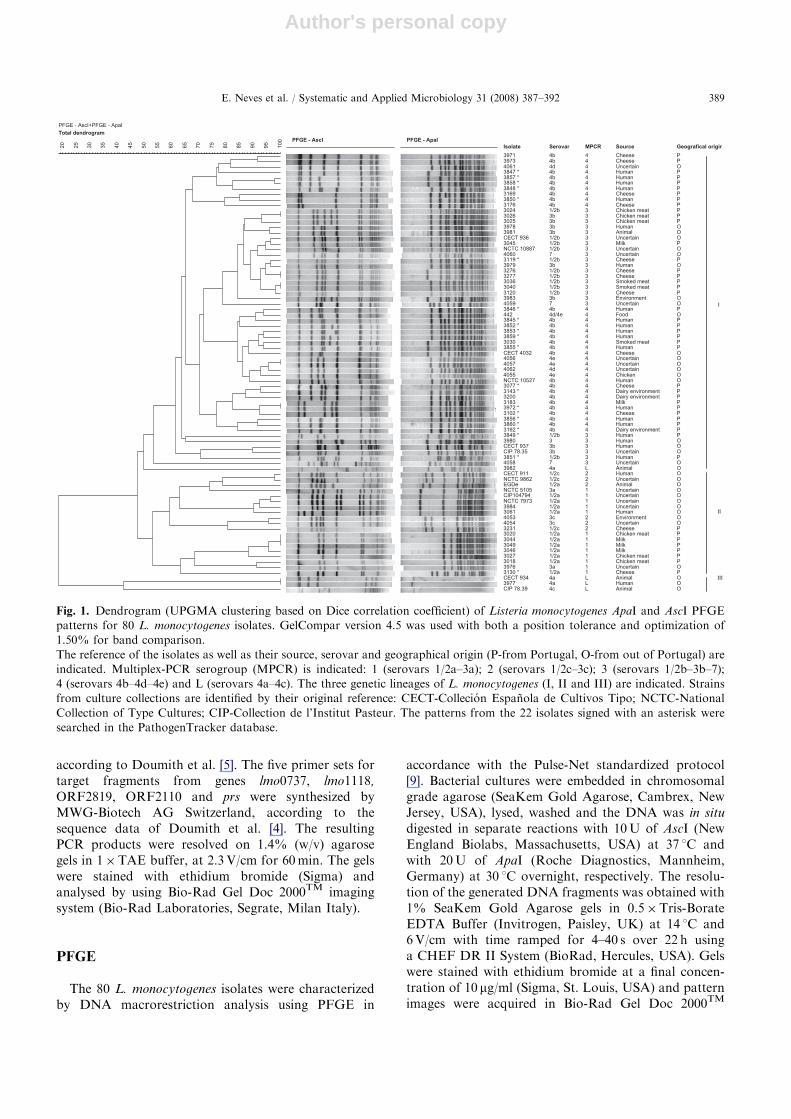
Fig. 1. Dendrogram (UPGMA clustering based on Dice correlation coefficient) of Listeria monocytogenes ApaI and AscI PFGE
patterns for 80 L. monocytogenes isolates. GelCompar version 4.5 was used with both a position tolerance and optimization of
1.50% for band comparison.
The reference of the isolates as well as their source, serovar and geographical origin (P-from Portugal, O-from out of Portugal) are
indicated. Multiplex-PCR serogroup (MPCR) is indicated: 1 (serovars 1/2a–3a); 2 (serovars 1/2c–3c); 3 (serovars 1/2b–3b–7);
4 (serovars 4b–4d–4e) and L (serovars 4a–4c). The three genetic lineages of L. monocytogenes (I, II and III) are indicated. Strains
from culture collections are identified by their original reference: CECT-Collecion Espanola de Cultivos Tipo; NCTC-National
Collection of Type Cultures; CIP-Collection de l’Institut Pasteur. The patterns from the 22 isolates signed with an asterisk were
searched in the PathogenTracker database.
E. Neves et al. / Systematic and Applied Microbiology 31 (2008) 387–392 389

Author's personal copy
imaging system (Bio-Rad Laboratories, Milan, Italy).The TIF images were normalized by aligning the peaksof the size standard strain Salmonella enterica serovarBraenderup H9812 which was loaded on three lanes ineach gel. This strain was kindly provided by CentreNational de Reference des Listeria, Institut Pasteur,France.
Data analysis
PFGE patterns were analyzed and compared usingthe GelCompar II version 4.5 (Applied Maths, Kortrijk,Belgium). The levels of similarity were based on the Dicecorrelation coefficient. For cluster analysis of thepatterns the unweighted-pair group matching algorithm(UPGMA) was used with a tolerance of 1.50% andoptimization of 1.50% as recommended by Martin et al.[13]. The quality of the cluster analysis was verified bycalculating the cophenetic correlation value (r) for thedendrogram. Eight control strains of L. monocytogenes
were used to determine the experimental variationbetween duplicate experiments. For both enzymes, theminimum level of repeatability of the macrorestrictionconditions was calculated by running DNA samplesfrom duplicated restrictions of the DNA of each strain.On the basis of the obtained results, a cut-off value ofsimilarity was established for typing identical strainswith identical outputs.
The Simpson’s index of discrimination (SID) ofPFGE, expressed as a percentage, was determined asdescribed by Hunter and Gaston [12].
Results and discussion
MPCR and PFGE analysis are currently used byseveral public and private laboratories for serogroupingand subtyping L. monocytogenes. The first methodconstitutes a pratical alternative to slide agglutinationserotyping and separates L. monocytogenes isolatesbelonging to the twelve serovars into five distinctserogroups. PFGE is considered the gold standardmethod for subtyping foodborne pathogens, becauseof its high discriminatory power and reproducibility. Inthis study, the combination of these methodologies withcomputer-assisted data analysis was applied to anisolate set representing considerable diversity (eightyisolates belonging to the twelve serovars representativesof the three genetic lineages of L. monocytogenes).
By using MPCR serogrouping, the 80 isolates wereassigned to five groups (1–4 and L) (Fig. 1) inaccordance with previous results from conventionalserotyping. The exception was the strain EGDe (serovar1/2a) that showed a profile corresponding to MPCRserogroup 2 (1/2c–3c) (Fig. 1) in agreement with a
previous report of its unusual behaviour in this respect [5].In accordance with this, the AscI and ApaI PFGEpatterns of this strain were closer to the ones displayedby two culture collection strains from serovar 1/2c (0.79similarity) than to the other patterns displayed by theserovar 1/2a isolates analysed (Fig. 1).
Strain 3980 given to us as serovar 3, displayed amultiplex pattern for group 3 (1/2b–3b–7), suggesting tobe a serovar 3b isolate. This result was confirmed by thelocation of this isolate in the dendrogram (Fig. 1).
The results of multiplex-PCR of strains from the rareserovars 4a and 4c (group L) showed a negativeinfluence of biomass excess in the cell lysate. In fact, inaddition to the expected 370 bp fragment, a faint bandof about 471 bp was present in the first amplificationprofiles of these strains (results not shown). This factcould lead into an incorrect assignment of these strainsto multiplex group 3 (serovars 1/2b–3b–7). Nightingaleet al. [17] also reported the misclassification of strainsfrom serovars 4a and 4c in multiplex group 3. In the lasttrials using a lysate with less biomass, this faint banddisappeared pointing out the need to work in stringentcompliance with the protocol.
PFGE of the DNAs of the 80 isolates digested withAscI showed 7–16 fragments ranging from approxi-mately 30 to 1130 kb in size, while 11–16 fragments of30–510 kb were obtained following digestion using ApaI.The genomic DNA from two out of the three strainsfrom serovar 4a (strain CECT 934 and isolate 3977) andstrain CIP 78.39 (serovar 4c) were not amenable todigestion with ApaI (Fig. 1). Similar results werepreviously reported by Brosch et al. [2] for strainATCC19116 (serovar 4c) equivalent to our strainCIP78.39.
The relationships among L. monocytogenes strainsbased on their combined ApaI and AscI PFGE profilesare shown in the dendrogram displayed in Fig. 1, andare supported by a cophenetic correlation coefficient (r)equal to 0.92. This value explained the goodness of fit ofthe clustering to the original data [21] and usually rangesbetween 0.60 and 0.95 and values higher than 0.80 areconsidered reasonable [18].
Based on data obtained from the duplicate experi-ments, a cut-off level of 0.95 similarity was establishedfor the definition of a PFGE pattern. PFGE of AscImacrorestriction fragments differentiated the 80 isolatesinto 33 different patterns. The PFGE of ApaI macro-restriction fragments of the 77 typeable isolates pro-duced 44 different patterns. A higher SID value wasachieved with ApaI (0.91) compared to AscI (0.85). Thecombination of patterns produced by both enzymesyielded 62 combined ApaI and AscI PFGE types(pulsotypes) increasing the discriminatory power up to0.97 (Fig. 1).
At about 0.42 similarity level L. monocytogenes
isolates could be divided into two major clusters. The
ARTICLE IN PRESSE. Neves et al. / Systematic and Applied Microbiology 31 (2008) 387–392390
Author's personal copy
smaller cluster corresponded to lineage III (three isolatesfrom serovars 4a and 4c typed only by AscI) wellseparated from the bigger cluster that gathered at 0.50 ofsimilarity two subgroups. In one of these subgroups the19 isolates from serovars 1/2a, 1/2c, 3a and 3c (lineage II)were clustered. The other subgroup gathered the58 isolates from serovars 1/2b, 3b, 4b, 4d, 4e and 7(lineage I). These results are in agreement with Doumithet al. [4] that used a DNA macroarray-based subtypingmethod to divide the three lineages of L. monocytogenes
into five phylogenetic groups, each correlated withserovars. In fact, clustering of the isolates according totheir pulsotypes is in accordance with MPCR serogroups.
Nevertheless, PFGE subtyping could not differentiateserovars within MPCR serogroups. Moreover, withinserogroups identical pulsotypes were shared by isolatesbelonging to different serovars: serogroup 3 (serovars1/2b and 3b, isolates 3024 and 3026) and serogroup 4(serovars 4e and 4d, isolates 4056, 4057 and 4062)(Fig. 1). Interestingly, two isolates belonging to MPCRserogroup 4 (serovars 4b-4d-4e) had significantly differ-ent levels of pathogenicity (evaluated both in vivo andin vitro) [16]: human isolate 3846 (4b) was significantlymore virulent than food isolate 442 (4d/4e) althoughtheir pulsotypes had about 0.94% similarity.
For some isolates, similarity clustering analysis ofPFGE patterns suggested association between moleculartypes and specific sources or Portuguese origin, inaccordance with previous reports [3,7]. At 0.53 similaritylevel, 19 isolates from lineage II were clustered in twosubgroups (Fig. 1). In one of these subgroups seven outof eight isolates were from Portugal and collected fromfood (chicken meat and milk). There was no evidencethat the isolates from milk were related or not and therewas no acknowledged association between the chickenmeat and the milk. In the other subgroup, the elevenisolates were mainly from out of Portugal and fromdiverse sources.
At 0.73 similarity level (Fig. 1) the isolates fromlineage I were grouped in three major subgroups. Oneof these subgroups gathered nine L. monocytogenes
(serogroup 4) Portuguese isolates from milk, cheese,dairy environment and from human cases of listeriosis.Two of these isolates (3183 and 3972) collected in 2002from raw milk, and in 2004 from placenta, respectively,have identical pulsotypes. In this cluster, there areisolates from milk and cheese (3077 and 3143) persis-tently collected in two different cheese dairies.
At the same similarity level, the two other majorclusters with 18 and 14 isolates (serogroups 3 and 4,respectively) were diverse in what geographical origin(from Portugal and from out of Portugal) and source(type of food, humans, environment and animal) areconcerned. The wide distribution of some of thesepulsotypes was confirmed by querying the Pathogen-Tracker database for 22 selected isolates. Isolate 3845
collected in 2000 in Portugal from an infected personshared the same pulsotype (designated in Fugett et al. [6]by pulsotype 7) with 17 isolates in PT database: bovine(2), human (5), water (4), food (2), caprine (2),enviroment (2). Among this group were isolates involvedin two outbreaks related to cheese consumption, inLos Angeles (LA) and in USA (1985) and Switzerland(1983–1987) [6]. Portuguese isolates 3857 and 3858collected from two infected persons in 1997 and 1998,respectively, had a pulsotype indistinguishable frompulsotype 20 displayed by isolates involved in a humanepidemic case related to pate consumption in 1988–1990,in the UK [6].
Computer-assisted data analysis of PFGE datasimplifies the processing of a large number of sampleswhile allowing data sharing if a stringent complianceto standard protocols is achieved. This standardiza-tion includes not only a rigorous accomplishment ofthe Pulse-Net standardized protocol by Graves andSwaminathan [9] but also standardization of proceduresin computer-assisted data analysis for pattern normal-ization and transfers of information intra and interlaboratories.
PulseNet Europe [13] recommends settings of bothoptimization and position tolerance at 1.5% forband comparison in accordance with Pulsenet USA.Although complying with these settings, strains visuallyindistinguishable may still be considered differentaccording to the clustering analysis [11]. Aiming tominimize this, the determination of the experimentalvariation between duplicate experiments was evaluatedin this work, allowing an establishment of a cut-offvalue of similarity of 0.95 for typing identical PFGEpatterns. In spite of this, visual PFGE matches werefound different by computer analysis (e.g. serovar4b isolates 3102 (cheese), 3183 (milk) and 3972 (hu-man)). Owing to this, a causal relationship between foodand human isolates cannot be deduced without anadditional subtyping method, in preference amenable toautomation and based on DNA sequence. Nevertheless,to improve correct interpretation of subtyping data, theavailability of a large and diverse PFGE type database isneeded.
To our knowledge, this is the first report on clusteringanalysis of PFGE patterns of a diversity set of strainsfrom twelve out of the thirteen serovars representativeof all three genetically different L. monocytogenes
lineages.
Acknowledgments
The authors thank Dr. Alban Le Monnier (I.P.),Dr. Laurent Mereghetti (L’U.F.R.), Dr. Leonor Faleiro(UA) and Dr. Luca Cocolin (USU) for providing someof the L. monocytogenes isolates and reference strains
ARTICLE IN PRESSE. Neves et al. / Systematic and Applied Microbiology 31 (2008) 387–392 391
Author's personal copy
used in this study. The authors thank Dr. MartinWiedmann and Yesim Soyer for queries to thePathogenTracker database.
The financial support of Fundacao para a Cienciae Tecnologia (FCT) (Projects POCI/SAU-ESP/56243/2004and PPCDT/SAU-ESP/56243/2004, Project REEQ/348/AGR/2005 and PhD grant SFRH/BD/17914/2004 of ElsaNeves) is gratefully acknowledged.
References
[1] M.K. Borucki, D.R. Call, Listeria monocytogenes ser-
otype identification by PCR, J. Clin. Microbiol. 41 (2003)
5537–5540.
[2] R. Brosch, J. Chen, J.B. Luchansky, Pulsed-field finger-
printing of Listeriae: identification of genomic divisions
for Listeria monocytogenes and their correlation with
serovars, Appl. Environ. Microbiol. 60 (1994) 2584–2592.
[3] L. Cocolin, S. Stella, R. Nappi, E. Bozzetta, C. Cantoni,
G. Comi, Analysis of PCR-based methods for character-
ization of Listeria monocytogenes strains isolated from
different sources, Int. J. Food Microbiol. 103 (2005)
167–178.
[4] M. Doumith, C. Cazalet, N. Simoes, L. Frangeul, C.
Jacquet, F. Kunst, P. Martin, P. Cossart, P. Glaser, C.
Buchrieser, New aspects regarding evolution and viru-
lence of Listeria monocytogenes revealed by comparative
genomics and DNA arrays, Infect. Immun. 72 (2004)
1072–1083.
[5] M. Doumith, C. Buchrieser, P. Glaser, C. Jacquet, P.
Martin, Differentiation of the major Listeria monocyto-
genes serovars by multiplex PCR, J. Clin. Microbiol. 42
(2004) 3819–3822.
[6] E. Fugett, E. Fortes, C. Nnoka, M. Wiedmann, Interna-
tional Life Sciences Institute North America Listeria
monocytogenes strain collection: development of standard
Listeria monocytogenes strain sets for research and
validation studies, J. Food Prot. 69 (2006) 2929–2938.
[7] E.B. Fugett, D. Schoonmaker-Bopp, N.B. Dumas,
J. Corby, M. Wiedmann, Pulsed-Field Gel Electrophor-
esis (PFGE) analysis of temporally matched Listeria
monocytogenes isolates from human clinical cases, foods,
ruminant farms, and urban and natural environments
reveals source-associated as well as widely distributed
PFGE types, J. Clin. Microbiol. 45 (2007) 865–873.
[8] P. Gerner-Smidt, K. Hise, J. Kincaid, S. Hunter, S.
Rolando, E. Hyytia-Trees, E.M. Ribot, B. Swaminathan,
PulseNet USA: a five-year update, Foodborne Pathog.
Dis. 3 (2006) 9–19.
[9] L.M. Graves, B. Swaminathan, PulseNet standardized
protocol for subtyping Listeria monocytogenes by macro-
restriction and pulsed-field gel electrophoresis, Int.
J. Food Microbiol. 65 (2001) 55–62.
[10] M.M. Guerra, F. Bernardo, J. McLauchlin, Amplified
fragment length polymorphism (AFLP) analysis of
Listeria monocytogenes, Syst. Appl. Microbiol. 25 (2002)
456–461.
[11] T.M. Hamdi, M. Naım, P. Martim, C. Jacquet, Identi-
fication and molecular characterization of Listeria mono-
cytogenes isolated in raw milk in the region of
Algiers (Algeria), Int. J. Food Microbiol. 116 (2007)
190–193.
[12] P.R. Hunter, M.A. Gaston, Numerical index of
the discriminatory ability of typing schemes: an applica-
tion of Simpson’s index of diversity, J. Clin. Microbiol. 26
(1988) 2465–2466.
[13] P. Martin, C. Jacquet, V. Goulet, V. Vaillant, H. De Valk,
Pulse-Field Gel Electrophoresis of Listeria monocytogenes
strains: the PulseNet Europe feasibility study, Foodborne
Pathog. Dis. 3 (2006) 3003–3308.
[14] J. McLauchlin, R.T. Mitchell, W.J. Smerdon, K. Jewell,
Listeria monocytogenes and listeriosis: a review of hazard
characterization for use in microbiological risk assessment
of foods, Int. J. Food Microbiol. 92 (2004) 15–33.
[15] C.A. Nadon, D.L. Woodward, C. Young, F.G. Rodgers,
M. Wiedmann, Correlations between molecular subtyping
and serotyping of Listeria monocytogenes, J. Clin.
Microbiol. 39 (2001) 2704–2707.
[16] E. Neves, A.C. Silva, S.M. Roche, P. Velge, L. Brito,
Virulence of Listeria monocytogenes isolated from cheese,
dairy environment and from other foods and clinical
cases, J. Med. Microbiol. 57 (2008) 411–415.
[17] K. Nightingale, L. Bovell, A. Grajczyk, M. Wiedmann,
Combined sigB allelic typing and multiplex PCR provide
improved discriminatory power and reliability for Listeria
monocytogenes molecular serotyping, J. Microbiol. Meth-
ods 68 (2007) 52–59.
[18] F. Priest, B. Austin, Modern Bacterial Taxonomy, second
ed., Chapman and Hall, London, 1993.
[19] O.F. Rasmussen, P. Skouboe, L. Dons, L. Rossen, J.E.
Olsen, Listeria monocytogenes exists in at least three
evolutionary lines: evidence from flagellin, invasive
associated protein and listeriolysin O genes, Microbiology
141 (1995) 2053–2061.
[20] H.P.R. Seeliger, K. Hohne, Serotyping of Listeria
monocytogenes and related species, in: T. Bergan,
J. Norris (Eds.), Methods in Microbiology, Academic
Press, New York, 1979, pp. 33–48.
[21] P.H.A. Sneath, R.R. Sokal, Numerical Taxonomy—The
Principles and Practice of Numerical Classification, W. H.
Freeman, San Francisco, 1973.
[22] K. Wernars, P. Boerlin, A. Audurier, E.G. Russell, G.D.
Curtis, L. Herman, N. van der Mee-Marquet, The WHO
multicenter study on Listeria monocytogenes sub-typing:
random amplification of polymorphic DNA (RAPD),
Int. J. Food Microbiol. 32 (1996) 325–341.
[23] M. Wiedmann, J.L. Bruce, C. Keating, A.E. Johnson,
P.L. McDonough, A. Batt, Ribotypes and virulence
gene polymorphisms suggest three distinct Listeria
monocytogenes lineages with differences in pathogenic
potential, Infect. Immun. 65 (1997) 2707–2716.
[24] W. Zhang, S.J. Knabel, Multiplex PCR assay simplifies
serotyping and sequence typing of Listeria monocytogenes
associated with human outbreaks, J. Food Prot. 68
(2005) 1907–1910.
ARTICLE IN PRESSE. Neves et al. / Systematic and Applied Microbiology 31 (2008) 387–392392