Implications of the Inability of Listeria monocytogenes EGD-e To Grow Anaerobically Due to a...
10
JOURNAL OF BACTERIOLOGY, June 2011, p. 2931–2940 Vol. 193, No. 12 0021-9193/11/$12.00 doi:10.1128/JB.01405-10 Copyright © 2011, American Society for Microbiology. All Rights Reserved. Implications of the Inability of Listeria monocytogenes EGD-e To Grow Anaerobically Due to a Deletion in the Class III NrdD Ribonucleotide Reductase for Its Use as a Model Laboratory Strain † Amos Ofer, 1 Ju ¨rgen Kreft, 2 Derek T. Logan, 3 Gerald Cohen, 1 Ilya Borovok, 1 * and Yair Aharonowitz 1 * Tel Aviv University, Department of Molecular Microbiology and Biotechnology, Ramat Aviv, 69978, Israel 1 ; University of Wu ¨rzburg, Biozentrum-Mikrobiologie, Am Hubland 97074 Wu ¨rzburg, Germany 2 ; and Department of Biochemistry and Structural Biology, Lund University, S-221 00 Lund, Sweden 3 Received 23 November 2010/Accepted 3 April 2011 Listeria monocytogenes is a Gram-positive facultative intracellular bacterium that causes life-threatening diseases in humans. It grows and survives in environments of low oxygen tension and under conditions of strict anaerobiosis. Oxygen-limiting conditions may be an important factor in determining its pathogenicity. L. monocytogenes serovar 1/2a strain EGD-e has been employed intensively to elucidate the mechanisms of intracellular multiplication and virulence. Listeria possesses genes encoding class I aerobic and class III anaerobic ribonucleotide reductases (RNRs). The class III RNR consists of a catalytic subunit NrdD and an activase NrdG. Surprisingly, L. monocytogenes EGD-e, but not other L. monocytogenes strains or other listerial species, is unable to grow under strict anaerobic conditions. Inspection of listerial NrdD amino acid sequences revealed a six-amino acid deletion in the C-terminal portion of the EGD-e protein, next to the essential glycyl radical domain. Nevertheless, L. monocytogenes EGD-e can grow under microaerophilic conditions due to the recruitment of residual class Ia RNR activity. A three-dimensional (3D) model based on the structure of bacteriophage T4 NrdD identified the location of the deletion, which appears in a highly conserved part of the NrdD RNR structure, in the / barrel domain near the glycyl radical domain. The deleted KITPFE region is essential either for interactions with the NrdG activase or, indirectly, for the stability of the glycyl radical loop. Given that L. monocytogenes EGD-e lacks a functional anaerobic RNR, the present findings are relevant to the interpretation of studies of pathogenesis with this strain specifically, in particular under conditions of low oxygen tension. Listeria monocytogenes is a Gram-positive, facultative intra- cellular bacterium that can live as a saprophyte, primarily in decaying vegetation in soil and as a pathogen in the tissues of mammals and birds, in which it can cause life-threatening disease (16, 29, 60). L. monocytogenes infects a variety of phagocytic and nonphagocytic mammalian cells. Following in- ternalization, the bacteria escapes from the vacuole/phago- some by membrane lysis into the cytosol, where it proliferates. Actin polymerization propels the bacterium to the host cell membrane, where it spreads to neighboring cells (10). The genes essential for these processes have been investigated thor- oughly (60). L. monocytogenes has the capacity to cross three tight barriers, the intestinal, blood-brain, and fetoplacental barriers. These features are considered to be central to the pathophysiology of listeriosis. During the last few years considerable information has been accumulated about L. monocytogenes metabolism and its mechanism of pathogenesis. However, relatively few studies have been performed to characterize growth, physiology, and virulence under defined conditions of low oxygen tension or under anaerobic conditions (reviewed in reference 36). In the anaerobic environment of the gastrointestinal tract, L. mono- cytogenes induces expression of numerous genes, among which are those responsible for production of vitamin B 12 and etha- nolamine utilization, suggesting that ethanolamine can, when oxygen is limited, serve as a carbon and nitrogen source for intracellular growth (6, 29). Low oxygen tension also facilitates increased expression and secretion of factors that promote in vitro adhesion to intestinal epithelial cells (8). Prior adaptation to low oxygen tension significantly enhances the in vivo infec- tive potential of the L. monocytogenes clinical serovar 4b iso- late Scott A (5). Moreover, an anaerobically grown strain, L. monocytogenes strain F4244 (wild type [WT], serovar 4b) ex- hibited greater translocation to the liver and spleen relative to aerobically grown organisms (8). These and other observa- tions, for example the recent study of Marteyn et al. (38) of the modulation of Shigella virulence in response to available oxy- gen in vivo which explores the influence of virulence determi- nants in response to localized microenvironments in the host, in particular to various oxygen concentrations in the gastroin- testinal tract, emphasize the importance of oxygen deprivation on the growth, survival, and pathogenesis of L. monocytogenes. In this report we focus on the nature of the listerial ribonucle- otide reductase (RNR) system necessary for anaerobic growth. RNRs are essential enzymes that provide the sole de novo pathway for synthesis of the deoxyribonucleotides (deoxy- nucleoside triphosphates [dNTPs]) required for DNA synthe- * Corresponding author. Mailing address: Tel Aviv University, De- partment of Molecular Microbiology and Biotechnology, Ramat Aviv 69978, Israel. Phone for Yair Aharonowitz: 972-3-6409411. Fax: 972- 3-6422245. E-mail: [email protected]. Phone for Ilya Borovok: 972- 3-6407504. Fax: 972-3-6422245. E-mail: [email protected]. † Supplemental material for this article may be found at http://jb .asm.org/. Published ahead of print on 8 April 2011. 2931
-
Upload
independent -
Category
Documents
-
view
0 -
download
0
Transcript of Implications of the Inability of Listeria monocytogenes EGD-e To Grow Anaerobically Due to a...

JOURNAL OF BACTERIOLOGY, June 2011, p. 2931–2940 Vol. 193, No. 120021-9193/11/$12.00 doi:10.1128/JB.01405-10Copyright © 2011, American Society for Microbiology. All Rights Reserved.
Implications of the Inability of Listeria monocytogenes EGD-e To GrowAnaerobically Due to a Deletion in the Class III NrdD Ribonucleotide
Reductase for Its Use as a Model Laboratory Strain�†Amos Ofer,1 Jurgen Kreft,2 Derek T. Logan,3 Gerald Cohen,1 Ilya Borovok,1* and Yair Aharonowitz1*Tel Aviv University, Department of Molecular Microbiology and Biotechnology, Ramat Aviv, 69978, Israel1; University of Wurzburg,
Biozentrum-Mikrobiologie, Am Hubland 97074 Wurzburg, Germany2; and Department of Biochemistry andStructural Biology, Lund University, S-221 00 Lund, Sweden3
Received 23 November 2010/Accepted 3 April 2011
Listeria monocytogenes is a Gram-positive facultative intracellular bacterium that causes life-threateningdiseases in humans. It grows and survives in environments of low oxygen tension and under conditions of strictanaerobiosis. Oxygen-limiting conditions may be an important factor in determining its pathogenicity. L.monocytogenes serovar 1/2a strain EGD-e has been employed intensively to elucidate the mechanisms ofintracellular multiplication and virulence. Listeria possesses genes encoding class I aerobic and class IIIanaerobic ribonucleotide reductases (RNRs). The class III RNR consists of a catalytic subunit NrdD and anactivase NrdG. Surprisingly, L. monocytogenes EGD-e, but not other L. monocytogenes strains or other listerialspecies, is unable to grow under strict anaerobic conditions. Inspection of listerial NrdD amino acid sequencesrevealed a six-amino acid deletion in the C-terminal portion of the EGD-e protein, next to the essential glycylradical domain. Nevertheless, L. monocytogenes EGD-e can grow under microaerophilic conditions due to therecruitment of residual class Ia RNR activity. A three-dimensional (3D) model based on the structure ofbacteriophage T4 NrdD identified the location of the deletion, which appears in a highly conserved part of theNrdD RNR structure, in the �/� barrel domain near the glycyl radical domain. The deleted KITPFE region isessential either for interactions with the NrdG activase or, indirectly, for the stability of the glycyl radical loop.Given that L. monocytogenes EGD-e lacks a functional anaerobic RNR, the present findings are relevant to theinterpretation of studies of pathogenesis with this strain specifically, in particular under conditions of lowoxygen tension.
Listeria monocytogenes is a Gram-positive, facultative intra-cellular bacterium that can live as a saprophyte, primarily indecaying vegetation in soil and as a pathogen in the tissues ofmammals and birds, in which it can cause life-threateningdisease (16, 29, 60). L. monocytogenes infects a variety ofphagocytic and nonphagocytic mammalian cells. Following in-ternalization, the bacteria escapes from the vacuole/phago-some by membrane lysis into the cytosol, where it proliferates.Actin polymerization propels the bacterium to the host cellmembrane, where it spreads to neighboring cells (10). Thegenes essential for these processes have been investigated thor-oughly (60). L. monocytogenes has the capacity to cross threetight barriers, the intestinal, blood-brain, and fetoplacentalbarriers. These features are considered to be central to thepathophysiology of listeriosis.
During the last few years considerable information has beenaccumulated about L. monocytogenes metabolism and itsmechanism of pathogenesis. However, relatively few studieshave been performed to characterize growth, physiology, and
virulence under defined conditions of low oxygen tension orunder anaerobic conditions (reviewed in reference 36). In theanaerobic environment of the gastrointestinal tract, L. mono-cytogenes induces expression of numerous genes, among whichare those responsible for production of vitamin B12 and etha-nolamine utilization, suggesting that ethanolamine can, whenoxygen is limited, serve as a carbon and nitrogen source forintracellular growth (6, 29). Low oxygen tension also facilitatesincreased expression and secretion of factors that promote invitro adhesion to intestinal epithelial cells (8). Prior adaptationto low oxygen tension significantly enhances the in vivo infec-tive potential of the L. monocytogenes clinical serovar 4b iso-late Scott A (5). Moreover, an anaerobically grown strain, L.monocytogenes strain F4244 (wild type [WT], serovar 4b) ex-hibited greater translocation to the liver and spleen relative toaerobically grown organisms (8). These and other observa-tions, for example the recent study of Marteyn et al. (38) of themodulation of Shigella virulence in response to available oxy-gen in vivo which explores the influence of virulence determi-nants in response to localized microenvironments in the host,in particular to various oxygen concentrations in the gastroin-testinal tract, emphasize the importance of oxygen deprivationon the growth, survival, and pathogenesis of L. monocytogenes.In this report we focus on the nature of the listerial ribonucle-otide reductase (RNR) system necessary for anaerobic growth.RNRs are essential enzymes that provide the sole de novopathway for synthesis of the deoxyribonucleotides (deoxy-nucleoside triphosphates [dNTPs]) required for DNA synthe-
* Corresponding author. Mailing address: Tel Aviv University, De-partment of Molecular Microbiology and Biotechnology, Ramat Aviv69978, Israel. Phone for Yair Aharonowitz: 972-3-6409411. Fax: 972-3-6422245. E-mail: [email protected]. Phone for Ilya Borovok: 972-3-6407504. Fax: 972-3-6422245. E-mail: [email protected].
† Supplemental material for this article may be found at http://jb.asm.org/.
� Published ahead of print on 8 April 2011.
2931

sis (28). RNRs catalyze the controlled reduction of all fourribonucleotides (nucleoside triphosphates [NTPs]) to maintaina balanced pool of dNTPs during the cell cycle. Three majorclasses of RNRs have been characterized (41). Class I RNRsare oxygen-dependent enzymes that are divided into two mainsubclasses, Ia and Ib (28, 41). Class Ia RNRs are found ineukaryotes, in a wide range of bacteria, and in some Archaea;they are encoded by operons containing nrdA and nrdB genesthat specify the NrdA subunit containing the catalytic andallosteric regulatory sites and the NrdB radical-generating sub-unit, respectively. Class Ib RNRs are encoded by operonsconsisting of nrdE and nrdF genes that specify the correspond-ing subunits NrdE and NrdF, respectively. Both class I RNRsrequire oxygen for the generation of the essential diferric ty-rosyl radical cofactor, which initiates radical propagation to theactive-site cysteine of NrdA or NrdE. In Escherichia coli andmany enterobacteria, the class Ib RNR operon contains twoadditional genes: nrdH, coding for NrdH, an �9-kDa thio-disulfide redox protein that functions as a specific electrondonor (55), and nrdI, coding for an �15-kDa flavodoxin pro-tein NrdI that functions as an electron donor in the mainte-nance of the NrdF diferric-tyrosyl radical (13, 47). The NrdABand NrdEF RNRs have limited sequence identity but sharemany catalytic properties (28). With few exceptions, all eu-karyotes possess just the class Ia RNR. Class II RNRs areoxygen-independent enzymes encoded by the nrdJ gene anduse coenzyme B12 (adenosylcobalamin) to generate a transientthiyl radical. The cofactor fulfills the function of the radical-generating subunit in class I enzymes. Class III RNRs areoxygen-sensitive enzymes encoded by nrdD, which often occursin an operon containing nrdG, determining a specific iron-sulfur containing activase NrdG. The activase mediates cleav-age of S-adenosylmethionine to generate a stable oxygen-sen-sitive glycyl radical close to the NrdD active site (28, 35, 41). InE. coli the glycyl radical is located on residue G681 of the712-amino acid polypeptide (18, 56). The importance of theanaerobic RNR system for bacterial pathogenesis has alreadybeen suggested for Staphylococcus aureus by Masalha et al.(39), and more recently the Streptococcus sanguinis anaerobicRNR was suggested to be a virulence determinant in infectiveendocarditis (43) and in Proteus mirabilis to contribute to uri-nary tract infection and colonization of the bladder (7).
L. monocytogenes has not been well characterized in terms ofits anaerobic growth. We supposed that, like most facultativeaerobes, it contains a class III RNR (18). Bioinformatic anal-ysis of L. monocytogenes and several other listerial speciesconfirmed that all contain an open reading frame (ORF) pre-dicted to encode the NrdD catalytic subunit of class III RNRs.We have characterized the L. monocytogenes anaerobic RNRsystem in the serovar 1/2a EGD-e strain, which is a commonlyused model laboratory reference organism and which is inten-sively studied to elucidate mechanisms of intracellular multi-plication and pathogenicity (19, 51). The L. monocytogenesEGD-e genome has been fully sequenced (21). Recently,global expression profiling of L. monocytogenes EGD-e has ledto important new insights into the regulatory processes thatoperate in response to different environmental conditions, in-cluding gastrointestinal infection, transition from saprophyticgrowth to pathogenic infection in animals, adaptation from theextracellular to the intracellular environment, and response to
stress agents (9, 59). Here we show that L. monocytogenesEGD-e, but not other L. monocytogenes strains or listerialspecies, contains a nonfunctional class III RNR due to a de-letion in NrdD near the glycyl radical domain. The EGD-estrain cannot grow under strict anaerobic conditions; neverthe-less, EGD-e can grow under conditions of low oxygen tensionby recruitment of the class Ia RNR aerobic system.
MATERIALS AND METHODS
Bacterial strains, plasmids, culture conditions, and chemicals. L. monocyto-genes serovar 1/2a EGD-e is the sequenced strain (21), donated to us by T.Chakraborty (University of Giessen, Germany). It has been provided to T.C. byH. Hahn (Berlin). In the Berlin laboratory it was routinely propagated throughmice (30). Other listerial species and strains used in this study are shown in Table1. All strains were routinely cultured at 37°C in brain heart infusion (BHI)medium (Becton, Dickinson and Co.). E. coli strain XL1-Blue was used forplasmid construction, and E. coli strain BL21(�DE3) for protein overexpression.E. coli strains were cultured in Luria-Bertani (LB; Difco) broth and at 25°C or37°C for protein overexpression. E. coli K-12 MG1655 and its isogenic menAmutant strain were grown in minimal medium on plates for evaluating anaerobicgrowth conditions in jars. pET14b and pET28a(�) expression vectors (Table 1)were used for protein overexpression. pKSV7 vector (54) was used for mutantconstruction, pGEM-T Easy vector (Promega) was used for cloning PCR prod-ucts. Chemicals were purchased from Sigma Chemical Co. (St. Louis, MO)unless otherwise noted.
Preparation of L. monocytogenes competent cells. Overnight cultures of L.monocytogenes strains grown in BHI medium were diluted to a 0.1 absorbance at600 nm in the same medium supplemented with 0.1 to 0.2% of glycine andincubated at 37°C, 250 rpm, to an absorbance of 0.5 at 600 nm. Penicillin G wasadded to a final concentration of 5 �g/ml, and the culture incubated to anabsorbance of 0.7 at 600 nm. Cells were harvested by centrifugation at 4,000 �g for 10 min at 4°C, washed twice with SMHEM 3.5 buffer (925 mM sucrose, 3.5mM MgCl2, 7 mM HEPES [pH 7.2]), and resuspended in 2.5 ml of the samebuffer for aliquoting (100 �l/microtube). Bacteria were stored at �70°C.
Construction of mutants. The temperature-sensitive pKSV7 shuttle vector(54) was used for creating mutations in L. monocytogenes strains EGD-e andF2365. Briefly, the upstream and downstream flanking regions of the gene to bemutated were PCR amplified from genomic DNA, purified by gel electrophore-sis, and cloned together into pKSV7. Vectors used in the constructions are listedin Table 1; see also Table S4 in the supplemental material for the primers used.The DNA fragments of 1 to 1.2 kb size were designed such that the 3� end in theupstream flanking region is able to anneal to the 5� end of the downstreamflanking region. The DNA fragments contained suitable restriction endonucleasesites for cloning. The two DNA fragments were mixed and used as the templatefor a second PCR to create a larger fragment containing the expected mutation.PCR products were gel purified, digested with the appropriate restriction en-zymes, ligated into pGEM-T Easy, and electroporated into E. coli XL1-Blue cellsas previously described (48). Transformants were isolated on LB plates supple-mented with 100 �g/ml ampicillin, 0.5 mM IPTG (isopropyl-�-D-thiogalactopy-ranoside), and 80 �g/ml X-Gal (5-bromo-4-chloro-3-indolyl-�-D-galactoside).Positive transformants were detected using blue-white screening and colonyPCR. DNA inserts were sequenced to verify their integrity. pGEM constructswere digested with the appropriate restriction enzymes, and L. monocytogenesDNA inserts purified and ligated into the digested pKSV7 vector. The resultingconstruct was electroporated into E. coli XL1-Blue and grown overnight at 30°Con LB plates supplemented with 100 �g/ml of ampicillin. Positive transformantswere detected using colony PCR, and constructs were isolated. Constructs wereintroduced into L. monocytogenes competent cells by electroporation (2 to 5 �gplasmid DNA, 200 , 2.5 �F, 2.5 Kv, using a Bio-Rad Gene Pulser II and 0.1-cmcuvettes). Transformants were selected for growth on BHI plates containingchloramphenicol (10 �g/ml) at 30°C for 2 days. The following procedure wasused to create point, deletion, and substitution mutations in L. monocytogenesstrains EGD-e and F2365. Mutant DNA fragments were introduced into the Lmonocytogenes chromosome by homologous recombination. Transformants werecultured on BHI medium supplemented with chloramphenicol at 30°C for 2 to 3days, diluted, spread on plates of the same medium, and incubated for 2 to 3 daysat 42°C, the nonpermissive temperature for pKSV7 replication. Several trans-formants were isolated on BHI plates containing chloramphenicol. Integrationwas confirmed in one transformant by PCR using primers flanking the pKSV7multiple cloning site and primers flanking the insert. To select for segregation of
2932 OFER ET AL. J. BACTERIOL.
the wild-type allele, integrants were cultured in liquid BHI medium withoutantibiotics for 2 to 3 days at 30°C, spread on BHI plates without antibiotics, andgrown at 37°C overnight. Colonies were picked and plated on BHI plates with/without chloramphenicol (10 �g/ml), grown at 37°C overnight, and screened forloss of the antibiotic marker. Chloramphenicol-sensitive clones were analyzed byPCR and DNA sequencing to confirm the presence of the mutation and loss ofthe pKSV7 vector.
Anaerobic growth. Anaerobic growth of bacteria on plates was performedusing anaerobic jars containing dispensable anaerobic sachets and an anaerobicindicator (Oxoid, Hampshire, United Kingdom) as previously described (39).Two sachets were used per jar. Bacteria were serially diluted, plated on BHIplates with or without hydroxyurea (HU), and incubated for 3 days at 37°C. E.coli MG1655 and menA (57) strains were used to verify anaerobiosis. Fiftymicroliters of a 10�4 dilution of an overnight culture grown on minimal mediumsupplemented with 1 mM MgSO4 (Merck), 0.2% Casamino Acids (Sigma,), 0.5mM tryptophan, 0.6% glycerol, 40 mM trimethylamine N-oxide dihydrate, and 5�g/ml thiamine were incubated on plates in an anaerobic jar. Under the standardanaerobic conditions, E. coli MG1655 grows well on this medium, while themenA mutant is unable to grow. Anaerobic growth of L. monocytogenes in liquidmedia was performed in Wheaton serum bottles (100-ml capacity) containing 60ml of BHI medium or in the case of prfA induction (62) LB supplemented with50 mM MOPS (morpholinepropanesulfonic acid), 25 mM glucose-1-phosphate,and 0.2% activated charcoal, supplemented with 5.7 mM cysteine (Sigma) toscavenge traces of oxygen and 0.0001% of resazurin as a redox indicator (24, 39).The bottle contents were boiled for 5 min to remove dissolved oxygen in themedium, immediately purged with nitrogen for 5 min (0.75 atm), and cappedprior to being autoclaved. Overnight aerobic and anaerobic cultures were sub-cultured (0.5 ml) in anaerobic bottles with or without 50 mM HU.
Bioinformatics. Primary DNA sequence analyses and ORF searches wereperformed with the National Center for Biotechnology Information server ORFFinder (http://www.ncbi.nlm.nih.gov/gorf/gorf.html) and the Clone Manager 7program (Scientific & Educational Software, Durham, NC). The ATP-cone mod-ule of lmo2155 (NrdA) was identified using several domain motifs, includingInterPro IPR005144 (http://www.ebi.ac.uk/interpro/IEntry?acIPR005144),PROSITE PS51161 (http://www.expasy.org/cgi-bin/nicedoc.pl?PS51161), andPfam PF03477 (http://pfam.sanger.ac.uk/family?accPF03477). Deduced aminoacid sequences of L. monocytogenes EGD-e RNR proteins were used as BLAST(1) queries to mine public databases, including those at the National Center forBiotechnology Information (http://www.ncbi.nlm.nih.gov/) and the Broad Insti-tute (http://www.broadinstitute.org/). A putative L. monocytogenes NrdI(lmo2153) protein was identified using the Simple Modular Architecture Tool(SMART) (32) (http://smart.embl-heidelberg.de/), the Pfam protein family da-tabase (17) (http://pfam.sanger.ac.uk), the integrated resource of Protein Do-mains (InterPro) (26) (http://www.ebi.ac.uk/interpro/), the database of proteinfamilies and domains PROSITE (52) (http://www.expasy.ch/prosite/), and theSUPERFAMILY database of structural and functional annotation for all pro-teins and genomes (22) (http://supfam.cs.bris.ac.uk/SUPERFAMILY/). The pa-rameters for molecular mass, theoretical pI, and amino acid composition werecomputed using the ProtParam Tool on the ExPASy server (http://www.expasy.org/tools/protparam.html). Pairwise and multiple sequence alignments wereperformed with the ClustalW program (25) using the EBI ClustalW2 server(http://www.ebi.ac.uk/Tools/clustalw2/index.html). Phylogenetic and molecularevolutionary analyses were conducted using MEGA version 5 (58) (http://www.megasoftware.net/).
Modeling. Three-dimensional homology models for NrdD from L. monocyto-genes strains 1/2a EDG-e and HCC23 were generated using the Modeler pro-
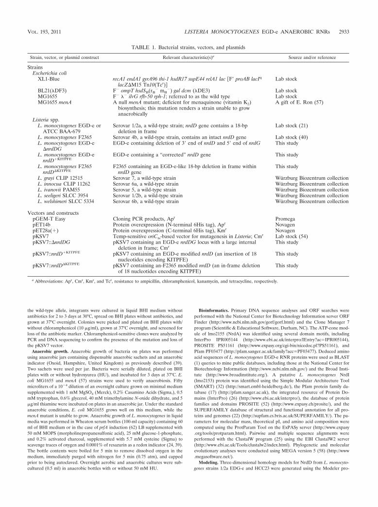
TABLE 1. Bacterial strains, vectors, and plasmids
Strain, vector, or plasmid construct Relevant characteristic(s)a Source and/or reference
StrainsEscherichia coli
XL1-Blue recA1 endA1 gyrA96 thi-1 hsdR17 supE44 relA1 lac �F� proAB lacIq
lacZ�M15 Tn10(Tcr) Lab stock
BL21(�DF3) F� ompT hsdSB(rB� mB
�) gal dcm (�DE3) Lab stockMG1655 F� �� ilvG rfb-50 rph-1; referred to as the wild type Lab stockMG1655 menA A null menA mutant; deficient for menaquinone (vitamin K2)
biosynthesis; this mutation renders a strain unable to growanaerobically
A gift of E. Ron (57)
Listeria spp.L. monocytogenes EGD-e or
ATCC BAA-679Serovar 1/2a, a wild-type strain; nrdD gene contains a 18-bp
deletion in frameLab stock (21)
L. monocytogenes F2365 Serovar 4b, a wild-type strain, contains an intact nrdD gene Lab stock (40)L. monocytogenes EGD-e
�nrdDGEGD-e containing deletion of 3� end of nrdD and 5� end of nrdG This study
L. monocytogenes EGD-enrdD�KITPFE
EGD-e containing a “corrected” nrdD gene This study
L. monocytogenes F2365nrdD�KITPFE
F2365 containing an EGD-e-like 18-bp deletion in frame withinnrdD gene
This study
L. grayi CLIP 12515 Serovar 7, a wild-type strain Wurzburg Biozentrum collectionL. innocua CLIP 11262 Serovar 6a, a wild-type strain Wurzburg Biozentrum collectionL. ivanovii PAM55 Serovar 5, a wild-type strain Wurzburg Biozentrum collectionL. seeligeri SLCC 3954 Serovar 1/2b, a wild-type strain Wurzburg Biozentrum collectionL. welshimeri SLCC 5334 Serovar 6b, a wild-type strain Wurzburg Biozentrum collection
Vectors and constructspGEM-T Easy Cloning PCR products, Apr PromegapET14b Protein overexpression (N-terminal 6His tag), Apr NovagenpET28a(�) Protein overexpression (C-terminal 6His tag), Kmr NovagenpKSV7 Temp-sensitive oriCts-based vector for mutagenesis in Listeria; Cmr Lab stock (54)pKSV7::�nrdDG pKSV7 containing an EGD-e nrdDG locus with a large internal
deletion in frame; CmrThis study
pKSV7::nrdD�KITPFE pKSV7 containing an EGD-e modified nrdD (an insertion of 18nucleotides encoding KITPFE)
This study
pKSV7::nrdD�KITPFE pKSV7 containing an-F2365 modified nrdD (an in-frame deletionof 18 nucleotides encoding KITPFE)
This study
a Abbreviations: Apr, Cmr, Kmr, and Tcr, resistance to ampicillin, chloramphenicol, kanamycin, and tetracycline, respectively.
VOL. 193, 2011 LISTERIA MONOCYTOGENES EGD-e ANAEROBIC RNRs 2933

gram (15) through the ModWeb server (https://modbase.compbio.ucsf.edu/scgi/modweb.cgi). The best template identified by the server was the crystal structureof NrdD from bacteriophage T4 (33), Protein Data Bank [PDB] ID 1H7B.Residues 126 to 685 of the listerial proteins could be modeled based on residues26 to 585 of T4 NrdD. The sequence identity was 50%, and the model was ofgood quality as judged by relevant criteria. When the structures were alignedusing Pymol (http://www.pymol.org), the root mean square deviation in C-alphapositions between the EGD-e model and the template was 0.16 Å for 425positions, while it was 0.14 Å for 423 positions in the HC233 model.
RESULTS AND DISCUSSION
Listeria contains genes encoding class I and class III RNRs.A search of the complete L. monocytogenes serovar 1/2a strainEGD-e genome (accession number NC_003210) (21) for genesencoding putative RNR proteins was carried out by the use ofstandard BLAST procedures (1) via the NCBI server (http://www.ncbi.nlm.nih.gov/BLAST). Two gene clusters that areannotated as containing ORFs predicted to belong to class Iaand class III of the RNR family of proteins were found (Fig. 1).One gene cluster consisting of lmo2155 and lmo2154 is pre-dicted to encode the class Ia RNR NrdA- and NrdB-like pro-teins (Fig. 1A). The listerial class I RNR large subunits containan ATP-cone domain in their N-terminal regions similar tothat present in the NrdA subunit of class Ia RNRs of entero-bacteria and shown to regulate overall enzyme activity by in-teraction with ATP/dATP (2). Immediately downstream oflmo2154 are found two genes, lmo2153 and lmo2152, which arepredicted to encode putative flavodoxin (NrdI-like) andthioredoxin (TrxL)-like proteins, respectively, suggesting thatall four genes are likely to form an operon. Reverse transcrip-tase (RT) PCR analysis of the nrdABI-trxL gene cluster re-vealed that nrdA, nrdB, nrdI-like, and trxL genes are cotrans-cribed and form an operon (see Fig. S1 in the supplementalmaterial). The lmo2151 gene immediately downstream of trxLis predicted to encode a MazG-like protein (63) which mightbe cotranscribed with nrdABI-trxL as well (see Fig. S1). Thededuced amino acid sequence of Lmo2153 resembles that of
the E. coli flavodoxin NrdI, which is involved in the mainte-nance of the diferric-tyrosyl radical cofactor (4, 12, 13). Thi-oredoxin-like Lmo2152 was denoted as TrxL to distinguish itfrom the TrxA ortholog encoded by lmo1233. Class I RNRenzyme activity was characterized employing NrdA, NrdB,NrdI, and TrxL Ni affinity purified His6-tagged recombinantproteins (see Fig. S2A to F in the supplemental material).RNR activity was assayed by monitoring the conversion of[3H]CDP to [3H]dCDP (14). The results establish that the L.monocytogenes aerobic RNR conforms to the typical patternobserved for the class Ia RNR subdivision (28). TrxL, thethioredoxin-like protein encoded by the class Ia RNR operon,can serve as an electron donor to the NrdAB reaction. In thepresence of 1 to 2 mM dithiothreitol (DTT), TrxL significantlystimulated RNR activity (see Fig. S3 in the supplemental ma-terial), suggesting that in vivo it can function with thioredoxinreductase and NADPH as an electron donor for the aerobicclass I RNR, in a manner similar to that of E. coli and S. aureusNrdH (39, 45).
A similar gene organization is found in the genomes of otherL. monocytogenes strains and Listeria species, including non-pathogenic L. innocua (21) and L. welshimeri (23). Systematicanalysis of other bacterial genomes revealed that several spe-cies of the family Xanthomonadaceae (gammaproteobacteria),plant-pathogenic Xanthomonas and Xylella (44, 53), andStenotrophomonas maltophilia, an opportunistic human patho-gen causing nosocomial infections (50), contain a class I RNRgene cluster similar to that in Listeria species (Fig. 1A). Phy-logenetic analysis demonstrates that bacteria possessing thenrdABI-trxL operon comprise a well-defined clade that is wellseparated from other class Ia and Ib RNRs (see Fig. S4 in thesupplemental material). Besides an unusual nrdABI-trxL locus,genomes of all listerial species contain a second gene clusterthat encodes the “classic” anaerobic class III RNR proteinsNrdD (reductase) and NrdG (activase) (Fig. 1B).
FIG. 1. Organization of the class I and III RNR genes of L. monocytogenes. (A) Organization of the L. monocytogenes EGD-e class InrdABI-trxL RNR genes and comparison with some other bacterial class Ia and Ib RNR gene organizations. (B) Organization of the L.monocytogenes EGD-e class III nrdDG RNR genes.
2934 OFER ET AL. J. BACTERIOL.

The L. monocytogenes EGD-e class III RNR system does notsupport anaerobic growth due to a deletion in NrdD. L. mono-cytogenes is a facultative anaerobe able to grow in aerobic andanaerobic conditions. We therefore expected that L. monocy-togenes EGD-e would grow under anaerobic conditions andthat growth would be unaffected by hydroxyurea (HU), whichis a specific inhibitor of the class I RNR scavenging the freeoxygen-dependent tyrosyl radical (46). Figure 2 shows that L.monocytogenes EGD-e (WT) does indeed grow on BHI in solidmedium under anaerobic conditions. An E. coli menA mutantstrain that is unable to grow anaerobically served as a controlfor anaerobiosis (57). To verify that growth is dependent on afunctional class III RNR, we created an in-frame deletion inthe nrdDG genes (Material and Methods) and were surprisedto find that the mutant strain grew almost as well as the WT(Fig. 2). We therefore presumed that growth under anaerobicconditions was due to the class Ia RNR. To test this hypothesis,we supplemented BHI plates with 50 mM HU and observedthat growth of EGD-e and the �nrdD mutant was totally abol-ished, indicating that under the anaerobic conditions employedEGD-e growth was due to the activity of the class Ia RNR. Incontrast, L. monocytogenes strain F2365 (serovar 4b) is able togrow under the same anaerobic conditions in the presence ofHU (not shown). Moreover, the listerial species L. ivanovii, L.welshimeri, L. innocua, L. seeligeri, and L. grayi are all also ableto grow anaerobically in the presence of HU (Fig, 2). Theseobservations imply that the L. monocytogenes EGD-e class IIIRNR is nonfunctional.
Comparison of the predicted L. monocytogenes EGD-eNrdD amino acid sequence with those of some 25 L. monocy-togenes strains, including F2365 (see Table S2 in the supple-mental material), revealed that EGD-e NrdD alone contains adeletion of six amino acids—KITPFE—in the C-terminal partof the polypeptide (Fig. 3). Furthermore, all other listerialspecies, including L. grayi, L. innocua, L. ivanovii, L. seeligeri,and L. welshimeri, contain ORFs coding for an intact NrdDprotein. Based on the PROSITE profile of the glycine radicaldomain GLY_RADICAL_2 (PS51149) and on the reports of
Luttringer et al. (37) on the Zn center in the E. coli anaerobicribonucleotide reductase, the position of the deletion lies closeto the glycyl radical-containing domain (see below), indicatingthat it is likely to be critical for the formation of the stableradical necessary for enzyme activity.
To test experimentally the hypothesis that the KITPFE de-letion in L. monocytogenes EGD-e is responsible for creating anonfunctional class III RNR, we introduced an 18-nucleotideDNA fragment into the EGD-e nrdD gene to generate theprecise coding sequence present in L. monocytogenes F2365and in other listerial nrdD genes. In a reciprocal experiment,we deleted the corresponding 18-nucleotide DNA fragmentcoding for KITPFE in strain F2365. The constructions wereperformed using a temperature-sensitive replication vector,pKSV7, as described in Materials and Methods. The pairs ofisogenic strains EGD-e nrdD�KITPFE (WT) and EGD-enrdD�KITPFE and strains F2365 nrdD�KITPFE (WT) and F2365nrdD�KITPFE were tested for growth in BHI medium underaerobic and anaerobic conditions.
In BHI liquid medium there was no discernible difference inthe growth of the 4 strains under aerobic conditions as judgedby the rates and extents of growth (data not shown). Figure 4shows growth in liquid BHI medium under anaerobic condi-tions. F2365 grew well and reached an absorbance (A600) of 1.6after 8 h, whereas EGD-e grew poorly and reached an A600 of0.3. The two complemented strains behaved in the oppositeway; EGD-e nrdD�KITPFE grew equally well as F2365, whileF2364 nrdD�KITPFE grew poorly and reached an A600 of 0.3.The four strains were also tested for growth on BHI plates inanaerobic jars with and without HU (Fig. 5). In the absence ofHU all four strains grew equally well on the plates (Fig. 5, top).When the plates were supplemented with 50 mM HU, bothstrains carrying the KITPFE deletion, EGD-e nrdD�KITPFE
(WT) and F2365 nrdD�KITPFE, failed to grow, whereas bothstrains possessing the KITPFE sequence, EGD-e nrdD�KITPFE
and F2365 nrdD�KITPFE, grew equally well and to the sameextent as they did in the absence of HU (Fig. 5, bottom). Theseobservations establish that the L. monocytogenes nrdD gene is
FIG. 2. Anaerobic growth of Listeria spp. in solid medium. Listeria species were grown on BHI plates with and without 50 mM HU. Strainstested were L. monocytogenes EGD-e WT (L. mo EGD-e), an isogenic mutant possessing a deletion in nrdDG (L. mo EGD-e �DG), L. innocua(L. in), L. seeligeri (L. se), L. welshimeri (L. we), L. grayi (L. gr), and L. ivanovii (L. iv). L. monocytogenes EGD-e WT and the nrdDG mutant strainfailed to grow under anaerobic conditions in the presence of HU. Anaerobic conditions were verified using E. coli MG1655 (WT) and its menAmutant strain grown on the minimal M9 medium; in anaerobic conditions the menA mutant is unable to grow.
VOL. 193, 2011 LISTERIA MONOCYTOGENES EGD-e ANAEROBIC RNRs 2935

essential for anaerobic growth. They recall similar studies withE. coli, Lactococcus lactis, and S. aureus in which the growthdeficiency of an nrdD mutant was apparent only when strictanaerobic growth conditions were employed (20, 27, 39).
Our original observation of the growth of L. monocytogenesEGD-e (WT) on BHI plates in an anaerobic jar is most likelydue to the residual activity of the class I RNR under themicroaerophilic conditions existing rather than strict anaero-biosis. Thus, E. coli, L. lactis, and S. aureus nrdD mutants werefound to grow at very low oxygen concentrations but failed togrow when strict anaerobic conditions were defined (20, 27,39). Possibly, expression of the class I RNR genes may beupregulated at a low oxygen concentration. In Pseudomonasaeruginosa the class Ia RNR activity is significantly increasedunder microaerophilic conditions (61).
The L. monocytogenes NrdD deletion is located next to theglycyl radical domain. The L. monocytogenes EGD-e NrdDprotein contains 710 amino acids. It shares 53% and 63%sequence identity with the E. coli and L. lactis orthologs, re-
spectively, and about 50% identity with the bacteriophage T4NrdD. Phylogeny of the L. monocytogenes NrdD protein withother bacterial class III RNR NrdD proteins shows that itclusters together with NrdDs of Gram-positive bacteria and iswell separated from NrdDs of Gram-negative bacteria (Fig. 6).Figure 3, top, shows the EGD-e NrdD (Lmo0279) domainstructure according to the Pfam database (2, 17). The N-ter-minal portion of the protein, comprising amino acids 9 to 96,exhibits high sequence identity (E value of 2.4e�22) with theATP-cone regulatory domain found in the NrdA subunit ofclass Ia RNR proteins (2). The NrdD C-terminal portion con-tains the glycyl radical domain (55) consisting of residues 637to 686. The glycyl radical is located on G680, corresponding toG681 in E. coli NrdD and G580 of bacteriophage T4 NrdD.Four conserved cysteine residues in a CXXC(X)14CXXC motif(34, 37) in the glycyl radical domain upstream of the glycylradical site, involved in Zn binding, are conserved in the liste-rial NrdD protein.
We have attempted to locate the KITPFE deletion site by
FIG. 3. Multiple sequence alignment of NrdD proteins. (Top) The ATP-cone and glycyl radical-containing domains of L. monocytogenesEGD-e NrdD, which are according to PF03477 and PF01128, respectively. Amino acid numbering corresponds to that for L. monocytogenes EGD-eNrdD (accession number NP_463810). The six-amino acid deletion in EGD-e NrdD occurs between residues 588 and 589. Two pairs ofzinc-binding cysteines are located between residues 642 and 681. (Bottom) Partial amino acid sequence of an alignment of NrdD proteins fromListeria and selected bacteria. The position in Listeria species of the KITPFE region that is deleted in EGD-e is marked in light gray. The startregion of the glycyl radical domains in bacteriophage T4 and E. coli NrdD, according to Luttringer et al. (37), is shown in dark gray; that deducedfor the L. monocytogenes NrdD is shown as well. The glycyl radical domain in L. monocytogenes starts at position R587. The KITPFE amino aciddeletion is located at the beginning of the domain. Asterisks, colons, and dots indicate identical, highly similar, and weakly similar residues,respectively.
2936 OFER ET AL. J. BACTERIOL.

employing a three-dimensional (3D) homology model of theEGD-e NrdD protein based on the structure established forthe bacteriophage T4 NrdD (18) (Fig. 7), the only existing 3Dstructure for an anaerobic class III RNR protein. Surprisingly,as shown in Fig. 7A, it turns out that the deleted sequence isnot a part of the C-terminal glycyl radical domain—either themetal-binding part or the loop containing the glycyl radical—aspredicted by the protein domain databases. However, it isfound nearby at the beginning of the second-last helix in the(�/�)10 barrel domain characteristic of the RNR family, a veryhighly conserved part of the structure. The deletion occursbetween the two K residues in the sequence FHYDVRKKIDF. This deletion is likely to shorten significantly the loopbetween the end of this helix and the preceding �-strand.These are important interaction areas for the extreme C ter-minus of both pyruvate formate lyase and glycerol dehydratase(GD) (49), and we suppose that a similar interaction may occurin NrdD, although we have been unable to model it until now,as the last 20 residues of T4 NrdD are disordered in all thecrystal structures (33). This might be because the C terminus isordered only when in complex with the NrdG activase, or atsome other specific point in the activation cycle.
In the EGD-e NrdD model, the shortening of the loop andthe helix (the latter by about one turn) due to the deletion isevident (Fig. 7B). In contrast, when the NrdD protein of L.monocytogenes strain HCC23 possessing a complete NrdD (ac-cession number YP_002351305) was modeled, the structurewas essentially identical to that of bacteriophage T4, apartfrom a few variations in side-chain conformations (not shown).
FIG. 4. Anaerobic growth of L. monocytogenes EGD-e WT, EGD-enrdD�KITPFE, F2365 WT, and F2365 nrdD�KITPFE in liquid medium.Aerobic and anaerobic overnight cultures of all four strains were usedto inoculate Wheaton serum anaerobic bottles containing BHI, resaz-urin, and cysteine and the liquid cultures grown at 37°C (see Materialsand Methods). EGD-e WT and F2365 nrdD�KITPFE possess a mutantNrdD protein; EGD-e nrdD�KITPFE and F2365 nrdD�KITPFE contain afunctional NrdD protein (38). The data shown are representative oftwo experiments; error bars show the average variation.
FIG. 5. Anaerobic growth of L. monocytogenes EGD-e WT, EGD-e nrdD�KITPFE, F2365 WT, and F2365 nrdD�KITPFE on solid BHI mediumin the presence or absence of HU. Exponentially growing cultures were aliquoted on BHI plates with or without 50 mM HU. Plates were incubatedfor 48 h at 37°C in an anaerobic jar.
FIG. 6. Molecular phylogeny of bacterial NrdD proteins. Theneighbor-joining (NJ) method was applied to estimate relationshipsamong aligned sequences by using the MEGA 4 program. Pyrococcusfuriosus was used as the outgroup in the tree. Bootstrap analysis usingNJ was conducted with 500 replicates, and values above 50% are givenat nodes. Bacterial names and accession numbers of NrdD proteins areshown in Table S5 in the supplemental material.
VOL. 193, 2011 LISTERIA MONOCYTOGENES EGD-e ANAEROBIC RNRs 2937

The deletion is one of only a few deviations between the twostructures. There is also a 10-residue insertion relative to theT4 template structure in the nearby loop (residues 511 to 521in Listeria) preceding the long helix associated with strand H ofthe �/� barrel, which could also be involved in interactionswith the C terminus (Fig. 7B). However, since the insertion iscommon to both Listeria strains modeled, it cannot be respon-sible for differences in activity between them.
The homology model suggests that the remaining residues inEGD-e NrdD repack into a different compact conformation inthat area that shifts the loop significantly. Pro486, which de-fines the N terminus of the �-helix, is one of the residuesdeleted in EGD-e NrdD. If we assume that the C terminus ofNrdD interacts with the N terminus of this helix, then thepresence of a secondary-structure-breaking proline there isvery important, which explains its extreme conservation in allbacterial NrdD sequences. Our current hypothesis is that theKITPFE region is either essential for interactions with theNrdG activase or, indirectly, for the stability of the glycyl rad-ical loop. Figure 7C shows the C terminus of the visible part ofNrdD, which points in the general direction of this helix. Thereare 15 more residues in the sequence than we can see in thestructure, so it is possible that interactions are made with the Cterminus under certain conditions, perhaps when the activaseis bound. Indeed, in pyruvate formate lyase (3, 31), the car-boxylate group of the extreme C terminus interacts with theend of the equivalent helix (Fig. 7C). Significantly, a verysimilar interaction is found in GD (42). Moreover, a minimalpoint mutation (R782K) in the same three-dimensional region
of glycerol dehydratase results in a tight, unproductive complexwith the GD activase protein, critically reducing enzyme activ-ity and implying that conformational changes in this area arevery important for activation (42).
Concluding remarks. In order to establish infection, L.monocytogenes, in common with all food-borne pathogens,must be capable of sensing and responding to the hostile en-vironment of the human gastrointestinal tract which may in-clude nutrient deprivation, acidity, iron limitation, bile, andoxygen limitation. It has been reported that L. monocytogenescan grow as a biofilm which contains oxygen gradients andanaerobic pockets (11). Here we show that L. monocytogenesserovar 1/2a EGD-e, but not other L. monocytogenes strains orlisterial species, contains a nonfunctional anaerobic class IIIRNR system due to a deletion in the C-terminal portion of theprotein, in the essential glycyl radical domain. The EGD-estrain has been employed extensively as a model laboratorylisterial pathogen to elucidate the mechanisms of intracellularmultiplication and virulence. Because L. monocytogenesEGD-e lacks a functional anaerobic RNR, it is relevant to askwhether the findings presented here affect interpretation ofstudies of pathogenesis, particularly those with conditions oflow oxygen tension. Plausibly, EGD-e can continue to serve asa suitable reference strain as long as anaerobic/microaerophilicconditions are not used, which is the case in many in vitro orcell culture experiments. However, the EGD-e strain wouldnot be suitable for studies such as gastrointestinal infections orenvironmental conditions of very low oxygen tension. Animal
FIG. 7. Three-dimensional homology modeling of L. monocytogenes NrdD. (A) Mapping of the six-amino acid deletion onto the structure ofNrdD from bacteriophage T4. This NrdD is shown in green, with the C-terminal domain in a darker shade. The second NrdD monomer of thedimeric enzyme is shown in blue. The deletion sequence is marked in red. The Gly radical site (G580, mutated to Ala in this structure) and theradical cysteine can also be seen, in stick representation. A plausible position for the C-terminal residue is sketched. (B) Comparison of the crystalstructure of T4 NrdD (green) with the homology models for L. monocytogenes NrdD from strains HCC23 (blue) and EGD-e (dark pink). Side chainpacking for the shortened loop in the EGD-e model, as suggested by Modeler, is shown. However, this is only one of several plausible packingsthat can be generated. The nearby loop t511-521, which is 10 residues longer in L. monocytogenes than in T4, is labeled. (C) Importance ofinteractions of the extreme C terminus of glycyl radical proteins with the deletion region. The structure of pyruvate formate lyase is shown in darkblue, with its C terminus in gray. The structure of glycerol dehydratase is shown in light blue. The loop and helix homologous to the deletion regionin NrdD are highlighted in red. The extreme C-terminal amino acid of PFL (M759) and a residue important for conformational changes of theC terminus in activation of GD (R782) are shown as sticks and labeled.
2938 OFER ET AL. J. BACTERIOL.

model studies are under way to clarify whether the anaerobicRNR system is a virulence determinant.
ACKNOWLEDGMENTS
This work was supported by the German Federal Ministryfor Education and Research in the framework of the ERA-NET PathoGenoMics, project SPATELIS (grant no. BMBF/PTJ0313939), and partially by the EU- funded Network of ExcellenceEuroPathoGenomics (NoE EPG). D.T.L. was supported by a grant(2006-4387) from the Swedish Research Council (VR).
We thank Michaela Yanku and Batia Gorovitz-Harris for technicalassistance.
REFERENCES
1. Altschul, S. F., and D. J. Lipman. 1990. Protein database searches formultiple alignments. Proc. Natl. Acad. Sci. U. S. A. 87:5509–5513.
2. Aravind, L., Y. I. Wolf, and E. V. Koonin. 2000. The ATP-cone: an evolu-tionarily mobile, ATP-binding regulatory domain. J. Mol. Microbiol. Bio-technol. 2:191–194.
3. Becker, A., and W. Kabsch. 2002. X-ray structure of pyruvate formate-lyasein complex with pyruvate and CoA. How the enzyme uses the Cys-418 thiylradical for pyruvate cleavage. J. Biol. Chem. 277:40036–40042.
4. Boal, A. K., J. A. Cotruvo, Jr, J. Stubbe, and A. C. Rosenzweig. 2010.Structural basis for activation of class Ib ribonucleotide reductase. Science329:1526–1530.
5. Bo Andersen, J., B. B. Roldgaard, B. B. Christensen, and T. R. Licht. 2007.Oxygen restriction increases the infective potential of Listeria monocytogenesin vitro in Caco-2 cells and in vivo in guinea pigs. BMC Microbiol. 7:55.
6. Buchrieser, C., C. Rusniok, F. Kunst, P. Cossart, and P. Glaser. 2003.Comparison of the genome sequences of Listeria monocytogenes and Listeriainnocua: clues for evolution and pathogenicity. FEMS Immunol. Med. Mi-crobiol. 35:207–213.
7. Burall, L. S., et al. 2004. Proteus mirabilis genes that contribute to patho-genesis of urinary tract infection: identification of 25 signature-tagged mu-tants attenuated at least 100-fold. Infect. Immun. 72:2922–2938.
8. Burkholder, K. M., et al. 2009. Expression of LAP, a SecA2-dependentsecretory protein, is induced under anaerobic environment. Microbes Infect.11:859–867.
9. Camejo, A., et al. 2009. In vivo transcriptional profiling of Listeria monocy-togenes and mutagenesis identify new virulence factors involved in infection.PLoS Pathog. 5:e1000449.
10. Cossart, P., and P. J. Sansonetti. 2004. Bacterial invasion: the paradigms ofenteroinvasive pathogens. Science 304:242–248.
11. Costerton, J. W., Z. Lewandowski, D. E. Caldwell, D. R. Korber, and H. M.Lappin-Scott. 1995. Microbial biofilms. Annu. Rev. Microbiol. 49:711–745.
12. Cotruvo, J. A., Jr., and J. Stubbe. 2010. An active dimanganese(III)-tyrosylradical cofactor in Escherichia coli class Ib ribonucleotide reductase. Bio-chemistry 49:1297–1309.
13. Cotruvo, J. A., Jr., and J. Stubbe. 2008. NrdI, a flavodoxin involved inmaintenance of the diferric-tyrosyl radical cofactor in Escherichia coli classIb ribonucleotide reductase. Proc. Natl. Acad. Sci. U. S. A. 105:14383–14388.
14. Engstrom, Y., S. Eriksson, L. Thelander, and M. Akerman. 1979. Ribonu-cleotide reductase from calf thymus. Purification and properties. Biochem-istry 18:2941–2948.
15. Eswar, N., et al. 2006. Comparative protein structure modeling using Mod-eller. Curr. Protoc. Bioinformatics Chapter 5:Unit 5.6.
16. Farber, J. M., and P. I. Peterkin. 1991. Listeria monocytogenes, a food-bornepathogen. Microbiol. Rev. 55:476–511.
17. Finn, R. D., et al. 2008. The Pfam protein families database. Nucleic AcidsRes. 36:D281–D288.
18. Fontecave, M., E. Mulliez, and D. T. Logan. 2002. Deoxyribonucleotidesynthesis in anaerobic microorganisms: the class III ribonucleotide reduc-tase. Prog. Nucleic Acid Res. Mol. Biol. 72:95–127.
19. Freitag, N. E., G. C. Port, and M. D. Miner. 2009. Listeria monocytogenes—from saprophyte to intracellular pathogen. Nat. Rev. Microbiol. 7:623–628.
20. Garriga, X., et al. 1996. nrdD and nrdG genes are essential for strict anaer-obic growth of Escherichia coli. Biochem. Biophys. Res. Commun. 229:189–192.
21. Glaser, P., et al. 2001. Comparative genomics of Listeria species. Science294:849–852.
22. Gough, J., and C. Chothia. 2002. SUPERFAMILY: HMMs representing allproteins of known structure. SCOP sequence searches, alignments and ge-nome assignments. Nucleic Acids Res. 30:268–272.
23. Hain, T., C. Steinweg, and T. Chakraborty. 2006. Comparative and func-tional genomics of Listeria spp. J. Biotechnol. 126:37–51.
24. Hartig, E., A. Hartmann, M. Schatzle, A. M. Albertini, and D. Jahn. 2006.The Bacillus subtilis nrdEF genes, encoding a class Ib ribonucleotide reduc-tase, are essential for aerobic and anaerobic growth. Appl. Environ. Micro-biol. 72:5260–5265.
25. Higgins, D. G., J. D. Thompson, and T. J. Gibson. 1996. Using CLUSTALfor multiple sequence alignments. Methods Enzymol. 266:383–402.
26. Hunter, S., et al. 2009. InterPro: the integrative protein signature database.Nucleic Acids Res. 37:D211–D215.
27. Jordan, A., et al. 1996. The ribonucleotide reductase system of Lactococcuslactis. Characterization of an NrdEF enzyme and a new electron transportprotein. J. Biol. Chem. 271:8779–8785.
28. Jordan, A., and P. Reichard. 1998. Ribonucleotide reductases. Annu. Rev.Biochem. 67:71–98.
29. Joseph, B., et al. 2006. Identification of Listeria monocytogenes genes con-tributing to intracellular replication by expression profiling and mutantscreening. J. Bacteriol. 188:556–568.
30. Kaufmann, S. H., M. M. Simon, and H. Hahn. 1979. Specific Lyt 123 cells areinvolved in protection against Listeria monocytogenes and in delayed-typehypersensitivity to listerial antigens. J. Exp. Med. 150:1033–1038.
31. Lehtio, L., V. M. Leppanen, J. W. Kozarich, and A. Goldman. 2002. Structureof Escherichia coli pyruvate formate-lyase with pyruvate. Acta Crystallogr. DBiol. Crystallogr. 58:2209–2212.
32. Letunic, I., et al. 2004. SMART 4.0: towards genomic data integration.Nucleic Acids Res. 32:D142–144.
33. Logan, D. T., J. Andersson, B. M. Sjoberg, and P. Nordlund. 1999. A glycylradical site in the crystal structure of a class III ribonucleotide reductase.Science 283:1499–1504.
34. Logan, D. T., et al. 2003. A metal-binding site in the catalytic subunit ofanaerobic ribonucleotide reductase. Proc. Natl. Acad. Sci. U. S. A. 100:3826–3831.
35. Lundin, D., E. Torrents, A. M. Poole, and B. M. Sjoberg. 2009. RNRdb, acurated database of the universal enzyme family ribonucleotide reductase,reveals a high level of misannotation in sequences deposited to Genbank.BMC Genomics 10:589.
36. Lungu, B., S. C. Ricke, and M. G. Johnson. 2009. Growth, survival, prolif-eration and pathogenesis of Listeria monocytogenes under low oxygen oranaerobic conditions: a review. Anaerobe 15:7–17.
37. Luttringer, F., E. Mulliez, B. Dublet, D. Lemaire, and M. Fontecave. 2009.The Zn center of the anaerobic ribonucleotide reductase from E. coli. J. Biol.Inorg. Chem. 14:923–933.
38. Marteyn, B., et al. 2010. Modulation of Shigella virulence in response toavailable oxygen in vivo. Nature 465:355–358.
39. Masalha, M., I. Borovok, R. Schreiber, Y. Aharonowitz, and G. Cohen. 2001.Analysis of transcription of the Staphylococcus aureus aerobic class Ib andanaerobic class III ribonucleotide reductase genes in response to oxygen. J.Bacteriol. 183:7260–7272.
40. Nelson, K. E., et al. 2004. Whole genome comparisons of serotype 4b and1/2a strains of the food-borne pathogen Listeria monocytogenes reveal newinsights into the core genome components of this species. Nucleic Acids Res.32:2386–2395.
41. Nordlund, P., and P. Reichard. 2006. Ribonucleotide reductases. Annu. Rev.Biochem. 75:681–706.
42. O’Brien, J. R., et al. 2004. Insight into the mechanism of the B12-indepen-dent glycerol dehydratase from Clostridium butyricum: preliminary biochem-ical and structural characterization. Biochemistry 43:4635–4645.
43. Paik, S., et al. 2005. Identification of virulence determinants for endocarditisin Streptococcus sanguinis by signature-tagged mutagenesis. Infect. Immun.73:6064–6074.
44. Qian, W., et al. 2005. Comparative and functional genomic analyses of thepathogenicity of phytopathogen Xanthomonas campestris pv. campestris.Genome Res. 15:757–767.
45. Rabinovitch, I., et al. 2010. Staphylococcus aureus NrdH redoxin is a reduc-tant of the class Ib ribonucleotide reductase. J. Bacteriol. 192:4963–4972.
46. Reichard, P., and A. Ehrenberg. 1983. Ribonucleotide reductase—a radicalenzyme. Science 221:514–519.
47. Roca, I., E. Ballana, A. Panosa, E. Torrents, and I. Gibert. 2008. Fuma-rate and nitrate reduction (FNR) dependent activation of the Escherichiacoli anaerobic ribonucleotide reductase nrdDG promoter. Int. Microbiol.11:49–56.
48. Sambrook, J., E. F. Fritsch, and T. Maniatis. 1989. Molecular cloning: alaboratory manual, 2nd ed. Cold Spring Harbor Laboratory Press, ColdSpring Harbor, NY.
49. Selmer, T., A. J. Pierik, and J. Heider. 2005. New glycyl radical enzymescatalysing key metabolic steps in anaerobic bacteria. Biol. Chem. 386:981–988.
50. Senol, E. 2004. Stenotrophomonas maltophilia: the significance and role as anosocomial pathogen. J. Hosp. Infect. 57:1–7.
51. Seveau, S., J. Pizarro-Cerda, and P. Cossart. 2007. Molecular mechanismsexploited by Listeria monocytogenes during host cell invasion. Microbes In-fect. 9:1167–1175.
52. Sigrist, C. J. et al. 2010. PROSITE, a protein domain database for functionalcharacterization and annotation. Nucleic Acids Res. 38:D161–D166.
53. Simpson, A. J., et al. 2000. The genome sequence of the plant pathogenXylella fastidiosa. The Xylella fastidiosa Consortium of the Organization forNucleotide Sequencing and Analysis. Nature 406:151–159.
54. Smith, K., and P. Youngman. 1992. Use of a new integrational vector to
VOL. 193, 2011 LISTERIA MONOCYTOGENES EGD-e ANAEROBIC RNRs 2939

investigate compartment-specific expression of the Bacillus subtilis spoIIMgene. Biochimie 74:705–711.
55. Sun, X., et al. 1993. A possible glycine radical in anaerobic ribonucleotidereductase from Escherichia coli: nucleotide sequence of the cloned nrdDgene. Proc. Natl. Acad. Sci. U. S. A. 90:577–581.
56. Sun, X., et al. 1996. The free radical of the anaerobic ribonucleotide reduc-tase from Escherichia coli is at glycine 681. J. Biol. Chem. 271:6827–6831.
57. Suvarna, K., D. Stevenson, R. Meganathan, and M. E. Hudspeth. 1998.Menaquinone (vitamin K2) biosynthesis: localization and characterization ofthe menA gene from Escherichia coli. J. Bacteriol. 180:2782–2787.
58. Tamura, K., J. Dudley, M. Nei, and S. Kumar. 2007. MEGA4: MolecularEvolutionary Genetics Analysis (MEGA) software version 4.0. Mol. Biol.Evol. 24:1596–1599.
59. Toledo-Arana, A., et al. 2009. The Listeria transcriptional landscape fromsaprophytism to virulence. Nature 459:950–956.
60. Vazquez-Boland, J. A., et al. 2001. Listeria pathogenesis and molecular vir-ulence determinants. Clin. Microbiol. Rev. 14:584–640.
61. Wu, M., T. Guina, M. Brittnacher, H. Nguyen, J. Eng., and S. I. Miller. 2005.The Pseudomonas aeruginosa proteome during anaerobic growth. J. Bacte-riol. 187:8185–8190.
62. Zemansky, J., et al. 2009. Development of a mariner-based transposon andidentification of Listeria monocytogenes determinants, including the peptidyl-prolyl isomerase PrsA2, that contribute to its hemolytic phenotype. J. Bac-teriol. 191:3950–3964.
63. Zhang, J., and M. Inouye. 2002. MazG, a nucleoside triphosphate pyrophos-phohydrolase, interacts with Era, an essential GTPase in Escherichia coli. J.Bacteriol. 184:5323–5329.
2940 OFER ET AL. J. BACTERIOL.