
Diversity in Overall Activity Regulation of Ribonucleotide Reductase
25
1 Diversity in Overall Activity Regulation of Ribonucleotide Reductase * Venkateswara Rao Jonna 1 , Mikael Crona 2† , Reza Rofougaran 1† , Daniel Lundin 2 , Samuel Johansson 1 , Kristoffer Brännström 1 , Britt-Marie Sjöberg 2 and Anders Hofer 1 From the 1 Dept. of Medical Biochemistry and Biophysics, Umeå University, SE-901 87 Umeå, Sweden, and the 2 Dept. of Biochemistry and Biophysics, Stockholm University, SE-106 91 Stockholm, Sweden. *Running title: Pseudomonas aeruginosa ribonucleotide reductase To whom correspondence should be addressed: Anders Hofer, Dept. of Medical Biochemistry and Biophysics, Umeå University, SE-901 87 Umeå, Sweden. Phone: +46-70-2974096. Email: [email protected] Keywords: Pseudomonas aeruginosa (P. aeruginosa), ribonucleotide reductase, overall activity regulation, oligomerization, dATP inhibition, protein complex, enzyme inactivation, allosteric regulation ¯¯¯¯¯¯¯¯¯¯¯¯¯¯¯¯¯¯¯¯¯¯¯¯¯¯¯¯¯¯¯¯¯¯¯¯¯¯¯¯¯¯¯¯¯¯¯¯¯¯¯¯¯¯¯¯¯¯¯¯¯¯¯¯¯¯¯¯¯¯¯¯¯¯¯¯¯¯¯¯¯¯¯¯¯ Background. Ribonucleotide reductase (RNR) § makes DNA building blocks. Results. Binding of three dATP molecules to the Pseudomonas aeruginosa class I RNR α subunit inactivates the enzyme by inducing an inert α 4 complex. Conclusion. The number of bound dATP molecules and the tetrameric complex are unique among RNRs. Significance. The novel inhibition mechanism of P. aeruginosa RNR is a potential drug target. ABSTRACT Ribonucleotide reductase (RNR) catalyzes the reduction of ribonucleotides to the corresponding deoxyribonucleotides, which are used as building blocks for DNA replication and repair. This process is tightly regulated via two allosteric sites, the specificity site (s-site) and the overall activity site (a-site). The a-site resides in an N-terminal ATP cone domain that binds dATP or ATP and functions as an on/off switch, whereas the composite s-site binds ATP, dATP, dTTP, or dGTP and determines which substrate to reduce. There are three classes of RNRs, and class I RNRs consist of different combinations of α and β subunits. In eukaryotic and Escherichia coli canonical class I RNRs, dATP inhibits enzyme activity through the formation of inactive α 6 and α 4 β 4 complexes, respectively. Here we show that the Pseudomonas aeruginosa class I RNR has a duplicated ATP cone domain and represents a third mechanism of overall activity regulation. Each α polypeptide binds three dATP molecules, and the N-terminal ATP cone is critical for binding two of the dATPs because a truncated protein lacking this cone could only bind dATP to its s-site. ATP activates the enzyme solely by preventing dATP from binding. The dATP-induced inactive form is an α 4 complex, which can interact with β 2 to form a non-productive α 4 β 2 complex. Other allosteric effectors induce a mixture of α 2 and α 4 forms, with the former being able to interact with β 2 to form active α 2 β 2 complexes. The unique features of the P. aeruginosa RNR are interesting both from evolutionary and drug discovery perspectives. ¯¯¯¯¯¯¯¯¯¯¯¯¯¯¯¯¯¯¯¯¯¯¯¯¯¯¯¯¯¯¯¯¯¯¯¯¯¯ http://www.jbc.org/cgi/doi/10.1074/jbc.M115.649624 The latest version is at JBC Papers in Press. Published on May 13, 2015 as Manuscript M115.649624 Copyright 2015 by The American Society for Biochemistry and Molecular Biology, Inc. by guest on April 10, 2016 http://www.jbc.org/ Downloaded from
-
Upload
independent -
Category
Documents
-
view
7 -
download
0
Transcript of Diversity in Overall Activity Regulation of Ribonucleotide Reductase
1
Diversity in Overall Activity Regulation of Ribonucleotide Reductase * Venkateswara Rao Jonna1, Mikael Crona2†, Reza Rofougaran1†, Daniel Lundin2, Samuel Johansson1, Kristoffer Brännström1, Britt-Marie Sjöberg2 and Anders Hofer1 From the 1Dept. of Medical Biochemistry and Biophysics, Umeå University, SE-901 87 Umeå, Sweden, and the 2Dept. of Biochemistry and Biophysics, Stockholm University, SE-106 91 Stockholm, Sweden. *Running title: Pseudomonas aeruginosa ribonucleotide reductase To whom correspondence should be addressed: Anders Hofer, Dept. of Medical Biochemistry and Biophysics, Umeå University, SE-901 87 Umeå, Sweden. Phone: +46-70-2974096. Email: [email protected] Keywords: Pseudomonas aeruginosa (P. aeruginosa), ribonucleotide reductase, overall activity regulation, oligomerization, dATP inhibition, protein complex, enzyme inactivation, allosteric regulation ¯¯¯¯¯¯¯¯¯¯¯¯¯¯¯¯¯¯¯¯¯¯¯¯¯¯¯¯¯¯¯¯¯¯¯¯¯¯¯¯¯¯¯¯¯¯¯¯¯¯¯¯¯¯¯¯¯¯¯¯¯¯¯¯¯¯¯¯¯¯¯¯¯¯¯¯¯¯¯¯¯¯¯¯¯ Background. Ribonucleotide reductase (RNR)§ makes DNA building blocks. Results. Binding of three dATP molecules to the Pseudomonas aeruginosa class I RNR α subunit inactivates the enzyme by inducing an inert α4 complex. Conclusion. The number of bound dATP molecules and the tetrameric complex are unique among RNRs. Significance. The novel inhibition mechanism of P. aeruginosa RNR is a potential drug target.
ABSTRACT
Ribonucleotide reductase (RNR) catalyzes the reduction of ribonucleotides to the corresponding deoxyribonucleotides, which are used as building blocks for DNA replication and repair. This process is tightly regulated via two allosteric sites, the specificity site (s-site) and the overall activity site (a-site). The a-site resides in an N-terminal ATP cone domain that binds dATP or ATP and functions as an on/off switch, whereas the composite s-site binds ATP, dATP, dTTP, or dGTP and determines which
substrate to reduce. There are three classes of RNRs, and class I RNRs consist of different combinations of α and β subunits. In eukaryotic and Escherichia coli canonical class I RNRs, dATP inhibits enzyme activity through the formation of inactive α6 and α4β4 complexes, respectively. Here we show that the Pseudomonas aeruginosa class I RNR has a duplicated ATP cone domain and represents a third mechanism of overall activity regulation. Each α polypeptide binds three dATP molecules, and the N-terminal ATP cone is critical for binding two of the dATPs because a truncated protein lacking this cone could only bind dATP to its s-site. ATP activates the enzyme solely by preventing dATP from binding. The dATP-induced inactive form is an α4 complex, which can interact with β2 to form a non-productive α4β2 complex. Other allosteric effectors induce a mixture of α2 and α4 forms, with the former being able to interact with β2 to form active α2β2 complexes. The unique features of the P. aeruginosa RNR are interesting both from evolutionary and drug discovery perspectives.
¯¯¯¯¯¯¯¯¯¯¯¯¯¯¯¯¯¯¯¯¯¯¯¯¯¯¯¯¯¯¯¯¯¯¯¯¯¯
http://www.jbc.org/cgi/doi/10.1074/jbc.M115.649624The latest version is at JBC Papers in Press. Published on May 13, 2015 as Manuscript M115.649624
Copyright 2015 by The American Society for Biochemistry and Molecular Biology, Inc.
by guest on April 10, 2016
http://ww
w.jbc.org/
Dow
nloaded from

2
Ribonucleotide reductase (RNR) catalyzes the rate-limiting step in the only known de novo pathway for synthesis of DNA building blocks by reducing ribonucleoside di- or triphoshates (NDPs or NTPs) to the corresponding deoxyribonucleoside di- or triphosphates (dNDPs or dNTPs). High requirements for dNTPs in cancer cells and proliferating pathogens together with the lack of alternative pathways make RNR an interesting therapeutic target. RNRs are divided into three classes sharing a common fold but differing in how they generate the free radical that is essential for catalysis (1-3). Class I RNR enzymes – the dominating class in eukaryotes, common in bacteria, and also found in some archaea and double-stranded DNA viruses – consist of a catalytic protein, R1 (or α2) and a smaller free radical-generating protein, R2 (or β2). The minimal active form is an α2β2 complex, but larger oligomeric complexes can also be formed (1). In contrast, class II RNR enzymes have only one subunit, and the free radical is generated from adenosylcobalamin. Class III RNR enzymes are anaerobic and use a dedicated activase to generate a stable glycine radical in the catalytic subunit. The RNR genes are called nrdA and nrdB for the α and β subunits in class I, nrdE and nrdF for the corresponding subunits in the non-canonical subclass Ib, nrdJ for class II, nrdD for class III, and nrdG for the associated activase.
Sophisticated allosteric regulation of RNR controls both the absolute concentrations of dNTPs in cells and the relative ratios of the four different dNTPs (1). Both of these controls are important for replication fidelity and DNA repair, and unbalanced dNTP pools are mutagenic (4). The two allosteric controls are implemented separately in the enzyme (1). Ratios between different dNTPs are controlled by substrate specificity regulation that occurs by binding of effector nucleotides to the specificity site (s-site) in the catalytic subunit. In class I enzymes that use NDPs as substrates, the binding of dATP/ATP induces CDP/UDP reduction, whereas dTTP and
dGTP binding induce GDP and ADP reduction, respectively. The s-site and how it affects the active site to control substrate specificity is conserved in all three RNR classes. In contrast, the control of the absolute dNTP concentration by overall activity regulation of RNR is more unevenly distributed. This regulation has been found only in RNRs with an N-terminal ATP cone (5), which makes up the overall activity site (a-site).
Overall activity is regulated by competitive binding of ATP or dATP to the a-site (1). When levels of dNTPs are low, the ATP cone will preferentially bind ATP and the enzyme is active. Conversely, when dNTP levels are sufficiently high, dATP will bind the a-site and inhibit enzyme activity. Mechanistically, the process has been studied in the canonical class I enzymes from Escherichia coli, humans, mice, Saccharomyces cerevisiae, and the slime mold Dictyostelium discoideum (6–13). There are similarities, but also clear differences, in action between the bacterial E. coli RNR and the eukaryotic enzymes. In both mechanisms, high dNTP levels mediate oligomerization into tight complexes of larger size than the common α2β2 complex, and the β2 and α2n subunits can no longer interact in a productive way. In the eukaryotic class I enzymes, the overall activity regulation relies on two different types of α6 complexes depending on whether dATP or ATP binds to the a-site (8,9). The dATP-inhibited complex binds the β2 subunit in the center of the α6 ring in such a way that the electron transport chain between the β and α subunits is disrupted. Less is known about the ATP-induced α6 complex, which has not been crystallized, but mutagenesis studies have indicated that it is structurally different from the inactive complex (9) and gel filtration analysis shows that it might bind up to three β2 subunits (12).
The corresponding class I enzyme from E. coli uses another mechanism of overall activity regulation. In this case, the enzyme activity is turned off via dimerization of the active α2β2 form
by guest on April 10, 2016
http://ww
w.jbc.org/
Dow
nloaded from

3
into an inactive ring-shaped α4β4 complex that cannot support electron transport between the subunits (6,7,13). Thus in E. coli both subunits are needed for oligomerization, whereas the eukaryotic α subunit can form hexamers on its own. Furthermore, the E. coli enzyme can be inhibited by dATP as well as by combinations of ATP and high concentrations of dTTP or dGTP (7). The overall activity regulation of the E. coli enzyme thus appears to be governed by crosstalk between the a-site and the s-site, while the overall activity regulation of the eukaryotic enzyme seems to be entirely driven by the a-site.
The different overall activity mechanisms found in eukaryotes and E. coli raises the question of whether the picture we have today of one eukaryotic and one bacterial mechanism is complete or if there are additional mechanisms to be discovered. More mechanisms can possibly be anticipated based on sequence comparisons of RNRs, where phylogenetic trees from full-length RNR sequences support an extensive subclassification of RNRs (http://rnrdb.pfitmap.org). The N-terminal ATP cone is not found in all RNRs, suggesting that it has been gained and lost occasionally during evolution. Independent gains of an ATP cone could possibly lead to mechanistic differences in activity regulation. A particularly interesting example of an ATP cone gain is found in the class I enzyme from Pseudomonas aeruginosa, which has two consecutive ATP cones. Earlier mutagenesis studies have indicated that only the first ATP cone has a role in overall activity regulation because its removal (the NrdA-Δ147 mutant) abolishes dATP inhibition and the ability to oligomerize in the presence of ATP (14). The second ATP cone seems to be needed for core functions because the removal of both cones leads to complete loss of enzyme activity (14). An additional ATP cone in between the N-terminal ATP cone and the core of the protein might have implications for which inhibited complexes can be formed. In the previous analysis of the P.
aeruginosa class I RNR, the size determination of the ATP-induced oligomers was ambiguous (14), which is a common problem with gel filtration analysis of protein complexes in rapid equilibrium. In this study we have used gas-phase electrophoretic mobility macromolecule analysis (GEMMA), which is an alternative method better suited for equilibrating complexes that has successfully been used for studies of other RNRs (7,8,10).
We show that RNRs with multiple ATP cones are much more common among RNRs than previously realized and that the P. aeruginosa class I RNR indeed differs in several aspects from both the eukaryotic and the E. coli mechanistic models of overall activity regulation. The P. aeruginosa enzyme is the first known example of an RNR that can bind three dATP per α polypeptide. Furthermore, the dATP-inhibited form in this case is an α4 complex and ATP has a more passive role in the overall regulation than in the previously characterized models. The distinct oligomeric differences as well as the number of ATP cones among RNRs suggest that the mechanism of activity regulation might form the basis for the development of novel antibiotics.
EXPERIMENTAL PROCEDURES
ATP cone counting - All sequences for each subclass in RNRdb2 (http://rnrdb.pfitmap.org) release 0.9001 were downloaded from the NCBI (this functionality was still lacking from RNRdb2 when this work was performed). The ATP cone domain (PF03477) from the protein family database Pfam (http://pfam.xfam.org) was used to search all unique sequences in RNRdb2 using the Hidden Markov Modeling-based software HMMER (http://hmmer.janelia.org) with its default settings. The identified domains in unique sequences were counted.
Purification of P. aeruginosa RNR - The P. aeruginosa α (107.1 kDa), α-Δ147 (91.3 kDa), and β (47.4 kDa) subunits were expressed and purified
by guest on April 10, 2016
http://ww
w.jbc.org/
Dow
nloaded from

4
as described previously (14). The specific activities of the α, α-Δ147 and β subunits (assayed with 0.3 μM polypeptide) were 123, 100, and 250 nmol × min−1 × mg−1, respectively, as measured with 0.7 mM CDP as the substrate, 3 mM ATP as the allosteric effector, and the non-limiting subunit at 5.6 times molar excess.
Enzyme assays and filter binding experiments – Unless otherwise indicated, the RNR enzyme assays were performed with the α and β subunits at 0.3 μM and 0.6 μM, respectively, in a buffer containing 30 mM dithiothreitol (DTT), 10 μM FeCl3, 30 mM magnesium acetate, 0.5 mM substrate, allosteric effectors (see figure legends), and 30 mM Tris-HCl (pH 7.6) at 25 °C for 20 min. The enzyme reaction was stopped by heating the sample to 100 °C for 2 min, and the products were separated from substrates by boronate chromatography. The products in the flow-through were either measured by scintillation counting (in the radioactivity assay) or by HPLC (in the four-substrate assay) (7). In the filter binding assays, which were performed at 4 °C as described previously (15), the α subunit was incubated with 2 mM DTT, 30 mM magnesium acetate, 50 mM Tris-HCl (pH 7.6), and radiolabeled and unlabeled nucleotides at concentrations indicated in the figure legends.
GEMMA - For GEMMA analysis, which was essentially performed as described previously (7), the α subunits and/or β subunits (at 0.4 μM unless otherwise stated) were incubated in a buffer containing 20 mM ammonium acetate, 0.005% Tween-20 (only for the α subunit), and nucleotides as indicated in the figure legends. GEMMA analysis was performed at reduced pressure (1.4–2 psi) to minimize interference from nucleotides and Mg2+ on the size measurement. Each sample was scanned 3–5 times, and a density of 0.58 g/cm3 was used for conversion from diameter to molecular mass.
Gel filtration – To confirm the GEMMA data for the β subunit, gel filtration analysis was performed on a Superdex-200 column with a
mobile phase containing 150 mM KCl and 50 mM Tris-HCl (pH 7.6) and a flow rate of 0.5 ml/min. The β subunit (30 μg) was loaded using a 100 μl injection volume. The molecular mass was determined by comparing the retention time to that of a standard consisting of myoglobin (16.9 kDa), ovalbumin (45 kDa), transferrin (78 kDa), IgG (150 kDa), ferritin (440 kDa), and thyroglobulin (670 kDa). The proteins were mixed with mobile phase before loading.
Isothermal calorimetry - Binding experiments were performed using an Auto-iTC200 (MicroCal, GE Healthcare) at 25 °C. Protein concentrations were 20 µM dATP-induced α4 tetramer in the cell and 325 µM β2 dimer in the syringe. For the control experiment, 325 µM β2 dimer was titrated into the cell with buffer (0.5 mM dATP, 30 mM magnesium acetate, and 30 mM Tris-HCl (pH 7.6)). For each experiment, we performed 19 automated injections of 2 µl each (duration 0.8 s) with 300 s intervals between each injection and with a stirring speed of 1000 rpm. A small pre-injection (the first 0.4 µl) was performed 1500 s prior to the main injection to reduce the background signal. The titrations were carried out with high feedback and a filter period of 5 s.
RESULTS
P. aeruginosa class I RNR belongs to a subclass with varying numbers of ATP cones - A bioinformatics analysis was performed to determine how common multiple ATP cones are in RNRs. We first searched all unique RNR sequences found in NCBI’s non-redundant database with the Pfam ATP cone domain (PF03477) to determine the number of ATP cones in the three RNR classes as well as in the new subclasses proposed in version 2 of the RNRdb (http://rnrdb.pfitmap.org) (Table 1). Confirming common wisdom in the field, ATP cones, and thus activity regulation, are rare in class II RNRs (7%), more common in class I (47%), and most common in class III sequences where the majority (76%)
by guest on April 10, 2016
http://ww
w.jbc.org/
Dow
nloaded from

5
has at least one ATP cone. Class I RNRs lacking ATP cones are dominated by the NrdE subclass (identical to the traditional subclass Ib) containing more than a third of the known class I sequences (37%), none of which have any identifiable ATP cones. Among the remaining class I RNRs, 78% have at least one ATP cone. Interestingly, multiple ATP cones are fairly common and are found in all RNR classes and many of the subclasses. They are especially common in the NrdAz subclass that includes the P. aeruginosa and the Chlamydia trachomatis class I RNRs, both of which are well-known for carrying multiple ATP cones (14,16).
The N-terminal ATP cone in the P. aeruginosa α subunit was found to be much more similar to the Pfam profile than the second ATP cone (Table 2). In fact, all Pseudomonas spp. (only P. aeruginosa is shown in the table) have very similar scores for their two N-terminal ATP cones (average HMMER scores of 70.0 ± 4.5 and 27.9 ± 4.4). Amino acid sequence analysis showed important differences in the second ATP cone compared to the Pfam profile, with the most striking one being an Arg to Ala substitution in the highly conserved KRD/N motif (Fig. 1). The Arg residue is involved in the binding of the ATP/dATP phosphate moieties (9). The NrdAz subclass also includes Chlamydia and Chlamydophila species, which all have three ATP cones (only C. trachomatis is shown in Table 2). The first two ATP cones appear functional judging by their high similarities to the profile (average HMMER scores of 71.1 ± 10.7 and 66.0 ± 14.6). The N-terminal ATP-cone in most NrdAz sequences is the one that is best preserved judging by its similarity to the profile (the N-terminal ATP cone has the highest HMMER score in 478 out of 485 NrdAz sequences compared with only 7 for the second ATP cone and 0 for the third). Twenty-one NrdAz sequences have a second ATP cone that is almost as similar to the profile as the first, or in a few cases more similar, and thus appears to be functional.
The P. aeruginosa α subunit can bind up to three dATP molecules per polypeptide - All allosterically regulated RNRs characterized so far can bind either one or two dATP molecules to each α polypeptide (1), one in the s-site and for those that have an ATP cone, a second one in the a-site. To our surprise, a Scatchard plot from the filter binding assay with radiolabeled dATP showed that the P. aeruginosa α subunit could bind approximately three dATP molecules per polypeptide (n = 2.62 ± 0.06; KD = 1.51 ± 0.10 µM) (Fig. 2A). This analysis, which is strongly dependent on the fraction of active protein, showed a significantly higher n value than reported before (14). All three allosteric sites seemed to have similar affinities, which is also a difference from other characterized RNRs where the s-site generally has much higher dATP affinity than the a-site.
Further characterization of the dATP binding sites showed that dTTP could compete out one of the three dATP molecules, whereas dGTP and ATP seemed to be able to compete out considerably more of the dATP (Fig. 2B). In one experimental series (open squares in Fig. 2B) where a constant high concentration of unlabeled dTTP was present (4 mM) to prevent the binding of one of the three dATP molecules, an increasing concentration of unlabeled ATP was shown to compete out dATP from the two remaining sites.
A Scatchard plot of dGTP binding to the P. aeruginosa α subunit showed that the nucleotide was bound to one site with high affinity as indicated by the straight line in the beginning of the curve and that it was bound to the remaining sites with much lower affinity (Fig. 2C). Assuming one high-affinity site and two low-affinity sites, KD values could be calculated as 1.15 ± 0.22 µM and 97 ± 9 µM, respectively. In a competition experiment using labeled dGTP at a concentration (80 μM) high enough to saturate the high-affinity site and partially fill up the low affinity sites, dTTP could compete out one dGTP
by guest on April 10, 2016
http://ww
w.jbc.org/
Dow
nloaded from

6
molecule whereas dATP competed out all label (Fig 2D). ATP could only compete out a minor fraction of the dGTP, which was consistent with the experiment in Fig. 2B (compare open and filled squares) where ATP was primarily competing for the type of sites where dTTP was not able to bind. We interpret these results as one typical s-site binding dGTP, dTTP, and dATP (and perhaps ATP) plus two a-sites binding dATP, ATP, and to some extent dGTP. The low affinity of ATP for the s-site in comparison to the dNTPs is typical for all studied RNRs and reflects ATP’s much higher concentration in the cell (1).
To better understand the nature of the two a-sites, binding experiments with the α-Δ147 subunit, which lacks the N-terminal ATP cone, were performed. The mutant subunit had only one dATP binding site (n = 1.16 ± 0.05; KD = 2.29 ± 0.27 µM), which was concluded to be an s-site because the dATP was readily competed out with dTTP (Figs. 2E and 2F). Removal of the first of the two ATP cones in the P. aeruginosa α subunit will, therefore, lead to loss of both a-site dATP molecules.
P. aeruginosa β subunit is predominantly a dimer whereas the α subunit is in a nucleotide-dependent equilibrium between monomers, dimers, and tetramers - GEMMA analysis of the β subunit showed that it is predominantly a dimer, although small amounts of monomers were also observed (Fig. 3A). We also performed gel filtration analysis of the protein (Fig. 4). Although the position of the main peak indicated a larger size than the 94 kDa dimer (calculated as 127 kDa from a standard curve based on the retention times of the standard globular proteins), it was still not large enough to indicate a tetrameric quaternary structure, which was what had been concluded in the earlier P. aeruginosa RNR study (14). The observed deviation between GEMMA and gel filtration could instead be due to that the protein is not entirely globular. Altogether, our GEMMA and gel filtration data indicate that the P. aeruginosa β subunit quaternary structure does not
differ from the dimeric form found in other species.
The α subunit was in a monomer-dimer-tetramer equilibrium (Fig. 3A-B). The tetramer was not detected in the α-Δ147 mutant showing that this complex cannot be formed in the absence of the N-terminal a-site. In the wild-type α subunit, tetramer formation was strongly stimulated in the presence of allosteric effectors. Also dTTP, which only binds to the s-site, was able to stimulate tetramer formation. However, in the presence of physiologically relevant Mg2+ concentrations (in the mM range), dimers dominated over tetramers, and this effect was even more pronounced in the presence of a reducing agent (DTT) that preserves the active form of the α subunit (Fig. 3B). In contrast, the dATP-induced tetramer was stable in the presence of Mg2+ and DTT. GEMMA is normally not very compatible with non-volatile chemicals, but because all experiments were performed at a reduced pressure to minimize the effect on the molecular mass of non-specifically bound Mg2+ and nucleotides, the concentration of Mg2+ could be raised up to 1 mM with only a slight shift of the peak positions to the right. A conclusion from the experiments with the α subunit is that the a-site is an absolute requirement for tetramer formation and that the stability of the tetramer is affected by the nucleotide that is bound to it.
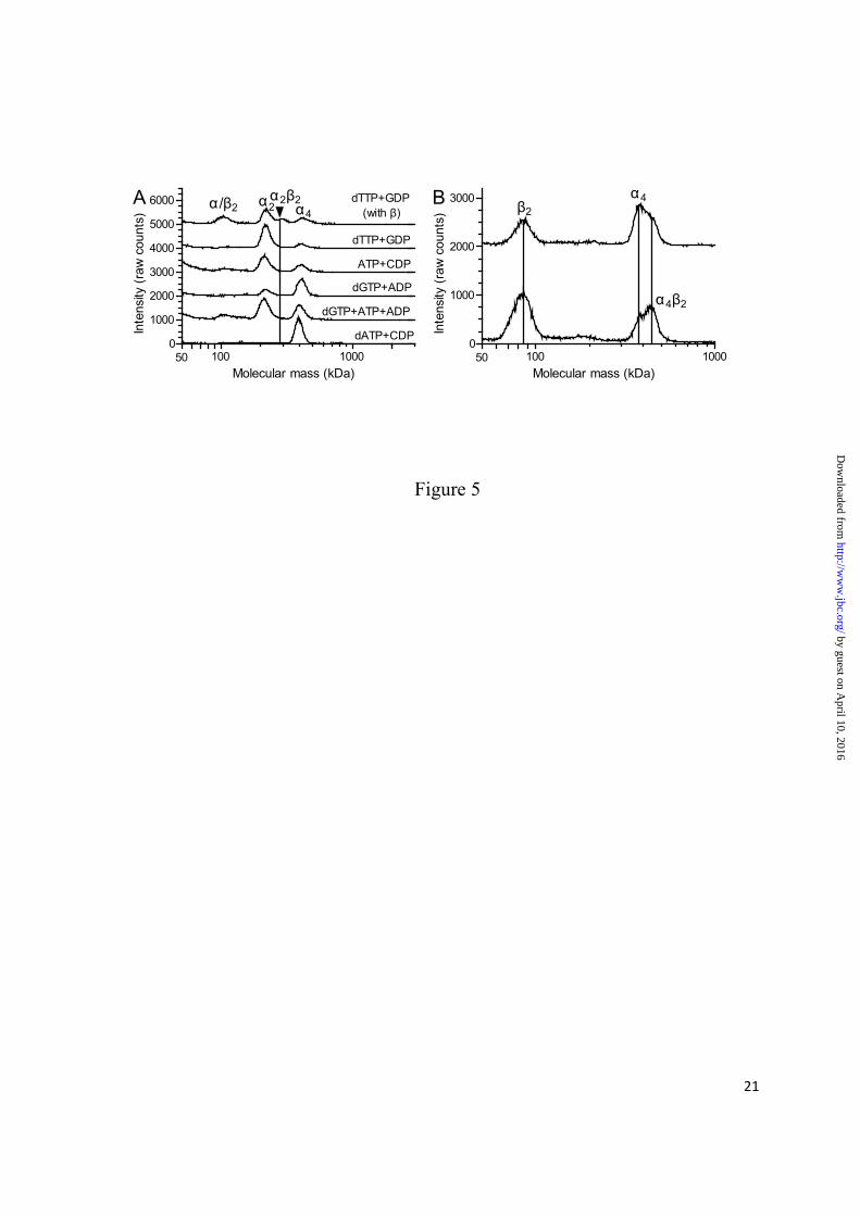
P. aeruginosa α4 and α4β2 complexes predominate under conditions in which the protein is inactive - To determine which are the active and inactive forms of the P. aeruginosa RNR, a series of GEMMA experiments were undertaken where the enzyme was incubated with combinations of allosteric effectors and substrates (Fig. 5A). All experiments were performed in the presence of Mg2+ (1 mM) and reducing agent (30 mM DTT) to resemble the conditions when the enzyme is normally active. As seen in the top trace where both subunits were incubated with dTTP and GDP, two peaks appeared that were not observed when the α subunit was tested under similar conditions
by guest on April 10, 2016
http://ww
w.jbc.org/
Dow
nloaded from

7
in the absence of the second subunit (2nd trace). One of the peaks is the free β2 subunit, and the other has the molecular mass of an α2β2 complex. In analogy with all characterized class I RNRs to date, this can be assumed to be an active form of the enzyme. In the remaining traces, the α protein was tested alone in the presence of different allosteric effector/substrate combinations. In all cases, there were dimer–tetramer equilibria, but in the presence of the activating allosteric effectors (dTTP and ATP) the equilibria were shifted towards dimers, and with the inhibiting allosteric effector (dATP) the equilibrium was shifted toward tetramers. Interestingly, dGTP, which has some a-site affinity (see Fig. 2), favored tetramers (Fig. 5A, 4th trace). If the dGTP was combined with ATP, which only competes readily for the a-site (see Fig. 2D), the equilibrium shifted toward dimers (Fig. 5A, 5th trace). The combination of dGTP in the s-site and ATP in the a-site thus seems to favor dimers. Tetramers induced by dATP were very stable as indicated by the fact that dimers were hardly visible (Fig. 5A, bottom trace).
Fig. 5B shows the interaction between the dATP-induced α4 complexes and the β subunit. These experiments were performed with 50 µM dATP concentrations with equimolar Mg2+ and without DTT to get more exact size measurements. The gradual addition of more β subunit led to a mass shift of ~65 kDa, which suggests that only one β2 dimer binds to each α4 tetramer. The shift remained the same even with higher concentrations of β subunit (data not shown). The α4β2 structure was also supported by isothermal titration calorimetry where the β2 dimer was titrated into a solution with dATP-induced α4 tetramer (Fig. 6). The experiment was performed three times with similar results (Fig. 6 shows one of them). An average of the three experiments showed that one β2 dimer (n = 1.28 ± 0.03 SEM) binds each α4 tetramer, and this supports our conclusion that the inhibited form of the enzyme is an α4β2 complex. The two subunits had a moderate affinity (KD = 2.28 ± 0.05 µM) for each other, and
their interaction was primarily ΔH-driven (ΔH = −10.3 ± 0.5 kcal × mol−1, ΔS = −8.6 ±1.8 cal × mol−1 × K−1). In this case, the solution contained 0.5 mM dATP and 30 mM magnesium acetate.
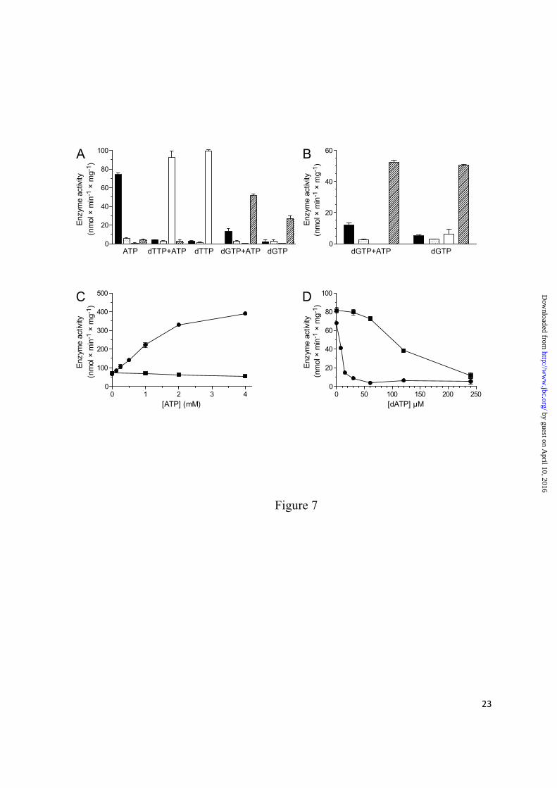
Role of a-site ATP in enzyme activity regulation - A four-substrate assay showed that the P. aeruginosa class I RNR has similar specificity regulation as most other characterized RNRs (1), with ATP stimulating CDP reduction and dTTP and dGTP stimulating GDP and ADP reduction, respectively (Fig. 7A). However, UDP seems to be a very poor substrate under all circumstances. In order to specifically study the effect of ATP binding to the a-site, experiments were also performed where ATP was combined with dGTP or dTTP at high enough concentration to exclude ATP binding to the s-site (Fig. 7A–D). As expected from its low affinity to the s-site, ATP addition did not affect the GDP and ADP specificity induced by 2 mM dTTP or dGTP, respectively (Fig. 7A). ATP instead increased the dGTP-induced ADP reduction. Because no such ATP effect was observed with dTTP-induced GDP reduction, we wanted to investigate whether this effect was related to the ability of dGTP to bind to the a-site. In Fig. 7B, the concentration of dGTP was reduced to 0.5 mM to decrease a-site occupancy. ATP did not have any obvious effect on dGTP-stimulated ADP reduction under these conditions, suggesting that the role of ATP is indirect, i.e. to compete out inhibitory dGTP from the a-site rather than having an effect on its own. The lack of direct ATP effects on enzyme activity via the a-site is illustrated in Fig. 7C where a comparison with the mouse enzyme was made. In the mouse RNR, the dTTP-promoted GDP reduction was strongly stimulated by ATP via the a-site whereas it was not stimulated in the P. aeruginosa enzyme. The role of ATP as a passive regulator was further confirmed in Fig. 7D, where a higher concentration of dATP was needed to inhibit the enzyme in the presence of ATP than in its absence. The passive activation of ATP is ascribed to the a-site because any ATP binding to
by guest on April 10, 2016
http://ww
w.jbc.org/
Dow
nloaded from
8
the s-site would lead to inhibition of GDP reduction rather than activation.
DISCUSSION
Overall RNR activity regulation has until now been believed to occur by the binding of a single dATP molecule to the N-terminal ATP cone and thereby causing the formation of an inactive α6 (eukaryotes) or α4β4 (E. coli) complex. We now present a third model by showing that P. aeruginosa class I RNR binds two dATP molecules to its ATP cone and that the inactive complex is an α4 tetramer.
The fact that regulation of RNR activity always involves an N-terminal ATP cone would perhaps suggest that it is an ancestral trait present in the common ancestor of modern RNRs. Speaking against this, however, is the patchy distribution of activity regulation and ATP cones among modern RNRs and that there is more than one mechanism to achieve overall activity regulation. We believe that evolution has repeatedly discovered the ATP cone as a way of regulating RNR activity, possibly aided by recombination between RNR genes. In this scenario, the ATP cone acts like a module that can be gained or lost depending on the organism’s requirement to exert control over the absolute concentration of dNTPs beyond transcriptional and other non-allosteric mechanisms. Supporting this scenario, we observed a much larger heterogeneity than expected in the distribution of RNR sequences with ATP cones, RNR sequences without ATP cones, and RNR sequences with multiple ATP cones among the different RNR classes and subclasses. In contrast, the substrate specificity regulation is much more conserved and common to all studied RNRs with the exception of Herpesviridae (33). It is, therefore, likely to represent the mechanism present in the ancestor of all modern RNRs. The regulation of P. aeruginosa RNR specificity is also similar to other species with the exception that UDP is a very poor substrate. It is therefore likely that P. aeruginosa
gets most of its dTTP via CDP reduction followed by deamination rather than via UDP reduction. This alternative pathway is common in many species such as E. coli and mammalian cells (1).
A surprisingly large number of RNR sequences have multiple ATP cones. In most cases the second ATP cone appears nonfunctional, like in P. aeruginosa, but some, in particular the Chlamydia spp. and Chlamydophila spp. sequences, contain a second ATP-cone with high HMMER scores indicating functionality (C. trachomatis is shown in Table 2). If the ATP cone and the activity regulation have been gained multiple times, details of the mechanism are expected to differ between RNRs because a recently gained ATP cone needs to adapt to the surrounding amino acid sequence in order to be able to form dATP-inhibited complexes. The different mechanisms identified in the eukaryotic, E. coli, and now P. aeruginosa RNRs support this hypothesis. Horizontal transfer of RNR genes and paralogy both in the form of multiple copies of proteins from the same class and encoding of several classes in the same genome are commonly found in organisms (34) (RNRdb2: http://rnrdb.pfitmap.org), and this provides a plausible scenario for how the ATP cone could have been transferred between different classes and subclasses of RNRs.
Common for the class I RNR family is that the α subunit can dimerize and interact with the β2 subunit to form an active α2β2 complex, although this tetramer is considered a minor form of the eukaryotic enzymes under physiological ATP concentrations (1). The α dimerization and the interaction between the two subunits are generally stimulated by allosteric effectors, whereas the a-site-binding effectors can also stimulate the formation of higher order complexes. To date, the overall activity regulation mechanism has only been studied in a few class I RNRs (1) representing three of the ten subclasses, including NrdAe from mammals and other eukaryotes, NrdAg from E. coli, and, as reported here, NrdAz from P. aeruginosa. All have N-terminal ATP-
by guest on April 10, 2016
http://ww
w.jbc.org/
Dow
nloaded from

9
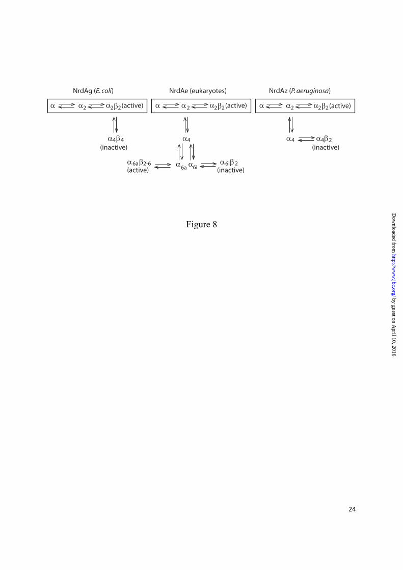
cones that appear functional based on their HMMER score (Table 2). As shown in Fig. 8, P. aeruginosa and eukaryotes have an α-driven oligomerization process in which two or three α2 subunits associate to form a tetramer or a hexamer, respectively. In eukaryotes, ATP bound to the ATP cones of the α subunits opens the conformation of the hexamer to allow one, two, or three β2 dimers to bind and form an active α6β2–6 complex, whereas in P. aeruginosa ATP (or empty a-sites) destabilizes the tetramer allowing the formation of α2 and α2β2 complexes. While the active form in E. coli is the same α2β2 complex as in P. aeruginosa, the equilibrium between active α2β2 and inactive α4β4 complexes requires interaction with the β subunit in E. coli. There are thus clear similarities between P. aeruginosa and eukaryotic class I RNRs in terms of how higher complexes are formed, but also similarities between the P. aeruginosa and E. coli class I enzymes in that the active form is an α2β2 complex.
An interesting feature of the P. aeruginosa RNR is that although studies on the a-site mutant (α-Δ147) show that the a-site itself is a prerequisite for tetramer formation, it does not need to be occupied with nucleotides for this to occur. Tetramers form to some extent even in the absence of nucleotides or in the presence of only s-site effectors, but the tetramers formed in the presence of dATP are more stable and probably represent a tighter structure. The tetramers induced by other effectors than dATP are destabilized by Mg2+, and the dimers could, therefore, be even more predominating under enzyme assay conditions (30 mM Mg2+) than in the GEMMA experiments, which are limited to Mg2+ concentrations ≤ 1 mM. It can therefore be concluded that tight α4 tetramers are inactive and that the α2β2 complex is the major active form, whereas it is less clear whether the looser α4
tetramers have any enzymatic activity or not.
Perhaps the most striking feature discovered in the P. aeruginosa RNR is that the enzyme could
bind two dATP molecules in the a-sites and that it lost the ability to bind both of them if the first ATP cone was removed. Because HMMER scores and amino acid comparisons supported the interpretation that only the first ATP cone is functional, this seems to be the first example of an ATP cone that can bind more than one dATP molecule. Interestingly, dATP binds to the s-site and a-sites with similar KD values and yet it is known from before (14) that low concentrations of dATP activate CDP reduction (KL = 3.2 μM), whereas higher concentrations were needed to inhibit it (KL = 17 μM), just like in the enzymes that have an affinity difference between the sites. However, it should be remembered that KD and KL values are not entirely equivalent because the latter is measured in the presence of substrate and is also influenced by the efficiency of oligomer formation, which occurs on a slower time scale than the rapid exchange of nucleotides in the allosteric sites. In addition, higher dATP occupancy might be needed to cause inhibition in the case of P. aeruginosa because there are three allosteric sites that need to be filled up.
Another unique feature found in the P. aeruginosa enzyme is that the ATP seems to only have a passive role in the overall activity regulation. This conclusion is based on the observation that the addition of this nucleotide did not result in any change in overall activity under conditions when it was only allowed to bind to the a-site (when the s-site was saturated by dTTP). In contrast, the mouse enzyme was strongly activated by a-site ATP under these conditions (Fig. 7C), which stimulates the formation of α6β2 complexes (8). The corresponding E. coli enzyme is instead inhibited by combinations of ATP and dGTP/dTTP (7). The only effect of a-site ATP in the P. aeruginosa enzyme was that it could prevent dATP from binding and thereby decrease enzyme inhibition. The role of ATP is thus more passive in P. aeruginosa RNR than in the other two enzymes because it only requires that ATP
by guest on April 10, 2016
http://ww
w.jbc.org/
Dow
nloaded from
10
binds and not that it mediates any effect on its own.
The P. aeruginosa class I RNR is interesting from a drug development viewpoint, as demonstrated in a previous study (35). Some clinically used drugs that target the allosteric and catalytic sites of RNR, such as clofarabine and gemcitabine, have direct effects on oligomerization as well as the affinity between α and β subunits in different RNRs (12,36,37). The allosteric a-site of the P. aeruginosa enzyme seems to have a broader specificity than most other members of class I RNRs. Not only can
dATP inhibit the enzyme, but so can high concentrations of dGTP (in the mM range), indicating that the site is more flexible than in other RNRs studied to date. This could be an advantage for the development of nucleotide analogs that specifically bind the a-site and inhibit the P. aeruginosa enzyme. The broader specificity, and perhaps also the differences in mechanism between the P. aeruginosa and eukaryotic RNRs, suggests that activity regulation could be a target for inhibition of DNA precursor synthesis in P. aeruginosa.
REFERENCES
1. Hofer, A., Crona, M., Logan, D. T., and Sjöberg, B.-M. (2012) DNA building blocks: keeping control of manufacture. Crit Rev Biochem Mol Biol 47, 50-63
2. Lundin, D., Berggren, G., Logan, D. T., and Sjöberg, B.-M. (2015) The origin and evolution of ribonucleotide reduction. Life (Basel) 5, 604-636
3. Nordlund, P., and Reichard, P. (2006) Ribonucleotide reductases. Annual Review of Biochemistry 75, 681-706
4. Mathews, C. K. (2006) DNA precursor metabolism and genomic stability. Faseb J. 20, 1300-1314
5. Aravind, L., Wolf, Y. I., and Koonin, E. V. (2000) The ATP-cone: an evolutionarily mobile, ATP-binding regulatory domain. J. Mol. Microbiol. Biotechnol. 2, 191-194
6. Brown, N. C., and Reichard, P. (1969) Ribonucleoside diphosphate reductase. Formation of active and inactive complexes of proteins B1 and B2. J. Mol. Biol. 46, 25-38
7. Rofougaran, R., Crona, M., Vodnala, M., Sjöberg, B.-M., and Hofer, A. (2008) Oligomerization status directs overall activity regulation of the Escherichia coli class Ia ribonucleotide reductase. J. Biol. Chem. 283, 35310-35318
8. Rofougaran, R., Vodnala, M., and Hofer, A. (2006) Enzymatically active mammalian ribonucleotide reductase exists primarily as an α6β2 octamer. J. Biol. Chem. 281, 27705-27711
9. Fairman, J. W., Wijerathna, S. R., Ahmad, M. F., Xu, H., Nakano, R., Jha, S., Prendergast, J., Welin, R. M., Flodin, S., Roos, A., Nordlund, P., Li, Z., Walz, T., and Dealwis, C. G. (2011) Structural basis for allosteric regulation of human ribonucleotide reductase by nucleotide-induced oligomerization. Nature Structral & Molecular Biology 18, 316-322
10. Crona, M., Avesson, L., Sahlin, M., Lundin, D., Hinas, A., Klose, R., Söderbom, F., and Sjöberg, B.-M. (2013) A rare combination of ribonucleotide reductases in the social amoeba Dictyostelium discoideum. J. Biol. Chem. 288, 8198-8208
11. Kashlan, O. B., Scott, C. P., Lear, J. D., and Cooperman, B. S. (2002) A comprehensive model for the allosteric regulation of mammalian ribonucleotide reductase. Functional consequences of ATP- and dATP-induced oligomerization of the large subunit. Biochemistry 41, 462-474
by guest on April 10, 2016
http://ww
w.jbc.org/
Dow
nloaded from

11
12. Wang, J., Lohman, G. J., and Stubbe, J. (2009) Mechanism of inactivation of human ribonucleotide reductase with p53R2 by gemcitabine 5'-diphosphate. Biochemistry 48, 11612-11621
13. Ando, N., Brignole, E. J., Zimanyi, C. M., Funk, M. A., Yokoyama, K., Asturias, F. J., Stubbe, J., and Drennan, C. L. (2011) Structural interconversions modulate activity of Escherichia coli ribonucleotide reductase. Proc. Natl. Acad. Sci. U.S.A. 108, 21046-21051
14. Torrents, E., Westman, M., Sahlin, M., and Sjöberg, B.-M. (2006) Ribonucleotide reductase modularity: Atypical duplication of the ATP-cone domain in Pseudomonas aeruginosa. J. Biol. Chem. 281, 25287-25296
15. Ormö, M., and Sjöberg, B.-M. (1990) An ultrafiltration assay for nucleotide binding to ribonucleotide reductase. Anal. Biochem. 189, 138-141
16. Roshick, C., Iliffe-Lee, E. R., and McClarty, G. (2000) Cloning and characterization of ribonucleotide reductase from Chlamydia trachomatis. J. Biol. Chem. 275, 38111-38119
17. Reichard, P., Eliasson, R., Ingemarson, R., and Thelander, L. (2000) Cross-talk between the allosteric effector-binding sites in mouse ribonucleotide reductase. J. Biol. Chem. 275, 33021-33026
18. Eriksson, S., Thelander, L., and Åkerman, M. (1979) Allosteric regulation of calf thymus ribonucleoside diphosphate reductase. Biochemistry 18, 2948-2952
19. Thelander, L., Eriksson, S., and Åkerman, M. (1980) Ribonucleotide reductase from calf thymus. Separation of the enzyme into two nonidentical subunits, proteins M1 and M2. J. Biol. Chem. 255, 7426-7432
20. Eriksson, S., Gudas, L. J., Ullman, B., Clift, S. M., and Martin, D. W., Jr. (1981) DeoxyATP-resistant ribonucleotide reductase of mutant mouse lymphoma cells. Evidence for heterozygosity for the protein M1 subunits. J Biol Chem 256, 10184-10188
21. Domkin, V., Thelander, L., and Chabes, A. (2002) Yeast DNA damage-inducible Rnr3 has a very low catalytic activity strongly stimulated after the formation of a cross-talking Rnr1/Rnr3 complex. J Biol Chem 277, 18574-18578
22. Hofer, A., Ekanem, J. T., and Thelander, L. (1998) Allosteric regulation of Trypanosoma brucei ribonucleotide reductase studied in vitro and in vivo. J. Biol. Chem. 273, 34098-34104
23. Hofer, A., Schmidt, P. P., Gräslund, A., and Thelander, L. (1997) Cloning and characterization of the R1 and R2 subunits of ribonucleotide reductase from Trypanosoma brucei. Proc. Natl. Acad. Sci. U.S.A. 94, 6959-6964
24. Hendricks, S. P., and Mathews, C. K. (1998) Allosteric regulation of vaccinia virus ribonucleotide reductase, analyzed by simultaneous monitoring of its four activities. J Biol Chem 273, 29512-29518
25. Berglund, O. (1972) Ribonucleoside diphosphate reductase induced by bacteriophage T4. II. Allosteric regulation of substrate sepecificity and catalytic activity. J Biol Chem 247, 7276-7281
26. Berglund, O., and Eckstein, F. (1972) Synthesis of ATP- and dATP-substituted sepharoses and their application in the purification of phage-T4-induced ribonucleotide reductase. Eur J Biochem 28, 492-496
27. Hendricks, S. P., and Mathews, C. K. (1997) Regulation of T4 phage aerobic ribonucleotide reductase. Simultaneous assay of the four activities. J. Biol. Chem. 272, 2861-2865
28. Brown, N. C., and Reichard, P. (1969) Role of effector binding in allosteric control of ribonucleoside diphosphate reductase. J. Mol. Biol. 46, 39-55
by guest on April 10, 2016
http://ww
w.jbc.org/
Dow
nloaded from

12
29. Riera, J., Robb, F. T., Weiss, R., and Fontecave, M. (1997) Ribonucleotide reductase in the archaeon Pyrococcus furiosus: a critical enzyme in the evolution of DNA genomes? Proc Natl Acad Sci U S A 94, 475-478
30. Eliasson, R., Pontis, E., Jordan, A., and Reichard, P. (1999) Allosteric control of three B12-dependent (class II) ribonucleotide reductases. Implications for the evolution of ribonucleotide reduction. J Biol Chem 274, 7182-7189
31. Eliasson, R., Pontis, E., Sun, X., and Reichard, P. (1994) Allosteric control of the substrate specificity of the anaerobic ribonucleotide reductase from Escherichia coli. J Biol Chem 269, 26052-26057
32. Torrents, E., Buist, G., Liu, A., Eliasson, R., Kok, J., Gibert, I., Gräslund, A., and Reichard, P. (2000) The anaerobic (class III) ribonucleotide reductase from Lactococcus lactis. Catalytic properties and allosteric regulation of the pure enzyme system. J Biol Chem 275, 2463-2471
33. Averett, D. R., Lubbers, C., Elion, G. B., and Spector, T. (1983) Ribonucleotide reductase induced by herpes simplex type 1 virus. Characterization of a distinct enzyme. J. Biol. Chem. 258, 9831-9838
34. Lundin, D., Gribaldo, S., Torrents, E., Sjöberg, B.-M., and Poole, A. M. (2010) Ribonucleotide reduction - horizontal transfer of a required function spans all three domains. BMC Evol. Biol. 10, 383
35. Tholander, F., and Sjöberg, B.-M. (2012) Discovery of antimicrobial ribonucleotide reductase inhibitors by screening in microwell format. Proc. Natl. Acad. Sci. U.S.A. 109, 9798-9803
36. Aye, Y., and Stubbe, J. (2011) Clofarabine 5'-di and -triphosphates inhibit human ribonucleotide reductase by altering the quaternary structure of its large subunit. Proc. Natl. Acad. Sci. U.S.A. 108, 9815-9820
37. Zimanyi, C. M., Ando, N., Brignole, E. J., Asturias, F. J., Stubbe, J., and Drennan, C. L. (2012) Tangled up in knots: structures of inactivated forms of E. coli class Ia ribonucleotide reductase. Structure 20, 1374-1383
FOOTNOTES
*This work was supported by the Kempe Foundation (to AH), Carl Trygger´s Foundation (to AH and BMS), the Swedish Cancer Foundation (to BMS), the Swedish Research Council (to AH and BMS), and the Wenner-Gren Foundations (to BMS). †These two authors have contributed equally to this work. §Abbreviations used: RNR, ribonucleotide reductase; NDPs, nucleoside diphosphates; NTPs, nucleoside triphosphates; dNDPs, deoxyribonucleoside diphosphates; dNTPs, deoxyribonucleoside triphosphates; s-site, specificity site; a-site, overall activity site; GEMMA, gas-phase electrophoretic mobility macromolecule analysis; DTT, dithiothreitol
by guest on April 10, 2016
http://ww
w.jbc.org/
Dow
nloaded from
13
FIGURE LEGENDS
FIGURE 1. Amino acid sequence alignment of the two ATP cones of the P. aeruginosa α subunit against the Pfam profile consensus sequence. Important differences in the second ATP cone from the Pfam profile are highlighted, including an Arg to Ala substitution in the KRD/N motif, two deletions, and a hydrophobic residue substituted for a Thr. Only the first 63 amino acids out of the 90 amino acids of the Pfam ATP cone are shown.
FIGURE 2. Nucleotide binding experiments. (A) Scatchard plot showing the number of dATP binding sites in the α subunit. Experiments were performed with 0.5 μM (●), 1.25 μM (■), and 2.5 μM (▲) α subunit in panels A and E. (B) Competition experiments where 60 μM 3H-dATP is competing with varying concentrations of unlabeled dTTP (▼), dGTP (▲), ATP (■), dATP (●), or a combination of ATP with 4 mM dTTP (□) for binding to 5 µM α protein. (C) Scatchard plot showing the binding of dGTP to 1.25 μM (●), 2.5 μM (■), and 5 μM (▲) α subunit. (D) Competition experiments where 80 μM 3H-dGTP is competing with varying concentrations of unlabeled dTTP (▼), dGTP (▲), ATP (■) or dATP (●) for binding to 15 µM α subunit. (E) Scatchard plot showing the binding of dATP to the α-Δ147 subunit. (F) Competition experiments where 30 μM 3H-dATP is competing with varying concentrations of unlabeled dTTP for binding to 3 µM α-Δ147 subunit. All graphs are based on the data from three separate experiments with standard errors indicated. The linear regression lines for panels A, C, and E (based on the first four points in C) are for visual purposes only, and the calculations of binding constants and number of sites in the text are based on non-linear regression of corresponding bound vs. free ligand graphs.
FIGURE 3. GEMMA analysis of the P. aeruginosa β, α, and α-Δ147 subunits. (A) Analysis of the three proteins in the absence of nucleotide effectors. The lines centered below the peaks indicate masses of 42 kDa (β), 85 kDa (β2 and α-Δ147), 103 kDa (α), 171 kDa (α2-Δ147), 201 kDa (α2), and 378 kDa (α4). (B) The distribution of α, α2, and α4 peaks in the presence of 50 μM dTTP (only equimolar Mg2+ was present), 50 µM dTTP + 1 mM Mg2+ (dTTP + Mg), 50 µM dTTP + 1 mM Mg2+ + 30 mM DTT (dTTP + Mg + DTT), or 50 µM dATP + 1 mM Mg2+ + 30 mM DTT (dATP + Mg + DTT). The lines centered below the peaks of the upper trace indicate molecular masses of 100 kDa (α), 206 kDa (α2), and 376 kDa (α4). The slight shift of the lower traces in B is due to non-specific binding of Mg2+. The assignments of the peaks in A and B were based on the theoretical masses of 107 kDa (α), 91 kDa (α-Δ147), and 47 kDa (β). The traces are spaced 3000 (A) and 1000 or 2000 (B) units apart in order to fit many experiments in the same graph.
FIGURE 4. Gel filtration analysis of the P. aeruginosa β subunit (upper trace) in comparison to a molecular mass standard of globular proteins (lower trace). The masses of the standard proteins are indicated. The traces are spaced 250 units apart to fit both experiments in the same graph.
FIGURE 5. GEMMA analysis to determine the stability of the P. aeruginosa α4 complexes in the presence of different nucleotides (A) and how the α4 complexes interact with the β subunit (B). The proteins were incubated with 30 mM DTT and 1 mM magnesium acetate. The NDP substrate (100 µM) and allosteric effector (50 µM dNTP or 200 µM ATP) are indicated. The vertical line indicates the position of the α2β2 complex, which was determined to be 286 kDa, whereas the positions of other peaks were similar as in Fig. 3B. (B) The α subunit in the presence of dATP was incubated together with 1:1 and 1:2 molar ratios with the β subunit (top and bottom traces, respectively). The lines in B indicate
by guest on April 10, 2016
http://ww
w.jbc.org/
Dow
nloaded from

14
masses of 85 kDa, 375 kDa, and 439 kDa. The traces are spaced 1000 (A) or 2000 (B) units apart to fit many experiments in the same graph.
FIGURE 6. Isothermal calorimetry analysis showing α4β2 formation. The β2 dimer was titrated into a solution of dATP-induced α4 tetramer. Both solutions contained 0.5 mM dATP and 30 mM magnesium acetate. Calorimetric data were plotted and fitted using the standard single-site binding model (Origin, MicroCal).
FIGURE 7. Allosteric regulation of the P. aeruginosa class I RNR activity. (A) Enzyme activity was measured with the indicated allosteric effectors (2 mM of each) and all four substrates present simultaneously (0.5 mM of each). The activities are shown with CDP (solid bar), UDP (stippled), GDP (open), and ADP (hatched). (B) Similar to the corresponding experiments in A, but the concentration of dGTP was only 0.5 mM. (C) GDP reduction with the mouse (●) and P. aeruginosa (■) RNRs in the presence of 2 mM dTTP and increasing concentrations of ATP. (D) GDP reduction with the P. aeruginosa RNR in the presence of 2 mM dTTP and increasing concentrations of dATP. The experiment was performed in the presence (■) and absence (●) of 3 mM ATP. Experiments were performed in triplicate (A and B) or duplicate (C and D) with the standard errors indicated.
FIGURE 8. Oligomerization of the E. coli, eukaryotic, and P. aeruginosa class I RNRs. The squares indicate general complexes in the class I RNR family. The eukaryotic hexamers are indicated with a and i to highlight active and inactive structural conformations, respectively. The eukaryotic α4 complex is a hypothetical intermediate form.
by guest on April 10, 2016
http://ww
w.jbc.org/
Dow
nloaded from

15
Table 1. ATP cone distribution in different RNR classes and subclasses. The right column shows how many sequences the frequency is based on. The predominant number of ATP cones in each subclass is boldfaced.
Class Subclass Frequency of number of ATP cones (%) Number of
sequences 0 1 2 3
I (NrdA/E) 53 33 13 1 4186 I NrdAe 9 90 1 0 630 I NrdAg 4 96 0 0 688 I NrdAh 37 31 32 0 326 I NrdAi 100 0 0 0 122 I NrdAk 100 0 0 0 158 I NrdAn 58 37 4 0 89 I NrdAz 10 3 82 5 541 I NrdE 100 0 0 0 1532
II (NrdJ) 93 6 1 0.1 1800 II NrdJd 100 0.4 0 0 541 II NrdJf 89 9 2 0.1 1002 II NrdJm 95 4 0.4 1 257
III (NrdD) 24 70 6 0.3 2426 III NrdDa 18 36 43 3 74 III NrdDb 33 58 9 0 457 III NrdDc 13 83 4 0.2 1378 III NrdDd 33 64 3 0 351 III NrdDf 100 0 0 0 4 III NrdDh 69 31 0 0 134 III NrdDi 100 0 0 0 28
by guest on April 10, 2016
http://ww
w.jbc.org/
Dow
nloaded from

16
Table 2. Pfam profile HMMER score similarity of ATP cones in RNRs. The table shows the number of ATP cones and their HMMER scores as well as data (with indicated references) on dATP binding, dATP inhibition, and if any quaternary structure is associated with dATP inhibition. Inhibited complexes that can bind β2 but are not dependent on it for their formation are written in their α form (e.g. α4 rather than α4β2 for P. aeruginosa). Included in the list are all RNRs known to have an ATP cone that binds dATP/ATP and a few additional RNRs of relevance.
Class Subclass Species No of ATP cones
HMMER scores dATP
Reference Binding Inhibition
I NrdAe D. discoideum 1 63.1 Yes Yes, α6 (10) I NrdAe Mammalia 1 65.0 Yes Yes, α6 (9,12,17-20)
I NrdAe S. cerevisiae RNR1
1 56.9 Yes* Yes, α6 (9,21)
I NrdAe S. cerevisiae RNR3
1 55.3 n.d.** No (21)
I NrdAe Trypanosoma brucei
1 41.7 Yes No (22,23)
I NrdAe Vaccinia virus 1 62.9 Yes* Yes (24) I NrdAg Bacteriophage T4 1 64.6 Yes No (25-27) I NrdAg E. coli 1 54.2 Yes Yes, α4β4 (6,7,13,28) I NrdAz P. aeruginosa 2 70.6; 31.8 Yes Yes, α4 (14), this study I NrdAz C. trachomatis 3 73.1; 68.3; 12.0 Yes* Yes (16)
II NrdJf Pyrococcus furiosus
1 79.4 Yes (?)* Yes(?)* (29)
II NrdJf Thermoplasma acidophilum
1 90.9 Yes No (30)
III NrdDc E. coli 1 73.0 Yes*** (31) III NrdDc Lactococcus lactis 1 74.7 Yes*** (32) III NrdDd P. aeruginosa 1 82.2 n.d.**
*Binding experiments have not been performed, but enzymatic data show that they have general dATP inhibition. The RNR from P. furiosus is marked with a question mark because the data behind the conclusion were not shown in the reference.
**Not determined.
***The effect of dATP is dependent on which nucleotide is occupying the s-site.
by guest on April 10, 2016
http://ww
w.jbc.org/
Dow
nloaded from

19
Figure 3
10 100 10000
2000
4000
6000
8000
10000
β
α
α- Δ147
β2β
α2
αα4
αα2
Molecular mass (kDa)
Inte
nsity
(raw
cou
nts)
100 10000
1000
2000
3000
4000
5000
6000
200 500
α4α2α
α2
50
dATP+Mg+DTT(α- Δ147)
dATP+Mg+DTT
dTTP
dTTP+Mg
dTTP+Mg+DTT
Molecular mass (kDa)
Inte
nsity
(raw
cou
nts)
A B
by guest on April 10, 2016
http://ww
w.jbc.org/
Dow
nloaded from

Johansson, Kristoffer Brännström, Britt-Marie Sjöberg and Anders HoferVenkateswara Rao Jonna, Mikael Crona, Reza Rofougaran, Daniel Lundin, Samuel
Diversity in Overall Activity Regulation of Ribonucleotide Reductase
published online May 13, 2015J. Biol. Chem.
10.1074/jbc.M115.649624Access the most updated version of this article at doi:
Alerts:
When a correction for this article is posted•
When this article is cited•
to choose from all of JBC's e-mail alertsClick here
http://www.jbc.org/content/early/2015/05/13/jbc.M115.649624.full.html#ref-list-1
This article cites 0 references, 0 of which can be accessed free at
by guest on April 10, 2016
http://ww
w.jbc.org/
Dow
nloaded from