MIXED MATRIX MEMBRANE ADSORBERS FOR PROTEIN ...
175
MIXED MATRIX MEMBRANE ADSORBERS FOR PROTEIN AND BLOOD PURIFICATION
-
Upload
khangminh22 -
Category
Documents
-
view
2 -
download
0
Transcript of MIXED MATRIX MEMBRANE ADSORBERS FOR PROTEIN ...

MIXED MATRIX MEMBRANE ADSORBERS
FOR PROTEIN AND BLOOD PURIFICATION

This work was financially supported by the Indonesian Ministry of Education and Cultureand the Membrane Technology Group of The University of Twente, The Netherlands
Mixed Matrix Membrane Adsorbersfor Protein and Blood Purification
PhD Thesis, University of Twente
ISBN: 978-90-365-2507-7
© Saiful, Enschede (The Netherlands), 2007
No part of this work may be reproduced by print, photocopy, or any other means withoutpermission of the author
CoverFront cover illustrates bacterias and endotoxins adsorption during hemodialysisBacterias and red blood cells pictures are obtained from 3D SCIENCE.com
Printed by Wöhrmann Print Service, Zutphen

Mixed Matrix Membrane Adsorbersfor Protein and Blood Purification
DISSERTATION
to obtainthe doctor’s degree at the University of Twente,
on the authority of the rector magnificus,prof. dr. W.H.M. Zijm,
on account of the decision of the graduation committee,to be publicly defended
on Thursday 24th May 2007 at 13.15
by
Saiful
born on 22nd September 1969
in Beureugang, NAD-Indonesia

This dissertation has been approved by:
Promotor: Prof. Dr.–Ing. M. Wessling
Assistant-promotor: Dr.–Ing. Z. Borneman

Dedicated to my beloved wife and daughter:Tahiya and Iza


ACKNOWLEDGEMENTS
I have finished my PhD thesis. This thesis is the result of a four-year research projectwhich was conducted at the Membrane Technology Group, Faculty of Science andTechnology, University of Twente. Numerous colleges have contributed in accomplishing theresearch reported in this thesis. I would like to take the opportunity to thank all those whomade my work possible and enjoyable. The research reported in this thesis was conductedunder the auspices of through the Membrane Technology Group of the University of Twenteand the Ministry of Education of the Republic of Indonesia. These combined supports havemade my work possible and are therefore gratefully acknowledged.
I am indebted to Prof. Dr.–Ing. Matthias Wessling for the opportunity he gave me towork here in this wonderful research group. My stay in Netherlands was started frominvitation that was arranged and set by him. I gratefully acknowledge the continued supportsand encouragements of Prof Matthias to become an independent scientist and to introducethe author to interdisciplinary application of chemical engineering and membrane technologyto biochemical and biomedical problems. I enjoyed working in your groups very much, bothfrom professional as a personal point of view.
I express deeply my sincere gratitude to my research advisor, Dr.–Ing. ZandrieBorneman, for his encouragements, guidance and inspirations throughout this work,especially in the beginning of my study, for all his cares and all valuable discussions made thehard of a beginner PhD a little more manageable. I have exceedingly benefited from his vastknowledge, lasting enthusiasm and exceptional personality, but also for his friendshipwithout which this dissertation would not be possible to be done. You are very supportiveand give a freedom in doing the research. Zandrie, thank you very much for your input in thethesis: reading, correcting and improving the thesis. Many thank for translating the summaryinto the samenvatting. I am also indebt to other staff members of the group: Dimitris, Rob,Kitty and Antoine for nice and collegial research atmosphere with me.
I would also like to thank the members of my promotion committee for reading thefinal draft of the thesis, giving approval to publicly defend this thesis and being part of thisspecial day. It is an honour for me that Prof. Dr. J.F.J. Engbersen, Prof. Dr. J. Feijen andspecially Prof. Ing. Giulio C. Sarti and Dr.-Ing. Bernd Krause (who will make such a longjourney) can attend the promotion ceremony. I am also proud that two special people, JoàoM. Sousa André de and M. Srivatsa Bettahalli Narasimha, can be my paranimfs and canaccompany me in such important moment.
A ton gratitude for all my friends for constantly supporting me and for making mefeels at home when times are tough. Former members of my roommates: Nela, Kitty andAnna for many discussion and support during the initial phase of my PhD, made meconfident during the in beginning of the project. It is not easy to work when my feelingdepart to my family in Indonesia. Saskia, thank’s a lot for sharing your knowledge andexperiences about MMM in beginning of the project. I want to thank my new roommates:Miriam, Srivatsa, Magda, Katja and Clara with humor, joviality and kindness. No daywithout cookies and chocolate as well as a week of fruits. Special thanks are extended toMagda, the mother’s of MMM, for her invaluable discussions and supports. Many thanks areowed to Joao for his helpful suggestions and continuous encouragement. Your inputs andcorrections are really appreciated: muitíssimo agradecido. Spending one month in the newbuilding Meander was very exciting. All are in new: new laboratory, new office and new

roommates with new spirit. I would like to thank Szymon, Jorrit, and Schwan for nice talksand supports during the last month.
It has not been completely easy and smooth for four years study in this country. Ifound a difficult time when the strong earth quake and tsunami disaster were hit and swapmy hometowns where my beloved family and my parent are living. Fortunately, my familyand my parent are safe. However, we are very sad and shock because my family and I werepassed away for peace by some of close relatives and friends. Sadly approximately 173.741people were died in my province. It destroyed and wiped everything in the town. On behalfof the whole family, I would like to thank all members of Membrane Technology Groups foryour sympathy and support in this hard situation. I extend special thankful to Matthias andhis family and Zandrie and his family for their empathy and support. I spent one month inthe “ground zero” in difficult situation and then came back to Enschede with my family tocontinue my work in the lab.
Of course this work can not be success without an important and extremely helpfulfrom Greet, the Secretary of Membrane Technology Group. I can not account how manypapers and letters were translated to English. May be I always complicated. Greet, thank youvery much for your friendship, encouragement, kindness and help with various tasks overthe years. I would also like to acknowledge the valuable assistance of Mr. John Heek, ErikRolevin, Marcel and Herman during my research for their invaluable discussions andassistances as well as providing an interesting working environment.
In last four years I have been enjoying a very pleasant research atmosphere, I thenrecognize that the membrane technology group was not only good place for scientific of lifebut also many sportive and social events. Running the Bata Race, my ankle was bruits in firstBatavarien. Unfortunately, I can not joint the Bata Race afterward. I enjoy riding once a yearwith the bicycle through the neighborhood of Enschede-Germany and end up with BBQ. It isalso very nice to have a cup drink or a piece of delicious cake after each new publication inthe group, birthday, friday afternoon and farewell borrel, etc. There are many otherswonderful activities in the group; unfortunately I wasn’t able to join all of them. Thanks to allof you who organized these events.
I would especially express my thanks to Om Stand and Tante Hadi for theirsupports. A big thanks goes to Nazaruddin for his friendship and invaluable assistances withendotoxin experiment. Along way to come to Enschede with cold weather made you difficultin the beginning. I thank Iqbal (begundal) for fruit discussions about protein and enzymeproperties in the beginning of project. Terima kasih banyak for the entire Indonesiancommunity in Enschede, especially Eko dan Ella sekeluarga. I apologize for not being able tomention all of you personally here. All of you have made my stay in the Netherlands feel likehome.
I want to dedicate a few words in Indonesia (Aceh) to my family in Aceh wheresome of my loved ones are; who have supported me from the beginning. Terimong geunasehbeurayeuk that untuk: Mak and Ayah di Meulaboh; abang Mukhlis and Munzir sekeluarga diMeulaboh dan Idar sekeluarga di Banda Aceh ateuh dukungan dan doa selamanyo.Terimong geunaseh cit lon peutrok untuk Ayah dan Bunda sekeluarga di Darussalam ateuhdukungan dan doa selamanyo.
My loving wife, Tahiya, deserve the most recognition by far. I could not have madeit without her love and understanding in this long journey, for her contribution in all areastoo numerous to mention. I would like to thank my daughter Iza, who unknowingly hasrejuvenated my pleasure in the simple things for making me smile when times were tough, Ilove you girl. Akhirnya dan untuk selamanya, saya memanjatkan puji dan syukur kehadiratAllah SWT atas segala nikmat dan karunianya sehingga dapat menyelesaikan salah satuperjuangan berat di Enschede.

VII
CONTENTS
ACKNOWLEDGMENTS
CHAPTER-1 General Background and Scope of the Thesis 1
1.1. General Introduction 1
1.1.1. Bioproducts separation 1
1.1.2. Extracorporeal blood purification 2
1.2. Outline of the thesis 4
1.3. List of Symbols and Abbreviations 7
References 7
CHAPTER-2 Introduction of Biological Separation and Blood purification 9
2.1. Membrane and Chromatography 10
2.2. Application of Ion exchange Membrane Chromatography 13
2.3. Extracorporeal Blood Purification 16
2.4. Mixed Matrix Membrane Adsorbers 21
2.5. Conclusion 23
2.6. List of Abbreviations 24
References 24
CHAPTER-3 Enzyme Capturing and Concentration with MMM adsorbers 33
Abstract 33
3.1. Introduction 34
3.2. Experimental 36
3.2.1. Materials 36
3.2.2. Membrane preparation and characterization 36
3.2.2.1. Membrane preparation 36
3.2.2.2. Scanning electron microscopy 37
3.2.2.3. Membrane porosity and swelling degree 37
3.2.2.4. Clean water flux 38
3.2.3. Adsorption experiments 38
3.2.3.1. Measurement of zeta potential of Lysozyme 38
3.2.3.2. Batch adsorption experiments 38

Contents
VIII
2.3.3. Adsorption kinetics 39
3.2.3.4. Adsorption isotherm 39
3.2.3.5. Desorption 39
3.2.3.6. Regeneration 40
3.2.3.7. Dynamic experiments 40
3.2.3.8. Fluorescence measurements 41
2.3.9. Enzymatic activity 41
3.3. Results and Discussion 41
3.3.1. Net Charge of Lysozyme 42
3.3.2. Mixed matrix membrane adsorbers characterization 42
3.3.3. Effect of ionic strength on LZ desorption 48
3.3.4. Regeneration and Reuse the membrane Lew CNP80 WS 49
3.3.5. Dynamic adsorption of LZ on membrane Lew CNP80 WS 50
3.3.6. Effect of the flux on dynamic adsorption 51
3.3.7. Lysozyme Stability and Biological Activity 54
3.4. Conclusion 55
3.5. Acknowledgements 56
3.6. List of Symbols and Abbreviation 56
References 57
CHAPTER-4 Enzyme Capturing and Concentration from Binary Mixtures 61
Abstract 61
4.1. Introduction 62
4.2. Experimental 64
4.2.1. Materials 64
4.2.2. Membrane preparation 65
4.2.3. Membrane Characterization 65
4.2.3.1. Scanning electron microscopy 65
4.2.3.2. Membrane porosity and swelling degree 65
4.2.3.3. Clean water flux 66
4.2.4. Adsorption experiments 66
4.2.4.1. Measurement of zeta potential of pure BSA and LZ 66
4.2.4.2. Adsorption isotherm 66

Chapter 1
IX
4.2.4.3. Dynamic adsorption experiment 66
4.2.4.4. Regeneration 67
4.2.4.5. LZ Stability and Activity assay 67
4.3. Results and Discussions 68
4.3.1. Zeta potentials BSA and LZ 68
4.3.2. Membrane characterization 69
4.3.3. Protein adsorption isotherm 70
4.3.4. Breakthrough curves for BSA and LZ at flux 50 Lm-2h-1 72
4.3.5. Purity BSA and LZ at flux 50 Lm-2h-1 73
4.3.6. Effect of permeation rate 75
4.3.7. Effect of ionic strength 76
4.3.8. Reuse of membrane 77
3.9. Stability and activity of LZ 78
4.4. Conclusion 79
4.5. Acknowledgements 80
4.6. List of Symbols and Abbreviation 80
References 80
CHAPTER-5 Preparation of Double Layer Mixed Matrix Membrane 85
Abstract 85
5.1 Introduction 86
5.2. Experimental 89
5.2.1. Materials 89
5.2.2. Membrane preparations and characterizations 89
5.2.2.1. Membrane preparations 89
5.2.2.2. Scanning electron microscopy 91
5.2.2.3. Membrane porosity and swelling degree 91
5.2.2.4. Clean water flux 91
5.2.2.5. Pore size measurements (Coulter porometer) 91
5.3 Results and Discussion 92
5.3.1. Mixed matrix membrane adsorbers preparation 92
5.3.2. Membrane morphology 93
5.3.2. Membrane characteristics 96

Contents
X
5.3.4. Effect of concentration in the co-casting layer 98
5.4. Conclusion 100
5.5. Acknowledgements 101
5.6. List of Symbols and Abbreviation 101
References 101
CHAPTER-6 Carbon Base Mixed Matrix Membrane for Blood Toxin
Removal
103
Abstract 103
6.1. Introduction 104
6.2. Experimental 106
6.2.1. Materials 106
6.2.2. Membrane preparation and Characterizations 107
6.2.3. Adsorption experiments 107
6.2.3.1. Batch experiments 107
6.2.3.2. Adsorption isotherm 108
6.2.3.3. Non-specific BSA adsorption 108
6.2.3.4. Dynamic adsorption experiments 109
a. Dead-end filtration 109
b. Cross flow filtration 109
c. Dialysis 109
6.3. Results and Discussion 111
6.3.1. Static adsorption of creatinine 111
6.3.2. Adsorption isotherm and effectiveness use of adsorbent 113
6.3.3. Non specific adsorption BSA 114
6.3.4. Dynamic creatinine adsorption 116
a) During dead-end filtration 116
b) Cross flow mode 117
c) Dialysis 118
6.4. Conclusion 120
6.5. Acknowledgements 120
6.6. List of Symbols and Abbreviation 120
References 121

Chapter 1
XI
CHAPTER-7 Mixed Matrix Membrane Adsorbers for Endotoxin
adsorption
125
Abstract 125
7.1. Introduction 126
7.1.1. Endotoxin removal 126
7.1.2. Back Transport in hemodialysis 127
7.2. Experimental 129
7. 2.1. Materials 129
7.2.2. Removing Endotoxin Contamination 130
7.2.3. Assay Endotoxin Concentration 130
7.2.4. Membrane preparation and characterization 131
7.2.5. Adsorption experiments 131
7.2.5.1. Batch experiments 131
7.2.5.2. Dialysis experiments 132
7.2.5.3. Dead-end filtration 132
7.2.6. Regeneration of MMM M500 132
7.3. Result and Discussion 133
7.3.1. Membrane preparation and characterization 133
7.3.2. Endotoxin adsorption by different loading 134
7.3.3. Adsorption in time 136
7.3.4. Adsorption isotherm of endotoxin 137
7.3.5. Effect of ionic strength 138
7.3.6. Endotoxin adsorption during dialysis 140
7.3.7. Dead end filtration of endotoxin 141
7.3.8. Reuse MMM M500 142
7.4. Conclusion 143
7.5. Acknowledgements 144
7.6. List of Symbols and Abbreviation 144
References 144

Contents
XII
Summary 149
Samenvatting 153
Ringkasan 157
About the Author 161

General Background and Scope of the Thesis
1.1. General Background
1.1.1. Bioproducts separation
Biotechnology has contributed to and developed new sources of many valuable
healthcare and life science products. These bioproducts are produced by gene expression in
various biological systems; both prokaryotic and eukaryotic systems[1]. The products include
therapeutic proteins and polysaccharides, monoclonals, vaccines, diagnostics, pharmaceutical
chemicals and enzymes for the food and consumer markets. The production of bioproducts
continue to grow progressively[2]. Current estimates predict that global bio-market is worth
over US $48 billion and by the end of this decade global biopharmaceutical products could
easily reach over US $100 billion with the annual growth rate of 19%[3].
Various types of bioproducts have to be isolated and purified from mixtures before
they can be used in order to ensure the safety and efficacy[4, 5]. The ideal separation process
optimizes yield and maintains low manufacturing costs. However, isolation and purification
of bioproducts are difficult and expensive. Biologics separation typically involves several
consecutive steps to reach the desired purity. It causes products loss, long process times and
denaturation of fragile products. The isolation and purification account for 50 to 80 % of the
total production costs[6, 7]. Almost all large scale process configurations are based on packed
bed chromatographic system because a high capacity and product purity can be obtained.
However, these methods have severe major drawbacks and limitations[8, 9] including high
pressure drop, low throughput, sensitive to fouling and plugging, channelling, time
consuming and bed compression. Innovations and improvements of the existing methods are
needed to overcome the shortcomings of the classical technologies and for economical
bioproducts recovery.
Adsorptive membrane chromatography has been introduced as integrative approach
to reduce the number of steps within a purification process and to bypass the fundamental
limitations of packed bed system. It is especially suited for treatment of large volumes of
liquid containing low concentrations of target biomolecules. This approach is able to unite the
1Chapter

Chapter 1
2
coarse and the fine purification steps by embedding in microfiltration membrane[8].
Nowadays, adsorptive membrane chromatography can be prepared by two different
approaches:
I. By chemically coupled ligands to the internal surface of microporous filtration
membranes [8, 10, 11]. The activated membrane has been reported as a potentially
advantageous tool to purify proteins at a laboratory, pilot and industrial scale. The main
limitation of this adsorptive membrane is the low binding capacity[9, 10, 12].
II. By entrapping functionalized particles (sorbent) into a macroporous polymeric
support[13-15]. A polymer solution with dispersed functionalized particles is cast as a
flat film or spun into a fiber and then solidified by a phase inversion process to form a
so-called Mixed Matrix Membrane (MMM) or Particles Loaded Membrane (PLM). The
MMM demonstrates high adsorption capacity, comparable to packed bed systems[15,
16]. By choosing the particles and the supports one can establish various functionalities
such as affinity, ion exchange, adsorption, catalysis and reactive chromatography.
The potential for continues progress and tremendous advances in sorbent
technologies offer a broad application of the MMM in biotechnology and biomedical fields.
As new approach in art chromatographic techniques, there are still many interesting
application of the MMM that remain to be investigated with respect to their performance in
adsorptive membrane chromatography. In this thesis, the latest development and application
of adsorptive chromatographic media based on particles loaded membrane adsorber will be
discussed.
1.1.2. Extracorporeal blood purification
Biological fluids purification is nowadays very important in biomedical fields. A
growing population of patients with kidney and liver failure, poisoning, sepsis shock, and
multi-organs failure by more complex medical problems require technological innovations
and improvements to enhance the safety, reliability and efficiency of extracorporeal organs
support for removing harmful substances from blood. Kidney and liver failure have become a
major threat to public health and expected to rise steeply over the next decade due to ageing
and population increase. More than 1.1 million patients throughout the world undergo
kidney chronic dialysis[17]. In Germany, approximately 70.000 patients require hospital
treatment for severe liver diseases[18] and in the United States, there are more than 360.000
hospitalizations due to chronic liver disease and cirrhosis[19]. Kidney and liver

General background and scope of the thesis
3
transplantation can not be routinely provided to this group of patients because of the steadily
increasing lack of donor organs[18, 20]. Furthermore, replacing the malfunctioning kidney
with a healthy one stimulates the rejection mechanisms of the living body against foreign
organ, unless the donor is a near relative[21]. Thus, there is a considerable drive to develop
improved therapies for kidney and liver failure with the capacity to replace a wider range of
the organ functions, thereby reducing morbidity, mortality and the overall economic impact
associated with this condition.
Several techniques have been applied in blood purification (including hemodialysis,
peritoneal dialysis, hemodiafiltration, hemofiltration, and plasmapheresis) but such
techniques have limitations in removal of harmful substances. In order to achieve higher
capacities and mass removal rates, new strategies that combine hemodialysis
(hemodiafiltration) with hemoperfusion (sorbent system) and/or plasma-filtration with
plasma-adsorption have been studied in both acute and chronic renal failure as well as liver
failure[22-24]. Adsorption might in fact be a new form of solute removal, to be used in
conjunction with hemodialysis[25]. Historically, charcoal (activated carbon) and resins were
used for their adsorptive properties and ability to remove harmful substances from blood.
Activated carbon and resins are the most widely used sorbents in commercially available
hemoperfusion cartridges due to its huge adsorption capacity and low cost[26]. In clinical
practice, they have played a main role in the acute poisoning treatment or drug overdose, in
view of their ability to remove protein-bound and lipid-soluble drugs from blood more
efficiently than hemodialysis. Sorbent systems have potential applications in different disease
states, including sepsis/SIRS, uremia, autoimmune diseases, hyperlipemia, hepatic failure,
cardiopulmonary bypass, intoxication of drug over doses and poisonous and multi-organs
failure[22, 27, 28]. However, enthusiasm for the use activated carbon and resins in medical
applications is often counterbalanced for safety concerns due to the release of small micro-
particles, particles coalition and possibly fragmentation, poor homogeneity and bio-
incompatibility.
Blood-compatible chromatographic procedures are required to be developed, that do
not alter the complex composition of biological fluids. The MMM concept offers opportunities
to develop a high adsorption capacity membrane chromatography for the removal of blood
harmful substances. Taking the benefits of membrane chromatography can improve blood
flow rate in hemoperfusion and improve the safety of sorbents. Therefore, entrapping small
sorbent particles into biocompatible porous matrix support can improve surface area and

Chapter 1
4
shorten the diffusional distance of toxic compounds to the active sites. Hence, the MMM can
be used for integration of hemofiltration (hemodialysis) with hemoperfusion in which
harmful substances are removed in one step.
1.2. Outline of the thesis
This thesis can be divided into two major parts. In the first part (Chapter 3 and
chapter 4), the applications of MMM for enzyme capturing from single and binary mixtures
are investigated. The adsorptive MMM prepared by dispersing Lewatit CNP80 WS resin (a
weak cation exchange resin, carboxyl functional group) in a homogenous polymer solution
followed by a wet phase separation process. The newly developed MMMs offer new
possibilities, especially for capturing, concentrating and purifying enzymes (proteins) in one
step. This separation method combines the specificity of functionalized small particles or
adsorbents with the convenience of filtration membranes. The second part (Chapter 5, 6 and
7) describes two types of sorbents that are applied in the preparation of the MMM to
investigate their performance for blood toxins removal. First, activated carbon based
membranes (MMM AC) are prepared by incorporating activated carbon particles into porous
cellulose acetate polymeric matrix. Second, an anion exchange resins Lewatit M500 (a strong
anion exchange, quaternary amine functional group) particles have been used to form anion
exchanger MMM. The developed MMMs are applied to remove creatinine (Chapter 6) as well
as endotoxins (Chapter 7). MMMs improve capacity and safety of hemodialysis membranes.
Moreover, entrapping particles in the membrane is an alternative approach to protect micro-
particles release and to increase biocompatibility of adsorbent for extracorporeal blood
purification.
Chapter 2
An overview in developments and applications of ion exchange membrane chromatography
are discussed in this chapter. The discussion focuses on biologics processing with the
applications of anion and cation exchange membrane chromatography. The chapter finally
reviews with the adsorption and membrane based application in extracorporeal blood
purification treatments.

General background and scope of the thesis
5
Chapter 3
This chapter describes the use of membrane adsorbers for capturing and concentrating
enzymes, one of the greatest challenges in downstream purification. A weak cation exchange
resin, Lewatit CNP80 WS, is incorporated as particulate material into a macroporous EVAL
matrix to prepare a membrane adsorber with a high particles loading. The performance of the
prepared ion exchange MMM as enzyme adsorbers was investigated using Lysozyme (LZ) as
a model enzyme. We demonstrated that the adsorptive membrane features both a high static
as well as a high dynamic LZ adsorption capacity. Dynamic LZ adsorption capacity of the
MMM is significantly higher than the equivalent commercial Sartobind C membrane. The
MMM can be reused in multiple adsorption/desorption cycles maintaining the high binding
capacity performance. Enzyme activity is maintained after an adsorption and desorption
cycle.
Chapter 4
In chapter 4, the MMM concept is proposed as a process with an increased selectivity,
capacity and throughput. The application of capturing LZ from BSA-LZ mixtures is studied.
The MMM adsorber features a high adsorption capacity in both static and dynamic modes.
High separation factors and purities of BSA and LZ are obtained in effluents and elution
buffers. LZ, the retained protein, is recovered in the elution buffer in a five-fold increased
concentration. At permeate fluxes above 20 Lm-2h-1, the adsorption capacity and purification
power is independent of the permeate flux. The MMM can be reused in multiple
adsorption/desorption cycles thereby maintaining the high binding capacity. This membrane
is a mild media for LZ purifier and concentrator.
Chapter 5
In this chapter, we propose the preparation of Mixed Matrix Membrane (MMM) adsorbers for
extracorporeal blood purification by incorporating activated carbon into a biocompatible
macroporous polymeric support. The MMM adsorbers are prepared by solvent evaporation
or by water vapor induced phase separation followed by an immersion precipitation step.
Integral MMM double layer membranes with porous sub-structures are obtained by single
step co-casting of two polymer solutions on glass plate. The active support layer contains of
activated carbon particles that are embedded in cellulose acetate; the separating layer is
prepared from a particle free cellulose acetate. Skinned films are obtained by direct

Chapter 1
6
immersion of the polymer solution into the coagulation bath containing non-solvent. Porous
membranes are formed when solvent evaporation in humid air takes place preceding the
immersion precipitation. The best membranes obtained are the double layer MMM that are
formed by solvent evaporation in humid air before immersion precipitation. They show a
more porous structure, higher flux and better mechanical strength than other obtained MMM.
The co-casting process opens the possibility to improve the mechanical stability, the
biocompatibility and prevention of particle loss during preparation and processing.
Chapter 6
Chapter 6 describes the application of the MMM adsorbers as extracorporeal blood
purification media for the elimination of model blood uremic toxins such as creatinine.
MMMs are prepared by embedding activated carbon particles into macroporous cellulose
acetate membranes without decreasing the activity of the activated carbon. To improve
biocompatibility and to avoid small particles of activated carbon being mixed into the blood
stream, the MMMs are co-cast with a particle-free cellulose acetate. Creatinine (Crt) is used as
model uremia toxic compound to investigate the membrane adsorber performance using
different process configuration and as function of different process parameters. The MMM
features a high static as well as a high dynamic creatinine removal. The MMM is able to
integrate hemodialysis as well as hemofiltration with adsorption, in which blood toxins are
removed in single process step.
Chapter 7
In this chapter, integration of adsorption and filtration properties is adopted for the removal
of endotoxin (Et) from solutions. The aim is to examine and compare the endotoxin
adsorption capacity of two different type adsorptive membranes. The first MMM contains
embedded activated carbon (AC) particles and the second one contains embedded anion
exchange resins in cellulose acetate matrix. The extended application of the MMM for
removing large molecular weight harmful substances such as endotoxins are of particular
interest in the treatment of sepsis and multi-organ failure. The MMM AC displays a higher
adsorption capacity than the MMM M500. The high endotoxin adsorption capacity improves
safety of hemodialysis by blocking endotoxin transport across the membrane by adsorption.
The developed MMM is able to remove harmful substances by filtration, diffusion,
convection and adsorption in single process step.

General background and scope of the thesis
7
1.3. List of Symbols and Abbreviations
SymbolsLewatit CNP80 WS A weak cation exchange resin, carboxyl functional groupLewatit M500 A strong cation exchange resin, quartenary amine functional groupSartobind C A weak cation exchange membrane, carboxyl functional group
AbbreviationsAC Activated carbon Et EndotoxinBSA Bovine serum albumin MMM Mixed matrix membraneCA Cellulose acetate LZ LysozymeCrt Creatinine PLM Particles loaded membraneEVAL Ethylene vinyl alcohol SIRS System inflammatory response
syndrome
References
1. Dechow, F.J., Separation and Purification Techniques in Biotechnology, 1989, New Jersey,Noyes Publications.
2. Gavrilescu, M. and Y. Chisti, Biotechnology - A sustainable alternative for chemical industry,Biotechnology Advances, 23(7-8) (2005), p. 471-499.
3. Granader, R., et al., Biopharmaceuticals - Current Market Dynamics and Future Outlook,Research and Market Report 2005, AS Insights.
4. Dennison, C., A Guide to Protein Isolation, 2002, New York, Kluwer Academic Publishing.5. Committee on Bioprocess Engineering, N.R.C., Putting biotechnology to work: Bioprocess
engineering, 1992, Washington D.C., National Academy of Science, p.50-85.6. Karumanchi, R.S.M.S., et al., Field-assisted extraction of cells, particles and macromolecules,
Trends in Biotechnology, 20(2) (2002), p. 72-78.7. Kawai, T., K. Saito, and W. Lee, Protein binding to polymer brush, based on ion-exchange,
hydrophobic, and affinity interactions, Journal of Chromatography B, 790(1-2) (2003), p. 131-142.
8. Thommes, J. and M.R. Kula, Review: Membrane Chromatography-An Integrative Concept inthe Downstream Processing of Proteins, Biotechnology Progress, 11 (1995), p. 357-367.
9. Ghosh, R., Protein separation using membrane chromatography: opportunities and challenges,Journal of Chromatography A, 952(1-2) (2002), p. 13-27.
10. Roper, D.K. and E.N. Lightfoot, Separation of Biomolecules Using Adsorptive Membranes,Journal of Chromatography A, 702(1-2) (1995), p. 3-26.
11. Zeng, X. and E. Ruckenstein, Membrane chromatography: Preparation and applications toprotein separation, Biotechnology Progress, 15(6) (1999), p. 1003-1019.
12. Zhou, J.X. and T. Tressel, Basic concepts in Q membrane chromatography for large-scaleantibody production, Biotechnology Progress, 22(2) (2006), p. 341-349.
13. Lensmeyer, G.L., et al., Use of particle-loaded membranes to extract steroids for high-performance liquid chromatographic analyses improved analyte stability and detection, Journalof Chromatography A, 691(1-2) (1995), p. 239-246.
14. Lingeman, H. and S.J.F. Hoekstra-Oussoren, Particle-loaded membranes for sampleconcentration and/or clean-up in bioanalysis, Journal of Chromatography B: BiomedicalSciences and Applications, 689(1) (1997), p. 221-237.
15. Avramescu, M.-E., et al., Preparation of mixed matrix adsorber membranes for proteinrecovery, Journal of Membrane Science, 218(1-2) (2003), p. 219-233.

Chapter 1
8
16. Saiful, Z. Borneman, and M. Wessling, Enzyme capturing and concentration with mixedmatrix membrane adsorbers, Journal of Membrane Science, 280(1-2) (2006), p. 406-417.
17. Lysaght, M.J., Maintenance Dialysis Population Dynamics: Current Trends and Long-TermImplications, Journal of American Society of Nephrology, 13 (2002), p. S37-40.
18. Vienken, J. and H. Christmann, How can liver toxins be removed? Filtration and adsorptionwith the Prometheus system, Therapeutic Apheresis and Dialysis, 10(2) (2006), p. 125-131.
19. Rozga, J., Liver support technology - An update, Xenotransplantation, 13(5) (2006), p. 380-389.
20. Vienken, J., et al., Regenerative Medicine-Membranes and Scaffolds, Artificial Organs, 30(10)(2006), p. 727-729.
21. Davankov, V.A., Artificial Kidney, in United States Application 5545131. 1996: USA.22. Mikhalovsky, S.V., Emerging technologies in extracorporeal treatment: Focus on adsorption,
Perfusion, 18(SUPPL. 1) (2003), p. 47-54.23. Ash, S.R., T.A. Sullivan, and D.J. Carr, Sorbent suspensions vs. sorbent columns for
extracorporeal detoxification in hepatic failure, Therapeutic Apheresis and Dialysis, 10(2)(2006), p. 145-153.
24. Ash, S.R., D.J. Carr, and T.A. Sullivan, Sorbent suspension reactor for extracorporealdetoxification in hepatic failure or drug overdose, American Society for Artificial InternalOrgans Journal, 50(6) (2004), p. Iix-Ixv.
25. Ronco, C. and J.F. Winchester, eds. Dialysis, dialyzer and sorbent: Where are we going?Contribution to Nephrology. Vol. 133. 2002, Karger: Basel. 1-199.
26. Erturk, E., M. Haberal, and E. Piskin, Towards the commercialization of a hemoperfusioncolumn. Part I: Selection of activated carbon, Clinical Materials, 2(1) (1987), p. 55-65.
27. Ronco, C., V. Bordoni, and N.W. Levin, Adsorbents: from basic structure to clinicalapplication, Contributions to Nephrology, (137) (2002), p. 158-164.
28. Ash, S.R. and J.F. Winchester, Introduction: Sorbents in extracorporeal blood therapy,Advances in Renal Replacement Therapy, 9(1) (2002), p. 1-2.

Introduction in Biomolecules Separation and
Blood Purification
Biomolecules cover a wide range of products made inside living cells. They have a
large, complex and inherently heterogeneous molecular structure and comprise among others
drugs, toxins, antitoxins, monoclonals, vaccines, viruses and blood products[1, 2]. Biological
products are normally present in low concentrations in complex mixtures. They consist of
different proteins with different biological functions, various combinations of cells and cell
fragments[3]. Biomolecules that are required for clinical purposes must be ultrapure. The
degree of purity of injectable proteins is set by the Food and Drug Administration (F.D.A.). In
general, when contaminants can be detected they must be removed or proven to be harmless.
Most therapeutic proteins are purified above 99.99 % purity.
Biomolecules can be separated from each other and from other molecules based on
differences in size, solubility, charge and binding affinity. Techniques that are applied are
precipitation, crystallization, column chromatography and electrophoresis. Most
bioseparation processes can be categorized into four main separation classes: (a) removal of
insoluble materials (clarification), (b) product isolation, (c) purification and (d) polishing[4].
The overall product yield is reduced by each process step. The key factor for successful and
efficient protein purification is to select the most appropriate techniques and to combine them
in a logical way to minimize the amount of steps and to maximize the yield[5]. The selection
of the applied techniques is based on the properties of the feed stock and the desired product.
For intracellular products, the first purification step is cell harvesting. Subsequently, cell lysis
takes place to open the host cells and to release the intracellular product. Then the
supernatant containing the biomolecules is separated from the cell debris (clarification) by
centrifugation or microfiltration.
The isolation and purification of biomolecular targets is difficult. Many purification
steps are involved, which cause product loss, require long process times and are expensive.
The isolation and purification steps account for 50 to 80 % of the total production costs[6, 7].
Besides purity the biomolecules have to maintain their native structure or biological
2Chapter

Chapter 2
10
activity[3, 8]. Since many biologics, especially proteins (enzymes) are fragile molecules that
cannot handle harsh conditions; an effective and reliable separation technique is required to
eliminate product denaturation. Minimizing the amount of purification steps and speeding-
up the purification process increases the compactness, improves the economics and reduces
labor costs and the time to market.
2.1. Membrane and Chromatography
Membrane filtration and chromatography are most widely used and an essential tool
in biologics isolation and purification, to obtain the required purity of biotherapeutic and
diagnostic products[9]. The advantages and disadvantages of membrane filtration and
chromatography are complementary to each other[10]. Membrane filtration is mainly used in
downstream processing to remove cell debris, colloidal materials or suspended solids and
viruses. Membranes are also applied for the separation of large sized biomolecules from small
ones. The major limitation of microfiltration and ultrafiltration is the relatively low sieving
resolution[11, 12]. Therefore, this technique is not used for high resolution separations. On the
other hand, column chromatography offers high resolution separations in the isolation and
purification of biomolecules out of crude mixtures in both capturing and polishing steps.
Among the downstream processing chromatographic techniques, ion exchange
chromatography is most often used[13, 14]. Chromatography is traditionally carried out using
packed beds columns, which have several major limitations[15, 16]. The pressure drop across
packed bed is high and tends to increase with time due to combined effects of bed
consolidation and column blinding caused by accumulated colloidal materials. Other
drawbacks are low throughput, sensitive to fouling and plugging, channelling and bed
compression. Moreover, packed bed chromatography is relatively time-consuming due to the
dependence on slow intra-particles diffusion transport of biomolecular targets to their
binding sites located within the pores of the packing material[17].
Some of the limitations of packed bed chromatography have been overcome using
newly developed monodisperse non-porous rigid chromatographic media. However, these
media are generally expensive and the solute binding capacity is greatly reduced since
binding only takes place at the external surface area. Also with these materials, the problem
of high-pressure drop still persists[15]. In parallel with the development of non-porous
chromatographic particles, new types of stationary phases including perfusive and super-
porous beads were investigated[18, 19]. Perfusive supports are characterized as having large

Introduction in Biomolecules separation and blood purification
11
and interconnected pores with a complement of smaller pores, which contribute significantly
to high surface area. Moreover, the binding capacity for perfusive media is higher than that
for nonporous supports leading in capturing applications to a more efficient separation
process.
During the last decade, membrane chromatography has been introduced as
alternative stationary phase to bypass the fundamental limitations of packed-bed
chromatography[20-23]. Membrane chromatographic systems (also called membrane
adsorbers) are produced by chemically coupling of functional groups to the internal surface
of macroporous membranes[16, 23, 24]. The adsorptive membrane is an attractive and
competitive alternative technique for a wide variety of separations in biotechnological and
medical fields. The membrane adsorber acts as a short wide chromatographic column with
minimal operating pressure and maximal throughput (Figure 2.1). Membrane adsorbers can
be employed as flat sheets, monolithic discs and hollow fibers. Since the adsorption capacity
of a single membrane layer or fiber is limited, and to average out membrane heterogeneities,
multiple membranes are stacked or put in series and housed within the filtration modules.
The membrane adsorber can be operated either in dead-end or in cross flow mode. Binding
selectivity and specificity are based on electrostatic, hydrophobic or affinity interactions
between the biomolecule and the ligand. The advantage of adsorptive membranes is the
absence of long diffusional path lengths, which are present in packed bed chromatographic
systems. By adsorptive membranes, the transport of the dissolved molecules to the active
sites in the membrane occurs by convective flow rather than by slow diffusional processes
through a stagnant fluid inside the pores of an adsorbent particle. Another major advantage
of the membrane adsorber when compared with packed bed systems is the relative ease of
scaling up. Further membrane adsorbers offer the possibility to operate sterile with a high
reproducibility[24, 25]. The main limitation of membrane adsorbers is the low binding
capacity due to low BET area, which makes that membrane adsorbers are especially suitable
for the treatment of large volumes with containing low concentrations of target molecules[23,
26, 27].

Chapter 2
12
Figure 2.1. Illustration of membrane chromatography. (Left side of dotted line) Schematic representation ofmembrane chromatography having ligands and porous structure. (Right side of dotted line) ThePrinciple of membrane chromatography: biomolecules target (ligates) are selectively captured bymembrane active sites (ligands) during the process load.
A second route for the preparation of adsorptive membranes is incorporating
functionalized particles into macroporous polymeric structures. Lensmeyer et al.[28] and
Lingeman et al.[29] have proposed an analytical separation media, so called solid-phase
extraction (SPE) using particles-loaded membranes. The adsorptive membrane can be applied
to isolate peptides, proteins, nucleic acids or other organic compounds from complex
mixtures. Recently, Avramescu et al.[30] have developed a simple preparation method to
produce particles loaded membranes, a robust and high throughput chromatographic media
for biomolecules separation. The so called Mixed Matrix Membrane (MMM) posseses a high
active area by embedding a high load of small functionalized particles, to capture the
molecular targets. The adsorptive MMM show, because of embedding small beads, a higher
adsorption capacity than the bigger sized particles containing packed bed systems. The MMM
can be prepared in different shapes and can be operated either as flat sheet stack or as module
containing fiber membranes. By independent particle and matrix material selection various
functions such as ion exchange, adsorption, catalysis or enzymatic activity can be
incorporated. Adsorptive MMMs combine the selectivity of chromatography resins with the
high throughput of filtration membranes. This results in rapid processing and greatly
improves the adsorption, washing, elution and regeneration steps. The particle loaded MMM
is commercialized by Mosaic Systems.
An alternative high throughput convective system that might be useful in
bioseparation is the monolithic column[31]. The advantages are similar with membrane
chromatography, but differ from the classical membrane media in terms of material,
preparation and morphology[32, 33]. A monolith is a continuous bed consisting of a single

Introduction in Biomolecules separation and blood purification
13
piece of a highly porous solid material. Similar to membranes, the most important feature of
monoliths is that the mobile phase is forced to flow through the big pores of the monolith.
The big pores of the monoliths have the advantage of a low flow resistance, however these
big pores are accompanied by a low active surface area and thus a low adsorption capacity
2.2. Application of Ion Exchange Membrane Chromatography
This thesis focuses on the development and application of ion exchange membrane
chromatography. Ion exchange membrane chromatography is the most widely used
chromatographic method for protein separation[15]. The principle of protein separation by
ion exchange is based on the electrostatic interaction between the charges of the protein and
membrane surface. The target molecule must displace the counter-ion of the exchanger in
order to attach to its surface. The protein adsorption capacity of ion exchange membranes can
be very high. The protein concentration and the adsorption conditions (pH and ionic
strength) determine the binding capacity. Ion exchange membranes can be prepared either by
chemically modification of commercially available microfiltration membranes or by
embedding of functionalized particles into a macroporous matrix.
Ion exchange membranes can be classified as cation or anion exchange and both
classes contain weak and strong varieties. The most common functional group used in anion
exchangers membrane are DEAE (diethyl amino ethyl group, a weak anion exchange), EA
(ethanol amino group, a weak anion exchange), DEA (diethyl amino, a weak anion exchange),
TEAE (Triethyl amino ethyl group, a strong anion exchange), Q (Quaternary ammonium, a
strong anion exchange) and QAE (Quaternary amino ethyl, strong anion exchange).
Therefore, the functional groups of cation exchangers membrane chromatography often being
used are C (carboxy group, a weak anion exchange), CM (carboxy methyl group, a weak
cation exchange), S (Methyl sulphonate group, strong cation exchange) and SP (sulphopropyl
group, strong cation exchange).
Membrane chromatography has been reported as an advantageous tool to purify
proteins at laboratory, pilot and industrial scale[34-37]. It is applicable at several stages in a
purification train: clarification, isolation, purification and polishing. Depending on the
membrane geometry and module design, adsorptive membranes can accommodate any of
these tasks.

Tabl
e 2.
1.A
pplic
atio
ns o
f ani
on ex
chan
ge m
embr
ane c
hrom
atog
raph
y w
ith d
iffer
ent t
ype o
f lig
ands
Mat
rix
supp
ort
Man
ufac
ture
Func
tion
al g
roup
Biom
olec
ule
targ
etG
eom
etry
Ref
.C
hito
san-
EG
DC
EN
on-c
omm
erci
alA
Cyt
ochr
ome
c, L
Z, o
valb
umin
, hu
man
ser
um a
lbum
in a
nd s
oybe
an t
ryps
inin
hibi
tor
FS[3
8]
Mill
ipor
eO
valb
umin
and
myo
glob
inFS
[39]
Mill
ipor
ePh
ospo
dies
tera
seFS
[40]
Mill
ipor
eLZ
, cyt
ochr
ome
c, c
onal
bum
in c
hym
otry
psin
ogen
, lac
talb
umin
and
ova
lbum
inFS
[41]
Cel
lulo
se
Wha
tman
LZ, B
SA a
ndg-
glob
ulin
FS[4
2]PE
Non
-com
mer
cial
BSA
HF
[43]
Mod
ified
cel
lulo
se
Sart
oriu
sA
edes
aeg
ypti
dens
onuc
leos
is v
irus
FS[4
4]PE
-GM
AA
sahi
Gel
solin
HF
[45]
GM
A-E
DM
AN
on-c
omm
erci
al
DEA
E
Soyb
ean
tryp
sin
inhi
bito
r, m
yogl
obin
and
con
albu
min
FS[4
6]G
MA
-ED
MA
Non
-com
mer
cial
Myo
glob
in, c
onal
bum
in, o
valb
umin
and
soy
abea
n tr
ysin
inhi
bito
rFS
[25]
Cel
lulo
seM
illip
ore
& s
arto
rius
DEA
Milk
pro
tein
FS[4
7]PE
Non
-com
mer
cial
DEA
& E
ABS
AH
F[4
8]C
ellu
lose
Wha
tman
DEA
E &
QA
BSA
FS[4
9]N
ylon
66
Non
-com
mer
cial
DEA
E, P
EI,
DA
H:D
OC
Endo
toxi
nFS
[50]
Nyl
on 6
6N
on-c
omm
erci
alD
EAE,
PEI
,A
H:D
OC
, PM
B,PL
LEn
doto
xin
FS[5
1]
EVA
LN
on-c
omm
erci
alBS
A a
nd H
emog
lobi
nFS
[52]
Sart
oriu
sA
ntis
ense
olig
onuc
leot
ide
FS[5
3]Sa
rtor
ius
Aed
es a
egyp
ti, ly
sozy
me,
BSA
and
thyr
oglo
bulin
FS[5
4]Sa
rtor
ius
BSA
, IgM
FS[5
5]Sa
rtor
ius
Plas
ma
prot
ein
FS[5
6]Sa
rtor
ius
Mon
oclo
nal a
ntib
ody
FS[3
4]Sa
rtor
ius
LZ, c
hym
otry
psin
ogen
and
soy
bean
tryp
sin
inhi
bito
rFS
[57]
Mill
ipor
eW
hey
prot
ein
FS[5
8]Sa
rtor
ius
Aed
es a
egyp
ti de
nson
ucle
osis
vir
usFS
[44]
Viv
a sc
ienc
eB-
lact
oglo
bulin
FS[5
9]Pa
llD
NA
FS[6
0]
Mod
ified
cel
lulo
se
Pall
Hem
oglo
bin
FS[6
1]PE
SPa
llD
NA
and
LZ
FS[6
2]PE
SPa
llD
NA
and
RN
AFS
[63]
GM
ASa
ulen
tech
nik
Q
Hum
an tu
mor
nec
rosi
sFS
[64]
CA
Kin
etek
QA
EΒ-
gala
ctos
idas
eFS
[65]
Chapter 2
14

Tabl
e 2.
2.A
pplic
atio
ns o
f cat
ion
exch
ange
mem
bran
e chr
omat
ogra
phy
with
diff
eren
t typ
es o
f lig
ands
Mat
rix
supp
ort
Man
ufac
ture
Func
tion
al g
roup
Biom
olec
ule
targ
etG
eom
etry
Ref
.N
on-c
omm
erci
alBS
AFS
, FB
[66]
EVA
LN
on-c
omm
erci
alLy
sozy
me
FS[6
7]M
odifi
ed c
ellu
lose
Sa
rtor
ius
C
Aed
es a
egyp
ti de
nson
ucle
osis
vir
usFS
[68]
Mill
ipor
eLy
sozy
me,
cyt
ochr
ome
c, c
hym
otrp
sino
gen,
lac
talb
umin
, co
nalb
umin
and
oval
bum
inFS
[41]
Mill
ipor
e
CM
Imm
unot
oxin
and
mon
oclo
nal a
ntib
ody
FS[6
9]
Cel
lulo
se
Wha
tman
PLy
zoym
e, B
SA a
ndg-
glob
ulin
FS[4
2]Sa
rtor
ius
Aed
es a
egyp
ti, ly
sozy
me,
BSA
and
thyr
oglo
bulin
FS[5
4]Sa
rtor
ius
Aed
es a
egyp
ti de
nson
ucle
osis
vir
usFS
[68]
Sart
oriu
sBo
vine
lact
ofer
rin,
lact
oper
oxid
ase
and
bovi
ne la
ctof
erri
cin
FS[7
0]Sa
rtor
ius
Hem
oglo
bin
and
lyso
zym
eFS
[71]
Sart
oriu
sLy
sozy
me,
ova
lbum
inFS
[55]
Sart
oriu
sPl
asm
a pr
otei
nFS
[56]
Sart
oriu
sM
onoc
lona
l ant
ibod
yFS
[34]
Sart
oriu
sLy
sozy
me,
chy
mot
ryps
inog
en A
and
soy
bean
tryp
sin
inhi
bito
rFS
[57]
Sart
oriu
sM
onoc
lona
l ant
ibod
yFS
[72]
Sart
oriu
sM
onoc
lona
l ant
ibod
y an
d an
tithr
ombi
n II
IFS
[73]
Mod
ified
cel
lulo
se
Sart
oriu
sLy
sozy
me
and
BSA
FS[7
4]M
illip
ore
Whe
y pr
otei
nFS
[75]
Mill
ipor
e an
d S
arto
rius
Milk
pro
tein
FS[4
7]
Sart
oriu
sLa
ctog
lobu
lin, l
ysoz
yme,
con
albu
min
, cyt
ochr
ome
c an
d ch
ymot
rips
inog
enFS
[76]
Met
ache
mBS
AFS
[77]
Cel
lulo
se
Mill
ipor
eLa
ctof
errin
and
lact
oper
oxid
ase
FS[7
8]PE
SPa
llLy
sozy
me
and
thyr
oglo
bulin
FS[7
9]EV
AL
Non
-com
mer
cial
BSA
and
Hb
FS[5
2]PE
-GM
AN
on-c
omm
erci
al
S
Lyso
zym
e[8
0]EV
AL
Non
-com
mer
cial
S -B
SABi
lirub
inFS
[81]
PE-G
MA
Non
-com
mer
cial
Lyso
zym
eFS
[82]
Cel
lulo
seM
illip
ore
SPLa
ctal
bum
in a
nd B
SAFS
[83]
Introduction in Biomolecules separation and Blood purification
15

Chapter 2
16
In table 2.1 and 2.2 ion exchange membrane adsorbers are presented with various
functionalities and configurations, i.e. flat sheets, hollow fibers, full fibers and spiral wounds.
Within the class of ion exchange membranes, anion exchange membrane chromatography
(Table 2.1), is most widely applied. The main fields of interest are antibodies separation,
isolation of food proteins and enzymes recovery and plasma proteins separation. Quaternary
anion exchange membranes (strong basic) are applied in viral vaccine production and DNA
purification for gene therapeutic agent production[60]. Furthermore, anion exchange
chromatography has been successfully applied for endotoxin (LPS) removal from biological
products[50, 51].
Cation exchange membranes (Table 2.2), have been used to separate single
molecules from complex mixtures such as isolation of whey proteins, hemoglobin from blood,
enzymes from fermentation liquors and monoclonal antibodies recovery from biological
fluids. Monoclonal antibodies and human recombinant antithrombin III were subjected to
preparative purification on Sepharose® FF packed bed columns and Sartobind S units[73]. The
adsorptive membrane yielded higher throughputs (13-fold for monoclonal antibodies) and
comparable or higher recoveries than the Sepharose columns. However, higher concentration
factors were achieved on the Sepharose columns. The separation of the similar sized proteins
serum albumin and hemoglobin using cation or anion exchange resins loaded membrane
adsorbers was recently reported[84]. A comparing process for the isolation of HSA employing
stacks of flat-sheet membranes and ion exchange porous beads was reported by Gebauer et
al.[56]. At the laboratory scale, the productivity of the membrane based systems was up to
eight folds higher than the traditional packed bed systems.
2.3. Extracorporeal Blood Purification
Extracorporeal blood purification is an essential technique used in medicine for the
treatment of patients with acute or chronic kidney insufficiency, liver failure, detoxification,
septic shock and multiple organ failure (MOF)[85-87]. The techniques used include
hemodialysis, hemodiafiltration, hemofiltration, plasmapheresis and hemoperfusion. In
extracorporeal blood purification hemodialysis and hemoperfusion are the most widely
applied therapeutic treatments[88, 89].
A hemodialysis treatment replaces the function of the kidneys, which normally serve
as the body's natural filtration system. Through the use of a blood filter and a chemical

Introduction in Biomolecules separation and blood purification
17
solution known as dialysate, the treatment removes waste products and excess fluid from the
bloodstream, while maintaining the proper chemical balance of the blood.
The disadvantage of hemodialysis is that the removal depends only on the toxins
molecular weight with a lack of specificity; without any chemical selectivity of normal kidney
or liver function. Middle-sized molecules in the range of 300 to 2000 Dalton, which include
polypeptides that are suspects of causing uremic symptoms, diffuse poorly across
hemodialysis membranes. Larger molecules are even unable to pass through the membranes.
The failure of these therapies has stimulated the development of other treatments for blood
purification[90]. Hemodialysis is beneficial for correcting acute renal failure, pulmonary
edema, fluid and electrolyte disturbances that may accompany poisoning.
In the last three decades, sorbent technology (hemoperfusion) has been applied in
the treatment of severe intoxication and to increase the efficiency of hemodialysis, or replace
it, in renal replacement therapy and fulminant hepatic failure[87]. Hemoperfusion is a
treatment technique in which large volumes of the patient's blood are passed over an
adsorbent in order to remove toxic substances from the blood.
Sorbent hemoperfusion is gaining ground as a valuable adjunct to dialysis,
especially in regeneration of dialysate. There are two kinds of sorbents used in medical
treatments, i.e. natural sorbents (e.g. activated carbon) and the synthetic sorbents (e.g. ion
exchange resins). Activated carbon is an excellent sorbent for removing organic metabolic
wastes, drugs and other undesirable components from the blood. Resins have been applied in
severe intoxication and in removing endotoxin, ß2-microglobulin, leptin, retinol, angiogenin,
IL-1β, TNF-α, bilirubin and another harmful subtances. Activated carbons and ion exchange
resins are the most widely used sorbent and are as cartridges commercially available (Table
2.3). Other sorbents, for example various immunosorbents and more complex sorbent
systems; incorporated with bio-functional agents (e.g. antigens, antibodies, enzymes) are
utilized for clinical applications[91]. The pioneering work of Yatzidis[92] reported an effective
removal of creatinine, uric acid, phenols, organic acids and barbiraturates by direct
hemoperfusion through uncoated activated charcoal. Ever since, there has been growing
interest in the perfusion of blood through sorbents to remove toxic substances. Recently,
Winchester[93] reported detoxication by hemoperfusion for the removal of hypnotics and
sedatives (ethchlorvynol, gluthetimide, meprobamate); analgetics (aspirin and paracetamol);

Tabl
e 2.
3. A
pplic
atio
ns of
sorb
ent h
emop
erfu
sion
Prod
uct n
ame/
Syst
emM
anuf
actu
reFu
nctio
nal g
roup
or s
orbe
nt ty
peA
pplic
atio
n /
mol
ecul
es ta
rget
Ref.
Ads
orba
300
CG
ambr
oC
harc
oal (
coat
ed w
ith c
ellu
lose
)N
on-s
peci
fic[9
4]
Am
berli
te®
,A
mbe
rchr
ome®
Belc
o Sp
AA
mbe
rlite
XA
D, A
mbe
rchr
ome
Non
-spe
cific
[95]
Beta
Sorb
Rena
l Tec
h. In
t.Po
lyst
yren
e re
sin
(coa
ted
with
PV
P)N
on-s
peci
fic[8
7]
Bioc
ompa
tible
Sys
tem
Cla
rk R
&D
, Inc
.C
harc
oal (
coat
ed w
ith h
epar
in)
Non
-spe
cific
[96]
Biol
ogic
DT,
DTP
FH
emoc
lean
seC
harc
oal a
nd c
atio
n ex
chan
ge (u
ncoa
ted)
Non
-spe
cific
[97]
Cyt
oSor
bRe
nal T
ech.
Int.
Poly
styr
ene
ß 2-m
icro
glob
ulin
, lep
tin, r
etin
ol, a
ngio
geni
n, IL
-1β
and
TNF-α
[98]
DA
LI S
yste
mFr
esen
ius
Ant
i-Apo
ant
ibod
ies
Lipo
prot
ein
[99]
Hem
osor
baA
sahi
Med
. Co.
Ltd.
Cha
rcoa
l (co
ated
with
pol
y-H
EMA
)N
on-s
peci
fic[1
00]
HEL
P sy
stem
B. B
raun
Hep
arin
LDL
chol
este
rol,
lipop
rote
in a
nd fi
brin
ogen
[101
]
Hem
apur
260
Org
anon
-Tek
nika
Nor
it ex
trud
ed c
harc
oal (
coat
ed w
ith c
ellu
lose
ace
tate
)N
on-s
peci
fic[1
02]
Imm
unos
orba
Fres
eniu
sSt
aphy
loco
ccal
pro
tein
A (S
PA)
nt-B
NP
and
nt-A
NP,
and
FV
III a
ntib
odie
s[1
03]
Lipo
sorb
aK
anek
aD
extr
an su
lfate
Apo
lipop
rote
in B
, LD
L ch
oles
tero
l and
lipo
prot
ein
[104
]
Lixe
lleK
anek
aH
exad
ecyl
alk
ilβ 2
-mic
rogl
obul
in[1
05]
MA
RSTe
rakl
inA
ctiv
e ca
rbon
and
ani
on e
xcha
nge
resi
n (u
ncoa
ted)
Non
-spe
cific
[106
]
MA
TISS
EFr
esen
ius
Alb
umin
Endo
toxi
ns, c
ytok
ines
and
che
mok
ines
[107
]
Med
isor
ba M
G 5
0K
urar
ay M
ed. I
nc.
Ant
i-ace
tylo
chol
ine
Mya
sthe
nia
grav
is[1
08]
Med
isor
ba B
L-30
0K
arar
ay M
ed. I
nc.
Ani
on re
sin
coat
ed P
HEM
ABi
lirub
in[1
09]
Pros
orba
Kan
eka
Stap
hylo
cocc
al p
rote
in A
(SPA
)Ig
G a
nd lo
w-d
ensi
ty li
popr
otei
ns-c
hole
ster
ol[1
10]
PH-3
50A
sahi
Med
. Co.
Ltd.
Phen
ylal
anin
eA
nti-D
NA
Ab
and
imm
une
com
pone
nts
[111
]
Plas
orba
BR-
350
Asa
hi M
ed. C
o.Lt
d.A
nion
exc
hang
e re
sin
(unc
oate
d)En
doto
xin
and
bilir
ubin
[112
]
PMX-
20R
Tora
yPo
lym
ixin
BEn
doto
xins
, cyt
okin
es,c
hem
okin
es[1
13]
RED
Y sy
stem
Rena
l sol
utio
nC
harc
oal a
nd io
n ex
chan
ge (u
ncoa
ted)
Non
-spe
cific
[114
]
Rheo
sorb
Plas
maS
elec
tFi
brin
ogen
-bin
ding
pen
tape
ptid
eFi
brin
ogen
, fib
rin
and
fibrin
ogen
[115
]
Sele
sorb
Kan
eka
Dex
tran
sulfa
teA
ntib
odie
s an
d im
mun
e co
mpl
exes
[116
]Th
eras
orb
Baxt
erA
nti-I
gG a
ntib
odie
sFV
III i
nhib
itors
[117
]TR
-350
Asa
hi M
ed. C
o.Lt
d.Tr
ypth
opha
nm
yast
enia
, aut
oim
mun
e po
lineu
ropa
thy,
rheu
mat
oid
arth
ritis
[118
]
Det
oxyl
3Be
lco
SpA
Cha
rcoa
l (un
coat
ed)
Non
-spe
cific
[119
]M
DS
Uni
v. K
rem
sN
eutr
al re
sin,
cha
rcoa
l and
ani
on e
xcha
nge
(unc
oate
d)N
on-s
peci
fic[1
20]
Chapter 2
18

Introduction in Biomolecules separation and blood purification
19
agricultural chemicals (parathion, paraquat, polychlorinated and organophosphorus
compounds) and cardiovascular agents (digoxin). Many of these blood toxins are lipid-
soluble or protein-bound and are not or poorly dialyzable. Figure 2.2 demonstrates that all
harmful uremic toxins present in ultrafiltrate can be removed by 5 h perfusion through an
activated carbon column.
Figure 2.2. Chromatograms uremic toxins. HPLC chromatogram of uremic ultrafiltratesbefore(left) and after(right) perfusion through an activated carbon containingcolumn, replotted from [121]. IS is internal standard.
Hemoperfusion can not fully substitute hemodialysis because it limited in urea
sorption and cannot control the fluid balance since it is not able to remove excess water[87].
Davankov et al.[122] suggested that a more efficient artificial kidney might be produced by
combining the strengths of dialysis membranes with the adsorption power of high surface
area sorbents. The reason behind this idea is that filtration through semipermeable
membranes should remove excess water together with urea and other small toxins, whereas
hemoperfusion should remove larger molecules such as β2-microglobulin and pro-
inflammatory cytokines. Hemoperfusion is recently applied for the treatment of chronic
uremia in adjunction to hemodialysis or hemofiltration[123] (Figure 2.3). The regular use of
charcoal hemoperfusion as an adjunct to hemodialysis in chronic uremia is capable to
improve the patient's clinical and laboratory condition as well as reducing the weekly
treatment time [124]. However, the hemodialysis-hemoperfusion treatment is expensive and
more complex then a separate hemodialysis treatment.

Chapter 2
20
Figure 2.3. Possible application modes of sorbent,s replotted from[123]. Top: Hemoperfusion-hemodialysis (HPHD), the sorbent unit is placed in series before the hemofilter. Bloodcomes into direct contact with sorbent and high biocompatibility is required. Middle:Hemodiafiltration with endogenous ultrafiltrate regeneration (HFR), the sorbent unitis placed on-line in the ultrafiltrate produce from a hemofilter. The system is used foron-line hemodiafiltration. Bottom: Couple plasma filtration adsorption (CPFA), thesorbent unit is placed online in the plasma filtrate, produced by the plasma filter. Theplasma filter is placed in series with the hemodiafilter.
Apart from undesirable side effects of sorbent hemoperfusion such as removal of
essential plasma components and the relatively poor blood compatibility presents a major
challenge for the material scientists. The high affinity of activated carbon to blood

Introduction in Biomolecules separation and blood purification
21
components in combination with the fragmentation of activated carbon results in emboli
formation. This means that direct blood contact with activated carbon should be avoided.
These harmful effects stimulate the need for more biocompatible-activated carbon adsorbents.
Many attempts have been made to overcome these problems by sorbent coating with a
polymer solution and by encapsulating the activated carbon particles into a polymeric hull.
This approach was already initiated by Yatzidis[92], the “father” of carbon hemoperfusion.
The sorbent encapsulation has been further developed by Chang et al.[125], who used
cellulose nitrate (collodion) and demonstrated that encapsulation minimizes the generation of
microparticles and improves the blood compatibility. However, the additional layer reduces
the hemoperfusion efficiency and the coated sorbents still may be involved in micro emboli
formation due to not complete coverage of the coating, mechanical abrasion of the naked
carbon surfaces prior casting and the fragility of the capsule. Applying a double coating
solves this problem[126].
2.4. Mixed Matrix Membranes
Membrane characteristics can be tailored by embedding of functionalized particles.
These membranes are called Mixed Matrix Membranes (MMMs). The matrix material has to
confine the functionalized particles without interfering with the activity of the particles. These
membranes are expected to combine the selectivity of the filler materials with the low costs,
manufacturing ease and flow behaviour of polymer membranes[127]. Particularly, to achieve
high permeability and selectivity values for membrane separation processes. Hybrid
materials have been proved to be very useful in many separation areas. Most of the MMM
work described in the literature deals with the pervaporation of aqueous mixtures or with the
gases separation. The studies have been extended to porous membranes for ultrafiltration and
microfiltration application. Based on type of MMMs matrix support and filler, they can be
classified into 3 classes:
(a) Dense polymeric matrix
The first type of zeolite filled mixed-matrix membrane relies on relative sorption of
different permeants to obtain an improved pervaporation separation. Early work by te
Hennepe et al.[128] showed an improvement in alcohol selectivity and permeability by
embedding silicalite, a hydrophobic zeolite, as alcohol-selective adsorbent in silicone rubber.
In the second type, the adsorptive properties of molecular sieve are used to enhance
selectivity for given gas mixtures by increasing the sorption of the desired gas component

Chapter 2
22
within the mixed matrix membrane. Relatively permeation occurs by a combination of
diffusion through the polymer phase and by diffusion through the permeable molecular sieve
particles. Initial success in incorporating molecular sieves was achieved by embedding
zeolites in highly flexible rubbers such as polydimethylsiloxane and ethylene–propylene
diene rubber (EPDM)[129], and in glassy flexible polymers, such as cellulose acetate[130] and
polyvinyl acetate (PVAc)[131].
(b) Dense inorganic matrix
Introducing molecular sieving functions into inorganic thin films (membranes) is
another strategy to utilize chemical selectivity. Inorganic-zeolite membranes, where the
zeolite is the dispersed phase, were introduced to amorphous silica or to carbon molecular
sieve matrices[132]. If co-polyimide membranes are subjected to temperatures over 350 °C in
an inert atmosphere, decomposition of carbon takes place resulting Carbon Molecular Sieve
(CMS) membranes[133]. These materials combine high permeabilities with molecular sieve
properties and have shown to be promising membrane materials. To enhance the separation
properties of these CMS membranes the carbon matrix can be functionalized with materials
that show a high affinity towards one of the permeating gas species. For example adding Ag-
nanoclusters to increase the O2 selectivity over N2[134].
(c) Porous matrix support
In membrane applications, adsorbents like ion exchange particles[30], carbon[135],
and catalyst [136] have been dispersed within micro- and macroporous polymeric structures,
resulting in enhanced filtering capabilities. These membranes are available as alternative
stationary chromatographic phases as flat disks and fibers with a high cross-sectional surface
area[137]. The porous mixed matrix adsorber membranes have, depending upon the particle
selection, a wide variety of applications (Table 2.1 and Table 2.2). For example, applications
include peptide and protein isolation from fermentation broths, protein fractionation, ligand
immobilisation for affinity-based separations, immobilised enzymes for reactions,
detoxification.
Nowadays, Mixed Matrix Membranes can simply be prepared by phase inversion
methods[138]. Dispersions of dissolved polymer and particulate material can be solidified by
a phase inversion process into flat films or fibers. The phase inversion process can be
established by a change in temperature (thermal induced phase separation, TIPS) or by a
change in composition. The composition change can be induced by solvent evaporation,
immersion into a non-solvent coagulation bath (non-solvent induced phase separation, NIPS)

Introduction in Biomolecules separation and blood purification
23
or by exposure to non-solvent vapor phase (non-solvent vapor induced phase separation,
VIPS)[139]. Among these techniques, NIPS and VIPS are the most common methods.
The MMM fiber concept is nowadays commercialized by Mosaic Systems. The
adsorber membranes contain particles tightly held together within a polymeric matrix of
different shapes. An important parameter for the performance of adsorptive membranes is
the amount of embedded resins. By increasing the resin load, the adsorptive sites density
increases resulting in higher adsorption capacities. The polymer used should not interfere with
the activity of the particles. The polymer function is just to control the hydrodynamics and to
confine the particles within the three dimensional MMM structure. The developed concept is
extremely flexible, the geometry, the adsorption capacity, as well as the selectivity of the
adsorber membrane can easily be tailored.
2.5. Conclusions
The main advantage of membrane chromatographic systems is the absence of pore
diffusion, which is the main transport resistance in conventional column chromatography
using porous particles. As a result of the convective flow the mass transfer resistance is
tremendously reduced. This results in chromatographic systems that are characterized by
rapid processing, which greatly improves the adsorption, washing, elution, and regeneration
steps and decreases the probability of inactivation/denaturation of fragile components like
proteins.
Although many ligands and applications have already been introduced, there are
still many interesting applications that remain to be investigated with respect to their
performance in membrane chromatography. This is especially valid for mixed matrix
membranes as new alternative stationary phase in medical treatments. The application of
adsorptive membranes in extracorporeal medical therapies such as detoxification during
kidney and liver failure improves the quality and shortens the time of the medical treatment.
The dialysis membranes have reached their limit for transport of middle and large molecular
weight substances. The addition of sorbents to dialysis is a major advance in the removal of
middle and large sized molecules. By embedding the sorbents into hemofiltration
(hemodiafiltration) membranes, hemofiltration and sorbent technology are combined in one
single treatment step.

Chapter 2
24
2.6. Acknowledgements
We want to acknowledge the Membrane Technology Group of the University of
Twente and the Ministry of Education of the Republic of Indonesia for the financial support.
2.7. List of Abbreviations
A Amino LDL Low density lipoproteinAC Activated carbon LZ LysozymeBSA Bovine serum albumin LPS LipopolysaccharideC Carboxy β2M MicroglobulinCA Cellulose acetate MARS Molecular absorbent recirculating systemCM Carboxy methyl MMM Mixed matrix membraneCMS Carbon Molecular Sieve MOF Multi organs failureCPFA Couple plasma filtration adsorption NA Not availableDAH 1,6-diaminohexane NIPS Non-solvent induced phase separationDNA Deoxyribonucleic acid Nt-BNP N-terminal brain natriuretic peptideDTPF Detoxifier and plasma filter Nt-ANP N-terminal atrial natriuretic peptideDEA Diethylamino PDMS polydimethylsiloxaneDEAE Diethylaminoethyl PE PolyethyleneDOC deoxycholate PLL Poly-l-lysinEA (Hydroxyethyl)amino PLM Particle loaded membrane,EDMA Ethylene dimethacrylate PVAc polyvinyl acetateEGDCE Ethylene glycol diglycidyl ether PMB Polymyxin BEVAL Ethylene vinyl alcohol QAE Quaternary amino ethylEPDM ethylene–propylene diene rubber R RadialFB Full fiber REDY Recirculating dialysisFDA Food and drug administration RNA Ribonucleic acidFS Flat sheet SP SulfopropylIgG Immunoglobulin S Sulphonic acidIL Interleukin TIPS Thermal induced phase separationFV Factor TNF Tumor necrosis factorGMA Glycidyl methacrylate VIPS Vapor-induced phase separationHFR Hemofiltration filtrate regenerationHF Hollow fiber
References1. Webster, C., et al., Biologics: Can There Be Abbreviated Applications, Generics, or Follow-On
Products? BioPharm International, 1(July) (2003), p. 1-5.2. Korn, D.E., Legal Considerations for Generic Biologics, BioProcess International, 1(10)
(2003), p. 20-24.

Introduction in Biomolecules separation and blood purification
25
3. Deutscheur, M.P., Methods in Enzymology: Guide to protein purification, Vol. Volume 182,1998, London, Academic press.
4. Shaeiwitz, J.A. and D.H. Joseph, Biochemical Separations. Ullmann's Encyclopedia ofIndustrial Chemistry, 2002, Virginia, Wiley-VCH Verlag GmbH & Co.KgaA.
5. Biotech, A.P., Protein purification handbook, 2003, Buckinghamshire, Amersham ParmaciaBiotech Limited.
6. Karumanchi, R.S.M.S., et al., Field-assisted extraction of cells, particles and macromolecules,Trends in Biotechnology, 20(2) (2002), p. 72-78.
7. Kawai, T., K. Saito, and W. Lee, Protein binding to polymer brush, based on ion-exchange,hydrophobic, and affinity interactions, Journal of Chromatography B, 790(1-2) (2003), p.131-142.
8. Ahuja, S., Handbook of Bioseparation. Separation and Science Technology, Vol. 2, 2000,New York, Academic Press.
9. Sofer, G., Preparative chromatographic separations in pharmaceutical, diagnostic, andbiotechnology industries: current and future trends, Journal of Chromatography A, 707(1)(1995), p. 23-28.
10. Kale, A.V. and M. Cheyan, Membranes, Chromatography and Membrane Chromatography.Separation Technologies for the Biopharm and Nutraceutical Era, Applied Membrane Scienceand Technology, 1 (2005), p. 59-86.
11. Cherkasov, A.N. and A.E. Polotsky, The resolving power of ultrafiltration, Journal ofMembrane Science, 110(1) (1996), p. 79.
12. Santoso, F., et al., A novel technique for preparation of aminated polyimide membranes withmicrofiltration characteristics, Journal of Membrane Science, 223(1-2) (2003), p. 171.
13. DePhillips, P. and A.M. Lenhoff, Determinants of protein retention characteristics on cation-exchange adsorbents, Journal of Chromatography A, 933(1-2) (2001), p. 57-72.
14. Zhou, X., et al., Macroporous polymeric ion exchanger of high capacity for protein adsorption,Biochemical Engineering Journal, 11(1) (2002), p. 13-17.
15. Ghosh, R., Protein separation using membrane chromatography: opportunities and challenges,Journal of chromatography A., 952 (2002), p. 13-27.
16. Thommes, J. and M.R. Kula, Review: Membrane Chromatography-An Integrative Concept inthe Downstream Processing of Proteins, Biotechnology Progress, 11 (1995), p. 357-367.
17. Klein, E., Affinity membranes: a 10-year review, Journal of Membrane Science, 179(1-2)(2000), p. 1-27.
18. Afeyan, N.B., S.P. Fulton, and F.E. Regnier, Perfusion chromatography packing materials forproteins and peptides, Journal of Chromatography A, 544 (1991), p. 267-279.
19. Fulton, S.P., et al., Very high speed separation of proteins with a 20-[mu]m reversed-phasesorbent, Journal of Chromatography A, 547 (1991), p. 452-456.
20. Charcosset, C., Review: Purification of Proteins by Membrane Chromatography, J.Chem.Technol. Biotechnol, 71 (1998), p. 95-110.
21. Thommes, J. and M.R. Kula, Review: Membrane Chromatography-An Integrative Concept inthe Downstream Processing of Proteins, Biotechnol. Prog., 11 (1995), p. 357-367.
22. Zou, H., Q. Luo, and D. Zhou, Affinity membrane chromatography for the analysis andpurification of proteins, Journal of Biochemical and Biophysical Methods, 49(1-3) (2001),p. 199- 240.
23. Roper, D.K. and E.N. Lightfoot, Separation of Biomolecules Using Adsorptive Membranes,Journal of Chromatography A, 702(1-2) (1995), p. 3-26.
24. Zeng, X. and E. Ruckenstein, Membrane chromatography: Preparation and applications toprotein separation, Biotechnology Progress, 15(6) (1999), p. 1003-1019.
25. Tennikova, T.B., et al., High-performance membrane chromatography of proteins, a novelmethod of protein separation, Journal of Chromatography, 555(1-2) (1991), p. 97-107.

Chapter 2
26
26. Zhou, J.X. and T. Tressel, Basic concepts in Q membrane chromatography for large-scaleantibody production, Biotechnology Progress, 22(2) (2006), p. 341-349.
27. Ghosh, R., Protein separation using membrane chromatography: Opportunities and challenges,Journal of Chromatography A, 952(1-2) (2002), p. 13-27.
28. Lensmeyer, G.L., et al., Use of particle-loaded membranes to extract steroids for high-performance liquid chromatographic analyses improved analyte stability and detection, Journalof Chromatography A, 691(1-2) (1995), p. 239.
29. Lingeman, H. and S.J.F. Hoekstra-Oussoren, Particle-loaded membranes for sampleconcentration and/or clean-up in bioanalysis, Journal of Chromatography B: BiomedicalSciences and Applications, 689(1) (1997), p. 221.
30. Avramescu, M.E., et al., Preparation of mixed matrix adsorber membranes for proteinrecovery, Journal of Membrane Science, 218(1-2) (2003), p. 219-233.
31. Jungbauer, A. and R. Hahn, Monoliths for fast bioseparation and bioconversion and theirapplications in biotechnology, Journal of separation science, 27 (2004), p. p767-778.
32. Josic, D., A. Buchacher, and A. Jungbauer, Monoliths as stationary phases for separation ofproteins and polynucleotides and enzymatic conversion, Journal of Chromatography B,752(2) (2001), p. 191-205.
33. Zou, H., et al., Monolithic stationary phases for liquid chromatography and capillaryelectrochromatography, Journal of Chromatography A, 954(1-2) (2002), p. 5-32.
34. Knudsen, H.L., et al., Membrane ion-exchange chromatography for process-scale antibodypurification, Journal of Chromatography A, 907(1-2) (2001), p. 145-154.
35. Brandt, S., et al., Membrane-based affinity technology for commercial scale purifications,Bio/Technology, 6(7) (1988), p. 779-782.
36. Fulton, S.P., et al., Large-scale processing and high-throughput perfusion chromatography,Bio/Technology, 10(6) (1992), p. 635-639.
37. Gottschalk, U., S. Fischer-Fruehholz, and O. Reif, Membrane adsorbers, a cutting edgeprocess technology at the threshold, BioProcess Intl., 2 (2004), p. 56-65.
38. Zeng, X. and E. Ruckenstein, Cross-linked macroporous chitosan anion-exchange membranesfor protein separations, Journal of Membrane Science, 148(2) (1998), p. 195-205.
39. Shiosaki, A., M. Goto, and T. Hirose, Frontal analysis of protein adsorption on a membraneadsorber, Journal of Chromatography A, 679(1) (1994), p. 1-9.
40. Prpic, V., R.J. Uhing, and T.W. Gettys, Separation and assay of phosphodiesterase isoforms inmurine peritoneal macrophages using membrane matrix DEAE chromatography and[32P]cAMP, Analytical Biochemistry, 208(1) (1993), p. 155-160.
41. Gerstner, J.A., R. Hamilton, and S.M. Cramer, Membrane chromatographic systems forhigh-throughput protein separations, Journal of Chromatography, 596(2) (1992), p. 173-180.
42. Lin, S.-Y. and S.-Y. Suen, Protein separation using plate-and-frame modules with ion-exchange membranes, Journal of Membrane Science, 204(1-2) (2002), p. 37-51.
43. Tsuneda, S., et al., High-throughput processing of proteins using a porous and tentacle anion-exchange membrane, Journal of Chromatography A, 689(2) (1995), p. 211-218.
44. Han, B., et al., Binding Aedes aegypti densonucleosis virus to ion exchange membranes,Journal of Chromatography A, 1092(1-2) (2005), p. 114-124.
45. Hagiwara, K., et al., High-performance purification of gelsolin from plasma using anion-exchange porous hollow-fiber membrane, Journal of Chromatography B, 821(2) (2005), p.153-158.
46. Josic, D., et al., High-performance membrane chromatography of serum and plasma membraneproteins, J. Chromatogr., 590 (1992), p. 57-76.
47. Splitt, H., I. Mackenstedt, and R. Freitag, Preparative membrane adsorber chromatographyfor the isolation of cow milk components, Journal of Chromatography A, 729(1-2) (1996), p.87-97.

Introduction in Biomolecules separation and blood purification
27
48. Kubota, N., et al., Comparison of protein adsorption by anion-exchange interaction ontoporous hollow-fiber membrane and gel bead-packed bed, Journal of Membrane Science, 117(1-2) (1996), p. 135-142.
49. Manganaro, J.L. and B.S. Goldberg, Protein purification with novel porous sheets containingderivatized cellulose, Biotechnology Progress, 9(3) (1993), p. 285.
50. Anspach, F.B. and D. Petsch, Membrane adsorbers for selective endotoxin removal fromprotein solutions, Process Biochemistry, 35(9) (2000), p. 1005-1012.
51. Petsch, D., et al., Membrane adsorbers for selective removal of bacterial endotoxin, Journal ofChromatography B: Biomedical Applications, 693(1) (1997), p. 79-91.
52. Avramescu, M.-E., Z. Borneman, and M. Wessling, Mixed-matrix membrane adsorbers forprotein separation, Journal of Chromatography A, 1006(1-2) (2003), p. 171-183.
53. Deshmukh, R.R., et al., Large-scale purification of antisense oligonucleotides by high-performance membrane adsorber chromatography, Journal of Chromatography A, 890(1)(2000), p. 179-192.
54. Wickramasinghe, S.R., et al., Characterizing solute binding to macroporous ion exchangemembrane adsorbers using confocal laser scanning microscopy, Journal of MembraneScience, 281(1-2) (2006), p. 609-618.
55. Santarelli, X., et al., Characterization and application of new macroporous membrane ionexchangers, Journal of Chromatography B: Biomedical Applications, 706(1) (1998), p. 13-22.
56. Gebauer, K.H., J. Thommes, and M.-R. Kula, Plasma protein fractionation with advancedmembrane adsorbents, Biotechnology and Bioengineering, 54(2) (1997), p. 181-189.
57. Reif, O.-W. and R. Freitag, Characterization and application of strong ion-exchangemembrane adsorbers as stationary phases in high-performance liquid chromatography ofproteins, Journal of Chromatography, 654(1) (1993), p. 29-41.
58. Zietlow, M.F. and M.R. Etzel, Evaluation of sulfopropyl ion-exchange membrane cartridgesfor isolation of proteins from bovine whey, Journal of Liquid Chromatography, 18(5) (1995),p. 1001-1018.
59. Bhattacharjee, S., C. Bhattacharjee, and S. Datta, Studies on the fractionation of [beta]-lactoglobulin from casein whey using ultrafiltration and ion-exchange membranechromatography, Journal of Membrane Science, 275(1-2) (2006), p. 141-150.
60. Zhang, S., A. Krivosheyeva, and S. Nochumson, Large-scale capture and partialpurification of plasmid DNA using anion-exchange membrane capsules, Biotechnology andapplied biochemistry, 37(3) (2003), p. 245-249.
61. Lu, X., D. Zhao, and Z. Su, Purification of hemoglobin by ion exchange chromatography inflow-through mode with PEG as an escort, Artificial Cells, Blood Substitutes, andImmobilization Biotechnology, 32(2) (2004), p. 209-227.
62. Teeters, M.A., et al., Adsorptive membrane chromatography for purification of plasmid DNA,Journal of Chromatography A, 989(1) (2003), p. 165-173.
63. Tseng, W.-C., et al., Effect of alcohol on purification of plasmid DNA using ion-exchangemembrane, Journal of Membrane Science, 233(1-2) (2004), p. 161-167.
64. Luksa, J., et al., Purification of human tumour necrosis factor by membrane chromatography,Journal of Chromatography A, 661(1-2) (1994), p. 161-168.
65. Heng, M.H. and C.E. Glatz, Charged fusions for selective recovery of?-galactosidase from cellextract using hollow fiber ion-exchange membrane adsorption, Biotechnology andBioengineering, 42(3) (1993), p. 333-338.
66. Avramescu, M.-E., Z. Borneman, and M. Wessling, Dynamic Behavior of AdsorberMembranes for Protein Recovery, Biotechnology and Bioengineering, 84(5) (2003), p. 564-572.

Chapter 2
28
67. Saiful, Z. Borneman, and M. Wessling, Enzyme capturing and concentration with mixedmatrix membrane adsorbers, Journal of Membrane Science, 280(1-2) (2006), p. 406-417.
68. Han, B., et al., Binding Aedes aegypti densonucleosis virus to ion exchange membranes,Journal of Chromatography A: International Symposium on Preparative and IndustrialChromatography and Allied Techniques, 1092(1) (2005), p. 114-124.
69. Dosio, F., et al., Single-step purification of immunotoxins containing a high ionic chargeribosome inactivating protein clavin by carboxymethyl high-performance membranechromatography, Journal of Chromatography A, 830(2) (1999), p. 329.
70. Plate, K., et al., Isolation of bovine lactoferrin, lactoperoxidase and enzymatically preparedlactoferricin from proteolytic digestion of bovine lactoferrin using adsorptive membranechromatography, Journal of Chromatography A, 1117(1) (2006), p. 81-86.
71. Demmer, W. and D. Nussbaumer, Large-scale membrane adsorbers, Journal ofChromatography A, 852(1) (1999), p. 73-81.
72. Langlotz, P. and K.H. Kroner, Surface-modified membranes as a matrix for proteinpurification, Journal of Chromatography, 591(1-2) (1992), p. 107-113.
73. Lutkemeyer, D., et al., Membrane chromatography for rapid purification of recombinantantithrombin III and monoclonal antibodies from cell culture supernatant, Journal ofChromatography, 639(1) (1993), p. 57-66.
74. Gebauer, K.H., J. Thommes, and M.R. Kula, Breakthrough performance of high-capacitymembrane adsorbers in protein chromatography, Chemical Engineering Science, 52(3)(1997), p. 405-419.
75. Adisaputro, I.A., Y.-J. Wu, and M.R. Etzel, Strong cation and anion exchange membranesand beads for protein isolation from whey, Journal of Liquid Chromatography and RelatedTechnologies, 19(9) (1996), p. 1437-1450.
76. Freitag, R., H. Splitt, and O.W. Reif, Controlled mixed-mode interaction chromatography onmembrane adsorbers, Journal of Chromatography A, 728(1-2) (1996), p. 129.
77. Sarfert, F.T. and M.R. Etzel, Mass transfer limitations in protein separations using ion-exchange membranes, Journal of Chromatography A, 764(1) (1997), p. 3-20.
78. Chiu, C.K. and M.R. Etzel, Fractionation of lactoperoxidase and lactoferrin from bovine wheyusing a cation exchange membrane, Journal of Food Science, 62(5) (1997), p. 996-1000.
79. Teeters, M.A., T.W. Root, and E.N. Lightfoot, Performance and scale-up of adsorptivemembrane chromatography, Journal of Chromatography A, 944(1-2) (2002), p. 129-139.
80. Sasagawa, N., et al., Ionic crosslinking of SO3H-group-containing graft chains helps tocapture lysozyme in a permeation mode, Journal of Chromatography A, 848(1-2) (1999), p.161-168.
81. Avramescu, M.E., et al., Adsorptive membranes for bilirubin removal, Journal ofChromatography B-Analytical Technologies in the Biomedical and Life Sciences, 803(2)(2004), p. 215-223.
82. Tsuneda, S., et al., Binding of lysozyme onto a cation-exchange microporous membranecontaining tentacle-type grafted polymer branches, Biotechnology Progress, 10(1) (1994), p.76-81.
83. Weinbrenner, W.F. and M.R. Etzel, Competitive adsorption of?-lactalbumin and bovineserum albumin to a sulfopropyl ion-exchange membrane, Journal of Chromatography A,662(2) (1994), p. 414-419.
84. Avramescu, M.E., Z. Borneman, and M. Wessling, Mixed-matrix membrane adsorbers forprotein separation, Journal of Chromatography A, 1006(1-2) (2003), p. 171-183.
85. Bellomo, R., C. Tetta, and C. Ronco, Coupled plasma filtration adsorption, Intensive CareMedicine, 29(8) (2003), p. 1222-1228.
86. Rozga, J., et al., A novel plasma filtration therapy for hepatic failure: Preclinical studies,Therapeutic Apheresis and Dialysis, 10(2) (2006), p. 138-144.

Introduction in Biomolecules separation and blood purification
29
87. Winchester, J.F., et al., The next step from high-flux dialysis: Application of sorbenttechnology, Blood Purification, 20(1) (2002), p. 81-88.
88. Mikhalovsky, S.V., Emerging technologies in extracorporeal treatment: Focus on adsorption,Perfusion, 18(SUPPL. 1) (2003), p. 47-54.
89. Lysaght, M.J., Maintenance Dialysis Population Dynamics: Current Trends and Long-TermImplications, Journal of American Society of Nephrology, 13 (2002), p. S37-40.
90. Ash, S.R. and J.F. Winchester, Introduction: Sorbents in extracorporeal blood therapy,Advances in Renal Replacement Therapy, 9(1) (2002), p. 1-2.
91. Erturk, E., M. Haberal, and E. Piskin, Towards the commercialization of a hemoperfusioncolumn. Part I: Selection of activated carbon, Clinical Materials, 2(1) (1987), p. 55-65.
92. Yatzidis, H., A Convenient haemoperfusion Micro-apparatus for Treatment of Endegeneousand Exogenous Intoxicatons; its Use as an Effective artificial Kidney, Proc.Eur.Dial.Transplant.Assoc., 1 (1964), p. 83-87.
93. Winchester, F.J., Dialysis and Hemoperfusion in Poisoning, Advances in RenalReplacement Therapy, 9(1) (2002), p. 26-30.
94. Tefferi, A., et al., Charcoal hemofiltration for hepatic veno-occlusive disease after hematopoieticstem cell transplantation, Bone Marrow Transplantation, 28 (2001), p. 997-999.
95. Winchester, F.J., et al., Sorbent augmented dialysis systems, Contributions to Nephrology,(137) (2002), p. 170-180.
96. Rahman, M.H., S.S. Haqqie, and M.D. McGoldrick, Acute hemolysis with acute renalfailure in a patient with valproic acid poisoning treated with charcoal hemoperfusion,Hemodialysis International, 10(3) (2006), p. 256-259.
97. Ash, S.R., et al., Hepatic treatment by powered sorbent pheresis - The Biologic-DTPF system,American Society for Artificial Internal Organs Journal, 44 (1998), p. 92A.
98. Winchester, J.F., et al., Sorbents in acute renal failure and the systemic inflammatory responsesyndrome, Blood Purification, 21(1) (2003), p. 79.
99. Julius, U., et al., Intraindividual comparison of two extracorporeal LDL apheresis methods:Lipidfiltration and HELP, International Journal of Artificial Organs, 25(12) (2002), p.1180-1188.
100. Abe, T., et al., Extracorporeal albumin dialysis, Therapeutic Apheresis and Dialysis, 8(3)(2004), p. 217-222.
101. Jaeger, B.R., The HELP system for the treatment of atherothrombotic disorders: A review,Therapeutic Apheresis, 7(4) (2003), p. 391-396.
102. van Dijk, B., C.R. Vonk, and I.C. Dijkhuis, Detoxification of toxic agents from the blood.Clinical results with a new hemoperfusion system, de Hemopur 260, 32(16) (1979), p. 728-736.
103. Staudt, A., et al., Effects of immunoadsorption on the nt-BNP and nt-ANP plasma levels ofpatients suffering from dilated cardiomyopathy, Therapeutic Apheresis and Dialysis, 10(1)(2006), p. 42-48.
104. Nakamura, T., et al., Effects of low-density lipoprotein apheresis on plasma matrixmetalloproteinase-9 and serum tissue inhibitor of metalloproteinase-1 levels in diabetichemodialysis patients with arteriosclerosis obliterans, ASAIO Journal, 49(4) (2003), p. 430-434.
105. Suzuki, K., M. Shimazaki, and H. Kutsuki, Beta-2-microglobulin-selective adsorbent column(Lixelle) for the treatment of dialysis-related amyloidosis, in Therapeutic Apheresis. 2003. p.104-107.
106. Mitzner, S.R., et al., Albumin dialysis using the molecular adsorbent recirculating system,Current Opinion in Nephrology and Hypertension, 10(6) (2001), p. 777-783.

Chapter 2
30
107. Staubach, K.-H., et al., A new endotoxin adsorption device in Gram-negative sepsis: Use ofimmobilized albumin with the MATISSE® adsorber, Transfusion and Apheresis Science,29(1) (2003), p. 93-98.
108. Nakaji, S. and N. Hayashi, Adsorption column for myasthenia gravis treatment: MedisorbaMG-50, Therapeutic Apheresis, 7(1) (2003), p. 78-84.
109. Nakaji, S. and N. Hayashi, Bilirubin Adsorption Column Medisorba BL-300, TherapeuticApheresis and Dialysis, 7(1) (2003), p. 98-103.
110. Pineda, A.A., Selective therapeutic extraction of plasma constituents, revisited, Transfusion,39(7) (1999), p. 671-673.
111. Sugimoto, K., et al., Immunoadsorption plasmapheresis using a phenylalanine column as aneffective treatment for lupus nephritis, Therapeutic Apheresis and Dialysis, 10(2) (2006), p.187-192.
112. Ryan, C.J., et al., Multisorbent Plasma Perfusion in Fulminant Hepatic Failure: Effects ofDuration and Frequency of Treatment in Rats with Grade III Hepatic Coma, ArtificialOrgans, 25(2) (2001), p. 109-118.
113. Nakamura, T., et al., Polymyxin B-immobilized fiber hemoperfusion in patients with sepsis,Dialysis and Transplantation, 32(10) (2003), p. 602-609.
114. Eventov, V.L., Y.M. Andrianova, and E.A. Kukaeva, Regeneration hemodialysis systems,Biomedical Engineering, 35(2) (2001), p. 103-109.
115. Koll, R.A., J. Klinkmann, and W.O. Richter, RheoSorb: A specific adsorber for fibrinogenelimination in clinical situations with impaired rheology, Artificial Organs, 26(2) (2002), p.145-151.
116. Suzuki, K., The role of immunoadsorption using dextran-sulfate cellulose columns in thetreatment of systemic lupus erythematosus, Therapeutic Apheresis, 4(3) (2000), p. 239-243.
117. Freedman, J., et al., Immunoadsorption may provide a cost-effective approach to managementof patients with inhibitors to FVIII, Transfusion, 43(11) (2003), p. 1508-1513.
118. Taniguchi, T., et al., A novel adsorbent of circulating bacterial toxins and cytokines: The effectof direct hemoperfusion with CTR column for the treatment of experimental endotoxemia,Critical Care Medicine, 34(3) (2006), p. 800-806.
119. Cole, L., et al., The effect of coupled haemofiltration and adsorption on inflammatory cytokinesin an ex vivo model, Nephrology Dialysis Transplantation, 17(11) (2002), p. 1950-1956.
120. Falkenhagen, D., et al., Patient safety technology for microadsorbent systems in extracorporealblood purification, Artificial Organs, 26(2) (2002), p. 84-90.
121. LA Greca, G., et al., The concept of sorbent in hemodialysis, International Journal ofArtificial Organs, 21(6) (1998), p. 303-308.
122. Davankov, V., et al., Polymeric adsorbent for removing toxic proteins from blood of patientswith kidney failure, Journal of Chromatography B: Biomedical Sciences and Applications,739(1) (2000), p. 73-80.
123. Ronco, C., et al., Use of Sorbent in Acute Renal Failure and Sepsis, Contribution toNephrology, 133 (2001), p. 180-193.
124. Wang, Y.J., et al., Assessment of resin perfusion in hepatic failure in vitro and in vivo, WorldJournal of Gastroenterology, 10(6) (2004), p. 837.
125. Chang, T.M.S., Performance Characteristics Of The Microcapsule Artificial Kidney. RenalDialysis, 1974, London, Sector Publisher, 135-147.
126. Nakabayashi, N., et al., Preparation of Ploy-Hema Coated Spherical Activated Carbon forDirect Hemoperfusion. Hemoperfusion: Kidney and Liver Support and Detoxification,ed. S. Sideman and T.M.S. Chang, 1980, New York, Hemisphere PublishingCorporation, 57-61.
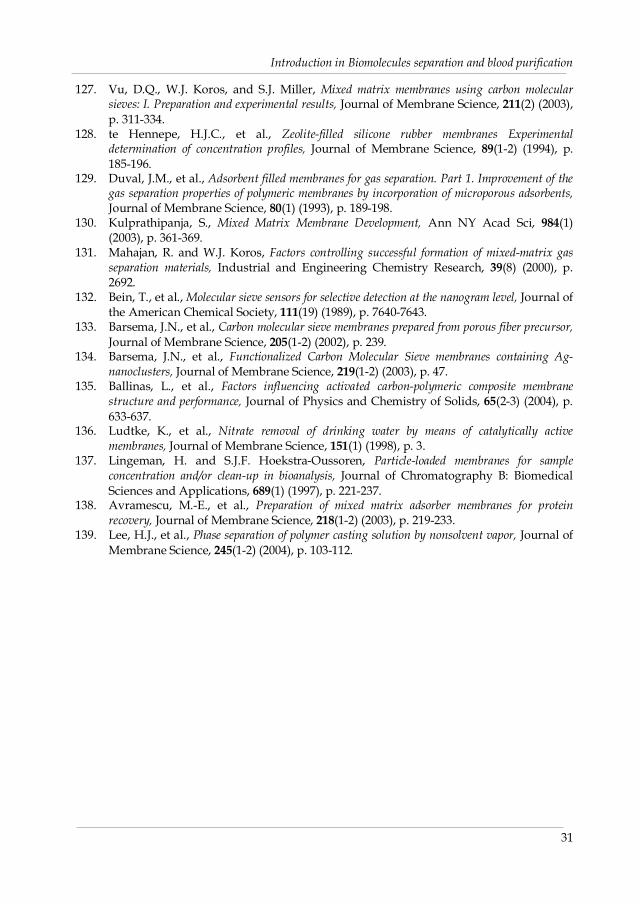
Introduction in Biomolecules separation and blood purification
31
127. Vu, D.Q., W.J. Koros, and S.J. Miller, Mixed matrix membranes using carbon molecularsieves: I. Preparation and experimental results, Journal of Membrane Science, 211(2) (2003),p. 311-334.
128. te Hennepe, H.J.C., et al., Zeolite-filled silicone rubber membranes Experimentaldetermination of concentration profiles, Journal of Membrane Science, 89(1-2) (1994), p.185-196.
129. Duval, J.M., et al., Adsorbent filled membranes for gas separation. Part 1. Improvement of thegas separation properties of polymeric membranes by incorporation of microporous adsorbents,Journal of Membrane Science, 80(1) (1993), p. 189-198.
130. Kulprathipanja, S., Mixed Matrix Membrane Development, Ann NY Acad Sci, 984(1)(2003), p. 361-369.
131. Mahajan, R. and W.J. Koros, Factors controlling successful formation of mixed-matrix gasseparation materials, Industrial and Engineering Chemistry Research, 39(8) (2000), p.2692.
132. Bein, T., et al., Molecular sieve sensors for selective detection at the nanogram level, Journal ofthe American Chemical Society, 111(19) (1989), p. 7640-7643.
133. Barsema, J.N., et al., Carbon molecular sieve membranes prepared from porous fiber precursor,Journal of Membrane Science, 205(1-2) (2002), p. 239.
134. Barsema, J.N., et al., Functionalized Carbon Molecular Sieve membranes containing Ag-nanoclusters, Journal of Membrane Science, 219(1-2) (2003), p. 47.
135. Ballinas, L., et al., Factors influencing activated carbon-polymeric composite membranestructure and performance, Journal of Physics and Chemistry of Solids, 65(2-3) (2004), p.633-637.
136. Ludtke, K., et al., Nitrate removal of drinking water by means of catalytically activemembranes, Journal of Membrane Science, 151(1) (1998), p. 3.
137. Lingeman, H. and S.J.F. Hoekstra-Oussoren, Particle-loaded membranes for sampleconcentration and/or clean-up in bioanalysis, Journal of Chromatography B: BiomedicalSciences and Applications, 689(1) (1997), p. 221-237.
138. Avramescu, M.-E., et al., Preparation of mixed matrix adsorber membranes for proteinrecovery, Journal of Membrane Science, 218(1-2) (2003), p. 219-233.
139. Lee, H.J., et al., Phase separation of polymer casting solution by nonsolvent vapor, Journal ofMembrane Science, 245(1-2) (2004), p. 103-112.

32

Enzyme Capturing and Concentration With
MMM Adsorbers*)
Abstract
This study reports the use of membrane adsorbers for Lysozyme (LZ) capturing and
concentration: the membrane adsorbers are prepared by incorporation of ion exchange resins
into an EVAL porous matrix. The Mixed Matrix Membrane (MMM) adsorber possesses an
open and interconnected porous structure with a large ion exchange surface available for
enzyme adsorption. The adsorptive membrane features both a high static as well as a high
dynamic LZ adsorption capacity. The measured LZ adsorption isotherm is of the Langmuir
type, with a maximum adsorption capacity of 147 mg LZ/mL membrane. Dynamic LZ
adsorption capacity at a flux of 25 Lm-2h-1 was 63 mg LZ/mL membrane, which is
significantly higher than the equivalent commercial membrane Sartobind C. Since the kinetics
of desorption processes are faster than the kinetics of adsorption processes, the performance
can be improved by exerting the desorption processes at higher fluxes than the adsorption
processes. The MMM can be reused in multiple adsorption/desorption cycles maintaining the
high binding capacity performance. Fluorescence spectra of the LZ after adsorption and
elution were similar to native LZ. This is confirmed by activity tests showing that the activity
of LZ was maintained after an adsorption and desorption cycle.
Keywords: Mixed Matrix Membrane (MMM), Sartobind C, adsorption, Lysozyme (LZ) andactivity
*) This chapter has been published in J. Membrane Sci., 280(1-2) (2006), 406-417
3Chapter

Chapter 3
34
3.1. Introduction
Chromatography is the method most widely used to obtain the required purity for
biotherapeutics and diagnostics[1]. Among all the chromatographic methods, ion exchange is
one of the most often used techniques in downstream processing. It is employed for recovery
and purification of proteins, polypeptides, nucleic acids, polynucleotides and other
biomolecules. Ion exchange is advantageous in terms of broad applicability, high resolution
and large adsorption capacity in large-scale protein purification processes[2]. Another
advantage of this technique is that the elution takes place under mild conditions so that the
proteins maintain their native conformation during the chromatographic processing. Protein
separation in ion-exchange chromatography is mainly determined by the electrostatic
interaction between the solute and the oppositely charged of the surface stationary phase[3].
Many types ion exchange resins are nowadays commercial available and have been applied
in protein capturing, purifying and polishing steps[4, 5].
The developments in bio downstream processes are promoted by reliable and
efficient separation and purification methods, for the many diverse biomolecules that are
produced and studied in biopharmaceutical and bioproduct industries. Large-scale
separation and purification processes must be cost efficient because they contribute for a large
extend to the total product manufacturing costs. In a typical protein production process from
fermentation to final product, isolation and purification account for 50 to 80% of the total
production costs[6, 7]. The desirable properties in protein purification are among others a
high throughput, a high capacity and the ability of the adsorbent to be used repeatedly[8].
In the recent years there is a considerable and increasing interest in developing
membrane adsorbers. The membrane adsorber acts as a short and wide chromatographic
column. Membrane adsorbers can exist in a variety of configurations[9-12] with a variety of
adsorptive mechanisms (e.g. ion exchange, hydrophobic, reversed-phase or affinity
interactions). The advantage of adsorptive membranes is the absence of a long diffusion path
length, which often occur in fixed bed chromatography. In adsorptive membranes, the
transport of the dissolved molecules to the active sites in the membrane occurs by convective
flow rather than by diffusion through a stagnant fluid inside the pores of an adsorbent
particle. Another major advantage of the membrane adsorbers is the relative ease of scaling
up when compared with packed bed systems[13]. Membranes with adsorptive
chromatography properties do exhibit technological progress in both membrane filtration and

Enzyme Capturing and Concentration with Mixed Matrix Membrane Adsorbers
35
fixed-bed liquid chromatography. Membrane adsorbers combine the selectivity of
chromatography resins with the high productivity of filtration membranes[10, 11, 13, 14].
Avramescu et al.[13] described a new chromatographic concept for a single-step
process for the preparation of ion exchange Mixed Matrix Membrane (MMM) adsorbers
having particulate material entrapped in a porous matrix. A mixture of dissolved polymers
and additives in which particulate material was dispersed, was cast/spun and solidified by a
phase inversion process into a flat/fibrous membrane. The prepared MMM contain ion
exchange particles tightly held together within a porous polymeric matrix, the latter not
interfering with the activity of the particles. Such membranes can be prepared in different
shapes and can be operated either as stacked microporous flat sheet membranes or as modules
containing fibrous membranes. By independent selection of particles and matrix material
various functions such as ion exchange, adsorption, catalysis or enzymatic activity can be
incorporated. Such membrane adsorbers may also serve as a platform to which an end-user can
couple a specific ligate[10].
MMM adsorbers have been applied for separation and recovery of BSA, Hb and
bilirubin[13, 15, 16]. Although several applications have been introduced to membrane
adsorbers already, there are still many interesting applications that remain to be explored
with respect to their performance in membrane chromatography. One of the greatest
challenges in downstream purification processing is the capturing and concentration of
enzymes. Many biomolecules are difficult to purify in a native and biological active form. An
effective and reliable separation technique is needed to achieve this requirement. Due to their
low pressure drop and mild process conditions, a MMM adsorbers is a good candidate to
maintain the native conformation and the biological activity of proteins or enzymes during
the isolation and purification processes. In this study we provide an extensive
characterisation of the MMM adsorbers in static and dynamic mode for enzyme recovery. A
weak cation exchange resin, Lewatit CNP80 WS, is incorporated as particulate material into a
porous EVAL matrix to prepare adsorber membranes with a high particle loading. The
performance of the prepared ion exchange MMM as enzyme adsorbers was investigated
using Lysozyme (LZ) as a model enzyme. System parameters such as membrane porosity,
swelling and permeability and process conditions like pH, ionic strength and membrane flux
were varied to study their effect on the performances of mixed membrane adsorber during
Lysozyme recovery.

Chapter 3
36
3.2. Experimental
3.2.1. Materials
EVAL (a random copolymer of ethylene and vinyl alcohol) with an average ethylene
content of 44 mol % was purchased from Aldrich and was used as membrane material
without further modification. Dimethylsulfoxide (DMSO, Merck) was employed as solvent
and 1-octanol (Fluka) as non-solvent-additive in the casting solution. Water was used as non-
solvent in the coagulation bath. Lewatit ion exchange resins CNP80 WS (Bayer) were used as
adsorbent particle. The particles were grinded and fractionated down to a fraction with an
average size of 10 µm. This size was chosen because of the high surface over volume ratio
while the pores in the membrane are still sufficient to maintain a high flux and to avoid
particle loss out of the matrix. Hydrochloric acid (HCl) is used for the regeneration of MMM
adsorbers. The enzyme used for the adsorption measurements was Lysozyme (LZ). LZ was
obtained by Fluka and has a Mw of 14600 g/mol and a pI around 11. Freeze-dried Micrococcus
lysodeikticus cells (ATCC strain # 4698) were used as enzyme substrate, purchased from Fluka
biochemical. Sartobind C membranes, kindly supplied by Sartorius, were used as a
representative of commercial membrane adsorbers.
Buffer solutions were freshly prepared in ultra pure water. Ultra pure water was
prepared using a Millipore purification unit Milli-Q plus. The buffers used for adsorption
were an acetate buffer for the pH 4-5 regime, a phosphate buffer for pH 3, 6-8 and 12 regime,
a Tris buffer for pH value 9 and a Carbonate buffer for pH values between 10 to11. The ionic
strength of the adsorption buffers was kept constant at 17 mM, which is close to physiological
conditions, by addition of NaCl. This low ionic strength results in maximum adsorption[17].
The elution buffer was prepared by increasing the ionic strength of the adsorption buffer pH
7.0 to 0.5 M using NaCl.
3.2.2. Membrane preparation and characterisation
3.2.2.1. Membrane preparation
To obtain membranes with protein adsorptive properties, Lewatit ion exchange
particles with an average diameter of 10 mm were added to a solution containing 10 %wt
EVAL and 10 % 1-octanol in DMSO. The 1-octanol was added to the casting solution in order
to improve the membrane morphology. The mixtures were stirred over night at 50 °C to
break down clusters of particles. The MMM were prepared by immersion precipitation. For
this the polymeric mixture was cast (casting knife 0.475 mm) on a glass plate and immediately

Enzyme Capturing and Concentration with Mixed Matrix Membrane Adsorbers
37
immersed into a water coagulation bath at 40-45 °C and the membranes were formed a few
moments after immersion. The membranes were washed with tap water at room temperature
to remove residual solvent and 1-octanol. After washing, the membranes were dried in the air
overnight and afterwards dried in a conventional oven at 50 °C.
An important parameter for the MMM adsorbers preparation is the resin loading, i.e.
the amount of resins contained in a membrane. The resin loading is calculated by equation (1),
where Wr is the amount of ion exchange resins (g) and Wp is the amount of EVAL polymer in
the casting solution (g).
100WW
WRrp
rloading ´
÷÷÷
ø
ö
ççç
è
æ
+= Eq . (3.1)
3.2.2.2. Scanning electron microscopy
The membranes were characterized by Scanning Electron Microscopy (SEM) using a
JEOL TM 220 A. The cross-section was scanned as well as the top and bottom surfaces of the
membrane. For the cross-section the membranes are fractured in liquid nitrogen. Before
examination the samples were gold-coated using Balzers Union SCD 040 sputter coater.
3.2.2.3. Membrane porosity and swelling degree
The membrane porosity was determined by the water uptake of a calibrated volume
of membrane. Average values were obtained from three different samples. The membrane
porosity, ε, and swelling degree, sd, were determined from the swelling experiments. A
micrometer of Mitutoyo was used to measure the thickness of the membrane. Average values
were obtained from three different samples. Porosity was calculated by the following
equation:
100V
VVε(%)
wetmembrane,
drymembrane,wetmembrane,×
-= Eq. (3.2)
The swelling degree is determined by the volume of water compared to the volume of the dry
membrane.
( ) 100V
VV%sd
drymembrane,
drymembrane,wetmembrane,×
-= Eq. (3.3)
Where Vmembrane,dry and Vmembrane,wet are respectively the volume of the dry membrane and the
volume of the swollen membrane after 24 hours immersing in a water bath at room

Chapter 3
38
temperature. Before weighing the attached water was removed by dry padding the
membrane with filter paper.
3.2.2.4. Clean water flux
Ultra pure water fluxes of the membranes are determined at room temperature by
using a nitrogen pressurized ‘Amicon type’ dead-end filtration cell. The applied pressure is 1
x 104 Pa (0.1 bar). The reported flux values are measured at steady state conditions, under 3.33
Hz (200 rpm). An average flux is calculated from several experiments using different pieces of
membrane.
3.2.3. Adsorption experiments
3.2.3.1. Measurement of zeta potential of Lysozyme
The net charge of Lysozyme as a function of pH is determined with a dynamic light
scattering method using a Malvern Zetasizer 2000. Acetate, phosphate and carbonate buffers,
all 17mM, are used to cover pH values ranging from 3 to 12. Lysozyme solutions were
prepared in the buffers in concentrations of 1 mg/mL. The sample is measured with the
Zetasizer for 20 seconds at least 5 times. All measurements were carried out at room
temperature (25 oC).
3.2.3.2. Batch adsorption experiments
The adsorption capacities of the prepared cation exchange membranes have been
determined by batch experiments. Protein concentrations of 1-2 mg/mL are employed in the
adsorption experiments. A known weight of membrane is immersed overnight in ultra-pure
water to ensure a steady inner structure during the experiments and afterwards transferred in
the protein solution. The protein solutions are prepared using different buffer solutions with
pH’s ranging from 3-12. The ionic strength of all buffer solutions applied for adsorption
experiments is kept constant at 17mM by adding NaCl. The samples containing a known
concentration of LZ are incubated with an exact amount of membranes in a sealed container
under continuous shaking at 25°C. The protein adsorbs on the membrane adsorbers thereby
reducing the LZ concentration in the bulk until equilibrium is reached. The equilibrium
protein concentration is determined by spectrophotometric analysis after 24 hours. Protein
absorption was measured at 280 nm and 2mm quartz cuvettes are used for measuring the

Enzyme Capturing and Concentration with Mixed Matrix Membrane Adsorbers
39
absorbance. The adsorbed protein amount, qeq [mg protein /g membrane], at equilibrium is
calculated by
membrane
eq0eq W
V)C(Cq
×-= Eq. (3.4)
Where C0 is the initial protein concentration [mg/mL], Ceq is the protein concentration at
equilibrium [mg/mL], V is the volume of the solution [mL] and Wmembrane is the weight of the
dry membrane [g].
3.2.3.3. Adsorption kinetics
Approximately 0.6 g of membrane adsorber was incubated in a LZ solution of pH 11
at 25°C in a shaking bath. The LZ concentration in the solution was monitored in time with a
Philips analytical PU 8720 UV-VIS Spectrophotometer. For the measurement on the pure
resins the solutions were filtered using a Spartan 30 filter (regenerated Cellulose) with a pore
size of 0.45µm to avoid possible disturbing of the UV-signal by resin particles.
3.2.3.4. Adsorption isotherm
In order to determine the adsorption isotherm batch adsorption experiments were
carried out with different initial protein concentration. The same amount of membrane was
used in the experiments to end up with different equilibrium concentrations. From literature
is known that the Langmuir adsorption model can be used for protein adsorption. The
Langmuir equation can be rewritten as:
eqmd
meq C1
qK
q1
q1
´+= Eq. (3.5)
The parameters qeq (adsorbed protein concentration into the membrane adsorbers) and Ceq
(the equilibrium protein concentration in the bulk solution) are experimental data. A plot of
1/Ceq versus 1/qeq allows determining the maximum adsorption capacity (qm) and the
dissociation constant (Kd), which have been estimated by fitting the equation to experimental
results using the least-square regression.
3.2.3.5. Desorption
After an adsorption and washing step the membranes are transferred into the
desorption buffer. Desorption was accomplished in static as well as in dynamic
measurements by using a phosphate buffer of pH=7, I= 0.5 M NaCl for static as well as

Chapter 3
40
dynamic experiments. The desorption was carried out for 24 hours in a shaking bath at 25 ºC.
The recovery is defined as the percentage of desorbed protein, the amount desorbed protein
(qdesorbed) divided by the amount of adsorbed protein (qadsorbed).
3.2.3.6. Regeneration
After an adsorption, washing and desorption cycle the membrane adsorbers were
washed using 0.50 M NaCl. Then 10 % HCl was used to guarantee complete removal of
residual LZ that might have been attached to the membrane by non-specific interactions and
cannot be eluted by a high ionic strength NaCl solution. The contact time between the
membranes and the HCl solution was about 24 h in total at room temperature. Before the next
adsorption/desorption cycle could start the membranes were conditioned by washing out the
remaining HCl with ultra pure water until neutral pH was obtained. The capacity of the
regenerated membrane was then re-measured. Since Sartobind C membranes are ready to use
(according to the producer) we did not test the effect of regeneration of these membranes.
3.2.3.7. Dynamic adsorption experiments
The dynamic adsorption performance of the flat sheet MMM was measured at
constant permeation rate using a continuous feeding stirred dead-end filtration cell. To
overcome the limited adsorption capacity and to average out inhomogeneities[18], 10 sheets
of membranes were stacked. 2 mg/mL LZ solution in Carbonate buffer pH 11 was loaded
through the stack of 10 membranes with a flux of 25 Lm-2h-1. The permeate was collected and
fractionated in samples of 2 mL using a fraction collector and the LZ concentration in the
permeate samples was followed spectrophotometrically at 280 nm. After reaching the
breakthrough point the experiment was stopped and the membrane stack was washed with
fresh buffer to release the unbounded proteins and subsequently a phosphate buffer
containing 0.5 M sodium chloride were permeated through the membranes for elution of the
adsorbed proteins. The protein mass adsorbed per unit of membrane bed was calculated by
numerical integration over the filtration run up to a breakthrough concentration 10% of the
feed concentration. The effect of the flux on both the loading and the elution were
investigated at five different fluxes (25, 50, 100, 300, 500 Lm-2h-1). A comparison of the effect of
the flux was also obtained for MMM and commercial membrane adsorbers. Sartobind C
membranes were used as representative commercial membrane.

Enzyme Capturing and Concentration with Mixed Matrix Membrane Adsorbers
41
3.2.3.8. Fluorescence measurements
In order to evaluate the effect of the process conditions on the LZ activity,
fluorescence spectra of the native LZ, heat denaturated LZ and desorbed LZ were measured.
LZ fluorescence emission spectra were recorded from 300 to 400 nm after excitation at 284 nm
using an Edinburgh Analytical Instruments F900. All measurements were carried out at 25 °C
at a LZ concentration of 2 mg/mL in the presence of the desired salts concentration and pH 7.
Native LZ aqueous solution (2 mg/mL, pH 11) was denaturated at 85 oC for 2 hours. LZ
fluorescence emission mainly stems from two buried Tryptophan (Trp) residues, which can
be selectively excited at 284 nm[19-21].
3.2.3.9. Enzymatic activity
The enzyme activity of LZ was determined by a spectrometric method using
Micrococcus lysodiekticus as substrate [22-24]. This assay is based on the decrease in Abs (450
nm) of a M. lysodeikticus cell suspension when the cells are digested by LZ. The amount of LZ
containing sample should be adjusted so that a decrease in absorbance of 0.02 to 0.04 per
minute is obtained (LZ concentration is about 0.0125 mg/mL). LZ was first dissolved either in
pure water or phosphate buffer (0.025 M) and kept on ice. 3 mg of lyophilized M. lysodeikticus
cells were dissolved in 10 mL 0.1 M phosphate buffer (pH 7), kept on ice. Optical density of
the cell suspension was measured at 450 nm. To obtain an optical density between 0.5 and 0.7
lyophilized cells or phosphate buffer was added to a cell suspension, final substrate
concentration about 0.3 mg/mL. Thereafter, 100 ml of the enzyme solution was added to 1.4
mL cell suspension of M. lysodeikticus and the change in the A450 was recorded every 15
seconds for 2 minutes. Temperature was kept constant at 25 oC. One activity unit (U.A.) is
equivalent to a decrease of 0.001 absorbance unit per minutes. Specific activity is the ratio of
enzyme activity to mass of protein in the sample, usually expressed as units of activity per
mg protein (U/mg).
3.3. Results and Discussion
Experimental results are presented and discussed in this section. First the charge
density of LZ as function of pH is described. Thereafter, a detailed membrane
characterization in terms of morphology, porosity and clean water flux is presented, followed
by LZ adsorption capacity in both static and dynamic mode. Most relevant is the dynamic
capacity, which represents the functional capacity at defined fluxes. The influences in both

Chapter 3
42
adsorption and desorption processes will demonstrate the applicability of MMM in the
isolation LZ maintaining its biological activity.
3.3.1. Net Charge of Lysozyme
The pH of a polypeptide (enzyme in our case) influences the behavior and amount
of adsorption in ion exchange processes. Identifying the electrostatic properties of a protein
surface is useful for the interpretation of biochemical data and for a better understanding the
ion exchange adsorption behavior. Figure 3.1 shows the net charge of LZ at different pH’s.
The values are in agreement with literature data[25-28]. The pI, the pH where the LZ net
charge is zero, is 11.0.
3.3.2. Mixed matrix membrane adsorbers characterization
Lewatit CNP80 WS, a weak cation exchange resin, was employed in the preparation
of mixed matrix membranes (MMM). The membrane containing Lewatit CNP80 WS has
carboxylic functional groups as active binding sites. An important parameter in the
preparation of MMM is the amount of resins incorporated into the polymeric matrix. Both a
high loading and the application of small particles favor a high adsorption capacity of the
MMM.
Figure 3.1. Zeta potential of LZ at different pH’s in buffered 17 mM ionic strength solutions.Experimental data were compare with data reported from literature.
0 2 4 6 8 10 12-8
-4
0
4
8
12
16
20
Zp (m
V)
pH
Kuehner et al. Hayne et al This work
pI

Enzyme Capturing and Concentration with Mixed Matrix Membrane Adsorbers
43
The viscosity of the casting solution increases with increasing resin loading. The
maximum resin loading that could be obtained was 65%; above this loading the viscosity of
the casting solution was too high for a proper casting of the polymer solution. The membrane
with a 65 % resin loading has a desired set of properties. The membrane posses an open and
interconnected porous structure (Figure 3.2). There is no evidence of finger-like macrovoids
and the resin particles are uniformly distributed in the membrane matrix across the entire
cross-section (Figures 3.2A and 3.2B). The ion exchange particles are tightly held together
within the porous polymeric matrix and were uniformly distributed. No significant loss of
particles was observed during the membrane formation process on the glass plate and the
membrane possesses sufficient mechanical strength. The porosity of these membranes was
70% and swelling degree was 22 %. Clean water flux was 100 Lm-2h-1 at a trans-membrane
pressure of 1 x 10⁴ Pa (0.1 bar).
Figure 3.2. SEM micrograph of the membrane prepared out of a 10% EVAL, 10% 1-octanol, in DMSOsolution containing 65% loading Lew CNP80 WS. Temperature of the coagulation bath 40-45oC and the membranes were dried by at room temperature and conventional oven at 50 oC. A).Cross section, magnification x350, the size bar indicates 50mm; B) Cross section,magnification x750, the size bar indicates 10 mm; C) Bottom surface, magnification x1500, thesize bar indicates 10 mm; D) Top surface, magnification 10000x, the size bar indicates 1 mm.
A B
C D

Chapter 3
44
Ideal membrane adsorbers possess a low non-selective protein adsorption onto the
polymer surface and a high adsorption onto resin particles. By incorporating resin particle in
the matrix polymer the non-specific adsorption will decrease. Since EVAL is a hydrophilic
polymer the protein adsorption on the particle free membrane is 5.5 mg/mL membrane,
which is less than 4 % of the lowest obtained total adsorption capacity at pH=9 in Figure 3.4.
The adsorption of LZ on particle-free membranes was mainly attributed to hydrophobic
interaction and dipol-dipol interaction.
The adsorption of LZ was measured in time for suspensions of resins and MMM to
investigate the accessibility of the active sites (Figure 3.3). The adsorption measurements were
performed at pH 11. We found that the adsorption capacity increased in time during 12 hours
where after equilibrium was reached. The obtained equilibrium capacity after 24 hours was
305 mg LZ /g resin and for the MMM 205 mg/g membrane which is equal to 315 mg/g resin
in the membrane. The equivalent values for the LZ adsorption capacity of pure resins and
resins incorporated into the polymeric structure proved that the active sites in the membrane
are well accessible for LZ.
0 4 8 12 16 20 240
50
100
150
200
250
300
350
Ads
orbe
d LZ
(mg
/g a
dsor
bent
)
Time (hour)
MMM Resin in MMM Pure resin
Figure 3.3. Static adsorption of LZ onto suspension resin Lew CNP80 WS and MMM LewCNP80 WS in time at pH 11.0 and temperature 25oC. LZ initial concentration was 2mg/mL at ionic strength 17 mM and equilibrium time 24 hours.
The total protein adsorption depends on the accessibility of the adsorptive resins in
the membrane. Figure 3.4 compares the LZ adsorption capacity of the pure resins, the mixed

Enzyme Capturing and Concentration with Mixed Matrix Membrane Adsorbers
45
matrix membrane in total and the resin as estimated to be in the MMM at different pH’s. First
of all, this graph clearly shows that all particles are well accessible in the MMM adsorber. It
also can be seen that LZ adsorption declines up to a pH=9 where after a maximum adsorption
is obtained at pH=11. Above pH=11 the sorption of LZ drops in capacity, which can be
related to repulsive charges and instability of the protein. At pH=3 the adsorption capacity of
the pure resins was 260 mg LZ per gram of adsorbent: this number decreased gradually to
220 at pH=9 and after that it increased again and reached a maximum at pH=11 of 305 mg LZ
per gram resin where after the capacity drops till 190 at pH=12. The adsorption profile of LZ
in MMM adsorbers has a similar tendency.
Proteins have no net charge at their isoelectric points and therefore the maximum
adsorption from aqueous solutions is usually observed at their isoelectric point [29]. This
nonlinear dependence is inconsistent with an adsorption mechanism dominated by net
electrostatic forces between enzyme and adsorbent: otherwise the adsorption capacity would
be highest at extreme acid pH and decrease monotonically with increasing pH. Around the
isoelectric point where the protein carries as many positively charged group as negatively
charged ones, the net intramolecular coulombic interaction is attractive and dispersive (or
London-van der Waals) interaction favors a compact structure. At pI, there exists no net
Figure 3.4. LZ adsorption on suspended Lewatit CNP80 WS resins and MMM Lewatit 80 WSadsorbers at different pH’s and temperature 25 oC. LZ initial concentration was 2mg/mL at ionic strength 17 mM.
2 4 6 8 10 120
50
100
150
200
250
300
350
Ads
obed
LZ
(mg/
g ad
sorb
ent)
pH
MMMResin in MMMPure resin

Chapter 3
46
electrostatic repulsion between the proteins in the adsorption layer. Therefore the proteins
can be tightly packed on the surface[30] and this leads to increased adsorption at pI.
Additional contribution may be related to interaction of the hydrogen bonding between the
charged resin and neutral protein. Haynes et al.[27] also found that LZ adsorption on a
negatively (-SO3- group) charged polystyrene latex shows a maximum at or near the pI of the
protein. Shen et al.[31] developed the stoichiometric displacement model, which accounts for
the effects of hydrogen ion Donnan equilibrium (or protein charge regulation) during the ion
exchange adsorption particularly for case of adsorption near the pI of the protein. The net
charge of a protein depends not only on pH and ionic strength of the surrounding fluid, but
also on the proximity of protein to other charge surfaces, resulted from electrostatic
interaction between the charged surface and the ionogenic functional groups on the protein.
When the pH is near the protein pI, protein charge regulation is an important factor, which
causes discrepancy between the characteristic binding charge and the electrical charge on the
protein in free solution. The adsorption of protein near pI of protein contributes from
hydrogen ion Donnan equilibrium effect. The ion Donnan equilibrium also explains the
ability of proteins to adsorb onto a surface with the same charge sign because the protein in
the fluid and adsorbed phase can have an opposite charge signs due to hydrogen Donnan
equilibrium effect. Kang et al.[32] reported that adsorption near the pI could improve
resolution of proteins when using ion-exchange chromatography focusing with salt gradient
elution. The resolution of protein in IEC is highest when the pH of liquid phase is near the pI
of protein since this condition again enhances relative adsorption affinity. The investigator
suggests that the adsorption of protein near the pI controlled by charge distribution on the
surface. This argument is also reasonable to rationalize adsorption of LZ near the pI. A LZ
molecule has 20 positively charged groups, 1 is histidine, 8 lysine and 11 arginine groups.
Besides these positively charged groups LZ has also 10 negatively charged groups namely 2
glutamic acid and 8 aspartic acid[33]. Rasmol molecular-graphic visualization of Lysozyme
shows that most residue groups reside on or near the molecular surface[25]. The present of
positively charged lysine and arginine groups on the surface favor positive charge around pI
of LZ, which can interact with a cation exchange surface. An additional explanation can be
found in the solubility of lysozyme, which is dependant on the pH and the buffers used. Since
the chaotropic effect of phosphate and acetate buffers used in this research is similar (same
position in the Hofmeister series) the difference can only be explained by the pH dependant
solubility. Ladisch [34] reported data about the solubility of lysozyme in his book

Enzyme Capturing and Concentration with Mixed Matrix Membrane Adsorbers
47
Bioseparations Engineering, Table 4.5. The maximum solubility is between pH=6 and 7
(almost 50 % higher than at pH 5 and 8). Expected is that the adsorption follows this behavior.
The measured LZ adsorption isotherm on MMM is used to characterize the
interaction with the adsorbent. This provides a relationship between the concentration of the
protein in solution and the amount of protein adsorbed on the solid phase when the two
phases are in equilibrium. Adsorption isotherm data were obtained for LZ at pH 11 and 17
mM ionic strength, as shown in Figure 3.5. Equilibrium data were fit with the Langmuir
model by nonlinear least-squares regression analysis. The solid line in the figure is the
Langmuir isotherm that best fit the data. The maximum adsorption capacity (qm) is 147 mg
LZ/mL membrane and the dissociation constant (Kd) is 4.4x10⁻² mg/mL. This value prove
that the mixed matrix membrane adsorbers posses a good accessibility of LZ to the adsorptive
site. The adsorption capacities are high compared with published data in literature. Tejeda-
Mansir et al.[35] investigated adsorption of LZ on dye cellulosics. Their Langmuir isotherm
values
0.0 0.4 0.8 1.2 1.6 2.00
50
100
150
Kd = 0.044 mg/mlqm= 147 mg LZ/ ml membrane
Ads
orbe
d LZ
(mg/
ml m
embr
ane)
Ceq
(mg/ml)
Figure 3.5. Correlation between Experimental data and Langmuir model of the LZ adsorptionisotherm at ionic strength 17 mM, equilibrium time 24 hours
were qm=2.0 mg/mL and Kd=5.9x10⁻² mg/mL. LZ adsorption capacities of Zn(II), Cu(II) and
Ni(II)-chelated hollow fibers were different. The maximum capacities of Zn(II), Cu(II) or
Ni(II)-chelated hollow fibers were 144, 75 and 69 mg/g, respectively [26]. LZ adsorption
capacity of a dye-immobilized membrane was 13.3 mg/mL [36]. The high capacity of the

Chapter 3
48
described MMM adsorbers can be attributed to the fact that the surface over volume is high
when small sized ion exchange particles in high loadings are embedded. Lysozyme
adsorption takes place mainly at the outer surface of the particles: when instead of small
particles big porous particles are used the area available for adsorption is much lower.
3.3.3. Effect of ionic strength on LZ desorption
A complete separation sequence should include an adsorption, desorption, washing
and regeneration step. Desorption (elution) of ion exchange adsorbent is commonly
performed by increasing of the salt concentration or by changing the pH of the elution buffer.
A phosphate buffer at pH=7 with varying concentration of NaCl was used in desorption
experiments. The desorption of the adsorbed LZ from MMM was studied in a batch system.
Figure 3.6 gives the amount of LZ desorbed using different salt concentrations. Desorption of
LZ from the MMM can be improved by increasing the ionic strength of the desorption buffer.
In operations, this results in a higher productivity.
0.0 0.5 1.0 1.5 2.00
65
70
75
80
85
90
95
100
Des
orpt
ion
(%)
NaCl (M)
Figure 3.6. Effect of salt on static LZ desorption from membrane Lew CNP80 WS. Desorptionexperiments were perform at pH 7 and in buffered 0.5 M NaC, equilibrium time 24hours at temperature 25 oC
Elution involves displacement of the protein from fixed charges by other counterions
with a greater binding affinity for the fixed charge than the protein and which then becomes
the new counter ion. The ability of counter ions (salts) to displace the protein bound to fixed

Enzyme Capturing and Concentration with Mixed Matrix Membrane Adsorbers
49
charge is a function of the difference in affinities between the fixed charges and non-fixed
charges of both the protein and the salt. Affinities in turn are affected by several variables,
including of magnitude of the net charge of protein and the concentration and type of salt
used for displacement [37]. As the salt concentration is stepped up, salt diffuses faster to the
particle creating very large driving forces for desorption. By these conditions a fraction of the
adsorbed protein becomes rapidly unbound and diffuses away from the particle surface[38].
Dickman and Proctor[39] indicated that the LZ activity increases by increasing the
NaCl concentration from 0 till 0.5 M but declines rapidly when the NaCl concentration is
higher than 0.5 M. For our system, desorption of LZ increases with the ionic strength. At 0.5
M NaCl more than 98 % of the adsorbed LZ was recovered and recovery stays high. In all
desorption or elution experiments 0.5 M NaCl solutions were applied.
3.3.4. Regeneration and reuse the membrane Lew CNP80 WS
It is important to characterize the adsorption and desorption capacity as function of
sequence of adsorption and desorption cycles. After an adsorption and desorption step,
regeneration was accomplished by washing the membranes with 10% HCl to guarantee
thorough removal of non-specific bound components which were not eluted by the
desorption buffer. The contact time between the membrane and the HCl solution was about
0
30
60
90
120
150
First Second Third Fourth
(mg
LZ/m
l mem
bran
e)
Adsorption Desorption
Figure 3.7. Repeated static adsorption-desorption LZ cycles onto MMM Lew CNP80 WS.Adsorption at pH 11 and temperature 25°C. Initial LZ concentration was 2 mg/mLat ionic strength 17 mM. Desorption at pH 7 in buffered 0.5 M NaCl. Theadsorption capacity was measured after 24 hours

Chapter 3
50
24 hour in total at room temperature. After the regeneration step the HCl was washed out
with ultra pure water until neutral pH. During the regeneration the enzyme was completely
removed from the resin. Regenerating the membrane for three times showed no loss in
adsorption capacity as shown in Figure 3.7. The adsorption after the first regeneration is
slightly higher than in the first adsorption cycle. An explanation for this phenomenon may be
contamination or partly inactivation of the unconditioned resins. After the first regeneration
step the particles are clean and the intrinsic adsorption capacity can be obtained.
3.3.5. Dynamic adsorption of LZ on membrane Lew CNP80 WS
The breakthrough and elution curves of the cation-exchange porous membrane
adsorbers for LZ are shown in Fig. 3.8 as a function of permeate volume of the protein
solution. A LZ feed solution (2mg/mL) was loaded at pH 11. In the desorption step a buffer
of pH 7 with ionic strength 0.5 M NaCl was applied. The dynamic binding capacity is defined
as the amount of protein bound when the effluent concentration reaches 10% of the feed
concentration. The dynamic LZ adsorption capacity at 10% breakthrough was 58 mg LZ/mL
membrane at a flux 50 Lm-2h-1. This is about 2.5 times lower than the maximum static binding
capacity. Elution of the adsorbed protein-MMM complex with phosphate buffer pH 7 at a flux
Figure 3.8. Typical breakthrough and elution curves of LZ for a stack of 10 MMM Lew CNP80WS operated at a feed concentration of 2 mg/mL LZ and a constant filtration flux of50 Lm-2h-1.
0 20 40 60 80 100 120 140 1600.0
0.2
0.4
0.6
0
2
4
6
8
10D
esor
ptio
n, C
/C0 (-
)
Ads
orpt
ion,
C/C
0 (-)
Permeate Volume (ml)
Loading Washing Elution

Enzyme Capturing and Concentration with Mixed Matrix Membrane Adsorbers
51
of 50 Lm-2h-1, leads to LZ recovery values up to 98 %. The protein was concentrated up to 8
fold into the desorptive buffer, as indicated on right-hand y-axis of Figure 3.8.
The obtained dynamic LZ adsorption capacity was lower than the corresponding
static capacity because convective contribution to mass transport dominates over the diffusive
contribution. Liquid containing the protein molecules flows preferentially through the large
pores. Adsorption in smaller pores can only occur by slower diffusional transport, hence
these sites are less likely to be accessed. Bypassing and uneven flow distributions are others
sources, which lead to lower dynamic capacities. Full adsorption capacity can only be
achieved if the protein residence time in the MMM adsorber is long enough to allow each
adsorbate molecules to diffuse to the adsorptive site and to rearrange/unfold their structure
at the adsorptive interface.
3.3.6. Effect of the flux on dynamic adsorption
A lysozyme solution (2 mg LZ/mL) prepared in a binding buffer pH 11 was used to
study the effect of flux on the adsorption capacity. By changing the flux from 25 up to 500
Lm-2h-1 a capacity loss of a factor 3 was found. We can conclude from Figure 3.9 that the
dynamic adsorption capacity is constant using fluxes above 300 Lm-2h-1. Although the loss in
capacity between 25 and 300 Lm-2h-1 is significant, this drop in capacity is acceptable,
considering the 20-fold increase in throughput. Nevertheless, the independent ratio of flux
and adsorption in the high flux regimes indicates that in this convective controlled regime LZ
had sufficient time to bind to the binding sites in the mixed matrix membranes. Furthermore,
we can conclude from the breakthrough curves that the effect of the flux is much bigger in the
adsorption mode than in the elution mode (Figure 3.10), especially when fluxes above 100
Lm-2h-1 were applied. Adsorption of proteins on solid surfaces is a complex process and is
influenced by many factors such as re-arrangement and conformational changes of the
protein where as desorption is strongly dependant on the ionic strength and the kinetics the
salt ions.
Dynamic adsorption capacity is strongly correlated with the accessible area and is
influenced to a large extent by mean pore size, pore size distribution, pore interconnectivity
and the protein diffusion coefficient and residence time in the adsorber membrane. Sartobind
C membrane adsorbers that posses the same carboxylic functionality as the CNP80 containing
MMM are selected as a commercial reference. The morphology of Sartobind membranes is
very different: the cation exchange functionality is grafted onto the surface of the starting
membrane. The MMM adsorber obtain their functionality through the mixing of cation

Chapter 3
52
exchange particles into the bare membrane. Hence it is difficult to relate any difference in
adsorption performance to morphological characteristics. Rather, we like to compare the two
different membranes in order benchmark the MMM adsorbers to an industrial reference.
0 100 200 300 400 5000
20
40
60
80
100
120
140
160D
BC (m
g/m
l mem
bran
e)
Permeate flux (Lm-2h-1)
Mixed Matrix membrane Sartobind C membrane
Solid points represent static experiments
Figure 3.9. Dynamic binding capacity of LZ on MMM Lew CNP80 WS and Sartobind Cmembrane at different flux and room temperature. LZ loading concentration was 2mg/mL at ionic strength 17 mM. (DBC=dynamic binding capacity)
The dynamic binding capacity of the Sartobind membranes is almost independent of
the flux (Figure 3.9 and Table 3.1). Convection controls the adsorption and only at low fluxes
there is an additional adsorption due to diffusion of LZ to the active sites in diffusional pores.
This result is in agreement with the data published by Santarelli et al.[40]. They studied the
dynamic capacity of strong cation exchange Sartobind S membranes at different permeate
rates. They found that by increasing the permeate flux from 120 Lm-2h-1 to 1200 Lm-2h-1 a
slight decrease in adsorption from 1.33 till 1.20 mg/cm2 membrane. Our MMM adsorbers
show a bigger flux dependent adsorption behavior. The MMM adsorbers start with an up to 7
times higher adsorption capacity compared to the Sartobind C membranes. The Sartobind
membranes show an almost flux independent capacity, but the capacity of the MMM
adsorbers is flux sensitive. Nevertheless, the dynamic capacity is twice as large at a flux of 500
Lm-2h-1.

Enzyme Capturing and Concentration with Mixed Matrix Membrane Adsorbers
53
Table 3.1. Dynamic binding capacities of LZ on membrane Lew CNP80 WS and Sartobind C at10 % breakthrough point
Flux(Lh-1m-2)
Dynamic binding(mg LZ/mL membrane)
Concentrated Recovery (%)
MMM SC MMM SC MMM SC25 63 16 9 fold 3.5 fold 98 9950 58 11 8 fold 3.5 fold 98 99100 42 11 7 fold 3.5 fold 98 99300 22 11 6.5 fold 3.5 fold 98 99500 20 11 6 fold 3.5 fold 98 99
MMM= mixed matrix membrane, SC = sartobind C
The throughput during elution is an important parameter for the efficient
productivity of LZ from the membranes. Fig. 3.10 presents the elution profiles of LZ at
different fluxes. A 0.025 M sodium phosphate buffer containing 0.5 M NaCl could elute LZ
bound to the membrane quantitatively. Sharp elution peaks were obtained at all fluxes. The
capacities at different fluxes of the CNP80 WS containing adsorbers are summarized in Table
1. From this we can conclude that highest productivity can be obtained by adsorbing at
moderate fluxes followed by desorption at high fluxes. The MMM adsorber shows much
higher concentration factors, 9 fold, during elution compared to the Sartobind membrane, 3
fold, This can be contributed to the higher adsorption capacity of the MMM adsorber where
small embedded IEX-particles are responsible for a high active surface area per volume of
Figure 3.10. Typical elution curve of LZ at different flux on MMM Lew CNP80 WS andSartobind C membrane. Phosphate buffer pH=7 containing 0.5 M NaCl was usedas elution buffer
.
0 5 10 15 20 25 300
2
4
6
8
10
C/C
0(-)
Permeate volume (ml)
25 lm2h-1
50 lm2h-1
100 lm2h-1
300 lm2h-1
500 lm2h-1
Mixed matrix membrane
0 5 10 15 20 25 300
1
2
3
4
C/C
0(-)
Permeate volume (ml)
25 lm-2h-1
50 lm-2h-1
100 lm-2h-1
300 lm-2h-1
500 lm-2h-1
Sartobind C membrane

Chapter 3
54
membrane where as the Sartobind membranes possess a low active area because of the
presence of big go through pores. And since almost all the active sites in the Sartobind
membranes are located on the pore wall of these convective pores, the Sartobind membranes
are less sensitive to the rate of the permeate flux.
3.3.7. Lysozyme stability and biological activity
Stability and activity of enzymes are essential during their production, storage and
use to promote their industrial applicability. However, environmental conditions such as
high temperature, shear, extreme pH and strong salinity can cause protein denaturation,
leading to protein aggregation[23]. Native and denatured states of a protein can be
investigated by fluorescence spectroscopy[20-22, 41-43]. LZ is a protein that contains six
tryptophan residues, but only two of them, Trp62 and Trp108, appear to dominate the
fluorescence spectrum. Trp62 and Trp108 are arranged close to the substrate-binding site (the
protein split). They play an important role in substrate binding to an inhibitor and in the
protein stabilization. Fluorescence analyses of these tryptophan residues have provided
information on the LZ-ligand interaction and ligand-induced conformational change around
the binding site[44, 45].
The fluorescence spectra of native LZ, recovered from elution step and denaturated
LZ were recorded (Figure 3.11). A significant change can be observed with respect to the
maximum wavelength of 339 nm: the denaturated LZ shows a much broader maximum as
compared to native one. In contrast, the fluorescence spectra of the LZ obtained from the
elution step resemble the one of the native LZ very well and no significant shift of maximum
wavelength was detected in the spectra of these samples relative to the native protein. This
indicates that no detectable conformational change of LZ in mixed membrane adsorbers
separation processes.
Specific activity and total activity are critical parameters in enzyme purification. The
influence adsorption and elution process on enzymatic activity was also evaluated. A
suspension of M. luteus bacteria is subjected to LZ fraction, the cell walls are broken down
and the optical density of this suspension decreases with time. This occurs because the LZ
induces bacterial cell lysis by hydrolyzing b-1,4 linked glycosidic bond of cell wall’s
peptidoglycan. The rate of this reaction depends on the amount of active enzyme present, so
the rate of decrease in optical density can be used to measure enzyme activity[23, 24, 46]. LZ
obtained by MMM was used to compare the activity properties with native one.

Enzyme Capturing and Concentration with Mixed Matrix Membrane Adsorbers
55
300 320 340 360 380 400
Flou
resc
ence
Inte
nsity
(a.u
)
Wavelength (nm)
LZ in Water LZ in BPS pH7 LZ fraction at pH7 LZ denaturated by heating
Figure 3.11. Fluorescence spectra obtained for LZ before adsorption, after desorption anddenaturated. All measurement were carried out at 25 oC and LZ concentration 2mg/mL. (BPS=buffer phosphate)
The adsorption and elution process did not significantly affect to the activity of LZ.
The specific activity of native LZ was 79000 unit/mg and the specific activity of recovered LZ
was 73000 unit/mg. This activity test demonstrates that fraction of LZ was maintained above
90 % activity after the separation process. This confirms that the MMM process is a very
gentle process; the active LZ was maintained above 90% of the native Lysozyme.
3.4. Conclusions
The performance of MMM adsorbers for enzyme capturing and concentration was
studied. The MMM adsorbers are prepared by a phase inversion method. The capacity is
established by embedding of small particles into a microporous polymeric membrane. A
membrane with higher resin loading is preferable for providing a higher adsorption capacity.
The MMM adsorbers demonstrate a high static and dynamic protein adsorption capacity and
an easy scale up for the separation of LZ. The dynamic binding was found to be dependent on
the flux. Where the dynamic elution of LZ is independent of the flux and leads to several fold
concentration and LZ recovery values up to 98 %. The fluorescence spectra of the LZ from the
elution step indicate that no obvious conformational change of LZ took place. Activity test
with Micrococcus lysodeikticus as the substrate demonstrate that the activity of isolated LZ was

Chapter 3
56
maintained for more than 90 % after separation processes. This indicates that the MMM
adsorbers process is a mild capturing and concentration process for LZ recovery. A
comparison between MMM and commercial membranes demonstrates that MMM posses a
higher dynamic adsorption capacity. The MMM adsorbers concept combines robustness, with
high capacities and high throughput and mild process conditions.
3.5. Acknowledgements
We want to acknowledge the financial support for this work through the Membrane
Technology Group of the University of Twente and the Ministry of Education of the Republic
of Indonesia. We thank Sartorius kindly for supplying the Sartobind C membranes.
3.6. List of Symbols and Abbreviations
Symbols
C0 Initial protein concentration mg/mL
Ceq Protein concentration at equilibrium mg/mLI Ionic strength mol/lKd Dissociation constant mg/mLMw Molecular weight g/molQ Adsorption capacity mg/g membraneqdesorbed Amount of protein desorbed mg/g membraneqeq Adsorption capacity at equilibrium mg/g membraneqm Maximum adsorption capacity mg/g membraneR Recovery %sd Swelling degree %V Volume mLW Weight gWr Amount of ion exchange resin gWp Amount of polymer gzi Charge number -e Porosity %# Number -
AbbreviationsATCC American type culture collectionBSA Bovine serum albuminCER Cation exchange resinDMSO Dimethylsulfoxide

Enzyme Capturing and Concentration with Mixed Matrix Membrane Adsorbers
57
EVAL Ethylene-vinyl copolymerHCl Hydrochloric acidLZ LysozymeLew LewatitMMM Mixed matrix membranepI Isoelectric pointU.A Unit activityTrp Tryptophan
References1. Sofer, G., Preparative chromatographic separations in pharmaceutical, diagnostic, and
biotechnology industries: current and future trends, Journal of Chromatography A, 707(1)(1995), p. 23-28.
2. DePhillips, P. and A.M. Lenhoff, Determinants of protein retention characteristics on cation-exchange adsorbents, Journal of Chromatography A, 933(1-2) (2001), p. 57-72.
3. Hallgren, E., et al., Protein retention in ion-exchange chromatography: effect of net charge andcharge distribution, Journal of Chromatography A, 877(1-2) (2000), p. 13-24.
4. Zhou, X., et al., Macroporous polymeric ion exchanger of high capacity for protein adsorption,Biochemical Engineering Journal, 11(1) (2002), p. 13-17.
5. Biotech, A.P., Protein purification handbook, 2003, Amersham Parmacia Biotech Limited.6. Kawai, T., K. Saito, and W. Lee, Protein binding to polymer brush, based on ion-exchange,
hydrophobic, and affinity interactions, Journal of Chromatography B, 790(1-2) (2003), p. 131-142.
7. Karumanchi, R.S.M.S., et al., Field-assisted extraction of cells, particles and macromolecules,Trends in Biotechnology, 20(2) (2002), p. 72-78.
8. Koguma, I., et al., Multilayer Binding of Proteins to Polymer Chains Grafted onto PorousHollow-Fiber Membranes Containing Different Anion-Exchange Groups, Biotechnol. Prog.,16(3) (2000), p. 456-461.
9. Charcosset, C., Review: Purification of Proteins by Membrane Chromatography, J.Chem.Technol. Biotechnol, 71 (1998), p. 95-110.
10. Klein, E., Affinity membranes: a 10-year review, Journal of Membrane Science, 179(1-2)(2000), p. 1-27.
11. Roper, D.K. and E.N. Lightfoot, Separation of biomolecules using adsorptive membranes,Journal of Chromatography A, 702(1-2) (1995), p. 3-26.
12. Thommes, J. and M.R. Kula, Review: Membrane Chromatography-An Integrative Concept inthe Downstream Processing of Proteins, Biotechnol. Prog., 11 (1995), p. 357-367.
13. Avramescu, M.-E., et al., Preparation of mixed matrix adsorber membranes for proteinrecovery, Journal of Membrane Science, 218(1-2) (2003), p. 219-233.
14. Van Reis, R. and A. Zydney, Membrane separations in biotechnology, Current Opinion inBiotechnology, 12(2) (2001), p. 208-211.
15. Avramescu, M.-E., Z. Borneman, and M. Wessling, Mixed-matrix membrane adsorbers forprotein separation, Journal of Chromatography A, 1006(1-2) (2003), p. 171-183.
16. Avramescu, M.E., et al., Adsorptive membranes for bilirubin removal, Journal ofChromatography B, 803(2) (2004), p. 215-223.
17. Avramescu, M.-E., et al., Dinamic Behavior of Adsorber Membranes for Protein Recovery,Biotechnology and Bioengineering, 84(5) (2003), p. 564-572.
18. Liu, H.-C. and J.R. Fried, Breakthrough of lysozyme through an affinity membrane of cellulose-cibacron blue, AIChE Journal, 40(1) (2003), p. 40-49.

Chapter 3
58
19. Imoto, T., et al., Fluorescence of Lysozyme: emission from trypthophan residues 62 and 108 andenergy migration, Proc Nat Acad Sci USA, 69 (1971), p. 1151-1155.
20. Pérez, C. and K. Griebenow, Effect of salts on lysozyme stability at the water-oil interface andupon encapsulation in poly(lactic-co-glycolic) acid microspheres, Biotechnology andBioengineering, 82(7) (2003), p. 825 - 832.
21. Senel, S., et al., Dye-affinity hollow-fibres and their lysozyme adsorption-desorptioncharacteristics, Polymer International, 50(10) (2001), p. 1143-1149.
22. Malzert, A., et al., Spectroscopic studies on poly(ethylene glycol)-lysozyme interactions,International Journal of Pharmaceutics, 260(2) (2003), p. 175-186.
23. Colombie, S., A. Gaunand, and B. Lindet, Lysozyme inactivation under mechanical stirring:effect of physical and molecular interfaces, Enzyme and Microbial Technology, 28(9-10)(2001), p. 820-826.
24. Lee, Y.C. and D. Yang, Determination of lysozyme activities in a microplate format, AnalyticalBiochemistry, 310(2) (2002), p. 223-224.
25. Kuehner, D.E., et al., Lysozyme Net Charge and Ion Binding in Concentrated AqueousElectrolyte Solutions, J. Phys. Chem. B, 103(8) (1999), p. 1368 -1374.
26. Lee, H.-M., Y.W. Kim, and J.K. Baird, Electrophoretic mobility and zeta-potential of lysozymecrystals in aqueous solutions of some 1: 1 electrolytes, Journal of Crystal Growth, 232(1-4)(2001), p. 294-300.
27. Haynes, C.A., E. Sliwinsky, and W. Norde, Structural and Electrostatic Properties ofGlobular Proteins at a Polystyrene-Water Interface, Journal of Colloid and Interface Science,164(2) (1994), p. 394-409.
28. Palacio, L., et al., Fouling with protein mixtures in microfiltration: BSA-lysozyme and BSA-pepsin, Journal of Membrane Science, 222(1-2) (2003), p. 41-51.
29. Etheve, J., P. Dejardin, and M. Boissiere, Influence of pH on the adsorption of lysozyme on asulfonated membrane with and without poly(ethyleneimine), Desalination, 146(1-3) (2002), p.111-113.
30. Norde, W. and J. Lyklema, Why protein prefer interfaces, J. Biomater. Sci. Polymer Edn.,2(3) (1991), p. 183-202.
31. Shen, H. and D.D. Frey, Charge regulation in protein ion-exchange chromatography:Development and experimental evaluation of a theory based on hydrogen ion Donnanequilibrium, Journal of Chromatography A, 1034(1-2) (2004), p. 55-68.
32. Kang, X. and D.D. Frey, High-performance cation-exchange chromatofocusing of proteins,Journal of Chromatography A, 991(1) (2003), p. 117-128.
33. Lu, J.R., T.J. Su, and B.J. Howlin, The Effect of Solution pH on the Structural Conformation ofLysozyme Layers Adsorbed on the Surface of Water, J. Phys. Chem. B, 103(28) (1999), p. 5903 -5909.
34. Ladisch, M.R., Bioseparations engineering: Principle, practice, and economic, 2001, New York,Wiley.
35. Tejeda-Mansir, A., et al., Breakthrough performance of stacks of dye-cellulosic fabric in affinitychromatography of lysozyme, Bioprocess and Biosystems Engineering, 25(4) (2003), p. 235-242.
36. Senel, S., et al., Comparison of adsorption performances of metal-chelated polyamide hollow fibremembranes in lysozyme separation, Colloids and Surfaces B: Biointerfaces, 24(3-4) (2002), p.265-275.
37. Deutscheur, M.P., Methods in Enzymology: Guide to protein purification, Vol. Volume 182,1998, London, Academic press.
38. Hunter, A.K. and G. Carta, Protein adsorption on novel acrylamido-based polymeric ion-exchangers: III. Salt concentration effects and elution behavior, Journal of Chromatography A,930(1-2) (2001), p. 79-93.

Enzyme Capturing and Concentration with Mixed Matrix Membrane Adsorbers
59
39. Dickman, S. and C.M. Proctor, Factors Affecting the activity of egg white lysozyme, Arch.biochem. biophys., 40 (1952), p. 364-371.
40. Santarelli, X., et al., Characterization and aplication of new macroporous membrane ionexchangers, J. of Chromatography B, 706 (1998), p. 13-22.
41. Buijs, J. and V. Hlady, Adsorption Kinetics, Conformation, and Mobility of the GrowthHormone and Lysozyme on Solid Surfaces, Studied with TIRF, Journal of Colloid andInterface Science, 190(1) (1997), p. 171-181.
42. Czeslik, C. and R. Winter, Effect of temperature on the conformation of lysozyme adsorbed tosilica particles, Phys. Chem. Chem. Phys., 3 (2001), p. 235-239.
43. Clark, S.R., P. Billsten, and H. Elwing, A fluorescence technique for investigating proteinadsorption phenomena at a colloidal silica surface, Colloids and Surfaces B: Biointerfaces, 2(5)(1994), p. 457-461.
44. Nishimoto, E., et al., Internal Motion of Lysozyme Studied by Time-Resolved FluorescenceDepolarization of Tryptophan Residues, Biochemistry, 37(16) (1998), p. 5599-5607.
45. Lehrer, S.S., Solute Perturbation of Protein Fluorescence.The Quenching of the TryptophylFluorescence of Model Compounds and of Lysozyme by Iodide Ion, Biochemistry, 10(17) (1971),p. 3254-3263.
46. Malzert, A., et al., Influence of some formulation parameters on lysozyme adsorption and on itsstability in solution, International Journal of Pharmaceutics, 242(1-2) (2002), p. 405-409.

60

Enzyme Capturing and Concentration from
Binary Mixtures
Abstract
Developments in bioseparations are necessary to fulfill the demands of higher throughputs,
higher selectivity’s and higher capacities. The Mixed Matrix Membranes (MMM) concept is
proposed as a moderate process with an increased throughput and dynamic adsorption
capacity. This study presents the application of EVAL-based adsorptive MMM containing
weak cation exchange resins for the separation and concentration of protein mixtures. The
MMM are prepared by a wet phase inversion method. The adsorptive membranes feature a
high adsorption capacity in both static and dynamic modes. High separation factors and
purities of Bovine Serum Albumin (BSA) and Lysozyme (LZ) are obtained in effluent and
elution buffers. LZ, the retained protein, is recovered with a high purity in the elution buffer
in a five fold increased concentration. At fluxes above 20 Lm-2h-1, the adsorption capacity and
purification power are independent of the flux. The MMM can be reused in multiple
adsorption/desorption cycles thereby maintaining the high binding capacity. The operational
conditions using MMM adsorbers are mild and do not affect the three-dimensional structure
of the LZ molecules, which is proven by fluorescence spectroscopy and biological activity
tests. The MMM act in protein separations both as protein purifier and concentrator and is an
attractive alternative for packed bed systems because of its high capacity, high throughput,
robustness, ease of scaling up and mild separation conditions.
Keywords: Membrane adsorbers, Bovine Serum Albumin (BSA), Lysozyme (LZ) and adsorption
4Chapter

Chapter 4
62
4.1. Introduction
Bio-downstream processing by means of protein isolation and purification plays an
important role in the formulation of biopharmaceutical products and contributes significantly
to overall production costs[1, 2]. An improvement of existing methods or the development of
a new technology is necessary to overcome the shortcomings of the classical bio-downstream
processes.
In the early days of protein purification, protein separation from complex mixtures
was based on protein precipitation by water miscible organic solvents[3]. Alternative
processes including adsorption, gel filtration, liquid phase partitioning, electrophoretic
methods and membrane technology have been developed for protein purification. Membrane
filtration is used primarily for purification and concentration of biomolecules. Membrane
filtration offers a fast route for removal of cell debris, colloidal or suspended solids and
viruses from homogenized suspensions of bacterial cells[4-6]. One of the major limitation
membranes based on sieving properties is their relatively low resolution[7, 8]. Therefore, this
technique is not used for fine separation. The another drawbacks of membrane filtration in
protein filtration is concentration polarization and membrane fouling[9-11]. Adsorption
techniques often result in purification steps with greatest increase in protein purity.
Therefore, these techniques have become widely used especially when adopted in
combination with chromatographic [12-14] and membrane processes[15-19].
Several authors have described the fractionation of Bovine serum albumin (BSA) and
Lysozyme (LZ) mixtures by means of membrane filtration and adsorptive membrane
chromatography. Iritani et al.[20] reported the fractionation of BSA and LZ by membrane
filtration using 30 kDa polysulfone membranes. The membranes were assumed to be
permeable for LZ and almost completely retentive for BSA. The LZ permeation was found to
be very strongly depending on the BSA concentration. An increase in BSA concentration
resulted in a decrease in the LZ flux. Millesime et al.[21] reported the fractionation of BSA and
LZ using both modified and unmodified inorganic membranes. Since protein fractionation is
affected by protein - membrane surface interactions, the separation selectivity may be
improved by modification of the membrane surface. Several authors[22-24] tried to improve
the membrane performance by optimizing the process parameters such as membrane
pretreatments, and adjusting the pH and ionic strength of the protein solution. Kanani et
al.[25] reported a favorable protein-protein interaction by adding BSA in the UF process for
the separation of LZ from myoglobin. A binary mixture of LZ and myoglobin was

Enzymes Capturing and Concentration from Binary Mixtures
63
ultrafiltered using a 30 kDa MWCO polyethersulfone membrane. LZ was permeated
significantly faster than myoglobin. However, when BSA was added to the feed solution, LZ
was largely retained while the permeation of myoglobin was significantly increased. The
decrease in LZ permeation rate was explained by the electrostatic BSA–LZ interaction, while
the simultaneous increase in myoglobin permeation was contributed to the Donnan effect in
combination with size exclusion.
Fang et al.[26] investigated the separation of BSA and LZ by using polysulfone-
based cation-exchange membranes. In batch experiments at pH 7.4, the adsorption capacity of
LZ was 15.6 mg/mL membrane, where BSA showed no adsorption at all. In a dynamic
adsorption process using a 10 layered membrane stack, BSA and LZ were effectively
separated with a LZ recovery of about 93 %. Lin et al.[27] investigated the separation of BSA
and LZ using ion-exchange membranes in plate-and-frame modules. In batch mode
experiments they found that using cation-exchange membranes the maximum LZ adsorption
capacity was 46.5±17.8 mg/mL membrane and the maximum BSA adsorption capacity onto
anion-exchange membrane was 47.5±20.0 mg/mL membrane. In a dynamic filtration process,
the separation performance is for large extend influenced by the operating conditions.
Furthermore a critical factor that limits the application of adsorptive membrane systems in
protein fractionation is a low binding capacity[26, 28].
In bioseparation processes, chromatography is another essential technique for
obtaining the required purity of biotherapeutic and diagnostic products. By using
chromatographic methods, very good separations and high purities can be achieved.
Chromatographic processes are traditionally carried out using packed beds. However, these
methods have some severe major drawbacks[19, 29]. An approach to overcome the limitations
associated with packed beds is to use synthetic microporous or macroporous membranes as
alternative chromatographic stationary phase[15, 19]. Avramescu et al.[30] proposed a simple
method to produce macroporous adsorptive membranes, the so called Mixed Matrix
Membranes (MMM) adsorbers concept. The MMM adsorbers concept is an effective approach
to increase the selectivity and capacity of membrane chromatographic systems.
Functionalization of the macroporous membranes takes place by entrapping small affinity
particles, it results in an uniform distribution of ligands through the whole matrix and a
capacity comparable with that of packed bed systems[31, 32]. The MMM adsorber captures
selectively proteins of interest using the affinity properties of the adsorbent. Unbound
materials and interferences are washed out where after the bound species are eluted in high

Chapter 4
64
concentration and purity. An additional advantage of the MMM adsorbers is the robustness.
This makes that an intensive pretreatment, such as commonly used in packed bed
chromatography, is unnecessary. The MMM-technique combines the selectivity and capacity
of chromatographic resins (particles) with the low flow resistance of macroporous
membranes[33]. Important is that the nature and activity of the proteins (enzymes) is not
affected by the purification process[31].
Applications of the MMM have been investigated for the separation and recovery of
single components, i.e. BSA, Hb, LZ, bilirubin[30, 31, 34, 35]. Recently, the use of MMM
adsorbers was reported for fractionation of BSA-Hb mixtures[33]. In the present work, new
applications of the MMM for fractionation of BSA and LZ mixtures are studied. BSA and LZ
are representing two extremes with respect to their physicochemical properties[36, 37]. At
physiological pH LZ is positively charged whereas BSA is a negatively charged.
In this chapter, we present an extensive performance study of the MMM application
for the fractionation of BSA and LZ in both static and dynamic mode. To prepare weak cation
exchange MMM adsorbers, Lewatit CNP80 WS particles are incorporated into a porous EVAL
matrix. System parameters such as membrane properties and the adsorption conditions in
chromatographic separations of proteins are investigated. Adsorption selectivity of BSA and
LZ from mixtures is evaluated and compared with literature data.
4.2. Experiments
4.2.1. Materials
EVAL (a random copolymer of ethylene and vinyl alcohol) with an average ethylene
content of 44 mol % was purchased from Aldrich and used as a membrane material without
further modification. Dimethylsulfoxide (DMSO, Merck) was employed as solvent and 1-
octanol (Fluka) as non-solvent additive in the casting solution. Water was used as a non-
solvent in the coagulation bath. Ion exchange functionality was introduced by embedding
Lewatit CNP80 WS (a weak acidic macroporous acrylic-based cationic exchange resin), kindly
supply by Caldic, Belgium. The particles were milled and air classified to obtain a fraction
with an average particles diameter of 10 micron. Hydrochloric acid (HCl) was used for the
regeneration of the MMM adsorbers. Bovine Serum Albumin (BSA) and Lysozyme (LZ)
obtained from Fluka were used for the adsorption measurements.
Buffer solutions were freshly prepared in ultra-pure water. Ultra-pure water was
prepared using a Millipore purification unit Milli-Q plus. The 17 mM ionic strength of buffers

Enzymes Capturing and Concentration from Binary Mixtures
65
used for zeta potential measurements were an acetate buffer for the pH 4-5 regime, a
phosphate buffer for pH 3, 6-8 and 12 regimes, a tris buffer for pH value 9 and a carbonate
buffer for pH values between 10 to 11. The ionic strength of the adsorption buffers (7.5 mM
phosphate buffer pH 7) was kept constant at 17 mM by the addition of NaCl. pH 7 was
chosen as the operational pH for adsorption, which is close to the physiological pH. The
elution buffer was prepared by increasing the ionic strength of the adsorption buffer to 0.5 M
using NaCl.
4.2.2. Membrane preparation
The membranes used in this study are prepared by the phase inversion method. The
membranes are prepared out of a DMSO solution, containing 10 % EVAL, 10 % 1-octanol and
65 wt% of resin loading. The 1-octanol was added to the casting solution in order to improve
the membrane morphology. The mixture was stirred over night at 50 °C to break down
possible particles clusters. The MMM were prepared by immersion precipitation. For this, the
polymeric mixture was cast (casting knife 0.475 mm) on a glass plate and immediately
immersed into a water coagulation bath at 40-45 °C and the membranes were formed a few
moments after immersion. Then the membranes were washed with tap water at room
temperature to remove residual solvent. After washing, the membranes were overnight air
dried and afterwards dried in a conventional oven at 50 °C.
4.2.3. Membrane Characterization
4.2.3.1. Scanning electron microscopy
The cross sections, as well as the top and bottom surfaces of the membrane were
characterized by Scanning Electron Microscopy (SEM, JEOL TM 220 A). For the
characterization of the top and bottom surfaces, pieces of membranes are air dried and glued
with double side adhesive tape on sample stubs. For the cross-section the wet membranes are
fractured in liquid nitrogen. After preparation the samples are dried at 30 ºC in a vacuum
oven and before examination gold-coated using a Balzers Union SCD 040 sputter coater.
4.2.3.2. Membrane porosity and swelling degree
The membrane porosity (ε) and swelling degree (sd) were determined from swelling
experiments by calculating the weight and the volume of both dry and wet membranes. Their
porosity was determined from the water uptake of a calibrated volume of dry membrane. The

Chapter 4
66
membranes were submersed for 24 hour at room temperature in water and the change in
weight was recorded. Before weighing the attached water was removed from the surface of
the swollen membrane by padding it dry using a filter paper. A digital micrometer
(Mitutoyo) was used to measure the membrane thickness. The reported values are averages
of three different samples.
4.2.3.3. Clean water flux
Water fluxes of the membranes are determined at room temperature by using a
nitrogen pressurized stirred dead-end filtration cell. The applied pressure is 1x10⁴ Pa (0.1
bar). The reported flux values are measured at steady state conditions, at 3.33 Hz (200 rpm).
The average flux was calculated from numerous experiments using different pieces of
membrane.
4.2.4. Adsorption experiments
4.2.4.1. Zeta potential measurement of pure BSA and LZ
The net charges of BSA and LZ as a function of pH were determined using a
Malvern Zetasizer 2000. Acetate, phosphate and carbonate buffers, all 17 mM ionic strength,
were used to cover pH values ranging from 3 to 12. Protein solutions are prepared in the
buffers in concentrations of 1 mg/mL. The Zeta potential was measured during 20 seconds
for at least five times. All measurements were carried out at room temperature (25 oC).
4.2.4.2. Adsorption isotherm
Protein adsorption onto ion exchange resins follows the Langmuir adsorption
isotherm[17, 38]. To determine the adsorption isotherm, batch adsorption experiments were
carried out using different initial protein concentrations. After 24 hours, the protein depletion
was determined spectrophotometrically at 280 nm in 2mm quartz cuvettes. The equilibrium
data are plotted and fitted with the linear form of the Langmuir model by the using least-
squares regression analysis. From this plot the maximum adsorption capacity (qm) and the
dissociation constant (Kd) can be calculated.
4.2.4.3. Dynamic adsorption experiment
The dynamic adsorption performance of flat sheet EVAL adsorber membranes is
measured at a constant permeation rate using a nitrogen-pressurized stirred dead-end

Enzymes Capturing and Concentration from Binary Mixtures
67
filtration cell. To average out membrane inhomogeneities, 6 membrane sheets were stacked.
A solution containing BSA and LZ in 17 mM ionic strength of phosphate buffer at pH 7 was
permeated with a flux of 50 Lm-2h-1 through the membrane stack. Permeate was collected and
fractionated in samples of 2 mL. After the breakthrough point was reached, the experiment
was stopped and the stack was washed with binding buffer to rinse out the unbounded
proteins. Subsequently, a phosphate buffer containing 0.5 M sodium chloride was permeated
through the membrane stack to elute the adsorbed proteins. The BSA and LZ concentrations
in the feed and permeate samples were analyzed by HPLC (Waters 717 plus) attached with a
Waters 2487 dual l absorbance detector. An analytical protocol based on gradient
chromatography using a cation exchange column (Water HPLC Column, BiosuiteTM CM 10
mm CXC) is used. As binding buffer 20 mM phosphate buffer pH 6.1 and as eluting buffer 20
mM phosphate buffer pH 6.1 containing 0.5 M NaCl is used. The protein mass adsorbed per
unit of membrane volume is calculated by numerical integration over the filtration run until a
breakthrough concentration of 10 % was reached. The effect of permeation rate of both
loading and elution was investigated at 3 different permeation rates (25, 50, 100 Lm-2h-1). For
the 50 Lm-2h-1 the effect of the ionic strength on the adsorber performance is also investigated.
4.2.4.4. Regeneration
After an adsorption, washing and desorption cycle, the membrane adsorbers were
washed with 10 % HCl to guarantee complete removal of residual BSA and LZ that might
have been attached to the membrane surface by non-specific interactions and was not eluted
by the high ionic strength NaCl solution in the desorption step. Before the next
adsorption/desorption cycle could start, the membranes were pre-conditioned by washing
out the remaining HCl with ultra pure water until neutral pH. The adsorption capacity of the
regenerated membrane was then re-measured.
4.2.4.5. LZ stability and enzymatic activity
Effect of adsorption and desorption conditions on LZ stability and enzymatic
activity were determined. The LZ stability was obtained by measuring the fluorescence
spectra of native LZ, heat-denaturated LZ and desorbed LZ using Edinburgh Analytical
Instruments F900. The LZ fluorescence emission spectra were recorded from 300 to 400 nm at
284 nm excitation wavelength. The enzyme activity of LZ was determined by a spectrometric

Chapter 4
68
method using Micrococcus lysodiekticus as substrate. This assay is based on the decrease in
absorbance (450 nm) of a M. lysodeikticus cell suspension when LZ digests the cells.
3 mg of lyophilized M. lysodeikticus cells were dissolved in 10 mL 0.1 M phosphate
buffer (pH 7), kept on ice. LZ was dissolved in 0.025 M phosphate buffer and kept on ice. The
amount of LZ in the sample had to be adjusted so that a decrease in absorbance of 0.02 to 0.04
per minute was obtained (LZ concentration is about 0.0125 mg/mL). Optical density of the
cell suspension was measured at 450 nm. To obtain an optical density between 0.5 and 0.7,
lyophilized cells or phosphate buffer were added to a cell suspension to reach a final
substrate concentration about 0.3 mg/mL. Thereafter, 100 ml of the enzyme solution was
added to 1.4 mL cell suspension of M. lysodeikticus and the change in the A450 was recorded
every 15 seconds for 2 minutes. Temperature was kept constant at 25 oC. One activity unit
(U.A.) is equivalent to a decrease of 0.001 absorbance unit per minutes. Specific activity is the
ratio of enzyme activity to mass of protein in the sample, usually expressed in units of activity
per mg protein (U/mg).
4.3. Results and Discussions
Experimental results are presented and discussed in this section. First, charge
densities of pure BSA and LZ as function of pH are described. Followed by a detailed
membrane characterization in terms of morphology, porosity and clean water flux. Then, the
protein adsorption and fractionation using the MMM adsorbers is evaluated in both static
and dynamic mode. Finally, the protein separation efficiency was evaluated by determining
the separation factor and purity of the isolated product.
4.3.1. Zeta potentials BSA and LZ
The zeta potential gives information about the overall surface charges of
components in relation to its environment. Insight in the protein surface charge improves the
understanding of the ion exchange adsorption behavior. The density of charges of single BSA
and LZ were determined between pH 3 until 12 in 1 mg/mL solutions. Figure 4.1 presents the
net charge of BSA and LZ at different pH values. The figure illustrates that the electrostatic
properties differ significantly as function of pH. The IEP for which the net charges are zero
can be deduced from the graph as 4.7 and 11.0 for BSA and LZ respectively. These values are
in agreement with literature data [10, 39]. The graph further shows that at pH 7, close to
physiological pH, the BSA has a net negative charge where LZ has a net positive charge. This

Enzymes Capturing and Concentration from Binary Mixtures
69
discrepancy in electrostatic properties can be exploited in adsorptive separation using cation
exchange materials. Consequently, pH 7 was chosen as operational pH in the fractionation of
BSA and LZ process.
2 4 6 8 10 12 14
-60
-40
-20
0
20
40
BSApHZp
(mV
)
LZ
Figure 4.1. Zeta potentials of pure BSA and LZ at difference pH’s, measured in buffered 17 mMionic strength solutions.
4.3.2. Membrane characterization
Figure 4.2 presents SEM images of Mixed Matrix Membrane (MMM) Lewatit CNP
80WS prepared by direct immersion precipitation in a water containing coagulation bath. An
important parameter for the preparation of membrane adsorbers is the amount of active
groups that can be incorporated into the polymeric matrix. The adsorption capacity increases
proportionally with IEX resin loading[30]. The maximum resin load applied in this research
was 65 wt%. Above this level, the viscosity of the casting solution is too high for a proper
casting. In addition, a very high particles loading reduces the mechanical strength of the
membrane, which can lead to mechanical failure under pressure. The membranes containing
a 65 wt% IEX resin loading are well structured and posses an open and interconnected porous
structure.
There are no evident finger-like macrovoids present in the membrane and the resins
are uniformly distributed over the entire cross-section. The absence of macrovoids is required
in membrane chromatography because macrovoids create mal-distribution, leading to lower
dynamic adsorption capacities. The ion exchange particles are tightly held together within the
porous polymeric matrix and no significant loss of the particles was observed during the
membrane formation. The membranes mechanical strength is sufficient, the porosity is 70 %

Chapter 4
70
and the swelling degree 20 %. The membranes demonstrate high clean water fluxes due to a
high pore interconnectivity and porosity. The clean water fluxes are on average 1000
Lm-2h-1bar-1. This high permeability allows MMM adsorbers stacking to increase the module
adsorption capacity and to minimize flow mal-distribution.
Figure 4.2. SEM micrographs of an EVAL based membrane adsorber containing 65wt% LewatitCNP80 particles: A) and B) Bottom-surface, magnification x50 and x1500; C) and D)Cross section, magnification x350 and x2000. The membranes were prepared by directimmersion precipitation in water containing coagulation batch at room temperature.
4.3.3. Protein adsorption isotherm
The protein adsorption isotherms are measured to characterize the interaction of
each protein with the adsorbent at a certain pH and ionic strength. The outcome provides the
relationship between the equilibrium concentration of the protein in the bulk solution and the
amount of protein adsorbed on to the solid phase. The adsorption isotherm for single BSA
and LZ are determined in batch experiments at pH 7 and 17 mM ionic strength. The
measured adsorption isotherms appear to be Langmuir type for both proteins. The obtained
adsorption isotherms are presented in Figure 4.3. The maximum adsorption capacities (qm) of
A
C
B
D

Enzymes Capturing and Concentration from Binary Mixtures
71
BSA and LZ are 8 and 91 mg/mL membrane respectively (equal to 11.70 and 133.10 mg/g
membrane for BSA and LZ respectively). The dissociation constants (Kd) are 3.2 10-1 mg/mL
for BSA and 4.5 10-2 mg/mL for LZ, which confirms a stronger LZ than BSA interaction with
the membrane adsorber. The obtained static capacities of the adsorptive membranes for BSA
and LZ are higher than the reported values in literatures that range from 6 to 40 mg per mL
membrane[17]. Fang et al.[26] reported 15.6 mg LZ/mL membrane for the adsorption
capacity with a cation-exchange activated polysulfone-based membranes, the membranes did
not show any measurable BSA adsorption.
Based on the assumption that the MMM and proteins mainly interact through
electrostatic interactions, the adsorption behavior of the different mixtures can be anticipated.
At an operational pH (pH=7), the MMM demonstrates higher adsorption for LZ than for BSA.
Since at pH 7 the MMM is negatively charged, the effective interaction takes place
predominantly with the positively charged LZ molecules and not with the negatively charged
BSA molecules. BSA adsorption mainly takes place at positive domains in the overall
negative charged BSA molecules and by non-specific interaction. When the pH is above the
isoelectric point BSA changes into a more extended conformation with more hydrophobic
Figure 4.3. Adsorption isotherms for pure BSA and LZ on mixed matrix membrane adsorbersat pH7 and ionic strength 17 mM, equilibrium time 24 hours. Experimental dataare correlated with the Langmuir adsorption isotherm model. The solid line figure isbest fit with the Langmuir isotherm.
0 1 2 3 40
20
40
60
80
100
Kd = 0.32 mg/ml
Kd = 0.045 mg/ml
qm = 8 mg/ml membraneq,
eq(m
g/m
l mem
bran
e)
C,eq
(mg/ml)
LZ
BSA
qm = 91 mg/ml membrane

Chapter 4
72
groups exposed to the outer part of the molecule leading to higher adsorption values on solid
interfaces[40]. This nonspecific protein adsorption also plays a role in the protein separation
process.
3.4. Breakthrough curves for BSA and LZ at flux 50 lm-2h-1
The typical breakthrough and elution curves of BSA and LZ mixtures using cation-
exchange porous membrane adsorbers as a function of permeate volume of the protein
solution are shown in Figure 4.4. In the dynamic adsorption experiments, a stack of 6
membranes was used to increase the adsorption capacity and to minimize flow
mal-distribution by averaging out membrane heterogeneities. The dynamic binding capacity,
0 20 40 60 80 100 120 1400
1
2
3
4
5
LZ
C/C
o (-)
Permeat volume (ml)
BSA
Adsorption Washing Elution
Figure 4.4. Typical breakthrough and elution curves of BSA and LZ mixtures through a 6membranes stack at pH 7 and ionic strength 17 mM and a constant filtration flux of 50Lm-2h-1. Feed concentration was 1 mg/mL for both BSA and LZ. Elution steps wereperformed at pH7 with 7.5 mM phosphate buffer containing 0.5 M sodium chloride.
DBC 10 %, defined as the amount of bound protein per volume membrane when the effluent
concentration reaches 10 % of the feed concentration, is 7 mg/mL membrane for BSA and 20
mg/mL membrane for LZ. The breakthrough curve also confirms that most adsorbed BSA
elutes during the washing step, which increases the LZ purity in the elution step. The amount
of eluted LZ, calculated by numerical integration over the elution curve, shows that 98 % of the
loaded LZ is recovered. Besides a higher purification grade, the LZ is also more than five fold

Enzymes Capturing and Concentration from Binary Mixtures
73
concentrated into the desorptive buffer. Biomolecular targets are generally present in low
concentrations in complex mixtures, which mean that often-large volumes have to be treated.
The adsorptive MMM technology is because of the low flow resistance and the robustness is
very suitable for handling large volumes containing low concentrations of target proteins.
The adsorptive MMM acts both as purifier and concentrator in this LZ recovery process.
The obtained dynamic LZ adsorption capacity (at DBC 10 %) is lower than the
corresponding static adsorption capacity. This can be explained by the fact that in the static
mode slow diffusional processes control the adsorption where as in dynamic mode there is a
balance between convection and diffusional processes. Since not all the adsorptive sites are
located in big convective go-through-pores the adsorption capacity of the system is to a
certain extend flow rate dependent. Besides this also membrane stack flow mal-distribution
contributes to a lower DBC 10 % compared to the static adsorption. These lower dynamic
adsorption capacities are in agreement with data published by Avramescu et al. [30] and
Roper et al [17],who also found lower dynamic than static adsorption capacities.
4.3.5. Purity BSA and LZ at flux 50 Lm-2h-1
The separation efficiency of BSA and LZ is evaluated by the separation factor. The
separation factor SBSA/LZ is defined as:
LZf,f,BSA
LZp,p,BSA
BSA/LZ
CC
CC
S = (4.1)
where Cf,BSA and Cf,LZ are the protein concentrations in the feed solution and Cp,BSA and Cp,LZ
are the protein concentrations in the collected permeate fractions during filtration, calculated
by numerical integration of the permeation curve over the filtration run until the 10 %
breakthrough. Figure 4.5 presents the purity of the individual proteins during permeation of
BSA – LZ mixtures in the adsorption, washing and elution step.
At the operating pH (pH=7), the membrane adsorbs preferential LZ due to opposite
protein/membrane charges and allows BSA to freely pass the membrane due to repulsive
charge forces. During the first part of filtration run, the LZ concentration in permeate is below
the HPLC detection limit (40 µg/mL). Hypothetical LZ concentrations, being the detection
limit of the HPLC, are used to calculate a minimal separation factor. In the first filtration
fractions this separation factor, SBSA/LZ, is above 1100, which indicates a BSA purity of at least
99.9 % (Figure 4.5). In the wash step weakly and unbound BSA is rinsed out in low

Chapter 4
74
concentration with high purities. Finally, the bound products (LZ) are dissociated from the
adsorptive IEX resins and eluted. The eluted LZ fractions have a high purity of more than
95% and are initially more than 5 times concentrated when compared with the feed solution.
The overall LZ process recovery is more than 98 %.
0 20 40 60 80 100 120 1400
20
40
60
80
100
LZPuri
ty (%
)
Permeat volume (ml)
BSA
Adsorption Washing Elution
Figure 4.5. The protein purity in the different process steps. The feed contains equal amounts ofBSA and LZ. In the adsorption and washing steps, the BSA purity is high and the LZpurity is high in the elution step. Initial BSA and LZ concentrations were 1 mg/mL,ionic strength 17 mM and the filtration flow rate 50 lm-2h-1.
Separations of BSA and LZ mixtures by ultrafiltration and adsorptive membranes
have been investigated in several studies. The BSA and LZ purity obtained by ultrafiltration
strongly depends on the membrane solute rejection and process parameters. Bellare et al.[41]
reported an almost complete BSA retention where the LZ retention varies with the amount of
treated volume. The obtained membrane selectivity was 20 and the LZ purity in the permeate
was above 95 %. Fang et al.[26] reported BSA and LZ separation with a cation exchange
polysulfone based membrane, ten membrane layers were stacked in a plate and frame
module. The BSA and LZ were effectively separated at permeate fluxes of 74 Lm-2h-1 with a
LZ recovery above 93 %. The main advantages of MMM systems in the fractionation of
proteins compared to ultrafiltration and another adsorptive membranes are the combination
of both a high static and dynamic adsorption capacity. Furthermore, the MMM can operate at

Enzymes Capturing and Concentration from Binary Mixtures
75
higher flow rates than ultrafiltration membranes without loosing their capacity. Moreover,
higher purities and higher concentrations can be obtained during elution.
4.3.6. Effect of the permeation rate
The influence of the permeation rate, which is proportional to the protein residence
time in the adsorber, on the dynamic LZ binding capacity and the separation factor SBSA/LZ are
examined by increasing the flux from 25–100 Lm-2h-1. Figure 4.6 shows that the LZ dynamic
binding capacity slightly depends on the LZ residence time in the membrane. When the flux
increased from 25 to 50 Lm-2h-1 the dynamic LZ binding capacity decreases only 5 %. A
further decrease in the LZ residence time does not result in any further decline in the
dynamic adsorption capacity.
Figure 4.6 also shows a slight initial dependency of the separation factor SBSA/LZ on
the flux. At a flux of 25 Lm-2h-1, the separation factor SBSA/LZ is 1200 and decreases by only 8 %
when the filtration flux increased by 100 % (50 Lm-2h-1). A further flux increase has no impact
on the separation factor SBSA/LZ.
0 20 40 60 80 1000
10
20
30
40
0
400
800
1200
1600S
BSA/LZ
DBC
(mg
LZ/m
l mem
bran
e)
Flux (l/m2/h)
DBC S,BS
A/L
Z
Figure 4.6. The influence of the flux on the dynamic LZ binding capacity and BSA-LZ separationfactor. Feed concentration for LZ and BSA 1 mg/mL, ionic strength 17 mM.
The adsorption process involves the transport of proteins from the feed solution to
the interface, their binding to the surface usually via electrostatic and hydrophobic
interactions and their refolding on the surface via conformational changes[42]. Full utilization

Chapter 4
76
of the adsorption capacity can be achieved only if the protein residence time in the membrane
adsorbers is sufficient to allow the protein molecules to diffuse to the adsorptive sites. In
traditional used packed bed systems containing gel type beads the adsorption process is for a
big extend diffusive controlled, which means flow rate dependent. The advantage of the
described MMM adsorbers is that almost all the active sites are reached by convection, which
means that at high flow rates the MMM adsorbers are more effective than packed bed
systems.
4.3.7. Effect of ionic strength
Electrostatic interactions are the dominating driving forces for protein adsorption
onto ion exchange surfaces. The protein adsorption behavior can be controlled by varying the
ionic strength of the protein solution. The effect of the ionic strength on the dynamic
adsorption capacity and the BSA purity in the effluent fractions is represented in Figure 4.7.
The LZ adsorption capacity slowly decreases with increasing ionic strength and the
separation factor follows the same tendency.
0 20 40 60 80 1000
5
10
15
20
25
0
25
50
75
100
BSA purity
DBC
(mg
LZ/m
l mem
bran
e)
Ionic strength (mM)
LZ DBC
BSA
pur
ity (%
)
Figure 4.7. The effect of the ionic strength on the BSA purification. The experiments are performedat room temperature, pH 7, ionic strength 17 mM, permeate flow rate 50l/m2/h withinitial. BSA and LZ concentrations of 1 mg/mL
This means that the BSA adsorption capacity is dependent of the ionic strength.
When the ionic strength increases from 17 to 50 mM the adsorption capacity of LZ and purity

Enzymes Capturing and Concentration from Binary Mixtures
77
of BSA decreases by 20 %. The effect of ionic strength on the protein adsorption onto ion
exchange resins is a very complex phenomenon and can be considered from different
aspects[43]. Charged proteins and surfaces are always attracting counter ions to their surfaces
to form an electrical double layer. As the ionic strength increases the screening of the surface
charges is more effective and the attractive interaction between the positively charged protein
and the negatively charged functional groups weakens. The increase of the ionic strength
causes a decrease in the ligand accessibility by the target proteins[44] and reduces the
porosity of the resin due to shrinkage network of the resin beads[43]. Typically, high salt
concentrations are used to desorb and elute the adsorbed proteins from ion exchange
surfaces[44]. This effect is confirmed by Avramescu et al.[33], they reported that high salt
concentrations cause a decrease in the BSA and Hb adsorption capacity on the MMM
containing weak cation exchange resins. Their obtained separation factors also follow the
same trend. Several other researchers have reported similar phenomena for different protein
adsorbent pairs[45, 46].
4.3.8. Reuse of membrane
Repetitive use of membrane adsorbers that maintain their adsorption performance is
of significant concern in adsorption technology and membrane chromatography. After
adsorption and desorption steps, regeneration is accomplished by washing the membranes
with 10% HCl to guarantee thorough removal of remaining protein residues, which are
bound to the membrane adsorber by other then electrostatic interaction forces and cannot be
eluted by a salt gradient. Then, the HCl was rinsed out with ultra pure water until neutral
pH. Where after the membranes were used in the next adsorption /desorption cycle. The
capacity of regenerated membrane was measured and no loss in adsorption capacity was
observed. Figure 4.8 shows that the dynamic LZ binding capacity after regeneration is even
slightly increased. This can be explained by the fact that fresh resins/membrane are partly
inactivated by contamination and are supplied in sodium form, which is less active compared
to the hydrogen form. The binding interaction of a hydrogen ion is lower than the binding of
a sodium ion[47, 48]. Adsorption on ion exchange resins involves a displacement of counter
ions from fixed charges by proteins.

Chapter 4
78
4.3.9. LZ stability and activity
In order to evaluate the effect of the separation processes on the LZ stability and
activity, fluorescence spectra and enzymatic activities were determined. Figure 4.9 presents
spectra from native, eluted and denaturated LZ. The fluorescence spectra of the LZ from the
elution step are very close to those of native LZ and no shift in the maximum wavelength is
visible. The fluorescence spectra showed that there is no denaturation or conformational
change the during separation processes.
These findings are confirmed with activity tests using Micrococcus lysodiekticus as
substrate. The activity tests proved that more than 90 % of the LZ activity is preserved,
indicating that the LZ is not denaturated in the purification and concentration steps. This
proves that the operational conditions using MMM adsorbers are mild and do not affect the
three dimensional structure of the LZ molecules.
Figure 4.8. Repeated dynamic binding capacity and separation factor of BSA and LZ ontomembrane Lew CNP80 WS. Adsorption at pH 7 and room temperature. InitialBSA and LZ concentrations were 1 mg/mL and ionic strength 17 mM.
0 1 2 30
10
20
30
40
50
0
400
800
1200
1600S,BSA/LZ
DBC
(mg
LZ/m
l mem
bran
e)
Reuse (-)
DBCLZ
S,BS
A/L
Z

Enzymes Capturing and Concentration from Binary Mixtures
79
300 320 340 360 380 400
Fluo
resc
ence
Inte
nsity
(a.u
)
Wavelength (nm)
Starting LZ and BSA LZ in fraction LZ denaturated by heating
Figure 4.9. Fluorescence spectra obtained for LZ in different condition
4.4. Conclusions
The MMM adsorbers containing weak cation exchange resins are prepared using a
phase inversion method. The developed MMM adsorbers demonstrate both a high static and
dynamic protein adsorption capacity and selectivity in the fractionation and purification of
BSA - LZ mixtures. High separation factors and high degrees in purity for both BSA and LZ
are obtained in both effluents and elution buffers. At permeate fluxes above 20 Lm-2h-1, the
adsorption capacity and purification power becomes independent of the permeate flux. The
operational conditions using MMM adsorbers are mild and are not affecting the three
dimensional structure of the LZ molecules, which is proven by fluorescence spectroscopy and
biological activity tests.
The MMM concept is flexible and can be applied in many applications by entrapping
different type of particles or by adjusting the process conditions. The membranes can be
applied in both protein capturing and polishing steps. The MMM are attractive in protein
separation because of its high capacity, high throughput, robustness, and ease of scaling up.
4.5. Acknowledgements
We want to acknowledge the Membrane Technology Group of the University of
Twente and the Ministry of Education of the Republic of Indonesia for the financial support.

Chapter 4
80
4.6. List of Symbols and Abbreviations
SymbolsC0 Initial protein concentration mg/mLCeq Protein concentration at equilibrium mg/mLI Ionic strength mol/lKd Dissociation constant mg/mLMw Molecular weight g/molQ Adsorption capacity mg/g membraneqdesorbed Amount of protein desorbed mg/g membraneqeq Adsorption capacity at equilibrium mg/g membraneqm Maximum adsorption capacity mg/g membraneR Recovery %sd Swelling degree %V Volume mLW Weight gWr Amount of ion exchange resin gWp Amount of polymer gzi Charge number -e Porosity %# Number -
Abbreviations
TCC American type culture collectionBSA Bovine serum albuminCER Cation exchange resinDMSO DimethylsulfoxideEVAL Ethylene-vinyl copolymerHCl Hydrochloric acidLZ LysozymeLew LewatitMMM Mixed matrix membranepI Isoelectric pointU.A Unit activityTrp Tryptophan
References1. Karumanchi, R.S.M.S., et al., Field-assisted extraction of cells, particles and macromolecules,
Trends in Biotechnology, 20(2) (2002), p. 72-78.2. Nikolov, Z.L. and S.L. Woodard, Downstream processing of recombinant proteins from
transgenic feedstock, Current Opinion in Biotechnology, 15(5) (2004), p. 479-486.3. Scopes, R.K., Separation by adsorption: general principles. In: Protein purification: Principle
and Practice. C.R. Cantor ed, 1994, New York, Springer-Verlag, 102-145.

Enzymes Capturing and Concentration from Binary Mixtures
81
4. Claudia Sousa, A., J.M.S. Cabral, and M. Mateus, Microfiltration of cutinase and Escherichiacoli cell fragment suspensions: The role of the electrolyte environment on the development ofdynamic cake layers, Journal of Membrane Science, 207(2) (2002), p. 171-187.
5. Czekaj, P., F. Lopez, and C. Guell, Membrane fouling during microfiltration of fermentedbeverages, Journal of Membrane Science, 166(2) (2000), p. 199-212.
6. Parnham, C.S. and R.H. Davis, Protein recovery from bacterial cell debris using crossflowmicrofiltration with backpulsing, Journal of Membrane Science, 118(2) (1996), p. 259-268.
7. Cherkasov, A.N. and A.E. Polotsky, The resolving power of ultrafiltration, Journal ofMembrane Science, 110(1) (1996), p. 79.
8. Santoso, F., et al., A novel technique for preparation of aminated polyimide membranes withmicrofiltration characteristics, Journal of Membrane Science, 223(1-2) (2003), p. 171.
9. Kelly, S.T. and A.L. Zydney, Mechanisms for BSA fouling during microfiltration, Journal ofMembrane Science, 107(1-2) (1995), p. 115-127.
10. Palacio, L., et al., Fouling with protein mixtures in microfiltration: BSA-lysozyme and BSA-pepsin, Journal of Membrane Science, 222(1-2) (2003), p. 41-51.
11. van Eijndhoven, R.H.C.M., S. Saksena, and A.L. Zydney, Protein fractionation usingelectrostatic interactions in membranel filtration, Biotechnol Bioeng, 48 (1995), p. 406-414.
12. Sofer, G., Preparative chromatographic separations in pharmaceutical, diagnostic, andbiotechnology industries: current and future trends, Journal of Chromatography A, 707(1)(1995), p. 23-28.
13. Levison, P.R., et al., Performance comparison of low-pressure ion-exchange chromatographymedia for protein separation, Journal of Chromatography A, 760(1) (1997), p. 151-158.
14. Boschetti, E., Advanced sorbents for preparative protein separation purposes, Journal ofChromatography A, 658(2) (1994), p. 207-236.
15. Charcosset, C., Review: Purification of Proteins by Membrane Chromatography, J.Chem.Technol. Biotechnol, 71 (1998), p. 95-110.
16. Klein, E., Affinity membranes: a 10-year review, Journal of Membrane Science, 179(1-2)(2000), p. 1-27.
17. Roper, D.K. and E.N. Lightfoot, Separation of biomolecules using adsorptive membranes,Journal of Chromatography A, 702(1-2) (1995), p. 3-26.
18. Thommes, J. and M.R. Kula, Review: Membrane Chromatography-An Integrative Concept inthe Downstream Processing of Proteins, Biotechnol. Prog., 11 (1995), p. 357-367.
19. Ghosh, R., Protein separation using membrane chromatography: opportunities and challenges,Journal of Chromatography A, 952(1-2) (2002), p. 13-27.
20. Iritani, E., Y. Mukai, and T. Murase, Separation of binary protein mixtures by ultrafiltration,Filtration & Separation, 34(9) (1997), p. 967-973.
21. Millesime, L., J. Dulieu, and B. Chaufer, Protein retention with modified and unmodifiedinorganic ultrafiltration membranes: model of ionic strength controlled retention, Journal ofMembrane Science, 108(1-2) (1995), p. 143-159.
22. Ghosh, R. and Z.F. Cui, Fractionation of BSA and lysozyme using ultrafiltration: effect of pHand membrane pretreatment, Journal of Membrane Science, 139(1) (1998), p. 17-28.
23. Ghosh, R., Q. Li, and Z. Cui, Fractionation of BSA and Lysozyme Using Ultrafiltration: Effectof Gas Sparging, AIChE Journal, 44(1) (1998), p. 61-67.
24. Saksena, S. and A.L. Zydney, Effect of solution pH and ionic strength on the separation ofalbumin from immunoglobulins (IgG) by selective filtration, Biotechnology andBioengineering, 43(10) (1994), p. 960-968.
25. Kanani, D.M., R. Ghosh, and C.D.M. Filipe, A novel approach for high-resolution protein-protein separation by ultrafiltration using a dual-facilitating agent, Journal of MembraneScience, 243(1-2) (2004), p. 223-228.

Chapter 4
82
26. Fang, J.K., et al., Preparation of polysulfone-based cation-exchange membranes and theirapplication in protein separation with a plate-and-frame module, Reactive and FunctionalPolymers, 59(2) (2004), p. 171-183.
27. Lin, S.Y. and S.Y. Suen, Protein separation using plate-and-frame modules with ion-exchangemembranes, Journal of Membrane Science, 204(1-2) (2002), p. 37-51.
28. Zhou, J.X. and T. Tressel, Basic concepts in Q membrane chromatography for large-scaleantibody production, Biotechnology Progress, 22(2) (2006), p. 341-349.
29. Thommes, J. and M.R. Kula, Review: Membrane Chromatography-An Integrative Concept inthe Downstream Processing of Proteins, Biotechnology Progress, 11 (1995), p. 357-367.
30. Avramescu, M.E., et al., Preparation of mixed matrix adsorber membranes for protein recovery,Journal of Membrane Science, 218(1-2) (2003), p. 219-233.
31. Saiful, Z. Borneman, and M. Wessling, Enzyme capturing and concentration with mixedmatrix membrane adsorbers, Journal of Membrane Science, 280(1-2) (2006), p. 406-417.
32. Avramescu, M.-E., et al., Preparation of mixed matrix adsorber membranes for proteinrecovery, Journal of Membrane Science, 218(1-2) (2003), p. 219-233.
33. Avramescu, M.E., Z. Borneman, and M. Wessling, Mixed-matrix membrane adsorbers forprotein separation, Journal of Chromatography A, 1006(1-2) (2003), p. 171-183.
34. Avramescu, M.E., et al., Dinamic Behavior of Adsorber Membranes for Protein Recovery,Biotechnology and Bioengineering, 84(5) (2003), p. 564-572.
35. Avramescu, M.E., et al., Adsorptive membranes for bilirubin removal, Journal ofChromatography B, 803(2) (2004), p. 215-223.
36. Kuehner, D.E., et al., Lysozyme Net Charge and Ion Binding in Concentrated AqueousElectrolyte Solutions, J. Phys. Chem. B, 103(8) (1999), p. 1368 -1374.
37. Coradin, T., A. Coupe, and J. Livage, Interactions of bovine serum albumin and lysozymewith sodium silicate solutions, Colloids and Surfaces B: Biointerfaces, 29(2-3) (2003), p. 189-196.
38. Yoon, J.Y., J.H. Kim, and W.S. Kim, The relationship of interaction forces in the proteinadsorption onto polymeric microspheres, Colloids and Surfaces. A: Physiochemical andEngineering Aspects, 153 (1999), p. 413-419.
39. Rezwan, K., L.P. Meier, and L.J. Gauckler, Lysozyme and bovine serum albumin adsorptionon uncoated silica and AlOOH-coated silica particles: the influence of positively and negativelycharged oxide surface coatings, Biomaterials, 26(21) (2005), p. 4351-4357.
40. Magdassi, S., Surface Activity of Proteins: chemical and physiochemical modification, 1996,New York, p.36-56.
41. Bellara, S.R., Z.F. Cui, and D.S. Pepper, Gas sparging to enhance permeate flux inultrafiltration using hollow fibre membranes, Journal of Membrane Science, 121(2) (1996), p.175-184.
42. Norde, W. and C.E. Giacomelli, BSA structural changes during homomolecular exchangebetween the adsorbed and the dissolved states, Journal of Biotechnology, 79(3) (2000), p. 259-268.
43. Can, H.K. and A. Güner, Investigation of adsorption-desorption dynamism of bovine serumalbumin on crosslinked N,N-diethylaminoethyl dextran microbeads: Solution phase, Journal ofApplied Polymer Science, 99(5) (2006), p. 2288-2299.
44. Hunter, A.K. and G. Carta, Protein adsorption on novel acrylamido-based polymeric ion-exchangers: III. Salt concentration effects and elution behavior, Journal of Chromatography A,930(1-2) (2001), p. 79-93.
45. Finette, G.M.S., Q.-M. Mao, and M.T.W. Hearn, Comparative studies on the isothermalcharacteristics of proteins adsorbed under batch equilibrium conditions to ion-exchange,immobilised metal ion affinity and dye affinity matrices with different ionic strength andtemperature conditions, Journal of Chromatography A, 20th International symposium on

Enzymes Capturing and Concentration from Binary Mixtures
83
high performance liquid phase separations and related techniques, 763(1-2) (1997), p. 71-90.
46. Weaver, L.E. and G. Carta, Protein Adsorption on Cation Exchangers: Comparison ofMacroporous and Gel-Composite Media, Biotechnol. Prog., 12(3) (1996), p. 342-355.
47. Bonner, O.D. and W.H. Payne, Equilibrium studies of some monovalent ions on Dowex 50,The Journal of Physical Chemistry, 58(2) (1954), p. 183-185.
48. Reichenberg, D., Properties of ion exchange resins in relation to their structure. III. Kinetics ofexchange, Journal of the American Chemical Society, 75(3) (1953), p. 589-597.

84

Preparation of Double Layer
Mixed Matrix Membrane
Abstract
In this chapter, we propose the preparation of Mixed Matrix Membrane (MMM) adsorbers for
extracorporeal blood purification by incorporating activated carbon into a biocompatible
porous polymeric support. The MMM adsorbers are prepared by solvent evaporation or by
water vapor induced phase separation followed by an immersion precipitation step. Integral
mixed matrix double layer membranes consisting of an active support and a separating layer
are prepared by co-casting two polymer solutions onto a glass plate. The active support layer
consists of activated carbon particles embedded in macroporous cellulose acetate; the
separating layer consists of particle free cellulose acetate. Dense or open porous structures can
be prepared depending on the applied process parameters. Dense films are obtained by direct
immersion of the polymer solution into a water containing coagulation bath. Porous
membranes are formed when solvent evaporation in humid air takes place preceding the
immersion precipitation. The co-casting process opens the possibility to improve the
mechanical stability and the biocompatibility of the membrane adsorber while preventing
particle loss during preparation and processing.
Keywords: Mixed Matrix Membrane (MMM), activated carbon, phase inversion, double layersand flux.
5Chapter

Chapter 5
86
5.1. Introduction
Porous Mixed-Matrix Membrane (MMM) adsorbers offer a wide variety of
applications, depending on the particle selection. Applications include peptide and protein
isolation from fermentation broths, protein fractionation, ligand immobilization for affinity-
based separations, immobilization of catalysts and enzymes, blood detoxification, product
protection and drug release systems[1]. In particular, for blood purification several types of
sorbent particles with various affinities are nowadays available. The affinity of adsorptive
particles for specific molecules can be defined in terms of hydrophobic, hydrophilic,
electrostatic, molecular recognition, or other specific interactions. Historically in
extracorporeal sorbent hemoperfusion, activated carbon and resin are used for their
adsorptive capacity and ability to remove harmful substances[2].
In the previous chapter we demonstrated that the MMM concept is a promising
alternative approach to chemical surface activation. By incorporating small particles, high
binding capacity membrane adsorbers can be obtained comparable or better than packed bed
systems[1, 3]. We propose that the MMM concept can be applied in blood purification by
incorporating hemoperfusion sorbents into a hemocompatible porous polymeric supports. It
is important that the polymeric matrix does not interfere with the particles accessibility. The
developed membranes can be applied in multiple therapeutic modalities (hemodialysis,
hemodiafiltration, hemofiltration and hemoperfusion) by tailoring the pore size or flux.
Nowadays, non-solvent induced phase separation (wet casting) and non-solvent
vapor induced phase separation (dry wet casting) are used to produce polymeric membranes
with different characteristics. In wet casting, a polymer solution is brought into contact with a
non-solvent. By exchange of the solvent and non-solvent, the solution becomes unstable and
phase separates in a polymer rich phase and a polymer lean phase. The polymer rich phase
solidifies and forms the polymer network of the membrane where as the polymer lean phase
forms the pores. Dry wet casting involves the residence of an initially homogeneous polymer
solution film in a non-solvent vapor environment before immersion into a non-solvent bath to
induce phase separation. When the polymer solution is exposed to dry air, the volatile
components evaporate from the polymer solution, which results in an increase in the polymer
concentration leading to the development of a dense skin, i.e., barrier layer. Vapor-induced
phase separation (VIPS) occurs when the cast polymer solution is exposed to a non-solvent
vapor. The non-solvent molecules are diffusing into the polymer solution bringing the
solution closer to demixing, thereby enhancing the speed of phase separation. Evaporation of

Preparation of Double Layer Mixed Matrix Membrane
87
solvent from the polymer solution in combination with the penetration of non-solvent vapor
into the polymer solution yields a variety of membrane structures including asymmetric or
symmetric, anisotropic or isotropic, with a dense or porous surface.
Selectivity, mechanical stability and surface chemistry are very important membrane
parameters. These characteristics can be tailored by producing double layer (multilayer)
membranes. Double layer membranes are basically composed of two main components, i.e.
outer layer (surface layer) and inner layer (support layer). The surface layer is a thin layer that
controls the high permeability and high selectivity membrane. The function of support layer
is to provide maximum membrane mechanical strength. Kools et al.[4] and He et al.[5] used a
simple method to produce integral multilayer porous membranes by simultaneously co-
casting a plurality of polymer solutions (different polymer types or different polymer
concentration) onto a support and then immersing the total assembly into a coagulation bath.
The term "co-casting" means that the individual polymer solution layers are cast
simultaneously, minimizing the delay time between the casting of the layers. By this way,
membranes with a continuous change in structure can be formed without a discontinuity in
between the layers. Holzki et al.[6] reported the production of multilayered membranes from
polymer solutions with different viscosities. The viscosity of the lower layer should be higher
than the viscosity of the upper layer. By varying the viscosity of the polymer solution and the
casting thickness, selective layers with different characteristics can be formed.
The double layer membrane concept can be combined with the concept of adsorptive
particle-loaded membranes[7, 8]. The particle containing layer can be co-cast with a particle
free polymer solution to obtain the desired performances in blood purifications. The new
membrane structure then has good blood-compatibility, high flux, and good mechanical
strength can be obtained by co-casting the MMM layer with a blood-compatible polymer
solution. Ideally, the co-casting layer should be highly porous in order to facilitate high
molecule transport to active sites in the underlying adsorption layer. The particle free layer
also acts as a selective barrier to prevent adsorption of large proteins and to minimize
activation blood complement system by the particles. Another advantage of developing
double layer MMM is preventing micro-particles loss during membrane preparation and
processing. The issue of small particles being mixed into the blood stream is inherent in the
use of sorbent hemoperfusion[2, 9]. The MMM that has surface and porous structure
modified could be prevented the particles release into blood stream during treatment without
affecting the accessibility the active sites of particles in the MMM.

Chapter 5
88
Figure 5.1. A) Co-casting knife; B) Schematic representation of the co-casting process and C)Resulting membrane structure comprising a porous surface layer and a layercontaining activated carbon particles.
A)
B)
C)

Preparation of Double Layer Mixed Matrix Membrane
89
In this chapter we study the preparation of double layer Mixed Matrix Membrane
(MMM) adsorbers with activated carbon particles incorporated into a porous cellulose acetate
structure. The membranes are prepared by phase inversion. A double casting knife is used to
cast two different polymer solution layers simultaneously, as shown in Figure 5.1. The first
slit of the casting knife is used to cast the polymer solution containing the adsorptive particles
and the second slit is used to cast the particle free polymer solution on top of the first
solution.
The membranes are prepared after a residence time in air with different water
activities or by direct immersion in a water bath. Cellulose acetate was selected as
biocompatible polymer for the backbone structure. The resulting membrane structures are
characterized in terms of morphology, clean water flux, pore size, swelling degree and
porosity. The performance of this membrane for blood toxic substances removal will be
presented in next chapters (chapters 6 and 7).
5.2. Experimental
5.2.1. Materials
Cellulose acetate (MW @ 30.000) with an average acetate content of 38.8 mol% was
purchased from Aldrich and used as membrane material without further modification.
Acetone (Merck) was employed as solvent. Ultra pure water (Milli-Q water is prepared using
a Milli-Q plus (Millipore) purification unit) was used as both additive and non-solvent in the
coagulation bath. Activated carbon (Norit A Supra EUR kindly supplied by Norit
Netherlands B.V.) was used as adsorbent particles. The adsorbent particles were fractionated
down to a particle size below 40 µm with average size of 20 µm.
5.2.2. Membrane preparation and characterization
5.2.2.1. Membrane preparation
To obtain membranes with adsorptive properties, activated carbon was dispersed
into a homogenous solution containing 12 wt% cellulose acetate (CA) and 20 wt% water in
acetone. The water, being a non-solvent additive was added to the casting solution in order to
facilitate instantaneous demixing, which improves the membrane morphology. The
dispersion was stirred over night at room temperature to break down possible particles
clusters. The MMM were prepared by immersion precipitation, air drying, water vapor
induced phase separation or by a combination of these methods. Figure 5.2 illustrates the

Chapter 5
90
schematic process. The polymer solutions were co-cast on a glass plate using a double casting
knife with adjustable height. The height of the first and the second slit was 475 and 550 mm
Figure 5.2. Schematic processes of MMM preparation. I) Membrane is prepared after a certainresidence time in water vapor before immersing into a water coagulation bath. II)Membrane is prepared by direct immersion into the water containing coagulation bath.
respectively. In the direct immersion process the polymeric mixture immediately after casting
immersed into a water containing coagulation bath of 22-23 °C. In the air dry process, the cast
polymer solution was allowed to solvent evaporate before immersing into a water
coagulation bath. In the water vapor induced phase separation process, the cast polymer
solution was exposed to water vapor (temperature 30-33 oC and relative humidity of 98 %)
prior to immersion in a water containing coagulation bath. After the formation process, the
membranes were washed with milli-Q water at room temperature to remove the residual
solvent traces. The membranes were overnight dried in air and stored in a vacuum oven at 30
°C.
To study the influence of the casting thickness and polymer concentration of the co-
casting layer on the membrane forming process, polymer solutions containing different CA
concentrations 10, 12 and 14 wt% were cast at two different co-casting thicknesses (75 mm and
125 mm).
I
II

Preparation of Double Layer Mixed Matrix Membrane
91
5.2.2.2. Scanning electron microscopy
The membrane structures were characterized by Scanning Electron Microscopy
(SEM) using a JEOL TM 220 A. The membrane top surfaces with and without co-casting layer
as well as the cross sections and bottom surfaces were examined. Cross-section samples were
prepared by cryogenic breaking fresh samples in liquid nitrogen. The samples were allowed
to dry overnight under vacuum at room temperature and then gold-coated using a Balzers
Union SCD 040 sputter coater.
5.2.2.3. Membrane porosity and swelling degree
The membrane porosity was determined from the water uptake of a calibrated
volume of dry membrane. Average values were obtained from three different samples. The
membrane porosity, ε, and swelling degree, sd, were determined from swelling experiments.
A digital micrometer (Mitutoyo) was used to measure the thickness of the membrane.
Average values were obtained from three different samples (refer to equation 3.2 and 3.3).
5.2.2.4. Clean water flux
Clean water fluxes of the membranes were determined using a nitrogen pressurized
stirred dead-end ultra filtration cell. The applied pressure was 1x10⁵ Pa (1 bar). The reported
flux values were measured at steady state conditions, under 3,33 Hz (200 rpm). The average
flux was calculated from several experiments using different membrane pieces of 3.14 cm2
surface area.
5.2.2.5. Pore size measurements
The membrane pore size distribution was determined using a Coulter Porometer II.
The membranes were impregnated with Porofil®, a commercial wetting agent (Aldrich). By
applying gas pressure on the membrane, the wetting agent can be pushed out the pores,
leading to a gas flow through the membrane. The required pressure depends on the pore
radius (Laplace law). By plotting gas flow rate versus the pressure and applying Darcy’s law,
the pore size distribution (minimum and maximum pore size as well as mean flow pore size)
can be calculated.

Chapter 5
92
5.3. Results and Discussion
Experimental results are presented and discussed in this section. First, the
preparation and characterization of single layer membranes using the direct immersion
method is discussed followed by dry air and water vapor induced preparation methods. The
membrane characterization in terms of morphology, clean water flux, pore size, swelling
degree and porosity is presented. In the last part of the results and discussion section, the co-
casting process will be discussed.
5.3.1. Mixed matrix membrane adsorbers preparation
A proper casting of a particle dispersed polymer solution depends on the amount of
polymer and sorbent particles added to the solution. A high particle load is preferred to
obtain a high density of active sites in the Mixed Matrix Membrane adsorber. On the other
hand, the particle load and polymer matrix are dictating the mechanical strength and
morphology of the MMM. Based on previous knowledge[1, 3], the optimal polymer
concentration to prepare MMM ranges from 10 to 14 (wt%). Above this amount, the polymer
solution becomes too viscous, which limits the dispersing of high amounts of particles. The
maximum allowable particle load is limited to approximately 65 wt%. Above this loading, the
viscosity of the casting solution is too high for proper casting. Furthermore, the final structure
of the MMM exhibits mechanical instabilities and becomes brittle. Therefore, for the
preparation of activated carbon based mixed matrix membranes (MMM AC), we used a
casting solution containing 12 wt% of cellulose acetate to incorporate 60 wt% of active carbon
particles.
The challenge of embedding activated carbon in mixed matrix membranes
application is related to the activity of the activated carbon. Many possible organic solvents
and additives are adsorbing so strongly to the activated carbon, that desorption can only be
accomplished in very low yields[11]. This means that the remaining adsorption capacity is
strongly reduced. Therefore, Milli-Q water rather than tap water was used as pore former to
maintain the activity of the activated carbon as much as possible. Acetone was used as
solvent since it can be very easily eliminated from activated carbon due to its high volatility.
Furthermore, Milli-Q water was added to the casting solution to force the instantaneous onset
of liquid-liquid demixing which forms the ‘’fine’’ porous structure. Membranes with a water-
acetone ratio above of 12-% (v/v) in the casting solution showed that phase separation occurs
instantaneously[12]. According to Zeman et al.[13], binary systems of CA/Acetone yield

Preparation of Double Layer Mixed Matrix Membrane
93
dense films, while the ternary systems with water as non-solvent additive in casting solution,
yield skinned membranes with very low water permeabilities.
5.3.2. Membrane morphology
Images of cross sections and surface sections of prepared membranes are presented
in Figure 5.3 and Figure 5.4 respectively. The results demonstrate that the particles show a
good dispersion and are held tightly together within the porous polymeric structure (Figure
5.3). No particle loss is observed during membrane formation. The particles are uniformly
dispersed throughout the porous substructure. The addition of the particles increases the
viscosity of the polymer solution and thereby restricting the growth of the polymer lean
domains, which results in the formation of a porous layer containing a small pore
network[10]. No macrovoids are observed in the particle containing substructure for all
membranes types. The MMM without co-casting layer, which are formed by direct immersion
and air drying show a dense top surface. A more open top surface is obtained by exposing the
casting solution to water vapor (Figure 5.4). The membrane morphology of the co-casting
layer varies from asymmetric to symmetric, with either a dense or porous skin layer,
supported by an interconnected porous layer with an open porous structure at the bottom
surface.
Figure 5.3 and 5.4 show the SEM pictures of MMM prepared by co-casting with a
particle-free cellulose acetate solution followed by: A) direct immersion into a water
containing coagulation bath, B) 1 minute residence time in dry air before immersing into a
water containing coagulation bath, C) 6 minutes residence time in water vapor before
immersing into a water containing coagulation bath. In the case that the MMM are prepared
merely by applying a direct immersion precipitation process, a dense top layer is formed
covering the macrovoids containing co-casting layer. The finger-like macrovoids are formed
only in solutions that start to demix instantaneously[12]. It would be desirable to improve the
mechanical strength and the flow profile through the membrane by preparing integral
multilayer MMM with a microporous structure without macrovoids. SEM analysis revealed
that no macrovoids were present in any MMM prepared by air casting or water vapor
induces phase separation. Exposure to highly humid nitrogen causes phase separation of a
polymer solution, which leads to the formation of porous films. The MMM AC spongy
structure was obtained in an environment with 98 % humidity (Figure 5.4 C2).

Chapter 5
94
Figure 5.3. Effect of different induced phase separation methods on the resulting membranemorphology. The membranes are prepared with a 12 % CA, 20 % water and 60 %loading of AC in acetone; A) MMM prepared by direct immersion precipitation in awater coagulation bath. B) MMM prepared after 1 minute evaporation step in dry airat ambient temperature (25oC) before immersing in a water coagulation bath. C)MMM prepared by water vapor induced phase separation, 6 minutes, before immersingin a water coagulation bath. 1) Single layer membranes, cross sections, magnificationx350, the size bar indicates 50 mm; 2) Dual layer membranes, cross section,magnification x350, the size bar indicates 50 mm. Co-casting solution 10 wt% CA,20wt% water in acetone. Temperature of the coagulation bath: 22-24 oC. Vapor bathtemperature 30 to 33 oC. Casting thicknesses for sub layer and top layer were 475 and75 mm respectively. White lines in images (1) indicate the transition between MMMlayer and co-casting layer.
A1 A2
B1 B2
C1 C2

Preparation of Double Layer Mixed Matrix Membrane
95
Figure 5.4. Effect of different induced phase separation methods on the resulting membranemorphology. The membranes are prepared with a 12 % CA, 20 % water and 60 %loading of AC in acetone; A) MMM prepared by direct immersion precipitation in awater coagulation bath. B) MMM prepared after a 1 minute evaporation step in dry airat ambient temperature (25oC) before immersing in water coagulation bath. C) MMMprepared by water vapor induced phase separation, 6 minutes, before immersing in awater coagulation bath. 1) Single layer membranes top surfaces, magnification x1500,the size bar indicates 10mm; 2) Dual layer top surfaces, magnification x1500, the sizebar indicates 10 mm. Co-casting solution containing 10 wt% CA, 20 wt% water inacetone.
B1 B2
C1 C2
A1 A2
Chapter 5
96
Matsuyama et al. [14, 15] demonstrate that an increase in relative humidity can change the
morphology of PVDF membranes from dense to porous structures. The water vapor
influences the solvent and non-solvent mass transfer in the polymer solution, resulting in a
variety of different polymeric structures. Park et al.[16] reported that by using non-volatile
solvents, porous polysulfone membrane morphologies can be obtained after 3 h exposure by
using relative humidities ranging from 65 % to 100 %. On the other hand, phase separation in
VIPS with volatile solvent takes places in shorter times. Yip et al. [17] reported precipitation
times of 207 s and 151 s for relative humidities of 68 % and 98.5 %, respectively. In the MMM
preparation, a volatile solvent (acetone) is used. During evaporation the polymer
concentration of the solution increases, resulting in solidification of the polymer solution. A
high relative humidity enables a high in diffusion rate of water vapor into the polymer
solution, thereby enhancing the speed of the phase separation resulting in the formation of
larger sized pores. Coarsening phenomena play an important role in the formation of larger
pore sizes. Coalescence of the polymer-lean phases is the main coarsening process and leads
to the formation of open porous structures[16]. The co-cast MMM prepared by water vapor
induced phase separation, possess higher clean water fluxes as well as better mechanical
properties than membranes formed by air drying or by direct immersion into a water
coagulation bath. The VIPS membranes combine large pores at the top surface with good
interconnected pores throughout the entire cross section.
5.3.3. Membrane characteristics
Membranes with a highly interconnected porous sub-layer can be obtained by water
addition to the casting solution, which clearly shows the pore former strength of water. An
increase of the water content in the dope solution may affect pore sizes and clean water
fluxes. We observed that the maximal amount of water that can be added to the casting
solution is 20 wt%, using higher water concentrations the casting solution becomes unstable
and starts to demix.
Even with water additions as pore former, the MMM prepared by solvent
evaporation (air drying) or by direct immersion, yield membranes with more dense top layer
and low clean water fluxes. However, both water addition to the polymer solution and 6
minutes residence time in water vapor before immersion in a water containing coagulation
bath results in membranes with high fluxes. The exposure of the cast polymer solution to
humid nitrogen has the most pronounced effect on the membrane performance. Pore size and

Preparation of Double Layer Mixed Matrix Membrane
97
clean water flux were significantly increased under high humidity (Table 5.1). Furthermore,
membranes prepared by vapor induced phase separation in combination with immersion
precipitation also display a very good mechanical strength. These membranes have a swelling
degree of 6% which is a half of swelling degree of bare CA membranes.
Table 5.1. Porosities, pore sizes and clean water fluxes of prepared cellulose acetate membranesand with activated carbon embedded MMM.
Porosity (%) Mean Pore size(µm) Clean water flux(Lm-2h-1)*)
Membrane Type Water inco-castingsolution
(%) WB WV WB DA WV WB DA(1 min)
WV(6 min)
CA (bare) - 68 73 0.000 0.000 0.262 2 0 1200MMM - 55 55 0.076 0.037 0.114 20 10 500MMM co-casting 10 52 52 0.000 0.000 0.115 0 0 300MMM co-casting 15 52 52 0.022 0.000 0.152 8 0 500MMM co-casting 20 53 53 0.083 0.091 0.234 40 160 800
WB= water bath, WV= water vapor, DA= dry air and *) applied pressure 1bar.
The maximum pore size in the top layer of a membrane strongly depends on the
water vapor activity and the residence time of the polymer solution in the vapor
environment. Figure 5.5 presents the clean water fluxes of both bare cellulose acetate
membranes and the MMM AC, prepared with different water vapor residence times.
0 2 4 60
300
600
900
1200
MMM
CA bare
Cle
an w
ater
flux
(Lm
-2h-1
)
Time exposure (minute)
Figure 5.5. The influence of the water vapor residence time on the clean water flux. Temperature ofthe water vapor varies between 30 and 33 0C. The temperature of the coagulation bathwas 22-24 oC.

Chapter 5
98
Membranes with dense top layers with macrovoids are formed by direct immersion
of polymer solution in a water containing coagulation bath where instantaneously demixing
takes place. The resulted membranes show very low clean water fluxes. When the water
vapor residence time increases from 0 to 2 min, the membrane structure changes from an
asymmetrical structure with a dense layer and finger-like macrovoids, to a structure with a
porous top layer without macrovoids in the cross section. The clean water permeability
increases with the surface porosity. By increasing the water vapor residence even further
coarse porous structures are formed due to coarsening of the polymer-lean phase. This pore
size enlargement results in higher clean water fluxes.
The water vapor residence time and the vapor temperature have a pronounced effect
on the membrane formation. After casting the polymer solution on a glass plate, the solvent
starts to evaporate and non-solvent starts to penetrate into the polymer solution. The
originally transparent polymer solution becomes turbid. We observed that the change from a
clear film solution to a turbid solution starts after 25 s. When the water vapor residence time
proceeds for a longer period, an opaque film is formed. The water vapor residence time at
30 0C necessary to obtain a very open top surface was six minutes. By solvent evaporation, the
polymer solution volume decreases, which results in a formation of a thinner MMM. We also
observed that increasing the water vapor temperature results in the formation of membranes
with larger pores. When membranes formed at 35-40 °C some pinholes appear at the surface,
indicating that under these evaporation conditions, the polymer lean domains grow and
merge to pores that comprise the entire cross section of the membrane.
5.3.4. Effect of the polymer concentration in the co-casting layer
The co-casting layer of the MMM appears to be a key factor to tailor the clean water
flux of the membranes. Adjustment of the MMM flow resistance can be achieved by varying
the polymer concentration of the co-casting layer. Experiments are conducted using co-
casting solutions with different initial polymer concentrations, ranging from 10-14 wt% CA.
The main morphology differences that can be observed with SEM are related to the top
surface, in terms of porosity and pore size distribution (Figure 5.6). The membrane porosity
was decreased by 6 % when increasing CA concentration from 10 to 14 wt%. The clean water
fluxes are significantly reduced by increasing the polymer concentration of the co-casting
layer. The obtained clean water flux at 14 wt% CA was 535 Lm-2h-1bar-1 which was
correspond to flux decline by 33 %.

Preparation of Double Layer Mixed Matrix Membrane
99
Figure 5.6. The effect of the polymer concentration in the co-casting layer on the surface porosity.MMM are co-cast with different cellulose acetate concentrations in 20 % water inacetone. A) 10 %, B) 12 % and C) 14 % CA. 1) Cross sections, magnification x350, thesize bar indicates 50 mm; 2) Top surfaces, magnification x1500, the size bar indicates 10mm. Temperature in vapor bath 30 to 33 oC. The sub-layer of all membranes is preparedout solution containing 12 % CA, 20 % water, 60 wt% loading of AC, with acetone asa solvent; 6 minutes residence in water vapor before immersing into a water containingcoagulation bath. Casting thicknesses for sub layer and top layer were 475 and 75 mmrespectively. White lines in images (1) indicate the transition between MMM layer andco-casting layer.
A1 A2
B1 B2
C2C1

Chapter 5
100
SEM characterization showed that an increase in CA concentration lead to a decrease
of the pore size of the top surface. The bigger pores on the top surface from membranes cast
from 10 wt% CA solution resulted from the lower viscosity, higher mobility of molecules and
faster in diffusion of water vapor. Irregular pore sizes and denser surface structures are
observed when membranes are cast from a 14 wt% CA solution.
Co-cast membranes from dilute polymer solutions possess thin and porous skin
layers with high clean water fluxes. Furthermore, membranes co-cast with low polymer
concentrations show no significant transition layer between the membrane co-cast layer and
the adsorption layer. On the other hand, membranes prepared from casting solutions with a
high polymer concentration exhibited a discrete thicker skin layer. By increasing the polymer
concentration in the co-casting layer fewer pores are formed. This phenomenon was also
observed by Matsuyama[14].
The thickness of the co-casting layer is hardly affecting the MMM permeability.
However, to enhance the active sites density in the whole MMM, a thin co-casting layer is
preferred. The main task of the co-casting layer is to prevent particle loss, improve the
biocompatibility and control transport of molecules to the active sites in the MMM.
MMM co-casting can be useful for any application field where membranes with
adsorptive properties are concerned. Changing the surface pore size opens the possibility to
develop MMM in which transport of solute by diffusion, convection and combination of
diffusion and convection can be controlled. Altering the surface chemistry of the MMM by
polymer co-casting, opens the possibility to obtain MMM with different hydrophilic and
hydrophobic moieties. The presence of hydrophilic and hydrophobic domains improves the
biocompatibility of the adsorber, which is desirable in blood purification applications[18, 19].
We report the use and performance of these membranes for blood toxins removal in the next
chapters (chapter 6 and 7).
5.4. Conclusions
Various types of the MMMs AC have been prepared by direct immersion
precipitation, and by a combination of immersion precipitation preceding with evaporation
or vapor induced phase separation. We have demonstrated that the resulting membrane
morphology can be tailored from dense asymmetric to porous symmetric. Dense films are
obtained by direct immersion of the polymer solution into a water containing coagulation
bath or by dry air solvent evaporation. When solvent evaporation takes place in humid air,

Preparation of Double Layer Mixed Matrix Membrane
101
membranes containing porous structures without macrovoids are formed. MMM AC with
open top surfaces and high clean water fluxes are obtained by co-casting the MMM layer with
a particles-free cellulose acetate solution. Co-casting with a particle free polymer solution is
especially useful to tailor the membrane permeability independent of the composition and
particles load of the active layer. Besides this particle loss during production and direct
particles - blood contact during processing can be avoided, which enormously increase the
blood compatibility of the Mixed Matrix Membrane. The best membranes obtained are the
double layer MMMs that are formed by solvent evaporation in humid air before immersion
precipitation. They demonstrate more porous structure, higher flux and better mechanical
strength than other obtained MMMs.
5.5. Acknowledgements
We want to acknowledge the Membrane Technology Group of the University of
Twente and the Ministry of Education of the Republic of Indonesia for the financial support.
5.6. List of Symbols and Abbreviations
SymbolsMw Molecular weight g/molsd Swelling degree %Wt Weight ge Porosity %
AbbreviationsAC Activated carbon WV Water vaporCA Cellulose acetate MMM Mixed Matrix Membrane(s)DA Dry air PVDF Polyvinylidene fluorideNIPS Non-solvent induce phase separation VIPS Vapor induce phase separationWB Water batch
References1. Avramescu, M.-E., et al., Preparation of mixed matrix adsorber membranes for protein
recovery, Journal of Membrane Science, 218(1-2) (2003), p. 219-233.2. Bellomo, R., C. Tetta, and C. Ronco, Coupled plasma filtration adsorption, Intensive Care
Medicine, 29(8) (2003), p. 1222-1228.3. Saiful, Z. Borneman, and M. Wessling, Enzyme capturing and concentration with mixed
matrix membrane adsorbers, Journal of Membrane Science, 280(1-2) (2006), p. 406-417.4. Kools, W., Process of forming multilayered structures, in United States Application
20050040100. 2005: USA.

Chapter 5
102
5. He, T., et al., Preparation of composite hollow fiber membranes: co-extrusion of hydrophiliccoatings onto porous hydrophobic support structures, Journal of Membrane Science, 207(2)(2002), p. 143-156.
6. Holzki, U., H.-J. Muller, and T. Renner, Multi-layer microfiltration membrane having anintegrated prefiltration layer and method of making same, in United States Patent: 5,620,790.1997: USA.
7. Jiang, L.Y., T.S. Chung, and S. Kulprathipanja, Fabrication of mixed matrix hollow fiberswith intimate polymer-zeolite interface for gas separation, AIChE Journal, 52(8) (2006), p.2898-2908.
8. Huai-Min, G., et al., Poly(vinyl alcohol) multilayer mixed matrix membranes for thedehydration of ethanol-water mixture, Journal of Membrane Science, 268 (2) (2006), p. 113-122.
9. Sandeman, S.R., et al., Assessing the in vitro biocompatibility of a novel carbon device for thetreatment of sepsis, Biomaterials, 26(34) (2005), p. 7124-7131.
10. Wara, N.M., L.F. Francis, and B.V. Velamakanni, Addition of alumina to cellulose acetatemembranes, Journal of Membrane Science, 104(1-2) (1995), p. 43.
11. Matisova, E. and S. Skrabakova, Carbon sorbents and their utilization for the preconcentrationof organic pollutants in environmental samples, Journal of Chromatography A, 707(2) (1995),p. 145-179.
12. Smolders, C.A., et al., Microstructures in phase-inversion membranes. Part 1. Formation ofmacrovoids, Journal of Membrane Science, 73(2-3) (1992), p. 259-275.
13. Zeman, L. and T. Fraser, Formation of air-cast cellulose acetate membranes. Part I. Study ofmacrovoid formation, Journal of Membrane Science, 84(1-2) (1993), p. 93-106.
14. Matsuyama, H., et al., Membrane formation via phase separation induced by penetration ofnonsolvent from vapor phase. II. Membrane morphology, Journal of Applied Polymer Science,74(1) (1999), p. 171-178.
15. Matsuyama, H., et al., Membrane formation via phase separation induced by penetration ofnonsolvent from vapor phase. I. Phase diagram and mass transfer process, Journal of AppliedPolymer Science, 74(1) (1999), p. 159-170.
16. Park, H.C., et al., Membrane formation by water vapor induced phase inversion, Journal ofMembrane Science, 156(2) (1999), p. 169-178.
17. Yip, Y. and A.J. McHugh, Modeling and simulation of nonsolvent vapor-induced phaseseparation, Journal of Membrane Science, 271(1-2) (2006), p. 163-176.
18. Hoenich, N.A., et al., Biocompatibility of membranes used in the treatment of renal failure,Biomaterials, 16(8) (1995), p. 587-592.
19. Krause, B., et al., Polymeric Membranes for Medical Applications, Chemie-Ingenieur-Technik, 75(11) (2003), p. 1725-1732.

Carbon Based Mixed Matrix Membranes for
Blood Toxin Removal
Abstract
The new platform technology, adsorptive Mixed Matrix Membranes (MMM), combines high
throughput, high capacity, and robust operation. Any sorbent or functionalized particle can
be incorporated into a macroporous membrane structure. In this study, MMM adsorbers are
developed for blood toxin removal by incorporation of activated carbon into cellulose acetate
macroporous membranes. The membranes are prepared by water vapor induced phase
separation followed by an immersion precipitation step. To improve biocompatibility and to
avoid small activated carbon particles being in direct contact with the blood stream, the
MMM are co-cast with a particle-free cellulose acetate solution. The MMM possess an open
and interconnected macroporous structure with a high loading of activated carbon available
for blood toxins adsorption. The prepared membranes show a high static as well as dynamic
Creatinine (Crt) adsorption capacity. The Crt adsorption isotherm is of the Langmuir type,
with a maximum adsorption capacity of 15 mg Crt/g membrane (equal to 27 mg Crt/g
embedded activated carbon). The dynamic adsorption capacity is evaluated in different
process configurations with various process parameters. High dynamic Crt removal is
obtained in dead-end, cross flow filtration, as well as in dialysis mode. The MMM AC has
characteristics of hemodialysis as well as hemofiltration integrated with adsorption, in which
blood toxins are removed in one step.
Keywords: Mixed Matrix Membrane (MMM), activated carbon, creatinine, adsorption,hemodialysis and hemoperfusion
6Chapter

Chapter 6
104
6.1. Introduction
A hemoperfusion system, extracorporeal blood purification based on sorbent
adsorption, has been developed with the use of activated carbon (charcoal) as adsorbent.
Activated carbon has recently been applied to increase the efficiency of hemodialysis, to
remove toxic compounds during kidney or liver failure, autoimmune diseases and
encephalopathy[1, 2]. Activated carbon is also effective in regeneration of dialysate[3-5].
Furthermore, the use of activated carbon hemoperfusion columns are introduced to improve
protein-bound and water soluble toxins removal, detoxification by drugs overdoses and
poisonous and endotoxin removal[6-10]. Activated carbon based hemoperfusion is attractive,
since it provides large adsorptive surface areas (up to 500 m2/g of adsorbent) for low
costs[11].
Because sorbent systems are not able to the control fluid balance (no removal of
excess water) and due to its limited urea sorption, hemoperfusion cannot fully substitute
hemodialysis[2]. Hemodialysis on the other hand purifies only based on molecular weight
with a lack of molecular recognition. Dialysis has a high removal rate of low molecular
weight substances, a poor removal rate of middle molecular weight substances and is unable
to remove high molecular weight substances. The idea of developing a therapy that combines
two or more techniques to achieve separations that can not be achieved with one single
technique is desired. Davankov et al.[12] suggest that by combining the strengths of dialysis
membranes with the adsorption power of high surface area adsorbents, a more efficient
artificial kidney might be produced. Furthermore, plasma filtration could be coupled with
adsorption, as in CPFA (coupled plasma filtration adsorption)[13]. CPFA is a newly
developed technique that uses a plasma filter membrane in series with a charcoal and resin
cartridge. The plasma filtrate is circulated through the adsorptive cartridges and re-infused in
the extracorporeal circuit. Ash et al. [14] have developed the Biologic-DTPF (detoxifier plasma
filter) system. This device combines hemodiadsorption (dialysis of blood against powdered
sorbents, the biologic-DT system) with push-pull sorbent pheresis (plasma filter with a
separated sorbent system, the PF add-on module). The DT system is capable in removing low
molecular weight compounds while the PF system is able to remove large molecules by
allowing a direct contact between plasma and sorbent. By this method protein-bound toxins
and endotoxins can be removed from the blood stream. The DTPF system was tested
clinically with a limited number of patients with liver failure, sepsis and multi-organ
failure[7]. Mitzner et al.[15] have developed the so called MARS (Molecular Adsorbent

Carbon Based Mixed Matrix Membranes for Blood Toxin Removal
105
Recirculating System) membrane, which has been clinically used as extracorporeal support
during liver and kidney failures. It relies on dialysis across an albumin-impregnated high flux
polysulfone membrane against an albumin-rich (20 %) dialysate. The albumin dialysate is in
turn regenerated by a combination of a sorbent system and a hemodialysis membrane. The
MARS membrane system aims to eliminate bilirubin, bile acids, aromatic amino acids,
medium chain fatty acids and cytokines. The MARS membrane system was also clinically
used by brain dysfunction and multiple organ failure[16].
The use of adsorbents in medical applications are often excluded for safety concerns,
such as release of small micro-particles, poor homogeneity and biocompatibility, as evidenced
by thrombocytopenia and neutropenia[13, 17-19]. Therefore, direct contact of activated carbon
with blood components in a hemoperfusion circuit should be prevented. Many attempts have
been made to overcome these problems by coating the particles with a polymer or by
encapsulating the activated carbon particles in a polymeric shell. However, the additional
layer reduces the efficiency of the hemoperfusion treatment[7, 20] and the coated sorbents
still may be involved in microemboli formation due to non-uniformity, incomplete coating,
mechanical abrasion of the naked carbon surfaces prior to casting and the fragility of the
capsule. Applying double coatings could solve these problems[21]. The hemoperfusion
treatment uses large palletized granular sorbents, which are at least 500 µm till 1000 µm in
diameter[22-24]. The large particle size of sorbent granules in hemoperfusion columns limits
the available active surface area and are therefore rapidly exhausted[13]. To increase the
effective active surface area hemoperfusion should be carried out with smaller sized particles.
However, small particles are not applicable in hemoperfusion because of several drawbacks
and limitations including column plugging, channeling, column packing and high pressure
drop[7]. Moreover, smaller particles are more difficult to effectively deal with in circuits and
difficult to encapsulate.
In clinical practice of blood purification therapies, membrane separations are
combined with adsorption processes. Therefore, it is essential to develop or modify a novel
hemodialysis (hemofiltration) membrane which integrates filtration and adsorption. Most of
the extracorporeal blood detoxification treatments including kidney and liver support do not
work without adsorptive purification systems[7, 13, 20, 25, 26].
A new approach to obtain adsorptive filtration membranes is integration of small
functionalized particles (sorbents) with membrane filtration. This concept is reliable and
efficient in biologics separation processes. During past few years the Mixed Matrix

Chapter 6
106
Membranes (MMM) technology is developed to capture proteins, enzymes and bilirubin[27-
31]. As described in the previous chapters, these membranes are established by embedding
adsorbents (functionalized particles) in polymeric macroporous structures, formed by a phase
inversion method. The MMM concept can be applied for small particles, thereby increasing
the active surface area. This approach prevents mechanical particle stress by coalition and
agitation, which might result in fragmentation of the activated carbon particles. The
advantage of particle loaded membrane chromatography is the high blood flow rate, since the
molecular transport is predominantly controlled by convection. It overcomes the limitations
in conventional hemoperfusion, including inhomogeneous blood flow distribution and
stagnation phenomena within the unit[24, 32]. Moreover, the pressure drop across the MMM
is lower than in similar particle sized packed bed systems. The MMM adsorbers concept
offers possibilities to integrate hemoadsorption and hemofiltration in one unit operation.
In this chapter, we are applying the MMM adsorbers concept, which offers an
alternative approach for blood toxins removal by blood purification membranes meanwhile
increasing the biocompatibility of hemoperfusion sorbents (activated carbon). The activated
membranes are prepared by incorporating activated carbon into a biocompatible
macroporous polymeric matrix without affecting the activity. The MMM adsorbers are
prepared by phase inversion methods. To improve the blood-compatibility, and to avoid
mixing of small activated carbon particles with the blood stream, the MMM is co-cast with a
particle-free cellulose acetate solution with a particle free top layer preventing blood
components getting in contact with adsorbent particles. Creatinine (Crt) is used as model
blood toxin to characterize the membrane adsorber performance. The MMM are evaluated in
different configurations as a function of various process parameters, in both static and
dynamic mode.
6.2. Experimental
6.2.1. Materials
Cellulose acetate with an average acetate content of 38.8 mol % was purchased from
Aldrich ([email protected]) and used as membrane material without further modification. Acetone
(Merck) was employed as solvent. Ultra pure water, prepared using a Millipore Milli-Q plus
purification unit, was used as additive in the polymer solution and as non-solvent in the
coagulation bath. Activated carbon Norit A Supra EUR, kindly supplied by Norit
Netherlands B.V. was used as adsorbent particles. Creatinine was used as a model blood

Carbon Based Mixed Matrix Membranes for Blood Toxin Removal
107
uremic-toxin[33, 34] to investigate the adsorption capacity of the MMM. BSA obtained from
Fluka was used to evaluate non specific protein adsorption.
The buffer solutions were freshly prepared in ultrapure water. Tyrode buffer was
chosen as representing adequately the ionic solutes, pH and glucose concentrations of the
blood plasma. The Tyrode buffer composed of 137 mM NaCl, 5.4 mM KCl, 1.8 mM CaCl2, 0.5
mM MgCl2, 11,9 mM NaHCO3, 0.47 mM NaH2PO4, 5.5 mM and glucose (pH=7.4). The
dialysate buffer contained of 140 mM NaCl, 2 mM KCl, 1.5 mM CaCl2, 0.25 mM MgCl2, 35
mM NaHCO3, 4 mM acetate and 5.5 mM glucose (pH=7.4).
6.2.2. Membrane preparation and characterization
The Mixed matrix membranes containing activated carbon (MMM AC) were
prepared by water vapor induced phase separation followed by immersion in a water
containing coagulation bath as described in chapter 5 (section 5.2.2.). The membranes were
characterized in terms of morphology, porosity, clean water fluxes, pore sizes and Crt
adsorption capacity.
6.2.3. Adsorption experiments
6.2.3.1. Batch experiments
The creatinine adsorption capacity of the prepared MMM AC was determined by
batch experiments. Creatinine (Crt) concentrations of 0.10-0.12 mg/mL were employed in the
adsorption experiments. Known membrane weights were 24 hours equilibrated in a
phosphate buffer to ensure a steady inner structure during the adsorption experiments and
afterwards transferred in the Crt solution. The Crt solutions were prepared in the Tyrode
buffer as described above. The samples containing a known Crt concentration were incubated
with an exact amount of membranes in sealed containers under continuous shaking at 25 °C.
The MMM AC adsorbs the Crt thereby reducing the Crt concentration in the bulk. The
equilibrium Crt concentration after 24 hours was determined by spectrophotometric (Philips
analytical PU 8720 UV-VIS spectrophotometer) analysis. The Crt depletion was measured at
230 nm with 2 mm quartz cuvettes. The amount of adsorbed Crt, qeq [mg Crt /g membrane],
at equilibrium is calculated by:
membrane
eqeq W
VCCq
×-=
)( 0Eq. (6.1)

Chapter 6
108
Where C0 is the initial Crt concentration [mg/mL], Ceq is the Crt concentration at equilibrium
[mg/mL], V is the volume of the Crt solution [mL] and Wmembrane is the weight of the dry
membrane [g]. For the Crt absorbance on pure activated carbon particles experiments, the Crt
solutions were pre-filtered before the UV-measurements to avoid possible light scattering by
dispersed carbon particles using 0.45µm Spartan 30 (regenerated cellulose) filters.
6.2.3.2. Adsorption isotherm
In order to determine the Crt adsorption isotherm, batch adsorption experiments
were carried out with different amounts of the MMM AC using the same initial Crt
concentration to end up with different equilibrium concentrations. From literature, it is well
known that the Langmuir adsorption model can be applied to determine the adsorption
isotherm of solutes on activated carbon[35-37]. The Langmuir equation can be written in
linear form as:
eqm
d
meq CqK
qq111 ´+= Eq. (6.2)
The parameters qeq, the adsorbed Crt concentration into the membrane adsorbers [mg/g] and
Ceq, the equilibrium Crt concentration in the bulk solution [mg/mL] are experimental data. A
plot of 1/Ceq versus 1/qeq allows determining qm, the maximum adsorption capacity [mg/g]
and Kd, the dissociation constant [mg/mL], which can be calculated from a linear curve fit.
6.2.3.3. Non-specific BSA adsorption
BSA adsorption of suspended AC particles, the MMM AC, the particles-free CA
membrane was investigated to determine effect of non-specific adsorption of the blood
plasma protein which is of prime importance for the biocompatibility of the materials. The
adsorption of bovine serum albumin (BSA) was performed by incubation an exact amount of
the activated carbon, the particle-free cellulose acetate membrane and the MMM AC in a
protein solution. BSA concentrations of 2 mg/mL in a Tyrode buffer pH=7.4 are employed in
the adsorption experiments. A known weight of sorbent is transferred in the protein solution.
The samples were kept under continues shaking at 25 ºC for 24 hours. After 24 hours the
absorbance of the protein solutions were measured spectrophotometrically at 280 nm. For
pure activated carbon experiments the solution was pre-filtered with 0.45 µm Spartan 30
filters before measuring the UV absorbance.

Carbon Based Mixed Matrix Membranes for Blood Toxin Removal
109
6.2.3.4. Dynamic adsorption experiments
The dynamic adsorption experiments were investigated in three different modes: a)
dead-end filtration, b) cross flow filtration and c) dialysis. Figure 6.1 presents the schematics
of the used dynamic experimental set-ups.
a. Dead-end filtration
The dynamic adsorption performance of flat sheet MMM was measured at constant
permeation rate using a stirred dead-end filtration cell. To overcome the limited adsorption
capacity and to average out membrane inhomogeneities, 57.7 cm2 (6 layers of 0.787 g) of
adsorptive membranes were stacked. 125 mL Crt solution (0.1 mg/mL in Tyrode buffer pH
7.4) was recirculated through the membrane stack. During the experiment, samples from the
feed reservoir were taken to determine the Crt depletion as function of time. After the UV-
absorption measurement the samples were returned to the feed reservoir. The experiments
were evaluated at three different fluxes (42, 85, 212 Lm-2h-1) during six hours operation.
b. Cross flow filtration
The cross flow filtration was performed at constant permeate flux at (22-23 oC). Both
retentate and permeated were recirculated back to the feed reservoir. The membrane area
used was 58.8 cm2 (single layer of 0.802 gram). The cross flow filtrations were performed
using a Minitan-S cross flow module (Millipore) with an effective membrane cross flow area
of 30 cm2. Before starting the experiment, the membranes were wetted with phosphate buffer
pH 7.4. Crt solutions, 125 mL of 0.1 mg/mL in Tyrode buffer, were used as feed solution.
During the experiments, samples of the feed solution (reservoir) were taken to determine the
Crt depletion as function of time by measuring the UV-absorption at 230 nm. After measuring
the UV-absorption, the samples were returned to the feed reservoir. The cross flow filtration
experiments were evaluated for six hours before the process was stopped. The effect of the
permeate rate on the Crt adsorption was investigated with permeate fluxes ranging from 30
to 100 Lm-2h-1.
c. Dialysis test
The diffusive transport of Crt was investigated using a two-compartment dialysis
cell at room temperature (Figure 6.1C). The first compartment was filled with Crt in the
Tyrode buffer and the second compartment was filled with dialysate buffer. The
compartments were separated by the MMM AC. The volume of each compartment was
105 mL and the active membrane area was 19.6 cm2 (0.25 gram). Both solutions were
recirculated at flow rate of 3 mL/minute using a peristaltic pump. During the experiment,

Chapter 6
110
1 mL samples were taken as function of time to determine the Crt concentration in both
compartments. Immediately after measuring the UV absorbance, at 230 nm the samples were
returned to the compartments. The dialysis experiments were evaluated for six hours before
the process were stopped.
A)
B)
C)
Figure 6.1. Schematic representation of the experimental set-ups used in the dynamic adsorptionexperiments. A) Dead-end filtration. B) Cross flow filtration. C) Dialysis. All systemshave complete retentate and permeate recirculation.

Carbon Based Mixed Matrix Membranes for Blood Toxin Removal
111
6.3. Results and Discussions
The results and discussions section firstly present the membrane performance,
which is evaluates in both static and dynamic modes as function of different process
parameters and different process configurations. Followed by the feasibility of these
membranes in extracorporeal blood purification applications.
6.3.1. Static adsorption of creatinine
Static creatinine (Crt) adsorption capacities for AC particles, AC particles embedded
in cellulose acetate membranes and bare cellulose acetate membranes are studied batch wise.
For this the sorbents are immersed for 24 h into a 0.1 mg Crt/mL Tyrode buffer solution, pH
7.4 at room temperature. The adsorbed Crt amount is presented in Table 6.1. Bare CA does
not adsorb any creatinine. The activated carbon particles adsorb 26 mg Crt/g adsorbent. The
activated carbon load for the membranes is 60wt% for the membranes without a co-cast layer;
the membranes with a co-cast layer have because of the additional layer an AC load of
55wt%. The for AC weight normalized adsorption capacity of activated carbon embedded in
the membranes is similar to the adsorption capacity of free AC particles, being 26 mg/g
sorbent.
Table 6.1 Static Crt adsorption onto pure cellulose acetate membranes, activated carbon (AC)particles, and different type of MMM AC membranes.
Adsorbed creatininePreparation method
(mg/g membrane) (mg/g adsorbent)
Pure CA 0.0 0.0
AC particles 26.0 26.0
MMM without co-casting Water vapor 14.5 24.2
MMM without co-casting Water bath 14.0 23.3
MMM co-casting Water vapor 13.5 24.5
MMM co-casting Water bath 13.0 23.6
Overall, the entrapped activated carbon particles are very accessible for Crt
adsorption. The high porosities and the good pore interconnectivity in entire cross section of
the MMM AC contributes to entire accessibility of the activated carbon particles in the MMM.

Chapter 6
112
The additional co-cast layer prevents particle loss and possesses an open structure that acts as
selective barrier for large molecules, but does not show any retention for small sized
molecules like Crt. The slightly lower Crt adsorption capacity of the co-cast layer MMM AC is
related to the contribution of the non adsorbing co-cast layer to the total adsorbent weight.
Table 6.1 confirms that the cellulose acetate matrix does not contribute to the Crt adsorption
capacity of the MMM AC.
The unrestricted accessibility of activated carbon particles in the MMM can also be
visualized by measuring the Crt adsorption in time of suspended activated carbon particles
and activated carbon particles entrapped in the MMM Figure 6.3. The Crt adsorption
capacities are measured during 24 hours. The figure clearly shows the fast adsorption kinetics
within the first 3 hours, which cover approximately 50 % of the total adsorbed Crt. The Crt
adsorption kinetics of embedded activated carbon particles in the MMM is close to that of
suspended activated carbon particles. Indicating that the macroporous polymer matrix hardly
contributes to the Crt diffusion resistance. The obtained equilibrium capacity after 24 hours is
13.3 mg Crt/g MMM, which is equal to 24.2 mg Crt/ g activated carbon in the membrane
(co-cast layer contributes to 5 % of the total MMM AC weight).
Figure 6.3. A) Kinetic Crt adsorption onto the MMM AC co-cast with a particle-free CA layer,the membranes are prepared by vapor induce phase separation preceding the immersionin a water coagulation bath,. B) Reduction of creatinine concentration in time duringincubation of the MMM AC. Starting concentration of 0.1 mg Crt/mL in Tyrodebuffers.
0 5 10 15 20 250
10
20
30A)
Normalized
MMM AC
Ads
orbe
d C
rt (m
g/g
sorb
ent)
Time (hour)
Pure AC
0 5 10 15 20 25
0.2
0.4
0.6
0.8
1.0 B)
C/C
0(-)
Time (hour)
Pure AC MMM AC

Carbon Based Mixed Matrix Membranes for Blood Toxin Removal
113
6.3.2. Crt adsorption isotherm and adsorbent effectiveness
The Crt adsorption isotherm is determined to appoint the maximum Crt adsorption
capacity of MMM AC. The solid line in figure 6.4 presents the best Crt adsorption Langmuir
isotherm curve fit. The maximum adsorption capacity (qm) is 15 mg Crt/gram MMM AC,
which is equal to 27 mg Crt/g AC in the MMM. The dissociation constant (Kd) value is 0.04
mg/mL. The obtained maximum Crt adsorption capacity is comparable with highest
literature data. Lee et al. [37] reported a maximum Crt adsorption capacity being 30 mg Crt/g
activated carbon in a phosphate buffer of 37 oC. Tsai et al. [38] used molecular imprinted
(MIP) for Crt adsorption. Using the Langmuir isotherm adsorption model he found a
maximum adsorption of 5 mg Crt/g MIP. Malchesky et al.[39] reported total Crt removal
using extracorporeal implantable blood interchange devices with activated carbon as
adsorption material. Using a 0.94 mg Crt /mL starting concentration they found a total
adsorption capacity of 26.0 mg Crt/g charcoal. This value is in agreement with the data
reported by Lee et al.[37] and our own mixed MMM AC data.
Figure 6.4. A) Crt adsorption isotherm of MMM AC. The solid line indicates that there is a goodagreement between the experimental data and the best curve fit using the Langmuirequation. B) Efficiency of Crt adsorption versus the mass ratio of the MMM AC overcreatinine. The MMM AC are prepared by water vapor induced co-casting,temperature 30-33 oC.
The Crt adsorption efficiency can be reported as the percentage removed Crt as
function of the adsorbent weight divided by the initial Crt concentration. These studies are
executed in batch experiments with initial Crt concentrations of 0.1 mg/mL in the Tyrode
0.00 0.05 0.10 0.15 0.20 0.250
3
6
9
12
15A)
Kd = 0.0403 mg/ml
qm= 15 mg Crt/g membrane
q eq (m
g/m
l mem
bran
e)
Ceq
(mg/ml)0 20 40 60 80 100 120
0
20
40
60
80
100B)
Effe
cien
cy (1
00 %
)
K = Madsorbent
/MCrt
(-)
Crtinitial=0.1 mg/ml

Chapter 6
114
buffer. Figure 6.4B proves that the Crt removal efficiency improves using higher adsorbent -
Crt concentration ratios. By increasing the sorbent - Crt ratio up 80, the Crt removal efficiency
reaches 90 %.
The high Crt adsorptive capacity of activated carbon is not only determined by its
porosity (physical nature) but also by its chemical composition (functional groups on the
surface), both factors are contributing to the adsorption performance. Therefore, large
activated carbon surface areas are needed to obtain high Crt adsorption. Since Crt is a rather
small molecule, 0.5 nm, there is hardly any steric hindrance effect that limits intraparticle Crt
pore penetration in activated carbon. Functional groups, especially oxygen containing
functionalities and delocalized electrons in the structure determine the chemical behaviour of
activated carbon surfaces[40]. Since Crt molecules are hydrophilic, hydrogen bonding and
dipole-dipole interactions are assumed to be a major interaction forces between Crt and
activated carbon[38, 41].
6.3.3. Non specific BSA adsorption
Protein adsorption at blood-biomaterial interfaces is of prime importance for the
biocompatibility of the material. The main factor promoting protein adsorption on surfaces is
the hydrophobic interaction force between the sorbent surface and the protein molecule. BSA
adsorption on particle-free CA membranes, MMM AC and AC particle suspensions is
investigated to determine non-specific BSA (a bovine blood plasma protein) adsorption on
entrapped active carbon particles. We have chosen BSA rather than HSA in order to compare
the adsorption behavior with previous developed membranes (sorbents). Table 6.2 presents
the BSA adsorption at the different substrates. Activated carbon particles show a high non
specific BSA adsorption. The BSA adsorption capacity from a 2 mg BSA/mL solution is up to
50 mg/g activated carbon particles. Contrary, the BSA adsorption capacity of entrapped
activated carbon particles in cellulose acetate membrane, drops by a factor 50 down about
1 mg/ sorbent, which is equal to the pure cellulose acetate BSA adsorption capacity. This
value is in agreement with data reported by Liu et al.[42], who measured 0.3 mg BSA/g
membrane onto pure CA membranes, feed concentration 0.5 mg BSA/mL. The low BSA
adsorption of the MMM AC demonstrates that entrapped activated carbon particles in the
MMM are not accessible for high molecular weight BSA molecules where low molecular
weight Crt molecules have free access. This makes that the MMM AC is very suitable for
removal of toxic Crt out BSA containing (plasma) solutions.

Carbon Based Mixed Matrix Membranes for Blood Toxin Removal
115
The improved blood compatibility of active carbon after embedding into porous
cellulose acetate can be explained by formation of a cellulose acetate shell (coating) around
the entrapped particles. SEM micrographs (Figure 5.4 and Figure 6.5) confirmed that
activated carbon particles in the MMM are coated by cellulose acetate. The formation of a CA
coating is a major additional advantage of entrapping activated carbon particles in a CA
support, particularly in blood purification processes where the blood biocompatibility of
sorbents is a major concern.
Table 6.2 Non-specific BSA adsorption onto pure CA membranes, activated carbon particles andMMM AC
Material Adsorbed BSA (mg/g adsorbent)
Pure cellulose acetate membranes 1
Activated carbon particles 50
MMM AC 1
Covering particles with cellulose acetate also increases the hydrophilic nature of
carbon particles. It is well known that cellulose acetate has been used as polymeric support
for activated carbon coatings (and another sorbents) in order to improve the
biocompatibility[43-46]. Meanwhile, the membrane possesses a porous structure and a very
good pore interconnectivity so that harmful substances can utilize all active sites. The effects
Figure 6.5. The effect of embedded particles on surface coating activated carbon particles. SEMmicrographs of pure activated carbon particles (A) and activated carbon embedded in aCA support, cross section (B), magnification 1500x. Because of particle coating and theco-cast top layer there is no direct blood contact with the embedded activated carbonparticles in the MMM AC.
A B

Chapter 6
116
of a cellulose acetate coating layer has also been demonstrated by Tijssen et al.[47]. The BSA
adsorption of coated particles was approximately 0.8 mg BSA/g sorbent which is close to the
non-specific adsorption value of the MMM AC. These results proof that the biocompatibility
problems of sorbent hemoperfusion processes can be solved with the MMM approach.
Furthermore, the biocompatibility of the blood side of the MMM adsorber can even be further
improved by co-casting the MMM with a biocompatible layer.
6.3.4. Dynamic creatinine adsorption
a) Dead-end filtration
Dynamic adsorption of Crt from a plasma mimicking solution (in absence of BSA)
was examined. A tyrode buffer containing 0.1 mg Crt /mL is perfused through a dead-end
ultrafiltration module containing a 6 stack of MMM AC. The adsorptive membranes are
stacked to average out membrane heterogeneities, porosity and thickness variations. Figure
6.6 presents the Crt decrease in solution and the amount of adsorbed Crt onto the MMM AC
during 6 hours filtration at different fluxes: 42, 85 and 212 Lm-2h-1 as function of permeate
volume.
Figure 6.6. A). Crt removal rate using different filtration fluxes. B) Crt adsorption as function ofthe filtrated volume using different fluxes. The initial Crt concentration isapproximately 0.1 mg/mL. Total feed volume 125 mL. Experiments are performed at 25oC. Duration of all filtration runs is 6 hours.
Total blood toxin removal is a very important demand in hemoperfusion
(adsorption). The total Crt removal in a recirculation mode can be controlled by the flux or by
recirculation of the plasma. The Figure 6.6 demonstrates that the dynamic Crt depletion
0 200 400 600 800 10000.0
0.2
0.4
0.6
0.8
1.0 A)
C/C
o (-)
Filtration volume (ml)
42 85 212
0 2 4 60
4
8
12
16B)
Ads
orbe
d (m
g C
rt/g
mem
bran
e)
Time (hour)
42 lm-2h-1
85 lm-2h-1
212 lm-2h-1
Maximum capacity

Carbon Based Mixed Matrix Membranes for Blood Toxin Removal
117
(removal) from the feed solution depends on the permeate flux. By changing the filtration
flux the Crt residence time in the membrane structure also changes. At low fluxes, Crt
molecules have more time to reach active sites that are located in diffusive pores, resulting in
more a pronounced decrease in Crt concentration. On the other hand the low fluxes restrict
the volume of plasma treated. By increasing the flux the treated volume increases and the
membrane residence time is shorter. The results depicted in figure 6.8B indicate that total
amount Crt removal in six hours recirculation is almost independent of the filtration flux.
b) Cross flow filtration
Dynamic Crt removal rate by MMM AC is investigated in a complete recycle cross
flow mode by filtrating a Tyrode buffer containing 0.1 mg Crt /mL for six hours. The
influence of the permeate flux on the adsorption behavior is investigated from 30 to 100
Lm-2h-1 at cross flow velocity of 1.5x10⁻² m/s to 3.7x10⁻² m/s (stage cut was lower than 0.05).
Figure 6.7A) presents the Crt depletion in the feed solution as function of the filtrated volume.
Figure 6.7. Crt removal by MMM AC adsorption during cross flow filtration. A) The influence ofthe permeation on the Crt removal rate. B) Crt adsorption as function of the filtratedvolume for different fluxs. The initial Crt concentration is 0.1 mg/mL and the total feedvolume is 125 mL. The experiments were performed at 25 oC for six hours.
We observed that changing the flux from 30 to 100 Lm-2h-1 influences the kinetic Crt
adsorption. By increasing the flux up to 100 Lm-2h-1 the decrease in Crt adsorption capacity is
significantly. This can be contributed to flow maldistribution influenced by the MMM AC
morphology, i.e. inhomogeneity within a single membrane layer. The toxins containing feed
permeates preferentially through the large pores. Adsorption in smaller pores only occurs by
0 500 1000 1500 20000.0
0.2
0.4
0.6
0.8
1.0 A)
C/C
o (-)
Filtration volume (ml)
30 60100
0 2 4 60
4
8
12
16B)
Ads
orbe
d (m
g C
rt/g
mem
bran
e)
Time (hour)
30 lm-2h-1
60 lm-2h-1
100 lm-2h-1
Maximum capacity

Chapter 6
118
slow diffusional processes, for which the membrane residence time is not sufficient, hence
these sites are less likely accessed by Crt. Using a complete recirculation mode for 6 hours the
Crt adsorption capacity for all filtration fluxes is almost completely utilized. However the
filtration flux of 60 Lm-2h-1 is most effective, since with this flux the Crt adsorption capacity is
utilized within 4 hours of filtration (figure 6.7B)).
For a successful application in blood purification, an adsorbent should be capable of
removing Crt in the presence of serum albumin. To investigate the Crt adsorption of MMM
AC in the presence of BSA a Tyrode buffer containing 0.1 mg Crt /mL and 2 mg BSA /mL
was filtered in cross flow mode through a single layer of the MMM AC. Figure 6.8 presents
the Crt adsorption capacity in presence of BSA during six hours of cross flow filtration. The
results prove that the MMM AC is indeed capable to selectively capture Crt out of a BSA
containing solution. Since Crt is an un-bounded uremia blood toxin, its adsorption capacity is
only slightly influenced by presence of BSA[48].
0 2 4 60
4
8
12
Flux=100 Lm-2h-1
Crt
Ads
orbe
d (m
g C
rt/g
mem
bran
e)
Time (hour)
Crt-BSA mixtures
Figure 6.8. Creatinine adsorption onto MMM AC in the presence of BSA. ConcentrationBSA 2 mg/mL and creatinine concentration 0.1 mg/mL.
c) Dialysis
The MMM AC offers the possibility to integrate adsorption and hemodialysis
treatments in one step. We investigated the performance of the MMM AC for this purpose.
The adsorptive membrane was placed in a dialysis cell, which separates two compartments:

Carbon Based Mixed Matrix Membranes for Blood Toxin Removal
119
one containing the Crt solution and the other containing the dialysate. The total Crt removal
from the sample solution and the Crt concentration in the dialysate side were determined in
time. The amount of Crt removed by adsorption onto the MMM AC can be calculated by
subtraction the increase in Crt in the dialysate cell from the Crt that is removed from the
sample side. Figure 6.11 presents the Crt removal during six hours of dialysis. Crt is
significantly removed during the dialysis process by a combination of adsorption in the
MMM AC and diffusion across the membrane.
0 2 4 60
4
8
12
16
Total removal
Adsorption
Dialysis
Rem
oval
(mg
Crt
/g m
embr
ane)
Time (hour)
Figure 6.9. Creatinine removal during adsorptive dialysis. The MMM AC was prepared by vaporinduced phase separation, 6 minutes, before immersion into water coagulation bath.Starting concentration of Crt was approximately 0.1 mg/mL.
The relative contribution of adsorption and dialysis to the total removal of Crt can
clearly be observed (Figure 6.9). This indicates that most of Crt is captured by active carbon
when diffusion through the membrane. The Crt removal by adsorption contributes to amore
than 83 % of the total removal. Even after 6 dialyzing hours, the adsorption capacity of the
MMM AC is not fully utilized yet, which means that Crt cross over to the other compartment
still is minimized. An increase in Crt removal by diffusion (dialysis) across the membrane can
be expected when all of the active sites in the MMM AC are occupied (breakthrough).
These results proved that the integration of adsorption functions in dialysis membranes
significantly improves the performance of a dialysis treatment by preventing the diffusion of
toxins between the two fluid streams (compartments). The adsorptive MMM can be adapted

Chapter 6
120
to different extracorporeal blood purification treatments, i.e. hemoperfusion, hemofiltration
and hemodialysis. The total amount Crt removed during a six hour treatment depends solely
on the MMM adsorption capacity. The MMM concept is ensure as safe treatment since only
parts of the blood perfuses through the adsorbent and blood cellular component do not
interact with the embedded sorbents.
6.4. ConclusionMixed matrix membrane (MMM) adsorbers are studied for removal of blood toxins.
The MMM adsorbers are prepared by incorporating activated carbon into macroporous
cellulose acetate membranes. The membranes prepared by direct immersion precipitation in a
water containing coagulation bath contain a skin layer covering a macroporous substructure.
Membranes prepared after a vapor residence time prior to immersion into a water containing
coagulation bath contain a porous top layer covering a macroporous substructure. The
biocompatibility of the membrane adsorber can be improved by co-casting with a particle-free
cellulose acetate solution. The prepared membranes show a high static as well as dynamic
Creatinine (Crt) adsorption capacity. The measured Crt adsorption isotherm is of the
Langmuir type, with a maximum adsorption capacity of 15 mg Crt/g membrane, equivalent
to 27 mg Crt/g embedded activated carbon. High dynamic Crt removal rates are obtained in
dead-end and cross flow filtration, as well as in dialysis mode. The Crt removal is for all
techniques more dependant on the Crt residence time in the MM AC than on the filtration
flux. After six hours of adsorptive dialysis using a 0.1 mg Crt /mL solution the adsorption
capacity is not fully utilized yet, which means a minimal Crt cross over between the dialysis
compartment. The MMM AC technology is suitable for integration of hemofiltration and
dialysis with adsorption.
6.5. Acknowledgements
We want to acknowledge the financial support of this work through the Membrane
Technology Group of the University of Twente and the Ministry of Education of the Republic
of Indonesia.
6.6. List of Symbols and Abbreviations
SymbolsMw Molecular weight g/molqdesorbed Amount of protein desorbed mg/g membrane

Carbon Based Mixed Matrix Membranes for Blood Toxin Removal
121
qeq Adsorption capacity at equilibrium mg/g membraneqm Maximum adsorption capacity mg/g membraneR Loading %V Volume mLW Weight gWr Amount of adsorbent gWp Amount of polymer g
AbbreviationsAC Activated carbon MMM Mixed matrix membraneCA Cellulose acetate NA Not availableBSA Bovine serum albumin NIPS Non-solvent induce phase separationCrt Creatinine VIPS Vapor-induced phase separationKd Dissociation constant WB Water bathMIP Molecular imprinted polymer WV Water vapor
References
1. Winchester, J.F., et al., History of sorbents in uremia:Dialysis, dialyzers and sorbents. Whereare we going? Contributions to Nephrology, (133) (2001), p. 131-139.
2. Winchester, J.F., et al., The next step from high-flux dialysis: Application of sorbent technology,Blood Purification, 20(1) (2002), p. 81-88.
3. Eventov, V.L., Y.M. Andrianova, and E.A. Kukaeva, Regeneration hemodialysis systems,Biomedical Engineering, 35(2) (2001), p. 103-109.
4. Botella, J., P.M. Ghezzi, and C. Sanz-Moreno, Adsorption in hemodialysis, KidneyInternational, Supplement, 58(76) (2000), p. S60-S65.
5. Roberts, M., The regenerative dialysis (REDY) sorbent system, Nephrology, 4(4) (1998), p.275-278.
6. Botella, J., P.M. Ghezzi, and C. Sanz-Moreno, Adsorption in hemodialysis, KidneyInternational, Supplement, 58(76) (2000).
7. Ash, S.R., T.A. Sullivan, and D.J. Carr, Sorbent suspensions vs. sorbent columns forextracorporeal detoxification in hepatic failure, Therapeutic Apheresis and Dialysis, 10(2)(2006), p. 145-153.
8. Mikhalovsky, S.V., et al., Investigation of structural and adsorptive characteristics of variouscarbons, Adsorption, 11(1 SUPPL.) (2005), p. 657.
9. Ash, S.R., D.J. Carr, and T.A. Sullivan, Sorbent suspension reactor for extracorporealdetoxification in hepatic failure or drug overdose, American Society for Artificial InternalOrgans Journal, 50(6) (2004), p. Iix-Ixv.
10. Castro, R., et al., Paraquat intoxication and hemocarboperfusion, Intoxicato por paraquato eterapeutica com hemocarboperfusato, 18(6) (2005), p. 423-432.
11. Winchester, F.J., et al., Sorbent augmented dialysis systems, Contributions to Nephrology,(137) (2002), p. 170-180.
12. Davankov, V., et al., Polymeric adsorbent for removing toxic proteins from blood of patientswith kidney failure, Journal of Chromatography B: Biomedical Sciences and Applications,739(1) (2000), p. 73-80.
13. Bellomo, R., C. Tetta, and C. Ronco, Coupled plasma filtration adsorption, Intensive CareMedicine, 29(8) (2003), p. 1222-1228.

Chapter 6
122
14. Ash, S.R., Powdered Sorbent Liver Dialysis and Pheresis in Treatment of Hepatic Failure,Therapeutic Apheresis and Dialysis, 5(5) (2001), p. 404-416.
15. Mitzner, S.R., et al., Albumin dialysis using the molecular adsorbent recirculating system,Current Opinion in Nephrology and Hypertension, 10(6) (2001), p. 777-783.
16. Sen, S., R. Williams, and R. Jalan, Emerging indications for albumin dialysis, AmericanJournal of Gastroenterology, 100(2) (2005), p. 468-475.
17. Pond, S.M., Extracorporeal techniques in the treatment of poisoned patients, Medical Journalof Australia, 154(9) (1991), p. 617-622.
18. Winchester, J.F., J. Salsberg, and E. Yousha, Removal of middle molecules with sorbents,Artificial Cells, Blood Substitutes, and Immobilization Biotechnology, 30(5-6) (2002), p.547.
19. Sandeman, S.R., et al., Assessing the in vitro biocompatibility of a novel carbon device for thetreatment of sepsis, Biomaterials, 26(34) (2005), p. 7124-7131.
20. Mikhalovsky, S.V., Emerging technologies in extracorporeal treatment: Focus on adsorption,Perfusion, 18(SUPPL. 1) (2003), p. 47-54.
21. Nakabayashi, N., et al., Preparation of Poly-Hema Coated Spherical Activated Carbon forDirect Hemoperfusion. Hemoperfusion: Kidney and Liver Support and Detoxification, ed.S. Sideman and T.M.S. Chang, 1980, New York, Hemisphere Publishing Corporation, 57-61.
22. Erturk, E., M. Haberal, and E. Piskin, Towards the commercialization of a haemoperfusiocolumn. Part I: Selection of activated carbon, Clinical Materials, 2(1) (1987), p. 55.
23. Winchester, J.F., et al., Sorbent augmented dialysis: Minor addition or major advance intherapy? Blood Purification, 19(2) (2001), p. 255.
24. Polaschegg, H.D., C. Ronco, and M. Soli, Characterization of flow-dynamic pattern in a newsorbent cartridge for combined hemoperfusion-hemodialysis, Contributions to Nephrology,(133) (2001), p. 154-165.
25. Winchester, J.F. and C. Ronco, Sorbent hemoperfusion in end-stage renal disease: An in-depthreview, Advances in Renal Replacement Therapy, 9(1) (2002), p. 19-25.
26. Winchester, F.J., Dialysis and Hemoperfusion in Poisoning, Advances in Renal ReplacementTherapy, 9(1) (2002), p. 26-30.
27. Avramescu, M.E., et al., Adsorptive membranes for bilirubin removal, Journal ofChromatography B-Analytical Technologies in the Biomedical and Life Sciences, 803(2)(2004), p. 215-223.
28. Avramescu, M.-E., Z. Borneman, and M. Wessling, Dynamic Behavior of AdsorberMembranes for Protein Recovery, Biotechnology and Bioengineering, 84(5) (2003), p. 564-572.
29. Avramescu, M.-E., Z. Borneman, and M. Wessling, Mixed-matrix membrane adsorbers forprotein separation, Journal of Chromatography A: International Symposium onPreparative and Industrial Chromatography and Allied Techniques, 1006(1-2) (2003), p.171-183.
30. Avramescu, M.-E., et al., Preparation of mixed matrix adsorber membranes for proteinrecovery, Journal of Membrane Science, 218(1-2) (2003), p. 219-233.
31. Saiful, Z. Borneman, and M. Wessling, Enzyme capturing and concentration with mixedmatrix membrane adsorbers, Journal of Membrane Science, 280(1-2) (2006), p. 406-417.
32. Ronco, C., et al., Blood flow distribution in sorbent beds: Analysis of a new sorbent device forhemoperfusion, International Journal of Artificial Organs, 23(2) (2000), p. 125.
33. Vanholder, R. and R. De Smet, Pathophysiologic Effects of Uremic Retention Solutes, J AmSoc Nephrol, 10(8) (1999), p. 1815-1823.
34. Ziolko, M., J.A. Pietrzyk, and J. Grabska-Chrzastowska, Accuracy of hemodialysis modeling,57(3) (2000), p. 1152-1163.

Carbon Based Mixed Matrix Membranes for Blood Toxin Removal
123
35. McKay, G., Adsorption Of Dyestuffs From Aqueous Solutions Using The Activated CarbonAdsorption Model To Determine Breakthrough Curves, Chemical Engineering Journal, 28(2)(1984), p. 95-104.
36. Ko, D.C.K., et al., Applications of Multipore Model for the Mechanism Identification during theAdsorption of Dye on Activated Carbon and Bagasse Pith, Langmuir, 19(3) (2003), p. 722-730.
37. Lee, C.J., S.T. Hsu, and S.C. Hu, A one-compartment single-pore model for extracorporealhemoperfusion, Computers in Biology and Medicine, 19(2) (1989), p. 83-94.
38. Tsai, H.A. and M.J. Syu, Synthesis of creatinine-imprinted poly(-cyclodextrin) for the specificbinding of creatinine, Biomaterials, 26(15) (2005), p. 2759-2766.
39. Malchesky, P.S. and Y. Nose, Device and method for effecting fluid interchange functions, inUnited States Patent 4,127,481. 1978: USA.
40. Matisova, E. and S. Skrabakova, Carbon sorbents and their utilization for the preconcentrationof organic pollutants in environmental samples, Journal of Chromatography A, 707(2) (1995),p. 145-179.
41. Sreenivasan, K. and R. Sivakumar, Interaction of molecularly imprinted polymers withcreatinine, Journal of Applied Polymer Science, 66(13) (1997), p. 2539-2542.
42. Liu, C. and R. Bai, Preparation of chitosan/cellulose acetate blend hollow fibers for adsorptiveperformance, Journal of Membrane Science, 267(1-2) (2005), p. 68-77.
43. Chamuleau, R.A.F.M., et al., Problems in treating experimentally induced acute hepatic failureby hemoperfusion or cross circulation, Hepatology, 3(5) (1983), p. 696-700.
44. Piskin, E., C. Cakmakli, and A. Ozdural, The best coating material for hemoperfusion:comparation of cellulose nitrate with cellulose acetate and derivatives, International Journal ofArtificial Organs, 4(2) (1981), p. 86-88.
45. de Koning, H., R. Chamuleau, and A. Bantjes, Coating membranes for a sorbent-basedartificial liver: adsorption characteristics, Artificial Organs, 6(3) (1982), p. 280-7.
46. Tijssen, J., et al., A hemoperfusion column based on activated carbon granules coated with anultrathin membrane of cellulose acetate, Artificial Organs, 3(1) (1979), p. 11-14.
47. Tijssen, J., A Hemoperfusion Column Based on Coated Activated Carbon. 1980, HogeschoolTwente: Netherlands. p. 72.
48. Vanholder, R., et al., Review on uremic toxins: Classification, concentration, andinterindividual variability, Kidney International, 63(5) (2003), p. 1934-1943.

124

Mixed Matrix Membrane Adsorbers for
Endotoxin adsorption
Abstract
This chapter presents Mixed Matrix Membrane (MMM) adsorbers for endotoxin removal. The
MMM are prepared by water vapor induced phase separation followed by immersion
precipitation. The adsorptive capacity is obtained by embedding activated carbon (AC)
particles and anion exchange resins (Lewatit M500) into porous cellulose acetate membranes.
The MMMs were co-cast with particles-free cellulose acetate solutions to improve the
biocompatibility and mechanical strength. The co-casting layer acts as selective layer that
avoids particle loss and controls the transport of toxic compound to the active sites. The
optimized MMM adsorber possesses a porous structure and contains a high particle load
available for endotoxin adsorption. The membranes feature both high static as well as high
dynamic endotoxin removal capacities. MMM AC displays a higher adsorption capacity than
MMM M500. The high endotoxin adsorption capacity of MMM improves safety and modality
of hemodialysis by blocking endotoxin membrane passage. The developed MMM is able to
remove harmful substances by filtration, diffusion, convection and adsorption in one step
during a hemodialysis or hemofiltration treatment.
Keywords: Mixed Matrix Membrane (MMM), activated carbon, anion exchange, endotoxin,adsorption, hemodialysis, hemofiltration, hemoperfusion
7Chapter

Chapter 7
126
7.1. Introduction
Endotoxins or lipopolysaccharides (LPS) are components of the outer cell wall of
gram negative bacteria. LPS consists of a lipid A anchor, a polysaccharide core and chains of
repetitive carbohydrates. The repeating oligosaccharides are strain-specific for diverse gram-
negative species and appear to be highly variable[1]. This leads to a wide range of molecular
weights, 2 to 20 kDa, with an average molecular weight of 10-12 kDa. It is known that the
lipid A component is responsible for the endotoxic action such as induction of fever, chill,
hypotension, nausea, adult respiratory distress syndrome, vasodilatation, diarrhoea,
disseminated intravascular coagulation and multi-organ failure[2, 3]. LPS can activate several
pathways in the humoral and cellular immune defence resulting in the activation of blood
complement and contact system of plasmatic coagulation. An initial inflammatory response
syndrome (SIRS) is followed by three stages: sepsis, severe sepsis and septic shock. Sepsis
(infection in the bloodstream) is a major cause of morbidity and mortality among hospitalized
patients or in intensive care units[4]. With the increase of the stages from sepsis to septic
shock the organ failure and mortality rates increase[5]. Many patients with endotoxic
infections have progressive deterioration of organ functions which result in multiple organ
dysfunction syndrome (MODS) and finally in multiple organ failure (MOF), which can
eventually lead to death[4, 6]. Furthermore, LPS directly effect the activation of monocytes,
neutrophils, endothelial cells and lymphocytes, which leads to the release of several
cytokines, among them TNF-α, IL-1α, IL-1 β and IL-6[7] that appear to contribute to the onset
of SIRS, sepsis and mortality of patient. Therefore, IL-1 (α and β) and TNF-α are the major
contributing factors in enhancing the production of β2-microglobulin which is the main factor
in the complication of long-term hemodialysis[7, 8].
7.1.1. Endotoxin removal
High morbidity and mortality rates upon infection with endotoxins require suitable
treatments to detoxify endotoxin (Et) contained in infuse solutions. Different approaches are
possible to decrease the morbidity and mortality rates upon infection with endotoxin. When
endotoxins are neutralized the activation of the cytokines is stopped and the effects of the
endotoxin infections are minimized. The effects of endotoxin infections can be minimized
when patients are treated with antibodies, inhibition agents or neutralizing proteins[9].
However these approaches have been clinically applied with little success[10, 11]. Endotoxin
can also be detoxified by the administration of polymixin B[12], a widespread antibiotic active

Mixed Matrix Membrane Adsorbers for Endotoxin Adsorption
127
against most strains of gram-negative bacteria. A disadvantage of polymixin B (PMB) is that it
is neurotoxic, nephrotoxic and stimulates monocytes to release interleukin-1[13]. In view of
the fact that drug therapy has failed to provide convincing results as yet, another approach
has been gaining in treatment of sepsis[14].
Endotoxin, several pro- and anti-inflammatory mediators can be directly removed
from the bloodstream of the patients by extracorporeal blood purification, i.e. hemofiltration
(plasmafiltration) and hemoperfusion[15, 16]. The hemofilters used in these blood treatment
systems have molecular weight cutoffs of 30-50 kDa. Convection of fluid across these
membranes should carry molecules up to the cutoff weight. However, some endotoxins
(cytokines) which aggregate or bind to much larger plasma proteins have shown limited
removal[17]. In order to improve the efficacy of a blood purification system with septic
patients, unselective adsorption on a cartridge (hemoperfusion) was coupled to
plasmafiltration and conventional hemodiafiltration in a newly designed extracorporeal
device, so called coupled plasmafiltration-adsorption (CPFA). Studies of this technique
recently reported improved hemodynamics and survival in animal and human sepsis[18].
Recently, the in vitro study of the heparin induced extracorporeal lipoprotein fibrinogen
precipitation (HELP) system[11] has shown to be efficient in removing endotoxins and TNF-α
from plasma. Ash et al.[19] have introduced the BioLogic-DTPF (detoxification plasma
filtration) detoxification system, which is able to remove excess cytokines and endotoxins
from bovine plasma. Lately, Nakamura et al.[20] have reported the use of polymyxin B
immobilized fiber (PMX-F) as a specific sorbents to remove endotoxins by direct
hemoperfusion. However, the patient has to accept the risk that if polymyxin B, which is
nephrotoxic and neurotoxic, leaches out the fiber kidney damage occurs[15, 21]. Furthermore,
PMB has quite poor hemocompatibility for thrombocytes[22].
7.1.2. Back transport in hemodialysis
Therapy during renal failure requires the ongoing treatment of patients with
hemodialysis. In the hemodialysis treatment, the blood stream containing uremic toxic
compounds is dialyzed against a countercurrent flow of dialysate separated by a
semipermeable membrane. The dialysate is proportioned in each hemodialysis unit and is
inevitably contaminated with bacterial substances and endotoxins[23]. The issue of bacterial
contamination of dialysate still represents a challenge for the attending nephrologists[24]
despite the availability of elaborate water treatment facilities for many countries and centers

Chapter 7
128
including the introduction of reverse osmosis, dialysate ultrafiltration, single-pass hydraulic
circuit in dialysis machine. Investigations in dialysis centers revealed that a high percentage
of the centers tested were using dialysis water and dialysis fluid exhibiting microbial
contamination at levels above the standards set by the Association for the Advancement of
Medical Instrumentation (AAMI). The main contributors to contamination were proven to be
the water used for preparing the dialysis fluid, the bicarbonate concentrate, which promotes
the growth of water-born microorganisms commonly detected in water and dialysate
including several Pseudomonas species as well as other gram-negative bacteria such as
Alcaligenes species, Moraxella and Corynebacteri[25]. Furthermore, biofilm may be developed in
the dead spaces in the fluid distribution system[26]. Hence, inadequate maintenance of tanks,
improper design and inadequate microbiological monitoring of water treatment systems
promotes biofilm growth on various conduits of the hemodialysis machines and the water
treatment systems[23], resulting on high level contamination of dialysate. Even when dialysis
machines are disinfected daily, biofilm contamination may not be completely eradicated[27].
Several studies have shown that both in low- and high-flux hemodialysis
membranes back-transport phenomenon are unavoidable, i.e. Endotoxin transfer from
contaminated dialysate fluids to the blood stream are leading to potential risks[24, 25, 28-31].
Production of pro-inflammatory cytokines such as tumor necrosis factor alpha (TNF-α),
interleukin-2 and -6 (IL-2, IL-6) and interleukin-1 beta (IL-1b) play a pivotal role in
inflammatory complications in HD-patients. The consequence is a state of micro-
inflammation that may contribute to progressive inflammatory diseases in chronic renal
failure such as β2-microglobulin amyloidosis, protein catabolism and atherosclerosis[25, 32].
Long-term hemodialysis often has complicating conditions such as dialysis-related
amyloidosis as consequences of middle molecule substances like β2-microglobulin not being
removed from the blood in patients by low flux hemodialysis membrane[33, 34]. Therefore,
high flux dialysis membranes (larger pore size) are needed to enable these proteins be
successfully removed during hemodialysis[33, 35]. Due to greater efficiency and the reduced
dialysis treatment time high-flux membranes are increasingly used. An accompanying
disadvantage of high-flux membranes is the increased contribution of back transport.
Consequently, the high-flux dialysis membrane should offer additional properties,
more definitive protection against endotoxins[23]. Various kinds of Et-blocking filtration
membranes are presently used to remove Et from the dialysate[35]. However, not all present
hemodialysis membranes are able to prevent endotoxin passage or show a limited endotoxin

Mixed Matrix Membrane Adsorbers for Endotoxin Adsorption
129
adsorption capacity. Et-blocking membranes remove Et and their fragments by both retention
and adsorption. Adsorption is most pronounced for hemodialysis membranes made of
polymethyl methacrylate and acrylonitrile69[36]. Recently, polysulfone and polyether sulfone
hemodialysis membranes prepared with different skin layers properties are able to trap
endotoxins[35, 37]. Due to the wide molecular weight range distribution of endotoxins, they
are difficult to remove (block) by size exclusion, i.e. by membrane filtration. Nowadays, the
hemodialysis membrane concept focuses not only on the diffusion process and mass transfer
across a semipermeable membrane but also on adsorption properties[24, 38, 39]. The demand
for newly developed hemodialysis membranes is that they combine a high Et blocking while
maintaining a high diffusive or convective permeability for low and middle molecular weight
substances. These highly functional membranes are made of materials that show physical or
chemical endotoxin interactions. The pores of the membrane are allowed to be bigger because
the Et and other harmful compounds such as interleukin-1, tumor necrosis factor, peptides,
interleukin-6, β-2-microglobulin will not pass the membrane but adsorb to its surface.
Dialysis membranes that contain such adsorptive properties contribute to a decrease in
morbidity and mortality of patients undergoing both acute and chronic dialysis[38]. High
adsorption capacities can only be achieved when the membranes combine large surface areas
with a high active site density[40]. The development or improvement of dialysis membrane
for this application is of continuing interest.
In this chapter, integration of adsorption properties and high-flux filtration are
deployed for endotoxin removal. The aim of the present study is to develop and characterize
the efficiency of two different tailor made endotoxin adsorbing membranes. The adsorptive
functionality of the membranes is established by embedding activated carbon (AC) particles
or anion exchange resins Lewatit M500 into macroporous cellulose acetate membranes. The
efficiency in endotoxin removal and the prevention of endotoxin back transport of high-flux
MMM AC and MMM M500 is esteemed in both static and dynamic experiments.
7.2. Experimental
7. 2.1. Materials
Cellulose acetate with average acetate content 38.8 % was purchased from Aldrich
([email protected]) and used as membrane material without further modification. Acetone (Merck)
was employed as solvent. Water was used as pore former and non-solvent in the coagulation
bath. Activated carbon Norit A Supra EUR (particle size < 40 µm, kindly supplied by Norit)

Chapter 7
130
and strong anion exchange resin M500 (particles size < 50 µm, kindly supplied by Caldic BV.
Belgium) were used as adsorbent particles.
Buffer solutions were freshly prepared in ultra-pure water. Ultrapure water was
prepared using a Millipore purification unit Milli-Q plus. Endotoxin assays were performed
using the Limulus Amebocyte Lysate (LAL) Chromogenic end-point method from Cambrex Bio
Science Walkersville. LAL reagent and control standard endotoxin (CSE) used to generate
standard curves were supplied by Cambrex. The endotoxin bulk source used for the
feedstock was Escherichia coli (0111:b4) lipopolysaccharide (LPS), purchased from Sigma-
Aldrich. Pyrogen-free water used for buffer preparations was obtained from Cambrex,
England. All materials that were used to collect water samples, to dilute standard and sample
solutions and to run assays procedure were endotoxin-free and disposable (Cambrex).
7.2.2. Removing endotoxin contamination
Endotoxin can strongly adhere to glassware and is hard to remove by conventional
washing. To remove endotoxin from the surfaces, all glassware used was washed in a cycle
wash of solutions: in NaOH (1 N), HCl (1 N) and then 70 % ethanol, in an ultrasonic bath.
Rinsing with ultrapure water was performed between each solution and after the last ethanol
washing step. Afterwards the cleaned glassware was dried and stored endotoxin-free by
heating at 180 °C for more than 3 hours[41]. All depyrogenated glassware was store in closed
containers to protect it for environmental contamination. Before measuring, the samples were
stored at 2-8 °C and used within 24 hours. Samples that could not be measured within 24
hours were frozen until used.
7.2.3. Assay endotoxin concentration
Purified LPS originating from E. coli O 111:B4 was used as the endotoxin-containing
samples. In most studies, the bacterial concentration used was around 100-fold higher than
those encountered in the majority of clinical situations[34, 42, 43]. In this study, endotoxin
challenge concentrations were approximately 500 EU/mL (equivalent to 50 ng/mL). The
endotoxin solutions were prepared in 10 mM phosphate buffer pH 7.4. The ionic strength of
the buffer solutions was adjusted to 150 mM by adding NaCl. Endotoxin-free water was used
as control.
The endotoxin content was determined by the quantitative chromogenic Limulus
amebocyte lysate assay (QCL-1000; Cambrex Bio Science Walkersville, Inc.) according to the

Mixed Matrix Membrane Adsorbers for Endotoxin Adsorption
131
manufacturer’s instructions. The released amount of p-nitroaniline (pNA) was measured
spectrophotometrically at λmax = 405 nm and endotoxin from E. coli O111:B4 was used as
standard. The method is sensitive (0.0005 EU/mL) and give a linear correlation between the
pNA release and endotoxin concentration in the 0.1 to 1.0 EU/mL range. All samples were
measured in duplicate. If the deviation exceeded 10%, the measurement was repeated. To
increase the sensitivity of the QCL-1000 reagent kit, the LAL assay time was extended to 30
min. For calibration curves, the standard endotoxin solution was diluted to 0.1, 0.25, 0.5 and
1.0 EU/mL. The samples out of range of the calibration curve were diluted using pyrogen-
free water (Cambrex). The temperature of incubator was adjusted to 37 °C for the LAL assay.
Spectrophotometric detection was performed using disposable micro-volume cuvettes (500
µL).
7.2.4. Membrane preparation and characterization
The MMM preparation and characterization is described in chapter 5 (section 5.2.2.).
Both MMM AC and MMM M500 are prepared after 6 minutes water vapor induced phase
separation followed by immersion precipitation in a water containing coagulation bath. The
MMM AC contains 60wt% activated carbon and the MMM M500 contains 50wt% strong
anion exchange resins. The membranes are characterized in terms of morphology, porosity,
clean water fluxes and pore sizes.
7.2.5. Adsorption experiments
7.2.5.1. Batch experiments
The endotoxin adsorption capacity of the prepared MMM AC and MMM M500 was
determined by batch experiments. The membranes were cycle washed with NaOH 0.1 N, HCl
0.1 N and ethanol 70 % before used. Afterwards, the membranes were rinsed with pyrogen-
free water. The membranes were dried in a vacuum oven. All membranes used in the
endotoxin experiments were treated in same manner. Known membranes weight were
equilibrated with equilibrium buffers and afterwards transferred into the endotoxin solution,
in sealed container under continuous shaking at 25 °C. The endotoxin adsorbs on the
adsorptive membrane thereby reducing the endotoxin concentration in the bulk until
equilibrium was reached. The equilibrium endotoxin concentration was determined by LAL
test as described in section 7.2.3.
Static endotoxin adsorption was also investigated in time. This experiment was
similar to the batch process as described above, except that the measurement was followed in

Chapter 7
132
time. For pure activated carbon and ion exchange particles experiments the endotoxin
solution was filtered before LAL test using Spartan 30 (regenerated cellulose membrane). In
order to determine the adsorption isotherm batch adsorption experiments were carried out
with different initial endotoxin concentration as described in chapter 3.
7.2.5.2. Dialysis experiment
In the dialysis mode, the experiments were carried out in a cell which has two
identical compartments; the dialysate compartment and the endotoxin solution compartment.
The compartments were separated by the MMM membrane. The volumes of each solution
were 65 mL and the membrane area was 19.6 cm2. The solutions were gently stirred. During
the experiment, 10 µl samples of the solution undergoing dialysis were taken to determine the
concentration of endotoxin from both compartments as a function of time. The concentration
of endotoxin was determined by the LAL test as described in section 7.2.3. The dialysis
process was evaluated for six hours.
7.2.5.3. Dead-end filtration
In dead-end filtration, the dynamic adsorption performance of the flat sheet MMM
was measured at constant permeation rate. To overcome the limited adsorption capacity and
to average out membrane inhomogeneities, three sheets of adsorptive membranes were
stacked. The membrane areas used were 14.73 cm2 (the membrane thicknesses are 0.32 mm
and 0.27 mm for MMM AC and MMM M500 respectively). The filtrations are performed
using a plastic syringe filter holder of diameter 25 mm (Sartorius). 30 mL endotoxin solution
of 500 EU/mL in phosphate buffer pH 7.4 was permeated with complete recirculation
through the membranes module with 55 Lm-2h-1. During 6 hours 10 µl samples of the solution
undergoing filtration were taken to determine the concentration of endotoxin as a function of
time. The Et concentration was determined by LAL test as described in section 7.2.3.
7.2.6. Regeneration of MMM M500
Regeneration use of MMM M500 is investigated in order to check its repetitive
performance, which is a significant concern in non-clinical membrane chromatographic
applications and in adsorption technology. After an adsorption, washing and desorption
cycle, the MMM 500 were washed in a cycle wash of solutions: in NaOH (0.1 N), HCl (0.1 N)
and then 70 % ethanol, in continues shaking bath to guarantee complete removal of residual

Mixed Matrix Membrane Adsorbers for Endotoxin Adsorption
133
endotoxin. Before the next adsorption/desorption cycle could start, the membranes were pre-
conditioned by rinsing with endotoxin free water. The adsorption capacity of the regenerated
membrane was then re-measured.
7.3. Result and Discussion
Experimental results are presented and discussed in this section. Firstly, membrane
formation and a detailed membrane characterization in terms of morphology, porosity and
clean water flux are presented. Then, the endotoxin adsorption capacity of MMM AC and
MMM M500 are evaluated and compared in both static and dynamic mode. Finally, an
important characteristic: endotoxin adsorption by dialysis membrane is discussed.
7.3.1. Membrane preparation and characterization
Figure 7.1 shows SEM images of the MMM, which have been prepared with two
different types of sorbent, i.e. activated carbon (AC) and strong anion exchange resins M500.
The MMM AC was prepared from a solution of 12 % CA, 20% water in acetone containing 60
wt% loading activated carbon, under exposure to water vapor environment. On other hand,
MMM M500 was prepared from a solution of 14 % CA, 20 % water, in acetone containing 50
wt% loading M500. Regular membrane structures were obtained by means of these
compositions. The prepared MMM AC and MMM M500 show good mechanical properties
and no particle loss is observed during membrane formation. All MMM types possess open
and interconnected macroporous structures. The membranes display open structures both at
bottom and top surface.
The top layer of the membranes consists of a highly interconnected macroporous
structure, which forms a continuous structure with the sub-layer. The average pore size of the
membranes as determined by bubble point measurement is 0.234 and 0.212 µm for MMM AC
and MMM M500, respectively. The MMM AC has a swelling degree of 6.5 %, porosity of 53 %
and clean water flux of 800 Lm-2h-1bar-1. On the other hand, MMM M500 has a swelling
degree of 22 %, porosity of 70 % and a clean water flux of 750 Lm-2h-1bar-1. The higher
swelling degree and porosity of the MMM M500 compared to the MMM AC is due to the
high water uptake of the embedded M500 particles. SEM pictures confirm this by showing
the particle shrinkage upon drying by SEM sample preparation.

Chapter 7
134
Figure 7.1. SEM micrographs of co-cast MMM prepared by water vapor induced phase separationand immersion precipitation. A) MMM with embedded activated carbon particles. B)MMM with embedded M500 particles. 1) Cross section, magnification x350, the sizebar indicates 50mm; 2) Top surface, magnification x1500, the size bar indicates 10 mm;3) Left side, bottom surface, magnification x350, the size bar indicates 50 mm. Rightside, bottom surface, magnification x1500, the size bar indicates 10 mm.
7.3.2. Endotoxin adsorption by different loading
Endotoxins are amphiphilic; they bear both hydrophobic (the fatty acids part in lipid
A) and hydrophilic (the phosphoric acid groups and sugar part) properties[44]. The
amphiphilic nature of endotoxin initiates both hydrophobic and electrostatic interactions
A1 B1
A2 B2
A3 B3

Mixed Matrix Membrane Adsorbers for Endotoxin Adsorption
135
between endotoxin molecules and adsorbent materials[3]. Accordingly, endotoxin adsorption
even improves by making use of both interactions types simultaneously using different
functionalized particles. By comparing two different adsorbents, activated carbon and strong
anion exchange resins, we intend to see the adsorption behavior and particles accessibility in
the membrane. The effect of the particle load on the endotoxin adsorption capacity of
prepared MMM AC and MMM M500 is presented in Figure 7.2. It shows that the endotoxin
adsorption capacity scales with the particle load. Pure Cellulose acetate membranes possess
an adsorption capacity of 750 EU/g membrane, which is less than 2.3 % and 3.4 % of the total
adsorption capacity of MMM AC and MMM M500 respectively. The endotoxin adsorption
capacity of the embedded activated carbon and M500 particles is close to the adsorption
capacity particles suspensions. This leads to the conclusion that the embedded sorbent
particles are very well accessible and determine the endotoxin adsorption capacity of the
prepared MMM without interference of the matrix polymer.
0 20 40 60 80 1000
10000
20000
30000
40000
M500 particles
AC particles
pure cellulose acetate
MMM M500MMM AC
Ads
orbe
d (E
U/g
mem
bran
e)
Sorbent loading (%)
Figure 7.2. The effect of particle load, incorporated into a cellulose acetate matrix, on the endotoxinadsorption capacity of prepared MMM AC and MMM M500. Experiments werecarried out at pH 7.4, I 150 mM and temperature 25 oC. Initial endotoxinconcentration 500 EU/mL.
In aqueous solvents, hydrophobic patches on endotoxin will preferentially interact
with the hydrophobic surface of the sorbent, where the negatively part of endotoxin mainly
interacts with the hydrophilic part of the sorbent. Hydrophobic interaction predominantly
takes place between endotoxin and activated carbon. Since at pH 7.4 the endotoxin is

Chapter 7
136
negatively charged and the anion exchange M500 resin is positively charged, electrostatic
attractive forces are favoring endotoxin adsorption on M500 resins. Figure 7.2 demonstrates
that MMM AC membranes display a slightly higher endotoxin adsorption capacity than
MMM M500. This difference can be contributed to the particle size (different outer surface),
the density difference of the particles (dactive carbon=0.48 g/mL and dM500=1.08 g/mL) and the
particles porosity. Both the hydrophobic interaction and heterogeneity in surface chemistry
lead to a high endotoxin adsorption capacity of MMM AC. Activated carbon contains oxygen
functionalities and delocalized electrons in their structure, which determine the chemical
behaviour of the carbon surface[45]. The hydrophilic sites of endotoxin are able to interact by
electrostatic or hydrogen binding with the hydrophilic sites of activated carbon. Hayama et
al.[35] reported that hydrophobic properties are more pronounced for endotoxin adsorption
than hydrophilic properties.
7.3.3. Adsorption in time
The endotoxin removal is measured for particle suspensions and MMM during six
hours, which is longer than a normal dialysis treatment. Figure 7.3 presents endotoxin
adsorption by free and embedded particles by six hours static adsorption. Within this time
interval, the amount endotoxin adsorption increases and reaches equilibrium. During the first
Figure 7.3. Endotoxin adsorption by free and embedded sorbents by six hours static adsorption.A)Free ACparticles and AC embedded in the membrane. B) Free M500 resinparticles and M500 embedded in the membrane. Endotoxin initial concentrationwas 500 EU /mL. Experiment were performed at pH 7.4 and temperature 25 oC.
0 2 4 60
10000
20000
30000
40000
A) AC particles
Particles in the MMM
MMM AC
Ads
orbe
d (E
U/g
ads
orbe
nt)
Time (hour)
0 2 4 60
10000
20000
30000
40000
B)
Particles in the MMM
MMM M500
M500 particles
Ads
orbe
d (E
U/g
ads
orbe
nt)
Time (hour)

Mixed Matrix Membrane Adsorbers for Endotoxin Adsorption
137
three hours the free particles and the MMM adsorb more than 90 % of the total amount
endotoxin that is removed after six hour. The total endotoxin removal after six hours are
19.131 and 12.593 EU/g membrane for MMM AC and MMM M500 respectively or equivalent
to 32.000 and 25000 EU/gram particles in the MMM for AC and M500 correspondingly. The
equivalent values for the endotoxin adsorption capacity of pure particles and entrapped
particles into the membrane demonstrated that the adsorptive sites are very accessible for
endotoxin adsorption.The endotoxin adsorption kinetics are faster on MMM AC than on
MMM M500, which may be contributed to the smaller size of AC particles, which
accompanies with a larger external particle surface area.
7.3.4. Adsorption isotherm of endotoxin
The endotoxin adsorption isotherm for MMM AC and MMM M500 is determined
batch wise by varying the concentration of endotoxin (E. coli 0lll: B4 LPS) in a phosphate
buffer at physiological conditions (pH 7.4, I = 150 mM). The endotoxin adsorption isotherm is
obtained by plotting the equilibrium endotoxin concentration versus the amount endotoxin
adsorbed on the sorbent. Figure 7.4 illustrates the endotoxin adsorption isotherm onto both
MMM structures. The solid line is the Langmuir adsorption isotherm that best fit the data.
0 200 400 6000
10000
20000
30000
q eq (E
U/g
mem
bran
e)
Ceq
(EU/ml)
Kd = 188 EU/mlq
max= 22000 EU/g MMM M500
qmax
= 33000 EU/g MMM ACKd = 200 EU/ml
Figure 7. 4. Correlation between experimental endotoxin adsorption data and the Langmuirisotherm model onto MMM AC and MMM M500. At pH 7.4 and 25 oC.

Chapter 7
138
The plot of 1/Ceq versus 1/qeq allows determining qm, the maximum endotoxin adsorption
capacity. The maximum endotoxin adsorption capacity is 33000EU/g MMM AC (@ 3.3 µg
endotoxin/g MMM AC) and 22000 EU/g MMM M500 (@ 2.2 µg endotoxin/g MMM M500).
The resulting dissociation constants (Kd) are 200 EU/mL and 188 EU/mL for MMM AC and
MMM M500, respectively.
Endotoxin adsorption isotherms display a high adsorption capacity for both types of
membrane. It proves that the mixed matrix membrane adsorbers posses a good endotoxin
accessibility. The endotoxin adsorptive capacity onto activated carbon was 50 % higher than
that of the MMM M500 although both membranes showed similar Kd values, which indicates
that the binding kinetics between the two MMM’s are comparable.
The MMM Et adsorption capacities are comparable with published data. Anspach et
al.[46] reported a maximum endotoxin adsorption capacity on immobilized DEAE being 4.05
mg/mL membrane at initial concentration of 8000 EU/mL. Recently, the endotoxin
adsorption capacity of Lixelle® was reported[47]. Lixelle® adsorbs endotoxin by hydrophobic
interaction; it is comprised of beads with a ligand of hexadecyl alkyl chain bound on the
surface of porous cellulose beads. At initial Et concentration of 504.8 EU/mL, the Et
adsorption capacity was 1.18 µg Et/mL sorbent. Minobe et al.[48] applied immobilized
histamine for endotoxin adsorption. The Et adsorption capacity was 0.74 mg LPS/g sorbent.
LPS adsorption on carbon sieves Carboxen 1003 and Carboxen 1010 has been studied by
Gun’Ko et al.[49]. The LPS adsorption capacity of Carboxen 1003 is higher (9 µg/g sorbent)
than that of Carboxen 1010 (6 µg/g sorbent). This difference originates from the bigger
Carboxen 1003 pores that also accommodate large LPS molecules and their agglomerates.
Because of the small pore size Carboxen 1010 adsorbs LPS mainly on the external surface.
Hence, we could improve the performance of the MMM by using the carbons in the
membrane matrix.
7.3.5. Effect of the ionic strength
The influence of the ionic strength on the endotoxin adsorption by both MMM
adsorber types is examined. The endotoxin adsorption capacity onto MMM AC increases
significantly with increasing ionic strength. This proves that hydrophobic interaction
dominates the endotoxin adsorption mechanism onto AC. Higher ionic strengths decrease the
solubility of endotoxin thereby favoring the endotoxin adsorption process. Indeed, figure 7.5
demonstrates that by increasing the ionic strength to physiological conditions the endotoxin

Mixed Matrix Membrane Adsorbers for Endotoxin Adsorption
139
adsorption capacity increases almost by a factor 3. The endotoxin adsorption involves the
interaction between the LPS lipid-A group and the hydrophobic activated carbon surface.
This mechanism is confirmed by many data published in refereed journals[35, 50-53].
0 30 60 90 120 1500
10000
20000
30000
MMM AC
Ads
orbe
d (E
U/g
mem
bran
e)
Ionic strength (mM)
MMM M500
Figure 7.5. Effect of salt on static endotoxin adsorption desorption from membrane Lew CNP80WS. Desorption experiments were performed at pH 7 and in buffered 0.5 M NaC,equilibrium time 24 hours at temperature 25 oC.
Figure 7.5 also illustrates that the endotoxin adsorption capacity onto MMM M500
decreases slightly with increasing ionic strength. The principle of ion exchange is the
interaction between the charges of the endotoxin and the adsorbate surface. The endotoxin
molecules are displacing the counter ions of the ion exchanger and become attached on the
sorbent surface. At higher ionic strength solutions, there is more competition between the
negative groups located on the endotoxin surface and the counter ions. This results in a lower
endotoxin adsorption capacity. The primary interaction between endotoxin and strong basic
anion exchange groups from the Lewatit M500 particles are electrostatic interaction forces.
The interactions take place between the anionic phosphate groups of the endotoxin molecule
and the cationic ligands on the sorbents. Generally, a higher ionic strength decreases the
binding efficiency of ion exchange materials as is observed for almost all affinity ligands[3, 54,
55]. When an endotoxin molecule in addition to its hydrophobic character is negatively
charged, the adsorption of endotoxin can be also due to the simultaneous effects of the
cationic properties originating from the ligands and the hydrophobic properties originating

Chapter 7
140
from the MMM[35, 50, 56]. This means that in a mixed mode the endotoxin adsorption takes
place both by electrostatic as well as hydrophobic interaction.
7.3.6. Endotoxin adsorption during dialysis
The ability of MMM to increase modality and safety of hemodialysis by preventing
endotoxin back transport from dialysate side to the blood stream is very attractive. This
feasibility is evaluated by simulating a dialysis treatment. In this experiment, the starting
dialysate endotoxin concentration and the endotoxin in the sample solution was 500 and 3
EU/mL respectively. The adsorption side of the membrane was oriented towards the low
endotoxin concentration liquid (blood side). However, screening experiments show that
orientation not affects the endotoxin adsorption capacity. In hemodialysis treatment, facing
co-casting layer to the blood side is preferred in order to increase the blood compatibility. The
dialysis adsorption process was monitored in time for both pool solutions. Figure 7.6
demonstrates the decrease in endotoxin concentration in both the dialysate and blood side of
the dialysis membrane. This indicates that during the dialysis treatment no endotoxins cross
over takes place from the high endotoxin concentration side to the low endotoxin
concentration side.
Figure 7.6. Endotoxin adsorption during dialysis tests onto MMM AC and M500. Startingconcentration of endotoxin in dialysate and sample (blood side) is 3 EU and 500 EUrespectively.
Both co-cast MMM have a high endotoxin adsorption capacity during the dialysis
treatment. Yamamoto[37] and Hayama[35] reported that double layer hemodialysis
0 2 4 60.6
0.8
1.0MMM M500
C/C
0(-)
Time (h)
MMM AC
A) Sample
0 2 4 60.6
0.8
1.0
MMM M500C/C
0(-)
Time (h)
MMM AC
B) Dialysate

Mixed Matrix Membrane Adsorbers for Endotoxin Adsorption
141
membranes which comprised of a hydrophilic inner skin layer and completely hydrophobic
void and outer skin layers preventing endotoxin passage from the dialysate. Endotoxin is
adsorbed at the outer skin facing the dialysate. The MMM presented here show endotoxin
adsorption occuring from both the dialysate and the blood side. This gives raise to additional
advantages of the MMM since harmful substances from the blood side can be removed by
diffusion, convection and adsorption in one step. A number of studies report that endotoxin
adsorption properties improves the outcome of the patient in both acute and chronic kidney
and liver failure[18, 19, 38, 57].
7.3.7. Dead-end filtration of endotoxin
The effect of endotoxin removal by permeation through a MMM AC and MMM
M500 is presented in Figure 7.8. The endotoxin solution was permeated with a flow rate
55 Lm-2h-1 in complete recirculation mode through a dead-end module containing a 3 stack of
MMM. Endotoxin adsorption during the filtration onto MMM significantly lowers the
endotoxin concentration in the feed solution. The results show a fast adsorption of endotoxin
during the first two filtration hours, which is amply within the four till six hours of a normal
clinical hemodialysis treatment. In the perspective of a clinical hemoperfusion treatment, fully
Figure 7.7. Endotoxin removal and adsorption by dead-end filtration. A) Reduction ofendotoxin concentration in reservoir as function of treatment time. B) Thecomplementary adsorption curve. The solution was recirculated at permeate flux 50l/m2/h. The initial concentration of endotoxin was approximately 500 EU/mL.Experiment was performed at 25 oC for six hours.
0 2 4 60.6
0.8
1.0 A)
MMM AC
C/C
0(-)
Time (hour)
MMM M500
0 2 4 60
10000
20000
B)
MMM M500
Ads
orbe
d (E
U/g
mem
bran
e)
Time (hour)
MMM AC

Chapter 7
142
endotoxin removal should be completed as quickly as possible. Figure 7.7 shows that in six
filtration hours the endotoxin removal per gram of adsorbent is 17.000 EU and 20.000 EU for
the MMM M500 and MMM AC respectively. The difference between the two MMM can be
contributed to difference in particle loading, particle density, pore size, pore size distribution,
particle size, and adsorption mechanism.
7.3.8. Reuse MMM M500
Most adsorbents and membranes applied in blood purification systems are single
use. Lowrie et al.[58] proposed for economic reasons the reprocessing of (disposable)
dialysers. Wolff et al. [67] and Lacson et al. [68] however, identified several potential
problems with reuse including the possibility of exposure to toxic reprocessing chemicals,
alterations in solute clearance because of membrane modification, the presence of residual
plasma proteins and endotoxin on or within the membrane and changes in biocompatibility.
In this research, we are investigating the regeneration of MMM M500. In medical treatments,
single use is the current standard. However, in adsorption technology and non-clinical
membrane chromatographic applications sequential use is necessary to be competitive with
alternative methods like packed bed chromatography and adsorber columns. Therefore, the
MMM M500 performance in sequential adsorption and desorption cycles is determined.
0
4000
8000
12000
First Second Third Fourth
(Et E
U/g
mem
bran
e)
Adsorption Desorption
Figure 7.8. Repeated static endotoxin adsorption-desorption-regeneration cycles onto MMMM500. Adsorption performed at pH 7.4, ionic strength 150 mM and 25°C. Initialendotoxin concentration 500 EU/mL. Desorption at pH 7.4 in buffered 0.5 M NaCl.The adsorption capacity was measured after 24 hours.

Mixed Matrix Membrane Adsorbers for Endotoxin Adsorption
143
Electrostatic interaction forces are dominating endotoxin adsorption onto strong
basic anion exchange MMM M500. Increasing the ionic strength of the phosphate binding
buffer weakens the electrostatic interactions between the endotoxins and MMM M500, as
presented already in figure 7.6, and desorption takes place. After the desorption step
complete cleaning is accomplished by a cycle wash with NaOH (0.1 N), HCl (0.1 N) followed
by 70 % ethanol to guarantee thorough removal of non-specific bound components which do
not elute by the desorption buffer. Figure 7.9 proves that regenerated membranes maintain
their endotoxin adsorption capacity even after several cleaning treatments.
In contrast, with MMM M500 regeneration of MMM AC by increasing the salt
concentration or by using organic solvent is not possible. Endotoxin adsorption onto
MMM AC takes place predominantly by non specific interaction, i.e. Van der Waals and
hydrophobic interaction. Cycle wash with NaOH, HCl and ethanol restores less than 20 % of
the endotoxin adsorption capacity. Therefore, MMM AC membranes are contrary to
MMM M500 not suitable for multiple uses.
7.4. Conclusions
We proposed a concept where small particles with endotoxin adsorptive properties
are embedded in hemodialysis membranes: the so called Mixed Matrix Membranes (MMM)
adsorbers. The MMM adsorbers concept offers the possibility to capture endotoxin (LPS)
molecules during a hemodialyis or hemofiltration treatment. The adsorptive functions are
established by embedding high particle loads, activated carbon particles or strong anion
exchange resins into macroporous cellulose acetate membranes.
MMMs prepared with a particle free cellulose acetate co-cast offer additional
advantages. The most important ones are preventing particles loss, increase in
biocompatibility and an additional tool to tailor the adsorber porosity. The adsorptive MMMs
feature both a high static as well as a high dynamic endotoxin adsorption capacity. The high
endotoxin adsorption capacity makes that MMMs are extremely suitable for the integration of
hemofiltration and hemodialysis with hemoperfusion. Integration of these techniques by
applying the MMM concept improves the quality of the treatment since it removes both small
and middle sized toxins in a single treatment step. MMM hemodialysis is effective in blocking
endotoxin back transfer from dialysate to the blood stream. The membranes can be applied in
extracorporeal blood purification devices for the elimination of toxic substances from the
blood during kidney or liver failure.

Chapter 7
144
7.5. Acknowledgements
We want to acknowledge the Membrane Technology Group of the University of
Twente and the Ministry of Education of the Republic of Indonesia for the financial support.
7.6. List of Symbols and AbbreviationsSymbolsI Ionic strength mol/lKd Dissociation constant mg/mLMw Molecular weight g/molqmax Maximum adsorption capacity EU/g
Abbreviations
AC Activated carbon LAL Limulus Amebocyte lysateCA Cellulose acetate LPS lipopolysacharidesCPFA Couple plasma filtration adsorption MMM Mixed matrix membraneCSE Control standard endotoxin MOF Multi organ failureDTPF Detoxification plasma filtration MODS Multiple organ dysfunction syndromeEt Endotoxin NIPS non-solvent induce phase separationEU Endotoxin unit PMP Polymixin BHD Hemodialysis PMX-F Polymixin B immobilized fiberHELP Heparin induced extracorporeal
lipoprotein fibrinogen precipitationSIRS System inflammatory response
syndromeIL interleukin TNF tumor necrosis factorKd Dissociation constant
References
1. Caroff, M. and D. Karibian, Structure of bacterial lipopolysaccharides, CarbohydrateResearch, 338(23) (2003), p. 2431-2447.
2. Haeffner-Cavaillon, N., et al., Molecular aspects of endotoxins relevant to their biologicalfunctions, Nephrology Dialysis Transplantation, 14(4) (1999), p. 853-860.
3. Darkow, R., et al., Functionalized nanoparticles for endotoxin binding in aqueous solutions,Biomaterials, 20(14) (1999), p. 1277-1283.
4. Riedemann, N.C., R.F. Guo, and P.A. Ward, The enigma of sepsis, Journal of ClinicalInvestigation, 112(4) (2003), p. 460-467.
5. Klosterhalfen, B. and R.S. Bhardwaj, Septic shock, General Pharmacology, 31(1) (1998), p.25-32.
6. Devi Ramnath, R., et al., Inflammatory mediators in sepsis: Cytokines, chemokines, adhesionmolecules and gases, Journal of Organ Dysfunction, 2(2) (2006), p. 80-92.
7. Gesualdo, L., et al., Cytokines and bioincompatibility, Nephrology DialysisTransplantation, 13(7) (1998), p. 1622-1626.
8. Genctoy, G., et al., TNF alpha-308 genotype and renin-angiotensin system in hemodialysispatients: An effect on inflammatory cytokine levels? Artificial Organs, 29(2) (2005), p. 174-178.

Mixed Matrix Membrane Adsorbers for Endotoxin Adsorption
145
9. Lynn, W.A., Anti-endotoxin therapeutic options for the treatment of sepsis, Journal ofAntimicrobial Chemotherapy, 41 (1998), p. 71-80.
10. Hirasawa, H., et al., Endotoxin adsorption or hemodiafiltration in the treatment of multipleorgan failure, Current Opinion in Critical Care, 6(6) (2000), p. 421-425.
11. Bengsch, S., et al., Extracorporeal plasma treatment for the removal of endotoxin in patientswith sepsis: Clinical results of a pilot study, Shock, 23(6) (2005), p. 494-500.
12. Pappo, I., et al., Polymyxin B reduces cecal flora, TNF production and hepatic steatosis duringtotal parenteral nutrition in the rat, Journal of Surgical Research, 51(2) (1991), p. 106-112.
13. Hou, G., et al., Preparation of adsorbents for the removal of endotoxin, Artificial Cells, BloodSubstitutes, and Immobilization Biotechnology, 33(2) (2005), p. 227-237.
14. Zimmermann, M., et al., Endotoxin adsorbent based on immobilized human serum albumin,Clinical Chemistry and Laboratory Medicine, 37(3) (1999), p. 373-379.
15. Fortenberry, J.D. and M.L. Paden, Extracorporeal Therapies in the Treatment of Sepsis:Experience and Promise, Seminars in Pediatric Infectious Diseases, 17(2) (2006), p. 72-79.
16. De Vriese, A.S., et al., Can inflammatory cytokines be removed efficiently by continuous renalreplacement therapies? Intensive Care Medicine, 25(9) (1999), p. 903-910.
17. Steczko, J., et al., Cytokines and Endotoxin Removal by Sorbents and Its Application in Push-Pull Sorbent-Based Pheresis: The Biologic-DTPF System, Artificial Organs, 23(4) (1999), p.310-318.
18. Bellomo, R., C. Tetta, and C. Ronco, Coupled plasma filtration adsorption, Intensive CareMedicine, 29(8) (2003), p. 1222-1228.
19. Ash, S.R., et al., Treatment of systemic inflammatory response syndrome by push-pull powderedsorbent pheresis: A phase 1 clinical trial, Therapeutic Apheresis, 5(6) (2001), p. 497-505.
20. Nakamura, T., et al., Polymyxin B-immobilized fiber hemoperfusion with low priming volumein an elderly septic shock patient with marked endotoxemia, American Society for ArtificialInternal Organs Journal, 51(4) (2005), p. 482-484.
21. Yuan, Z., et al., Endotoxin adsorbent using dimethylamine ligands, Biomaterials, 26(15)(2005), p. 2741-2747.
22. Fang, H., J. Wei, and Y. Yu, In vivo studies of endotoxin removal by lysine-celluloseadsorbents, Biomaterials, 25(23) (2004), p. 5433-5440.
23. Weber, V., et al., Pyrogen transfer across high- and low-flux hemodialysis membranes,Artificial Organs, 28(2) (2004), p. 210-217.
24. Panichi, V., et al., Cytokine production in hemodiafiltration: A multicentre study, NephrologyDialysis Transplantation, 13(7) (1998), p. 1737-1744.
25. Lonnemann, G., The quality of dialysate: An integrated approach, Kidney International,Supplement, 58(76) (2000), p. S112-S119.
26. Bender, H., et al., Membranes for endotoxin removal from dialysate: Considerations onfeasibility of commercial ceramic membranes, Artificial Organs, 24(10) (2000), p. 826-829.
27. Oie, S., et al., Microbial contamination of dialysate and its prevention in hemodialysis units,Journal of Hospital Infection, 54(2) (2003), p. 115-119.
28. Weber, V., et al., Pyrogen transfer across high- and low-flux hemodialysis membranes,Artificial Organs, 28(2) (2004), p. 210.
29. Bowry, S.K., Dialysis membranes today, International Journal of Artificial Organs, 25(5)(2002), p. 447.
30. Schindler, R., et al., Induction of interleukin-1 and interleukin-1 receptor antagonist duringcontaminated in-vitro dialysis with whole blood, Nephrology Dialysis Transplantation, 11(1)(1996), p. 101-108.
31. Lonnemann, G., S. Krautzig, and K.M. Koch, Quality of water and dialysate in hemodialysis,Nephrology Dialysis Transplantation, 11(6) (1996), p. 946-949.

Chapter 7
146
32. Perez-Garcia, R. and P.O.C. Rodriguez-Benitez, Why and how to monitor bacterialcontamination of dialysate? Nephrology Dialysis Transplantation, 15(6) (2000), p. 760-764.
33. Takemoto, Y., et al., Endotoxin Adsorption of Various Dialysis Membranes: In Vitro Study,Artificial Organs, 27(12) (2003), p. 1134-1137.
34. Lonnemann, G., et al., Pyrogen retention by highly permeable synthetic membranes during invitro dialysis, Artificial Organs, 25(12) (2001), p. 951-960.
35. Hayama, M., et al., Optimum dialysis membrane for endotoxin blocking, Journal ofMembrane Science, 219(1-2) (2003), p. 15-25.
36. Boure, T. and R. Vanholder, Which dialyser membrane to choose? Nephrology DialysisTransplantation, 19(2) (2004), p. 293-296.
37. Yamamoto, K.I., et al., Evaluation of the activity of endotoxin trapped by a hollow-fiber dialysismembrane, Journal of Membrane Science, 272(1-2) (2006), p. 211-216.
38. Pascual, M., N. Tolkoff-Rubin, and J.A. Schifferli, Is adsorption an important characteristicof dialysis membranes? Kidney International, 49(2) (1996), p. 309-313.
39. De Vriese, A.S., et al., Cytokine removal during continuous hemofiltration in septic patients,Journal of the American Society of Nephrology, 10(4) (1999), p. 846.
40. Ash, S.R., T.A. Sullivan, and D.J. Carr, Sorbent suspensions vs. sorbent columns forextracorporeal detoxification in hepatic failure, Therapeutic Apheresis and Dialysis, 10(2)(2006), p. 145-153.
41. Gorbet, M.B. and M.V. Sefton, Endotoxin: The uninvited guest, Biomaterials, 26(34) (2005),p. 6811-6817.
42. Nube, M.J. and M. P. C. Grooteman, Impact of contaminated dialysate on long-termhemodialysis-related complications: is it really that important? Nephrology DialysisTransplantation, 16(10) (2001), p. 1986-1991.
43. Harding, G., et al., Endotoxin and bacterial contamination of dialysis center water anddialysate; a cross sectional survey., International Journal of Artificial Organs, 13 (1990), p.39–42.
44. Hanora, A., et al., Capture of bacterial endotoxins using a supermacroporous monolithic matrixwith immobilized polyethyleneimine, lysozyme or polymyxin B, Journal of Biotechnology,118(4) (2005), p. 421-433.
45. Matisova, E. and S. Skrabakova, Carbon sorbents and their utilization for the preconcentrationof organic pollutants in environmental samples, Journal of Chromatography A, 707(2) (1995),p. 145-179.
46. Anspach, F.B. and D. Petsch, Membrane adsorbers for selective endotoxin removal fromprotein solutions, Process Biochemistry, 35(9) (2000), p. 1005-1012.
47. Tsuchida, K., et al., Adsorption of endotoxin by beta-2-microglobulin adsorbent column(Lixelle): The new approach for endotoxinemia, Therapeutic Apheresis, 6(2) (2002), p. 116-118.
48. Minobe, S., et al., Specific assay for endotoxin using immobilized histidine and Limulusamebocyte lysate, Analytical Biochemistry, 198(2) (1991), p. 292-297.
49. Gun'Ko, V.M., et al., Adsorption of lipopolysaccharide on carbon sieves, Carbon, 44(7) (2006),p. 1258-1262.
50. Hirayama, C. and M. Sakata, Chromatographic removal of endotoxin from protein solutions bypolymer particles, Journal of Chromatography B: Analytical Technologies in theBiomedical and Life Sciences, 781(1-2) (2002), p. 419-432.
51. Tsuchida, K., et al., Adsorption of microorganism components by Lixelle Beads, ArtificialOrgans, 24(7) (2000), p. 577-580.
52. Malik, D.J., et al., Structured carbon hemoadsorbents for the removal of middle molecular weighttoxins, Carbon, 43(11) (2005), p. 2317-2329.

Mixed Matrix Membrane Adsorbers for Endotoxin Adsorption
147
53. Hou, K.C. and R. Zaniewski, The effect of hydrophobic interaction on endotoxin adsorption bypolymeric affinity matrix, Biochimica et Biophysica Acta - General Subjects, 1073(1) (1991),p. 149-154.
54. Sakata, M., et al., Effect of cationic polymer adsorbent pKa on the selective removal of endotoxinfrom an albumin solution, Chromatographia, 53(11-12) (2001), p. 619-623.
55. Avramescu, M.E., Z. Borneman, and M. Wessling, Mixed-matrix membrane adsorbers forprotein separation, Journal of Chromatography A, 1006(1-2) (2003), p. 171-183.
56. Matsumae, H., et al., Specific removal of endotoxin from protein solutions by immobilizedhistidine, Biotechnology and applied biochemistry, 12(2) (1990), p. 129-140.
57. Tetta, C., R. Bellomo, and C. Ronco, Artificial organ treatment for multiple organ failure,acute renal failure, and sepsis: Recent new trends, Artificial Organs, 27(3) (2003), p. 202-213.
58. Lowrie, E.G., et al., Reprocessing dialysers for multiple uses: Recent analysis of death risks forpatients, Nephrology Dialysis Transplantation, 19(11) (2004), p. 2823-2830.

148

SUMMARY
Biotechnology and bio-manufacturing markets are continuously growing,
generating new sources of many valuable healthcare and life science products including
therapeutic proteins and polysaccharides, monoclonals, vaccines, diagnostics, pharmaceutical
chemicals and enzymes. These bioproducts have to be isolated and purified from complex
mixtures before they can be used to ensure safety and efficacy. Nowadays, there are different
techniques available for biomolecules isolation and purification. Membrane filtration and
chromatography methods are the most widely techniques used to obtain the required purity
for biotherapeutic and diagnostic products. The advantages and disadvantages of these two
methods are complementary to each others. Therefore, if these methods can be integrated into
a one single unit operation, maximal yield can be combined with minimal product loss.
Furthermore, the integration of different separation steps into one single unit operation
increases the compactness, economics, and reduces the time to market.
Biological fluids purification is very important in food as well in the biomedical
field. A growing population of patients with kidney and liver failure, poisoning, and septic
shock by more complex medical problems require technological innovations to improve the
safety, reliability and efficiency of the treatment. Standard hemodialysis, although
supportive, has not provided a treatment modality which completely replaces kidney
function to remove all harmful substances. Hemodialysis membrane contains pores that allow
small molecules, including water to pass easily, the passage of middle molecule is more
restricted, where large molecules are completely rejected by the membrane. Lately, it has been
demonstrated that hemoperfusion over variety of sorbents might remove toxic waste
products, which are poorly or not removed by standard hemodialysis membranes. However,
the use of sorbents in medical applications is often counterbalanced for safety concerns due to
the release of small micro-particles, poor homogeneity and bio-incompatibility. Clearly, there
is a room for innovations and improvements in capacity and efficacy of hemodialysis
membrane as well as in the safety of sorbent systems.

Summary
150
In this thesis we developed an alternative biological separation process: instead of
using sorbent packed beds we integrate small sized sorbents and membrane filtration, being
the so called Mixed Matrix Membranes (MMMs). This technique combines the
chromatographic advantages being a high adsorption capacity and selectivity with the high
throughput of macroporous membranes. The MMM concept offers the opportunity to remove
blood toxins by embedding hemoperfusion sorbents into a blood-compatible porous
polymeric membranes. This background and the scope of the research are described in
chapter 1.
The recent developments and applications of membrane chromatography in
biological separations are reviewed in chapter 2. The discussion focuses mainly on ion
exchange membrane chromatography in biotechnology and medical fields. The chapter
finalizes with an overview of adsorptive and membrane based extracorporeal blood
purification treatments.
The performance of MMM adsorbers for enzyme capturing and concentrating has
been described in chapter 3. The MMM adsorbers are prepared by a phase inversion method.
The adsorption capacity is established by embedding small Lewatit CNP80 WS (a weak cation
exchange, carboxyl functional group) particles into a macroporous polymeric membrane. The
MMM adsorber demonstrates a high static and dynamic Lysozyme (LZ) adsorption capacity
and an easy scale up. The dynamic binding is dependent on the flux, residence time in the
membrane, where the elution of LZ is flux independent and leads to a multiple increase in LZ
concentration with recovery values up to 98 %. Fluorescence spectra of eluted LZ indicate that
there are no obvious conformational changes. Activity tests with Micrococcus lysodeikticus as
substrate demonstrate that the activity of the isolated LZ is maintained for more than 90 %
after the separation processes, which indicate that capturing and concentration with MMM
adsorbers is a mild process. A comparison between MMMs and commercial Sartobind
Smembranes demonstrates that MMM posses a higher adsorption capacity.
In chapter 4, an extensive characterisation of a new mixed matrix membrane adsorber
application, the separation of bovine serum albumin (BSA) and LZ is studied. The developed
MMM adsorbers demonstrate a high static and dynamic protein adsorption capacity and
selectivity in the fractionation of BSA and LZ. The MMMs allow the adsorptive membranes to
function both as protein purifier and concentrator. High values for the separation factors and
purities of BSA and LZ are obtained in effluents and elution buffers.

151
A major advantage of the membrane adsorbers concept is the ability to create
particle-loaded membranes using any type of particles and almost any type of polymeric
material. In chapter 5 we propose that the MMM concept can be applied in blood purification
by embedding activated carbon into a biocompatible porous polymeric support. Integral
double layers MMM with porous sub-structures are obtained by single step co-casting of two
polymer solutions with different compositions on a glass plate. Double layer mixed matrix
membranes consist out of an active support and a separating layer. The active support layer
consists of activated carbon particles that are embedded in cellulose acetate; the separating
layer consists of particle free cellulose acetate. The porosity of the MMM surface layer
depends on the applied process parameters. Skinned films are obtained by direct immersion
of the polymer solution into water containing coagulation bath. When preceding the
immersion precipitation solvent evaporation takes place in humid air, membranes with
porous surfaces are formed. The highest clean water fluxes are obtained by membranes
prepared via water vapor induced phase separation. The co-casting process opens the
possibility to improve the mechanical stability, the biocompatibility and prevents particles
loss during membrane preparation and processing.
The adsorption of creatinine (Crt) as a model of uremia blood toxin for MMM AC
has been investigated in chapter 6. The MMM adsorber captures toxic molecules during
hemodialysis or hemofiltration processes. The prepared membranes show a high static as
well as dynamic Crt adsorption capacity. The measured Crt adsorption isotherm is of the
Langmuir type, with a maximum adsorption capacity of 15 mg Crt/g membrane. High
dynamic Crt removals are obtained in dead-end and cross flow filtration, as well as in dialysis
mode. The dynamic binding capacity depends on the permeate flux. However, six hours
recirculation is sufficient to utilize the available MMM adsorption capacity in all adsorption
modes. In Crt dialysis process, Crt cross transport can be avoided by using Crt adsorptive
MMM. The MMM presents an alternative route to increase the biocompatiblity of activated
carbon. The MMM AC has characteristics of hemodialysis as well as hemofiltration integrated
with adsorption, in which blood toxins are removed in one step.
Proof-of-concept that the MMMs can improve the safety and capacity of
extracorporeal blood purification membrane are presented in chapter 7. The adsorptive
membranes are prepared by embedding activated carbon particles and anion exchange resins
Lewatit M500 (a strong anion exchange, quaternary amine functional group) into porous
cellulose acetate membranes. MMM dialysis is effective in blocking endotoxin transfer from

Summary
152
dialysate to the blood stream by adsorption. Integration of hemofiltration and/or
hemodialysis with hemoperfusion by applying the MMM concept improves the quality of the
treatment since it removes small, middle and large sized toxins in a single treatment step. The
membranes can be applied in extracorporeal blood purification devices for the elimination of
toxic substances from blood during kidney or liver failure, poisoning and sepsis shock.

SAMENVATTING
De biotechnologie zorgt voor economische groei door het genereren van nieuwe
bronnen voor de productie van waardevolle biowetenschappelijke en verzorgings-
producten zoals: therapeutische eiwitten en polysachariden, monoklonalen, vaccins,
diagnostica, farmaceutische chemicaliën en enzymen voor de voedings- en
consumentenmarkt. Al deze producten moeten uit complexe mengsels worden geïsoleerd
en gezuiverd om de productveiligheid en werkzaamheid te garanderen. Momenteel zijn er
voor de zuivering en isolering van biomoleculen meerdere technieken beschikbaar, met elk
haar eigen voor- en nadelen. De meest toegepaste technieken om de vereiste zuiverheid van
biotherapeutica en diagnostische producten te krijgen zijn membraanfiltratie en
chromatografie. De voor- en nadelen van deze technieken zijn complementair aan elkaar.
Het integreren van deze technieken in een processtap is daarom een logische keuze om de
productopbrengst te maximaliseren. Daarnaast zorgt de integratie voor compactheid, betere
economie, kortere marktintroductietijd en lagere loonkosten.
De zuivering van biologische vloeistoffen is met name belangrijk in voedings- en
biomedische toepassingen. Het groeiend aantal patiënten met nier- en leveraandoeningen,
vergiftigingsverschijnselen en septische shock vragen om technologische innovaties om de
veiligheid, betrouwbaarheid en efficiëntie van medische behandelingen te verhogen.
Standaard hemodialyse is slechts ondersteunend en is geen behandeling die de nierfunctie
volledig kan vervangen bij de verwijdering van schadelijke stoffen. Hemodialyse
membranen bevatten poriën die kleine moleculen, inclusief water, doorlaten. De
doorlaatbaarheid van middelgrote moleculen is meer beperkt waar grote moleculen in het
geheel niet worden doorgelaten. Recentelijk is aangetoond dat hemodialyse door een
grotere variëteit aan sorbentia in staat is toxische afvalproducten te verwijderen die slechts
gedeeltelijk of in het geheel niet door standaard hemodialyse-membranen worden
verwijderd. Daar tegenover staan echter enkele veiligheidsnadelen zoals: mogelijke afgifte
van kleine microdeeltjes, een slechte homogeniteit en biocompatibiliteit. Het staat daarom

154
onomstotelijk vast dat er ruimte is voor innovaties om de doelmatigheid van hemodialyse
membranen, als ook de veiligheid van sorbentsystemen te verhogen.
In dit proefschrift staan alternatieve biologische scheidingsprocessen beschreven
die in plaats van sorbentia in gepakte bedden gebruik maken van kleine gefunctionaliseerde
deeltjes die zijn ingebed in poreuze membraanstructuren, de zogenoemde “Mixed Matrix
Membranes” (MMMs). Deze techniek combineert de voordelen van chromatografie, een
hoge adsorptiecapaciteit, met de voordelen van membraansystemen, een hoge doorzet. Het
MMMs-concept biedt de mogelijkheid om giftige stoffen uit bloed te verwijderen door het
inbouwen van hemoperfusiesorbentia in bloedcompatibele poreuze membranen. Het doel
en de achtergrond van dit onderzoek staan beschreven in hoofdstuk 1 van dit proefschrift.
De recente ontwikkelingen en toepassingen van membraanchromatografie in biologische
scheidingen zijn beschreven in hoofdstuk 2. De discussie richt zich met name op
ionenwisselingsmembraanchromatografie in biologische en medische toepassingen. Het
hoofdstuk eindigt met een overzicht van adsorptieve membranen de gebruikt worden bij
bloedzuiveringsbehandelingstechnieken.
De prestaties van MMMs adsorptiesystemen voor het afvangen en concentreren
van enzymen is beschreven in hoofdstuk 3. De MMMs adsorptiesystemen zijn gemaakt
door middel van een faseinversieproces. De adsorptiecapaciteit wordt verkregen door het
inbouwen van kleine Lewatit CNP80 WS (een zwakke kationwisselaar) deeltjes in
macroporeuze polymeermembranen. De adsorptieve MMMs bezitten een hoge statische en
dynamische lysozyme (LZ) adsorptiecapaciteit en zijn gemakkelijk op te schalen. De
dynamische LZ-adsorptiecapaciteit is afhankelijk van de permeatiesnelheid, de verblijftijd in
het membraan. De LZ-elutie daar in tegen is onafhankelijk is van de permeatiesnelheid en
een levert een veelvoudige toename in de LZ concentratie op met een rendement van meer
dan 98 %. Fluorescentiemicroscopie toont aan dat er tijdens het zuiveren en concentreren
geen LZ-conformatieverandering optreedt. Dit wordt bevestigd door activiteitstesten met
Micrococcus Lysodeikticus als substraat, waarmee wordt aangetoond dat de activiteit van
geïsoleerd LZ voor meer dan 90 % blijft behouden. Dit betekent dat het afvangen en
concentreren met adsorptieve MMMs systemen een mild proces is. Een vergelijk tussen de
eigen MMMs en de commercieel beschikbare Sartobind S membranen laat zien dat MMMs
systemen een hogere statische adsorptiecapaciteit bezitten.

Samenvatting
155
In hoofdstuk 4 worden de nieuwe MMMs uitvoerig onderzocht op de geschiktheid
om runderalbumine (BSA) en lysozyme (LZ) van elkaar te scheiden. Het ontwikkelde
adsorptieve MMM heeft een hoge statische en dynamische adsorptiecapaciteit als ook een
hoge selectiviteit in de BSA - LZ fractionering. De MMMs hebben in dit proces tegelijkertijd
de functie van zuiveraar en concentrator, waarbij hoge scheidingsfactoren en zuiverheden
worden behaald in effluent- en elutiebuffers.
Een groot voordeel van het MMM-concept is de mogelijkheid om verschillende
typen deeltjes met bijna alle beschikbare polymeren te combineren. In hoofdstuk 5 toetsen
we het MMMs-concept op haar geschiktheid in bloedzuiveringstoepassingen door het
inbouwen van actiefkooldeeltjes in een biocompatibele poreuze polymere drager. Integrale
MMM-dubbellagen worden verkregen door twee polymeeroplossingen met verschillende
samenstellingen tegelijkertijd te strijken op een glasplaat. Het dubbellaags-MMM bestaat uit
een actieve steunlaag met daarop een scheidingslaag. De actieve steunlaag bevat
actiefkooldeeltjes; de scheidingslaag bestaat uit een deeltjesvrije celluloseacetaat. De
porositeit van de MMM-scheidingslaag wordt bepaald door de gebruikte procescondities.
Films met een dichtere scheidingslaag worden verkregen door directe onderdompeling van
de polymeeroplossing in een water bevattend coagulatiebad. Wanneer de onderdompeling
vooraf wordt gegaan door een verblijftijd in waterdamp wordt een meer open laag, met
hogere schoonwaterfluxen, verkregen. Het co-casting proces biedt daarnaast de
mogelijkheid om de mechanische stabiliteit en de biocompatibiliteit te verhogen, en
voorkomt tevens deeltjesuitval tijdens de bereiding en procesvoering.
De creatinine (Crt)-adsorptie, een modelstof voor bloedgifstoffen, aan MMMs is
beschreven in hoofdstuk 6. De MMMs-adsorber vangt toxische moleculen af tijdens
hemodialyse en hemofiltratie. De bereide membranen hebben een hoge statische en
dynamische Crt-adsorptiecapaciteit. Het adsorptiegedrag is te beschrijven met een
Langmuir isotherm, waarbij de maximale adsorptiewaarde 15 mg Crt/g membraan
bedraagt. De hoge adsorptiewaarden worden verkregen in zowel “dead-end” en “cross-
flow” als ook in de dialyse uitvoering. De dynamische bindingscapaciteit hangt af van de
permeatiesnelheid. Het blijkt dat 6 uur recirculatie van de voedingsvloeistof voor alle
uitvoeringsvormen voldoende is om de adsorptiecapaciteit van de membranen volledig te
benutten. In het dialyseproces kan Crt-terugtransport worden voorkomen door gebruik te
maken van MMMs met adsorptiefuncties. Tijdens inbouwen in een MMM krijgt het

156
actiefkooldeeltje een deklaag waardoor de biocompatibiliteit wordt verhoogd. Het actieve
kooldeeltjes bevattende MMM heeft de karakteristieken van hemodialyse en hemofiltratie in
combinatie met adsorptie, verenigd in een processtap.
Bewijs dat dit MMMs-concept de veiligheid en capaciteit van extracorporale
bloedzuivering verhoogd wordt gepresenteerd in hoofdstuk 7. Het bereide adsorptieve
membraan bevat zowel actiefkool als ook Lewatit M500 anionenwisselingsdeeltjes in poreus
celluloseacetaat. Hierdoor wordt tijdens de dialysebehandeling endotoxine transport van de
dialysaat- naar de bloedzijde door adsorptie effectief geblokkeerd. Integratie van
hemofiltratie en/of hemodialyse met hemoperfusie door gebruik te maken van het MMMs-
concept verbetert de kwaliteit van de medische behandeling omdat het zowel kleine,
middelgrote als ook grote toxische moleculen verwijdert in een enkelvoudige processtap. De
MMMs kunnen worden ingezet voor het verwijderen van toxische stoffen uit bloed tijdens
nier- of leverfunctieverlies, vergiftiging en tijdens het verkeren in een septische shock
waardoor de patiënt sneller zal herstellen.

RINGKASAN
Kemajuan pesat dalam bidang bioteknologi dan bioindustri telah menghasilkan
banyak senyawa dan produk yang sangat berguna dalam bidang kesehatan dan life science
seperti senyawa obat-obatan dari protein dan karbohidrat, antibodi, hormon, vaksin,
senyawa diagnosis, bahan kimia farmasi, dan enzim untuk industri makanan and minuman.
Untuk dapat digunakan bagi keperluan tertentu, semua senyawa produk bioteknologi ini
harus dipisahkan dan dimurnikan terlebih dahulu dari campuran dan pengotornya agar
terjamin keamanan dan khasiatnya. Ada berbagai macam teknik yang dapat digunakan
untuk pemisahan molekul-molekul tersebut dimana setiap teknik memiliki keunggulan dan
kelemahan masing-masing. Metode membrane filtrasi dan kromatografi merupakan dua
teknik yang sangat penting dalam pemisahan dan pemurnian produk bioteknologi.
Kelebihan dan kekurangan kedua metode ini saling melengkapi satu sama lainnya. Jika
kedua metode ini bisa disatukan maka akan sangat berguna sekali karena bisa
mempersingkat proses pemisahan dan pemurnian, maksimal hasilnya, lebih sederhana, dan
ekonomis.
Metode pemisahan senyawa-senyawa produk biologi sangat penting pula dalam
bidang medis (pengobatan) yaitu untuk memisahkan dan membuang racun dan bahan
pengotor yang terdapat dalam aliran darah. Seiring dengan meningkatnya jumlah pasien
yang menderita penyakit ginjal, hati, keracunan, dan sepsis serta dengan permasalahan yang
sangat komplek diperlukan suatu inovasi dan pengembangan teknik pemisahan agar
terjamin keamanan, tahan uji, dan efesien dalam terapi. Metode hemodialisis, meskipun
dapat membantu pasien tetapi tidak dapat berfungsi sebagaimana kerjanya ginjal atau hati
dalam membuang semua bahan kotoran dan racun dari dalam tubuh. Pemisahan dengan
membran hemodialisis hanya bergantung pada ukuran pori. Membran ini hanya mampu
membuang air dan racun dalam ukuran kecil, kurang mampu membuang racun yang ukuran
sedang dan tidak mampu membuang racun dalam ukuran besar. Untuk dapat membuang
semua bahan racun dalam tubuh maka metode hemodialisis harus digabungkan dengan
sistem adsorben (bahan penyerap), dimana adsorben mampu menyerap semua racun yang
tidak bisa dibuang dengan metode hemodialisis. Namun demikian, adsorben juga

158
mempunyai kekurangan yaitu tidak cukup aman dalam aplikasi karena mempunyai efek
samping (kurang biocompatibel) dan adsorben dapat pecah menjadi partikel kecil dan bisa
masuk kedalam darah. Untuk itu sangat perlu untuk dipelajari dan dikembangkan lebih
lanjut agar dapat dihasilkan membran hemodialisis yang mempunyai kemampuan handal
dan sistem adsorben yang aman dalam aplikasinya.
Dalam tesis ini telah dipelajari suatu metode alternatif untuk pemisahan dan
pemurnian senyawa-senyawa produk biologi dengan menyatukan metode membran dan
adsorben menjadi satu metode yang disebut Mixed Matrix Membrane (MMM, disebut juga
membran kromatografi). Membran ini mempunyai dua kinerja sekaligus yaitu menyaring
dan menyerap dalam satu tahap. Membran ini mampu menyaring senyawa dengan baik dan
sekaligus mampu menyerap senyawa-senyawa yang ditargetkan. Untuk aplikasi pada bidang
medis maka membran dan adsorben yang digunakan harus aman dan tidak menimbulkan
efek samping bagi komponen-komponen penting yang ada dalam darah. Latar belakang dan
batasan masalah dalam tesis ini telah digambarkan dengan jelas dalam bab 1.
Gambaran mengenai perkembangan dan aplikasi membran kromatografi dalam
pemisahan dan pemurnian senyawa-senyawa biologi seperti protein, enzim dan senyawa
beracun dijelaskan dalam bab 2. Dalam tesis ini, pembahasan hanya difokuskan pada
membran kromatografi penukar ion dalam bidang bioteknologi dan medis. Kemudian
beberapa metode yang telah digunakan dalam pemurnian darah beserta kelemahannya juga
dijelaskan pada bab ini.
Kinerja MMM dalam menyerap dan mengkonsentrasikan suatu enzim telah
dipelajari dalam bab 3. MMM dibuat dengan metode pembalikan fasa. Kemampuan adsorpsi
membran ini diperoleh dengan menempatkan partikel adsorben Lewatit CNP80 WS (suatu
resin penukar kation) didalam membran yang berpori. Membran yang diinginkan adalah
membran dengan kandungan partikel resin yang tinggi agar mempunyai kemampuan
adsorpsi yang maksimum. Hasil uji menunjukkan bahwa membran ini mempunyai daya
adsorpsi yang tinggi untuk senyawa Lysozyme (LZ) dan mudah dalam penanganan proses
pemisahannya. Kemampuan membran ini dalam menyerap senyawa LZ dipengaruhi oleh
kecepatan alir dari larutan tetapi kecepatan alir tidak mempengaruhi dalam recoveri LZ.
Konsentrasi LZ yang diperoleh 5 kali lebih tinggi dari konsentrasi awalnya dengan recoveri
mencapai 98%. Berdasarkan analisa spektrum floresen, molekul LZ hasil pemisahan stabil
dan hal ini dibuktikan dengan hasil uji aktivitas menggunakan Micrococcus lysodeikticus
sebagai subtrat, lebih dari 90% molekul LZ masih aktiv. Ini menunjukkan bahwa proses

Ringkasan
159
pemisahan dengan MMM dapat mempertahankan stabilitas dan aktivitas dari senyawa
sejenis LZ. Membran ini juga mempunyai kemampuan adsorpsi yang lebih baik
dibandingkan dengan membran sejenis yang sudah dijual di pasaran.
Dalam bab 4, suatu aplikasi baru MMM untuk pemisahan campuran protein bovine
serum albumin (BSA) dan Lysozyme (LZ) telah dipelajari secara detil. Membran yang telah
dikembang menunjukkan kemampuan adsorpsi and selektivitas yang tinggi dalam
fraksionasi BSA dan LZ. Membran ini dapat berfungsi sebagai media pemisah dan sekaligus
sebagai pemekat dari protein. Nilai faktor pemisahan dan kemurnian yang dicapai sangat
tinggi. Dengan cara yang fleksible, membran adsorpsi dapat diterapkan untuk berbagai
aplikasi yaitu dengan cara memilih adsorben yang cocok atau mengontrol kondisi proses
pemisahan.
Manfaat yang sangat utama dari konsep membran adsorpsi yang telah
dikembangkan yaitu kemudahan untuk membuat membran adsorpsi dengan berbagai sifat
fisika dan kimia yang berbeda: partikel adsorben jenis apapun dapat ditempatkan dalam
berbagi jenis membran polimer yang berbeda. Dalam bab 5 konsep MMM telah
dikembangkan untuk aplikasi dalam penghilangan senyawa racun darah dengan
menempatkan karbon aktiv dalam membran polimer yang hemocompatible (aman bagi
komponen darah). Untuk lebih aman dan kuat, membran dengan sistem dua lapis telah
dikembangkan dalam bab ini. Membran dua lapis dicetak sekaligus secara bersamaan dengan
komposisi larutan polimer yang berbeda; lapisan pertama yaitu lapisan aktiv yang
mengandung aktiv karbon dan polimer pendukung sedangkan yang satu lagi adalah lapisan
penyaring tanpa karbon aktiv. Membran berpori dan tidak berpori dapat dibuat tergantung
kondisi dan parameter dalam pencetakan. Membran jenis dua lapis ini dapat meningkat daya
tahan dari membran, lebih baik biocompatibiliti, dan melindungi partikel untuk tidak keluar
dari dalam membran.
Adsorpsi suatu model senyawa racun dalam darah, pada kasus ini adsorpsi creatinine
(Crt) pada MMM AC telah dipelajari dalam bab 6. Membran ini bisa menangkap racun selama
proses hemodialisis dan hemofiltrasi. Dia mampu mengadsorpsi molekul Crt baik dalam
kondisi statis atau dinamik dengan kemampuang yang tinggi. Kemampuan membran yang
tinggi dalam menghilangkan racun secara dinamis dibuktikan dalam dead-end filtrasi, cross-flow
filtrasi dan dialisis. Dalam kondisi dinamis, kemampuan membran untuk menangkap racun
dipengaruhi oleh kecepatan alir dari larutan. Tetapi karena prosesnya sirkulasi secara kontinu
maka jumlah racun yang diserap oleh membran adalah sama. Dalam dialisis, sifat adsorpsi dari
160
membran lebih utama perannya dalam penghilangan racun Crt daripada difusi. Secara umum
MMM AC mempunyai sifat yang khas yaitu mampu melakukan dua kinerja yaitu adsorpsi dan
filtrasi secara bersamaan dalam satu tahap.
Pembuktian bahwa MMM lebih aman dan memiliki kemampuan lebih tinggi dalam
pemisahan senyawa beracun telah dipelajari dalam bab 7. Untuk ini telah dibuat dua jenis
membran dengan menempatkan aktiv karbon dan resin penukar anion Lewatit M500
kedalam cellulose asetat membran. Membran ini menunjukkan kemampuan yang tinggi
dalam mengadsorpsi endotoxin baik dalam keadaan statis atau dinamis. Membran ini sangat
efektiv dalam mengadsorpsi endotoxin baik dari bagian dialisat maupun dari bagian cairan
darah. Kemampuannya ini menunjukkan bahwa MMM adalah cocok dalam menyatukan
membran hemofiltrasi dan hemodialisis dengan hemoperfusi (adsorpsi). Penyatuan teknik ini
memungkinkan untuk meningkatkan kualitas dari terapi karena membran mempunyai
kapasitas yang tinggi untuk menghilang baik racun ukuran kecil, sedang, maupun besar dari
dalam darah. Membrane ini juga mampu memblok endotoxin dari dialisat untuk tidak
masuk ke dalam cairan darah. Dengan kemampuannya ini, akan mempunyai prospek yang
sangat baik untuk diaplikasikan pada pasien yang menderita penyakit ginjal, hati, keracunan,
dan sepsis dalam membantu pemisahan dan penghilangan semua bahan beracun dan
pengotor dari dalam darah.

About the Author
Curriculum VitaeSaiful was born on 22 September 1969 in Beureugang-Meulaboh-Nangroe Aceh Darussalam, acity located in Northern of Sumatra Island, Indonesia. He graduated with bachelor degree inChemistry at the Institute Technology Sepuluh Nopember Surabaya, Indonesia. In 1999, heobtained his Master of Science in Analytical Chemistry in the Institute Technology ofBandung, Indonesia. After graduation he became a junior lecturer at Chemistry Department,Syiah Kuala University Banda Aceh. In February 2003 he started as a PhD student atMembrane Technology Group of the University of Twente, the Netherlands. His project wasfocusing mainly on developing Mixed Matrix Membrane for Biologics separation, specificallyfor protein (enzyme) capturing and concentration and blood toxins removal. The project wassupported by the Membrane Technology Group of the University of Twente and the Ministryof Education of the Republic Indonesia. He will back to Syiah Kuala University Banda Aceh,to continue his carrier in research and education.
Papers1. Saiful, Z. Borneman, and M. Wessling, Enzyme Capturing and Concentration With MMM
Adsorbers, J. Membrane Sci., 280(1-2) (2006), 406-4172. Saiful, Z. Borneman, and M. Wessling, Performance of Mixed matrix membrane adsorbers
for Lyzozyme Separation, The News Letter of The European Membrane Society, 66, 54-57,2004
3. Dimitrios F. Stamatialis, Bernke J. Papenburg, Miriam Gironés, Saiful, Srivatsa M.S.Bettahalli, Stephanie Schmitmeier and Matthias Wessling, Review article: Medicalapplications of membranes. Drug delivery, artificial organs and tissue engineering, J.Membrane Sci, Submitted for publication
4. Saiful, Z. Borneman, and M. Wessling, Carbon Based Mixed Matrix Membrane for BloodToxins Removal, J. Membrane Sci., in preparation
5. Saiful, Z. Borneman, and M. Wessling, Mixed Matrix Membrane for EndotoxinAdsorption, The International Journal of Artificial Organ, in preparation
6. Saiful, Z. Borneman, and M. Wessling, LZ capturing and Concentration from BinaryMixtures with MMM Adsorbers, Sep. Sci. Tech., in preparation
Conference Proceedings1. Saiful, Z. Borneman, and M. Wessling, Mixed Matrix Membrane for Extracorporeal Blood
purification, Proc. First of Dutch Conference on Bio-Medical Engineering 2007, 28,Netherland
2. Saiful, Z. Borneman, and M. Wessling, Fractionation of BSA and LZ by Mixed MatrixMembrane Adsorber, , Engineering With Membrane, Italy, The International Journal ofArtificial Organ Vol 28 No. 5, 2005
3. Saiful, Z. Borneman, and M. Wessling, Carbone Base Membrane Adsorbers, 5th The DutchProcess Technology Days, 2005, Arnhem-Netherlands
4. Saiful, Z. Borneman, and M. Wessling, Dynamic Performance of Mixed Matrix MembraneAdsorbers for Enzyme Separation, Proc. Euromembrane 2004, 540, Germany
5. Saiful, Z. Borneman, and M. Wessling, Mixed Matrix Membrane Adsorbers for EnzymeSeparation, Proc. Singapore Manufacturing Biologics Conference 2004, Singapore
6. Saiful, Z. Borneman, and M. Wessling, Separation of Lysozyme under Native Conditions,Proc. 11th of the Dutch Annual Conference on BioMedical Engineering 2004, 44-45,Netherland
![Matrix floating[1]](https://static.fdokumen.com/doc/165x107/63234342078ed8e56c0ac6f9/matrix-floating1.jpg)