Metal stress induces programmed cell death in aquatic fungi
7
Please cite this article in press as: Azevedo, M.-M., et al., Metal stress induces programmed cell death in aquatic fungi. Aquat. Toxicol. (2009), doi:10.1016/j.aquatox.2009.02.010 ARTICLE IN PRESS G Model AQTOX-2531; No. of Pages 7 Aquatic Toxicology xxx (2009) xxx–xxx Contents lists available at ScienceDirect Aquatic Toxicology journal homepage: www.elsevier.com/locate/aquatox Metal stress induces programmed cell death in aquatic fungi Maria-Manuel Azevedo a , Bruno Almeida b , Paula Ludovico b , Fernanda Cássio a,∗ a Centre of Molecular and Environmental Biology (CBMA), Department of Biology, University of Minho, Campus de Gualtar, 4710-057 Braga, Portugal b Life and Health Sciences Research Institute (ICVS), School of Health Sciences, University of Minho, Campus de Gualtar, 4710-057 Braga, Portugal article info Article history: Received 24 November 2008 Received in revised form 15 February 2009 Accepted 17 February 2009 Keywords: Aquatic fungi Metal cytotoxicity Programmed cell death Apoptotic markers abstract Aquatic hyphomycetes are a group of fungi that play a key role in organic matter turnover in both clean and metal-polluted streams. We examined the ability of Cu or Zn to induce programmed cell death (PCD) in three aquatic hyphomycete species through the evaluation of typical apoptotic markers, namely reactive oxygen species (ROS) accumulation, caspase-like activity, nuclear morphological alterations, and the occurrence of DNA strand breaks assessed by TUNEL assay. The exposure to both metals induced apoptotic events in all tested aquatic fungi. The most tolerant fungi either to Zn (Varicosporium elodeae) or Cu (Heliscus submersus) exhibited higher levels of PCD markers, suggesting that PCD processes might be linked to fungal resistance/tolerance to metal stress. Moreover, different patterns of apoptotic markers were found, namely a PCD process independent of ROS accumulation in V. elodeae exposed to Cu, or independent of caspase-like activity in Flagellospora curta exposed to Zn, or even without the occurrence of DNA strand breaks in F. curta exposed to Cu. This suggests that a multiplicity of PCD pathways might be operating in aquatic hyphomycetes. The occurrence of a tightly regulated cell death pathway, such as PCD, in aquatic hyphomycetes under metal stress might be a part of the mechanisms underlying fungal acclimation in metal-polluted streams, because it would allow the rapid removal of unwanted or damaged cells sparing nutrients and space for the fittest ones. © 2009 Elsevier B.V. All rights reserved. 1. Introduction Human activities contribute to a high release of metals in the environment at rates and concentrations sufficient to make them pollutants (Brown et al., 1999). Certain metals, such as Cu and Zn, are needed for the growth and metabolism of microorganisms (Gadd, 1993); however, above critical levels, they are known to inhibit a variety of metabolic activities affecting diverse cellular processes (Cobbett and Goldsbrough, 2002). The toxicity of metals can result from the generation of reac- tive oxygen species (ROS) that may cause damage to proteins, nucleic acids and lipids, eventually leading to cell death (Stohs and Bagchi, 1995). ROS can also act indirectly by modifying the cellular redox potential, which modulates key regulatory proteins involved in programmed cell death (PCD) (Mignotte and Vayssiere, 1998). Moreover, the ability of some antioxidant enzymes, such as catalase, to block apoptotic-PCD argues for the central role of oxida- tive stress in cell death processes (Buttke and Sandstrom, 1994). Madeo et al. (1999) showed for the first time that PCD could be triggered in yeasts by exposure to hydrogen peroxide. In fact, PCD can be induced by growing a glutathione yeast mutant (gsh1) ∗ Corresponding author. Tel.: +351 253604045; fax: +351 253678980. E-mail address: [email protected] (F. Cássio). in the absence of glutathione (Madeo et al., 1999). Programmed cell death, in which cells actively participate in their own death, is characterized by phenotypic alterations, such as DNA fragmen- tation, formation of membrane-enclosed cell fragments (apoptotic bodies) (Kerr et al., 1972) and caspase activation (Earnshaw et al., 1999). Metals are reported to induce PCD processes in various cell systems. For instance, DNA damage was caused by exposure to com- plexes of 1,10-phenanthroline and metals in yeast and mammalian cells (Barry et al., 2004), to Cd, Cu, Zn and Pb in tobacco and potato plants (Gichner et al., 2006) or to Cu in rat thymocytes (Wolfe et al., 1994). Also, caspase activation was observed after exposure of cancer cells to Zn (Rudolf et al., 2005). Although the molecular PCD pathways in filamentous fungi are only now starting to be uncovered, the available data have clearly demonstrated the presence of an ancestral apoptotic machinery in these organisms (Robson, 2006). Even though this cell death machinery has certain homologies with PCD in metazoans, it also involves some regulators that are absent in yeasts (Glass and Kanebo, 2003; Fedorova et al., 2005; Ludovico et al., 2005), indicating that the complex development and differentiation of fil- amentous fungi may require additional PCD pathways and/or their regulators (Koonin and Araving, 2002). The best studied form of PCD in filamentous fungi is the het- erokaryon incompatibility that can be triggered by cellular fusions between hyphae of incompatible individuals during vegetative growth or between incompatible germlings during the establish- 0166-445X/$ – see front matter © 2009 Elsevier B.V. All rights reserved. doi:10.1016/j.aquatox.2009.02.010
-
Upload
independent -
Category
Documents
-
view
6 -
download
0
Transcript of Metal stress induces programmed cell death in aquatic fungi

A
M
Ma
b
a
ARRA
KAMPA
1
epn1v(
tnaci1ctMtc
0d
ARTICLE IN PRESSG ModelQTOX-2531; No. of Pages 7
Aquatic Toxicology xxx (2009) xxx–xxx
Contents lists available at ScienceDirect
Aquatic Toxicology
journa l homepage: www.e lsev ier .com/ locate /aquatox
etal stress induces programmed cell death in aquatic fungi
aria-Manuel Azevedoa, Bruno Almeidab, Paula Ludovicob, Fernanda Cássioa,∗
Centre of Molecular and Environmental Biology (CBMA), Department of Biology, University of Minho, Campus de Gualtar, 4710-057 Braga, PortugalLife and Health Sciences Research Institute (ICVS), School of Health Sciences, University of Minho, Campus de Gualtar, 4710-057 Braga, Portugal
r t i c l e i n f o
rticle history:eceived 24 November 2008eceived in revised form 15 February 2009ccepted 17 February 2009
eywords:quatic fungietal cytotoxicity
rogrammed cell death
a b s t r a c t
Aquatic hyphomycetes are a group of fungi that play a key role in organic matter turnover in both cleanand metal-polluted streams. We examined the ability of Cu or Zn to induce programmed cell death(PCD) in three aquatic hyphomycete species through the evaluation of typical apoptotic markers, namelyreactive oxygen species (ROS) accumulation, caspase-like activity, nuclear morphological alterations, andthe occurrence of DNA strand breaks assessed by TUNEL assay. The exposure to both metals inducedapoptotic events in all tested aquatic fungi. The most tolerant fungi either to Zn (Varicosporium elodeae)or Cu (Heliscus submersus) exhibited higher levels of PCD markers, suggesting that PCD processes mightbe linked to fungal resistance/tolerance to metal stress. Moreover, different patterns of apoptotic markers
poptotic markers were found, namely a PCD process independent of ROS accumulation in V. elodeae exposed to Cu, orindependent of caspase-like activity in Flagellospora curta exposed to Zn, or even without the occurrenceof DNA strand breaks in F. curta exposed to Cu. This suggests that a multiplicity of PCD pathways mightbe operating in aquatic hyphomycetes. The occurrence of a tightly regulated cell death pathway, such asPCD, in aquatic hyphomycetes under metal stress might be a part of the mechanisms underlying fungal
uted sd spa
acclimation in metal-pollcells sparing nutrients an
. Introduction
Human activities contribute to a high release of metals in thenvironment at rates and concentrations sufficient to make themollutants (Brown et al., 1999). Certain metals, such as Cu and Zn, areeeded for the growth and metabolism of microorganisms (Gadd,993); however, above critical levels, they are known to inhibit aariety of metabolic activities affecting diverse cellular processesCobbett and Goldsbrough, 2002).
The toxicity of metals can result from the generation of reac-ive oxygen species (ROS) that may cause damage to proteins,ucleic acids and lipids, eventually leading to cell death (Stohsnd Bagchi, 1995). ROS can also act indirectly by modifying theellular redox potential, which modulates key regulatory proteinsnvolved in programmed cell death (PCD) (Mignotte and Vayssiere,998). Moreover, the ability of some antioxidant enzymes, such asatalase, to block apoptotic-PCD argues for the central role of oxida-
Please cite this article in press as: Azevedo, M.-M., et al., Metal stress(2009), doi:10.1016/j.aquatox.2009.02.010
ive stress in cell death processes (Buttke and Sandstrom, 1994).adeo et al. (1999) showed for the first time that PCD could be
riggered in yeasts by exposure to hydrogen peroxide. In fact, PCDan be induced by growing a glutathione yeast mutant (�gsh1)
∗ Corresponding author. Tel.: +351 253604045; fax: +351 253678980.E-mail address: [email protected] (F. Cássio).
166-445X/$ – see front matter © 2009 Elsevier B.V. All rights reserved.oi:10.1016/j.aquatox.2009.02.010
treams, because it would allow the rapid removal of unwanted or damagedce for the fittest ones.
© 2009 Elsevier B.V. All rights reserved.
in the absence of glutathione (Madeo et al., 1999). Programmedcell death, in which cells actively participate in their own death,is characterized by phenotypic alterations, such as DNA fragmen-tation, formation of membrane-enclosed cell fragments (apoptoticbodies) (Kerr et al., 1972) and caspase activation (Earnshaw et al.,1999). Metals are reported to induce PCD processes in various cellsystems. For instance, DNA damage was caused by exposure to com-plexes of 1,10-phenanthroline and metals in yeast and mammaliancells (Barry et al., 2004), to Cd, Cu, Zn and Pb in tobacco and potatoplants (Gichner et al., 2006) or to Cu in rat thymocytes (Wolfe etal., 1994). Also, caspase activation was observed after exposure ofcancer cells to Zn (Rudolf et al., 2005).
Although the molecular PCD pathways in filamentous fungi areonly now starting to be uncovered, the available data have clearlydemonstrated the presence of an ancestral apoptotic machineryin these organisms (Robson, 2006). Even though this cell deathmachinery has certain homologies with PCD in metazoans, italso involves some regulators that are absent in yeasts (Glassand Kanebo, 2003; Fedorova et al., 2005; Ludovico et al., 2005),indicating that the complex development and differentiation of fil-amentous fungi may require additional PCD pathways and/or their
induces programmed cell death in aquatic fungi. Aquat. Toxicol.
regulators (Koonin and Araving, 2002).The best studied form of PCD in filamentous fungi is the het-
erokaryon incompatibility that can be triggered by cellular fusionsbetween hyphae of incompatible individuals during vegetativegrowth or between incompatible germlings during the establish-
INA
2 tic Tox
mfbaRsatw(
vvbpah(oe2idtaDast
2
2
(csgtNeCsf<2
e
2s
uvo(aiwae
3
ARTICLEG ModelQTOX-2531; No. of Pages 7
M.-M. Azevedo et al. / Aqua
ent of fungal colonies (Glass et al., 2004). In addition, filamentousungi appear to possess a wide range of PCD responses triggeredy various death stimuli. In Aspergilli, apoptotic-like phenotypesre observed during entry into the stationary phase (Mousavi andobson, 2003) and sporulation (Thrane et al., 2004), and upon expo-ure to certain antifungal agents, such as amphotericin (Mousavind Robson, 2004), antifungal proteins (Leiter et al., 2005) and phy-osphingosines (Cheng et al., 2003), or in response to treatmentsith hydrogen peroxide (Mousavi and Robson, 2004) and farnesol
Semighini et al., 2006).Studies examining whether freshwater fungi undergo PCD are
irtually unknown. Aquatic hyphomycetes are an ecologically rele-ant group of freshwater fungi that play a key role as intermediariesetween plant detritus and invertebrates in either clean or metal-olluted streams (Sridhar et al., 2001; Bärlocher, 2005; Pascoal etl., 2005a). Previous reports showed that the exposure of aquaticyphomycetes to metals led to intracellular ROS accumulationAzevedo et al., 2007) and to shifts in the levels of glutathioner protein-bound SH compounds (Miersch et al., 2001; Jaeckelt al., 2005; Guimarães-Soares et al., 2006, 2007; Braha et al.,007). To test whether the stress imposed by Cu or Zn is able to
nduce PCD in aquatic hyphomycetes, we characterized the celleath process in three fungal species through the evaluation ofypical apoptotic markers, namely ROS accumulation, caspase-likectivity, nuclear morphological alterations, and the occurrence ofNA strand-breaks. Recognizing the existence of PCD processes inquatic hyphomycetes under metal stress will improve our under-tanding on the mechanisms of metal’s cytotoxicity and may helpo explain fungal survival in metal-polluted streams.
. Materials and methods
.1. Fungal species and conditions of maintenance
The aquatic hyphomycetes Heliscus submersus H.J. Huds.UMB-135.01), Flagellospora curta J. Webster (UMB-39.01) and Vari-osporium elodeae W. Kegel (UMB-142.01) were isolated fromingle spores collected from streams in the Northwest of Portu-al. The first two species were isolated from leaves collected inhe Este River, at a site with high nutrient loading (4.968 mg L−1
–NO3−, 0.249 mg L−1 N–NH4
+, and 0.176 mg L−1 P–PO43−; Pascoal
t al., 2005b) and heavy metals in the stream water (5.87 mg L−1
u, 2.02 mg L−1 Zn; Goncalves, 2001) due to urbanization, inten-ive agriculture and industrial activities. V. elodeae was isolatedrom foams collected in a clean stream (0.099 mg L−1 N–NO3
−,0.008 mg L−1 N–NH4
+, and 0.010 mg L−1 P–PO43−; Pascoal et al.,
005b) at the Peneda-Gerês National Park.Fungi were maintained on solid medium containing 2% malt
xtract and 1.5% agar, at 18 ◦C under artificial light.
.2. Growth conditions and preparation of fungal myceliumuspensions
Fungal spores (final concentration of 6 conidia mL−1) were inoc-lated in Erlenmeyer flasks containing sterile mineral medium withitamins and 2% glucose (van Uden, 1967) at pH 5.0, with or with-ut addition of Cu or Zn. Stock solutions of Cu (CuCl2) and ZnZnCl2), sterilized by filtration (Filtropur S, 0.2 mm; Sarstedt), wereseptically added to the growth medium at concentrations thatnhibited biomass production by 50% (EC50). Metal concentrations
Please cite this article in press as: Azevedo, M.-M., et al., Metal stress(2009), doi:10.1016/j.aquatox.2009.02.010
ere: 1.51 mM Cu and 0.47 mM Zn for H. submersus; 0.18 mM Cund 1.30 mM Zn for F. curta; 0.46 mM Cu and 7.32 mM Zn for V.lodeae.
The cultures were incubated on a shaker (160 rpm; Certomat BS, B. Braun Biotech International) at 18 ◦C under permanent artificial
PRESSicology xxx (2009) xxx–xxx
light, during 8 days. At this time fungal cultures were at the end ofexponential growth phase (not shown).
Fungal mycelia were harvested by filtration and homogenized inphosphate buffered saline (PBS; 0.12% Na2HPO4 anhydrous, 0.02%KH2PO4 anhydrous, 0.8% NaCl and 0.02% KCl). Mycelium suspen-sions were washed twice with cold PBS before the assays.
2.3. Assessment of intracellular reactive oxygen species
Reactive oxygen species (ROS) accumulation was monitoredwith MitoTracker Red CM-H2XRos (Molecular Probes, Eugene, OR)essentially as described elsewhere (Ludovico et al., 2002). Thereduced form of this dye does not fluoresce until entering anactively respiring cell, where it is oxidized by ROS to a red fluores-cent compound, which is sequestered in mitochondria. Myceliumsuspensions, prepared as above, were incubated with 0.25 �g �L−1
MitoTracker Red CM-H2XRos for 15 min at room temperature andthen scanned by epifluorescence microscopy (BX 61 Olympus, mag-nification 1000×).
2.4. Determination of caspase-like activity
The fluorochrome-labelled inhibitor of caspases (FITC-VAD-FMK) was used to detect active caspases in situ according to Madeoet al. (2002). Because this compound has affinity to the active centreof caspases, its binding to apoptotic cells can indicate caspase acti-vation. Since in yeast cells unspecific binding of FITC-VAD-FMK topropidium iodide-positive cells has been reported (Wysocki andKron, 2004), caspase activity was monitored only in propidiumiodide-negative cells.
Mycelium suspensions, prepared as above, were resuspended in200 �L of staining solution (50 �M FITC-VAD-FMK and 5 �g mL−1
propidium iodide) and incubated 40 min at 25 ◦C under dark. Afterthis, mycelia were washed twice in PBS (6200 g, 10 min), resus-pended in 20 �L of PBS and observed by epifluorescence microscopy(magnification 1000×).
2.5. Nuclear morphological alterations
The morphology of nuclei was assessed by 4′,6-diamidino-2-phenylindole (DAPI) staining. This compound is known to formfluorescent complexes with double-stranded DNA and thus local-izes in nuclei. In the normal phenotype, nuclei appear as singleround spots in cells. Apoptotic nuclei can be identified by the con-densed chromatin at the periphery of nuclear membranes or by theappearance of nuclear bodies.
Suspensions of fungal mycelium were fixed in ethanol 70% (v/v)during 30 min at 4 ◦C. Then, mycelium was centrifuged during 4 minat 11500 g (Bifuge-Pico-Heraeus) and the ethanol was discarded.After that, mycelium was incubated 20 min with 0.1 mg mL−1 ofDAPI (Sigma) under dark at room temperature. Subsequently,mycelium was washed twice, resuspended in 20 �L of PBS andanalysed by epifluorescence microscopy (magnification 1000×).
2.6. TUNEL and propidium iodide staining
DNA strand breaks were visualized by terminal deoxynu-cleotidyl transferase mediated dUTP nick end labelling (TUNEL) andpropidium iodide staining with the In situ Cell Death Detection Kit,Fluorescein (Boehringer Mannheim), essentially as described else-where (Madeo et al., 1999; Ludovico et al., 2001). This technique
induces programmed cell death in aquatic fungi. Aquat. Toxicol.
labels free 3′-OH termini with FITC-labelled deoxyuridine triphos-phate (dUTP), which was detected by epifluorescence microscopy.
Fungal mycelium was fixed with 3.7% (v/v) formaldehyde andcell walls digested with zymoliase during 2 h at 37 ◦C and 150 rpm(Med Line SI-600R). Then, mycelium suspensions were prepared as
ARTICLE IN PRESSG ModelAQTOX-2531; No. of Pages 7
M.-M. Azevedo et al. / Aquatic Toxicology xxx (2009) xxx–xxx 3
F elia ofa red flu
dawXiio2Rp(Mt
3
3o
Zb
TQa
F
HVF
−
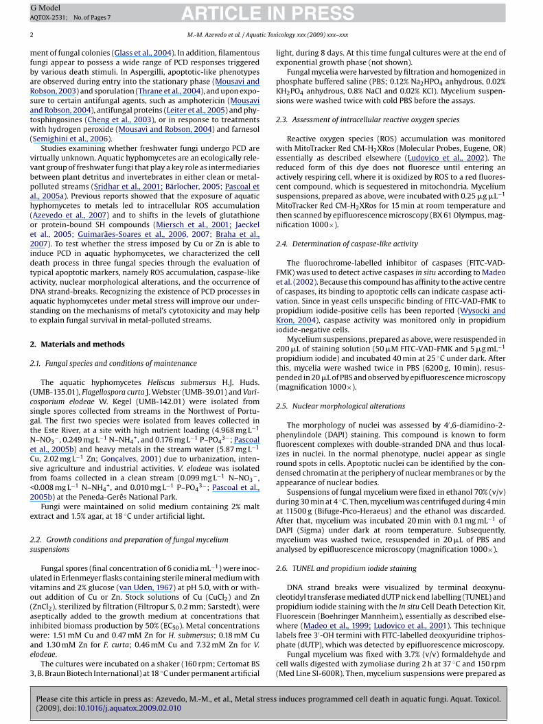
ig. 1. ROS accumulation, assessed by MitoTracker Red CM-H2XRos staining, in myct concentrations inhibiting biomass production by 50%. ROS-positive cells display
escribed above and cytospins of cell suspensions were done usingShandon Cytospin 2 cytocentrifuge at 1000 rpm for 5 min. Slidesere incubated in a permeabilization solution (0.1% (v/v) Triton-100 in 0.1% sodium citrate) for 10 min, rinsed twice in PBS and
ncubated with the TUNEL reaction mixture. Slides were incubatedn a humidified atmosphere under dark (1 h; 37 ◦C). Ten microlitresf a mixture containing 100 �L of the antifading agent Vectashield,�L of propidium iodide (50 �g mL−1), to co-localize DNA, and 2 �LNase (0.5 �g mL−1) was added to each slide. Positive controls wererepared by incubating the slides with 10 �L of DNase-10 units30 min; 37 ◦C), before incubation with the TUNEL reaction mixture.
ycelia were observed by epifluorescence microscopy (magnifica-ion 1000×).
. Results
.1. Copper and Zn induced oxidative stress through reactive
Please cite this article in press as: Azevedo, M.-M., et al., Metal stress(2009), doi:10.1016/j.aquatox.2009.02.010
xygen species accumulation
V. elodeae, H. submersus and F. curta mycelia exposed to Cu orn were stained with MitoTracker Red CM-H2XRos and analysedy epifluorescence microscopy to assess ROS accumulation. An
able 1ualitative analysis of reactive oxygen species (ROS) accumulation, caspase-like activityssay, in mycelia of aquatic fungi non-exposed or exposed for 8 days to Cu or Zn at concen
ungal species Cu
ROS Caspase-likeactivity
Nuclear morphologicalalterations
TU
. submersus ++ ++ + +. elodeae − + ++ +. curta + ++ + −, no detection; +, low detection; ++, high detection.
H. submersus, V. elodeae and F. curta non-exposed or exposed for 8 days to Cu or Znorescence.
increase in the red fluorescence indicative of cells with an increasedROS accumulation was detected in all species exposed to Zn (Fig. 1and Table 1). The exposure to Cu induced ROS accumulation in H.submersus and F. curta mycelia but not in V. elodeae (Fig. 1 andTable 1). Comparatively, the number of cells presenting increasedintracellular ROS accumulation was higher in H. submersus myceliaexposed to Cu than to Zn. The results indicated that oxidative stressseems to underlie the toxic effects of metals in the tested specieswith exception of V. elodeae under Cu exposure.
3.2. Copper and Zn induced caspase-like activity
Caspase activity can be monitored using FITC-VAD-FMK, a FITCconjugate of the caspase inhibitor Z-VAD-FMK that is delivered intothe cell where it binds to activated caspases. Although caspasesare not present in fungi, orthologues of the caspase family, termedmetacaspases, have been identified in fungi and plants (Uren et al.,
induces programmed cell death in aquatic fungi. Aquat. Toxicol.
2000), and their activity can be assessed using the same detectionkit (Madeo et al., 2002). According to our results, Cu or Zn exposureled to the detection of caspase-like activity in all species except inF. curta mycelia exposed to Zn (Fig. 2 and Table 1). A high numberof cells displaying caspase-like activity were found in H. submersus
, nuclear morphological alterations, and DNA strand-breaks, evaluated by TUNELtrations inhibiting biomass production by 50%.
Zn
NEL ROS Caspase-likeactivity
Nuclear morphologicalalterations
TUNEL
+ + + +++ ++ ++ ++ − + ++

Please cite this article in press as: Azevedo, M.-M., et al., Metal stress induces programmed cell death in aquatic fungi. Aquat. Toxicol.(2009), doi:10.1016/j.aquatox.2009.02.010
ARTICLE IN PRESSG ModelAQTOX-2531; No. of Pages 7
4 M.-M. Azevedo et al. / Aquatic Toxicology xxx (2009) xxx–xxx
Fig. 2. Caspase-like activity, assessed by FITC-VAD-FMK, in mycelia of H. submersus, V. elodeae and F. curta non-exposed or exposed for 8 days to Cu or Zn at concentrationsinhibiting biomass production by 50%. Caspase-positive cells display green fluorescence.
Fig. 3. Nuclear morphological alterations revealed by DAPI staining in H. submersus, V. elodeae and F. curta non-exposed or exposed for 8 days to Cu or Zn at concentrationsinhibiting biomass production by 50%. Arrows indicate nuclei with altered morphology. Inserts show detailed nuclear morphology, with nuclear alterations as half-ringarrangements or fragments.
INA
tic Tox
aa
3a
cmiasatAwdfoef
3T
Znapset
Fpd
ARTICLEG ModelQTOX-2531; No. of Pages 7
M.-M. Azevedo et al. / Aqua
nd F. curta exposed to Cu, and in V. elodeae exposed to Zn (Fig. 2nd Table 1).
.3. Copper and Zn induced nuclear morphological alterationsnd chromatin condensation
Alterations of nuclear morphology and evaluation of chromatinondensation, easily monitored by DAPI staining, are valuable hall-arks of a programmed cell death process and may give insights
nto the events associated with cellular toxicity induced by Cund Zn. This analysis allowed us to detect that Cu or Zn expo-ure induced nuclear alterations and chromatin condensation in allquatic hyphomycete species, while nuclei of control mycelia main-ained the homogeneous shape and density (Fig. 3 and Table 1).s a result of the alterations promoted by metals, nuclei appearedith arrangements in half-rings or nuclear fragments randomlyistributed (Fig. 3). These features were observed in cells of allungal species, particularly in V. elodeae exposed either to Cur Zn. These results showed that toxicity of Cu and Zn has anvident net negative consequence on the nuclear structure andunction.
.4. Copper and Zn induced DNA strand breaks revealed byUNEL assay
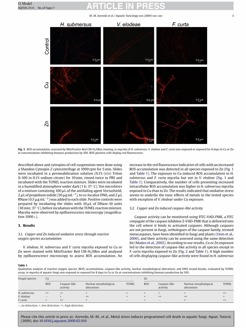
The exposure of V. elodeae, H. submersus and F. curta mycelia ton resulted in a TUNEL-positive phenotype, as shown by the yellowuclear fluorescence resulting from the superimposition of green
Please cite this article in press as: Azevedo, M.-M., et al., Metal stress(2009), doi:10.1016/j.aquatox.2009.02.010
nd red fluorescence due to simultaneous staining with TUNEL andropidium iodide (Fig. 4). This indicated the occurrence of DNAtrand breaks, a hallmark of the apoptotic process. Similarly, thexposure to Cu led to the appearance of DNA strand breaks withhe exception of F. curta mycelia, in which no TUNEL-positive phe-
ig. 4. DNA strand breaks visualized by TUNEL assay in H. submersus, V. elodeae and F. curtaroduction by 50%. TUNEL-positive cells are shown by yellow nuclear fluorescence, as theue to simultaneous staining with TUNEL and propidium iodide.
PRESSicology xxx (2009) xxx–xxx 5
notype was observed (Fig. 4 and Table 1). Beyond the occurrenceof this apoptotic marker upon metal exposure, it is noticeable thatZn led to a high number of cells with TUNEL-positive phenotypein H. submersus and F. curta mycelia. Results clearly demonstratethat a programmed cell death process characterized by an apop-totic phenotype was underlying the cytotoxic effects of Cu and Znin the tested aquatic hyphomycete species.
4. Discussion
The maintenance of cellular homeostasis is dependent on theability of cells to deal with environmental stressors. Metals cancause, directly or indirectly, an increase of ROS accumulation incells (Stohs and Bagchi, 1995) that may result in programmed celldeath (PCD). We have previously demonstrated that metal-inducedROS accumulation contributes noticeably to Cu and Zn toxicity inaquatic hyphomycetes (Azevedo et al., 2007). In the present workwe show, for the first time, a link between metal-induced oxidativestress and the occurrence of PCD in aquatic fungi.
In our study, it is clear that ROS accumulation upon Cu or Znexposure was associated with the occurrence of nuclear morpho-logical alterations, chromatin condensation, caspase-like activityand DNA strand breaks. Intracellular ROS are crucial signallingmolecules for the further occurrence of downstream apoptoticevents, such as caspase activation (Buttke and Sandstrom, 1994)and nuclear fragmentation (Masato et al., 1998). Our results showthat the exposure of V. elodeae mycelia to Cu promoted nuclear mor-phological alterations, caspase-like activity and DNA strand breaks,
induces programmed cell death in aquatic fungi. Aquat. Toxicol.
but did not lead to detectable intracellular ROS accumulation. Thissuggests that ROS might, in some cases, not be a prerequisite for PCDin aquatic hyphomycetes. Similarly, in Saccharomyces cerevisiae, dif-ferent scenarios of apoptosis independent of ROS signalling havebeen described, namely apoptosis induced by the antifungal agent
non-exposed or exposed for 8 days to Cu or Zn at concentrations inhibiting biomassresult of superimposition of green (FITC-labelled nucleotides) and red fluorescence

INA
6 tic Tox
c2
otbCcmcchmwamaiPfit
tacfifiaTPrwmoboufitCm
tParh
5
alocbomioPcbaf
ARTICLEG ModelQTOX-2531; No. of Pages 7
M.-M. Azevedo et al. / Aqua
iclopirox olamine (Almeida et al., 2007) or by aspirin (Balzan et al.,004).
Cells undergoing PCD do not always harbour all cardinal featuresf an apoptotic cell death (Schulze-Osthoff et al., 1994), fragmen-ation of nuclei with condensed chromatin and DNA strand breakseing the most characteristic traits (Mignotte and Vayssiere, 1998).onsistently, our results demonstrate that nuclear alterations withondensed chromatin and DNA strand breaks were present in theajority of the tested conditions. Nevertheless, the exposure of F.
urta mycelia to Cu did not result in DNA strand breaks. Under thisondition, the response of other apoptotic markers, particularly theigh amount of cells with caspase-like activity, suggests that cellsight eventually present DNA strand breaks later, although theyere not detected in the time course of the experiment. Addition-
lly, no caspase-like activity was found after exposure of F. curtaycelia to Zn. A reason for this observation might underlie on the
ctivation of a PCD pathway independent of metacaspase activityn this particular condition. In fact, several reports have describedCD processes independent of metacaspase activity in filamentousungi, such as in Aspergillus fumigatus treated with hydrogen perox-de or amphotericin B (Mousavi and Robson, 2004) or in A. nidulansreated with phytosphingosines (Cheng et al., 2003).
PCD has been assigned as an altruistic process that promoteshe sacrifice of some doomed cells in benefit of an entire cellularssemblage. This is the case of yeasts in which the PCD of someells will provide nutrients for others, probably the younger andtter cells (Herker et al., 2004), and it is most likely the case oflamentous fungi. For the first time, we provide evidence that Cund Zn stress can trigger apoptotic PCD in aquatic hyphomycetes.he occurrence of a tightly regulated cell death pathway, such asCD, in aquatic hyphomycetes under metal stress might play aole in fungal acclimation in metal-polluted streams, because itould allow the sacrifice of certain cells for benefit of the wholeycelium (Richie et al., 2007). In fact, if we hypothesize that PCD
ccurs in the older regions of the mycelium where nutrients haveeen exhausted and the older cells are more susceptible to Cur Zn stress, this cell death would allow the rapid removal ofnwanted or damaged cells sparing nutrients and space for thetter ones. Our results support this hypothesis because the mostolerant fungal species either to Zn (V. elodeae, EC50 7.32 mM) or tou (H. submersus, EC50 1.51 mM) exhibited the higher levels of PCDarkers.Moreover, our study demonstrates a different pattern of apop-
otic markers under different stressful conditions that might mirrorCD processes independent of ROS signalling, or of metacaspasectivity, or even without the appearance of DNA strand breaks,eflecting the plasticity of the PCD pathways operating in aquaticyphomycetes.
. Conclusion
For the first time, a link between metal-induced oxidative stressnd the occurrence of PCD in aquatic fungi was shown. ROS accumu-ation upon Cu or Zn exposure was associated with the occurrencef apoptotic markers, namely nuclear morphological alterations,hromatin condensation, caspase-like activity and DNA strandreaks. The most tolerant aquatic hyphomycete species to either Znr Cu exhibited higher levels of PCD markers, suggesting that PCDight allow the rapid removal of unwanted or damaged cells spar-
ng nutrients and space for the fitter ones. The different patternsf apoptotic markers found under different conditions, namely a
Please cite this article in press as: Azevedo, M.-M., et al., Metal stress(2009), doi:10.1016/j.aquatox.2009.02.010
CD process independent of ROS accumulation, or independent ofaspase-like activity, or even without the occurrence of DNA strandreaks, suggest that a multiplicity of PCD pathways might be oper-ting in aquatic hyphomycetes under metal stress, and is worthyor further studies.
PRESSicology xxx (2009) xxx–xxx
References
Almeida, B., Sampaio-Marques, B., Carvalho, J., Silva, M.T., Leão, C., Rodrigues, R.,Ludovico, P., 2007. An atypical active cell death process underlies the fungicidalactivity of ciclopirox olamine against the yeast Saccharomyces cerevisiae. FEMSYeast Res. 7, 404–412.
Azevedo, M.M., Carvalho, A., Pascoal, C., Rodrigues, F., Cássio, F., 2007. Responses ofantioxidant defenses to Cu and Zn stress in two aquatic fungi. Sci. Total Environ.377, 233–243.
Balzan, R., Sapienza, K., Galea, D.R., Vassallo, N., Frey, H., Bannister, W.H., 2004.Aspirin commits yeast cells to apoptosis depending on carbon source. Micro-biology 150, 109–115.
Bärlocher, F., 2005. Freshwater fungal communities. In: Deighton, J., White Jr., J.F.,Oudemans, P. (Eds.), The Fungal Community: its Organization and Role in theEcosystem. Taylor and Francis/CRC Press, Boca Raton, FL, pp. 39–59.
Barry, C., Kinsella, P., McCann, M., Devereux, M., O’Connor, R., Clynes, M., Kavanagh,K., 2004. Induction of apoptosis in yeast and mammalian cells by exposure to1,10-phenanthroline metal complexes. Toxicol. In Vitro 18, 63–70.
Braha, B., Tintemann, H., Krauss, G., Ehrman, J., Bärlocher, F., Krauss, G.-J., 2007. Stressresponse in two strains of the aquatic hyphomycete Heliscus lugdunensis afterexposure to cadmium and copper ions. Biometals 20, 93–105.
Brown Jr., G.E., Foster, A.L., Ostergren, J.D., 1999. Mineral surfaces and bioavailabilityof heavy metals: a molecular-scale perspective. Proc. Natl. Acad. Sci. U.S.A. 96,3388–3395.
Buttke, T.M., Sandstrom, P.A., 1994. Oxidative stress as a mediator of apoptosis.Immunol. Today 15, 7–10.
Cheng, J., Park, T.-S., Chio, L.C., Fischl, A.S., Ye, X.S., 2003. Induction of apopto-sis by sphingoid long-chain bases in Aspergillus nidulans. Mol. Cell. Biol. 23,163–177.
Cobbett, C., Goldsbrough, P., 2002. Phytochelatins and metallothioneins: rolesin heavy metal detoxification and homeostasis. Annu. Rev. Plant. Biol. 53,159–182.
Earnshaw, W.C., Martins, L.M., Kaufmann, S.H., 1999. Mammalian caspases: struc-ture, activation, substrates, and functions during apoptosis. Annu. Rev. Biochem.68, 383–424.
Fedorova, N.D., Badger, J.H., Robson, G.D., Wortman, J.R., Nierman, W.C., 2005. Com-parative analysis of programmed cell death pathways in filamentous fungi. BMCGenomics 6, 177–191.
Gadd, G.M., 1993. Interactions of fungi with toxic metals. New Phytol. 124, 25–60.Gichner, T., Patková, Z., Száková, J., Demnerová, K., 2006. Toxicity and DNA dam-
age in tobacco and potato plants growing on soil polluted with heavy metals.Ecotoxicol. Environ. Saf. 65, 420–426.
Glass, N.L., Kanebo, I., 2003. Fatal attraction: nonself recognition and heterokaryonincompatibility in filamentous fungi. Eukaryot. Cell 2, 1–8.
Glass, N.L., Rasmussen, C., Roca, M.G., Read, N.D., 2004. Hyphal homing, fusion andmycelial interconnectedness. Trends Microbiol. 12, 135–141.
Goncalves, M.A.P., 2001. Determinacão de metais pesados em águas superficiaisrecolhidas no Rio Este. M.Sc. thesis. University of Minho, Braga, Portugal.
Guimarães-Soares, L., Felícia, H., Bebianno, M.J., Cássio, F., 2006. Metal-binding pro-teins and peptides in aquatic fungi exposed to severe metal stress. Sci. TotalEnviron. 372, 148–156.
Guimarães-Soares, L., Pascoal, C., Cássio, F., 2007. Effects of heavy metals on theproduction of thiol compounds by the aquatic fungi Fontanospora fusiramosaand Flagellospora curta. Ecotoxicol. Environ. Saf. 66, 36–43.
Herker, E., Jungwirth, H., Lehmann, K.A., Maldener, C., Frohlich, K.U., Wissing, S.,Buttner, S., Fehr, M., Sigrist, S., Madeo, F., 2004. Chronological aging leads toapoptosis in yeast. J. Cell Biol. 164, 501–507.
Jaeckel, P., Krauss, G.-J., Krauss, G., 2005. Cadmium and zinc response of the fungiHeliscus lugdunensis and Verticillium cf. alboatrum isolated from highly pollutedwater. Sci. Total Environ. 346, 274–279.
Kerr, J.F., Wyllie, A.H., Currie, A.R., 1972. Apoptosis: a basic biological phenomenonwith wide-ranging implications in tissue kinetics. Br. J. Cancer 26, 239–257.
Koonin, E.V., Araving, L., 2002. Origin and evolution of eukaryotic apoptosis: thebacterial connection. Cell Death Differ. 9, 394–404.
Leiter, E., Szappanos, H., Oberparleiter, C., Kaiserer, L., Csernoch, L., Pusztahelyi, T.,Emri, T., Pócsi, I., Salvenmoser, W., Marx, F., 2005. Antifungal protein PAF severelyaffects the integrity of the plasma membrane of Aspergillus nidulans and inducesan apoptosis-like phenotype. Antimicrob. Agents Chemother. 49, 2445–2453.
Ludovico, P., Madeo, F., Silva, M., 2005. Yeast programmed cell death: an intricatepuzzle. IUBMB Life 57, 129–135.
Ludovico, P., Rodrigues, F., Almeida, A., Silva, T.M., Barrientos, A., Côrte-Real, M., 2002.Cytochrome c release and mitochondria involvement in programmed cell deathinduced by acetic acid in Saccharomyces cerevisiae. Mol. Biol. Cell 13, 2598–2606.
Ludovico, P., Sousa, M.J., Silva, T.M., Leão, C., Côrte-Real, M., 2001. Saccharomycescerevisiae commits to a programmed cell death process in response to aceticacid. Microbiology 147, 2409–2415.
Madeo, F., Fröhlich, E., Ligr, M., Grey, M., Sigrist, S.J., Wolf, D.H., Fröhlich, K.U., 1999.Oxygen stress: a regulator of apoptosis in yeast. J. Cell Biol. 145, 757–767.
Madeo, F., Herker, E., Maldener, C., 2002. A caspase-related protease regulates apop-tosis in yeast. Mol. Cell 9, 911–917.
induces programmed cell death in aquatic fungi. Aquat. Toxicol.
Masato, E., Hideki, S., Hideki, Y., Katsuya, O., Akihiro, I., Shigekazu, N., 1998. A caspase-activated DNase that degrades DNA during apoptosis, and its inhibitor ICAD.Nature 391, 43–50.
Miersch, J., Tschimedbalshir, M., Bärlocher, F., Grams, Y., Pierau, B., Schierhorn, A.,Krauss, G.-J., 2001. Heavy metals and thiol compounds in Mucor racemosus andArticulospora tetracladia. Mycol. Res. 105, 883–889.

INA
tic Tox
M
M
M
P
P
R
R
R
S
ARTICLEG ModelQTOX-2531; No. of Pages 7
M.-M. Azevedo et al. / Aqua
ignotte, B., Vayssiere, J.-L., 1998. Mitochondria and apoptosis. Eur. J. Biochem. 252,1–15.
ousavi, S.A.A., Robson, G.D., 2003. Entry into the stationary phase is associated witha rapid loss of viability and an apoptotic-like phenotype in the opportunisticpathogen Aspergillus fumigatus. Fungal Genet. Biol. 39, 221–229.
ousavi, S.A.A., Robson, G.D., 2004. Oxidative and amphotericin B-mediated celldeath in the opportunistic pathogen Aspergillus fumigatus is associated with anapoptotic-like phenotype. Microbiology 150, 1937–1945.
ascoal, C., Cássio, F., Marcotegui, A., Sanz, B., Gomes, P., 2005a. Role of fungi, bacteria,and invertebrates in leaf litter breakdown in a polluted river. J. N. Am. Benthol.Soc. 24, 784–797.
ascoal, C., Marvanová, L., Cássio, F., 2005b. Aquatic hyphomycete diversity instreams of Northwest Portugal. Fungal Divers. 19, 109–128.
ichie, D.L., Miley, M.D., Bhabhra, R., Robson, G.D., Rhodes, J.C., Askew, D.S., 2007.The Aspergillus fumigatus metacaspases CasA and CasB facilitate growth underconditions of endoplasmic reticulum stress. Mol. Microbiol. 63, 591–604.
Please cite this article in press as: Azevedo, M.-M., et al., Metal stress(2009), doi:10.1016/j.aquatox.2009.02.010
obson, G.D., 2006. Programmed cell death in the aspergilli and other filamentousfungi. Med. Mycol. 44, S109–S114.
udolf, E., Rudolf, K., Cervinka, M., 2005. Zn induced apoptosis in HEP-2 cancer cells:the role of oxidative stress and mitochondria. Biofactors 23, 107–120.
chulze-Osthoff, K., Walczak, H., Droge, W., Krammer, P.H., 1994. Cell nucleus andDNA fragmentation are not required for apoptosis. J. Cell Biol. 127, 15–20.
PRESSicology xxx (2009) xxx–xxx 7
Semighini, C.P., Hornby, J.M., Dumitru, R., Nickerson, K.W., Harris, S.D., 2006.Farnesol-induced apoptosis in Aspergillus nidulans reveals a possible mech-anism for antagonistic interactions between fungi. Mol. Microbiol. 59,753–764.
Sridhar, K.R., Krauss, G., Bärlocher, F., Raviraja, N.S., Wennrich, R., Baumbach, R.,Krauss, G.-J., 2001. Decomposition of alder leaves in two heavy metal pollutedstreams in Central Germany. Aquat. Microb. Ecol. 26, 73–80.
Stohs, S.J., Bagchi, D., 1995. Oxidative mechanisms in the toxicity of metal ions. FreeRadic. Biol. Med. 18, 321–336.
Thrane, C., Kaufmann, U., Stummann, B.M., Olsson, S., 2004. Activation of caspase-like activity and poly (ADP-ribose) polymerase degradation during sporulationin Aspergillus nidulans. Fungal Genet. Biol. 41, 361–368.
Uren, A.G., O’Rourke, K., Aravind, L.A., Pisabarro, M.T., Seshagiri, S., Koonin, E.V., Dixit,V.M., 2000. Identification of paracaspases and metacaspases: two ancient fam-ilies of caspase-like proteins, one of which plays a key role in MALT lymphoma.Mol. Cell. 6, 961–967.
induces programmed cell death in aquatic fungi. Aquat. Toxicol.
van Uden, N., 1967. Transport-limited fermentation and growth of Saccharomycescerevisiae and its competitive inhibition. Arch. Microbiol. 58, 155–168.
Wolfe, J.T., Ross, D., Cohen, G.M., 1994. A role for metals and free radicals in theinduction of apoptosis in thymocytes. FEBS Lett. 352, 58–62.
Wysocki, R., Kron, S.J., 2004. Yeast cell death during DNA damage arrest is indepen-dent of caspase or reactive oxygen species. J. Cell Biol. 166, 311–316.