The Genera of Fungi: fixing the application of type species of generic names

695
CARLOS A. INA; CIO AND J. C. DIANESE
Departamento de Fitopatologia Universidade de BrasıUlia, 70910-900 BrasıUlia, DF, Brasil
The following foliicolous fungi on Tabebuia species are described and illustrated : Anhelia tabebuiae sp. nov. and Dictyonella tabebuiae
sp. nov. (ascomycetes), Fumagospora tabebuiae sp. nov., Polychaeton tabebuiae sp. nov. and Septoria tabebuiae-impetiginosae sp. nov.
(coelomycetes), Cercospora tabebuiae-impetiginosae sp. nov. and Pseudocercospora tabebuiae-roseo-albae sp. nov. (hyphomycetes). Uncinula
peruviana was found and described on T. impetiginosa as a first record for Distrito Federal, Brazil.
Tabebuia Go! mes (Bignoniaceae) accommodates mostly trees
known in Brazil as ‘ ipe# s ’ which were previously treated as
species of Tecoma Juss. Tabebuia species are exploited from
native forests and also used as urban ornamentals throughout
Brazil and in the warmer regions of Latin America. Lorenzi
(1992) and Carvalho (1994) listed 15 Tabebuia species with
their common names from Brazil. In Brası! lia, Distrito Federal,
the following species are grown on street, parks, and in
private gardens : Tabebuia serratifolia (Vahl) Nich., T. impeti-
ginosa (Mart.) Standl., T. heptaphylla (Vell.) Tol., T. rosea DC,
and Tabebuia roseo-alba (Ridl.) Sandl. In the cerrado of DF and
Central Brazil the main native species are T. caraiba (Mart.)
Bur., and T. ochracea (Cham.) Standl.
Parasitic and superficial fungi occurring on leaves of
Tabebuia in Brazil have been reported by Vie! gas (1944, 1961),
Chupp (1953), Batista & Peres (1962), Batista & Bezerra
(1964), Dennis (1970), Ferreira & Alfenas (1980), Muchovej &
Ferreira (1981), Ferreira & Muchovej (1987), Schimitt & Veiga
(1987), Rezende & Ferreira (1988), Ferreira (1989), Gianasi &
Castro (1993), Dianese & Dianese (1994), Dianese, Medeiros
& Santos (1997), Dianese, Tessmann & Furlanetto (1994) and
Silva & Minter (1995).
This article reports on the occurrence of new fungi
assocociated with lesions on leaves of Tabebuia species in
gardens and parks of Brası! lia and also in parts of the native
cerrado vegetation still present in the area.
MATERIALS AND METHODS
Leaves of cultivated and native species of Tabebuia in the
cerrado were collected in urban areas of Brası! lia or from
natural reserves. Each sample was prepared, numbered,
registered, and deposited in the Mycological Reference
Collection of the Herbarium of the University of Brası! lia(Herbarium UB mycological collection).
Slides containing squash preparations of fungal fruiting
bodies or sections made by freezing microtome were used for
the morphological studies and microphotography. In most
cases the samples were stained with lacto-glycerol-cotton blue
or glycerol-KOH-phloxine B and the slides sealed with nail
polish.
Pieces of leaves with one or more lesions showing
representative samples of fruiting bodies were used for SEM
after being fixed in sodium cacodylate buffer, pH 7±4, 0±1 ,
containing glutaraldehyde 2%, for at least 24 h. The samples
were dehydrated in an aqueous series with increasing acetone
concentrations from 15, 30, 50, 75 to 100% of acetone, for
15 min in each concentration. Leaf pieces were then dried at
the critical point before being covered by a thin layer of gold
in a sputter coater for 2 min. Finally, the samples were
observed in a scanning electron microscope (Jeol, model JSM
840-A E).
DESCRIPTIONS AND DISCUSSION
The new species of fungi studied are described and illustrated
below. Further notes on Uncinula peruviana Syd. are also
reported.
Anhelia tabebuiae Ina! cio & Dianese, sp. nov. (Figs 1–6)
Laesiones 3–40 mm diam., primo rubello-brunneae ad brunneae vel
cinerero-brunneae cum brunneis marginibus tandem pallido-brunneae
ubi mortuae, coalescentes, amphigenae, irregulares, plerumque ad
marginem locatae vel inter nervos secundarios locatae. Ascomata
26–78¬51–158 µm, eustromatica, pulvinata, multilocularia, loculis
uniseriatis. Loculi 13–25¬13–18 µm, monoascales, parietibus ex
textura angulari, cum cellulis 3–8 µm diam. compositis. Hypostromata
plana, epidermalia, immersa. Asci 9–25¬8–19 µm, bitunicati, persis-
tentes, 4–8 sporis, obovoides vel subsphaerici, sessiles, vel curto-
pedicellati. Ascosporae 7–16¬4–7 µm, hyalinae, muriformes, 1–4
septatae transversim, 1–2 septatae in longitudinem, plerumque
phragmosporae, ad septa mediana constrictae.
Mycol. Res. 102 (6) : 695–708 (1998) Printed in the United Kingdom
Some foliicolous fungi on Tabebuia species

Some foliicolous fungi on Tabebuia species 696
Figs 1–6. Anhelia tabebuiae. Fig. 1. Leaf spots along the midrib and secondary veins on Tabebuia caraiba containing ascomata (bar,
10 mm). Fig. 2. Stromatic ascomata, SEM (bar, 10 µm). Fig. 3. Section of an erumpent ascoma in host leaf (bar, 50 µm). Fig. 4. Detail
of an ascoma showing texture of the stroma containing monoascal locules (bar, 5 µm). Figs 5, 6. Asci and ascospores (bar, 10 µm).

C. A. Ina! cio and J. C. Dianese 697
Table 1. Characteristics of other Anhelia species compared with A. tabebuiae
Host
Ascoma
(µm)
Asci
(µm)
Ascospores
(µm)
Hypostroma
(µm)
A. calami" Calamus, Araceae 100–150¬250–350 26–32¬17–19 10–12¬4–5 75–110
A. escharoides" Geissanthus, Myrsinaceae 400–1000 35–55¬30–42 16–28¬8–12 55–110
A. lantanae" Lantana, Verbenaceae 24–30¬19–23 12–15¬5–6 150–185
A. niger",$ Eupatorium, Compositae 250–400 11–16¬5–6
A. purpurascens" Machaerium, Leguminosae 180¬200–300 24–30¬20–26 13–17¬7–8 85–145
A. tetracerae" Tetracera alnifolia, Dilleniaceae 150¬500 30–38¬20–30 20–24¬4–9
A tristis",# Vaccinium teysmannianum, Ericaceae 270–360¬600–1500 35–48¬25–32 19–25¬7–10 170–240
A. tabebuiae Tabebuia caraiba, Bignoniaceae 26–78¬51–178 9–25¬8–19 7–16¬4–7 Flat
" Arx (1963) ; # Boedijn (1961) ; $ Vie! gas (1945a).
In foliis vivis Tabebuiae caraibae, in urbana Brasilia, DF, Ponte do
Bragueto, Eixo Rodovia! rio Norte, Plano Piloto ; 12 Sep. 95 ; leg.
C. A. Ina! cio no. 199 ; UB col. micol. 6443, holotypus.
Lesions 3–40 mm diam., reddish-brown to brown, or greyish-
brownwith brown borders, coalescent, amphigenous, irregular,
mostly located at leaf margins or between the secondary ribs
(Fig. 1). In a later phase it becomes pale brown and necrotic.
Ascomata 26–78¬51–158 µm, eustromatic, pulvinate, multi-
locular, forming one layer of locules. Locules monoascal,
13–25¬13–18 µm, wall with textura angularis of 3–8 µm
diam. cells (Figs 2–4). Hypostromata flat, epidermal, immersed
(Figs 3, 4). Asci 9–25¬8–19 µm, bitunicate, persistent, 4–8
spored, obovoidal to subsphaerical, sessile or short pedicellate
(Figs 4–6). Ascospores 7–16¬4–7 µm, hyaline, muriform, 1–4
transverse septa, 1–2 longitudinal septa, mostly phragmo-
sporic, constricted at median septum (Figs 4, 6).
Anhelia tabebuiae was compared with the known species of
the genus including the Brazilian species A. niger (Vie! gas) Arx,
found on Eupatorium sp. (Arx, 1963), which showed larger
ascomata than the new species, with asci and interascal
material shown as gelatinous matrix (Vie! gas, 1945a).
Arx (1963) also redescribed A. purpurascens (Rehm) Arx on
Mimosa flagellaris Benth. and on M. vellosiana Mart., and A.
lantanae (P. Henn.) Arx on Lantana sp. with conspicuous
hypostromata (150–185 µm diam.).
Anhelia tristis Racib., A. escharoides (Syd.) Arx, and A.
tetracerae (Hansf.) Arx, all with obviously larger dimensions
than A. tabebuiae, were found on Vaccinium teysmannianum
Miq., Geissanthus sp., and on Tetracera alnifolia Wiild. (Boedijn,
1961 ; Arx, 1963). Anhelia calami (Rac.) Arx is clearly different
from the new species and it was reported on Calamus sp. (Arx,
1963). The comparisons, which are summarized in Table 1,
lead to the conclusion that A. tabebuiae is a new species and
a first record of Anhelia on Tabebuia species.
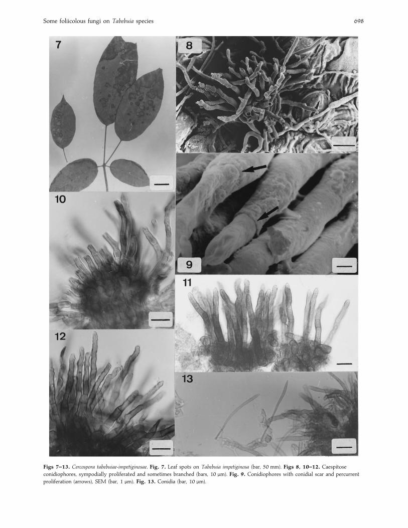
Cercospora tabebuiae-impetiginosae Ina! cio & Dianese,sp. nov. (Figs 7–13)
Laesiones 3–30 µm, amphigenae, cinereo-brunneae vel pallido-cinereae
cum marginibus purpurascentibus, coalescentes ubi mortuae. Stromata
24–107 µm diam., subepidermalia, erumpentia, ex textura angulari
cum cellulis 3–9 µm diam. composita. Conidiophora 24–58¬2–4 µm,
brunnea, caespitosa, amphigena, plerumque hypophylla, 1–8 septata,
geniculata, sympodialia, recta vel leviter incurvata, raro ramosa,
aliquando proliferationibus percurrentibus. Cellulae conidiogenae poly-
blasticae, sympodiales, integratae, terminales, cicatricibus conidiorum
inconspicuis sed per microscopium electronicum manifestis. Conidia
25–135¬2–4 µm, 1–10 septata, cylindrica ad acidularia, basibus
truncatis.
In foliis vivis Tabebuiae impetiginosae, Brası! lia, DF, University of
Brası! lia Campus, north of Faculty of Education, 17 July 94, leg. C. A.
Ina! cio no. 181, UB col. micol. 6283, holotypus.
Lesions 3–30 mm diam., amphigenous, greyish-brown to light
grey with purple borders, coalescent, necrotic (Fig. 7). Stromata
24–107 µm diam., subepidermal, erumpent, textura angularis of
polygonal cells, 3–9 µm diam. (Fig. 10). Conidiophores 24–58¬2–4 µm, brown, caespitose, amphigenous, mostly hypophyl-
lous (Figs 8–12), 1–8 septate, geniculate, sympodial, sometimes
percurrently proliferating, straight or slightly curved, rarely
branched, with inconspicuous scars, which are clearly seen in
SEM (Figs 8–12). Conidiogenous cells polyblastic, sympodial,
integrated, terminal, with inconspicuous scars which also are
clearly seen in SEM (Figs 9, 11, 12). Conidia 25–135¬2–4 µm,
1–10 septate, cylindrical to acicular, base truncate (Fig. 13).
Other specimens examined : On living leaves of Tabebuia impetiginosa
(Bignoniaceae) : University of Brası! lia Campus, Brası! lia, DF, north of
Faculty of Education, 4 May 94 ; leg. C. A. Ina! cio, no. 95, UB col.
micol. 6085 ; north of Faculty of Education, 29 May 94; leg. C. A.
Ina! cio, no. 147 ; UB col. micol. 6156 ; East of ICC in front of
University Restaurant, 4 May 94 ; leg. C. A. Ina! cio, no. 173 ; UB col.
micol. 6249 ; near Central Library, 17 July 94 ; leg. C. A. Ina! cio, no.
179 ; UB col. micol. 6280.
In Table 2 the Cercospora species found on Tabebuia are
compared with C. tabebuiae-impetiginosae. Cercospora tecomae
Chupp & Vie! gas has shorter, 25–50 (–100)¬3–5 µm, up to 7-
septate, conidia ; smaller stromata (20–25 µm) ; and larger
conidiophores (60–100¬4–6 µm) (Vie! gas, 1945 c ; Chupp,
1953) than those of C. tabebuiae-impetiginosae. The only other
species on Tabebuia which shows some similarity with the
new species is C. jahnii Syd. but this differs in size of conidia,
conidiophores and stromata. From Table 2, it is clear that C.
tabebuiae-impetiginosae is different from all species so far
described on bignoniaceous hosts.
Dictyonella tabebuiae Ina! cio & Dianese, sp. nov.(Figs 14–19)
Coloniae 4–15 mm diam., brunneae, circulares aut irregulares,
hypophyllae, plerumque singulares, raro gregariae, effusae, paginae
abaxiales foliolorum locatae. Mycelium superficiale, pallido-brunneum
vel brunneum, laxas infra-ascomatales pulvinas formantes ; hyphae
2–6 µm diam., pallido-brunneae, laeves, ramosae, hyphopodia nulla,
cum constrictionibus exiguiis ad parietes transversales. Ascomata
Some foliicolous fungi on Tabebuia species 698
Figs 7–13. Cercospora tabebuiae-impetiginosae. Fig. 7. Leaf spots on Tabebuia impetiginosa (bar, 50 mm). Figs 8, 10–12. Caespitose
conidiophores, sympodially proliferated and sometimes branched (bars, 10 µm). Fig. 9. Conidiophores with conidial scar and percurrent
proliferation (arrows), SEM (bar, 1 µm). Fig. 13. Conidia (bar, 10 µm).

C. A. Ina! cio and J. C. Dianese 699
Table 2. Main characteristics of Cercospora species on Bignoniaceae compared with those of C. tabeuiae-impetiginosae
Host
Lesion
(mm)
Stromatal diam.
(µm)
Conidiophores
(µm)
Conidia
(µm)
C. adenocalymae A. S. Mull. &
Chupp"
Adenocalyma
bullatum
0±5–3 40–60 20–65¬4±5–5±5 35–150¬3–4±5, subhyaline to pale
olivaceous brown
C. arrabidaceae Chupp & Vie! gas" Arrabidaea
platyphylla
0±5–6 Absent or small 10–175¬4–6 35–150¬3±5–5, pale brown to
medium olivaceous
C. bignoniaecola Speg." Bignonia sp. — Small 20–80 (80–160)¬4–7 25–70¬4–7, pale brown to
olivaceous
C. catalpae G. Winter" Catalpa
bignonioides
1–4 0–50 10–125¬3–5±5 40–120¬2±5–4±5, hyaline
C. catalparum Chupp" Catalpa
longissima
Rare 15–70¬4–7±5 35–125¬3–5±6, pale brown,
olivaceous, brown
C. cybistacis Henn." Cybistax
antisyphilitica
3–7 25–75 10–35¬2±5–4 20–70¬2±5–4, pale brown to
olivaceous-brown
C. dolichandrones Chupp" Dolichandrone
platycalyx
3–10 Small 10–30¬2–3±5 40–100¬1±5–3, subhyaline, pale
brown to yellowish-olivaceous
C. duplicata Ellis & Everh." Campsis
radicans
3–10 20–40 8–25¬2±5–4 20–100¬2–4±5, pale brown to
olivaceous-brown
C. hansfordi Chupp" Dolichandrone
platycalyx
0±5–2 Absent or very
reduced
60–300¬5–6±5 30–80¬4–5±5, subhyaline, pale
brown to olivaceous
C. jahnii Syd.",# Tabebuia spp. 3–20 Up to 50 10–40¬3–5 20–110¬3–5, pale brown to
olivaceous
C. sordida Sacc." Tecoma radicans — Absent 20–120¬3–5 20–200¬3–5, pale brown, medium to
dark olivaceous
C. stenolobiiola (Speg.) Chupp",# T. stans 2–10 Absent 50–250¬3–4 40–70¬3–6, hyaline
C. tecomae Chupp & Vie! gas" Tabebuia sp. — Reduced}not see 20–80¬4–5±5 25–50 (80)¬3–5, hyaline
C. tecomae Chupp & Vie! gas$ Tecoma sp. 1 or more 20–25 60–100¬4–6 35–100¬3–4, hyaline
C. tabebuiae-impetiginosae Tabebuia
impetiginosa
3–30 24–107¬31±5–80 24–58¬2–4 25–135¬2–4, hyaline to pale brown
" Chupp (1953) ; # Dennis (1970) ; $ Vie! gas (1945 c).
53–97 µm longa¬111–216 µm diam., supra mycelium superficiale
formata, brunnea, parietibus cum texturis angularibus formatis ;
cellulis 3–8 µm diam., isodiametris. Asci 47–95¬32–53 µm, bituni-
cati, clavati aut lato-clavati, persistentes, 4–8 sporis ; pseudo-
paraphyses adsunt. Pseudoparaphyses hyalinae, septatae. Ascosporae
26–39¬11–17 µm, brunneae, muriformes, constrictionibus septorum
medianorum manifestae, vaginis mucosis textae, paginis undulatae.
In foliis vivis Tabebuiae ochraceae ; Fazenda A; gua Limpa, University
of Brası! lia, Brası! lia, DF ; 1 May 95; leg. D. V. Rezende Santiago no.
2, UB col. micol. 8338, holotypus.
Colonies 4–15 mm diam., brown, circular or irregular, hypo-
phyllous, mostly single, seldom gregarious, spread throughout
the abaxial face of the leaflets (Fig. 14). Mycelium superficial,
forming a loose cushion under the ascomata, light brown to
brown; hyphae 2–6 µm diam, light brown, septate, smooth,
branched, non-hyphopodiate, with slight constriction at the
septa (Figs 15–17). Ascomata 53–97 µm long¬111–216 µm
diam., on top of the superficial mycelium, brown; wall with
textura angularis (pseudoparenchymatous) of isodiametric cells,
3–8 µm diam. (Figs 15, 16). Asci 47–95¬32–53 µm, bitunicate,
clavate to broad clavate, persistent, 4–8-spored, pseudo-
paraphysate (Figs 16, 18). Pseudoparaphyses hyaline, septate.
Ascospores 26–39¬11–17 µm, brown, muriform, clearly con-
stricted at median septa, covered by a mucilagenous sheath,
with undulate surface (Figs 18, 19).
Dictyonella tabebuiae is the second species of the genus on
Bignoniaceae and can be easily separated from D. dictyosporus
(Petr. & Cif.) Arx on Crescentia cujete L. (Arx, 1963) which has
much smaller ascospores and ascomata than D. tabebuiae
(Table 3).
Among other Dictyonella species only D. scabra (Syd.) Arx,
described on plants belonging to the Moraceae, shares some
characteristics with D. tabebuiae. It is, however, easily
distinguished by its smaller asci (55–80¬35–45 µm) and
larger ascomata (100–240 µm diam.). Furthermore, D. scabra
has ascospores with only two longitudinal septa while D.
tabebuiae has three or more. Finally, Batista & Nascimento
(1958) identified D. erysiphoides (Rehm) Ho$ hn. on Coccoloba
uvifera (L.) Jacq. This fungus was, however, clearly parasitic
with innate infective mycelium and immersed hypostroma
which probably belongs in the Myriangiaceae and is distinct
from Dictyonella species which are superficial and non-
parasitic.
Fumagospora tabebuiae Ina! cio & Dianese, sp. nov.(Figs 20–26)
Coloniae atro-brunneae aut nigrae, effusae, epiphyllae, velutinae,
irregulares, coalescentes, cum punctuationibus prominentibus. Myce-
lium atro-brunneum aut nigrum, superficiale, ramosum, hyphopodia
nulla. Hyphae 7–21¬4–11 µm, pallido-brunneae aut brunneae,
septatae, ramosae, constrictionibus septorum manifestae, cellulis
doliiformibus. Coniodiomata 86–219 alta¬44–122 µm diam., brun-
nea, conoidea, ostiolata, rostris truncatis ; parietibus texturis angulari-
bus ; cellulis 8–13¬4–10 µm. Rostrum fimbriatum, 14–44 µm diam.,
fimbriae 20–40¬2±5–4 µm. Cellulae conidiogenae ampulliformes,
hyalinae, parvae, phialidicae. Conidia 9–21¬4–10 µm, 1–4 septata,
muriformia, oblonga aut elliptica, leviter constricta ad septum
medianum.
In foliis vivis Tabebuiae impetiginosae ; Theatrum arenae, University
of Brası! lia, Campus Universita! rio, Brası! lia, DF, Brazil ; 21 July 95 ; leg.
C. A. Ina! cio no. 419 ; UB col. micol. 9252, holotypus.

Some foliicolous fungi on Tabebuia species 700
Figs 14–19. Dictyonella tabebuiae. Fig. 14. Hypophyllous colonies shown as patches formed by groups of small brown dots on leaves
of T. ochracea (bar, 10 mm). Fig. 15. Ascomata and ascospores, SEM (bar, 1 µm). Fig. 16. Section of an ascoma containing immature
asci (bar, 10 µm). Fig. 17. Exhyphopodiate septate hyphae (bar, 10 µm). Figs 18, 19. Asci and muriform ascospores (bars, 10 µm).
Table 3. Comparison of the other Dictyonella species with D. tabebuiae
Host
Ascomata diam.
(µm)
Asci
(µm)
Ascospores
(µm)
D. alangii Hansf. & Thirum" Alangium (¯Marlea) begoniifolium, Alangiaceae 100¬400 30–40¬25–30 19–24¬9–11
D. dictyosporus" Crescentia cujete, Bignoniaceae 50–150 — 18–26¬8–12
D. erysiphoides",# Coccoloba uvifera, Coccoloba sp., Polygonaceae 60–80¬160–250 34–55¬24–32 25–28¬9–12
D. mirabilis (Syd.) Arx" Premma odorata, Verbenaceae 90–115¬400–1100 34–44¬22–30 23–28¬10–13
D. scabra" Antiaris africana, Chlorophora excelsa, Moraceae 100–240 55–80¬35–45 30–38¬3–16
D. tabebuiae Tabebuia ochracea, Bignoniaceae 53–97¬111–216 47–95¬32–53 26–39¬11–17
" Arx (1963) ; # Batista & Nascimento (1958).

C. A. Ina! cio and J. C. Dianese 701
Figs 20–26. Fumagospora tabebuiae. Fig. 20. Epiphyllous colonies spreading on leaf blade of Tabebuia impetiginosa (bar, 10 mm). Fig. 21.
Fimbriate conidioma extruding conidia. Fig. 22. Texture of conidiomatal wall. Fig. 23. Monilioid superficial mycelium. Fig. 24. Detail
of fimbriate conidiomatal tip and multiseptate conidia. Fig. 25. Phialidic conidiogenous cell indicated by an arrow. Fig. 26. Hyaline, 2–3
septate conidia. All bars, 10 µm, except Fig. 20.

Some foliicolous fungi on Tabebuia species 702
Figs 27–32. Polychaeton tabebuiae. Fig. 27. Colonies on Tabebuia ipe (Bar, 50 mm). Fig. 28. Conidioma, SEM. Figs 29, 30. Monilioid
hyphae at the base of a non-fimbriate conidioma showing wall with textura varying from angularis to prismatica. Fig. 31. Phialidic
conidiogenous cell (arrow). Fig. 32. Conidia. All bars, 10 µm, except Fig. 27.
Colonies dark brown to black, spread throughout the leaf
blade, epiphyllous, velvety with raised dots, irregular,
coalescent (Fig. 20). Mycelium dark brown to black, superficial,
exhyphopodiate, branched. Hyphae 7–21¬4–11 µm, light
brown to brown, septate, branched, constricted at the septa
(monilioid), with doliiform cells (Fig. 23). Conidiomata
86–219 µm high¬44–122 µm diam., conical with a truncate
rostrum, ostiolate, brown; wall with textura angularis ; cells
8–13¬4–10 µm. Rostrum fimbriate, 14–44 µm diam. ; fimbriae
20–40¬2±5–4 µm (Figs 21, 22, 24). Conidiogenous cells
ampulliform, small, hyaline, phialidic (Fig. 25). Conidia
9–21¬4–10 µm, 1–4 septate, muriform, oblong-elliptical,
slightly constricted at the middlemost septum (Figs 24, 26).
Batista & Ciferri (1963) included in their monograph three
species of Fumagospora G. Arnaud, the type species, F.
capnodioides G. Arnaud (teleomorph : Capnodium salicinum
Mont., sensu Hughes, 1976) on Citrus, Olea, and Nerium in
France, F. gaultheriae Bat., A. F. Vital & Cif. on Gaultheria
procumbens L. (associated with Microxyphium [¯ Polychaeton
sensu S. Hughes] aciculiform Cif., Bat. & Nascim.), and F.
cistophila (Fr. sensu Maire) Bat. & Cif. (teleomorph : C.
cistophylum Maire) on Citrus sp. from North Africa.
Hawksworth et al. (1995) apparently recognized only F.
capnodioides. As the species descriptions given by Batista &
Ciferri (1963) were clearly presented and illustrated they were
compared with F. tabebuiae. There is only one close to the new
species. It is F. cistophila, which has different dimensions of
conidiomata (192–275¬82–110 µm) and conidia (13–17¬7–8±5 µm), thus justifying the proposal of a new species of
Fumagospora associated with T. impetiginosa.
Polychaeton tabebuiae Ina! cio & Dianese, sp. nov.(Figs 27–32)
Coloniae 1–6 mm, atro-brunneae aut nigrae, effusae, amphigenae,
circulares vel irregulares in intervenis locatae, ellipticae ubi proximae
venarum locatae. Mycelium atro-brunneum, tenue. Hyphae pallido-
brunneae, septatae, ramosae, constrictionibus septorum manifestae,
cum cellulis (6–14¬3–6 µm) doliiformibus, oblongis aut subglobosis.
Coniodiomata 105–255¬32–68 µm, pycnidialia, lageniformia, brun-
nea, rostrata, ostiolata. Parietes texturae angulares ad bases conidio-

C. A. Ina! cio and J. C. Dianese 703
Table 4. Main characteristics of Astragoxyphium, Leptoxyphium, and Microxyphium species sensu Batista & Ciferri (1963) [¯ Polychaeton sensu Hughes
(1976)] compared with those of Polychaeton tabebuiae
Conidiomata
(µm) Ostiole
Cells of conidiomatal
wall (µm)
Conidia
(µm)
Hyphal cells
(µm)
Astragoxyphium plumeriae 345–670¬35–55 Fimbriate 20–24±5¬4–8 4–15¬3–6 14–27¬2±5–15±5Leptoxyphium graminum 150–320¬30–60 Fimbriate 10–11±5¬3–4±5 4–5¬1±5–2 8–14¬4–5
Microxyphium aciculiforme 300–500¬40–60 Fimbriate 2±5–10¬2–6 to
10–14¬1±5–3
3–4±5¬1–1±5 8±5–15¬3±5
M. artocarpi 105–285¬19–46 Fimbriate 2±5–6±5¬3–5 (base)
2±5–6±5¬3–5 (rostrum)
3–5¬1–2 4±5–9¬4±5–6±5
M. coffeanum 200–260¬25–47 Fimbriate 3–9¬2–4 1–2 diam. 5±5–11¬3–4
M. columnatum 46–135¬24–37 Fimbriate 2±5–6±5¬2–6 2±5–4±5¬1–1±2 4±5–12±5¬3–5±5M. footii 550–2530¬73±5–93 Fimbriate 3–5¬2–2
M. jambosae 240–350¬28–45 Fimbriate 5–13¬3–5 2–5¬2–4 4–8¬2–4
M. leptospermi 150–600¬30–60 Non-fimbriate 4–10¬2 4±5–5¬1±5–2 7±5–9¬4±5–6
M. pinicola 660–4300¬78–125 Fimbriate 6±5–22±5¬2±5–5±5 3–4¬1–1 4–9¬3–5±5Polychaeton tabebuiae 105–255¬32–68 Non-fimbriate 4–10¬3–8 (base)
7–22¬3–8 (rostrum)
4–7¬1–2 6–14¬3–6
matum (cellulis 4–10¬3–8 µm) sed texturae prismaticae ad rostra
conidiomatum (cellulis 7–22¬3–8 µm). Rostra 58–146 longa¬13–
30 µm diam. Cellulae condidiogenae 2–5(–8)¬2–4(–9) µm, ampulli-
formes, hyalinae, parvae, phialidicae, aseptatae. Conidia 4–7¬1–2
µm, aseptata, muriformia, elliptica vel ovoida.
In foliis vivis Tabebiae roseo-albae, Ponte do Bragueto, Eixo
Rodovia! rio Norte, Plano Piloto, Brası! lia, DF, Brasiliae ; 12 Sep. 95 ;
leg. C. A. Ina! cio no. 200, UB col. micol. 6444, holotypus.
Colonies 1–6 mm diam., dark brown to black, amphigenous,
scattered on the leaf blade, circular or irregular, elliptical when
near the ribs (Fig. 27). Mycelium dark brown, thin. Hyphae light
brown, septate, monilioid ; cells 6–14¬3–6 µm, doliiform,
oblong to subglobose (Figs 29, 30). Conidiomata 105–255¬32–68 µm, pycnidial, lageniform, rostrate, ostiolate ; wall
textura angularis (cells 4–10¬3–8 µm) at conidiomatal base
and textura prismatica at rostrum (cells 7–22¬3–8 µm) ;
rostrum 58–146 long¬13–30 µm diam. (Figs 28–30). Conidio-
genous cells 2–5(–8)¬2–4(–9) µm, minute, phialidic, ampulli-
form, aseptate (Fig. 31). Conidia 4–7¬1–2 µm, hyaline,
aseptate, elliptical or ovoid (Fig. 32).
Hughes (1976) in his classical review of the sooty moulds
included Polychaeton (Pers.) Le! v. based on a lectotype that he
designated Polychaeton quercinum (Pers.) S. Hughes (¯ Fumago
quercina Pers.). The genus accommodates coelomycetes with
rostrate pycnidia with a wider base topped by a fimbriate or
smooth rostrum, and having a Scorias Fr. teleomorph. These
coelomycetes were placed under Microxyphium Sacc. by
Batista & Ciferri (1963) but this name was not accepted by
Hughes (1976) because of the previous existence of Micro-
xyphium (Harv. ex Berk. & Desm.) Thu$ men (teleomorph :
Dennisiella Bat. & Cif.), a dematiaceous hyphomycete. Batista
& Ciferri (1963) in their monograph on the Asbolisiaceae
which included the pycnidial anamorphs of the Capnodiales
placed under Microxyphium, Astragoxyphium, and Lepto-
xyphium, as typical coelomycetes. Since these might be
considered as Polychaeton spp. sensu Hughes (1976), P. tabebuiae
was compared with other species incorrectly placed in other
genera by Batista & Ciferri (1963), e.g. Astragoxyphium
plumeriae Bat. & Matta, Leptoxyphium graminum (Pat.) Speg.,
Microxyphium aciculiforme Bat., Cif. & Nascim., M. artocarpi
Bat., Nascim. & Cif., M. coffeanum Bat. & Matta, M. columnatum
Bat., Cif. & Nascim., M. foottii Harv., M. leptospermi E. Fisch.,
M. jambosae Bat., M. pinicola Bat. & Nascim. (Table 4) which
are all Polychaeton species sensu Hughes. Reference to Table 4
will allow those characters which distinguish P. tabebuiae from
the other species to be identified.
The only Microxyphium species reported on Tabebuia was
M. columnatum, with smaller conidiomata with walls of textura
globosa to textura angularis, containing smaller subglobose cells
(2±5–6±5¬2–6 µm). Based on these comparisons it was
concluded that there are major differences between the known
Microxyphium (¯ Polychaeton sensu S. Hughes) species and P.
tabebuiae.
Pseudocercospora tabebuiae-roseo-albae Ina! cio & Dianese,sp. nov. (Figs 33–39)
Laesiones usque ad 13 mm diam., amphigenae, circulares vel
irregulares, saepe hypophyllae, pallido-brunneae vel brunneo-griseae
marginibus cerasinis, coalescentes. Stromata 23–77 µm, brunneae,
subepidermalia, erumpentia, texturis angularibus, cellulis 3–8 µm
diam. Conidiophora 18–58¬2–5 µm, brunnea, 1–5 septata, ramosa,
flexuosa, interdum proliferationibus percurrentibus, dense fasciculata
vel caespitosa. Cellulae conidiogenae holoblasticae, sympodiales,
interdum proliferationibus percurrentibus, geniculatae, integratae,
terminales vel intercalares, cicatricibus inconspicuis sed conspicuis
ubicumque per microscopium electronicum examinatae. Conidia
12–57¬2–5 µm, hyalina vel pallido-brunnea, 1–8-septata, recta aut
curva, apicibus subulatis vel obtusis, basibus latis truncatis, cicatricibus
inconspicuis.
In foliis vivis Tabebuiae roseo-albae. Ponte do Bragueto, Eixo
Rodovia! rio Norte, Planto Piloto, Brası! lia, DF, Brasiliae ; 27 Jul. 95 ;
leg. C. A. Ina! cio no. 439, UB mycol. col. 9796, holotypus.
Lesions up to 13 mm diam., amphigenous, often hypophyllous,
light brown becoming brownish-grey with reddish-brown
borders, circular or irregular, coalescent (Fig. 33). Stromata
23–77¬26–65 µm diam., brown, subepidermal, erumpent,
textura angularis ; cells 3–8 µm diam. Conidiophores 18–58¬2–5 µm, brown, 1–5-septate, branched, flexuous, sometimes
proliferating percurrently, densely fasciculate to caespitose
(Figs 34–38). Conidiogenous cells holoblastic, sympodial,
sometimes proliferating percurrently, geniculate, integrated,
terminal becoming intercalary, with inconspicuous scars which

Some foliicolous fungi on Tabebuia species 704
Figs 33–39. Pseudocercospora tabebuiae-roseo-albae. Fig. 33. Leaf spots on Tabebuia roseo-albae (bar, 10 mm). Fig. 34. Caespitose
conidiophores and conidia, SEM. Fig. 35. Stomatal and erumpent conidiophores, SEM. Fig. 36. Conidiophores bearing immature
conidia, SEM. Fig. 37. Conidiophores (bar, 1 µm). Fig. 38. Sympodial proliferation on a conidiophore, SEM. Fig. 39. Conidia. All bars,
10 µm, except Figs 33 and 37.
Table 5. Main characteristics of Pseudocercospora species on Bignoniaceae in comparison with those of P. tabebuiae-roseo-albae
Host
Lesion
(mm)
Stroma
(µm)
Conidiophore
(µm)
Conidia
(µm)
P. sordida" Tecoma radicans None Absent 20–120¬3–5 20–200¬3–5, pale to medium
olivaceous
P. sordida# Campsis grandiflora 1–7 Absent 20–90¬3±5–5 20–165¬3–5±5, light brown to
olivaceous
P. tecomae-heterophyllae$ Tecomaria capensis 1–4 (15) Absent 4–25¬2–8 20–70¬2–3, subhyaline
P. oroxyligina% Oroxyli indici 2–5 Absent 50–100¬4–9 24–87¬7–9, light brown olive
P. stereospermicola% Stereospermun 5 25¬50 16–60¬3–6 50–110¬2–5, pale to light olivaceous
P. tabebuiae-roseo-albae Tabebuia roseo-alba Up to 13 23–77¬20–65 18–58¬2–5 12–57¬2–5, pale brown
" Chupp (1953) ; # Guo & Hsieh (1995) ; $ Yen, Kar & Das (1982) ; % Sriskantha & Sivanesan (1980).

C. A. Ina! cio and J. C. Dianese 705
Figs 40–45. Septoria tabebuiae-impetiginosae. Fig. 40. Circular leaf spots on Tabebuia impetiginosa (bar, 10 mm). Fig. 41. Conidial cirrus,
SEM. Fig. 42. Section of a subepidermal to intramesophyllic conidioma. Fig. 43. Distribution of conidiogenous cell lining the inner wall
of a conidioma. Fig. 44. Percurrent proliferation (arrow) of a conidiogenous cell which is sympodially forming two conidia. Fig. 45.
Multiseptate, guttulate, cylindrical conidia. All bars, 10 µm, except Fig. 40.
can be resolved in SEM (Figs 36–38). Conidia 12–57¬2–5 µm,
hyaline to light brown, 1–8-septate, straight or curved,
apex subacute or obtuse, base broad, truncate, not cicatrized
(Fig. 39).
Other specimens examined : On living leaves of T. roseo-alba, Brası! lia,DF, Ponte do Bragueto, Eixo Rodovia! rio Norte, Plano Piloto : 11 Sep.
95, leg. C. A. Ina! cio no. 432, UB col. micol 9931 ; 2 Oct. 95, leg.
C. A. Ina! cio no. 443, UB col. micol. 10294.
Among Pseudocercospora Speg. species reported on Bignoni-
aceae those which shared characteristics with P. tabebuiae-
roseo-albae were selected (Table 5) for comparison.
Pseudocercospora sordida (Sacc.) Deighton (Chupp, 1953 ;
Deighton, 1976 ; Guo & Hsieh, 1995) lacks stromata, forms
mostly epiphyllous colonies, has longer conidiophores (20–
120 µm) and conidia (20–200 µm) ; and P. tecomae-heterophyllae
(J. M. Yen) Y. L. Guo & X. J. Liu with only hypophyllous

Some foliicolous fungi on Tabebuia species 706
Table 6. Main characteristics of Septoria species on Bignoniaceae compared to S. tabebuiae-impetiginosae
Host
Conidiomata
(µm)
Conidiogenous
cells (µm)
Conidia
(µm)
Conidial
septation Ref.
S. catalpae Catalpa syringifolia 60–70 N 10–15¬1±5 Aseptate Saccardo (1884)
S. cremasti Bignonia pulchra 120–140 7–8¬3–3±5 40–55¬1±5–2 3–5 septate Vie! gas (1945b)
S. distictidis Distictis mensoana 80–90 N 40–80¬3–3±5 Septate Vie! gas (1945b)
S. ipirangae Unknown 60–80 N 30¬1 Aseptate Vie! gas (1961) ; Trotter
(1972)
S. tecomae Tecoma radicans 65–70 N 40–50¬2–2±5 N Saccardo (1895)
S. tecomaxochiti T. radicans 50–90 N 30–44¬1–5 Not seen Tehon & Stout (1929) ;
Trotter (1972)
S. cucutana Tabebuia pentaphylla ;
T. spectabilis
90–110 N 34–40¬0±8–1 N Dennis (1970) ; Kern &
Toro (1935)
S. tabebuiae T. berterii 50–80 (–100) N 18–40¬1±7–2±5 Septate Petrak & Ciferri (1930)
S. tabebuiae-impetiginosae T. impetiginosa 38–93 3–8¬3–7 25–67¬2–4 2–6-septate
N, no data.
colonies also does not form stromata, has slightly longer
conidia (20–70 µm) on shorter conidiophores (4–25 µm) (Guo
& Hsieh, 1995). Thus, both clearly differed from P. tabebuiae-
roseo-albae.
Pseudocercospora oroxyligena J. M. Yen, A. K. Kar & B. K.
Das is non-stromatic and shows major differences in conidial
dimensions and septation in relation to P. tabebuiae-roseo-albae.
P. stereospermicola Srisk. & Sivan. differs in its smaller stromata,
larger conidia, and smaller leaf spots (Table 5). As this is a first
record of a Pseudocercospora on Tabebuia clearly different from
other species found on Bignoniaceae it is considered a new
species.
Septoria tabebuiae-impetiginosae Ina! cio&Dianese, sp. nov.(Figs 40–45)
Laesiones usque ad 4 mm diam., amphigenae, circulares vel irregulares,
coalescentes, primo rubello-purpuraceae tandem pallido-cinnereae
cum marginibus rubello-purpuracis vel simpliciter pallido-brunneae.
Mycelium immersum, pallido-brunneum. Hyphae 2–5 µm diam.,
septatae. Conidiomata 38–93 µm diam. amphigena, pycnidialia,
globosa, ostiolata, immersa, subepidermalia, erumpentia, texturis
angularibus ; parietis ex cellulis 3–8 µm diam. composita. Cellulae
conidiogenae 3–8¬3–7 µm, ex cellulis in superficie interna parietis
conidiomatis compositae, holoblasticae, cylindraceae, proliferatio
percurrens unica raro adest. Conidia 25–67¬2–4 µm, hyalina,
guttulata, 2–6 septata, leniter curvata, fusiformia vel cylindracea,
basibus truncatis, apicibus subacutis.
In foliis vivis Tabebuiae impetiginosae, Ponte do Bragueto, Eixo
Rodovia! rio Norte, Plano Piloto, Brası! lia, DF, Brasiliae ; 27 Jul. 95 ; leg.
C. A. Ina! cio no : 178, UB col. micol. 6279, holotypus.
Lesions up to 4 mm diam., circular, coalescent, becoming
irregular, initially purplish-red becoming light grey with
purplish-red borders or light brown, amphigenous (Figs 40,
41). Mycelium immersed, pale brown; Hyphae septate ; 2–5 µm
diam. Conidiomata 38–93 µm diam., amphigenous, pycnidial,
globose, ostiolate, immersed, subepidermal, erumpent, textura
angularis ; cells 3–8 µm diam. (Figs 42, 43). Conidiogenous cells
3–8¬3–7 µm, holoblastic, cylindrical, rarely with one per-
current proliferation, originated from the inner layer of the
conidiomatal wall (Figs 43, 44). Conidia 25–67¬2–4 µm,
hyaline, guttulate, 2–6-septate, slightly curved, fusiform to
cylindrical with a truncate base ; apex subacute (Figs 44, 45).
Other specimens examined : On living leaves of Tabebuia impetiginosa in
Brası! lia, DF : University of Brası! lia Campus, 6 Mar. 95, leg. C. A.
Ina! cio no. 272, UB col. micol. 8343 ; University of Brası! lia Campus,
near Central Library, 7 Apr. 95, leg. C. A. Ina! cio, no. 274, UB col.
micol. UB 8346 ; University of Brası! lia Campus, near Bank of Brasil,
24 Apr. 95, leg. C. A. Ina! cio no. 277, UB col. micol. 8349 ; University
of Brası! lia Campus, ICC Sul, 13 Jun. 95, leg. C. A. Ina! cio no. 299, UB
col. micol. 8893 ; University of Brası! lia Campus, near Central Library,
13 Jun. 95, leg. C. A. Ina! cio no. 300, UB col. micol. 8349.
Several Septoria species have been found on bignoniaceous
hosts including Tabebuia (Table 6). Saccardo (1884) described
Septoria catalpae Sacc. on Catalpa syringifolia Bunge with small
bacilliform conidia (10–15¬1±5 µm), which cannot be con-
fused with S. tabebuiae-impetiginosae. The same is true with the
two species described by Vie! gas (1945b) in Brazil, S. cremasti
Vie! gas and Septoria distictidis. Septoria cremasti has larger
epiphyllous intramesophyllic conidiomata which form smaller
conidia than S. tabebuiae-impetiginosae while S. distictidis differs
from this new species by the black globose pycnidia which are
exclusively epiphyllous and longer hyaline conidia. Vie! gas
(1961) reported S. ipirangae Speg. on an unidentified Bignoni-
aceae which is also different because it forms aseptate, shorter
and thinner conidia (30¬1 µm) (Saccardo & Trotter, 1913).
The two Septoria species reported on Tecoma radicans (L.)
Juss. [¯Bignonia radicans L.,¯Campsis radicans (L.) Seem],
(Macbride, 1961), S. tecomaxochiti Tehon & G. L. Stout (1929)
with shorter aseptate conidia, and S, tecomae Ellis & Everh.
with smaller conidiomata and shorter conidia are both clearly
different from S. tabebuiae-impetiginosae.
Vie! gas (1961) recorded S. tabebuiae Petr. & Cif. on T. berterii
Britton and S. cucutana F. Kern & Toro (1935) on T. pentaphylla
Hemsl. and on T. spectabilis Planch. & Linden. These differ
from S. tabebuiae-impetiginosae because S. tabebuiae has aseptate
conidia and S. cucutana forms larger conidiomata, but has
shorter and thinner conidia. Ferreira (1989) mentioned a
Septoria on T. serratifolia, but without description.
Uncinula peruviana
Uncinula peruviana causes powdery mildew on Tabebuia species
(Zheng, 1985 ; Braun, 1987 ; Dianese & Dianese, 1994). Vie! gas
(1944) reported an Oidium sp. on Tabebuia sp. (¯Tecoma sp.),

C. A. Ina! cio and J. C. Dianese 707
Figs 46–51. Uncinula peruviana. Fig. 46. Amphigenous whitish colonies on leaves of Tabebuia impetiginosa (bar, 10 mm). Fig. 47.
Setose ascoma, SEM. Fig. 48. Circinate ascomatal appendages. Fig. 49. Ascoma showing wall with textura angularis. Fig. 50. An
ascoma extruding three asci. Fig. 51. Aseptate, hyaline, oblong ascospores (arrows). All bars, 10 µm, except Fig. 46.
in Cascata, State of Sa4 o Paulo, and Vie! gas (1961) recorded it
on T. ipe Mart. and T. grandiceps Kra$ nzl. in South America.
Rezende & Ferreira (1988) and Ferreira (1989) studied the
oidial symptoms present on T. rosea and T. ipe in Brası! lia,Goia# nia, Londrina, and also on T. serratifolia from Viçosa,
Minas Gerais. These authors concluded that the disease was
caused by an Uncinula species, probably U. peruviana. A
collection of the fungus is described and illustrated to
complement previous descriptions (Braun, 1987), and also
because this is a first record with a positive identification of
the species in the area around Brası! lia.
Uncinula peruviana Syd. Annales Mycologici 28, p. 433. 1930(Figs. 46–51)
Lesions powdery, white to grey, amphigenous, circular,
coalescing to cover most of the leaf blade (Fig. 46). Mycelium
superficial, amphigenous, hyaline, dense. Hyphae 4–8 µm
diam., superficial, hyaline, septate, branched (Fig. 47). Ascomata
87–129¬92–131 µm diam., on superficial mycelium, asto-
mous, globose, appendiculate, amphigenous, mostly hypo-
phyllous, dark brown to black ; walls with textura angularis ;
cells 6–22 µm diam. (Figs 47–50). Appendages 61–224¬5–7 µm, with uncinate apex, 25–60 per ascoma, mostly at the

Some foliicolous fungi on Tabebuia species 708
equatorial area of the ascoma, hyaline, thick-walled, aseptate,
unbranched, smooth (Figs 47, 48). Asci 22–60¬17–40 µm,
globose or subglobose, sessile or short-pedicellate, 1–3-
spored (Fig. 50). Ascospores 15–27¬10–17 µm, hyaline,
oblong to ovoid, smooth, guttulate (Fig. 51). Anamorphic
state : Oidium-like ; Hyphae 3–7 µm diam. ; conidiophores 5–10
diam.¬10–29 µm long ; conidia 27–39¬10–17 µm amphi-
genous, hyaline, doliiform, catenulate.
Specimens examined : On living leaves of T. impetiginosa, SQN 707,
Brası! lia, DF. Teleomorph : 7 May 95; leg. C. A. Ina! cio, no. 273, UB
col. micol. 8344. Anamorph : 28 May 95; leg. C. A. Ina! cio, no. 290,
UB col. micol. 8605 ; 28 Feb. 96 ; leg. C. A. Ina! cio, no. 446, UB col.
micol. 11120.
In addition to the amphigenous infection of the leaflets of
T. impetiginosa by Oidium sp., anamorph of U. peruviana, and
Ovulariopsis sp., anamorph of Phyllactinia, was simultaneously
present on the lower surface of the leaves. Another
hypophyllous Ovulariopsis sp. was found on T. heptaphylla in
Brası! lia, but the lack of ascomata did not allow further
elucidation of the taxonomy of these two powdery mildews.
The authors acknowledge the support of a grant from
Fundaça4 o Banco do Brasil and fellowships from CAPES and
CNPq}Brasil. They also thank Professor Mariza Sanchez for
the herbarium work, and Leila T. P. Santos for technical
assistance. This paper is a portion of an MS Thesis by the first
author.
REFERENCES
Arx, J. A. von (1963). Die gattu$ ngen der Myriangiales. Persoonia 2, 421–475.
Batista, A. C. & Bezerra, J. L. (1964). Ascomycetes de Flora Brası! lica.Publicaçah o Instituto de Micologia Universidade Federal de Pernambuco 438,
1–22.
Batista, A. C. & Ciferri, R. (1963). The sooty-molds of the Family Asbolisiaceae,
a taxonomical revision of the capnodiaceous pycnidial fungi Universidade do
Recife. Publicaça4 o 163, 1–229.
Batista, A. C. & Nascimento, M. L. (1958). Alguns fungos Myriangiales e seus
associados. Broteria 27, 173–175.
Batista, A. C. & Peres, E. P. (1962). Novos fungos Sphaeropsidaceae.
Universidade do Recife. Publicaça4 o 358, 1–30.
Boedijn, K. B. (1961). Myriangiales from Indonesia. Persoonia 2, 71–74.
Braun, U. (1987). A monograph of the Erysiphales (Powdery Mildews).
Beihefte zur Nova Hedwigia 89, 1–700.
Carvalho, P. E. R. (1994). EspeU cies Florestais Brasileiras, Recomendaçoh es Silvicul-
turais, Potencialidades e Uso da Madeira. EMBRAPA-CNPF}SPI : Curitiba.
Chupp, C. (1953). A Monograph of the Fungus Genus Cercospora. Published by
the author : Ithaca, New York.
Deighton, F. C. (1976). Studies on Cercospora and allied genera. VI.
Pseudocercospora Speg., Pantospora Cif. and Cercoseptoria Petr. Mycological
Papers 140, 1–168.
Dennis, R. W. G. (1970). Fungus flora of Venezuela and adjacent countries.
Kew Bulletin Additional Series III. William Clowes & Sons : London.
Dianese, J. C. & Dianese, A. C. (1994). Three Uncinula species from the
(Accepted 31 July 1997 )
Brazilian cerrado and a key to South American Uncinula species. Mycological
Research 96, 821–824.
Dianese, J. C., Medeiros, R. B. & Santos, L. T. P. (1997). Biodiversity of
microfungi found on native plants of the Brazilian cerrado. In Biodiversity
of Tropical Fungi (ed. K. D. Hyde), pp. 367–417, Hong Kong University
Press : Hong Kong.
Dianese, J. C., Tessmann, D. J. & Furlanetto, C. (1994). Reinstating Oswaldina
icarahyensis as the name of the anamorph of Apiosphaeria guaranitica.
Sydowia 46, 233–237.
Ferreira, F. A. (1989). Doenças dos ipe# s. In Patalogia florestal, Principais Doenças
Florestais no Brasil (ed. F. A. Ferreira), pp. 369–419. Sociedade de Investi-
gaço4 es Florestais : Viçosa.
Ferreira, F. A. & Alfenas, A. C. (1980). Nova mancha de folha do ipe# em
viveiros causada por Corynespora cassiicola. Revista Ao rvore 4, 103–110.
Ferreira, F. A. & Muchovej, J. J. (1987). Asteromidium tabebuiae sp. nov. from
Brazil. Mycotaxon 30, 97–100.
Gianasi, L. & Castro, H. A. (1993). Ocorre# ncia de doenças de ipe# na regia4 o de
Lavras, M.G. Fitopatologia Brasileira 18, 309.
Guo, Y. L. & Hsieh, W. H. (1995). The genus Pseudocercospora in China.
International Academic Publishers : Beijing.
Hawksworth, D. L., Kirk, P. M., Sutton, B. C. & Pegler, D. N. (1995).
Ainsworth & Bisby’s Dictionary of the Fungi, 8th ed. International Mycological
Institute : Egham, U.K.
Hughes, S. J. (1976). Sooty moulds. Mycologia 68, 693–820.
Kern, F. D. & Toro, R. A. (1935). Notes on some fungi from Colombia.
Mycologia 27, 615–617.
Lorenzi, H. (1992). Ao rvores Brasileiras, Manual de Identificaçah o e Cultivo de
Plantas ArboU reas Nativas do Brasil. Editora Plantarum Ltda : Nova Odess, Sa4 oPaulo.
Macbride, J. F. (1961). Flora of Peru. Field Museum Natural History, Botanical
series. Vol. III, part v, 1, Publication 930, 78–89.
Muchovej, J. J. & Ferreira, F. A. (1981). A new species of Phaeoramularia from
Brasil. Mycologia 73, 345–347.
Petrak, F. & Ciferri, R. (1930). Fungi dominicani. Annales Mycologici 28,
377–420.
Rezende, D. V. & Ferreira, F. A. (1988). Mancha prateada do ipe# (Tabebuia sp.)
associada a' fase anamo! rfica de Uncinula sp. (Oidium sp.) com hiper-
parasitismo de Ampelomyces sp. Fitopatologia Brasileira 13, 159.
Sarrardo, P. A. (1884). Sylloge Fungorum 3, 558.
Saccardo, P. A. (1895). Sylloge Fungorum 11, 544.
Saccardo, P. A. & Trotter, A. (1913). Sylloge Fungorum 22, 1103.
Schimitt, L. M. & Veiga, P. (1987). Ocorre# ncia de Ovulariopsis causando oı!dioem ipe# -roxo. Fitopatologia Brasileira 12, 011. (Abstr.).
Silva, M. S. & Minter, D. W. (1995). Fungi from Brazil recorded by Batista and
co-workers. Mycological Papers 169, 1–585.
Sriskantha, A. & Sivanesan, A. (1980). A new Pseudocercospora from Sri Lanka.
Transactions of the British Mycological Society 74, 431–433.
Tehon, L. R. & Stout, G. L. (1929). Notes on the parasitic fungi of Illinois. IV.
Mycologia 21, 190–192.
Trotter, A. (1972). Sylloge Fungorum 26, 188–375.
Vie! gas, A. P. (1944). Alguns fungos do Brasil. II. Ascomicetos. Bragantia 4,
15–16.
Vie! gas, A. P. (1945a). Uns poucos fungos do Brasil. Bragantia 5, 561–582.
Vie! gas, A. P. (1945a). Alguns fungos do Brasil. XI. Fungi Imperfecti,
Sphaeropsidales. Bragantia 5, 717–779.
Vie! gas, A. P. (1945 c). Alguns fungos do Brasil, Cercosporae. Bragantia.
Boletim da Sociedade Brasileira de Agronomia 8, 1–160.
Vie! gas, A. P. (1961). Iondice de fungos da AmeU rica do Sul. Instituo Agrono# mico
de Campinas : Campinas.
Yen, J. M., Kar, A. K. & Das, B. K. (1982). Studies on hyphomycetes from
West Bengal, India. II. Cercospora and allied genera of West Bengal, 2.
Mycotaxon 16, 58–79.
Zheng, R. Y. (1985). Genera of Erysiphales. Mycotaxon 22, 209–264.
Copyright © 2022 FDOKUMEN