Membrane traffic and turnover in TRP-ML1-deficient cells: a revised model for mucolipidosis type IV...
14
The Journal of Experimental Medicine ARTICLE © 2008 Miedel et al. The Rockefeller University Press $30.00 J. Exp. Med. Vol. 205 No. 6 1477-1490 www.jem.org/cgi/doi/10.1084/jem.20072194 1477 The gene MCOLN1 , coding for transient re- ceptor potential–mucolipin-1 (TRP-ML1), is mutated in the rare lysosomal storage disorder mucolipidosis type IV (MLIV) (1–4), which is clinically characterized by severe developmen- tal delays and psychomotor retardation, consti- tutive achlorohydria, and retinal degeneration and corneal opacities (5–8). As in all lysosomal storage disorders, MLIV is characterized at the cellular level by the buildup of membranous and electron-dense organelles containing un- digested lipid products (9, 10). However, the stored lipid products are more heterogeneous than in most storage disorders and include gangliosides, sphingolipids, phospholipids, acidic mucopolysaccharides, and cholesterol (11–14). Lipid accumulations in other lysosomal storage disorders are a result of the abnormal target- ing and/or activity of individual hydrolases involved in lipid processing. Although which lipid products accumulate in MLIV have been identified, there is no clear consensus as to which enzyme activities are compromised. A previous report suggested that there is deficient ganglio- side sialidase activity associated with MLIV (15), whereas others argued against this enzyme de- ficiency (12, 16). Although some reports dem- onstrate that both phospholipase (17) and acid lipase (18) activities are normal in MLIV pa- tient cells, other data suggest deficits in activ- ities of these enzymes in MLIV cells (13). It is possible that such differences reflect variations between the primary fibroblasts used in these studies or compensatory changes in enzyme ac- tivity at the gene expression level. Both immunofluorescence and subcellular fractionation studies demonstrate that TRP- ML1 is localized to lysosomes (19–22), and it is thought that lipid accumulations associated with MLIV are a result of imbalanced ion homeosta- sis along the endocytic pathway resulting from TRP-ML1 dysfunction (8, 12, 23). A consensus CORRESPONDENCE Ora A. Weisz: [email protected] OR Kirill Kiselyov: [email protected] Abbreviations used: apoB, apo- lipoprotein B; CO, cholesteryl oleate; HA, hemagglutinin; LacCer, BODIPY-C5-lactosyl- ceramide; LDL, low density lipoprotein; LPDS, lipoprotein- deficient serum; MLII, muco- lipidosis type II; MLIV, mucolipidosis type IV; NPC, Niemann-Pick type C; TCA, trichloroacetic acid; TMR, tetramethylrhodamine; TRP- ML1, transient receptor potential–mucolipin-1. Membrane traffic and turnover in TRP-ML1–deficient cells: a revised model for mucolipidosis type IV pathogenesis Mark T. Miedel, 1 Youssef Rbaibi, 3 Christopher J. Guerriero , 1 Grace Colletti, 3 Kelly M. Weixel, 1 Ora A. Weisz, 1,2 and Kirill Kiselyov 3 1 Renal-Electrolyte Division and 2 Department of Cell Biology and Physiology, University of Pittsburgh School of Medicine, Pittsburgh, PA 15261 3 Department of Biological Sciences, University of Pittsburgh, Pittsburgh, PA 15260 The lysosomal storage disorder mucolipidosis type IV (MLIV) is caused by mutations in the transient receptor potential–mucolipin-1 (TRP-ML1) ion channel. The “biogenesis” model for MLIV pathogenesis suggests that TRP-ML1 modulates postendocytic delivery to lyso- somes by regulating interactions between late endosomes and lysosomes. This model is based on observed lipid trafficking delays in MLIV patient fibroblasts. Because membrane traffic aberrations may be secondary to lipid buildup in chronically TRP-ML1–deficient cells, we depleted TRP-ML1 in HeLa cells using small interfering RNA and examined the effects on cell morphology and postendocytic traffic. TRP-ML1 knockdown induced gradual accumulation of membranous inclusions and, thus, represents a good model in which to examine the direct effects of acute TRP-ML1 deficiency on membrane traffic. Ratiometric imaging revealed decreased lysosomal pH in TRP-ML1–deficient cells, suggesting a disrup- tion in lysosomal function. Nevertheless, we found no effect of TRP-ML1 knockdown on the kinetics of protein or lipid delivery to lysosomes. In contrast, by comparing degradation kinetics of low density lipoprotein constituents, we confirmed a selective defect in choles- terol but not apolipoprotein B hydrolysis in MLIV fibroblasts. We hypothesize that the effects of TRP-ML1 loss on hydrolytic activity have a cumulative effect on lysosome func- tion, resulting in a lag between TRP-ML1 loss and full manifestation of MLIV.
Transcript of Membrane traffic and turnover in TRP-ML1-deficient cells: a revised model for mucolipidosis type IV...
The
Journ
al o
f Exp
erim
enta
l M
edic
ine
ARTICLE
© 2008 Miedel et al.
The Rockefeller University Press $30.00
J. Exp. Med. Vol. 205 No. 6 1477-1490 www.jem.org/cgi/doi/10.1084/jem.20072194
1477
The gene MCOLN1 , coding for transient re-ceptor potential – mucolipin-1 (TRP-ML1), is mutated in the rare lysosomal storage disorder mucolipidosis type IV (MLIV) ( 1 – 4 ), which is clinically characterized by severe developmen-tal delays and psychomotor retardation, consti-tutive achlorohydria, and retinal degeneration and corneal opacities ( 5 – 8 ). As in all lysosomal storage disorders, MLIV is characterized at the cellular level by the buildup of membranous and electron-dense organelles containing un-digested lipid products ( 9, 10 ). However, the stored lipid products are more heterogeneous than in most storage disorders and include gangliosides, sphingolipids, phospholipids, acidic mucopolysaccharides, and cholesterol ( 11 – 14 ). Lipid accumulations in other lysosomal storage disorders are a result of the abnormal target-ing and/or activity of individual hydrolases involved in lipid processing. Although which lipid products accumulate in MLIV have been
identifi ed, there is no clear consensus as to which enzyme activities are compromised. A previous report suggested that there is defi cient ganglio-side sialidase activity associated with MLIV ( 15 ), whereas others argued against this enzyme de-fi ciency ( 12, 16 ). Although some reports dem-onstrate that both phospholipase ( 17 ) and acid lipase ( 18 ) activities are normal in MLIV pa-tient cells, other data suggest defi cits in activ-ities of these enzymes in MLIV cells ( 13 ). It is possible that such diff erences refl ect variations between the primary fi broblasts used in these studies or compensatory changes in enzyme ac-tivity at the gene expression level.
Both immunofl uorescence and subcellular fractionation studies demonstrate that TRP-ML1 is localized to lysosomes ( 19 – 22 ), and it is thought that lipid accumulations associated with MLIV are a result of imbalanced ion homeosta-sis along the endocytic pathway resulting from TRP-ML1 dysfunction ( 8, 12, 23 ). A consensus
CORRESPONDENCE
Ora A. Weisz:
OR
Kirill Kiselyov:
Abbreviations used: apoB, apo-
lipoprotein B; CO, cholesteryl
oleate; HA, hemagglutinin;
LacCer, BODIPY-C5-lactosyl-
ceramide; LDL, low density
lipoprotein; LPDS, lipoprotein-
defi cient serum; MLII, muco-
lipidosis type II; MLIV,
mucolipidosis type IV; NPC,
Niemann-Pick type C; TCA,
trichloroacetic acid; TMR,
tetramethylrhodamine; TRP-
ML1, transient receptor
potential – mucolipin-1.
Membrane traffi c and turnover in TRP-ML1 – defi cient cells: a revised model for mucolipidosis type IV pathogenesis
Mark T. Miedel , 1 Youssef Rbaibi , 3 Christopher J. Guerriero , 1 Grace Colletti , 3 Kelly M. Weixel , 1 Ora A. Weisz , 1,2 and Kirill Kiselyov 3
1 Renal-Electrolyte Division and 2 Department of Cell Biology and Physiology, University of Pittsburgh School of Medicine,
Pittsburgh, PA 15261
3 Department of Biological Sciences, University of Pittsburgh, Pittsburgh, PA 15260
The lysosomal storage disorder mucolipidosis type IV (MLIV) is caused by mutations in the
transient receptor potential – mucolipin-1 (TRP-ML1) ion channel. The “ biogenesis ” model
for MLIV pathogenesis suggests that TRP-ML1 modulates postendocytic delivery to lyso-
somes by regulating interactions between late endosomes and lysosomes. This model is
based on observed lipid traffi cking delays in MLIV patient fi broblasts. Because membrane
traffi c aberrations may be secondary to lipid buildup in chronically TRP-ML1 – defi cient
cells, we depleted TRP-ML1 in HeLa cells using small interfering RNA and examined the
effects on cell morphology and postendocytic traffi c. TRP-ML1 knockdown induced gradual
accumulation of membranous inclusions and, thus, represents a good model in which to
examine the direct effects of acute TRP-ML1 defi ciency on membrane traffi c. Ratiometric
imaging revealed decreased lysosomal pH in TRP-ML1 – defi cient cells, suggesting a disrup-
tion in lysosomal function. Nevertheless, we found no effect of TRP-ML1 knockdown on
the kinetics of protein or lipid delivery to lysosomes. In contrast, by comparing degradation
kinetics of low density lipoprotein constituents, we confi rmed a selective defect in choles-
terol but not apolipoprotein B hydrolysis in MLIV fi broblasts. We hypothesize that the
effects of TRP-ML1 loss on hydrolytic activity have a cumulative effect on lysosome func-
tion, resulting in a lag between TRP-ML1 loss and full manifestation of MLIV.
1478 MEMBRANE TRAFFIC AND TURNOVER IN TRP-ML1 – DEFICIENT CELLS | Miedel et al.
TRP-ML1 – defi cient fi broblasts. It is possible that the buildup of lipids and other undigested materials in these cells even-tually impedes the entry of traffi cking markers into lyso-somes and manifests as delays in membrane traffi c. Indeed, exactly the same lipid traffi c delays were reported in several lysosomal storage disorders, whose main causes are entirely metabolic and are not directly related to membrane traffi c (GM1 and GM2 gangliosidoses, Fabry ’ s disease, and Niemann-Pick types A or B) ( 8, 38 ). To circumvent this issue, we used an siRNA approach to examine the consequences of acute down-regulation of TRP-ML1 function on postendocytic delivery to lysosomes.
Understanding whether TRP-ML1 regulates membrane traffi c or lipolysis is a key step in determining whether enzyme replacement therapies will be eff ective as treatment for MLIV. A fi nding that TRP-ML1 directly regulates membrane traffi c will make it unlikely that enzyme replacement therapies for MLIV will succeed. If, however, TRP-ML1 regulates lysosomal ion homeostasis, then replacement therapies, perhaps based on enzymes modifi ed to work in an MLIV-specifi c lysosomal environment, are likely to be useful.
RESULTS
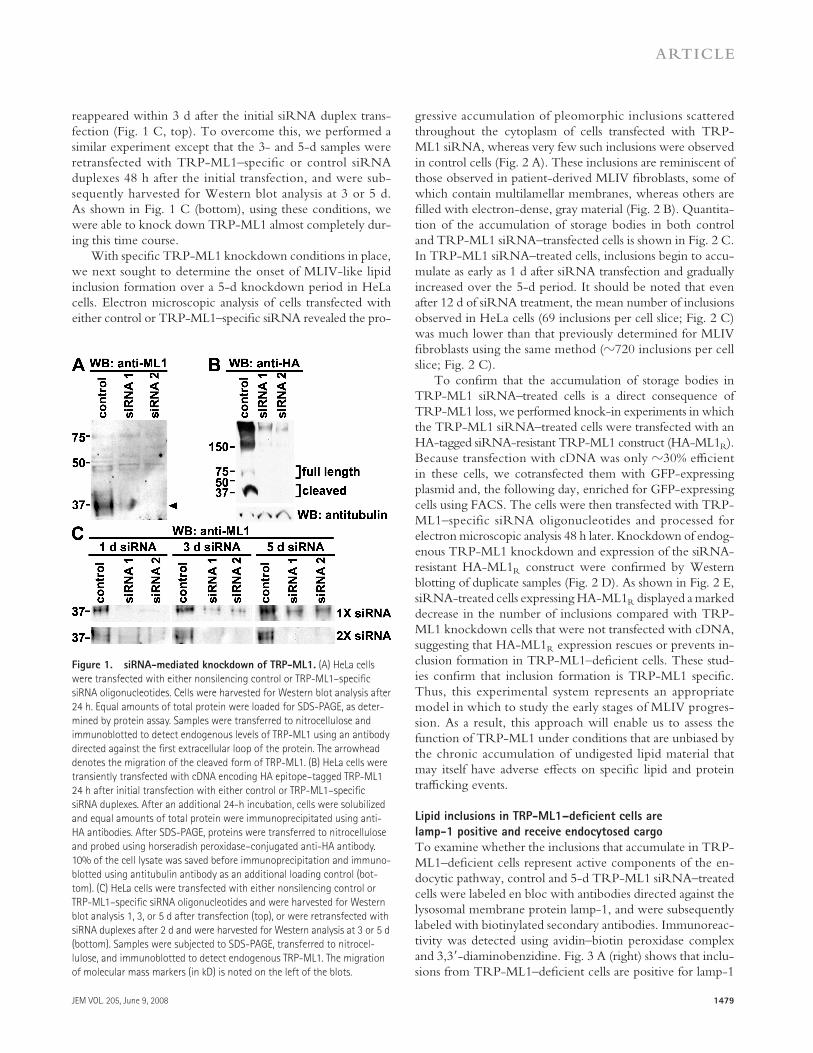
siRNA-mediated TRP-ML1 knockdown
We identifi ed two siRNA oligonucleotides specifi c for TRP-ML1 and tested their ability to knock down endog-enous as well as heterologously expressed hemagglutinin (HA) epitope – tagged TRP-ML1 in HeLa cells. Cells were transfected with control (nonsilencing) or with one of two TRP-ML1 – specifi c siRNA oligonucleotides. Cells were har-vested 24 h after transfection and subjected to Western blot analysis. We previously demonstrated that the 65-kD full-length TRP-ML1 is intracellularly cleaved into two frag-ments of roughly equal molecular mass (40 and 37 kD) ( 19, 20 ). Because it contains only a single transmembrane domain, the N-terminal fragment is less prone to aggregation than the full-length protein or C-terminal half, and is thus easier to detect upon SDS-PAGE. The arrowhead in Fig. 1 A marks the migration of the N-terminal fragment of TRP-ML1. Transfection with either of the TRP-ML1 – specifi c siRNA duplexes resulted in virtually complete knockout of native TRP-ML1. Similarly, knockdown of exogenously expressed TRP-ML1 was observed when cells transfected with siRNA were later retransfected with an HA-tagged version of TRP-ML1 ( Fig. 1 B ). Under these overexpression conditions, we could detect both the full-length protein and the HA-tagged cleaved fragments on Western blots of cells transfected with control but not with TRP-ML1 – specifi c siRNA duplexes ( Fig. 1 B ).
We next optimized conditions to knock down TRP-ML1 over longer time periods. To determine the duration of TRP-ML1 knockdown, cells were harvested for Western blot analysis at 1, 3, or 5 d after transfection with either a nonsilencing control siRNA duplex or with TRP-ML1 – spe-cifi c oligonucleotides. As before, knockdown was virtually com-plete within 1 d of transfection; however, native TRP-ML1
on TRP-ML1 permeability characteristics is only beginning to emerge, whereas native TRP-ML1 activity has not been studied. Recombinant TRP-ML1 was characterized in the plasma membrane, where it is targeted under overexpression conditions ( 19, 24 ), and in artifi cial lipid bilayers using TRP-ML1 purifi ed from overexpressing cells or synthesized in a cell-free system ( 25, 26 ). These experimental systems yielded outwardly rectifying monovalent cation-permeable channels. On the other hand, “ activating ” mutations in TRP-ML1 resulted in an inwardly rectifying current ( 27 ). Such mu-tants were permeable to Ca 2+ , which is similar to some previously published data on wild-type TRP-ML1 ( 24, 28 ), whereas other studies have demonstrated a Ca 2+ block of TRP-ML1 ( 13, 26 ).
The “ biogenesis ” model for MLIV progression suggests that TRP-ML1 regulates lipid traffi cking by mediating spe-cifi c fi ssion and/or fusion events between late endosomes and lysosomes that occur during the process of lysosome biogen-esis, a Ca 2+ -dependent process ( 29 – 34 ). In the absence of functional TRP-ML1, endocytosed material destined for degradation accumulates because of impaired access to the hydrolases necessary for catabolism. Moreover, this model predicts that there will be a global defect in the postendocytic delivery of both lipids and proteins to lysosomes. The notion that TRP-ML1 directly regulates membrane traffi c is based on the observations that TRP-ML1 – defi cient cells display aberrant mixing of lysosomal and endosomal content ( 28, 33 ) and that traffi cking of a fl uorescent conjugate of BODIPY-C5-lactosylceramide (LacCer) along the endocytic pathway is delayed in these cells ( 13, 21, 23 ). This model is also sup-ported by studies performed in Caenorhabditis elegans in which the functional TRP-ML1 orthologue CUP-5 has been iden-tifi ed ( 35, 36 ). Knockout of the cup-5 gene has been associ-ated with defects in lysosome biogenesis. There is increased colocalization of late endosomal and lysosomal markers in cup-5 mutants, and loss of this gene results in the abnormal accumulation of vacuolar structures that are interpreted to represent hybrid late endosomal – lysosomal structures ( 33 ). Again, the observed endocytic abnormalities observed in cup-5 mutants were alleviated by exogenous expression of func-tional human TRP-ML1.
The “ metabolic ” model suggests that, similar to the ClC channels, TRP-ML1 regulates lysosomal ion homeostasis and thus directly aff ects the activity of lysosomal digestive enzymes ( 37 ). It was hypothesized that TRP-ML1 functions as a H + leak pathway to prevent the overacidifi cation of the lysosomal lumen, and that the activity of lysosomal lipases are disrupted as a consequence of the ionic imbalance in TRP-ML1 – defi cient lysosomes ( 13 ).
An additional complexity that must be clarifi ed to prop-erly describe MLIV pathogenesis and TRP-ML1 function is whether any defects in membrane traffi c or lipid metabo-lism are the primary cause of MLIV, or are instead second-ary eff ects caused by the chronic accumulation of undigested lipids in these cells. The membrane traffi cking studies dis-cussed earlier in this section were performed in chronically
JEM VOL. 205, June 9, 2008
ARTICLE
1479
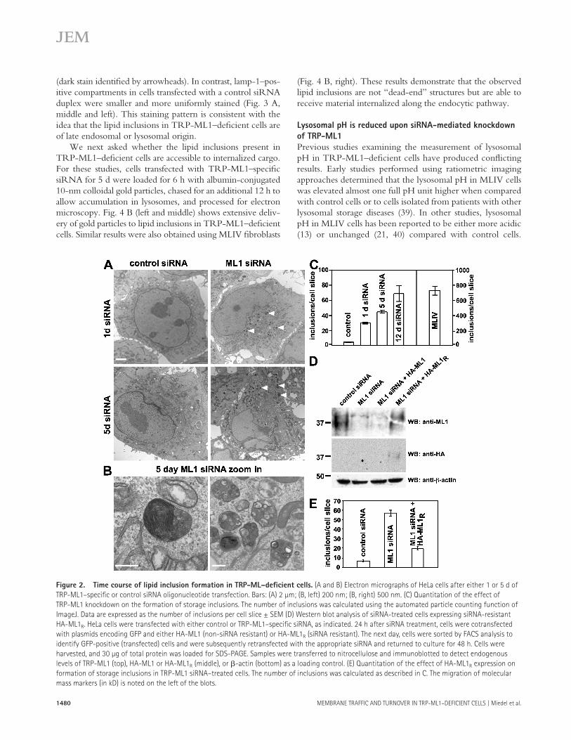
gressive accumulation of pleomorphic inclusions scattered throughout the cytoplasm of cells transfected with TRP-ML1 siRNA, whereas very few such inclusions were observed in control cells ( Fig. 2 A ). These inclusions are reminiscent of those observed in patient-derived MLIV fi broblasts, some of which contain multilamellar membranes, whereas others are fi lled with electron-dense, gray material ( Fig. 2 B ). Quantita-tion of the accumulation of storage bodies in both control and TRP-ML1 siRNA – transfected cells is shown in Fig. 2 C . In TRP-ML1 siRNA – treated cells, inclusions begin to accu-mulate as early as 1 d after siRNA transfection and gradually increased over the 5-d period. It should be noted that even after 12 d of siRNA treatment, the mean number of inclusions observed in HeLa cells (69 inclusions per cell slice; Fig. 2 C ) was much lower than that previously determined for MLIV fi broblasts using the same method ( � 720 inclusions per cell slice; Fig. 2 C ).
To confi rm that the accumulation of storage bodies in TRP-ML1 siRNA – treated cells is a direct consequence of TRP-ML1 loss, we performed knock-in experiments in which the TRP-ML1 siRNA – treated cells were transfected with an HA-tagged siRNA-resistant TRP-ML1 construct (HA-ML1 R ). Because transfection with cDNA was only � 30% effi cient in these cells, we cotransfected them with GFP-expressing plasmid and, the following day, enriched for GFP-expressing cells using FACS. The cells were then transfected with TRP-ML1 – specific siRNA oligonucleotides and processed for electron microscopic analysis 48 h later. Knockdown of endog-enous TRP-ML1 knockdown and expression of the siRNA-resistant HA-ML1 R construct were confi rmed by Western blotting of duplicate samples ( Fig. 2 D ). As shown in Fig. 2 E , siRNA-treated cells expressing HA-ML1 R displayed a marked decrease in the number of inclusions compared with TRP-ML1 knockdown cells that were not transfected with cDNA, suggesting that HA-ML1 R expression rescues or prevents in-clusion formation in TRP-ML1 – defi cient cells. These stud-ies confi rm that inclusion formation is TRP-ML1 specifi c. Thus, this experimental system represents an appropriate model in which to study the early stages of MLIV progres-sion. As a result, this approach will enable us to assess the function of TRP-ML1 under conditions that are unbiased by the chronic accumulation of undigested lipid material that may itself have adverse eff ects on specifi c lipid and protein traffi cking events.
Lipid inclusions in TRP-ML1 – defi cient cells are
lamp-1 positive and receive endocytosed cargo
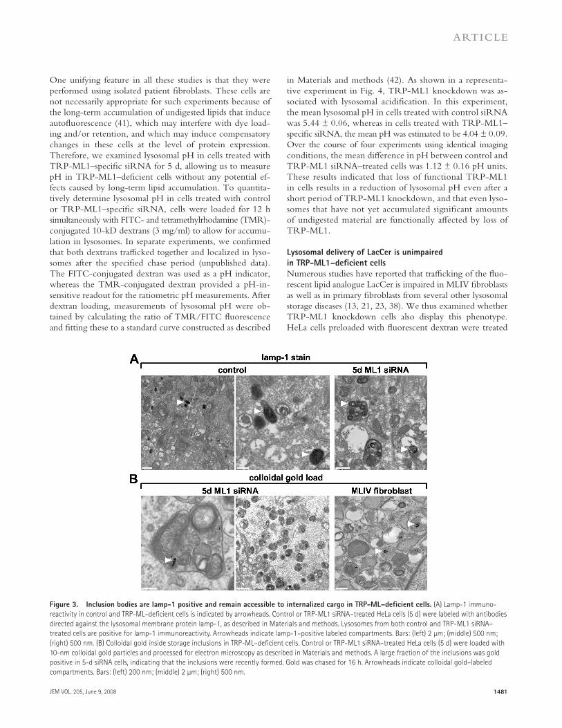
To examine whether the inclusions that accumulate in TRP-ML1 – defi cient cells represent active components of the en-docytic pathway, control and 5-d TRP-ML1 siRNA – treated cells were labeled en bloc with antibodies directed against the lysosomal membrane protein lamp-1, and were subsequently labeled with biotinylated secondary antibodies. Immunoreac-tivity was detected using avidin – biotin peroxidase complex and 3,3 � -diaminobenzidine. Fig. 3 A (right) shows that inclu-sions from TRP-ML1 – defi cient cells are positive for lamp-1
reappeared within 3 d after the initial siRNA duplex trans-fection ( Fig. 1 C , top). To overcome this, we performed a similar experiment except that the 3- and 5-d samples were retransfected with TRP-ML1 – specifi c or control siRNA duplexes 48 h after the initial transfection, and were sub-sequently harvested for Western blot analysis at 3 or 5 d. As shown in Fig. 1 C (bottom), using these conditions, we were able to knock down TRP-ML1 almost completely dur-ing this time course.
With specifi c TRP-ML1 knockdown conditions in place, we next sought to determine the onset of MLIV-like lipid inclusion formation over a 5-d knockdown period in HeLa cells. Electron microscopic analysis of cells transfected with either control or TRP-ML1 – specifi c siRNA revealed the pro-
Figure 1. siRNA-mediated knockdown of TRP-ML1. (A) HeLa cells
were transfected with either nonsilencing control or TRP-ML1 – specifi c
siRNA oligonucleotides. Cells were harvested for Western blot analysis after
24 h. Equal amounts of total protein were loaded for SDS-PAGE, as deter-
mined by protein assay. Samples were transferred to nitrocellulose and
immunoblotted to detect endogenous levels of TRP-ML1 using an antibody
directed against the fi rst extracellular loop of the protein. The arrowhead
denotes the migration of the cleaved form of TRP-ML1. (B) HeLa cells were
transiently transfected with cDNA encoding HA epitope – tagged TRP-ML1
24 h after initial transfection with either control or TRP-ML1 – specifi c
siRNA duplexes. After an additional 24-h incubation, cells were solubilized
and equal amounts of total protein were immunoprecipitated using anti-
HA antibodies. After SDS-PAGE, proteins were transferred to nitrocellulose
and probed using horseradish peroxidase – conjugated anti-HA antibody.
10% of the cell lysate was saved before immunoprecipitation and immuno-
blotted using antitubulin antibody as an additional loading control (bot-
tom). (C) HeLa cells were transfected with either nonsilencing control or
TRP-ML1 – specifi c siRNA oligonucleotides and were harvested for Western
blot analysis 1, 3, or 5 d after transfection (top), or were retransfected with
siRNA duplexes after 2 d and were harvested for Western analysis at 3 or 5 d
(bottom). Samples were subjected to SDS-PAGE, transferred to nitrocel-
lulose, and immunoblotted to detect endogenous TRP-ML1. The migration
of molecular mass markers (in kD) is noted on the left of the blots.
1480 MEMBRANE TRAFFIC AND TURNOVER IN TRP-ML1 – DEFICIENT CELLS | Miedel et al.
( Fig. 4 B , right). These results demonstrate that the observed lipid inclusions are not “ dead-end ” structures but are able to receive material internalized along the endocytic pathway.
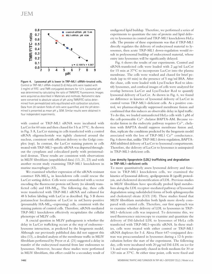
Lysosomal pH is reduced upon siRNA-mediated knockdown
of TRP-ML1
Previous studies examining the measurement of lysosomal pH in TRP-ML1 – defi cient cells have produced confl icting results. Early studies performed using ratiometric imaging approaches determined that the lysosomal pH in MLIV cells was elevated almost one full pH unit higher when compared with control cells or to cells isolated from patients with other lysosomal storage diseases ( 39 ). In other studies, lysosomal pH in MLIV cells has been reported to be either more acidic ( 13 ) or unchanged ( 21, 40 ) compared with control cells.
(dark stain identifi ed by arrowheads). In contrast, lamp-1 – pos-itive compartments in cells transfected with a control siRNA duplex were smaller and more uniformly stained ( Fig. 3 A , middle and left). This staining pattern is consistent with the idea that the lipid inclusions in TRP-ML1 – defi cient cells are of late endosomal or lysosomal origin.
We next asked whether the lipid inclusions present in TRP-ML1 – defi cient cells are accessible to internalized cargo. For these studies, cells transfected with TRP-ML1 – specifi c siRNA for 5 d were loaded for 6 h with albumin-conjugated 10-nm colloidal gold particles, chased for an additional 12 h to allow accumulation in lysosomes, and processed for electron microscopy. Fig. 4 B (left and middle) shows extensive deliv-ery of gold particles to lipid inclusions in TRP-ML1 – defi cient cells. Similar results were also obtained using MLIV fi broblasts
Figure 2. Time course of lipid inclusion formation in TRP-ML – defi cient cells. (A and B) Electron micrographs of HeLa cells after either 1 or 5 d of
TRP-ML1 – specifi c or control siRNA oligonucleotide transfection. Bars: (A) 2 μ m; (B, left) 200 nm; (B, right) 500 nm. (C) Quantitation of the effect of
TRP-ML1 knockdown on the formation of storage inclusions. The number of inclusions was calculated using the automated particle counting function of
ImageJ. Data are expressed as the number of inclusions per cell slice ± SEM (D) Western blot analysis of siRNA-treated cells expressing siRNA-resistant
HA-ML1 R . HeLa cells were transfected with either control or TRP-ML1 – specifi c siRNA, as indicated. 24 h after siRNA treatment, cells were cotransfected
with plasmids encoding GFP and either HA-ML1 (non-siRNA resistant) or HA-ML1 R (siRNA resistant). The next day, cells were sorted by FACS analysis to
identify GFP-positive (transfected) cells and were subsequently retransfected with the appropriate siRNA and returned to culture for 48 h. Cells were
harvested, and 30 μ g of total protein was loaded for SDS-PAGE. Samples were transferred to nitrocellulose and immunoblotted to detect endogenous
levels of TRP-ML1 (top), HA-ML1 or HA-ML1 R (middle), or � -actin (bottom) as a loading control. (E) Quantitation of the effect of HA-ML1 R expression on
formation of storage inclusions in TRP-ML1 siRNA – treated cells. The number of inclusions was calculated as described in C. The migration of molecular
mass markers (in kD) is noted on the left of the blots.
JEM VOL. 205, June 9, 2008
ARTICLE
1481
in Materials and methods ( 42 ). As shown in a representa-tive experiment in Fig. 4 , TRP-ML1 knockdown was as-sociated with lysosomal acidifi cation. In this experiment, the mean lysosomal pH in cells treated with control siRNA was 5.44 ± 0.06, whereas in cells treated with TRP-ML1 – specifi c siRNA, the mean pH was estimated to be 4.04 ± 0.09. Over the course of four experiments using identical imaging conditions, the mean diff erence in pH between control and TRP-ML1 siRNA – treated cells was 1.12 ± 0.16 pH units. These results indicated that loss of functional TRP-ML1 in cells results in a reduction of lysosomal pH even after a short period of TRP-ML1 knockdown, and that even lyso-somes that have not yet accumulated signifi cant amounts of undigested material are functionally aff ected by loss of TRP-ML1.
Lysosomal delivery of LacCer is unimpaired
in TRP-ML1 – defi cient cells
Numerous studies have reported that traffi cking of the fl uo-rescent lipid analogue LacCer is impaired in MLIV fi broblasts as well as in primary fi broblasts from several other lysosomal storage diseases ( 13, 21, 23, 38 ). We thus examined whether TRP-ML1 knockdown cells also display this phenotype. HeLa cells preloaded with fl uorescent dextran were treated
One unifying feature in all these studies is that they were performed using isolated patient fi broblasts. These cells are not necessarily appropriate for such experiments because of the long-term accumulation of undigested lipids that induce autofl uorescence ( 41 ), which may interfere with dye load-ing and/or retention, and which may induce compensatory changes in these cells at the level of protein expression. Therefore, we examined lysosomal pH in cells treated with TRP-ML1 – specifi c siRNA for 5 d, allowing us to measure pH in TRP-ML1 – defi cient cells without any potential ef-fects caused by long-term lipid accumulation. To quantita-tively determine lysosomal pH in cells treated with control or TRP-ML1 – specifi c siRNA, cells were loaded for 12 h simultaneously with FITC- and tetramethylrhodamine (TMR)- conjugated 10-kD dextrans (3 mg/ml) to allow for accumu-lation in lysosomes. In separate experiments, we confi rmed that both dextrans traffi cked together and localized in lyso-somes after the specifi ed chase period (unpublished data). The FITC-conjugated dextran was used as a pH indicator, whereas the TMR-conjugated dextran provided a pH-in-sensitive readout for the ratiometric pH measurements. After dextran loading, measurements of lysosomal pH were ob-tained by calculating the ratio of TMR/FITC fl uorescence and fi tting these to a standard curve constructed as described
Figure 3. Inclusion bodies are lamp-1 positive and remain accessible to internalized cargo in TRP-ML – defi cient cells. (A) Lamp-1 immuno-
reactivity in control and TRP-ML – defi cient cells is indicated by arrowheads. Control or TRP-ML1 siRNA – treated HeLa cells (5 d) were labeled with antibodies
directed against the lysosomal membrane protein lamp-1, as described in Materials and methods. Lysosomes from both control and TRP-ML1 siRNA –
treated cells are positive for lamp-1 immunoreactivity. Arrowheads indicate lamp-1 – positive labeled compartments. Bars: (left) 2 μ m; (middle) 500 nm;
(right) 500 nm. (B) Colloidal gold inside storage inclusions in TRP-ML – defi cient cells. Control or TRP-ML1 siRNA – treated HeLa cells (5 d) were loaded with
10-nm colloidal gold particles and processed for electron microscopy as described in Materials and methods. A large fraction of the inclusions was gold
positive in 5-d siRNA cells, indicating that the inclusions were recently formed. Gold was chased for 16 h. Arrowheads indicate colloidal gold – labeled
compartments. Bars: (left) 200 nm; (middle) 2 μ m; (right) 500 nm.
1482 MEMBRANE TRAFFIC AND TURNOVER IN TRP-ML1 – DEFICIENT CELLS | Miedel et al.
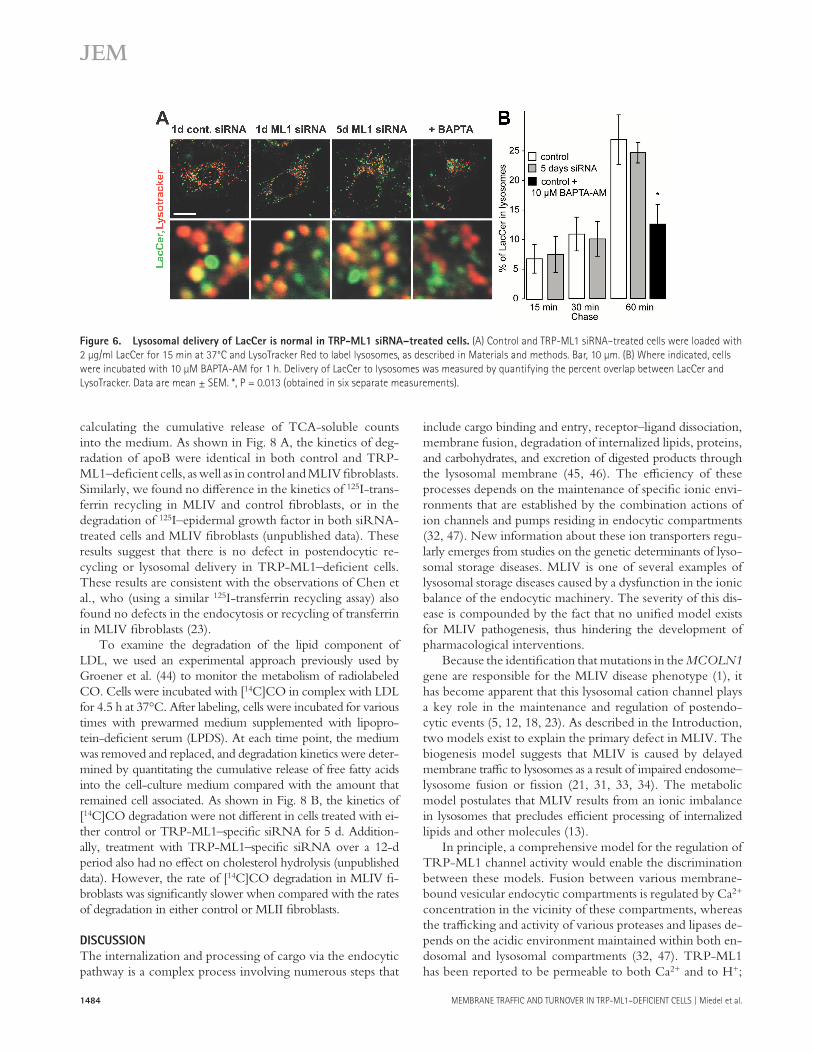
undigested lipid buildup. Therefore, we performed a series of experiments to quantitate the rate of protein and lipid deliv-ery to lysosomes in control and TRP-ML1 knockdown HeLa cells. The premise of these experiments was that if TRP-ML1 directly regulates the delivery of endocytosed material to ly-sosomes, then acute TRP-ML1 down-regulation would re-sult in prelysosomal buildup of endocytosed material, whose entry into lysosomes will be signifi cantly delayed.
Fig. 6 shows the results of our experiments. Control and siRNA-transfected cells were loaded with 2 μ g/ml LacCer for 15 min at 37 ° C to incorporate LacCer into the plasma membrane. The cells were washed and chased for brief pe-riods (up to 60 min) in the presence of 5 mg/ml BSA. After the chase, cells were loaded with LysoTracker Red to iden-tify lysosomes, and confocal images of cells were analyzed for overlap between LacCer and LysoTracker Red to quantify lysosomal delivery of LacCer. As shown in Fig. 6 , we found no diff erence in kinetics of lysosomal delivery of LacCer in control versus TRP-ML1 – defi cient cells. As a positive con-trol, we pharmacologically suppressed membrane fusion and confi rmed that this induces an observable delay in lipid traffi c. To do this, we loaded untransfected HeLa cells with 1 μ M of the cell-permeable Ca 2+ chelator BAPTA-AM. Because ve-sicular fusion in the endocytic pathway requires Ca 2+ , chela-tion with BAPTA should suppress membrane fusion and, thus, replicate the conditions predicted by the biogenesis model associated with the loss of TRP-ML1 Ca 2+ conductance. Fig. 6 shows that, unlike TRP-ML1 down-regulation, BAPTA-AM inhibited delivery of LacCer to lysosomal compartments. Therefore, the delivery of LacCer to lysosomes is unimpaired in TRP-ML1 – defi cient cells.
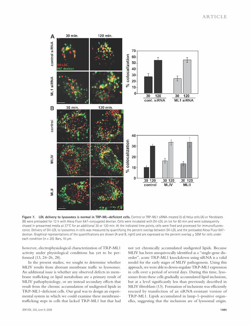
Low density lipoprotein (LDL) traffi cking and degradation
in TRP-ML1 – defi cient cells
To more quantitatively assess lysosomal delivery and func-tion in TRP-ML1 knockdown cells, we examined the kinetics of lysosomal delivery, apolipoprotein B (apoB) proteol-ysis, and cholesterol deesterifi cation of LDL. Previous studies in MLIV fi broblasts have specifi cally probed lipid metabo-lism along the LDL receptor – mediated pathway of lysosomal degradation using radiolabeled forms of both sphingomyelin and cholesteryl oleate (CO) ( 18 ). These studies found that MLIV fi broblasts metabolize both lipids more slowly com-pared with control cells. Therefore, our fi rst approach was to examine whether delivery of LDL to lysosomes in TRP-ML1 – deficient cells was impaired. To determine this, we used fl uorescence microscopy to examine and quantitate the delivery of DiI-labeled LDL to lysosomes of HeLa cells transfected with TRP-ML1 – specifi c siRNA. In these stud-ies, cells were treated with either control or TRP-ML1 siRNA duplexes for 5 d. Alexa Fluor 647 – conjugated dex-tran was preaccumulated in lysosomes during a 12-h prein-cubation before the start of the experiment. The following day, cells were incubated with 20 μ g/ml DiI-LDL on ice for 1 h and were subsequently chased in fresh medium for 30 or 120 min at 37 ° C. At either time point, cells were fi xed and
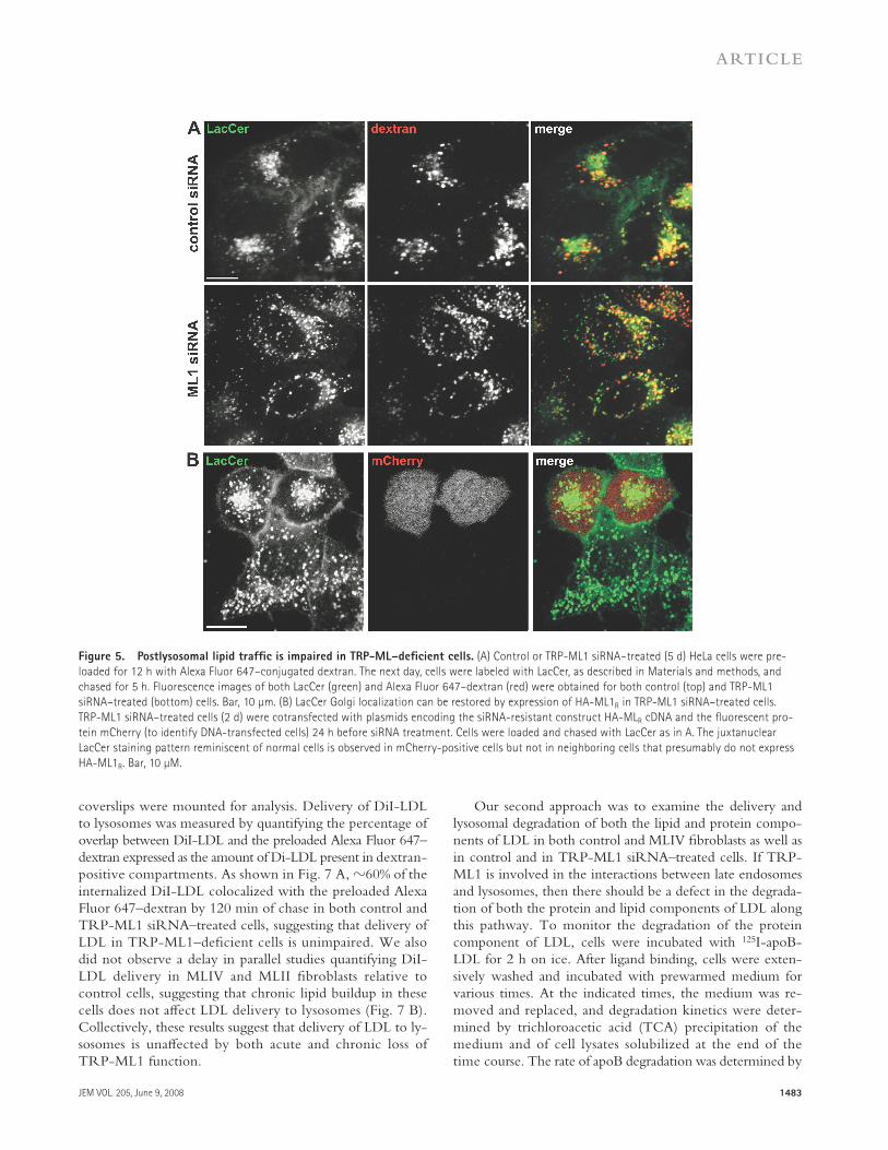
with control or TRP-ML1 siRNA were incubated with LacCer for 60 min and then chased for 5 h at 37 ° C. As shown in Fig. 5 A , LacCer staining in cells transfected with a control siRNA oligonucleotide was tightly clustered around the nucleus, consistent with effi cient delivery to the Golgi com-plex (top). In contrast, the LacCer staining pattern in cells treated with TRP-ML1 – specifi c siRNA was dispersed through-out the cytoplasm and colocalized partly with preinternal-ized dextran. These results are consistent with observations in MLIV fi broblasts (unpublished data) ( 13, 21, 23 ) and with another recent study examining TRP-ML1 knockdown in murine macrophages ( 43 ).
We examined whether expression of the siRNA-resistant construct HA-ML1 R in knockdown cells could rescue the LacCer sorting defect. Cells were cotransfected with a vector encoding the fl uorescent protein mCherry (to identify trans-fected cells) and HA-ML R . The following day, these cells were transfected with TRP-ML1 siRNA and cultured for 48 h before labeling with LacCer as described. Fig. 5 B shows juxtanuclear localization of LacCer in mCherry-positive (presumably HA-ML R – expressing) cells, consistent with the staining pattern of control cells. Therefore, siRNA-mediated TRP-ML1 knockdown eff ectively recapitulates the cellular phenotype of MLIV cells.
A crucial question in MLIV pathogenesis is whether the delay in LacCer traffi cking occurs at the step of endosome – lysosome interaction, as predicted by the biogenesis model. Although our previously published data did not support this idea ( 13 ), a detailed analysis of the membrane traffi c in MLIV fi broblasts performed by Pryor et al. ( 21 ) suggested a delay in transfer of the endocytosed material from late endosomes to lysosomes. However, because these studies were performed in MLIV fi broblasts, this eff ect could be a secondary result of
Figure 4. Lysosomal pH is lower in TRP-ML1 siRNA – treated cells.
Control or TRP-ML1 siRNA – treated (5 d) HeLa cells were loaded with
3 mg/ml of FITC- and TMR-conjugated dextrans for 12 h. Lysosomal pH
was determined by calculating the ratio of TMR/FITC fl uorescence. Images
were acquired as described in Materials and methods. Ratiometric data
were converted to absolute values of pH using TMR/FITC ratios deter-
mined from permeabilized cells equilibrated with calibration solutions.
Data from 20 random fi elds of cells were quantifi ed, and the pH deter-
mined is presented as mean pH ± SEM. Similar results were obtained in
four independent experiments.
JEM VOL. 205, June 9, 2008
ARTICLE
1483
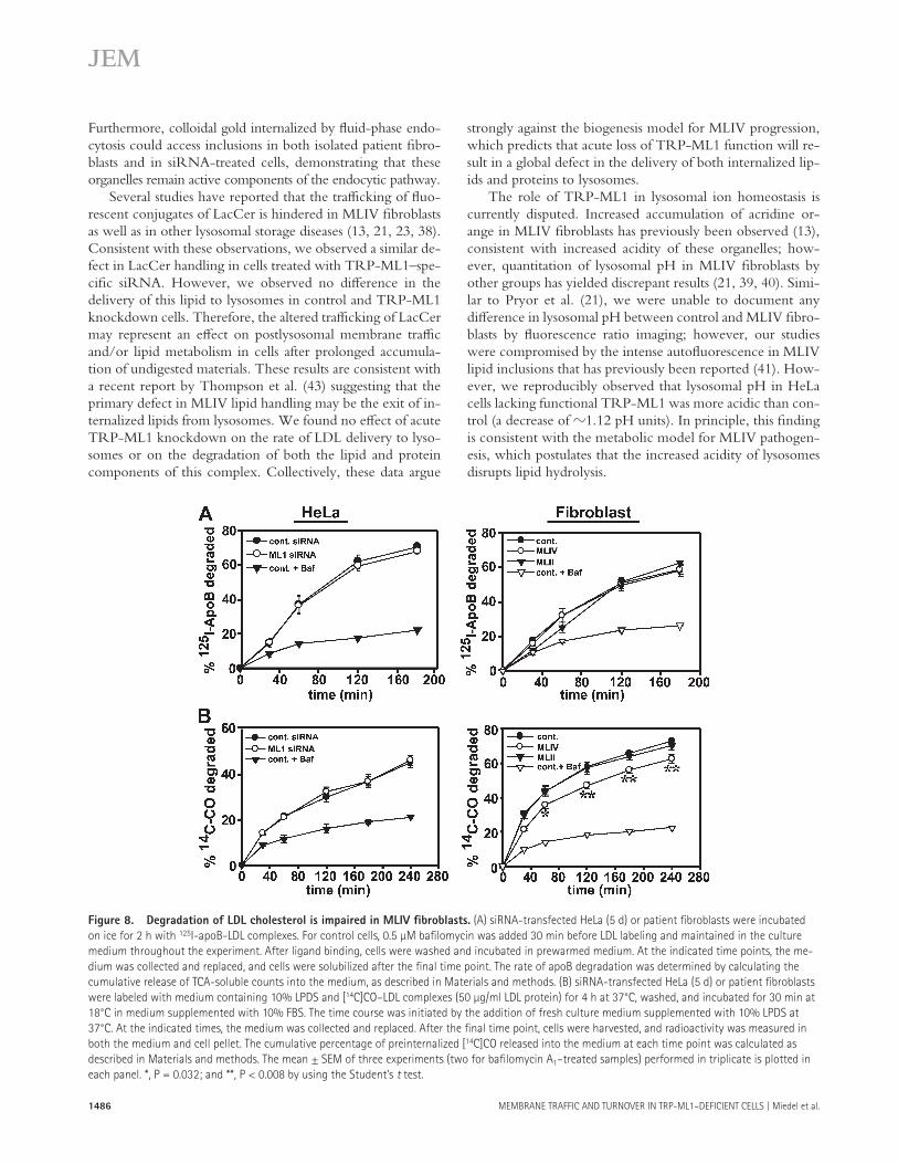
Our second approach was to examine the delivery and lysosomal degradation of both the lipid and protein compo-nents of LDL in both control and MLIV fi broblasts as well as in control and in TRP-ML1 siRNA – treated cells. If TRP-ML1 is involved in the interactions between late endosomes and lysosomes, then there should be a defect in the degrada-tion of both the protein and lipid components of LDL along this pathway. To monitor the degradation of the protein component of LDL, cells were incubated with 125 I-apoB-LDL for 2 h on ice. After ligand binding, cells were exten-sively washed and incubated with prewarmed medium for various times. At the indicated times, the medium was re-moved and replaced, and degradation kinetics were deter-mined by trichloroacetic acid (TCA) precipitation of the medium and of cell lysates solubilized at the end of the time course. The rate of apoB degradation was determined by
coverslips were mounted for analysis. Delivery of DiI-LDL to lysosomes was measured by quantifying the percentage of overlap between DiI-LDL and the preloaded Alexa Fluor 647 – dextran expressed as the amount of Di-LDL present in dextran-positive compartments. As shown in Fig. 7 A , � 60% of the internalized DiI-LDL colocalized with the preloaded Alexa Fluor 647 – dextran by 120 min of chase in both control and TRP-ML1 siRNA – treated cells, suggesting that delivery of LDL in TRP-ML1 – defi cient cells is unimpaired. We also did not observe a delay in parallel studies quantifying DiI-LDL delivery in MLIV and MLII fi broblasts relative to control cells, suggesting that chronic lipid buildup in these cells does not aff ect LDL delivery to lysosomes ( Fig. 7 B ). Collectively, these results suggest that delivery of LDL to ly-sosomes is unaff ected by both acute and chronic loss of TRP- ML1 function.
Figure 5. Postlysosomal lipid traffi c is impaired in TRP-ML – defi cient cells. (A) Control or TRP-ML1 siRNA – treated (5 d) HeLa cells were pre-
loaded for 12 h with Alexa Fluor 647 – conjugated dextran. The next day, cells were labeled with LacCer, as described in Materials and methods, and
chased for 5 h. Fluorescence images of both LacCer (green) and Alexa Fluor 647 – dextran (red) were obtained for both control (top) and TRP-ML1
siRNA – treated (bottom) cells. Bar, 10 μ m. (B) LacCer Golgi localization can be restored by expression of HA-ML1 R in TRP-ML1 siRNA – treated cells.
TRP-ML1 siRNA – treated cells (2 d) were cotransfected with plasmids encoding the siRNA-resistant construct HA-ML R cDNA and the fl uorescent pro-
tein mCherry (to identify DNA-transfected cells) 24 h before siRNA treatment. Cells were loaded and chased with LacCer as in A. The juxtanuclear
LacCer staining pattern reminiscent of normal cells is observed in mCherry-positive cells but not in neighboring cells that presumably do not express
HA-ML1 R . Bar, 10 μ M.
1484 MEMBRANE TRAFFIC AND TURNOVER IN TRP-ML1 – DEFICIENT CELLS | Miedel et al.
include cargo binding and entry, receptor – ligand dissociation, membrane fusion, degradation of internalized lipids, proteins, and carbohydrates, and excretion of digested products through the lysosomal membrane ( 45, 46 ). The effi ciency of these processes depends on the maintenance of specifi c ionic envi-ronments that are established by the combination actions of ion channels and pumps residing in endocytic compartments ( 32, 47 ). New information about these ion transporters regu-larly emerges from studies on the genetic determinants of lyso-somal storage diseases. MLIV is one of several examples of lysosomal storage diseases caused by a dysfunction in the ionic balance of the endocytic machinery. The severity of this dis-ease is compounded by the fact that no unifi ed model exists for MLIV pathogenesis, thus hindering the development of pharmacological interventions.
Because the identifi cation that mutations in the MCOLN1 gene are responsible for the MLIV disease phenotype ( 1 ), it has become apparent that this lysosomal cation channel plays a key role in the maintenance and regulation of postendo-cytic events ( 5, 12, 18, 23 ). As described in the Introduction, two models exist to explain the primary defect in MLIV. The biogenesis model suggests that MLIV is caused by delayed membrane traffi c to lysosomes as a result of impaired endosome – lysosome fusion or fi ssion ( 21, 31, 33, 34 ). The metabolic model postulates that MLIV results from an ionic imbalance in lysosomes that precludes effi cient processing of internalized lipids and other molecules ( 13 ).
In principle, a comprehensive model for the regulation of TRP-ML1 channel activity would enable the discrimination between these models. Fusion between various membrane-bound vesicular endocytic compartments is regulated by Ca 2+ concentration in the vicinity of these compartments, whereas the traffi cking and activity of various proteases and lipases de-pends on the acidic environment maintained within both en-dosomal and lysosomal compartments ( 32, 47 ). TRP-ML1 has been reported to be permeable to both Ca 2+ and to H + ;
calculating the cumulative release of TCA-soluble counts into the medium. As shown in Fig. 8 A , the kinetics of deg-radation of apoB were identical in both control and TRP-ML1 – defi cient cells, as well as in control and MLIV fi broblasts. Similarly, we found no diff erence in the kinetics of 125 I-trans-ferrin recycling in MLIV and control fi broblasts, or in the degradation of 125 I – epidermal growth factor in both siRNA-treated cells and MLIV fi broblasts (unpublished data). These results suggest that there is no defect in postendocytic re-cycling or lysosomal delivery in TRP-ML1 – defi cient cells. These results are consistent with the observations of Chen et al., who (using a similar 125 I-transferrin recycling assay) also found no defects in the endocytosis or recycling of transferrin in MLIV fi broblasts ( 23 ).
To examine the degradation of the lipid component of LDL, we used an experimental approach previously used by Groener et al. ( 44 ) to monitor the metabolism of radiolabeled CO. Cells were incubated with [ 14 C]CO in complex with LDL for 4.5 h at 37 ° C. After labeling, cells were incubated for various times with prewarmed medium supplemented with lipopro-tein-defi cient serum (LPDS). At each time point, the medium was removed and replaced, and degradation kinetics were deter-mined by quantitating the cumulative release of free fatty acids into the cell-culture medium compared with the amount that remained cell associated. As shown in Fig. 8 B , the kinetics of [ 14 C]CO degradation were not diff erent in cells treated with ei-ther control or TRP-ML1 – specifi c siRNA for 5 d. Addition-ally, treatment with TRP-ML1 – specifi c siRNA over a 12-d period also had no eff ect on cholesterol hydrolysis (unpublished data). However, the rate of [ 14 C]CO degradation in MLIV fi -broblasts was signifi cantly slower when compared with the rates of degradation in either control or MLII fi broblasts.
DISCUSSION
The internalization and processing of cargo via the endocytic pathway is a complex process involving numerous steps that
Figure 6. Lysosomal delivery of LacCer is normal in TRP-ML1 siRNA – treated cells. (A) Control and TRP-ML1 siRNA – treated cells were loaded with
2 μ g/ml LacCer for 15 min at 37 ° C and LysoTracker Red to label lysosomes, as described in Materials and methods. Bar, 10 μ m. (B) Where indicated, cells
were incubated with 10 μ M BAPTA-AM for 1 h. Delivery of LacCer to lysosomes was measured by quantifying the percent overlap between LacCer and
LysoTracker. Data are mean ± SEM. *, P = 0.013 (obtained in six separate measurements).
JEM VOL. 205, June 9, 2008
ARTICLE
1485
not yet chronically accumulated undigested lipids. Because MLIV has been unequivocally identifi ed as a “ single-gene dis-order ” , acute TRP-ML1 knockdown using siRNA is a valid model for the early stages of MLIV pathogenesis. Using this approach, we were able to down-regulate TRP-ML1 expression in cells over a period of several days. During this time, lyso-somes from these cells gradually accumulated lipid inclusions, but at a level signifi cantly less than previously described in MLIV fi broblasts ( 13 ). Formation of inclusions was effi ciently rescued by transfection of an siRNA-resistant version of TRP-ML1. Lipids accumulated in lamp-1 – positive organ-elles, suggesting that the inclusions are of lysosomal origin.
however, electrophysiological characterization of TRP-ML1 activity under physiological conditions has yet to be per-formed ( 13, 24 – 26, 28 ).
In the present studies, we sought to determine whether MLIV results from aberrant membrane traffi c to lysosomes. An additional issue is whether any observed defects in mem-brane traffi cking or lipid metabolism are a primary result of MLIV pathophysiology, or are instead secondary eff ects that result from the chronic accumulation of undigested lipids in TRP-ML1 – defi cient cells. Our goal was to design an experi-mental system in which we could examine these membrane-traffi cking steps in cells that lacked TRP-ML1 but that had
Figure 7. LDL delivery to lysosomes is normal in TRP-ML – defi cient cells. Control or TRP-ML1 siRNA – treated (5 d) HeLa cells (A) or fi broblasts
(B) were preloaded for 12 h with Alexa Fluor 647 – conjugated dextran. Cells were incubated with DiI-LDL on ice for 60 min and were subsequently
chased in prewarmed media at 37 ° C for an additional 30 or 120 min. At the indicated time points, cells were fi xed and processed for immunofl uores-
cence. Delivery of DiI-LDL to lysosomes in cells was measured by quantifying the percent overlap between DiI-LDL and the preloaded Alexa Fluor 647 –
dextran. Graphical representations of the quantifi cations are shown (A and B, right) and are expressed as the percent overlap ± SEM for cells under
each condition ( n = 20). Bars, 10 μ m.
1486 MEMBRANE TRAFFIC AND TURNOVER IN TRP-ML1 – DEFICIENT CELLS | Miedel et al.
strongly against the biogenesis model for MLIV progression, which predicts that acute loss of TRP-ML1 function will re-sult in a global defect in the delivery of both internalized lip-ids and proteins to lysosomes.
The role of TRP-ML1 in lysosomal ion homeostasis is currently disputed. Increased accumulation of acridine or-ange in MLIV fi broblasts has previously been observed ( 13 ), consistent with increased acidity of these organelles; how-ever, quantitation of lysosomal pH in MLIV fi broblasts by other groups has yielded discrepant results ( 21, 39, 40 ). Simi-lar to Pryor et al. ( 21 ), we were unable to document any diff erence in lysosomal pH between control and MLIV fi bro-blasts by fl uorescence ratio imaging; however, our studies were compromised by the intense autofl uorescence in MLIV lipid inclusions that has previously been reported ( 41 ). How-ever, we reproducibly observed that lysosomal pH in HeLa cells lacking functional TRP-ML1 was more acidic than con-trol (a decrease of � 1.12 pH units). In principle, this fi nding is consistent with the metabolic model for MLIV pathogen-esis, which postulates that the increased acidity of lysosomes disrupts lipid hydrolysis.
Furthermore, colloidal gold internalized by fl uid-phase endo-cytosis could access inclusions in both isolated patient fi bro-blasts and in siRNA-treated cells, demonstrating that these organelles remain active components of the endocytic pathway.
Several studies have reported that the traffi cking of fl uo-rescent conjugates of LacCer is hindered in MLIV fi broblasts as well as in other lysosomal storage diseases ( 13, 21, 23, 38 ). Consistent with these observations, we observed a similar de-fect in LacCer handling in cells treated with TRP-ML1 – spe-cifi c siRNA. However, we observed no diff erence in the delivery of this lipid to lysosomes in control and TRP-ML1 knockdown cells. Therefore, the altered traffi cking of LacCer may represent an eff ect on postlysosomal membrane traffi c and/or lipid metabolism in cells after prolonged accumula-tion of undigested materials. These results are consistent with a recent report by Thompson et al. ( 43 ) suggesting that the primary defect in MLIV lipid handling may be the exit of in-ternalized lipids from lysosomes. We found no eff ect of acute TRP-ML1 knockdown on the rate of LDL delivery to lyso-somes or on the degradation of both the lipid and protein components of this complex. Collectively, these data argue
Figure 8. Degradation of LDL cholesterol is impaired in MLIV fi broblasts. (A) siRNA-transfected HeLa (5 d) or patient fi broblasts were incubated
on ice for 2 h with 125 I-apoB-LDL complexes. For control cells, 0.5 μ M bafi lomycin was added 30 min before LDL labeling and maintained in the culture
medium throughout the experiment. After ligand binding, cells were washed and incubated in prewarmed medium. At the indicated time points, the me-
dium was collected and replaced, and cells were solubilized after the fi nal time point. The rate of apoB degradation was determined by calculating the
cumulative release of TCA-soluble counts into the medium, as described in Materials and methods. (B) siRNA-transfected HeLa (5 d) or patient fi broblasts
were labeled with medium containing 10% LPDS and [ 14 C]CO – LDL complexes (50 μ g/ml LDL protein) for 4 h at 37 ° C, washed, and incubated for 30 min at
18 ° C in medium supplemented with 10% FBS. The time course was initiated by the addition of fresh culture medium supplemented with 10% LPDS at
37 ° C. At the indicated times, the medium was collected and replaced. After the fi nal time point, cells were harvested, and radioactivity was measured in
both the medium and cell pellet. The cumulative percentage of preinternalized [ 14 C]CO released into the medium at each time point was calculated as
described in Materials and methods. The mean ± SEM of three experiments (two for bafi lomycin A 1 – treated samples) performed in triplicate is plotted in
each panel. *, P = 0.032; and **, P < 0.008 by using the Student ’ s t test.
JEM VOL. 205, June 9, 2008
ARTICLE
1487
To generate the siRNA-resistant construct HA-ML1 R , the codons encoding
amino acids IQEC starting at position 281 of TRP-ML1 (targeted by siRNA
no. 1) were mutated from CATCCAGGAGTG to TATTCAAGAATG to
create siRNA resistance without aff ecting the corresponding amino acid se-
quence. For knockdown of endogenous TRP-ML1, HeLa SS6 cells were
plated in 24-well dishes and allowed to grow to � 50 – 60% confl uence. Cells
were transfected with a nonspecifi c control siRNA duplex (Thermo Fisher
Scientifi c) or TRP-ML1 – specifi c siRNA duplexes. For each individual well,
3 μ l of 20 μ M siRNA oligonucleotide and 4.5 μ l TransIT-TKO oligonucleo-
tide transfection reagent (Mirus) were used. Cells were harvested for protein
concentration or Western blot analysis 1, 3, or 5 d after transfection. For cells
transfected with siRNA twice over the 5-d knockdown period, 2 d after ini-
tial transfection, cells were replated onto 24-well plates, retransfected as de-
scribed the same day, and harvested for analysis either 24 or 72 h later. For
knockdown of exogenously expressed HA epitope – tagged ML1 (HA-ML1)
( 19, 20 ), cells were plated and transfected with siRNA as described. 24 h af-
ter siRNA transfection, cells were transiently transfected with cDNA encod-
ing HA-ML1 using Lipofectamine 2000 (Invitrogen), according to the
manufacturer ’ s protocol, and harvested for Western blot analysis 24 h later.
Samples were immunoprecipitated and immunoblotted as previously de-
scribed ( 19, 20 ).
Electron microscopy. Cells grown on plastic dishes were fi xed by a 30-
min incubation with a solution containing 2.5% glutaraldehyde in 0.1M
Na-cacodylate, washed with 0.1M Na-cacodylate, postfi xed with a solution
containing 1% OsO4, washed with PBS, and stained en bloc for 30 min with
2% uranyl acetate. After dehydration by immersion in 30 – 100% ethanol, the
samples were embedded in resin by immersion in 30 – 100% resin/propylene
oxide mixtures. Fixed samples were mounted on grids and analyzed with a
transmission electron microscope (100CX; JEOL Ltd.). For immunostaining
with lamp-1 antibodies, after fi xation and freeze – thaw permeabilization of
the membranes at – 80 ° C in a cryoprotectant solution containing glycerol
and sucrose, cells were blocked in BSA with goat serum, incubated over-
night with monoclonal anti – lamp-1 antibodies (Santa Cruz Biotechnology,
Inc.), and rinsed, and biotinylated secondary antibodies were added. After an
extensive wash, the samples were incubated with avidin – peroxidase com-
plexes for 30 min, followed by another wash. Next, 3,3 � -diaminobenzidine
and H 2 O 2 were added for 4 – 10 min and, after another wash, the samples un-
derwent secondary fi xation with 1% OsO 4 for 1 h. This procedure yields the
dark stain associated with lamp-1 immunoreactivity.
For colloidal gold uptake experiments, 10-nm gold particles were incu-
bated with gelatin and BSA (pH 5), to coat the particles. Next, the particles
were washed and added to the cells for 1 h to load the endocytic pathway.
After a 6 – 16-h chase, cells were fi xed and processed for electron microscopy
as described in the previous paragraph.
Measurement of lysosomal pH. Determination of lysosomal pH was per-
formed as previously described ( 50, 51 ). HeLa cells treated with siRNA for
5 d were plated onto 0.17-mm � T live-cell cover glass dishes (Biotechs).
12 h before the start of imaging, cells were loaded with 3 mg/ml each of
FITC- and TMR-conjugated 10,000-kD dextrans (Invitrogen) to allow accu-
mulation in lysosomes. To measure pH diff erences between cells, a standard
curve was determined for each experiment. Before imaging for the standard
curve, cells were rinsed once with MES buff er (115 mM KCl, 5 mM NaCl,
1.2 mM MgSO 4 , 25 mM MES) that had been calibrated using a pH meter
(Accumet; Thermo Fisher Scientifi c) to 3, 4, 5, 6, 7, or 8. After rinsing, the
cells were imaged in the same buff er supplemented with 10 μ M nigericin
and 10 μ M monesin to equilibrate intracellular and extracellular pH. Experi-
mental dishes were imaged in DME without sodium bicarbonate. Images
were taken using a microscope (IX81; Olympus) with a 60 × 1.4 NA PlanApo
oil-immersion objective. Pairs of images were captured from random fi elds
of cells using a spinning disc confocal system (Perkin Elmer). Images were
acquired and analyzed with Metamorph software (MDS Analytical Technol-
ogies). For analysis, equivalent FITC and TMR images were subjected to a
morphological fi lter (Tophat) to remove background fl uorescence. A binary
Previous groups have demonstrated defects in lipase han-dling in MLIV fi broblasts, including delayed deesterifi cation of cholesterol esters and decreased lysosomal acid lipase activ-ity ( 13, 18 ). Consistent with this, we found that the release of free fatty acids from [ 14 C]CO-labeled LDL was slowed in MLIV fi broblasts. Surprisingly, however, we did not detect a defi cit in cholesterol metabolism in siRNA-treated cells (after 1, 5, or 12 d).
The apparently normal hydrolysis of LDL cholesterol upon acute loss of TRP-ML1 function demonstrates that the increased lysosomal acidity observed in these cells does not critically impair acid lipase activity. Rather, there appears to be a gradual eff ect on lysosomal hydrolysis that manifests as a lag period between loss of TRP-ML1 function and the full elaboration of the MLIV disease phenotype. It is possible that a minor defi cit in lipid hydrolysis that is undetectable early after TRP-ML1 loss has a cumulative eff ect whose conse-quences slowly develop as the disease progresses. Indeed, other lysosomal storage disease models such as Niemann-Pick type C (NPC) ( 48, 49 ) teach us that a defect in a single compo-nent of lysosomal machinery may sabotage processing of un-related classes of lipids. NPC disease is caused by the mutation in either the late endosomal membrane protein NPC1 or the soluble lysosomal protein NPC2, and results in abnormal cholesterol transport along the late endocytic pathway. Simi-lar to MLIV, NPC is caused by defective cholesterol traffi ck-ing rather than by a specifi c enzymatic abnormality. Although the primary defect is in cholesterol transport, the accumula-tion of other lipids, including sphingolipids, has also been demonstrated in NPC ( 48 ).
In summary, our results suggest that TRP-ML1 does not directly regulate membrane traffi c; thus, enzyme replacement therapies remain a potentially viable treatment option for MLIV. Future studies are needed to determine whether TRP-ML1 plays an essential role in maintaining lysosomal ion homeo-stasis directly so that replacement enzymes may be designed to operate in the unique environment of the TRP-ML1 – defi cient lysosome.
MATERIALS AND METHODS Cell lines and reagents. HeLa SS6 cells were maintained in DME (Sigma-
Aldrich) supplemented with 10% FBS (Atlanta Biologicals) and 100 μ g/ml
penicillin/streptomycin (Invitrogen). Skin fi broblasts from patients with
MLIV (WG0909), a heterozygous relative (WG0987), and MLII (WG0229)
were obtained from the Repository for Mutant Human Cell Strains. All skin
fi broblast cell lines were maintained in DME supplemented with 10% FBS
and 100 μ g/ml penicillin/streptomycin. All reagents were obtained from
Sigma-Aldrich unless indicated otherwise.
siRNA-mediated knockdown of TRP-ML1. Two double-stranded
siRNAs targeting the human form of ML1 (TRP-ML1; available from Gen-
Bank/EMBL/DDBJ under accession no. BC005149 ) were designed and
synthesized using Invitrogen ’ s proprietary BLOCK-iT RNAi protocol. The
fi rst TRP-ML1 – specifi c target sequence (ML1 siRNA no. 1) used was
5 � -CCCACATCCAGGAGTGTAA-3 � , corresponding to nucleotides 959 –
977 of the human TRP-ML1 mRNA. The second target sequence (ML1
siRNA no. 2) used was 5 � -CCGCTACCTGACCTTCTT-3 � , correspond-
ing to nucleotides 1328 – 1345 of the human TRP-ML1 mRNA. All experi-
ments, unless otherwise noted, were performed used TRP-ML1 siRNA no. 1.
1488 MEMBRANE TRAFFIC AND TURNOVER IN TRP-ML1 – DEFICIENT CELLS | Miedel et al.
Brown et al. ( 54 ). 600 μ g LDL (Biomedical Technologies, Inc.) and 120 mg
LPDS (Biomedical Technologies, Inc.) were mixed in 0.15 M NaCl con-
taining 0.3 mM thiomersol, 1 μ g/ml aprotinin, and 0.65 mM glutathione at
a fi nal volume of � 1.6 ml. 5 μ Ci [ 14 C]CO (GE Healthcare) in denatured
toluene solution was transferred to a 2.-ml conical tube and evaporated to
dryness under a steady stream of nitrogen for � 15 min, then resuspended in
50 μ l of absolute ethanol. The resuspended [ 14 C]CO was incubated for � 30
min in a 37 ° C water bath with frequent vortexing to ensure complete resus-
pension. The LDL – LPDS was then added to the resuspended [ 14 C]CO, and
this mixture was incubated overnight at 37 ° C under nitrogen. The next day,
the sample was transferred to a 0.5 – 3-ml Slide-A-Lyzer (10,000 kD) dialysis
cassette (Thermo Fisher Scientifi c) and dialyzed for 8 – 12 h at 4 ° C against 4
liter of buff er containing 0.15 M NaCl and 0.3 mM EDTA (pH 7). After di-
alysis, the solution was centrifuged in a benchtop microcentrifuge at 12,000
rpm for 5 min. The supernatant solution was collected, supplemented with
0.5% human serum albumin, and stored for up to 3 wk at 4 ° C.
Degradation of [ 14 C]CO – LDL. [ 14 C]CO – LDL degradation was per-
formed essentially as previously described by Groener et al. ( 44 ), with slight
modifi cation. HeLa SS6 cells or human skin fi broblasts were cultured in
DME supplemented with 10% FBS and 100 μ g/ml penicillin/streptomycin.
Cells were plated in 12-well plates at � 50% confl uence. 48h before the start
of an experiment, cells were preincubated with DME supplemented with
10% LPDS to up-regulate cell-surface expression of the LDL receptor. Cells
were then incubated with 0.4 ml DME containing 10% LPDS and [ 14 C]CO –
LDL (50 μ g/ml LDL protein) for 4h at 37 ° C. The labeling medium was re-
moved, and cells were washed with PBS and incubated for 30 min at 18 ° C
in DME supplemented with 10% FBS. The experiment was initiated by the
addition of fresh DME supplemented with 10% LPDS at 37 ° C to scavenge
the released free fatty acids ( 44 ). Where indicated, 0.5 μ M bafi lomycin A 1
(Sigma-Aldrich), was added to culture medium 30 min before labeling and
maintained throughout the time course. At various times (0.5 – 4 h), the me-
dium was collected and replaced. At the end of the time course, cells were
harvested in buff er containing 50 mM Tris-HCl (pH 7.4) and 1% Tx-100.
Radioactivity was measured in both the medium and the cell pellet by liquid
scintillation counting. The percentage of [ 14 C]CO – LDL degraded was cal-
culated as the amount of radioactivity present in the medium divided by the
total radioactivity present in the medium and cell pellet.
Degradation of 125 I-apoB-LDL. HeLa SS6 cells or human skin fi broblasts
were cultured in DME supplemented with 10% FBS and 100 μ g/ml penicil-
lin/streptomycin. Cells were plated in six-well plates at � 50% confl uence.
48 h before the start of an experiment, cells were preincubated with DME
supplemented with 10% LPDS to up-regulate cell-surface expression of the
LDL receptor. Cells were incubated in DME supplemented with 125 I-apoB-
LDL (25 μ g/ml LDL protein at 50 μ Ci/ml; Biomedical Technologies, Inc.)
on ice for 2h (0.6 ml). Cells were extensively washed in DME containing
BSA for three 10-mim periods. For control samples, 0.5 μ M bafi lomycin A 1
was added 30 min before LDL labeling and maintained throughout the time
course. At the start of the experiment, cells were incubated with 0.6 ml of
prewarmed DME per well at 37 ° C. At various times (10 – 180 min), the me-
dium was collected from cells and replaced. After the time course was com-
pleted, the cells were solubilized with buff er containing 50 mM Tris-HCl
(pH 7.4) and 1% Tx-100 for 15 min. After solubilization, TCA was added to
the medium collected over the time course and to the solubilized cells (fi nal
concentration = 10% [vol/vol]). The samples were incubated on ice for 20 min
and centrifuged at maximum speed in a microcentrifuge at 4 ° C for 15 min.
Radioactivity in the corresponding supernatants and pellets was counted using
a � counter (PerkinElmer). The rate of 125 I-apoB-LDL degradation was de-
termined by calculating the cumulative release of TCA-soluble counts into
the medium over the experimental time course.
This work was supported by an ML4 Foundation grant (to K. Kiselyov), by a seed
money award from the Pittsburgh Life Science Greenhouse (to K. Kiselyov), by
National Institutes of Health grant DK54407 (to O.A. Weisz), by an American
mask was applied so that only matched spots from each image were com-
pared. After applying a threshold, the total gray value for each image was re-
corded for both channels, and the TMR/FITC ratio was determined.
TMR/FITC ratios were plotted against pH values, and curves were fi tted us-
ing linear regression analysis. At least 20 images were analyzed for each condi-
tion in four independent experiments. The Student ’ s t test was used to
determine statistical signifi cance.
LacCer traffi c. To examine LacCer traffi cking at long chase times, HeLa
cells were labeled with 5 μ M LacCer complexed to BSA in serum-free DME
for 60 min at 37 ° C. Cells were gently washed three times with PBS and incu-
bated with DME plus 10% FCS for 5 h at 37 ° C. After chase, cell-surface Lac-
Cer was back-extracted by washing cells for 30 min in DME without
bicarbonate supplemented with 2% (wt/vol) fatty acid – free BSA at 4 ° C. Cells
were washed briefl y with PBS, and images were acquired in DME without
bicarbonate at 20 ° C. Images were acquired using the Olympus IX81 equipped
with a spinning disc confocal microscope system (UltraVIEW; PerkinElmer).
Cells were imaged using a 60 × objective, and the LacCer was excited using a
488-nm laser, whereas the dextran was excited using a 647-nm laser. These
two excitation fi lters were used to account for the aggregation-dependent shift
in red fl uorescence that is characteristic of the BODIPY fl uorophore. Under
these experimental conditions, little red LacCer fl uorescence was observed, so
LacCer images in Figs. 5 and 6 were acquired using only the 488-nm laser.
For shorter experiments examining lysosomal delivery kinetics, HeLa
cells were loaded with LacCer (Invitrogen) and analyzed as before ( 13 ). After
loading and chase, cells were incubated with the lysosomal marker Lyso-
Tracker Red (Invitrogen) and confocal images were taken. The images were
analyzed using the RGB colocalization add-in to ImageJ (available at http://
rsb.info.nih.gov/ij/), as previously described ( 13, 52 ). In each image, the
percentage of LacCer in lysosomes ( %laccer Lys ) was estimated by dividing the
number of pixels contained within the area of overlap between LacCer and
LysoTracker stains ( N over ) by the number of pixels covered by the LacCer
staining pattern ( N laccer ): %laccer Lys = 100 × N over /N laccer . To estimate N laccer , the
threshold settings of the LacCer image were adjusted to remove the signal
from the cytoplasm and binarized by assigning 1 to each LacCer-positive
pixel and 0 to LacCer-negative pixels. N laccer , the number of nonzero pixels,
was calculated using the analysis function of ImageJ. Next, to calculate N over ,
red (LysoTracker), green (LacCer), and blue (null) images of the same fi eld of
view were merged, and the resulting RGB image was subjected to the RGB
colocalization algorithm, yielding a single binary image in which each pixel
positive for both green (LacCer) and red (LysoTracker) signals has a value of
1 and each pixel negative for either green or red signals has a value of 0. N over
is the number of pixels with nonzero values.
DiI-LDL labeling. 48 h before imaging, cells were plated onto coverslips
and preincubated in DME supplemented with 10% LPDS to up-regulate
LDL receptor surface expression. Alexa Fluor 647 – conjugated dextran (Invi-
trogen) was preaccumulated in lysosomes during a 12-h incubation before the
start of the experiment. The next day, cells were incubated with 20 μ g/ml
DiI-LDL (Invitrogen) on ice for 60 min to allow LDL binding. Cells were
subsequently washed in PBS and chased in fresh DME for either 30 or 120
min at 37 ° C. At the end of the time course, cells were fi xed in 3.7% parafor-
maldehyde (Sigma-Aldrich) solution diluted in PBS for 10 min and mounted
for image analysis. Confocal imaging was performed on an Olympus IX81
equipped with an UltraVIEW spinning disc confocal head, and an argon – ion,
argon – krypton, and helium – cadmium laser combiner. Images were acquired
with a 100 × plan-apochromat objective (NA 1.4) and the appropriate fi lter
combination. The extent of colocalization between Alexa Fluor 647 – dextran
and DiI-LDL – positive compartments was determined using image analysis
software (Metamorph; MDS Analytical Technologies). The TIFF images
were imported into Photoshop (Adobe) to adjust contrast and image size.
Preparation of [ 14 C]CO – LDL complexes. In vitro preparation of cho-
lesteryl ester – radiolabeled lipoproteins was performed according to a modi-
fi ed version of the methods previously described by Terpstra et al. ( 53 ) and
JEM VOL. 205, June 9, 2008
ARTICLE
1489
20 . Miedel , M.T. , K.M. Weixel , J.R. Bruns , L.M. Traub , and O.A. Weisz . 2006 . Posttranslational cleavage and adaptor protein complex-depen-dent traffi cking of mucolipin-1. J. Biol. Chem. 281 : 12751 – 12759 .
21 . Pryor , P.R. , F. Reimann , F.M. Gribble , and J.P. Luzio . 2006 . Mucolipin-1 is a lysosomal membrane protein required for intracellular lactosylceramide traffi c. Traffi c . 7 : 1388 – 1398 .
22 . Vergarajauregui , S. , and R. Puertollano . 2006 . Two di-leucine motifs regulate traffi cking of mucolipin-1 to lysosomes. Traffi c . 7 : 337 – 353 .
23 . Chen , C.S. , G. Bach , and R.E. Pagano . 1998 . Abnormal transport along the lysosomal pathway in mucolipidosis, type IV disease. Proc. Natl. Acad. Sci. USA . 95 : 6373 – 6378 .
24 . LaPlante , J.M. , J. Falardeau , M. Sun , M. Kanazirska , E.M. Brown , S.A. Slaugenhaupt , and P.M. Vassilev . 2002 . Identifi cation and characteriza-tion of the single channel function of human mucolipin-1 implicated in mucolipidosis type IV, a disorder aff ecting the lysosomal pathway. FEBS Lett. 532 : 183 – 187 .
25 . Raychowdhury , M.K. , S. Gonzalez-Perrett , N. Montalbetti , G.A. Timpanaro , B. Chasan , W.H. Goldmann , S. Stahl , A. Cooney , E. Goldin , and H.F. Cantiello . 2004 . Molecular pathophysiology of muco-lipidosis type IV: pH dysregulation of the mucolipin-1 cation channel. Hum. Mol. Genet. 13 : 617 – 627 .
26 . Cantiello , H.F. , N. Montalbetti , W.H. Goldmann , M.K. Raychowdhury , S. Gonzalez-Perrett , G.A. Timpanaro , and B. Chasan . 2005 . Cation channel activity of mucolipin-1: the eff ect of calcium. Pfl ugers Arch. 451 : 304 – 312 .
27 . Xu , H. , M. Delling , L. Li , X. Dong , and D.E. Clapham . 2007 . Activating mutation in a mucolipin transient receptor potential channel leads to melanocyte loss in varitint-waddler mice. Proc. Natl. Acad. Sci. USA . 104 : 18321 – 18326 .
28 . LaPlante , J.M. , C.P. Ye , S.J. Quinn , E. Goldin , E.M. Brown , S.A. Slaugenhaupt , and P.M. Vassilev . 2004 . Functional links between mu-colipin-1 and Ca2+-dependent membrane traffi cking in mucolipidosis IV. Biochem. Biophys. Res. Commun. 322 : 1384 – 1391 .
29 . Luzio , J.P. , V. Poupon , M.R. Lindsay , B.M. Mullock , R.C. Piper , and P.R. Pryor . 2003 . Membrane dynamics and the biogenesis of lysosomes. Mol. Membr. Biol. 20 : 141 – 154 .
30 . Luzio , J.P. , P.R. Pryor , and N.A. Bright . 2007 . Lysosomes: fusion and function. Nat. Rev. Mol. Cell Biol. 8 : 622 – 632 .
31 . Piper , R.C. , and J.P. Luzio . 2004 . CUPpling calcium to lysosomal bio-genesis. Trends Cell Biol. 14 : 471 – 473 .
32 . Pryor , P.R. , B.M. Mullock , N.A. Bright , S.R. Gray , and J.P. Luzio . 2000 . The role of intraorganellar Ca2 + in late endosome – lysosome het-erotypic fusion and in the reformation of lysosomes from hybrid organ-elles. J. Cell Biol. 149 : 1053 – 1062 .
33 . Treusch , S. , S. Knuth , S.A. Slaugenhaupt , E. Goldin , B.D. Grant , and H. Fares . 2004 . Caenorhabditis elegans functional orthologue of human protein h-mucolipin-1 is required for lysosome biogenesis. Proc. Natl. Acad. Sci. USA . 101 : 4483 – 4488 .
34 . Schaheen , L. , H. Dang , and H. Fares . 2006 . Basis of lethality in C. ele-gans lacking CUP-5, the Mucolipidosis Type IV orthologue. Dev. Biol. 293 : 382 – 391 .
35 . Fares , H. , and I. Greenwald . 2001 . Genetic analysis of endocytosis in Caenorhabditis elegans : coelomocyte uptake defective mutants. Genetics . 159 : 133 – 145 .
36 . Fares , H. , and I. Greenwald . 2001 . Regulation of endocytosis by CUP-5, the Caenorhabditis elegans mucolipin-1 homolog. Nat. Genet. 28 : 64 – 68 .
37 . Jentsch , T.J. , M. Poet , J.C. Fuhrmann , and A.A. Zdebik . 2005 . Physiological functions of CLC Cl � channels gleaned from human ge-netic disease and mouse models. Annu. Rev. Physiol. 67 : 779 – 807 .
38 . Pagano , R.E. 2003 . Endocytic traffi cking of glycosphingolipids in sphingo-lipid storage diseases. Philos. Trans. R. Soc. Lond. B Biol. Sci. 358 : 885 – 891 .
39 . Bach , G. , C.S. Chen , and R.E. Pagano . 1999 . Elevated lysosomal pH in Mucolipidosis type IV cells. Clin. Chim. Acta . 280 : 173 – 179 .
40 . Kopitz , J. , C. Gerhard , P. Hofl er , and M. Cantz . 1994 . [14C]Methylamine accumulation in cultured human skin fi broblasts – a biochemical test for ly-sosomal storage and lysosomal diseases. Clin. Chim. Acta . 227 : 121 – 133 .
41 . Goldin , E. , E.J. Blanchette-Mackie , N.K. Dwyer , P.G. Pentchev , and R.O. Brady . 1995 . Cultured skin fi broblasts derived from patients with mucolipidosis 4 are auto-fl uorescent. Pediatr. Res. 37 : 687 – 692 .
Heart Association Predoctoral Fellowship (to C.J. Guerriero), and by the Clinical
and Translational Science Institute Multidisciplinary Predoctoral Fellowship
program, awarded through the Clinical and Translational Science Institute and
the Institute for Clinical Research Education at the University of Pittsburgh (grant
5TL1RR024155-02 to M.T. Miedel).
The authors have no confl icting fi nancial interests.
Submitted: 11 October 2007
Accepted: 28 April 2008
REFERENCES 1 . Bargal , R. , N. Avidan , E. Ben-Asher , Z. Olender , M. Zeigler , A.
Frumkin , A. Raas-Rothschild , G. Glusman , D. Lancet , and G. Bach . 2000 . Identifi cation of the gene causing mucolipidosis type IV. Nat. Genet. 26 : 118 – 123 .
2 . Bassi , M.T. , M. Manzoni , E. Monti , M.T. Pizzo , A. Ballabio , and G. Borsani . 2000 . Cloning of the gene encoding a novel integral membrane protein, mucolipidin – and identifi cation of the two major founder muta-tions causing mucolipidosis type IV. Am. J. Hum. Genet. 67 : 1110 – 1120 .
3 . Slaugenhaupt , S.A. , J.S. Acierno Jr ., L.A. Helbling , C. Bove , E. Goldin , G. Bach , R. Schiff mann , and J.F. Gusella . 1999 . Mapping of the mu-colipidosis type IV gene to chromosome 19p and defi nition of founder haplotypes. Am. J. Hum. Genet. 65 : 773 – 778 .
4 . Sun , M. , E. Goldin , S. Stahl , J.L. Falardeau , J.C. Kennedy , J.S. Acierno Jr ., C. Bove , C.R. Kaneski , J. Nagle , M.C. Bromley , et al . 2000 . Mucolipidosis type IV is caused by mutations in a gene encoding a novel transient receptor potential channel. Hum. Mol. Genet. 9 : 2471 – 2478 .
5 . Bach , G. 2001 . Mucolipidosis type IV. Mol. Genet. Metab. 73 : 197 – 203 . 6 . Schiff mann , R. , N.K. Dwyer , I.A. Lubensky , M. Tsokos , V.E. Sutliff ,
J.S. Latimer , K.P. Frei , R.O. Brady , N.W. Barton , E.J. Blanchette-Mackie , and E. Goldin . 1998 . Constitutive achlorhydria in mucolipido-sis type IV. Proc. Natl. Acad. Sci. USA . 95 : 1207 – 1212 .
7 . Slaugenhaupt , S.A. 2002 . The molecular basis of mucolipidosis type IV. Curr. Mol. Med. 2 : 445 – 450 .
8 . Zeevi , D.A. , A. Frumkin , and G. Bach . 2007 . TRPML and lysosomal function. Biochim. Biophys. Acta . 1772 : 851 – 858 .
9 . Bargal , R. , H.H. Goebel , E. Latta , and G. Bach . 2002 . Mucolipidosis IV: novel mutation and diverse ultrastructural spectrum in the skin. Neuropediatrics . 33 : 199 – 202 .
10 . Lubensky , I.A. , R. Schiff mann , E. Goldin , and M. Tsokos . 1999 . Lysosomal inclusions in gastric parietal cells in mucolipidosis type IV: a novel cause of achlorhydria and hypergastrinemia. Am. J. Surg. Pathol. 23 : 1527 – 1531 .
11 . Bargal , R. , and G. Bach . 1989 . Phosphatidylcholine storage in muco-lipidosis IV. Clin. Chim. Acta . 181 : 167 – 174 .
12 . Bargal , R. , and G. Bach . 1997 . Mucolipidosis type IV: abnormal trans-port of lipids to lysosomes. J. Inherit. Metab. Dis. 20 : 625 – 632 .
13 . Soyombo , A.A. , S. Tjon-Kon-Sang , Y. Rbaibi , E. Bashllari , J. Bisceglia , S. Muallem , and K. Kiselyov . 2006 . TRP-ML1 regulates lysosomal pH and acidic lysosomal lipid hydrolytic activity. J. Biol. Chem. 281 : 7294 – 7301 .
14 . Raghavan , S. , E. Leshinsky , and E.H. Kolodny . 1999 . G(M2)-ganglio-side metabolism in situ in mucolipidosis IV fi broblasts. Neurochem. Res. 24 : 475 – 479 .
15 . Bach , G. , M. Zeigler , T. Schaap , and G. Kohn . 1979 . Mucolipidosis type IV: ganglioside sialidase defi ciency. Biochem. Biophys. Res. Commun. 90 : 1341 – 1347 .
16 . Lieser , M. , E. Harms , H. Kern , G. Bach , and M. Cantz . 1989 . Ganglioside GM3 sialidase activity in fi broblasts of normal individuals and of patients with sialidosis and mucolipidosis IV. Subcellular distribu-tion and some properties. Biochem. J. 260 : 69 – 74 .
17 . Bargal , R. , and G. Bach . 1988 . Phospholipids accumulation in muco-lipidosis IV cultured fi broblasts. J. Inherit. Metab. Dis. 11 : 144 – 150 .
18 . Jansen , S.M. , J.E. Groener , W. Bax , and B.J. Poorthuis . 2001 . Delayed lysosomal metabolism of lipids in mucolipidosis type IV fi broblasts after LDL-receptor-mediated endocytosis. J. Inherit. Metab. Dis. 24 : 577 – 586 .
19 . Kiselyov , K. , J. Chen , Y. Rbaibi , D. Oberdick , S. Tjon-Kon-Sang , N. Shcheynikov , S. Muallem , and A. Soyombo . 2005 . TRP-ML1 is a lyso-somal monovalent cation channel that undergoes proteolytic cleavage. J. Biol. Chem. 280 : 43218 – 43223 .
1490 MEMBRANE TRAFFIC AND TURNOVER IN TRP-ML1 – DEFICIENT CELLS | Miedel et al.
42 . Belhoussine , R. , H. Morjani , S. Sharonov , D. Ploton , and M. Manfait . 1999 . Characterization of intracellular pH gradients in human multidrug-resistant tumor cells by means of scanning microspectrofl uorometry and dual-emission-ratio probes. Int. J. Cancer . 81 : 81 – 89 .
43 . Thompson , E.G. , L. Schaheen , H. Dang , and H. Fares . 2007 . Lysosomal traffi cking functions of mucolipin-1 in murine macrophages. BMC Cell Biol. 8 : 54 .
44 . Groener , J.E. , W. Bax , and B.J. Poorthuis . 1996 . Metabolic fate of oleic acid derived from lysosomal degradation of cholesteryl oleate in human fi broblasts. J. Lipid Res. 37 : 2271 – 2279 .
45 . Maxfi eld , F.R. , and T.E. McGraw . 2004 . Endocytic recycling. Nat. Rev. Mol. Cell Biol. 5 : 121 – 132 .
46 . Mukherjee , S. , R.N. Ghosh , and F.R. Maxfi eld . 1997 . Endocytosis. Physiol. Rev. 77 : 759 – 803 .
47 . Weisz , O.A. 2003 . Acidifi cation and protein traffi c. Int. Rev. Cytol. 226 : 259 – 319 .
48 . Ikonen , E. 2006 . Mechanisms for cellular cholesterol transport: defects and human disease. Physiol. Rev. 86 : 1237 – 1261 .
49 . Maxfi eld , F.R. , and I. Tabas . 2005 . Role of cholesterol and lipid orga-nization in disease. Nature . 438 : 612 – 621 .
50 . Di , A. , M.E. Brown , L.V. Deriy , C. Li , F.L. Szeto , Y. Chen , P. Huang , J. Tong , A.P. Naren , V. Bindokas , et al . 2006 . CFTR regulates phago-some acidifi cation in macrophages and alters bactericidal activity. Nat. Cell Biol. 8 : 933 – 944 .
51 . Holopainen , J.M. , J. Saarikoski , P.K. Kinnunen , and I. Jarvela . 2001 . Elevated lysosomal pH in neuronal ceroid lipofuscinoses (NCLs). Eur. J. Biochem. 268 : 5851 – 5856 .
52 . Jennings , J.J. , Jr ., J.H. Zhu , Y. Rbaibi , X. Luo , C.T. Chu , and K. Kiselyov . 2006 . Mitochondrial aberrations in mucolipidosis Type IV. J. Biol. Chem. 281 : 39041 – 39050 .
53 . Terpstra , A.H. , R.J. Nicolosi , and P.N. Herbert . 1989 . In vitro incorporation of radiolabeled cholesteryl esters into high and low den-sity lipoproteins. J. Lipid Res. 30 : 1663 – 1671 .
54 . Brown , M.S. , S.E. Dana , and J.L. Goldstein . 1975 . Receptor-depen-dent hydrolysis of cholesteryl esters contained in plasma low density lipoprotein. Proc. Natl. Acad. Sci. USA . 72 : 2925 – 2929 .