Multiferroicity and colossal magneto-capacitance in Cr-thiospinels

Magneto Immunoseparation of PathogenicBacteria and Electrochemical MagnetoGenosensing of the Double-Tagged Amplicon
Susana Liebana,† Anabel Lermo,† Susana Campoy,‡ Jordi Barbe,‡ Salvador Alegret,† andMarıa Isabel Pividori*,†
Grup de Sensors i Biosensors, Departament de Quımica, Universitat Autonoma de Barcelona, Bellaterra, Spain, andUnitat de Microbiologia, Departament de Genetica i Microbiologia, Universitat Autonoma de Barcelona,Bellaterra, Spain
A rapid and sensitive method for the detection of foodpathogenic bacteria is reported. In this approach, thebacteria are captured and preconcentrated from foodsamples with magnetic beads by immunological reactionwith the specific antibody against Salmonella. After thelysis of the captured bacteria, further amplification of thegenetic material by PCR with a double-tagging set ofprimers is performed to confirm the identity of thebacteria. Both steps are rapid alternatives to the time-consuming classical selective enrichment and biochemi-cal/serological tests. The double-tagged amplicon is thendetected by electrochemical magneto genosensing. The“IMS/double-tagging PCR/m-GEC electrochemical genos-ensing” approach is used for the first time for the sensitivedetection of Salmonella artificially inoculated into skimmilk samples. A limit of detection of 1 CFU mL-1 wasobtained in 3.5 h without any pretreatment, in LBbroth and in milk diluted 1/10 in LB. If the skim milkis pre-enriched for 6 h, the method is able to feasiblydetect as low as 0.04 CFU mL-1 (1 CFU in 25 g ofmilk) with a signal-to-background ratio of 20. More-over, the method is able to clearly distinguish betweenpathogenic bacteria such as Salmonella and Escheri-chia coli. The features of this approach are discussedand compared with classical culture methods andPCR-based assay.
INTRODUCTIONThe increasing incidence of food poisoning is a significant
public health concern for customers worldwide.1 Among foodpathogens such as Listeria monocytogenes, Vibrio vulnificus, andEscherichia coli O157:H7, Salmonella enteritidis has been thesource of many outbreaks, while Salmonella typhimurium andother antibiotic-resistant salmonellae have also recently becomea concern.2
Many factors have contributed to recent food emergencies,such as the increasingly complexity of the food production chainbecause of mass production. Food regulatory agencies have thusestablished control programs in order to avoid food pathogensfrom entering the food supply. One of the most effective ways forthe food sector to protect public health is to found their foodmanagement programs on hazard analysis and critical controlpoint (HACCP), which require rapid method for enabling manu-factures to take corrective actions immediately during the courseof the manufacturing process. In recent years significant improve-ments in the methodology for food microbiological analysis havebeen made. However, the development of new methods with theadvantages of rapid response, sensitivity and ease of multiplexingis still a challenge for food hygiene inspection.3 One improvementinvolves the use of “immunomagnetic separation” (IMS), that is,the use of magnetic beads to capture target bacteria -through animmunological reaction- from contaminating microflora and in-terfering food components, and to concentrate them into smallervolumes for further testing.4 Biosensing devices, especially thosebased on electrochemical transduction, can be also consideredas ideal tools to be implemented in HACCP programs, for beingused as an “alarm” to rapidly detect the risk of contamination byfood pathogens in a rapid, inexpensive and sensitive manner andin a wide variety of food matrixes.5
In this work, a rapid and sensitive strategy for the detectionof pathogenic bacteria in food is presented. In this approach, thebacteria are captured from food samples and preconcentrated byimmunomagnetic separation. After that, the bacteria attached tothe magnetic beads are lysed by a heating treatment and thegenomic DNA is thus released. The amplification of the geneticmaterial with a double-tagging set of primers is then performedto confirm the identity of the bacteria by an electrochemicalmagneto genosensing strategy. A real shortening of the analyticaltime is obtained by replacing the time-consuming tandem “selec-tive enrichment/differential plating culture steps” by the “immu-
* To whom correspondence should be addressed. Phone: +34 93 581 4937.Fax: +34 93 581 2379. E-mail: [email protected].
† Departament de Quımica.‡ Departament de Genetica i Microbiologia.
(1) Roberts, D. In Encyclopedia of Food Science and Nutrition; Caballero, B.;Trugo, L.; Finglas, P. M. , Eds.; Academic Press: New York, 2003; p 2654.
(2) Todd, E. In Encyclopedia of Food Science and Nutrition; Caballero, B.; Trugo,L.; Finglas, P. M. , Eds.; Academic Press: New York, 2003; p 1593.
(3) Upmann, M.; Bonaparte, C. In Encyclopedia of Food Microbiology; Robinson,R. K.; Batt, C. A.; Patel, P. , Eds.; Academic Press: New York, 2004; p 1887.
(4) Fratamico, P. M.; Crawford, C. G. In Encyclopedia of Food Microbiology;Robinson, R. K.; Batt, C. A.; Patel, P. , Eds.; Academic Press: New York,2004; p 654.
(5) (a) Mello, L. D.; Kubota, L. T. Food Chem. 2002, 77, 237–256. (b) Leonard,P.; Hearty, P.; Brennan, J.; Dunne, L.; Quinn, J.; Chakraborty, T.;O’Kennedy, R. Enzyme Microb. Technol. 2003, 32, 3–13. (c) Croci, L.;Delibato, E.; Volpe, G.; Palleschi, G. Anal. Lett. 2001, 34, 2597–2607.
Anal. Chem. 2009, 81, 5812–5820
10.1021/ac9007539 CCC: $40.75 2009 American Chemical Society5812 Analytical Chemistry, Vol. 81, No. 14, July 15, 2009Published on Web 06/18/2009
Dow
nloa
ded
by U
NIV
AU
TO
NO
MA
DE
BA
RC
EL
ON
A o
n A
ugus
t 31,
200
9 | h
ttp://
pubs
.acs
.org
P
ublic
atio
n D
ate
(Web
): J
une
18, 2
009
| doi
: 10.
1021
/ac9
0075
39

nomagnetic separation”, while the classical “biochemical/sero-logical testing assays” are replaced by the double-tagging PCRwith electrochemical magneto genosensing, for the confirmationof the bacteria. The PCR also allows the amplification of theanalytical signal in a rapid way instead of the classical cultureamplification of the bacteria. This methodology (IMS/double-tagging PCR/m-GEC electrochemical genosensing) is used forthe first time in the sensitive detection of Salmonella in skim milksamples. The results obtained with this new approach arecompared with classical cultured methods as well as with PCRstrategies.
EXPERIMENTAL SECTIONInstrumentation. Amperometric measurements were per-
formed with a LC-4C amperometric controller (BAS BioanalyticalSystems Inc., U.S.). A three-electrode setup was used comprisinga platinum auxiliary electrode (Crison 52-67 1), a double junctionAg/AgCl reference electrode (Orion 900200) with 0.1 mol L-1 KClas the external reference solution and a working electrode (themagneto electrode, mGEC). The detailed preparation of them-GEC electrodes has been extensively described by Pividoriet al.6 Temperature-controlled incubations in Eppendorf tubeswere performed in an Eppendorf Thermomixer compact. Themagnetic separation during the washing steps was performedusing a magnetic separator Dynal MPC-S (Prod. N° 120.20D,Dynal Biotech ASA, Norway). The PCR reaction was carriedout in an Eppendorf Mastercycler Personal thermocycler. TheSEM images were taken with the scanning electron microscopeHitachi LTD S-570 (Hitachi LTD, Tokyo, Japan). The goldsputtering of the samples was performed with the E5000Sputter Coater Polaron Equipment Limited (Watford, UK). TheK850 Critical Point Drier Emitech (Ashford, UK) was also usedfor the preparation of the samples for SEM.
Chemicals and Biochemicals. The graphite-epoxy compositewas prepared with graphite powder of 50 µm particle size (BDH,UK) and Epo-Tek H77 (epoxy resin and hardener both from EpoxyTechnology, U.S.). Dynabeads anti-Salmonella magnetic beads(Prod. N° 710.02) and streptavidin magnetic beads (Prod. N°112.06) were purchased from Invitrogen Dynal AS (Oslo, Norway).Anti-digoxigenin-POD (AntiDig-HRP) was purchased from RocheDiagnostics GmbH (Mannheim, Germany). Salmonella entericaserovar Typhimurium LT 2 and Escherichia coli K-12 strains weregrown at 37 °C in Luria-Bertoni (LB) pH 7.5 broth or agar.
The filters used as a support for SEM were Nucleopore Track-Etched Membranes, Whatman, (25 mm Ø, 0.2 µm pore size)(Product N° 110606, ALCO, Spain). The primers for the PCRamplification in the genosensing strategies were obtained fromTIB-MOLBIOL (Berlin, Germany). These primers were selectedfor the specific amplification of the IS200 insertion fragmentrelated to Salmonella spp. The primer sequences were BiotinylatedIS200 up: 5′ bio- ATG GGG GAC GAA AAG AGC TTA GC 3′, DIG-IS200 down: 5′ DIG- CTC CAG AAG CAT GTG AAT ATG 3′. TheExpand High Fidelity PCR System kit (Roche Molecular Bio-chemicals) was used for performing the PCR. All buffer solutionswere prepared with bidistilled water and all other reagents were
in analytical reagent grade (supplied from Sigma and Merck). Thecomposition of these solutions were 5 × SSC (0.75 mol L-1 NaCl,75 mmol L-1 trisodium citrate, pH 7.0); Tris Buffer (0.1 molL-1 Tris, 0.15 mol L-1 NaCl, pH 7.5); blocking Tris buffer (2%w/v BSA, 0.1% w/v Tween 20, 5 mmol L-1 EDTA, in TrisBuffer); PBST (10 mmol L-1 phosphate buffer, 0.8% w/v NaCl,pH 7.5, 0.05% v/v of Tween 20); PBSE (0.1 mol L-1 phosphatebuffer, 0.1 mol L-1 KCl, pH 7.0). Skim milk samples werepurchased in a local retail store.
Immunomagnetic Separation of Salmonella spp. Theschematic procedure is outlined in Figure 1. For the immuno-capturing of Salmonella enterica serovar Typhimurium LT 2,solutions from a concentration of 107 to 100 CFU mL-1 in bothLB broth and milk (diluted 1/10 in LB), were performed. Theexact concentration of the initial inoculum coming from anovernight culture in LB broth was found by dilution and platingin LB agar. The pure culture or skim milk diluted 1/10 in LBsamples (500 µL) were added to 10 µL of anti-Salmonellamagnetic bead (Figure 1A). An incubation step was performedfor 10 min slight agitation. After that, the magnetic beads withthe attached-bacteria were separated with a magnet, and thenwashed with PBST twice for 10 min. Finally, the collected modifiedMBs were resuspended in 150 µL of milli-Q water.
Evaluation of the IMS by SEM and Classical CultureMethods. The evaluation of the immunomagnetic separation(Figure 1A) was performed by SEM and by classical culturemethods, as schematically outlined in the Figure 2A. For themicroscopic evaluation of the attached bacteria on the magneticbead by SEM, the IMS was performed with a concentration ofbacteria of 104 CFU mL-1. After that, 5 mL of the solutioncontaining the modified magnetic bead was filtered through aNucleopore membrane (used as a support for the SEMmicroscopy). The filters were then fixed with glutaraldehydeand postfixed with osmium tetroxide.7
In order to study the efficiency of the immunomagneticseparation step, 500 µL of different concentration of bacteria(ranged from 2.9 × 10⁶ to 2.9 × 10⁻³ CFUs), in both LB brothand milk (diluted 1/10 in LB) were captured with 10 µL of anti-Salmonella magnetic beads, for 10 min in slight agitation andthe MB were resuspended in 50 µL milli-Q water. Finally, theinitial solutions, as well as 10-2 and 10-4 dilutions of the MBswith the attached bacteria were plated in LB agar and grownfor 18-24 h at 37 °C (Figure 2A).
Confirmation by Double-Tagging PCR Amplification andElectrochemical Magneto Genosensing of the Amplicon. Forevery concentration of bacteria in LB or milk samples, the lysisof the attached bacteria on the MBs was performed at 99 °C for20 min in order to break the cells and to achieve the releasing ofthe genomic DNA and the cellular debris to the solution for thePCR amplification (Figure 1B). The electrochemical genosensingstrategy for the confirmation of Salmonella spp. was preceded bythe amplification of a specific sequence for the IS200 insertionfragment related to Salmonella spp.8 (Figure 1C). The amplifica-tion was performed by a double-tagging polymerase chain reaction
(6) (a) Pividori, M. I.; Alegret, S. Anal. Lett. 2005, 38, 2541–2565. (b) Pividori,M. I.; Lermo, A.; Campoy, S.; Barbe, J.; Alegret, S. In Electrochemical SensorAnalysis; Alegret, S.; Merkoci, A., Eds.; Elsevier: Amsterdam, 2007; ppe221-e226.
(7) Van Harreveld, A.; Khattab, F. I. J. Cell Sci. 1968, 3, 579–594.(8) (a) Gibert, I.; Barbe, J.; Casadesus, J. J. Gen. Microbiol. 1990, 136, 2555–
2560. (b) Gibert, I.; Carroll, K.; Hillyard, D. R.; Barbe, J.; Casadesus, J.Nucleic Acids Res. 1991, 19, 1343–1344.
5813Analytical Chemistry, Vol. 81, No. 14, July 15, 2009
Dow
nloa
ded
by U
NIV
AU
TO
NO
MA
DE
BA
RC
EL
ON
A o
n A
ugus
t 31,
200
9 | h
ttp://
pubs
.acs
.org
P
ublic
atio
n D
ate
(Web
): J
une
18, 2
009
| doi
: 10.
1021
/ac9
0075
39
(PCR) using two labeled-primers with biotin and digoxigenin ineach extreme, respectively9 (detailed outlined in the expandedversion of Figure 1C in the Supporting Information). During thePCR, not only the amplification of pathogenic bacteria genomewas achieved, but also the double tagging of the amplicon endswith (i) the biotinylated capture primer to achieve the immobiliza-tion on streptavidin-modified magnetic beads and (ii) the digoxi-genin signaling primer to achieve the enzymatic detection throughAntiDig-HRP reporter.
The PCR was performed in a 50 µL of reaction mixturecontaining the DNA coming from Salmonella enterica serovarTyphimurium, previously attached onto the MBs. Each reactioncontained 200 µmol L-1 of each deoxynucleotide triphosphate(dATP, dGTP, dCTP, and dTTP), 0.5 µmol L-1 of the double-tagging set of primers and 5 U of polymerase. The reactionwas carried out in buffer containing 1.5 mmol L-1 MgCl2. Theamplification mixtures were exposed to an initial step at 95 °Cfor 120 s followed by 30 cycles of 95 °C for 30 s, 53 °C for 30 s,and 72 °C for 30 s, and a last step of 420 s at 72 °C.
All of these amplifications included a blank as a control, whichcontained the samples (LB broth or skim milk) without Salmonellaspp. template, as well as a positive control. The double-taggedamplicon was analyzed by electrochemical genosensing with them-GEC electrodes as well as with the classical gel electrophoresis.
The electrochemical genosensing strategy of the double-taggedamplicon (Figure 1D) comprises the follow steps, as detailedoutlined in Figure 1D in the Supporting Information: (a) Im-mobilization of the double-tagged amplicon in which the 5′ biotinend was immobilized on the streptavidin magnetic beads; (b)Enzymatic labeling with the antibody AntiDig-HRP able to bondthe 3′ digoxigenin end of the ds-DNA amplicon; (c) Magneticcapture of the modified magnetic beads by the m-GEC electrode;(d) Amperometric determination.
The immobilization of the double-tagged amplicon was achievedby adding 6.2 × 10⁶ streptavidin magnetic beads in an Eppendorftube with the diluted amplicon in 5 × SSC for 30 min at 42 °Cand at a final volume of 140 µL. Two washing steps were thenperformed with 140 µL of 5 × SSC for 2 min at 42 °C. Afterthat, the enzymatic labeling was performed by using antiDig-HRP (60 µg) in blocking Tris buffer at a final volume of 140µL for 30 min at 42 °C. Two washing steps were then performed
(9) Lermo, A.; Campoy, S.; Barbe, J.; Hernandez, S.; Alegret, S.; Pividori, M. I.Biosens. Bioelectron. 2007, 22, 2010–2017.
Figure 1. Schematic representation of the IMS/double-tagging PCR/m-GEC electrochemical genosensing approach. Details of parts C and Dare provided in the expanded version of the Figure in the Supporting Information.
5814 Analytical Chemistry, Vol. 81, No. 14, July 15, 2009
Dow
nloa
ded
by U
NIV
AU
TO
NO
MA
DE
BA
RC
EL
ON
A o
n A
ugus
t 31,
200
9 | h
ttp://
pubs
.acs
.org
P
ublic
atio
n D
ate
(Web
): J
une
18, 2
009
| doi
: 10.
1021
/ac9
0075
39

for 5 min at 42 °C in 140 µL of Tris buffer. After each incubationor washing step, the magnetic beads were separated from thesupernatant on the side wall by placing the Eppendorf tubesin a magnet separator until the beads were migrated to thetube sides and the liquid was clear. After the final washingstep, the modified magnetic beads were captured by dippingthe magneto electrode (m-GEC) inside the reaction tube (asshown in Figure 1D in the Supporting Information section). Themodified m-GEC electrode was immersed into the electrochemicalcell containing 20 mL of PBSE buffer with 1.81 mmol L-1
hydroquinone, and under continuous magnetic stirring, apotential of -0.100 V vs Ag/AgCl was applied. When a stablebaseline was reached, 1 mL of H2O2 was added into theelectrochemical cell to a final concentration of 4.90 mmol L-1
(which corresponds to the H2O2 concentration capable tosaturate the whole enzyme amount employed in the labelingprocedure), and the current was measured until the steady statecurrent was reached (normally after 1 min of H2O2 addition).This steady-state current was used for Figures 4, 5, and 6.
Safety Considerations. All the procedures involving themanipulation of potentially infectious materials or cultures wereperformed following the safe handling and containment of infec-tious microorganism’s guidelines.10 According to these guidelines,the experiments involving Salmonella typhimurium and E. coli
were performed in a Biosafety Level 2 Laboratory. Strict compli-ance with BSL-2 practices was followed and proper containmentequipment and facilities were used. Contaminated disposable pipettips were carefully placed in conveniently located puncture-resistant containers used for sharps disposal. All cultures, stocks,laboratory waste, laboratory glassware and other potentiallyinfectious materials were decontaminated before final disposal byautoclaving. The ultimate disposal was performed according tolocal regulations.
RESULTS AND DISCUSSIONImmunomagnetic Separation of Salmonella spp. Evalu-
ation by SEM and Classical Culture Methods. The first stepin the IMS/double-tagging PCR/m-GEC electrochemical genos-ensing strategy is the immunomagnetic separation of Salmonella.The microscopic characterization by SEM was performed for theevaluation of the immunological attachment of the bacteria to themagnetic beads. Figure 3 shows that the binding was achievedwith more than one specific binding site of the bacteria to themagnetic bead. In some cases, the whole surface of the bacteriumwas completely attached to the magnetic bead (Figure 3A and F).Moreover, a unique magnetic bead was able to attach more thanone bacterium (Figure 3B, C, and F). However, no more thanthree cells per magnetic beads were observed at a bacterialconcentration of 104 CFU mL-1. Finally, some aggregates wereobserved due to the binding of two different magnetic beadsby a unique bacterium cell (Figure 3E).
In order to evaluate the efficiency of the IMS procedure,classical culture method was also performed by growing thebacteria attached on magnetic beads for 18-24 h at 37 °C, as
(10) U.S. Department of Health and Human Services, Centers for Disease Controland Prevention and National Institutes of Health. Biosafety in Microbiologicaland Biomedical Laboratories; Chosewood, L. C. ; Wilson, D. E. , Eds.; U. S.Government Printing Office: Washington, DC, 2007; pp 44-49, Availaiblefree of charge online: http://www.cdc.gov/OD/ohs/biosfty/bmbl5/bmbl5toc.htm.
Figure 2. A: Evaluation of the IMS by classical culture method. Table A: Counted colony number after IMS and plating in LB for 18-24 h at37 °C. B: Culture plates of Salmonella cells attached to magnetic beads (concentrations ranged from 2.9 × 10⁻¹ (plate N° 6) to 2.9 × 10⁴ (plateN° 1) showing the typical colony features of Salmonella.
5815Analytical Chemistry, Vol. 81, No. 14, July 15, 2009
Dow
nloa
ded
by U
NIV
AU
TO
NO
MA
DE
BA
RC
EL
ON
A o
n A
ugus
t 31,
200
9 | h
ttp://
pubs
.acs
.org
P
ublic
atio
n D
ate
(Web
): J
une
18, 2
009
| doi
: 10.
1021
/ac9
0075
39
schematically outlined in Figure 2A. Colony counting was clearlydecreasing from 2.9 × 10⁶ to 2.9 × 100 CFUs, as shown in Figure 2,Table B. The corresponding plates from 2.9 × 10⁴ CFUs (plate 1) to2.9 × 10-1 CFUs (plate 6) are also shown in Figure 4B,displaying the characteristic colony features of Salmonella in LBmedia. An underestimation of the expected amount of bacteriawas observed in all the concentration range, except for the morediluted concentration corresponding to 2.9 × 100 CFU in 500 µLof sample (5 CFU mL-1). The counted colony number wasfound to be between 60 and 80% of the expected amount,perhaps due to the formation of the aggregates observed bySEM, formed by several bacterium cells but growing at aunique colony point in the agar plate. As low as 5 CFU mL-1
were effectively captured and detected with this strategy.Similar results were obtained when Salmonella was artificiallyinoculated into skim milk samples. In that case, not otheraccompanying flora was observed in the plate, as expected forUHT milk as well as for the IMS procedure.
Confirmation by Double-Tagging PCR Amplification andElectrochemical Magneto Genosensing of the Amplicon. Thesecond step in the IMS/double-tagging PCR/m-GEC electro-chemical genosensing approach is the double-tagging PCR forthe amplification of the Salmonella spp. genome for thegenosensing detection.9 Regarding the double-tagging PCR, thechosen set of primers amplified exclusively the IS200 insertionsequence, producing only the expected 201 bp fragment,according to the agarose gel electrophoresis shown in theexpanded version of Figure 4 in the Supporting Information,for the concentration range from 107 to 102 CFU mL-1 in skimmilk diluted 1/10 in LB broth and artificially inoculated withSalmonella. As shown in the expanded version of Figure 4
in Supporting Information, the LOD for the IMS/double-taggingPCR/electrophoresis was found to be 102 CFU mL-1 (lane N°6 in the gel electrophoresis). No bands were observed forthe negative controls (0 CFU mL-1) performed with skimmilk diluted 1/10 in LB broth (expanded version of Figure4, Supporting Information, lanes N° 9 and 10). In order toincrease the sensitivity of the assay, instead of the IMS/double-tagging PCR/electrophoresis approach, the proposed method-ology is based on the IMS/double-tagging PCR/m-GEC elec-trochemical genosensing, by replacing the electrophoresisdetection for the electrochemical magneto genosensing of thedouble-tagged amplicon9 (expanded version of Figure 1D,Supporting Information). The amperometric response of thedoubly labeled product was evaluated for artificially inoculatedbacteria in LB as well as in milk diluted 1/10 in LB (Figure 4Aand B). For the assay performed in LB broth the amperometricsignal corresponding to the LOD was estimated by processing35 negative control samples and performing seven differentsingle interday assays, and using six batches of magnetoelectrode devices, obtaining a mean value of 2.2 µA (Figure4A dotted line) with a standard deviation of 0.65 µA. For theassay performed in skim milk dilute 1/10 in LB, a mean valueof 2.2 µA (Figure 4B dotted line) with a standard deviation of0.35 µA was obtained. The amperometric signal correspondingto the LOD value was then extracted with a one-tailed t test ata 99% confidence level, giving a value of 3.78 and 3.10 µA,respectively (shown in Figure 4A and B as the solid horizontallines). As shown in Figure 4A and B for the samples artificiallyinoculated with Salmonella, the IMS/double-tagging PCR/m-GEC electrochemical genosensing approach is able to give a
Figure 3. Evaluation of the IMS by SEM at a Salmonella concentration of 104 CFU mL-1. The images show the Salmonella cells attached tothe magnetic beads. In all cases, identical acceleration voltage (15 KV) were used.
5816 Analytical Chemistry, Vol. 81, No. 14, July 15, 2009
Dow
nloa
ded
by U
NIV
AU
TO
NO
MA
DE
BA
RC
EL
ON
A o
n A
ugus
t 31,
200
9 | h
ttp://
pubs
.acs
.org
P
ublic
atio
n D
ate
(Web
): J
une
18, 2
009
| doi
: 10.
1021
/ac9
0075
39
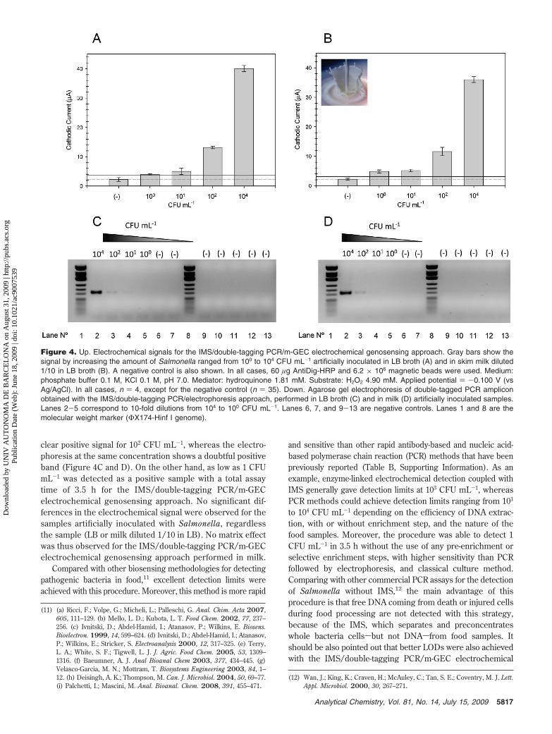
clear positive signal for 102 CFU mL-1, whereas the electro-phoresis at the same concentration shows a doubtful positiveband (Figure 4C and D). On the other hand, as low as 1 CFUmL-1 was detected as a positive sample with a total assaytime of 3.5 h for the IMS/double-tagging PCR/m-GECelectrochemical genosensing approach. No significant dif-ferences in the electrochemical signal were observed for thesamples artificially inoculated with Salmonella, regardlessthe sample (LB or milk diluted 1/10 in LB). No matrix effectwas thus observed for the IMS/double-tagging PCR/m-GECelectrochemical genosensing approach performed in milk.
Compared with other biosensing methodologies for detectingpathogenic bacteria in food,11 excellent detection limits wereachieved with this procedure. Moreover, this method is more rapid
and sensitive than other rapid antibody-based and nucleic acid-based polymerase chain reaction (PCR) methods that have beenpreviously reported (Table B, Supporting Information). As anexample, enzyme-linked electrochemical detection coupled withIMS generally gave detection limits at 103 CFU mL-1, whereasPCR methods could achieve detection limits ranging from 101
to 104 CFU mL-1 depending on the efficiency of DNA extrac-tion, with or without enrichment step, and the nature of thefood samples. Moreover, the procedure was able to detect 1CFU mL-1 in 3.5 h without the use of any pre-enrichment orselective enrichment steps, with higher sensitivity than PCRfollowed by electrophoresis, and classical culture method.Comparing with other commercial PCR assays for the detectionof Salmonella without IMS,12 the main advantage of thisprocedure is that free DNA coming from death or injured cellsduring food processing are not detected with this strategy,because of the IMS, which separates and preconcentrateswhole bacteria cellssbut not DNAsfrom food samples. Itshould be also pointed out that better LODs were also achievedwith the IMS/double-tagging PCR/m-GEC electrochemical
(11) (a) Ricci, F.; Volpe, G.; Micheli, L.; Palleschi, G. Anal. Chim. Acta 2007,605, 111–129. (b) Mello, L. D.; Kubota, L. T. Food Chem. 2002, 77, 237–256. (c) Ivnitski, D.; Abdel-Hamid, I.; Atanasov, P.; Wilkins, E. Biosens.Bioelectron. 1999, 14, 599–624. (d) Ivnitski, D.; Abdel-Hamid, I.; Atanasov,P.; Wilkins, E.; Stricker, S. Electroanalysis 2000, 12, 317–325. (e) Terry,L. A.; White, S. F.; Tigwell, L. J. J. Agric. Food Chem. 2005, 53, 1309–1316. (f) Baeumner, A. J. Anal Bioanal Chem 2003, 377, 434–445. (g)Velasco-Garcia, M. N.; Mottram, T. Biosystems Engineering 2003, 84, 1–12. (h) Deisingh, A. K.; Thompson, M. Can. J. Microbiol. 2004, 50, 69–77.(i) Palchetti, I.; Mascini, M. Anal. Bioanal. Chem. 2008, 391, 455–471.
(12) Wan, J.; King, K.; Craven, H.; McAuley, C.; Tan, S. E.; Coventry, M. J. Lett.Appl. Microbiol. 2000, 30, 267–271.
Figure 4. Up. Electrochemical signals for the IMS/double-tagging PCR/m-GEC electrochemical genosensing approach. Gray bars show thesignal by increasing the amount of Salmonella ranged from 100 to 104 CFU mL-1 artificially inoculated in LB broth (A) and in skim milk diluted1/10 in LB broth (B). A negative control is also shown. In all cases, 60 µg AntiDig-HRP and 6.2 × 10⁶ magnetic beads were used. Medium:phosphate buffer 0.1 M, KCl 0.1 M, pH 7.0. Mediator: hydroquinone 1.81 mM. Substrate: H2O2 4.90 mM. Applied potential ) -0.100 V (vsAg/AgCl). In all cases, n ) 4, except for the negative control (n ) 35). Down. Agarose gel electrophoresis of double-tagged PCR ampliconobtained with the IMS/double-tagging PCR/electrophoresis approach, performed in LB broth (C) and in milk (D) artificially inoculated samples.Lanes 2-5 correspond to 10-fold dilutions from 104 to 100 CFU mL-1. Lanes 6, 7, and 9-13 are negative controls. Lanes 1 and 8 are themolecular weight marker (ΦX174-Hinf I genome).
5817Analytical Chemistry, Vol. 81, No. 14, July 15, 2009
Dow
nloa
ded
by U
NIV
AU
TO
NO
MA
DE
BA
RC
EL
ON
A o
n A
ugus
t 31,
200
9 | h
ttp://
pubs
.acs
.org
P
ublic
atio
n D
ate
(Web
): J
une
18, 2
009
| doi
: 10.
1021
/ac9
0075
39
genosensing approach compared with LODs reported for IMS/real-time PCR systems.13 This fact can be ascribed to thesensitivity of the amplicon detection with the m-GEC electro-chemical genosensing strategy.
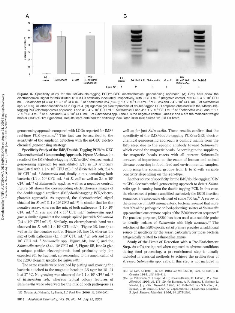
Specificity Study of the IMS/Double-Tagging PCR/m-GECElectrochemical Genosensing Approach. Figure 5A shows theresults of the IMS/double-tagging PCR/m-GEC electrochemicalgenosensing approach for milk diluted 1/10 in LB artificiallyinoculated with 1.1 × 10⁵ CFU mL-1 of Escherichia coli, 2.4 ×10⁵ CFU mL-1 Salmonella and, finally, a mix containing bothbacteria (1.1 × 10⁵ CFU mL-1 of E. coli as well as 2.4 × 10⁵
CFU mL-1 of Salmonella spp.), as well as a negative control.Figure 5B shows the corresponding electrophoresis images ofthe double-tagged amplicon (IMS/double-tagging PCR/electro-phoresis approach). As expected, the electrochemical signalobtained for E. coli (1.1 × 10⁵ CFU mL-1) is similar that for thenegative assay, whereas the mix of both pathogens (1.1 × 10⁵
CFU mL-1 E. coli and 2.4 × 10⁵ CFU mL-1 Salmonella spp.)gave a similar signal that the sample spiked just with Salmonella(2.4 × 10⁵ CFU mL-1). Similarly, no electrophoresis band wasobserved for E. coli 1.1 × 10⁵ CFU mL-1, (Figure 5B, lane 4) aswell as for the negative control (Figure 5B, lane 1), whereas themix of both pathogens (1.1 × 10⁵ CFU mL-1 E. coli and 2.4 ×10⁵ CFU mL-1 Salmonella spp., Figure 5B, lane 5) and theSalmonella sample (2.4 × 10⁵ CFU mL-1, Figure 5B, lane 3) givea unique positive electrophoresis band producing only theexpected 201 bp fragment, corresponding to the amplification ofthe IS200 element specific for Salmonella.
The same results were obtained by plating and growing thebacteria attached to the magnetic beads in LB agar for 18-24h at 37 °C. No growing was observed for 1.1 × 10⁵ CFU mL-1
of Escherichia coli, whereas typical colony features ofSalmonella were observed for the mix of both pathogens as
well as for just Salmonella. These results confirm that thespecificity of the IMS/double-tagging PCR/m-GEC electro-chemical genosensing approach is coming mainly from theIMS step, due to the specific antibody toward Salmonellawhich coated the magnetic beads. According to the suppliers,the magnetic beads reacts with all current Salmonellaserovars of importance as the cause of human and animaldisease occurring in food, feed and environmental samples,comprising the somatic groups from B to Z with variablereactivity depending on the serotype.
Another source of specificity for the IMS/double-tagging PCR/m-GEC electrochemical genosensing approach to detect Salmo-nella spp. is coming from the double-tagging PCR. In this case,the chosen set of primers amplified exclusively the IS200 insertionsequence, a transposable element of some 700 bp.14 A survey ofthe presence of IS200 among enteric bacteria revealed that morethan 90% of the pathogenic or food-poisoning isolates of Salmonellaspp contained one or more copies of the IS200 insertion sequence.8
For practical purposes, IS200 has been used as a suitable probeto identify isolates of Salmonella with high accuracy.15 Theselection of the IS200 specific set of primers provides an additionalsource of specificity for the assay, particularly for those bacteriaantigenically related to salmonellae genus.
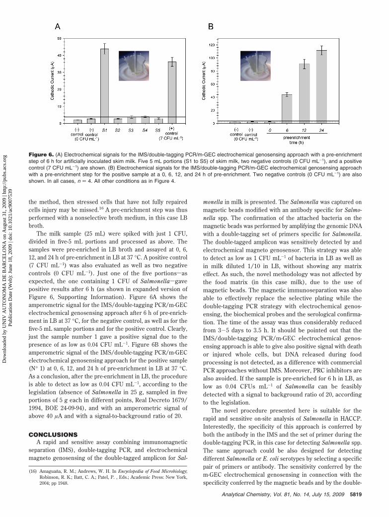
Study of the Limit of Detection with a Pre-EnrichmentStep. As cells are injured when exposed to adverse conditionsduring food processing, a pre-enrichment step is usuallyincluded in classical methods to achieve the proliferation ofstressed Salmonella spp. cells. If this step is not included in
(13) Notzon, A.; Helmuth, R.; Bauer, J. J. Food Prot. 2006, 12, 2896–2901.
(14) (a) Lam, S.; Roth, J. R. Cell 1983, 34, 951–960. (b) Lam, S.; Roth, J. R.Genetics 1983, 105, 801–811.
(15) (a) Millemann, Y.; Lesage, M. C.; Chaslus-Dancla, E.; Lafont, J. P. J. Clin.Microbiol. 1995, 33, 173–179. (b) Burnens, A. P.; Stanley, J.; Sechter, I.;Nicolet, J. J. Clin. Microbiol. 1996, 34, 1641–1645. (c) Schiaffino, A.;Beuzon, C. R.; Uzzau, S.; Leori, G.; Cappuccinelli, P.; Casadesus, J.; Rubino,S. Appl. Environ. Microbiol. 1996, 34, 2375–2380.
Figure 5. Specificity study for the IMS/double-tagging PCR/m-GEC electrochemical genosensing approach. (A) Gray bars show theelectrochemical signal for milk diluted 1/10 in LB artificially inoculated, respectively, with 0 CFU mL-1 (negative control, n ) 4); 2.4 × 10⁵ CFUmL-1 Salmonella (n ) 4); 1.1 × 10⁵ CFU mL-1 of Escherichia coli (n ) 5); 1.1 × 10⁵ CFU mL-1 of E. coli and 2.4 × 10⁵ CFU mL-1 of Salmonellaspp. (n ) 5). All other conditions as in Figure 4. (B) Agarose gel electrophoresis of double-tagged PCR amplicon obtained with the IMS/double-tagging PCR/electrophoresis approach. Lane 3: 2.4 × 10⁵ CFU mL-1 Salmonella; Lane 4: 1.1 × 10⁵ CFU mL-1 of Escherichia coli; Lane 5: 1.1× 10⁵ CFU mL-1 of E. coli and 2.4 × 10⁵ CFU mL-1 of Salmonella spp. Lane 1 is the negative control. Lanes 2 and 6 are the molecular weightmarker (ΦX174-Hinf I genome). Results were obtained for artificially inoculated skim milk diluted 1/10 in LB broth.
5818 Analytical Chemistry, Vol. 81, No. 14, July 15, 2009
Dow
nloa
ded
by U
NIV
AU
TO
NO
MA
DE
BA
RC
EL
ON
A o
n A
ugus
t 31,
200
9 | h
ttp://
pubs
.acs
.org
P
ublic
atio
n D
ate
(Web
): J
une
18, 2
009
| doi
: 10.
1021
/ac9
0075
39
the method, then stressed cells that have not fully repairedcells injury may be missed.16 A pre-enrichment step was thusperformed with a nonselective broth medium, in this case LBbroth.
The milk sample (25 mL) were spiked with just 1 CFU,divided in five-5 mL portions and processed as above. Thesamples were pre-enriched in LB broth and assayed at 0, 6,12, and 24 h of pre-enrichment in LB at 37 °C. A positive control(7 CFU mL-1) was also evaluated as well as two negativecontrols (0 CFU mL-1). Just one of the five portionssasexpected, the one containing 1 CFU of Salmonellasgavepositive results after 6 h (as shown in expanded version ofFigure 6, Supporting Information). Figure 6A shows theamperometric signal for the IMS/double-tagging PCR/m-GECelectrochemical genosensing approach after 6 h of pre-enrich-ment in LB at 37 °C, for the negative control, as well as for thefive-5 mL sample portions and for the positive control. Clearly,just the sample number 1 gave a positive signal due to thepresence of as low as 0.04 CFU mL-1. Figure 6B shows theamperometric signal of the IMS/double-tagging PCR/m-GECelectrochemical genosensing approach for the positive sample(N° 1) at 0, 6, 12, and 24 h of pre-enrichment in LB at 37 °C.As a conclusion, after the pre-enrichment in LB, the procedureis able to detect as low as 0.04 CFU mL-1, according to thelegislation (absence of Salmonella in 25 g, sampled in fiveportions of 5 g each in different points, Real Decreto 1679/1994, BOE 24-09-94), and with an amperometric signal ofabove 40 µA and with a signal-to-background ratio of 20.
CONCLUSIONSA rapid and sensitive assay combining immunomagnetic
separation (IMS), double-tagging PCR, and electrochemicalmagneto genosensing of the double-tagged amplicon for Sal-
monella in milk is presented. The Salmonella was captured onmagnetic beads modified with an antibody specific for Salmo-nella spp. The confirmation of the attached bacteria on themagnetic beads was performed by amplifying the genomic DNAwith a double-tagging set of primers specific for Salmonella.The double-tagged amplicon was sensitively detected by andelectrochemical magneto genosensor. This strategy was ableto detect as low as 1 CFU mL-1 of bacteria in LB as well asin milk diluted 1/10 in LB, without showing any matrixeffect. As such, the novel methodology was not affected bythe food matrix (in this case milk), due to the use ofmagnetic beads. The magnetic immunoseparation was alsoable to effectively replace the selective plating while thedouble-tagging PCR strategy with electrochemical genos-ensing, the biochemical probes and the serological confirma-tion. The time of the assay was thus considerably reducedfrom 3-5 days to 3.5 h. It should be pointed out that theIMS/double-tagging PCR/m-GEC electrochemical genos-ensing approach is able to give also positive signal with deathor injured whole cells, but DNA released during foodprocessing is not detected, as a difference with commercialPCR approaches without IMS. Moreover, PRC inhibitors arealso avoided. If the sample is pre-enriched for 6 h in LB, aslow as 0.04 CFUs mL-1 of Salmonella can be feasiblydetected with a signal to background ratio of 20, accordingto the legislation.
The novel procedure presented here is suitable for therapid and sensitive on-site analysis of Salmonella in HACCP.Interestedly, the specificity of this approach is conferred byboth the antibody in the IMS and the set of primer during thedouble-tagging PCR, in this case for detecting Salmonella spp.The same approach could be also designed for detectingdifferent Salmonella or E. coli serotypes by selecting a specificpair of primers or antibody. The sensitivity conferred by them-GEC electrochemical genosensing in connection with thespecificity conferred by the magnetic beads and by the double-
(16) Amaguana, R. M.; Andrews, W. H. In Encyclopedia of Food Microbiology;Robinson, R. K.; Batt, C. A.; Patel, P. , Eds.; Academic Press: New York,2004; pp 1948.
Figure 6. (A) Electrochemical signals for the IMS/double-tagging PCR/m-GEC electrochemical genosensing approach with a pre-enrichmentstep of 6 h for artificially inoculated skim milk. Five 5 mL portions (S1 to S5) of skim milk, two negative controls (0 CFU mL-1), and a positivecontrol (7 CFU mL-1) are shown. (B) Electrochemical signals for the IMS/double-tagging PCR/m-GEC electrochemical genosensing approachwith a pre-enrichment step for the positive sample at a 0, 6, 12, and 24 h of pre-enrichment. Two negative controls (0 CFU mL-1) are alsoshown. In all cases, n ) 4. All other conditions as in Figure 4.
5819Analytical Chemistry, Vol. 81, No. 14, July 15, 2009
Dow
nloa
ded
by U
NIV
AU
TO
NO
MA
DE
BA
RC
EL
ON
A o
n A
ugus
t 31,
200
9 | h
ttp://
pubs
.acs
.org
P
ublic
atio
n D
ate
(Web
): J
une
18, 2
009
| doi
: 10.
1021
/ac9
0075
39

tagging PCR result in a rapid, robust, and sensitive procedure.Future work will be focused on the possible application ofsuch method to screen printed electrodes allowing the develop-ment of disposable electrodes for in-field, low cost and user-friendly detection of multiple food pathogens affecting foodsafety.
ACKNOWLEDGMENTFinancial support from Ministry of Education and Science
(MEC), Madrid (Project BIO2007-63300), is acknowledged. MIP
also acknowledges the support from the Universidad Nacional delLitoral (Argentina).
SUPPORTING INFORMATION AVAILABLEExpanded version of Figures 1, 4, and 6, and Table B. This
material is available free of charge via the Internet athttp://pubs.acs.org.
Received for review April 7, 2009. Accepted May 22, 2009.
AC9007539
5820 Analytical Chemistry, Vol. 81, No. 14, July 15, 2009
Dow
nloa
ded
by U
NIV
AU
TO
NO
MA
DE
BA
RC
EL
ON
A o
n A
ugus
t 31,
200
9 | h
ttp://
pubs
.acs
.org
P
ublic
atio
n D
ate
(Web
): J
une
18, 2
009
| doi
: 10.
1021
/ac9
0075
39
Copyright © 2022 FDOKUMEN