
Magneto-optical response enhancement in 1D and 2D magnetoplasmonic crystals
10
Magneto-optical response enhancement in 1D and 2D magnetoplasmonic crystals A.A. Grunin a , A.G. Zhdanov a , B.B. Tsema a , A.A. Ezhov a , T.V. Dolgova a , E.A. Ganshina a , M.H. Hong b , A.A. Fedyanin a (a)Faculty of Physics, M.V. Lomonosov Moscow State University, Moscow 119991, Russia; (b)Department of Electrical and Computer Engineering, National University of Singapore, Singapore ABSTRACT The results of experimental observation of magneto-optical Kerr effect (MOKE) enhancement caused by surface plasmon-polaritons (SPP) excitation in 1D and 2D magnetoplasmonic crystals are presented. One-dimensional nickel magnetoplasmonic crystals have periodic structure formed by periodic nickel grooves made on nickel surface. The period of the structure is 320 nm and the depth of the grooves is 50 nm. The second group of the samples represents itself a 2D self-assembled hexagonally ordered monolayer of polystyrene (PS) microspheres with diameters from 500 to 760 nm and covered by 100- nm - thick nickel film. MOKE measurements performed in transversal configuration demonstrate that SPP excitation lead to transversal Kerr effect (TKE) enhancement resulting as a sharp peak in TKE spectrum. Keywords: magnetophotonic crystals, plasmonic crystals, magnetoplasmonic crystals, MOKE enhancement 1. INTRODUCTION The problem of light propagation control in various photonic devices stays important within the latest few years. Miniaturization of photonic circuits stimulates involving photonic micro- and nanostructures as unique materials providing an opportunity of more efficient light propagation control in comparison to conventional materials. Magneto-optical phenomena are widely used in a number of applications and remain as one of the most promising solutions of this problem. In this case magnetic field plays a role of external control of light propagation. For applications of magneto-optical phenomena in photonic micro- and nano- photonic devices are required looking for the ways of enhancement of magneto-optical phenomena. Magneto-optical effects are nonreciprocal effects and they can be enhanced by constructive interference. For example, well-known periodically structured dielectric elements with periods of the structure comparable with optical wavelength range and possessing magneto-optical properties are usually called magnetophotonic crystals by analogy with photonic crystals. 1, 2 Magneto-optical response in such structures is increased by several times in comparison with the single layer of the same thickness. One-dimensional magnetophotonic crystals have been studied thoroughly both theoretically and experimentally and the results have been reported in several works. 3, 4 In such structures significant Faraday angles are achieved practically with nor weakening of the transmitted light neither distorting its polarization. 5 At the other side there are well-known planar metal subwavelength gratings called plasmonic crystals. 6–8 Periodically patterned surface provides an opportunity to excite surface plasmon-polaritons (SPP) on the surface of plasmonic crystals. Previously such structures have been fabricated from noble metals, for example, from gold or silver, because in this case free path of surface plasmon polaritons reaches the largest values in optical range of wavelengths. Magnetoplasmonic crystals 9 represent plasmonic crystals which are fabricated from magnetic metal, for ex- ample, nickel and called by analogy with plasmonic and magnetophotonic crystals. Such structures possess both magneto-optical activity and surface plasmon resonances simultaneously. Fundamental issues in this area are Further author information: (Send correspondence to A.A. Grunin) A.A. Grunin: E-mail: [email protected], Telephone: +7 495 938 2210 Metamaterials IV, edited by Vladimir Kuzmiak, Peter Markoš, Tomasz Szoplik, Proc. of SPIE Vol. 7353, 73530F · © 2009 SPIE · CCC code: 0277-786X/09/$18 · doi: 10.1117/12.820704 Proc. of SPIE Vol. 7353 73530F-1
-
Upload
moscowstate -
Category
Documents
-
view
3 -
download
0
Transcript of Magneto-optical response enhancement in 1D and 2D magnetoplasmonic crystals
Magneto-optical response enhancement in 1D and 2Dmagnetoplasmonic crystals
A.A. Grunina, A.G. Zhdanova, B.B. Tsemaa, A.A. Ezhova, T.V. Dolgovaa, E.A. Ganshinaa,
M.H. Hongb, A.A. Fedyanina
(a)Faculty of Physics, M.V. Lomonosov Moscow State University, Moscow 119991, Russia;(b)Department of Electrical and Computer Engineering, National University of Singapore,
Singapore
ABSTRACT
The results of experimental observation of magneto-optical Kerr effect (MOKE) enhancement caused by surfaceplasmon-polaritons (SPP) excitation in 1D and 2D magnetoplasmonic crystals are presented. One-dimensionalnickel magnetoplasmonic crystals have periodic structure formed by periodic nickel grooves made on nickelsurface. The period of the structure is 320 nm and the depth of the grooves is 50 nm. The second group of thesamples represents itself a 2D self-assembled hexagonally ordered monolayer of polystyrene (PS) microsphereswith diameters from 500 to 760 nm and covered by 100- nm - thick nickel film. MOKE measurements performedin transversal configuration demonstrate that SPP excitation lead to transversal Kerr effect (TKE) enhancementresulting as a sharp peak in TKE spectrum.
Keywords: magnetophotonic crystals, plasmonic crystals, magnetoplasmonic crystals, MOKE enhancement
1. INTRODUCTION
The problem of light propagation control in various photonic devices stays important within the latest few years.Miniaturization of photonic circuits stimulates involving photonic micro- and nanostructures as unique materialsproviding an opportunity of more efficient light propagation control in comparison to conventional materials.Magneto-optical phenomena are widely used in a number of applications and remain as one of the most promisingsolutions of this problem. In this case magnetic field plays a role of external control of light propagation. Forapplications of magneto-optical phenomena in photonic micro- and nano- photonic devices are required lookingfor the ways of enhancement of magneto-optical phenomena. Magneto-optical effects are nonreciprocal effects andthey can be enhanced by constructive interference. For example, well-known periodically structured dielectricelements with periods of the structure comparable with optical wavelength range and possessing magneto-opticalproperties are usually called magnetophotonic crystals by analogy with photonic crystals.1,2 Magneto-opticalresponse in such structures is increased by several times in comparison with the single layer of the same thickness.One-dimensional magnetophotonic crystals have been studied thoroughly both theoretically and experimentallyand the results have been reported in several works.3,4 In such structures significant Faraday angles are achievedpractically with nor weakening of the transmitted light neither distorting its polarization.5
At the other side there are well-known planar metal subwavelength gratings called plasmonic crystals.6–8
Periodically patterned surface provides an opportunity to excite surface plasmon-polaritons (SPP) on the surfaceof plasmonic crystals. Previously such structures have been fabricated from noble metals, for example, from goldor silver, because in this case free path of surface plasmon polaritons reaches the largest values in optical rangeof wavelengths.
Magnetoplasmonic crystals9 represent plasmonic crystals which are fabricated from magnetic metal, for ex-ample, nickel and called by analogy with plasmonic and magnetophotonic crystals. Such structures possess bothmagneto-optical activity and surface plasmon resonances simultaneously. Fundamental issues in this area are
Further author information: (Send correspondence to A.A. Grunin)A.A. Grunin: E-mail: [email protected], Telephone: +7 495 938 2210
Metamaterials IV, edited by Vladimir Kuzmiak, Peter Markoš, Tomasz Szoplik, Proc. of SPIE Vol. 7353, 73530F · © 2009 SPIE · CCC code: 0277-786X/09/$18 · doi: 10.1117/12.820704
Proc. of SPIE Vol. 7353 73530F-1

Figure 1. a) Sketch of electromagnetic field configuration for SPP wave, b) dispersion properties of SPP: thin solid lineis dispersion law of SPP, dashed line is dispersion curve for the light in vacuum, thick solid line is dispersion curves formatching condition in Otto-Kretchmann scheme.
connected with the influence of surface plasmons excitation on magneto-optical response and with modification ofnon-diagonal elements of dielectric tensor of magnetic metal under surface plasmon excitation. For applications,magnetoplasmonics area presents a promising way of light propagation control in nano- and micro- photonicdevices. For comparison, magneto-optical effects in dielectric structures are limited by weak magneto-optical re-sponse of dielectrics in comparison with metals, at the same time, metals possess large absorption. Consequently,magnetoplasmonic crystals are attractive to applications in reflection geometry. Several works are devoted toinvestigation of optical and magneto-optical response of planar periodic metallic structures.3,10,11 In this work,we present experimental results of observation of magneto-optical Kerr effect enhancement caused by surfaceplasmon-polaritons (SPP) excitation in 1D and 2D magnetoplasmonics crystals.
2. MAGNETOPLASMONIC CRYSTALS
2.1 Surface plasmon-polaritons excitation on homogeneous metal surface
Surface plasmon-polaritons can be excited on metal-dielectric interface being specific solution of wave equationunder corresponding boundary conditions. This solution is a partially longitudinal TM- (p-polarized) wave.Excitation of TE- (s-polarized) SPP is prohibited in an ideal metal due to boundary conditions. SPP wavecomponents are given as:
U = U0 exp(±κ1,2z) cos(ωt− ksppx), (1)
where U0 is an amplitude of SPP, κ1,2 are damping constants for media at both sides of the interface, and kspp
is SPP wavevector. Electric field has both x− and z− components which are equal to:
Ex =1√−(ε1 + ε2)
H, Ez1 =√
ε2ε1(ε1 + ε2)
H, Ez2 =√
ε1ε2(ε1 + ε2)
H, (2)
respectively.
Such configuration of electromagnetic fields for SPP is shown in Fig.1a. SPP wave also demonstrates peculiardispersion properties. Its wave vector and damping factors are given as:
Proc. of SPIE Vol. 7353 73530F-2

Figure 2. a) Dispersion properties of SPP: thin solid line is dispersion curve of SPP, thick solid line is dispersion curve forthe light in vacuum, dashed line is dispersion curves for the −1th order of diffraction of incident light. b) Phase matchingscheme of SPP excitation, c)AFM image of the surface of 1D magnetoplasmonic crystal, scale bar is 1μm; d) spatialFourier transform of the AFM image.
kspp =√
ε1ε2
ε1 + ε2, κ1 = kspp
√−ε1
ε2, κ1 = kspp
√−ε2
ε1. (3)
Taking into consideration dispersion properties of ε1,2 one can obtain dispersion curves for SPP, which areshown schematically in Fig.1b. Solid thin curve corresponds to SPP, dashed one corresponds to light wavevec-tor. The two curves have only one crosspoint in zero and, consequently, SPP can not be excited by incidentelectromagnetic wave on homogeneous metal surface. To excite SPP it is necessary to fulfil phase matchingconditions corresponding to dispersion curves intersection on the ω − k plane. It can be achieved both utilizingprism-coupling schemes in traditional Otto and Kretchmann configurations (thick solid line in Fig.1b). Thedashed circle corresponds to the condition of of SPP excitation in this case.
2.2 Surface plasmon-polaritons excitation in periodical structures
Another way to satisfy the phase matching condition is using the periodically structured surface. Consider theone-dimensional periodical structure and the incident light with the plane of incidence perpendicular to thevector of reciprocal lattice. Incident wave is p-polarized. Periodically structured surface possesses reciprocalvector G. Phase matching condition for excitation of SPP by the −1th diffraction order is shown in Fig.2a. Thedashed circle corresponds to the SPP excitation in this case. Dashed line in Fig.2a corresponds to kx = G− k0
Fig.2b.
Matching conditions can be expresses in the following way:
Proc. of SPIE Vol. 7353 73530F-3

Figure 3. SEM image of closely-packed monolayer of PS microspheres with diameter of d = 500nm covered by 100-nm-thicknickel film
k0 sin α + nG = −kspp, n = ±1,±2..., (4)
and correspond to the case when the (±n)st order of diffraction coincides with the surface. SPP excitationmanifests in optical response as minimum in reflection spectrum that corresponds to energy flux redistributionbetween the 0th order of diffraction (specular reflection), the (-1)st order of diffraction and SPP propagationalong the metal surface. The dip in the spectrum taking place at the specified wavelength is called as Wood’sanomaly and is polarisation sensitive, as SPP excitation is possible only in p- polarisation.
3. EXPERIMENTAL SAMPLES
Two types of magnetoplasmonic crystals are considered. The first type is one-dimensional periodical nickelstructure. The second type is a quasi two-dimensional hexagonal periodical nickel structure.
Studied one-dimensional samples represent itself 1D periodically patterned surface of nickel. Periodic struc-ture is formed by periodic nickel grooves made on the nickel surface. These samples have been obtained bysputtering of nickel film with thickness about h = 100 nm on dielectric substrate with grooves. The period ofthe structure is 320 nm and the depth of the grooves is 40-50 nm. The filling factor of the nickel grooves isabout 0.5. The AFM image of the surface of experimental sample is shown in Fig.2c. White area on AFM imagecorresponds to the height of h = 50 nm and black ones correspond to the height of h = 0 nm. The scale baron the sketch is 1 μm. Put X axes collinear to reciprocal vector of structure. Then, Fourier transform coordi-nates X,Y are transformed in reciprocal vector coordinates GX , GY accordingly. In the Fig.2d spatial Fouriertransform of AFM image is shown. Wight area on spatial Fourier transform sketch corresponds to maximum ofFourier amplitude, that proves experimental structure have only one direction of periodicity parallel to X axis.
Two-dimensional samples are fabricated by using nanosphere lithography technique. First, hexagonally or-dered close-packed monolayer of polystyrene (PS) microspheres is formed on the glass substrate. PS microsphereswith diameter from d = 500 nm to d = 760 nm are used. At the second step, nickel film with thickness from30 to 90 nm is sputtered on surface of monolayer of PS microspheres. As result, two-dimensional hexagonallyordered magnetoplasmonic structures are obtained. SEM image of such structure with period d = 500 nm ispresented in Fig.3. The structures represent quasi 2D magnetoplasmonic crystals because these are not clearplane structures and in addition typical size of close-packed domain is about ∼ 50 μm.
Proc. of SPIE Vol. 7353 73530F-4

200
220
eooE
e2o
£00
uciquce SUäIG &sq
50 30 40 20 0420
fffJ(4!OLJ n£0 ai
Figure 4. Left: Dependence of reflection spectrum on incident angle, white area correspond to minimum, black areacorrespond to maximum of reflectivity. Right: reflection spectrum measured at incident angle α = 450.
4. EXPERIMENTAL RESULTS
4.1 Wood anomaly in 1D magnetoplasmonic crystals
The minimum in reflection spectrum of p− polarized incident light which often called as Wood’s anomaly showsenergy flux redistribution of incident light into the surface plasmon mode. The position of this minimum inreflection spectrum is defined by the phase matching conditions (4). For one-dimensional magnetoplasmoniccrystals Wood’s anomaly is observed under different phase-matching conditions. Phase-matching conditions canbe fulfilled by changing the incident angle or azimuthal angle. For azimuthal angle θ = 00, Wood’s anomalyposition is defined by the following equation:
λ = d
(√εNi
εNi + 1+ sin α
), (5)
where εNi is permittivity of nickel at this wavelength.
Experimental dependence of reflection spectrum on incident angle under azimuthal angle of θ = 00 in p−polarisation is shown in Fig.4. White strip on this sketch corresponds to minimum of reflectivity and black areacorrespond to maximum of reflectivity. Right sketch in Fig.4 shows the reflection spectrum for incident angleof α = 450. The position of Wood’s anomaly in Fig.4 fits well to theoretical dependence in Eq. (5). For s−polarization of incident light, SPP excitation is prohibited and there are no any features in reflection spectrum.
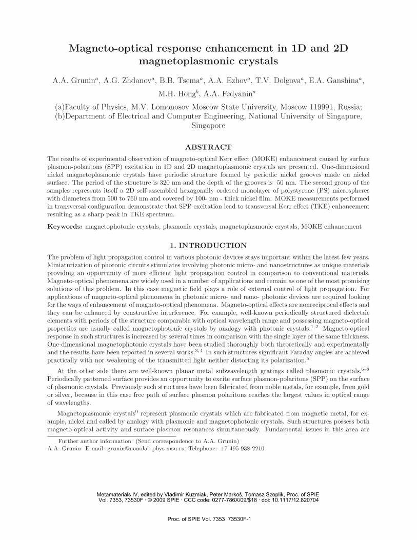
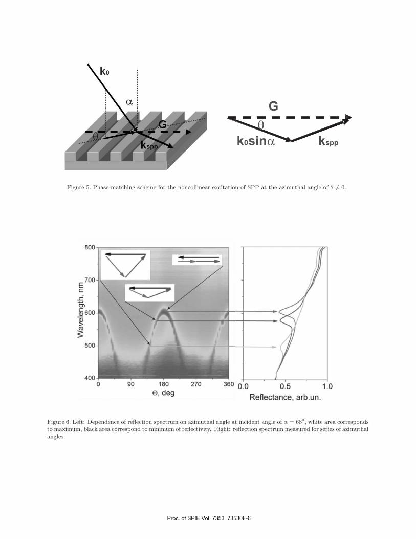
At noncollinear excitation of the surface plasmon-polaritons phase matching conditions are changed and wavevectors in this case are not located in plane of incident light as it is shown in Fig. 5. Experimental dependenceof reflection spectrum on azimuthal angle at incident angle of α = 680 measures in p− polarisation is shown inFig.6. White area on this sketch correspond to maximum of reflectivity and black area corresponds to minimumof reflectivity. The position of Wood’s anomaly is defined by the following equation obtained from geometricalcondition in Fig.5(left):
k2spp = G2 + k2
0 sin2 α− 2Gk0 sin α cos θ (6)
Proc. of SPIE Vol. 7353 73530F-5
or eo 00 °
.nu.dis 9OflBfO9ft91
o O8
p8b O oe 0
008
0o B
a,
ooa
001k
Figure 5. Phase-matching scheme for the noncollinear excitation of SPP at the azimuthal angle of θ �= 0.
Figure 6. Left: Dependence of reflection spectrum on azimuthal angle at incident angle of α = 680, white area correspondsto maximum, black area correspond to minimum of reflectivity. Right: reflection spectrum measured for series of azimuthalangles.
Proc. of SPIE Vol. 7353 73530F-6

A sharp dip in reflection spectra is observed for each angle of incidence and azimuth angle. This dip corre-sponds to the situation when the (-1)st diffraction order propogates along the plan of the sample and correspondto excitation of surface plasmon-polaritons.
4.2 Excitation of SPP in 2D samplesTwo-dimensional magnetoplasmonic crystals represent more complicated structures since both local Mie plas-mons and SPP can be excited. This magnetoplasmonic crystals represent multidomain structure that leads tofluctuations of the reciprocal vector and consequently azimuthal dependence is absent. Experimental results ofreflection spectra at different incident angles for s− and p− polarisation are shown in Fig.7. There are twogroups of features in experimental reflection spectra in p− polarisation. The black arrows in Fig.7 show dip inreflection spectrum which is in dependent on incident angle. This group of features corresponds to excitation oflocal Mie plasmons on the surface of sample. The grey arrows in Fig.7 show dip in reflection spectrum whichdepends on the incident angle and similar to Wood’s anomaly in case of 1D samples. This group of featurescorresponds to excitation of delocalized surface plasmon-polaritons on the surface of sample. These features inreflection spectrum of 2D magnetoplasmonic crystals are not so sharp and bright in comparison with 1D one.
4.3 Magneto-optical responseTransversal magneto-optical Kerr effect (TKE) was measured in specular reflection. The measurements werecarried out using lock-in technique for measuring the intensity of reflected signal. Magnetic field applied to thesamples was controlled by electromagnet in the range from 0 to 1.5 kOe. TKE value is defined as ratio δ = ΔI
I0,
where I0 is a intensity of reflected beam without magnetic field ΔI = I − I0, I is a intensity of reflected beamunder magnetic field B = 1.5 kOe. All TKE measurements were carried out at incident angle of α = 680.
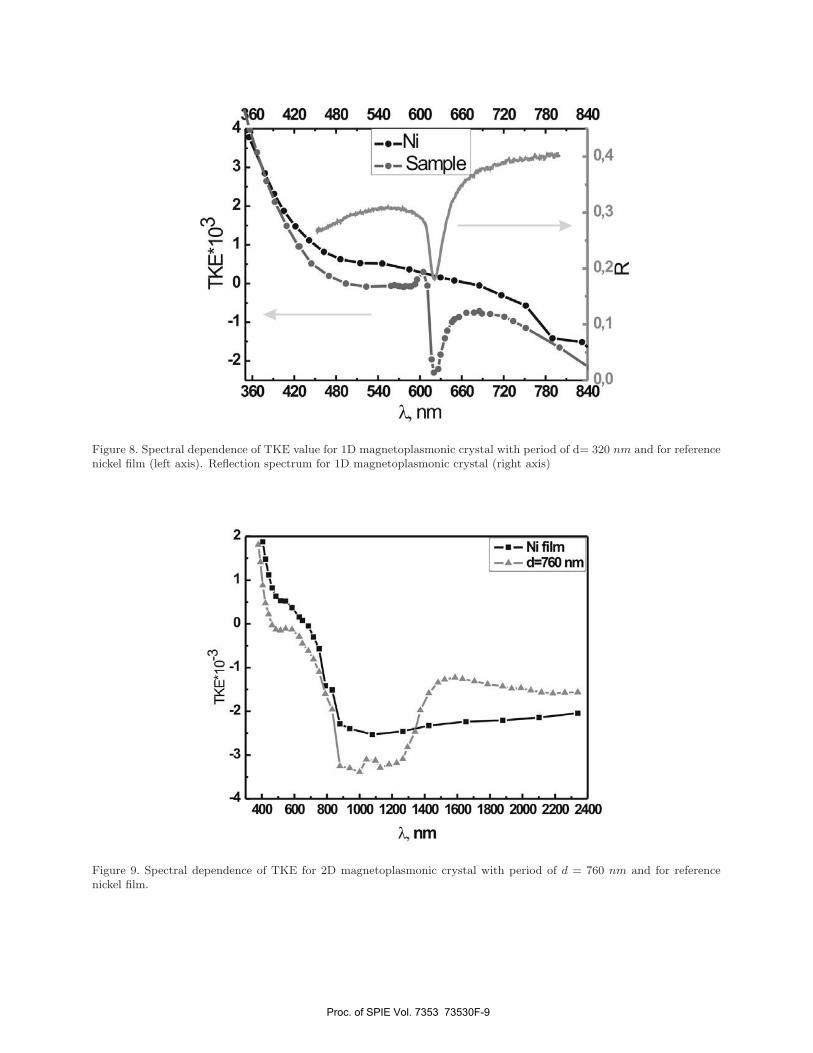
Experimental results of spectral dependence of TKE for 1D magnetolpasmonic crystal is shown in Fig.8 (greyline with circles) for the case of p− polarization of the incident light. For the reference, spectral dependenceof TKE for homogenous nickel film of equivalent thickness was measured, that is shown in Fig.8 as black linewith circles. Significant, about � 8 − 10 times, TKE enhancement is observed for magnetoplasmonic crystal incomparison with homogenous nickel film in narrow spectral range which corresponds to excitation of SPP Fig.8(grey line).
Experimental spectral dependence of TKE for 2D magnetolpasmonic crystal with period of d = 760 nm isshown in Fig.9(grey line) for the case of p− polarization. For the reference, spectral dependence of TKE forhomogenous nickel film of equivalent thickness was measured, Fig.9 (black line). In this case effect of TKEenhancement is not so significant and sharp as in 1D case. The reason that difference is a weak SPP excitationat large incidence angles (α = 680), that is shown in series of reflection spectra in Fig.7.
5. DISCUSSION
The series of observed effects in optical response of magnetoplasmonic crystals proves the possibility of excitationof surface plasmon-polaritons on surface of magnetoplasmonic crystals. The simple model of this process can bedivided on 3 steps:
1. periodically patterned structure acts like a subwavelength diffraction grating for incident light,
2. matching conditions between lateral component of incident light, reciprocal wavevector and SPP wave-vector are achieved in the case when the (-1)st order of diffraction lies in the plane of the surface,
3. SPP propagates along nickel plane in periodic structure.
Significant enhancement of transversal MOKE is observed experimentally. It is well correlated in spectrumwith excitation of SPP, proving plasmonic nature of the MOKE enhancement. The SPP excitation leads toincreasing of ”effective” distance of interaction between incidence light and magnetic media, since free path ofSPP on nickel surface is one order larger than free path of incidence light in nickel. The incident light is moreefficiently and interact with nickel by means of SPP excitation. Consequently, SPP excitation leads to MOKEenhancement. Another effect is selection only constructively interfering modes in MOKE response by periodicityof nickel surface. Apparently, combination of these two effects leads to significant MOKE enhancement.
Proc. of SPIE Vol. 7353 73530F-7

50
--b
uuJ400 200 @00 00 800 000 .1000
00
028:
.12
50
y uw400 200 @00 0J 800
00900 .1000
?-b
- uw200 @00 00 800 900 .1000
200 @00
- uw00 900 000 .1000
Figure 7. Reflection spectra at different incident angles. Period of the structure is d = 570 nm.
Proc. of SPIE Vol. 7353 73530F-8
YLJW3eo so o o eoo eeo so o io
o o-5
SII'I.I.I O4
It
_. - .0/
4,/ 10
3 2suJbJe
4-!eo iso io o eoo eeo so o 1O
3D;
yuwioo eco 800 1000 oo .iioo .ieoo oo 5000 ssoo soo
U -.
q=eo 'iw. II! thW
. .
Figure 8. Spectral dependence of TKE value for 1D magnetoplasmonic crystal with period of d= 320 nm and for referencenickel film (left axis). Reflection spectrum for 1D magnetoplasmonic crystal (right axis)
Figure 9. Spectral dependence of TKE for 2D magnetoplasmonic crystal with period of d = 760 nm and for referencenickel film.
Proc. of SPIE Vol. 7353 73530F-9
6. CONCLUSIONS
Wood’s anomaly and SPP excitation are experimentally observed in 1D and 2D nickel magnetoplasmonic crystals.It is proved by polarization-dependence and appears at wide range of incident and azimuthal angle. Wavelengthcorresponding to this spectral peculiarity satisfies phase-matching conditions for lateral component of the incidentwavevector, SPP wave vector and vector of reciprocal lattice. MOKE measurements in transversal geometrydemonstrate that SPP excitation lead to the TKE enhancement resulting in a sharp peak in TKE spectrum forcase of one-dimensional magnetoplasmonic crystals. Transversal Kerr effect enhancement in wide spectral rangeis observed for two-dimensional case.
REFERENCES1. M. Inoue, K. Arai, T. Fujii, and M.Abe, “Magneto-optical properties of one-dimensional photonic crystals
composed of magnetic and dielectric layers,” J. Appl. Phys. 83, p. 6768, 1998.2. M. Inoue, R. Fujikawa, A. Baryshev, A. Khanikaev, P. Lim, H. Uchida, O. Aktsipetrov, A. Fedyanin,
T. Murzina, and A. Granovsky, “Magnetophotonic crystals,” J. Phys. D: Appl. Phys. 39, p. R151, 2006.3. V. Belotelov and A. Zvezdin, “Magneto-optical properties of photonic crystals,” J. Opt. Soc. Am. B 22,
p. 286, 2005.4. A. A. Fedyanin, O. A. Aktsipetrov, D. Kobayashi, K. Nishimura, H.Uchida, and M. Inoue, “Enhanced Fara-
day and nonlinear magneto-optical Kerr effects in magnetophotonic crystals,” J. Magn. Magn. Mater. 282,p. 256, 2004.
5. A. Zhdanov, A. Fedyanin, O. Aktsipetrov, D. Kobayashi, H. Uchida, and M. Inoue, “Enhancement ofFaraday rotation at photonic-band-gap edge in garnet-based magnetophotonic crystals,” J. Magn. Magn.Mater. 300, p. e253, 2006.
6. S. I. Bozhevolnyi, J. Erland, K. Leosson, P. M. W. Skovgaard, and J. M. Hvam, “Waveguiding in surfaceplasmon polariton band gap structures,” Phys. Rev. Lett. 86, p. 308, 2001.
7. S. I. Bozhevolnyi and V. Coello, “Elastic scattering of surface plasmon polaritons: Modeling and experi-ment,” Phys. Rev. B 58, p. 10 899, 1998.
8. S. I. Bozhevolnyi, V. S. Volkov, K. Leosson, and A. Boltasseva, “Bend loss in surface plasmon polaritonband-gap structures,” Appl. Phys. Lett. 79, p. 1076, 2001.
9. A. Zhdanov, A. Fedyanin, T. Dolgova, A. Baryshev, A. Khanikaev, H. Uchida, and M. Inoue, “Wood’sanomaly in two-dimensional plasmon-assisted magnetophotonic crystals,” Proc. of SPIE 6728, pp. 67282v2–1, 2007.
10. V. Belotelov and A. Zvezdin, “Magnetooptics and extraordinary transmission of the perforated metallicfilms magnetized in polar geometry,” J. Magn. Magn. Mater. 300, p. e260, 2006.
11. L. Martin-Moreno and et al., “Theory of extraordinary optical transmission through subwavelength holearrays,” Phys. Rev. Lett. 86, p. 1114, 2001.
Proc. of SPIE Vol. 7353 73530F-10