Phagomagnetic Separation and Electrochemical Magneto-Genosensing of Pathogenic Bacteria
8
Phagomagnetic Separation and Electrochemical Magneto- Genosensing of Pathogenic Bacteria Susana Lie ́ bana, † Denis A. Spricigo, ‡ María Pilar Corte ́ s, ‡ Jordi Barbe ́ , ‡ Montserrat Llagostera, ‡ Salvador Alegret, † and María Isabel Pividori* ,† Grup de Sensors i Biosensors, † Departament de Química, and Unitat de Microbiologia, ‡ Departament de Gene ̀ tica i de Microbiologia, Universitat Autò noma de Barcelona, 08193 Cerdanyola del Valle ̀ s, Bellaterra, Spain * S Supporting Information ABSTRACT: This paper addresses the use of bacteriophages immobilized on magnetic particles for the biorecognition of the pathogenic bacteria, followed by electrochemical magneto- genosensing of the bacteria. The P22 bacteriophage specific to Salmonella (serotypes A, B, and D 1 ) is used as a model. The bacteria are captured and preconcentrated by the bacter- iophage-modified magnetic particles through the host inter- action with high specificity and efficiency. DNA amplification of the captured bacteria is then performed by double-tagging polymerase chain reaction (PCR). Further detection of the double- tagged amplicon is achieved by electrochemical magneto-genosensing. The strategy is able to detect in 4 h as low as 3 CFU mL −1 of Salmonella in Luria−Bertani (LB) media. This approach is compared with conventional culture methods and PCR-based assay, as well as with immunological screening assays for bacteria detection, highlighting the outstanding stability and cost-efficient and animal-free production of bacteriophages as biorecognition element in biosensing devices. B acteriophages (or phages) are natural host-specific, self- reproducing, and self-assembling nanostructured particles, with both structure and function encrypted in the genomic DNA. Bacteriophages bind to specific receptors on the bacterial surface in order to inject the genetic material inside the bacteria, using the host’s own replication machinery for multiplication. The replicated virions are eventually released, killing the bacteria and allowing the infection of other host cells. Beside the promising features of phage therapy, 1 bacteriophage- based diagnostic is attracting much interest 2 due to the high specificity of phages, which makes them ideal agents not only for the detection of bacteria, but also for the detection of almost all kinds of targets, ranging from small molecules to proteins and even cells, by using the phage display technique. 3 As phages have the ability to display peptides or proteins on their surface, those showing a very high affinity and specificity for a target can be selected out of a library. Unfortunately, as the use of phages as biorecognition elements is in its infancy, the range of commercially available bacteriophages is still limited. Another important advantage is the fast, cheap, and animal-friendly phage production, which is achieved by just infecting the host bacteria. 3 Moreover, phages are stable in a range of harsh conditions including pH and temperature. 3 Phages can even be used in the presence of nucleases or proteolytic enzymes, without degradation. The high stability of phages in a variety of environmental conditions makes them suitable for in situ monitoring of food and environmental contaminants. These naturally occurring nanoparticles have other interesting proper- ties in comparison with synthetic nanoparticles: all bacter- iophages are nearly identical, being monodisperse in shape and size, a fact difficult to achieve by laboratory synthesis. On the contrary, these nanoparticles are self-synthesized in their specific host, by producing a large amount of viral coat proteins with a large surface for further chemical modification. The reported methods for bacteria detection using bacteriophages include (i) expression of bacteriophage-encoded bioluminescent genes which produce visible products within the specific target cells (lux-bacteriophage strategy), 4 (ii) fluorescence-labeled phage, which can be combined with immunomagnetic separation (labeled phage strategy), 5 (iii) detection of bacteria by the intracellular replication of specific bacteriophages (named “phage amplification” strategy), 6,7 and the (iv) detection of the phage-mediated bacterial lysis and release of host enzymes (e.g., adenylate kinase) or ATP (termed “lysin-release ATP bioluminescence strategy”). 8 Bacteriophages recognize the bacterial receptors through their tail spike proteins. This biorecognition is highly specific and has been employed for the typing of bacteria. This level of specificity and selectivity opens avenues for the development of specific pathogen detection technologies and for the creation of biosensing platforms. Biosensing approaches based on quartz crystal microbalance (QCM) and surface plasmon resonance (SPR) as transduction platform were reported. 9−11 These early reports relied on physical adsorption of the bacteriophage on the sensor surface. Single-point, oriented, covalent attachment of the bacteriophages on different surfaces and transducers was Received: September 5, 2012 Accepted: February 13, 2013 Published: February 13, 2013 Article pubs.acs.org/ac © 2013 American Chemical Society 3079 dx.doi.org/10.1021/ac3024944 | Anal. Chem. 2013, 85, 3079−3086
Transcript of Phagomagnetic Separation and Electrochemical Magneto-Genosensing of Pathogenic Bacteria
Phagomagnetic Separation and Electrochemical Magneto-Genosensing of Pathogenic BacteriaSusana Liebana,† Denis A. Spricigo,‡ María Pilar Cortes,‡ Jordi Barbe,‡ Montserrat Llagostera,‡
Salvador Alegret,† and María Isabel Pividori*,†
Grup de Sensors i Biosensors, †Departament de Química, and Unitat de Microbiologia, ‡Departament de Genetica i de Microbiologia,Universitat Autonoma de Barcelona, 08193 Cerdanyola del Valles, Bellaterra, Spain
*S Supporting Information
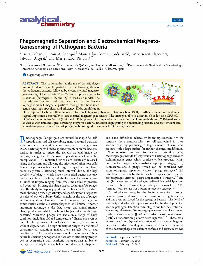
ABSTRACT: This paper addresses the use of bacteriophagesimmobilized on magnetic particles for the biorecognition ofthe pathogenic bacteria, followed by electrochemical magneto-genosensing of the bacteria. The P22 bacteriophage specific toSalmonella (serotypes A, B, and D1) is used as a model. Thebacteria are captured and preconcentrated by the bacter-iophage-modified magnetic particles through the host inter-action with high specificity and efficiency. DNA amplificationof the captured bacteria is then performed by double-tagging polymerase chain reaction (PCR). Further detection of the double-tagged amplicon is achieved by electrochemical magneto-genosensing. The strategy is able to detect in 4 h as low as 3 CFU mL−1
of Salmonella in Luria−Bertani (LB) media. This approach is compared with conventional culture methods and PCR-based assay,as well as with immunological screening assays for bacteria detection, highlighting the outstanding stability and cost-efficient andanimal-free production of bacteriophages as biorecognition element in biosensing devices.
Bacteriophages (or phages) are natural host-specific, self-reproducing, and self-assembling nanostructured particles,
with both structure and function encrypted in the genomicDNA. Bacteriophages bind to specific receptors on the bacterialsurface in order to inject the genetic material inside thebacteria, using the host’s own replication machinery formultiplication. The replicated virions are eventually released,killing the bacteria and allowing the infection of other host cells.Beside the promising features of phage therapy,1 bacteriophage-based diagnostic is attracting much interest2 due to the highspecificity of phages, which makes them ideal agents not onlyfor the detection of bacteria, but also for the detection of almostall kinds of targets, ranging from small molecules to proteinsand even cells, by using the phage display technique.3 As phageshave the ability to display peptides or proteins on their surface,those showing a very high affinity and specificity for a target canbe selected out of a library. Unfortunately, as the use of phagesas biorecognition elements is in its infancy, the range ofcommercially available bacteriophages is still limited. Anotherimportant advantage is the fast, cheap, and animal-friendlyphage production, which is achieved by just infecting the hostbacteria.3 Moreover, phages are stable in a range of harshconditions including pH and temperature.3 Phages can even beused in the presence of nucleases or proteolytic enzymes,without degradation. The high stability of phages in a variety ofenvironmental conditions makes them suitable for in situmonitoring of food and environmental contaminants. Thesenaturally occurring nanoparticles have other interesting proper-ties in comparison with synthetic nanoparticles: all bacter-iophages are nearly identical, being monodisperse in shape and
size, a fact difficult to achieve by laboratory synthesis. On thecontrary, these nanoparticles are self-synthesized in theirspecific host, by producing a large amount of viral coatproteins with a large surface for further chemical modification.The reported methods for bacteria detection using
bacteriophages include (i) expression of bacteriophage-encodedbioluminescent genes which produce visible products withinthe specific target cells (lux-bacteriophage strategy),4 (ii)fluorescence-labeled phage, which can be combined withimmunomagnetic separation (labeled phage strategy),5 (iii)detection of bacteria by the intracellular replication of specificbacteriophages (named “phage amplification” strategy),6,7 andthe (iv) detection of the phage-mediated bacterial lysis andrelease of host enzymes (e.g., adenylate kinase) or ATP(termed “lysin-release ATP bioluminescence strategy”).8
Bacteriophages recognize the bacterial receptors throughtheir tail spike proteins. This biorecognition is highly specificand has been employed for the typing of bacteria. This level ofspecificity and selectivity opens avenues for the development ofspecific pathogen detection technologies and for the creation ofbiosensing platforms. Biosensing approaches based on quartzcrystal microbalance (QCM) and surface plasmon resonance(SPR) as transduction platform were reported.9−11 These earlyreports relied on physical adsorption of the bacteriophage onthe sensor surface. Single-point, oriented, covalent attachmentof the bacteriophages on different surfaces and transducers was
Received: September 5, 2012Accepted: February 13, 2013Published: February 13, 2013
Article
pubs.acs.org/ac
© 2013 American Chemical Society 3079 dx.doi.org/10.1021/ac3024944 | Anal. Chem. 2013, 85, 3079−3086

also reported in order to yield better coverage and to improvethe performance of these devices. Streptavidin-mediatedattachment of bacteriophages that were genetically modifiedto directly express biotin on their capsid was reported.12,13
Covalent immobilization of bacteriophages on gold surface,14
screen-printed carbon electrode,15 and glass substrates16 forbiosensor application was also reported.This paper addresses the use of bacteriophage nanoparticles
as a highly specific biorecognition element for the capture andpreconcentration of pathogenic bacteria by using “phagomag-netic separation” (PMS), followed by electrochemical magneto-genosensing detection. The main advantage of using bacter-iophages relies on cost-efficient and animal-free production, aswell as their outstanding stability, overcoming thus the mainchallenges of the biorecognition elements in biosensing devices.The icosahedral-shaped bacteriophage (P22) specific to thepathogenic bacteria Salmonella was studied as a model.17,18 Theimmobilization of the native, non-modified, P22 phagenanoparticles on tosylated magnetic particles was achievedthroughout the amine moieties of the lysine residues in themain capsid monomeric protein (gp5)19 by covalent aminelinkage. After preconcentration of the bacteria on the magneticparticles by PMS, the bacteria were easily detected by double-tagging polymerase chain reaction (PCR) amplification of theDNA of the captured bacteria followed by electrochemicalmagneto-genosensing (PMS/double-tagging PCR/m-GECgenosensing).20
The main features of the PMS/double-tagging PCR/m-GECelectrochemical genosensing approach are compared withconventional culture methods and PCR-based assay.
■ EXPERIMENTAL SECTIONInstrumentation. Temperature-controlled incubations
were performed in an Eppendorf Thermomixer compact. Themagnetic separation during the washing steps was performedusing a magnetic separator Dynal MPC-S (product no.120.20D, Dynal Biotech ASA, Norway). The PCR reactionwas carried out in an Eppendorf Mastercycler personalthermocycler. Amperometric measurements were performedwith a LC-4C amperometric controller (BAS BioanalyticalSystems Inc., U.S.A.). A three-electrode setup was usedcomprising a platinum auxiliary electrode (Crison 52-67 1), adouble-junction Ag/AgCl reference electrode (Orion 900200)with 0.1 mol L−1 KCl as the external reference solution, and aworking electrode (the magneto-electrode, m-GEC). Thedetailed preparation of the m-GEC electrodes has beenextensively described by Pividori and co-workers21,22 (Figurei, Supporting Information). The scanning electron microscopy(SEM) images were taken with the scanning electronmicroscope Hitachi LTD S-570 (Hitachi LTD, Tokyo, Japan).Chemicals, Biochemicals, and Materials. Tosylactivated
magnetic particles (MP-Tosyl) (Dynabeads M-280, product no.142.03) as well as the streptavidin magnetic particles(Dynabeads M-280 Streptavidin, product no. 112.05) werepurchased from Life Technologies, Invitrogen Dynal AS (Oslo,Norway). AntiDig−HRP (antidigoxigenin−POD, product no.11.207.733.910) was purchased from Roche Diagnostics GmbH(Mannheim, Germany). The Bradford assay was performedwith the Coomasie Bradford protein assay kit, ref. 23200,Pierce, U.S.A.The Expand High Fidelity PCR System kit (Roche Molecular
Biochemicals) was used for performing the PCR. The primersfor the double-tagging PCR amplification in the genosensing
strategy were obtained from TIB-MOLBIOL (Berlin, Ger-many). These primers were selected for the specificamplification of the IS200 insertion sequence23 related toSalmonella spp. The primer sequences were biotin-IS200 up: 5′BIO-ATG GGG GAC GAA AAG AGC TTA GC 3′,digoxigenin-IS200 down: 5′ DIG-CTC CAG AAG CATGTG AAT ATG 3′.The buffer solutions were prepared with Milli-Q water. All
other reagents were analytical reagent grade supplied fromSigma and Merck. The composition of the solutions is detailedin the Supporting Information.
Bacterial Strains and P22 Bacteriophage. The P22bacteriophage (ATCC 19585-B1), Salmonella enterica serovarTyphimurium LT2, and Escherichia coli K12 strains were usedin this work. The bacteriophage lysate was obtained byinfecting exponential cultures of Salmonella TyphimuriumLT2 (108 CFU mL−1) with the P22 bacteriophage, and byfurther purification with cesium chloride gradient,24 as detailedin the Supporting Information. The bacteriophage titer wasdetermined by plating them using double agar layeredconventional method (Figure iv, part A, Supporting Informa-tion). The phage stock solutions were maintained in MgSO4 10mM in Milli-Q water solution at 4 °C retaining a constant titerfor several months. When specified, the P22 bacteriophageswere inactivated by exposure to a UV-C (254 nm) germicidallamp to avoid the lytic cycle.
Covalent Immobilization of P22 Bacteriophage onMagnetic Particles and Coupling Efficiency Study. Thenative P22 phage nanoparticles were covalently coupled for thefirst time to tosyl-activated magnetic particles by the reaction ofaminated aminoacidic lysine moieties of the main capsidmonomeric protein (gp5)18 (as schematically outlined inFigure ii, Supporting Information), by an amine linkage, inorder to obtain the P22 phage-modified magnetic particleconjugate (P22-MP). The binding was performed usingpurified P22 phage stock solution (200 μL) at a concentrationlevel of 2 × 10¹² PFU mL−1, reaching a concentration in theimmobilization solution of 4 × 10¹¹ PFU mL−1 as explained indetail in the Supporting Information.The coupling efficiency was evaluated by the Coomassie
Bradford protein assay,25 analyzing the protein concentration ofthe P22 phage capsid in the supernatant after the covalentattachment, and performing the calibration curve with thepurified P22 phage solution from 2 × 10¹⁰ to 5 × 10¹¹ PFUmL−1, as described in the Supporting Information, Figures iiand iii.A similar approach was performed by the double agar layered
conventional method for counting active phages. In thisapproach, 10-fold dilutions of the supernatant after the covalentattachment were plated through the double agar layeredmethod as described in the Supporting Information (Figure iv).
Evaluation of the Immobilized P22 Bacteriophage onMagnetic Particles by SEM and Conventional CultureMethods. The evaluation of the immobilized P22 bacter-iophages was performed by microscopic techniques (SEM) aswell as by conventional culture methods. For the microscopicevaluation by SEM, 10 μL of the P22 phage-modified magneticparticles (P22-MPs) in 5 mL of Milli-Q water (1924 PFU/MP)was filtered through a Nucleopore membrane (25 mm Ø, 0.2μm pore size). The filters were then fixed with glutaraldehyde,postfixed with osmium tetroxide, dehydratated with ethanol,and dried by CO2 critical point before gold metallization andobservation.26
Analytical Chemistry Article
dx.doi.org/10.1021/ac3024944 | Anal. Chem. 2013, 85, 3079−30863080

As previously addressed, the orientation of the bacterioph-ages on the solid support is an important issue to beconsidered. This orientation was studied by the double agarlayered method and enumeration of plaques by culturing theP22-MPs, since oriented phages immobilized on magneticparticles will produce bacteria attachment and further infectionof viable bacteria, producing the plaques (Figure v, SupportingInformation). A 10-fold dilution of the P22-MPs was platedthrough the double agar layered conventional method aspreviously explained (Figure iv, Supporting Information).Phagomagnetic Separation of Salmonella. Evaluation
by SEM and Conventional Culture Methods. Theprocedure for the PMS of the bacteria is schematically outlinedin Figure 1A. Inactivated bacteriophages by UV radiation wereused for the phagomagmetic separation to avoid the lytic cyclein order to keep the attached bacteria as a whole cell whilebeing captured, preconcentrated, and cultured since both SEMand culturing require non-infected bacteria.Bacterial solutions that ranged from 3.2 × 10⁶ to 3.2 × 100
CFU mL−1 in Luria−Bertani (LB) broth were performed forthe PMS of Salmonella Typhimurium LT2. A negative controlof LB broth was also processed. The culture in LB (500 μL)was mixed with 50 μL of P22-MPs (Figure 1A). An incubationstep was performed for 30 min at 37 °C without agitation. Afterthat, the magnetic particles with the attached bacteria wereseparated with a magnet and then washed with PBST for 5 min(3×) at room temperature. Finally, the modified magneticparticles were resuspended in 80 μL of Milli-Q water.The evaluation of the PMS (Figure 1A) was performed by
SEM and conventional culture methods. For the SEM study,the PMS was performed with a concentration of bacteria of 2.9× 10⁷ CFU mL−1, and the filters were treated as above-described. In order to study the efficiency of the PMS step byconventional culture method, 10 μL of modified magneticparticles of each solution that ranged from 3.2 × 10⁶ to 3.2 ×100 CFU mL−1 in LB broth including LB broth as negativecontrol was plated in LB agar and grown for 18−24 h at 37 °C.Phagomagnetic Separation, Double-Tagging PCR
Amplification, and Electrochemical Magneto-Genosens-ing. The procedure for the PMS of the bacteria followed by the
double-tagging PCR and the electrochemical magneto-genosensing of the attached bacteria (PMS/double-taggingPCR/m-GEC electrochemical genosensing) is schematicallyoutlined in Figure 1. In this case, UV-inactivated P22 phagenanoparticles were also used for the phagomagmetic separationto avoid the lytic cycle and the release of the genomic DNA ofthe bacteria while being captured and preconcentrated, sinceDNA is required for the double-tagging PCR amplification. Foreach concentration of bacteria in LB (from 3.2 × 10⁶ to 3.2 ×100 CFU mL−1), the lysis of the bacteria attached on theinactivated P22-MPs was performed at 99 °C for 20 min inorder to break the cells and to achieve the releasing of thegenomic DNA and the cellular debris to the solution for thePCR amplification (Figure 1B). The amplification of thespecific IS200 insertion sequence related to Salmonella spp. wasthus performed (Figure 1B) by a double-tagging PCR usingtwo labeled primers with biotin and digoxigenin27 (Figure vii,expanded version of Figure 1B, Supporting Information).During the PCR, not only the amplification of pathogenicbacteria genome was achieved, but also the double tagging ofthe amplicon ending with (i) the biotinylated capture primer toachieve the immobilization on streptavidin-modified magneticparticles and (ii) the digoxigenin signaling primer to achieve theenzymatic detection through antiDig−HRP reporter.All of these amplifications included not only a positive
control, but also a blank as a negative control, which containedLB broth without Salmonella spp. template. The double-taggedamplicon was analyzed by electrochemical genosensing with them-GEC electrodes as well as with the conventional gelelectrophoresis.The electrochemical genosensing strategy of the double-
tagged amplicon (Figure 1C) comprises the following steps, asoutlined in the Supporting Information and the Figure viii,expanded version of Figure 1C: (a) immobilization of thedouble-tagged amplicon in which the 5′ biotin end wasimmobilized on the streptavidin magnetic particles; (b)enzymatic labeling with the antibody antiDig−HRP able tobond the 5′ digoxigenin end of the ds-DNA amplicon; (c)magnetic capture of the modified magnetic particles by the m-GEC electrode; (d) amperometric determination.
Figure 1. Schematic representation of the phagomagnetic separation (PMS) (A) of the bacteria followed by the double-tagging PCR (B) and theelectrochemical magneto-genosensing (C) of the attached bacteria (PMS/double-tagging PCR/m-GEC electrochemical genosensing).
Analytical Chemistry Article
dx.doi.org/10.1021/ac3024944 | Anal. Chem. 2013, 85, 3079−30863081

Specificity Study of the “PMS/Double-Tagging PCR/m-GEC Electrochemical Genosensing” Approach. Inorder to verify the specificity of this approach, the aboveprocedure was also performed with 4.5 × 10⁵ CFU mL−1 of E.coli, Salmonella, and finally, a sample containing both bacterialspecies (4.3 × 10⁵ CFU mL−1 of each bacterial specie)artificially inoculated in LB, as well as a negative control.Safety Considerations. All the procedures involving the
manipulation of potentially infectious materials or cultures wereperformed following the guidelines for safe handling andcontainment of infectious microorganism.28 Strict compliancewith BSL-2 practices was followed in all experiments involvingSalmonella Typhimurium LT2, E. coli K12, and active P22bacteriophage, and proper containment equipment and facilitieswere used. The ultimate disposal was performed according tolocal regulations.
■ RESULTS AND DISCUSSION
Covalent Immobilization of P22 Bacteriophage onMagnetic Particles and Coupling Efficiency Study. Thenative P22 phage nanoparticles were covalently coupled for thefirst time to tosyl-activated magnetic particles by the reaction ofaminated aminoacidic moieties of the main capsid monomericprotein (gp5).18 The amount of viral protein present in thesupernatant before and after the immobilization step wasdetermined by the Bradford test to quantify the couplingefficieny of the capsid protein to the magnetic particle. Asshown in Figure iii, Supporting Information, a good calibrationcurve was obtained with the Bradford method of the P22 phage
nanoparticles showing good reproducibility at each concen-tration level (n = 3) and a linear range from 2 × 10¹⁰ to 5 ×10¹¹ PFU mL−1 (r = 0.968). By comparing the phageconcentration before and after immobilization, the couplingefficiency of nonmodified P22 bacteriophages (4 × 10¹¹ PFUmL−1) on tosyl-activated magnetic particles on both 7 × 10⁸
and 7 × 10⁷ magnetic particle units was found to be 100.4% and23.6%, respectively, with ratios of 626 and 1924 P22 phagenanoparticles (PFU) immobilized per magnetic particle,respectively. Moreover, the immobilization of an increasedamount of P22 phage nanoparticles (1 × 10¹² PFU) on thesame amount of magnetic particles (7 × 10⁷) showed a similarcoupling efficiency (25.6%), with a ratio of 2163 P22 phagenanoparticles (PFU) per each magnetic particle, indicating aplateau in the immobilization efficiency in approximately 2000PFU/MP. A similar approach for coupling efficiency study wasperformed by quantifying the plaque forming units (PFU) inthe supernatant before and after the immobilization step by thedouble agar layered conventional method for counting activephages (as explained in detail in the Supporting Information,Figure iv). After comparing the bacteriophage counting (PFU)before and after the immobilization, the coupling efficiency for1.44 × 1011 PFU on 7 × 10⁷ magnetic particle units was foundto be 37%, with a ratio of 757 phage nanoparticles (PFU)immobilized in each magnetic particle. The results arecomparable to those obtained by the Bradford method,considering that, in this last case, the starting amount ofphage for immobilization (1.44 × 10¹¹ PFU) was around 35%of the amount used for Bradford (4 × 10¹¹ PFU, the saturating
Figure 2. Evaluation of the immobilized P22 bacteriophage on magnetic particles by SEM (1924 PFU/MP). Images C, D, and F−H show, atdifferent resolution levels, the P22 bacteriophages attached to the magnetic particles. Panels A, B, and E show the magnetic particle withoutmodification as a negative control. In all cases, identical acceleration voltage (15 kV) was used.
Analytical Chemistry Article
dx.doi.org/10.1021/ac3024944 | Anal. Chem. 2013, 85, 3079−30863082

phage concentration), the immobilized phages on 7 × 10⁷ MPbeing thus also approximately 35% (757 PFU per MP) of thesaturated value (2000 PFU phage nanoparticles per magneticparticle) obtained by Bradford. The Bradford method showedthus good performance as a rapid alternative for the time-consuming microbiological methods in order to estimate thecoupling efficiency of phage nanoparticles, not only onmagnetic particles, but also in other supports. Finally, theoptimum ratio to achieve the higher covering of P22bacteriophages on 7 × 10⁷ magnetic particles was found tobe 4 × 10¹¹ PFU mL−1, reaching a coupling efficiency of around25% with approximately 2000 PFU per MPs.Evaluation of the Immobilized P22 Bacteriophage on
Magnetic Particles by SEM and Conventional CultureMethods. Figure 2 shows the microscopic characterization bySEM of non-modified (Figure 2, parts A, B, and E) andmodified (Figure 2, parts C, D, and F−H) magnetic particleswith P22 phages nanoparticles. Figure 2 shows the sphericalstructures of P22 bacteriophages (∼600−700 Å in diameter19)(Figure 2F−H) uniformly distributed on the surface of themagnetic particles (Figure 2, parts C and D).Although the successful in the immobilization of P22 phage
nanoparticles on magnetic particles was demonstrated bydifferent methodologies (Bradford, phage counting on thesupernatant by the double agar layered conventional method,and SEM), none of these methods can ensure the orientation ofthe tail spikes away from the solid support. This orientation wasstudied by the double agar layered method and enumeration ofplaques by culturing the P22 phage-modified magnetic particles(P22-MPs). The quantification of the number of bacterioph-ages per magnetic particle is not possible by plating the P22-MPs conjugates, due to the fact that all the bacteriophagesattached on the same magnetic particle will produce a uniqueplaque, as explained in Figure v (Supporting Information).However, the phage counting for the immobilization of 1.44 ×10¹¹ PFU on 7 × 10⁷ magnetic particle units was found to be
5.3 × 10⁷, which demonstrated lytic activity in at least 75% ofthe magnetic particles and, as such, confirmation of theoriented immobilization of the phages on the magneticparticles.
Phagomagnetic Separation of Salmonella. Evaluationby SEM and Conventional Culture Methods. Themicroscopic characterization by SEM was also performed forthe evaluation of the PMS, i.e., the bacteria attachment to themagnetic particles throughout the interaction between the tailspikes and the O-antigen polysaccharide receptor on thebacteria.17 In this case, instead of active P22 phages, UV-inactivated P22 phage nanoparticles were used for thephagomagmetic separation to avoid the lytic cycle in order tokeep the attached bacteria as a whole cell while being captured.The procedure for the PMS of the bacteria is schematicallyoutlined in Figure 1A.Figure 3 shows that the binding was achieved with more than
one specific binding site of the bacteria to the magnetic particle.Single-point attachment of the bacteria to the magnetic particlewas mostly observed. Moreover, a unique magnetic particle wasable to attach more than one bacterium. Finally, someaggregates were observed due to the binding of two or moredifferent magnetic particles by a unique bacterium cell.Conventional culture method was also performed by growing
the bacteria attached on magnetic particle for 18−24 h at 37°C, as schematically outlined in Figure vi, part A (SupportingInformation). Colony counting was clearly decreasing from 3.2× 10⁶ to 3.2 × 100 CFU mL−1. The corresponding plates arealso shown, displaying the characteristic colony features ofSalmonella in LB media. However, an underestimation of theexpected amount of bacteria was observed in all theconcentration range. The counted colony number was foundto be in all cases under 10% of the expected amount, perhapsdue to the formation of the aggregates observed by SEM,formed by several bacterium cells but growing at a uniquecolony point in the agar plate or, for instance, due to infection
Figure 3. Evaluation of the PMS by SEM at a Salmonella concentration of 2.9 × 10⁷ CFU mL−1. The images show the Salmonella cells attached tothe magnetic particles through the tail spikes. In all cases, identical acceleration voltage (15 kV) was used.
Analytical Chemistry Article
dx.doi.org/10.1021/ac3024944 | Anal. Chem. 2013, 85, 3079−30863083

of remaining active bacteriophages and, thus, under growing ofthe attached bacteria.Phagomagnetic Separation, Double-Tagging PCR
Amplification, and Electrochemical Magneto-Genosens-ing. The second step in the “PMS/double-tagging PCR/m-GEC electrochemical genosensing” approach is the double-tagging PCR for the amplification of the Salmonella spp.genome for the final genosensing detection.20,27 The chosen setof primers amplified exclusively the IS200 insertion sequence,producing only the expected 201 bp fragments, according tothe agarose gel electrophoresis shown in Figure 4, for the
concentration that ranged from 3.2 × 10⁶ to 3.2 × 100 CFUmL−1 (lanes 2−8) in LB broth artificially inoculated withSalmonella. As shown in Figure 4, the limit of detection (LOD)for the PMS/double-tagging PCR/electrophoresis was found tobe 3.2 × 10³ CFU mL−1 (lane 5 in the gel electrophoresis). Nobands were observed for the negative controls (0 CFU mL−1)performed in LB broth (Figure 4, lanes 10−13). In order toincrease the sensitivity of the assay, instead of the “PMS/double-tagging PCR/electrophoresis” approach, the proposedmethodology is based on the “PMS/double-tagging PCR/m-GEC electrochemical genosensing”, by replacing the electro-phoresis detection for the electrochemical magneto-genosens-ing of the double-tagged amplicon20,27 (Figure viii, expandedversion of Figure 1C, Supporting Information). The ampero-
metric response of the doubly labeled product was evaluated forartificially inoculated bacteria in LB (Figure 4). Theamperometric signal corresponding to the LOD was estimatedby processing the negative control samples of 0 CFU mL−1 inLB and performing three different single interday assays, usingsix magneto-electrode devices from different batches, obtaininga mean value of 0.75 μA with a standard deviation of 0.20 μA.The amperometric signal corresponding to the LOD value wasthen extracted with a one-tailed t test at a 99% confidence level,giving a value of 1.33 μA, respectively (shown in Figure 4 as thedotted horizontal line).As shown in Figure 4, the “PMS/double-tagging PCR/m-
GEC electrochemical genosensing” approach is able to give aclear positive signal (15.1 μA with a standard deviation of 2.08μA) and a signal-to-background ratio value of 20 for 3.2 × 10³
CFU mL−1, while the electrophoresis for the same concen-tration shows a weak positive band (Figure 4, lane 5). On theother hand, as low as 3 CFU mL−1 was clearly detected with atotal assay time of 4 h for the “PMS/double-tagging PCR/m-GEC electrochemical genosensing” approach, with an ampero-metric signal of 2.7 μA, a standard deviation of 0.20 μA, and asignal-to-background ratio value of 3.6. Compared with otherbiosensing methodologies for detecting pathogenic bacteria (ref27 and references therein, Table A, Supporting Information),excellent detection limits were achieved with this procedure. Inaddition, this method is more rapid and sensitive than otherrapid antibody-based and nucleic acid-based PCR methods thathave been previously reported (ref 27 and references therein,Table A, Supporting Information). Moreover, the procedure isable to detect at least 3 CFU mL−1 in 4 h without the use of anyculturing pre-enrichment or selective plating enrichment steps,with higher sensitivity than PCR followed by electrophoresis orplating by conventional culture method. Other rapidapproaches based on immunological recognition coupled withelectrochemical impedance spectroscopy or fluorescencedetection are able to detect the bacteria faster (ranging from6 min to 2.5 h) but with significantly higher LODs (from 102 to105 CFU mL−1) (Table A, Supporting Information). Regardingother rapid approaches based on genetic recognition, most ofthem are demonstrated with synthetic oligonucleotides, andonly few procedures are based on inoculated bacteria detectionobtaining LODs ranged from 10 to 104 CFU mL−1 (Table A,Supporting Information). To the best of our knowledge, onlydetection techniques based on fluorescence are able to obtainsimilar features to the “PMS/double-tagging PCR/m-GECelectrochemical genosensing” approach.Comparing the procedure for the bacteriophage-based and
the immunological magnetic separation27 coupled with double-tagging PCR/m-GEC electrochemical genosensing, the PMSapproach gave significantly lower background values for thenegative control (0.75 vs 2.2 μA, respectively), better standarddeviation values (0.2 n = 8 vs 0.65 n = 35), and thus loweramperometric signal corresponding to the LOD value (1.33 vs3.78 μA) allowing better discrimination at lower concentrationlevels. It should be also pointed out that remarkably improvedLOD was also achieved with the “PMS/double-tagging PCR/m-GEC electrochemical genosensing” approach compared withLODs reported for other biosensing approaches usingbacteriophages (Table A, Supporting Information). This factcan be ascribed to the combined used of the magneticseparation and the sensitivity of the amplicon detection withthe m-GEC electrochemical genosensing strategy.
Figure 4. Top: Electrochemical signals for the “PMS/double-taggingPCR/m-GEC electrochemical genosensing” approach. Gray bars showthe signal by increasing the amount of Salmonella ranged from 3.2 ×100 to 3.2 × 10⁵ CFU mL−1 artificially inoculated in LB broth. Twonegative controls (0 CFU mL−1 and PCR negative control) are alsoshown, respectively. In all cases, n = 4, except for the 0 CFU mL−1
negative control (n = 8). Bottom: Agarose gel electrophoresis ofdouble-tagged PCR amplicon obtained with the “PMS/double-taggingPCR/electrophoresis” approach. Lanes 2−8 are 10-fold dilutions thatranged from 3.2 × 10⁶ to 3.2 × 100 CFU mL−1. Lanes 10−13 are 0CFU mL−1 negative controls, while lane 14 is the PCR negativecontrol. Lanes 1 and 9 are the molecular weight marker (ΦX174-HinfI genome).
Analytical Chemistry Article
dx.doi.org/10.1021/ac3024944 | Anal. Chem. 2013, 85, 3079−30863084

Specificity Study of the “PMS/Double-Tagging PCR/m-GEC Electrochemical Genosensing” Approach. Figure5A shows the results of the “PMS/double-tagging PCR/m-
GEC electrochemical genosensing” approach for LB artificiallyinoculated, in all cases, with 4 × 10⁵ CFU mL−1 of E. coli,Salmonella, and both E. coli and Salmonella, as well as a negativecontrol. Figure 5B shows the corresponding electrophoresisimages of the double-tagged amplicon (“PMS/double-taggingPCR/electrophoresis” approach). As expected, the electro-chemical signal obtained for E. coli is similar to the negativecontrol signal, while the solution of both pathogens gave asimilar signal to that of the sample spiked just with Salmonella.Similarly, no electrophoresis band was observed for E. coli(Figure 5B, lane 4), while the mix of both pathogens (Figure5B, lane 2) and the Salmonella (Figure 5B, lane 5) gave aunique positive electrophoresis band producing only theexpected 201 bp fragments, corresponding to the amplificationof the IS200 insertion sequence specific for Salmonella, asconfirmed for the positive PCR control (lane 3). The sameresults were obtained by plating the bacteria attached to themagnetic particles in LB agar for 18−24 h at 37 °C. No growingwas observed for E. coli, while typical colony features ofSalmonella were observed for the mix of both pathogens as wellas for just Salmonella. These results confirm that the specificityof the “PMS/double-tagging PCR/m-GEC electrochemicalgenosensing” approach is coming mainly from the PMS step,due to the P22 bacteriophage specific to Salmonella whichcoated the magnetic particles whose tail-spike proteinsspecifically recognize the repetitive O-antigen part present inthe lipopolysaccharides (LPS) of Salmonella serotypes A, B, andD1 outer membrane.29 The selection of the IS200 specific set ofprimers for Salmonella spp.20,23,27 in the “PMS/double-taggingPCR/m-GEC electrochemical genosensing” approach providesan additional source of specificity, coming from the double-tagging PCR. This fact could be especially useful in otherapplications when bacteriophages with low specificity areinvolved in the PMS step.
■ CONCLUSIONSA rapid and sensitive assay combining PMS, double-taggingPCR, and electrochemical magneto-genosensing of the double-tagged amplicon for Salmonella is presented. This is the firsttime that native, whole bacteriophages are used as abiorecognition element for magnetic separation and bacteriapreconcentration. The main advantages of using phagomag-netic instead of the immunomagnetic separation rely on the useof the bacteriophages for biorecognition. Contrary to antibodygeneration, phages are animal-free, cost-efficiently produced bybacteria infection, taking only few hours. Another feature whichmakes them suitable as a biorecognition element is theiroutstanding stability. The specificity is mainly conferred by theP22 bacteriophage specific to serotypes A, B, and D1 during thePMS, being thus an extremely useful tool to trace the source ofoutbreaks by phage typing. A phage cocktail can be employedby using the same strategy to increase the host range of thisassay or for multiplexing the bacteria detection toward othersfood-borne pathogens, such as Listeria or E. coli.This strategy is able to detect in 4 h as low as 3 CFU mL−1 of
bacteria in LB media. As in the case of other rapid methods,such as PCR and immunological assays, the primary use of thisapproach is focused on screening out negative samples. As such,positive test results should be always considered presumptiveand must be confirmed by an approved culture method. Thehigh sensitivity of the approach conferred by the m-GECelectrochemical genosensing coupled with magnetic separationresults in an extremely specific, rapid, robust, and sensitiveprocedure, all of them promising features for beingimplemented as a microfluidic system mainly for food industryapplications.Future work will focus on further validation of this assay in
artificially inoculated as well as in naturally contaminated meats,poultry, dairy products, and environmental samples by assayingin parallel with standard plating techniques. Moreover, and inorder to reach the LODs according to the legislation (absenceof Salmonella in 25 g, sampled in five portions of 5 g each indifferent points, Real Decreto 1679/1994, BOE 24-09-94), apre-enrichment step of the sample in LB will be implemented.Other approaches based on PMS followed by electrochemicalimmunosensing, as well as the use of phage as tags to increasethe sensitivity of the detection, are currently being studied.
■ ASSOCIATED CONTENT*S Supporting InformationFigures i−viii, expanded version of Figure 1, parts B and C, andTable A. This material is available free of charge via the Internetat http://pubs.acs.org.
■ AUTHOR INFORMATIONCorresponding Author*Phone: +34 93 581 4937. Fax: +34 93 581 2473. E-mail:[email protected] authors declare no competing financial interest.
■ ACKNOWLEDGMENTSFinancial support from Ministry of Science and Innovation(MEC), Madrid (Project BIO2010-17566), and from General-itat de Catalunya (Projects SGR 323 and SGR1106), areacknowledged. D.A.S. acknowledges the support from theCoordenacao de Aperfeicoamento de Pessoal de Nivel Superior
Figure 5. (A) Specificity study for the “PMS/double-tagging PCR/m-GEC electrochemical genosensing” approach. Gray bars show theelectrochemical signal for LB artificially inoculated, respectively, with 0CFU mL−1 (negative control, n = 4), 4.3 × 10⁵ CFU mL−1 E. coli andSalmonella spp. (n = 4), 4.5 × 10⁵ CFU mL−1 E. coli (n = 4), and 4.5 ×10⁵ CFU mL−1 of Salmonella (n = 5). (B) Agarose gel electrophoresisof double-tagged PCR amplicon obtained with the “PMS/double-tagging PCR/electrophoresis” approach: lane 2, 4.3 × 10⁵ CFU mL−1
E. coli and Salmonella spp.; lane 4, 4.5 × 10⁵ CFU mL−1 E. coli; lane 5,4.5 × 105 CFU mL−1 of Salmonella. Lane 1 is the molecular weightmarker (ΦX174-Hinf I genome) while lane 3 is a PCR positivecontrol.
Analytical Chemistry Article
dx.doi.org/10.1021/ac3024944 | Anal. Chem. 2013, 85, 3079−30863085

(CAPES), Brazil. S.L. acknowledges the P.I.F. fellowshipreceived from Universitat Autonoma de Barcelona.
■ REFERENCES(1) Pirisi, A. Lancet 2000, 356, 1418.(2) Smartt, A. E.; Ripp, S. Anal. Bioanal. Chem. 2011, 400, 991−1007.(3) Van Dorst, B.; Mehta, J.; Bekaert, K.; Rouah-Martin, E.; De Coen,W.; Dubruel, P.; Blust, R.; Robbens, J. Biosens. Bioelectron. 2010, 26,1178−1194.(4) Loessner, M. J.; Rees, C. E.; Steward, G. S.; Scherer, S. Appl.Environ. Microbiol. 1996, 62, 1133−1140.(5) Goodridge, L.; Chen, J.; Griffiths, M. Int. J. Food Microbiol. 1999,47, 43−50.(6) Stewart, G. S. A. B.; Jassim, S. A. A.; Denyer, S. P.; Newby, P.;Linley, K.; Dhir, V. K. J. Appl. Microbiol. 1998, 84, 777−783.(7) Favrin, S. J.; Jassim, S. A.; Griffiths, M. W. Appl. Environ.Microbiol. 2001, 67, 217−224.(8) Blasco, R.; Murphy, M. J.; Sanders, M. F.; Squirrell, D. J. J. Appl.Microbiol. 1998, 84, 661−666.(9) Olsen, E. V.; Sorokulova, I. B.; Petrenko, V. A.; Chen, I.;Barbaree, J. M.; Vodyanoy, V. J. Biosens. Bioelectron. 2006, 21, 1434−1442.(10) Balasubramanian, S.; Sorokulova, I.; Vodyanoy, V. I.; Simonian,A. L. Biosens. Bioelectron. 2007, 22, 948−955.(11) Nanduri, V.; Sorokulova, I. B.; Samoylov, A. M.; Simonian, A.L.; Petrenko, V. A.; Vodyanoy, V. Biosens. Bioelectron. 2007, 22, 986−992.(12) Gervais, L.; Gel, M.; Allain, B.; Tolba, M.; Brovko, L.; Zourob,M.; Mandeville, R.; Griffiths, M.; Evoy, S. Sens. Actuators, B 2007, 125,615−621.(13) Tolba, M.; Minikh, O.; Brovko, L. Y.; Evoy, S.; Griffiths, M. W.Appl. Environ. Microbiol. 2010, 76, 528−535.(14) Singh, A.; Glass, N.; Tolba, M.; Brovko, L.; Griffiths, M.; Evoy,S. Biosens. Bioelectron. 2009, 24, 3645−3651.(15) Shabani, A.; Zourob, M.; Allain, B.; Marquette, C. A.; Lawrence,M. F.; Mandeville, R. Anal. Chem. 2008, 80, 9475−9482.(16) Handa, H.; Gurczynski, S.; Jackson, M. P.; Auner, G.; Walker, J.;Mao, G. Surf. Sci. 2008, 602, 1392−1400.(17) Lander, G. C.; Tang, L.; Casjens, S. R.; Gilcrease, E. B.;Prevelige, P.; Poliakov, A.; Potter, C. S.; Carragher, B.; Johnson, J. E.Science 2006, 312, 1791−1795.(18) Kang, S.; Hawkridge, A. M.; Johnson, K. L.; Muddiman, D. C.;Prevelige, P. E. J. Proteome Res. 2006, 5, 370−377.(19) Jiang, W.; Li, Z.; Zhang, Z.; Baker, M. L.; Prevelige, P. E.; Chiu,W. Nat. Struct. Biol. 2003, 10, 131−135.(20) Lermo, A.; Campoy, S.; Barbe, J.; Hernandez, S.; Alegret, S.;Pividori, M. I. Biosens. Bioelectron. 2007, 22, 2010−2017.(21) Zacco, E.; Pividori, M. I.; Alegret, S.; Galve, R.; Marco, M. P.Anal. Chem. 2006, 78, 1780−1788.(22) Pividori, M. I.; Alegret, S. In Electrochemical DNA Biosensors;Ozsoz, M., Ed.; Pan Stanford Publishing: Singapore, 2012; pp 57−93.(23) Gibert, I.; Carroll, K.; Hillyard, D. R.; Barbe, J.; Casadesus, J.Nucleic Acids Res. 1991, 19, 1343−1344.(24) Sambrook, J.; Russell, D. W. Molecular Cloning: A LaboratoryManual, 2nd ed.; Cold Spring Harbor Laboratory Press: Cold SpringHarbor, NY, 1989; pp 2.73−2.76.(25) Bradford, M. Anal. Biochem. 1976, 72, 248−254.(26) Van Harreveld, A.; Khattab, F. I. J. Cell Sci. 1968, 3, 579−594.(27) Liebana, S.; Lermo, A.; Campoy, S.; Barbe, J.; Alegret, S.;Pividori, M. I. Anal. Chem. 2009, 81, 5812−5820.(28) U.S. Department of Health and Human Services, Centers forDisease Control and Prevention and National Institutes of Health.Biosafety in Microbiological and Biomedical Laboratories; Chosewood, L.C., Wilson, D. E., Eds.; U. S. Government Printing Office:Washington, DC, 2007; pp 44−49. http://www.cdc.gov/OD/ohs/biosfty/bmbl5/bmbl5toc.htm.(29) Thouand, G.; Vachon, P.; Liu, S.; Dayre, M.; Griffiths, M. W. J.Food Prot. 2008, 71, 380−385.
Analytical Chemistry Article
dx.doi.org/10.1021/ac3024944 | Anal. Chem. 2013, 85, 3079−30863086