Real-time Surface Analysis and Tagged Material Cleansing for ...
8
IEEE/EG International Symposium on Volume Graphics (2010) R. Westermann and G. Kindlmann (Editors) Real-time Surface Analysis and Tagged Material Cleansing for Virtual Colonoscopy Christoph Russ 1,2 , Christoph Kubisch 2 , Feng Qiu 1 , Wei Hong 1 , Patric Ljung 1 1 Siemens Corporate Research, Princeton, NJ, USA 2 Department of Simulation und Graphics, Otto-von-Guericke University Magdeburg, Germany Abstract Virtual Colonoscopy is an important procedure for screening and detecting colorectal cancer. It increases patient comfort and reduces risks compared to optical colonoscopy. Oral contrast is used to emphasize the soft tissue border and avoid the need for physical colon cleansing. In order to ensure a reliable diagnosis, it is currently necessary to remove the fecal tagging in a time consuming pre-processing step. As the result can include artifacts and may effect polyp size, this paper proposes a novel technique that allows realistic visualization of the surface boundary based on unmodified CT images. A combined iso-surface reconstruction and direct volume rendering approach is developed to handle partial volume artifacts efficiently and allow on-the-fly surface reconstruction. The algorithm supports real-time analysis of detected surfaces and can differentiate material transitions between air, soft tissue and fluid. The surface-based rendering furthermore allows photo-realistic visualization through screen space shading to support procedure planning and interactive training. Categories and Subject Descriptors (according to ACM CCS): I.3.7 [Computer Graphics]: Three-Dimensional Graphics and Realism—Color, shading, shadowing and texture 1. Introduction Volume rendering techniques are of high interest to medical imaging. Approximately 70 million computed tomography (CT) scans are performed annually [dGMK * 09], which un- derlines the use of volumetric imaging in medical applica- tions and creates a need for advanced rendering methodolo- gies. Due to the amount of data and complexity of visible structures, research in this area is generally focusing on new visualization techniques to allow fast detection of diseases and enhancement of areas of interest. State-of-art medical procedures use volumetric datasets to analyze and visualize patients inner organs. An important application in this field is virtual endoscopy as described by Hong et al. [HMK * 97]. Instead of performing an actual intervention, a CT scan al- lows non-invasive diagnosis. Even though we apply our re- search to the lower abdomen, the proposed rendering meth- ods in this paper can also be used for other applications in- volving virtual endoscopy and organ visualization. Patient preparation for optical colonoscopy involves liq- uid diets and physical colon cleansing to ensure a clear view during the procedure. Insufflation of the organ is required to examine the extended surface and find polyps, which are abnormal and potentially cancerous tissue growths. A computer-generated, 3D flythrough to find such deforma- tions can prevent the traditional screening procedure and is considered more comfortable for patients and less difficult for physicians. However, virtual endoscopy also involves disadvantages. Besides the increased amount of radiation, only a traditional colonoscopy can unveil all surface details and allow treatment of diseases. Nevertheless, for the detec- tion of polyps larger than 5 mm, clinical studies indicate that sensitivity and specificity of virtual and real colonoscopy are similar [FNS * 99]. Within virtual colonoscopy patient preparation and colon insufflation are also necessary. To ensure minimal personal discomfort caused by physical colon cleansing and enhance the visible tissue boundary, oral contrast agents are given to patients. The resulting images show a more significant bor- der between air and soft tissue. Fluid and remains of stool in- side the colon are now represented by high intensity values, c The Eurographics Association 2010. DOI: 10.2312/VG/VG10/029-036
-
Upload
khangminh22 -
Category
Documents
-
view
3 -
download
0
Transcript of Real-time Surface Analysis and Tagged Material Cleansing for ...

IEEE/EG International Symposium on Volume Graphics (2010)R. Westermann and G. Kindlmann (Editors)
Real-time Surface Analysis and Tagged Material Cleansing
for Virtual Colonoscopy
Christoph Russ1,2, Christoph Kubisch2, Feng Qiu1, Wei Hong1, Patric Ljung1
1Siemens Corporate Research, Princeton, NJ, USA2Department of Simulation und Graphics, Otto-von-Guericke University Magdeburg, Germany
Abstract
Virtual Colonoscopy is an important procedure for screening and detecting colorectal cancer. It increases patient
comfort and reduces risks compared to optical colonoscopy. Oral contrast is used to emphasize the soft tissue
border and avoid the need for physical colon cleansing. In order to ensure a reliable diagnosis, it is currently
necessary to remove the fecal tagging in a time consuming pre-processing step. As the result can include artifacts
and may effect polyp size, this paper proposes a novel technique that allows realistic visualization of the surface
boundary based on unmodified CT images. A combined iso-surface reconstruction and direct volume rendering
approach is developed to handle partial volume artifacts efficiently and allow on-the-fly surface reconstruction.
The algorithm supports real-time analysis of detected surfaces and can differentiate material transitions between
air, soft tissue and fluid. The surface-based rendering furthermore allows photo-realistic visualization through
screen space shading to support procedure planning and interactive training.
Categories and Subject Descriptors (according to ACM CCS): I.3.7 [Computer Graphics]: Three-DimensionalGraphics and Realism—Color, shading, shadowing and texture
1. Introduction
Volume rendering techniques are of high interest to medicalimaging. Approximately 70 million computed tomography(CT) scans are performed annually [dGMK∗09], which un-derlines the use of volumetric imaging in medical applica-tions and creates a need for advanced rendering methodolo-gies. Due to the amount of data and complexity of visiblestructures, research in this area is generally focusing on newvisualization techniques to allow fast detection of diseasesand enhancement of areas of interest. State-of-art medicalprocedures use volumetric datasets to analyze and visualizepatients inner organs. An important application in this fieldis virtual endoscopy as described by Hong et al. [HMK∗97].Instead of performing an actual intervention, a CT scan al-lows non-invasive diagnosis. Even though we apply our re-search to the lower abdomen, the proposed rendering meth-ods in this paper can also be used for other applications in-volving virtual endoscopy and organ visualization.
Patient preparation for optical colonoscopy involves liq-uid diets and physical colon cleansing to ensure a clear view
during the procedure. Insufflation of the organ is requiredto examine the extended surface and find polyps, whichare abnormal and potentially cancerous tissue growths. Acomputer-generated, 3D flythrough to find such deforma-tions can prevent the traditional screening procedure and isconsidered more comfortable for patients and less difficultfor physicians. However, virtual endoscopy also involvesdisadvantages. Besides the increased amount of radiation,only a traditional colonoscopy can unveil all surface detailsand allow treatment of diseases. Nevertheless, for the detec-tion of polyps larger than 5 mm, clinical studies indicate thatsensitivity and specificity of virtual and real colonoscopy aresimilar [FNS∗99].
Within virtual colonoscopy patient preparation and coloninsufflation are also necessary. To ensure minimal personaldiscomfort caused by physical colon cleansing and enhancethe visible tissue boundary, oral contrast agents are given topatients. The resulting images show a more significant bor-der between air and soft tissue. Fluid and remains of stool in-side the colon are now represented by high intensity values,
c© The Eurographics Association 2010.
DOI: 10.2312/VG/VG10/029-036

C. Russ, C. Kubisch, F. Qiu, W. Hong, P. Ljung / Real-time Surface Analysis and Tagged Material Cleansing for VC
Figure 1: CT image taken for virtual colonoscopy. Red: air-
fluid transition, which is misclassified as a soft tissue bound-
ary due to the partial volume effect. Yellow: border between
air and soft tissue. Blue: fluid to soft tissue boundary.
leading to glaring artifacts and the displacement of the actualboundary. Figure 1 highlights the areas of interest to a physi-cian. Polyps could be located anywhere along the yellow andblue transition areas. The aim of digital bowel cleansing isto identify and remove the tagged material.
As the definition of an accurate surface boundary is im-portant for the detection of small geometric deformationsalong this border, we invent a novel method to reconstructand visualize a complete inner colon surface at real-time.Our method allows the detection of potential artifact areasand highlights them implicitly. Initial segmentation and fluidremoval is not required as we can visualize the surface with-out modifications to the data. Advanced shading methods al-low high quality rendering results and visual enhancementof polyps, which supports the diagnostic process and can beused for training or procedure planning purposes.
2. Previous and Related Work
The concept of virtual colonoscopy and the digital explo-ration of CT scans for polyp detection has been studied in-tensively. Early investigations by Hong et al. [HMK∗97]and Rubin et al. [RBA∗96] show that volume visualizationtechniques can be used to identify the colon surface and al-low the detection of geometrical deformations. Electroniccolon cleansing has been studied intensively [WLL∗06],[CLW∗00] and commonly involves image segmentation pro-cedures to detect or enhance the tissue boundary as shown byNäppi et al. [NY08]. Lakare et al. [LWSK00] introduces atechnique that allows ray-based analysis of a volume dataset to detect material boundaries. Their technique removescontrast-enhanced voxels as a pre-processing step based onsegmentation and ray-profile analysis.
Partial volume effects arising from the discretization ofthe captured images can lead to falsely classified voxelsand visualization artifacts. This effect has been analyzed bySerlie et al. [STF∗03] to create a multi-dimensional transi-tion model to improve polyp detection rates within virtualcolonoscopy. Their approach concentrates on three-materialtransition areas and uses similar characteristics for the anal-ysis as presented in Lum et al. [LM04], who uses 2D transferfunctions (TFs) with data sample pairs along the gradient di-rection for advanced volume lighting.
An adaptive empty space leaping procedure for translu-cency rendering in colonography was implemented by Lee et
al. [LLL∗09]. It operates on voxel level and improves thevisual quality and efficiency of volume ray-casting by al-ternating between empty space skipping and a refined ren-dering step. This approach is useful to display translucentareas, but does not handle partial volume effects or taggedmaterials in general. Scharsach et al. [SHN∗06] presents arendering procedure, which combines direct volume render-ing with surface shading. However, their approach simplyrenders any material hidden underneath an occluder. Theydo not allow the definition or distinction between materials,which limits the control over the transparency of the varioussurface layers, which is required for the removal of taggedmaterials. Hadwiger et al. [HSS∗05] proposes a techniqueto generate high quality iso-surfaces by using volume ray-casting and post-processing effects in screen space. Their al-gorithm is extended with additional screen-space effects inthis paper to add more realism to the visualization. A similartechnique was applied to endoscopic rendering by Krüger et
al. [KKSP08]. However, they do not handle partial volumeeffects and can not detect contrast-enhanced fluid.
Our proposed rendering framework does not only recon-struct partial volume layers and detects contrast-enhancedareas correctly, but also executes in real-time without theneed of initial segmentation. Thus, we eliminate tediouspre-processing steps and visualize the captured data withoutmodifications.
3. Soft-tissue boundary Detection and Rendering
Due to its flexibility and speed, ray-casting is used for ren-dering. The core algorithm that allows the real-time identifi-cation and reconstruction of contrast-enhanced areas withinthe volumetric dataset, consists of four main processingsteps:
1. An iso-surface is detected initially to represent the poten-tial soft tissue border.
2. These position coordinates are then analyzed to differen-tiate between different material transitions and effectivelyfind fluid boundaries.
3. Dependent on the estimated fluid probability, direct vol-ume rendering with a simple 2D TF setup is used to over-come partial volume artifacts and reconstruct misclassi-fied surface areas.
c© The Eurographics Association 2010.
30

C. Russ, C. Kubisch, F. Qiu, W. Hong, P. Ljung / Real-time Surface Analysis and Tagged Material Cleansing for VC
4. Finally, a second iso-surface defines the actual boundarybetween the fluid and soft tissue.
Assuming the camera position is located within the vol-ume and more specifically within air or fluid, the computa-tion of start and end positions for the ray-casting initializa-tion is not necessary. The origin of every ray is set to thecurrent view location, which can be inside fluid or air. Theindividual ray direction is determined by camera parameters.In this example a fisheye lens distortion setup is used for anauthentic, wide angle view as known from real endoscopes.In the following section we will explain the surface genera-tion in detail and then focus on the shading effects.
3.1. Potential Soft Tissue Detection
To allow the use of advanced surface rendering techniques(e.g. deferred shading, screen space filtering) the definitionof surface coordinates is required. Every ray is traversedwith a fixed sample distance and terminates as its intensityis larger than the soft tissue threshold. To avoid staircase ar-tifacts, hitpoint refinement [Sch05] enables the generationof a smooth surface by using a bisectional search algorithm.The final ray position then defines the surface coordinate forevery pixel. Experiments have shown that ten search stepscreate visual results with sufficient continuity.
However, this initial surface can not represent the soft tis-sue boundary accurately as a single threshold would classifysoft tissue and fluid samples the same way. Even though an-other higher threshold could separate them, the partial vol-ume border at the transition from air to fluid remains mis-classified. Figure 2 shows an example histogram for CTscans of the lower abdomen with a 1D trapezoid TF that dif-ferentiates air, soft tissue and fluid. Values within the low in-tensity range refer to air, while the medium intensity valuesare mostly related to soft tissue. The small peak within thehigh intensity range is related to contrast agents and bones.A transition from air to contrast-enhanced fluid will there-fore contain soft tissue samples from the intermediate in-tensity range, which leads to rendering artifacts. Visualizingthese areas appropriately requires the detection of a proba-bility with which every position belongs to the air-soft tissueboundary or the air-fluid boundary.
To generate high quality data values for every 3D sam-pling point, linear interpolation of the underlying data canlead to noise-related artifacts. We can generate smootheneddata samples, by using a convolution-based implementationof tri-cubical filtering on the graphics processing unit (GPU)presented in [SH05]. This method furthermore allows on-the-fly calculation of high quality first and second orderderivatives, which can significantly improve the surface vi-sualization and allow enhancement of polyps through curva-ture. Surface normals have also been generated via the cen-tral difference of surrounding samples. The results are con-sistent as the gradient is only calculated along robust mate-rial boundaries.
0 3000-1000 1000 2000
0.0
0.25
0.5
0.75
1.0[Air]
[Soft Tissue]
[Tagged Fluid]
Occurance
Intensity
Figure 2: The histogram of a lower abdomen CT scan shows
how a one dimensional TF can separate air (low intensity)
and contrast-enhanced fluid (high intensity). However, par-
tial volume samples at the intersection of air to fluid are lo-
cated within the soft tissue range (medium intensity).
3.2. Air-Fluid Border Detection
The previously generated surface coordinates are locatedwithin the local area of a material transition. It is there-fore possible to calculate a robust gradient for every lo-cation. Larger gradient magnitudes indicate higher contrastand therefore hint towards the air-fluid boundary. However,this measure alone is not considered reliable as it is influ-enced by noise and might not be differentiable to the actualsoft tissue boundary.
To detect high intensity values underneath the surface, weuse a maximum intensity projection of n samples along thenormal direction. Even though this method can suffer frominaccuracies in the gradient computation, it provides an ini-tial estimate of the air-fluid surface probability. For largeramounts of fluid, it can furthermore be assumed that the gra-dient of this transition is aligned with the gravity direction,which increases the likeliness of a partial volume layer inour algorithm. Even though noise effects this estimation, ex-periments have shown that the large intensity difference be-tween soft-tissue and contrast-enhanced fluid allows a robustdetection within four to six voxel layers.
Figure 3 shows an example of intensity and gradient mag-nitude profiles along a ray hitting an air to fluid boundarydirectly. To reconstruct thin surface layers, we combine theiso-surface rendering approach with an intermediate directvolume rendering step. Potentially misclassified surface ar-eas and partial volume artifacts are handled through a 2D TFas described in the following section.
3.3. Partial Volume Effect Removal
To successfully remove partial volume artifacts, which leadto undesirable surfaces at air to fluid transitions, we use a di-rect volume rendering approach. A simple 2D TF (Figure 5),using the intensity and gradient magnitude as input, is ap-plied for an adaptive number of samples into the volume.
c© The Eurographics Association 2010.
31

C. Russ, C. Kubisch, F. Qiu, W. Hong, P. Ljung / Real-time Surface Analysis and Tagged Material Cleansing for VC
Figure 3: This intensity profile relates to sample values
along a gradient at the air-fluid boundary. The transition
from low to high intensity is characterized through a large
gradient magnitude that increases and decreases consis-
tently.
viewpoint
soft tissue
partial volume layer
hidden surface
v
n
Figure 4: After the iso-surface detection, an air-fluid tran-
sition probability p is found by searching for high intensity
values along the local normal N. The partial volume recon-
struction and additional iso-surface definition is then exe-
cuted for areas with p > 0.
The actual ray direction is not changed to avoid distortion,while the number of samples s is weighted proportional tothe angle between normal N and view direction D:
s = ⌊k× (N ·D)⌋;{k ∈ Z,k = const} (1)
It is necessary to adjust the number of steps through thepartial volume layer to ensure the complete coverage of thethis area as shown in Figure 4. The generation of a well de-fined surface with direct volume rendering requires high fre-quencies in the TF. To increase the number of TF samples,but prevent expensive oversampling of the 3D data, we in-troduce a novel approach based on linear interpolation be-tween two sucessive tri-cubically filtered points along theray (Figure 5). The intermediate values are calculated with-out the need of additional tricubical interpolation, which re-duces the total number of all samples through the partial vol-
Figure 5: Linear interpolation between two successive data
samples can remove undersampling artifacts in the TF. Left:
Large points show tricubical sampling positions. A fixed
number of subsamples shown as black lines are calculated
through linear interpolation between those. Right: Corre-
sponding lookup in 2D TF to account for high frequencies.
Figure 6: Left: Areas colored in bright red are likely to be
fluid transitions. Right: Brown areas show the occurred sur-
face reconstruction, while all gray areas are defined by sur-
face coordinates.
ume area. This technique is comparable with pre-integratedTFs [EKE01], but does not require additional memory andworks well for 1D and 2D cases. It is furthermore less com-plex than local peak detection [KHW∗09]. By using a para-metric TF implicitly within the shader, we can even preventexpensive texture lookups. The 2D TF itself suppresses highgradient samples and renders medium intensity values. Itspecifically does not include the contrast-enhanced high den-sities and large gradient values. This procedure, to overcomepartial volume layers, is only applied to the previously iden-tified, potential fluid surface areas and allows the implicitreconstruction of soft tissue values that have been falselyclassified as shown in Figure 6.
The ray either terminates after an adaptively defined num-ber of steps s (Equation 1) or after full opacity is reached. Incase the sampling position actually traversed s times alongthe ray and no surface has been rendered, a secondary iso-surface search step is initiated, which starts at the last sam-pling position. Figure 7 describes how the partial volumelayer is passed and the different rendering techniques havebeen combined into a hybrid approach.
c© The Eurographics Association 2010.
32

C. Russ, C. Kubisch, F. Qiu, W. Hong, P. Ljung / Real-time Surface Analysis and Tagged Material Cleansing for VC
soft tissue
hidden surface
soft tissue
hidden surface
Figure 7: As a first step all visible iso-surface intersections
are defined and adjusted with hitpoint refinement. The sec-
ond stage (red arrows) show the surface analysis along the
gradients. The light red area has been reconstructed and the
final iso-surface is defined as an additional surface search.
3.4. Final Surface Composition
To define a continuous colon wall the initial iso-surface al-gorithm is applied to previously identified fluid areas. Everyindividual ray is traversed from its current sampling positionafter passing the partial volume layer. A second threshold isrequired to find the boundary between high intensity fluidvalues and the medium intensity soft tissue range. Thresh-old comparison and the calculated gradient have to be in-verted as we are now looking at the fluid to soft tissue tran-sition area. The presented surface detection algorithm re-quires final composition of the generated locations and gra-dients. The results are interpolated to ensure smooth transi-tions based on the following measures.
1. The secondary iso-surface between fluid and soft tissueis combined with the direct volume rendering results viainterpolation with the final opacity values for every rayafter s number of steps (Equation 1).
2. The result is then combined with the rendered color andposition values of the initial iso-surface detection by in-terpolating with the estimated fluid probability.
3. As the surface positions generated through the recon-struction process might include visual artifacts, their lo-cation is stored in a 2D mask (Figure 8), which allows theindividual coloration of these areas as part of the surfaceshading.
4. Photo-realistic Visualization Components
Besides the proposed rendering algorithm for colon cleans-ing, we also improve the surface rendering by creating aphoto-realistic on-the-fly visualization. The generated im-age buffers (Figure 8) are used for deferred shading as anefficient visualization method for polyp detection. As part ofour concept, we add screen space ambient occlusion to im-prove the visibility of polyps (Figure 9), while texturing andlighting effects add more realistic rendering features. Thiscan be especially helpful for training of physicians and pre-procedural planning.
Figure 8: a: Detected high intensity values create a mask
for further computation. b: The reconstructed boundary is
masked here. c: Initial iso surface rendering results. d:
Depth map of the completed fluid removal algorithm. e: Sur-
face normals. f : Position coordinates for every fragment.
Figure 9: Left: Basic Phong shading of the colon surface.
Center: Ambient occlusion generated from depth map in
screen space. Right: Screen space ambient occlusion applied
to final compositing.
4.1. Shading and Texturing
The diffuse light reflection is calculated after the Blinn-Phong shading model. Specular reflections in real colono-scopies however, appear more complex. A thin mucosa layercovers the surface and introduces distorted and slightly ar-bitrary light reflections. A similar effect can be created bydistorting the generated normals with Perlin noise.
To allow a photo-realistic visualization of the colon with-out an existing texture parameterization, two different tech-niques have been combined to include surface details. A pro-cedural formula to generate organ like structures from Perlinnoise was implemented for a smooth and continuous colorcontribution throughout the colon. As the surface location isknown for every pixel, a texture can be projected onto eachaxis and blended together based on the tri-planar mappingsdepended on the local normal [KKSP08]. This technique al-lows us to add blood vessels to the visualization, which aredifficult to generate procedurally.
c© The Eurographics Association 2010.
33

C. Russ, C. Kubisch, F. Qiu, W. Hong, P. Ljung / Real-time Surface Analysis and Tagged Material Cleansing for VC
Figure 10: Top left: A simple diffuse shading of the extracted
iso surfaces. Top right: We apply screen space ambient oc-
clusion. Bottom left: Adding detail textures and light attenu-
ation. Bottom right: Fluid can be visualized transparently.
4.2. Shadows, Occlusion and Translucency
To avoid additional render passes for shadow calculation wecan employ the fact that the only light source within colono-scopies is attached to the actual camera. The hereby createdshadow appears as a hallow around the geometric highlightsof the organ. Screen space occlusion testing [Mit07] is a fastmethod to estimate the ambient occlusion of arbitrary com-plex scenes in 2D. It requires a depth map as input and al-lows the fast calculation of a global illumination estimation.By using this procedure within virtual colonoscopy, we gainmultiple benefits. As polyps are naturally less occluded ob-jects, their appearance is highlighted and their visibility en-hanced. The lighting of the scene appears much more com-plex and gains significantly in depth. Finally, an alternativeparameterization of the algorithm allows to enhance ambientocclusion, caused by depth differences, and generate haloshadow areas around depth discontinuities, which imitatereal shadows very efficiently. Figure 10 shows the interme-diate rendering results after application of this technique.
The depth difference between the initial iso-surface ex-traction and the final surface definition is equivalent to thelocal depth of detected fluid layers. It can therefore be usedto visualize the present fluid areas as shown in Figure 10. Asthe depth of fluid is known through the detection of primaryand secondary iso-surfaces, a diffuse blending can simulateits translucent appearance.
Figure 11: Real-time, high quality curvature calculation al-
lows on-the-fly enhancement of possible polyp structures as
an example extension of our surface extraction algorithm.
5. Additional Features for Virtual Colonoscopy
The presented algorithm for real-time tagged material re-moval was developed within the focus of a medical appli-cation. Our prototype completes the proposed concept forvirtual colonoscopy and shows the useful implementation ofthis work. As a central aspect we focus on polyp localizationand enhancement.
5.1. Curvature Generation and Polyp Detection
Using a convolution-based filtering method [SH05], we cangenerate the curvature for the extracted surface coordinatesat runtime on the GPU. As shown by Zhao et al. [ZBB∗06]the curvature is a useful measure for polyp identification.As we are focusing on real-time solutions, curvature infor-mation can be used to enhance the polyp appearance. A 2Dtexture mapping can then identify polyp caps using the max-imum and minimum principle curvatures as input. Further-more, our fluid detection algorithm can support polyp iden-tification as they cover a slightly different intensity rangecompared to soft tissue as shown in Hong et al. [HQK06]and Näppi et al. [NY08]. The enhancement of identifiedpolyps can be seen in Figure 11.
5.2. Intensity Rendering
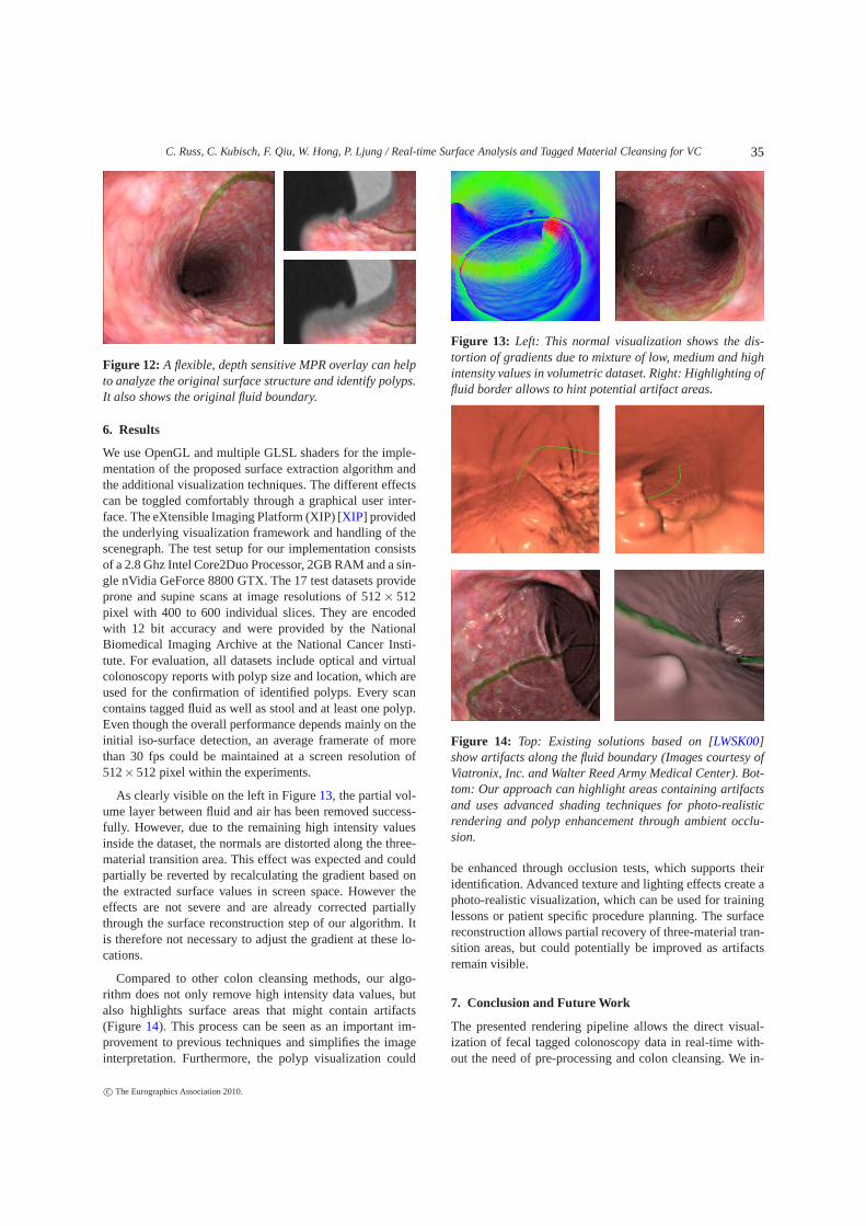
For manual surface analysis purposes, an interactive planecan be rendered within the three dimensional endoscopicview, which displays the original intensity values of the CTscan data. These values can be compared directly to the vi-sualized surface. The plane can be rotated interactively andmoves with the camera. A transparent border allows depthimpressions, while the center area cuts through the surfaceand displays the actual intensity values as the result of anapplied 1D TF. Figure 12 shows an example.
c© The Eurographics Association 2010.
34
C. Russ, C. Kubisch, F. Qiu, W. Hong, P. Ljung / Real-time Surface Analysis and Tagged Material Cleansing for VC
Figure 12: A flexible, depth sensitive MPR overlay can help
to analyze the original surface structure and identify polyps.
It also shows the original fluid boundary.
6. Results
We use OpenGL and multiple GLSL shaders for the imple-mentation of the proposed surface extraction algorithm andthe additional visualization techniques. The different effectscan be toggled comfortably through a graphical user inter-face. The eXtensible Imaging Platform (XIP) [XIP] providedthe underlying visualization framework and handling of thescenegraph. The test setup for our implementation consistsof a 2.8 Ghz Intel Core2Duo Processor, 2GB RAM and a sin-gle nVidia GeForce 8800 GTX. The 17 test datasets provideprone and supine scans at image resolutions of 512 × 512pixel with 400 to 600 individual slices. They are encodedwith 12 bit accuracy and were provided by the NationalBiomedical Imaging Archive at the National Cancer Insti-tute. For evaluation, all datasets include optical and virtualcolonoscopy reports with polyp size and location, which areused for the confirmation of identified polyps. Every scancontains tagged fluid as well as stool and at least one polyp.Even though the overall performance depends mainly on theinitial iso-surface detection, an average framerate of morethan 30 fps could be maintained at a screen resolution of512× 512 pixel within the experiments.
As clearly visible on the left in Figure 13, the partial vol-ume layer between fluid and air has been removed success-fully. However, due to the remaining high intensity valuesinside the dataset, the normals are distorted along the three-material transition area. This effect was expected and couldpartially be reverted by recalculating the gradient based onthe extracted surface values in screen space. However theeffects are not severe and are already corrected partiallythrough the surface reconstruction step of our algorithm. Itis therefore not necessary to adjust the gradient at these lo-cations.
Compared to other colon cleansing methods, our algo-rithm does not only remove high intensity data values, butalso highlights surface areas that might contain artifacts(Figure 14). This process can be seen as an important im-provement to previous techniques and simplifies the imageinterpretation. Furthermore, the polyp visualization could
Figure 13: Left: This normal visualization shows the dis-
tortion of gradients due to mixture of low, medium and high
intensity values in volumetric dataset. Right: Highlighting of
fluid border allows to hint potential artifact areas.
Figure 14: Top: Existing solutions based on [LWSK00]
show artifacts along the fluid boundary (Images courtesy of
Viatronix, Inc. and Walter Reed Army Medical Center). Bot-
tom: Our approach can highlight areas containing artifacts
and uses advanced shading techniques for photo-realistic
rendering and polyp enhancement through ambient occlu-
sion.
be enhanced through occlusion tests, which supports theiridentification. Advanced texture and lighting effects create aphoto-realistic visualization, which can be used for traininglessons or patient specific procedure planning. The surfacereconstruction allows partial recovery of three-material tran-sition areas, but could potentially be improved as artifactsremain visible.
7. Conclusion and Future Work
The presented rendering pipeline allows the direct visual-ization of fecal tagged colonoscopy data in real-time with-out the need of pre-processing and colon cleansing. We in-
c© The Eurographics Association 2010.
35
C. Russ, C. Kubisch, F. Qiu, W. Hong, P. Ljung / Real-time Surface Analysis and Tagged Material Cleansing for VC
troduced a virtual colonoscopy application that allows thephoto-realistic visualization of the colonic lumen and pro-vides various interactive tools for the viewer. The renderingprocedure is applicable to different medical applications re-lated to virtual endoscopy that require the removal of taggedmaterial areas.
A novel algorithm for surface analysis and automated ma-terial interpretation generates high quality iso-surface co-ordinates, gradients and curvature values at runtime. Addi-tional post-processing steps are used for advanced render-ing techniques, adding screen space ambient occlusion andshadow silhouettes at low computational costs.
Our implementation does not require to modify the taggedCT input data. Thus, we can directly display the correlationbetween the presence of high intensity data values and pos-sible distortions within the rendering. Critical areas aroundpotential partial volume layers and areas with three-materialtransitions can be identified and highlighted to visualize in-creased artifact probability. We would like to perform a clin-ical study to analyze the use of the implemented procedures.Additional feedback from a broader range of users wouldhelp to measure the success of our surface generation andthe acceptance of advanced visualization effects within vir-tual colonoscopy. It furthermore remains a challenging taskto actually identify all polyps in real-time. The current en-hancement through curvature values can be improved by tak-ing additional geometric parameters into account. So far, theimplemented application provides an appropriate platformfor the interactive exploration of fecal tagged CT data andimproves the current techniques in speed and visual quality.
References
[CLW∗00] CHEN D., LIANG Z., WAX M. R., LI L. L. B.,KAUFMAN A. E.: A novel approach to extract colon lumen fromct images for virtual colonoscopy. IEEE Transactions on Medical
Imaging 19, 12 (2000), 1220–1226. 2
[dGMK∗09] DE GONZÁLEZ A. B., MAHESH M., KIM K.-P.,BHARGAVAN M., LEWIS R., METTLER F., LAND C.: Pro-jected cancer risks from computed tomographic scans performedin the united states in 2007. Archives of internal medicine 169,22 (2009), 2071–2077. 1
[EKE01] ENGEL K., KRAUS M., ERTL T.: High-quality pre-integrated volume rendering using hardware-accelerated pixelshading. In HWWS ’01: Proceedings of the ACM SIG-
GRAPH/EUROGRAPHICS workshop on Graphics hardware
(New York, NY, USA, 2001), ACM, pp. 9–16. 4
[FNS∗99] FENLON H. M., NUNES D. P., SCHROY P. C., BAR-ISH M. A., CLARKE P. D., FERRUCCI J. T.: A comparisonof virtual and conventional colonoscopy for the detection of col-orectal polyps. N Engl J Med 341, 20 (1999), 1496–503. 1
[HMK∗97] HONG L., MURAKI S., KAUFMAN A. E., BARTZ
D., HE T.: Virtual voyage: interactive navigation in the humancolon. In SIGGRAPH (1997), pp. 27–34. 1, 2
[HQK06] HONG W., QIU F., KAUFMAN A.: A pipeline for com-puter aided polyp detection. IEEE Transactions on Visualization
and Computer Graphics 12 (2006), 861–868. 6
[HSS∗05] HADWIGER M., SIGG C., SCHARSACH H., BHLER
K., GROSS M.: Real-time ray-casting and advanced shadingof discrete isosurfaces. Computer Graphics Forum 24, 3 (Sep.2005), 303–312. 2
[KHW∗09] KNOLL A., HIJAZI Y., WESTERTEIGER R., SCHOTT
M., HANSEN C., HAGEN H.: Volume ray casting with peak find-ing and differential sampling. IEEE Transactions on Visualiza-
tion and Computer Graphics 15 (2009), 1571–1578. 4
[KKSP08] KRÜGER A., KUBISCH C., STRAUSS G., PREIM B.:Sinus Endoscopy - Application of Advanced GPU Volume Ren-dering for Virtual Endoscopy. IEEE Transactions on Visualiza-
tion and Computer Graphics 14, 6 (November/Dezember 2008),1491–1498. 2, 5
[LLL∗09] LEE T.-H., LEE J., LEE H., KYE H., SHIN Y. G.,KIM S. H.: Fast perspective volume ray casting method usingGPU-based acceleration techniques for translucency rendering in3D endoluminal CT colonography. Computers in biology and
medicine 39, 8 (Aug. 2009), 657–666. 2
[LM04] LUM E. B., MA K.-L.: Lighting transfer functions us-ing gradient aligned sampling. In VIS ’04: Proceedings of the
conference on Visualization ’04 (Washington, DC, USA, 2004),IEEE Computer Society, pp. 289–296. 2
[LWSK00] LAKARE S., WAN M., SATO M., KAUFMAN A.: 3Ddigital cleansing using segmentation rays. In VIS ’00: Proceed-
ings of the conference on Visualization ’00 (Los Alamitos, CA,USA, 2000), IEEE Computer Society Press, pp. 37–44. 2, 7
[Mit07] MITTRING M.: Finding next gen: Cryengine 2. In SIG-GRAPH ’07: ACM SIGGRAPH 2007 courses (New York, NY,USA, 2007), ACM, pp. 97–121. 6
[NY08] NÄPPI J., YOSHIDA H.: Adaptive correction of thepseudo-enhancement of CT attenuation for fecal-tagging CTcolonography. Medical Image Analysis 12, 4 (2008), 413 – 426.2, 6
[RBA∗96] RUBIN G., BEULLIEU C., ANNGIRO V., RINGLE H.,NORBASH A., FELLER J., DAKE M., JEFFEREY R., NAPLE S.:Perspective volume rendering of CT and MR images: Applica-tions for endoscopic imaging. Radiology 199 (1996), 321–330.2
[Sch05] SCHARSACH H.: Advanced gpu raycasting. In Proceed-
ings of the 9th Central European Seminar on Computer Graphics(May 2005). 3
[SH05] SIGG C., HADWIGER M.: Fast third-order texture filter-ing. Addison-Wesley Professional, 2005. 3, 6
[SHN∗06] SCHARSACH H., HADWIGER M., NEUBAUER A.,WOLFSBERGER S., BÜHLER K.: Perspective isosurface anddirect volume rendering for virtual endoscopy applications. InEuroVis (2006), pp. 315–322. 2
[STF∗03] SERLIE I., TRUYEN R., FLORIE J., POST F. H.,VAN VLIET L. J., VOS F.: Computed cleansing for virtualcolonoscopy using a three-material transition model. In MICCAI2003, 6th Intern. Conf., Montréal, Proceedings, Part II (2003),Springer, pp. 175–183. 2
[WLL∗06] WANG Z., LIANG Z., LI X., LI L., LI B., EREMINA
D., LU H.: An improved electronic colon cleansing method fordetection of colonic polyps by virtual colonoscopy. IEEE Trans-
actions on Biomedical Engineering 53, 8 (2006), 1635–1646. 2
[XIP] eXtensible Imaging Platform. http://openxip.org/. 7
[ZBB∗06] ZHAO L., BOTHA C., BESCOS J., TRUYEN R., VOS
F., POST F.: Lines of curvature for polyp detection in virtualcolonoscopy. IEEE Transactions on Visualization and Computer
Graphics 12 (2006), 885–892. 6
c© The Eurographics Association 2010.
36