Mac-1 Signaling via Src-Family and Syk Kinases Results in Elastase-Dependent Thrombohemorrhagic...
13
Immunity 25, 271–283, August 2006 ª2006 Elsevier Inc. DOI 10.1016/j.immuni.2006.05.014 Mac-1 Signaling via Src-Family and Syk Kinases Results in Elastase-Dependent Thrombohemorrhagic Vasculopathy Junichi Hirahashi, 1,6 Divya Mekala, 1,6 Jessica Van Ziffle, 3 Ling Xiao, 1 Simin Saffaripour, 4 Denisa D. Wagner, 4,5 Steven D. Shapiro, 2 Clifford Lowell, 3 and Tanya N. Mayadas 1, * 1 Department of Pathology Center for Excellence in Vascular Biology 2 Department of Medicine Brigham and Women’s Hospital and Harvard Medical School Boston, Massachusetts 02115 3 Department of Laboratory Medicine University of California, San Francisco San Francisco, California 94143 4 Center for Blood Research Institute for Biomedical Research 5 Department of Pathology Harvard Medical School Boston, Massachusetts 02115 Summary CD18 integrins promote neutrophil recruitment, and their engagement activates tyrosine kinases, leading to neutrophil activation. However, the significance of integrin-dependent leukocyte activation in vivo has been difficult to prove. Here, in a model of thrombohe- morrhagic vasculitis, the CD18 integrin Mac-1 on neu- trophils recognized complement C3 deposited within vessel walls and triggered a signaling pathway involv- ing the Src-family kinase Hck and the Syk tyrosine ki- nase. This led to neutrophil elastase release, causing hemorrhage, fibrin deposition, and thrombosis. Mice genetically deficient in any of these components (C3, Mac-1, Hck, Syk, or elastase) were resistant to disease despite normal tissue neutrophil accumulation. Dis- ease was restored in Mac-1-deficient mice infused with wild-type, but not kinase- or elastase-deficient, neutrophils. Elastase release in the inflamed tissue was reduced in Mac-1-deficient mice, and a deficiency of Mac-1 or the kinases blocked neutrophil elastase re- lease in vitro. These data suggest that Mac-1 engage- ment of complement activates tyrosine kinases to pro- mote elastase-dependent blood vessel injury in vivo. Introduction Neutrophils are key players of the innate immune re- sponse during both host defense and inflammation. Neutrophil functions are largely dependent on the leuko- cyte-specific CD18 integrins. In vivo, blockade or a defi- ciency of CD18 integrins limits inflammation, primarily by reducing neutrophil recruitment to the site of injury. In vitro, engagement of CD18 integrins signals activation of effector functions (ROS production and degranula- tion) (Harris et al., 2000). However, despite the clear evidence that neutrophil integrin signaling in vitro leads to cellular activation, it remains unclear to what extent this contributes to tissue injury or host defense in vivo. Indeed, in immune complex-mediated inflammatory dis- eases, it is often assumed that signaling through neutro- phil Fc receptors is primarily responsible for activation of cellular effector function, while the integrins serve mainly a supporting role to facilitate cellular recruitment. Given the complexity of most disease models, where infiltrating inflammatory cells are receiving signals through numerous ligands, it is often difficult to sort out the relative contribution of integrins alone to neutro- phil activation in vivo. Thus, defining a model of an in- flammatory disease that is strictly dependent on integrin signaling of neutrophil effector functions remains an elusive goal in the field of inflammation research. The CD18 integrins LFA-1 (CD11a/CD18, a L b 2 ), Mac-1 (CD11b/CD18, a m b 2 , CR3), p150,95 (CD11c/CD18, a c b 2 ), and CD11d/CD18 (a d b 2 ) are composed of unique CD11 subunits complexed to a common CD18 subunit. Pa- tients with a deficiency in the CD18 subunit have in- creased susceptibility to bacterial and fungal infections, which is associated with the inability of their neutrophils and macrophages to migrate and perform cytotoxic functions (Anderson and Springer, 1987). In mice, a defi- ciency in CD18 or a CD18 receptor intercellular adhesion molecule 1 (ICAM-1), present on the endothelium and leukocytes, attenuates neutrophil accumulation and as- sociated tissue injury in models of inflmmation (Harris et al., 2000; Kakkar and Lefer, 2004). Studies in mice de- ficient in LFA-1 suggest that it plays a dominant role in neutrophil vascular arrest and transendothelial migra- tion (Andrew et al., 1998; Ding et al., 1999; Henderson et al., 2003). In contrast, Mac-1, which is the predomi- nant CD18 integrin on neutrophils, is postulated to play a more important role in activation of effector func- tions (Coxon et al., 1996; Ding et al., 1999; Lu et al., 1997; Mayadas and Cullere, 2005; Tang et al., 1997). Mac-1-mediated adhesion to complement iC3b- coated microbes promotes neutrophil phagocytosis and the release of oxygen radicals and proteinases within phagolysosomes, which is important for host de- fense. However, when the opsonized target is too large to ingest, the extracellular release of these products may contribute to tissue injury during inflammation (Ar- naout, 1990; Ross, 2000). In vitro studies have delin- eated signaling cascades responsible for CD18 integ- rin-mediated adhesion, degranulation, and reactive oxygen species (ROS) generation (Lowell, 2004; Lowell and Berton, 1999). Integrin engagement results in acti- vation of Src-family and Syk tyrosine kinases, which co- localize with CD18 integrins to phosphorylate a number of downstream targets including Cbl, Pyk-2, PLCg1 and 2, ERK, Vav, and Paxillin. Of these substrates, Cbl, SLP- 76, and Vav 1,3 have been demonstrated to play a role in neutrophil and macrophage integrin signaling in vitro (Caveggion et al., 2003; Gakidis et al., 2004; Newbrough et al., 2003). In vitro neutrophils deficient in Src-family members Hck and Fgr have defects in adhesion-depen- dent secondary granule release (Mocsai et al., 1999) and *Correspondence: [email protected] 6 These authors contributed equally to this work.
-
Upload
independent -
Category
Documents
-
view
0 -
download
0
Transcript of Mac-1 Signaling via Src-Family and Syk Kinases Results in Elastase-Dependent Thrombohemorrhagic...

Immunity 25, 271–283, August 2006 ª2006 Elsevier Inc. DOI 10.1016/j.immuni.2006.05.014
Mac-1 Signaling via Src-Family and Syk KinasesResults in Elastase-DependentThrombohemorrhagic Vasculopathy
Junichi Hirahashi,1,6 Divya Mekala,1,6
Jessica Van Ziffle,3 Ling Xiao,1 Simin Saffaripour,4
Denisa D. Wagner,4,5 Steven D. Shapiro,2
Clifford Lowell,3 and Tanya N. Mayadas1,*1Department of PathologyCenter for Excellence in Vascular Biology2Department of MedicineBrigham and Women’s Hospital and Harvard
Medical SchoolBoston, Massachusetts 021153Department of Laboratory MedicineUniversity of California, San FranciscoSan Francisco, California 941434Center for Blood Research Institute for Biomedical
Research5Department of PathologyHarvard Medical SchoolBoston, Massachusetts 02115
Summary
CD18 integrins promote neutrophil recruitment, and
their engagement activates tyrosine kinases, leadingto neutrophil activation. However, the significance of
integrin-dependent leukocyte activation in vivo hasbeen difficult to prove. Here, in a model of thrombohe-
morrhagic vasculitis, the CD18 integrin Mac-1 on neu-trophils recognized complement C3 deposited within
vessel walls and triggered a signaling pathway involv-ing the Src-family kinase Hck and the Syk tyrosine ki-
nase. This led to neutrophil elastase release, causinghemorrhage, fibrin deposition, and thrombosis. Mice
genetically deficient in any of these components (C3,Mac-1, Hck, Syk, or elastase) were resistant to disease
despite normal tissue neutrophil accumulation. Dis-ease was restored in Mac-1-deficient mice infused
with wild-type, but not kinase- or elastase-deficient,neutrophils. Elastase release in the inflamed tissue
was reduced in Mac-1-deficient mice, and a deficiencyof Mac-1 or the kinases blocked neutrophil elastase re-
lease in vitro. These data suggest that Mac-1 engage-ment of complement activates tyrosine kinases to pro-
mote elastase-dependent blood vessel injury in vivo.
Introduction
Neutrophils are key players of the innate immune re-sponse during both host defense and inflammation.Neutrophil functions are largely dependent on the leuko-cyte-specific CD18 integrins. In vivo, blockade or a defi-ciency of CD18 integrins limits inflammation, primarilyby reducing neutrophil recruitment to the site of injury.In vitro, engagement of CD18 integrins signals activationof effector functions (ROS production and degranula-tion) (Harris et al., 2000). However, despite the clear
*Correspondence: [email protected] These authors contributed equally to this work.
evidence that neutrophil integrin signaling in vitro leadsto cellular activation, it remains unclear to what extentthis contributes to tissue injury or host defense in vivo.Indeed, in immune complex-mediated inflammatory dis-eases, it is often assumed that signaling through neutro-phil Fc receptors is primarily responsible for activationof cellular effector function, while the integrins servemainly a supporting role to facilitate cellular recruitment.Given the complexity of most disease models, whereinfiltrating inflammatory cells are receiving signalsthrough numerous ligands, it is often difficult to sortout the relative contribution of integrins alone to neutro-phil activation in vivo. Thus, defining a model of an in-flammatory disease that is strictly dependent on integrinsignaling of neutrophil effector functions remains anelusive goal in the field of inflammation research.
The CD18 integrins LFA-1 (CD11a/CD18, aLb2), Mac-1(CD11b/CD18, amb2, CR3), p150,95 (CD11c/CD18, acb2),and CD11d/CD18 (adb2) are composed of unique CD11subunits complexed to a common CD18 subunit. Pa-tients with a deficiency in the CD18 subunit have in-creased susceptibility to bacterial and fungal infections,which is associated with the inability of their neutrophilsand macrophages to migrate and perform cytotoxicfunctions (Anderson and Springer, 1987). In mice, a defi-ciency in CD18 or a CD18 receptor intercellular adhesionmolecule 1 (ICAM-1), present on the endothelium andleukocytes, attenuates neutrophil accumulation and as-sociated tissue injury in models of inflmmation (Harriset al., 2000; Kakkar and Lefer, 2004). Studies in mice de-ficient in LFA-1 suggest that it plays a dominant role inneutrophil vascular arrest and transendothelial migra-tion (Andrew et al., 1998; Ding et al., 1999; Hendersonet al., 2003). In contrast, Mac-1, which is the predomi-nant CD18 integrin on neutrophils, is postulated toplay a more important role in activation of effector func-tions (Coxon et al., 1996; Ding et al., 1999; Lu et al., 1997;Mayadas and Cullere, 2005; Tang et al., 1997).
Mac-1-mediated adhesion to complement iC3b-coated microbes promotes neutrophil phagocytosisand the release of oxygen radicals and proteinaseswithin phagolysosomes, which is important for host de-fense. However, when the opsonized target is too largeto ingest, the extracellular release of these productsmay contribute to tissue injury during inflammation (Ar-naout, 1990; Ross, 2000). In vitro studies have delin-eated signaling cascades responsible for CD18 integ-rin-mediated adhesion, degranulation, and reactiveoxygen species (ROS) generation (Lowell, 2004; Lowelland Berton, 1999). Integrin engagement results in acti-vation of Src-family and Syk tyrosine kinases, which co-localize with CD18 integrins to phosphorylate a numberof downstream targets including Cbl, Pyk-2, PLCg1 and2, ERK, Vav, and Paxillin. Of these substrates, Cbl, SLP-76, and Vav 1,3 have been demonstrated to play a role inneutrophil and macrophage integrin signaling in vitro(Caveggion et al., 2003; Gakidis et al., 2004; Newbroughet al., 2003). In vitro neutrophils deficient in Src-familymembers Hck and Fgr have defects in adhesion-depen-dent secondary granule release (Mocsai et al., 1999) and

Immunity272
Syk-deficient neutrophils exhibit a defect in CD18-in-duced ROS generation and secondary granule release(Mocsai et al., 2002).
Neutrophil-derived cytotoxic products play an impor-tant role in the inflammation of the vessel wall. The localShwartzman-like reaction (LSR), induced by repeatedcytokine injection in the skin, produces histopathologiclesions that are characteristic of thrombohemorrhagicvasculitis (Brozna, 1990) and resemble those observedin SLE in cases where vascular damage occurs in thepresence of complement deposition (Danning et al.,1998; Philips et al., 1989; Riemekasten et al., 2002).The LSR is dependent on neutrophils (Argenbright andBarton, 1992; Chang et al., 1993; Stetson and Good,1951; Zhou et al., 2003). Neutrophil aggregation, theirinteraction with the endothelium and platelets as wellas neutrophil-derived microparticles promote the gen-eration of occlusive thrombi, fibrin accumulation, andhemorrhage (Bouchard and Tracy, 2001; Brozna, 1990;Gasser and Schifferli, 2005).
In this work, we directly tested the contribution ofMac-1, its ligands, effector mechanisms, and signalingpathways to the LSR by using relevant gene mutantmice and intravenous reconstitution of mice with puri-fied neutrophils. While Mac-1-deficient mice showednormal infiltration of neutrophils into the inflammatorysite, these mice failed to develop significant hemor-rhagic vasculitis, thus separating the neutrophil recruit-ment versus activation functions of this leukocyte integ-rin. Mac-1 engagement of complement within the vesselwall signaled activation of Src-family and Syk kinasesin a signaling pathway that led to neutrophil elastaserelease, which in turn was responsible for blood vesselinjury. These data delineate an important physiologicalrole for Mac-1-mediated signaling of effector functionsin tissue damage associated with thrombohemorrhagicvasculitis.
Results
Mac-1 Is Required for the Formation of Hemorrhagic
LesionsThe local Shwartzman reaction (LSR) was elicited by aninitial intradermal injection of lipopolysaccharide (LPS)followed 20–24 hr later by a challenge with tumor necro-sis factor (TNF) at the same site. 24 hr after TNF injec-tion, hemorrhaging was observed in the intact skin.Microscopic analysis of skin tissue sections revealedsignificant erythrocyte extravasation, neutrophil accu-mulation, extravascular fibrin accumulation, and occlu-sive thrombi containing neutrophils. Mac-1-deficientmice subjected to LSR exhibited no detectable hemor-rhage (Figure 1). This was associated with a substantialdecrease in the number of thrombus-occluded vesselsand reduced fibrin deposition. The fibrin that was pres-ent formed a ring within the lumen of intact vessels incontrast to the predominantly extravascular fibrin accu-mulation observed in wild-type mice. Despite the ab-sence of hemorrhage in Mac-1-deficient mice, neutro-phil accumulation in the skin lesions of the mutantmice was essentially equivalent to that of wild-type ani-mals as assessed on tissue sections (Figure 1A). Inter-estingly, neutrophils adherent to the vessel wall ofwild-type animals tended to be flattened against the
lumen while those in the Mac-1-deficient animals wereround in appearance (Figure 1B, c and d), perhaps re-flecting altered adhesion-dependent spreading of theMac-1-deficient cells. Tissue neutrophil accumulationas assayed by tissue myeloperoxidase (MPO) activity(converted to number of neutrophils via a standardcurve) was shown to be comparable in both groups ofanimals (#neutrophils 3 104/mg tissue: wt, 2.13 6 0.17;Mac-1-deficient, 2.02 6 0.16). Neutrophil accumulationwas also similar in wild-type and Mac-1-deficient miceafter LPS injection alone or LPS followed by a 4 hr TNFstimulation (data not shown). These data indicate thatin the LSR model, the CD18 integrin Mac-1 is not criticalfor neutrophil recruitment to the inflammatory site but isrequired for tissue injury leading to the development ofthrombohemorrhagic vasculitis.
The LSR was evaluated in LFA-1-deficient mice to as-sess the relative contribution of this sister CD18 integrinin neutrophil accumulation and tissue injury. LFA-1-defi-cient mice exhibited only a 20% reduction in hemor-rhage (RBC as % of tissue area: wt, 11.7 6 0.32; LFA-1-deficient, 9.31 6 0.90, p < 0.05, n = 6 per group) anda 12% reduction in neutrophil accumulation (data notshown). We conclude that, in contrast to Mac-1, theCD18 integrin LFA-1 does not play a major role in dis-ease pathogenesis in the LSR model.
In vitro studies in macrophages suggest that LPS sig-naling through CD14 requires Mac-1 (Dobrovolskaia andVogel, 2002). Ribonuclease protection analysis of totalRNA in skin-tissue samples of LPS-injected mice re-vealed that cytokines upregulated by LPS (IL-1b, IL-1 re-ceptor antagonist, and IL-6) were similar in wild-typeand Mac-1-deficient mice. Others, including IFNg, IL-12(p35, p40), IL-18, MIF, and IL-1a, did not increase uponLPS treatment in either genotype (Figure 1C, data notshown), and the cytokine profile did not change uponadditional challenge with TNF (data not shown). Thus,the phenotype in Mac-1-deficient mice in LSR is likelynot the result of altered cytokine production after LPSinjection.
Mac-1 on Intravascular Neutrophils Promotes
Hemorrhagic LesionsMac-1 is present on murine neutrophils, eosinophils,mast cells, monocytes, and tissue macrophages. To ex-amine whether Mac-1 on neutrophils is responsible forhemorrhagic lesions, Mac-1-deficient mice were in-jected intravenously with purified bone marrow-derivedneutrophils harvested from wild-type or Mac-1-deficientmice 2 hr prior to TNF injection in the dorsal skin to in-duce the LSR. The adoptive transfer of wild-type neutro-phils into Mac-1-deficient mice restored the formationof hemorrhagic lesions while the transfer of Mac-1-defi-cient cells had no effect despite robust neutrophil re-cruitment to the site of inflammation (Figure 2). Theextent of hemorrhage, thrombus formation, and fibrindeposition was essentially the same in Mac-1-deficientmice injected with wild-type neutrophils as seen in nor-mal wild-type animals. In contrast to intravenous trans-fers, injection of wild-type neutrophils intradermally (atthe site of LPS and TNF injection) into Mac-1-deficientanimals was not effective in reconstituting disease(data not shown). Therefore, Mac-1 on intravascular

Mac-1 Signaling in Elastase-Dependent Vasculitis273
Figure 1. Mac-1-Deficient Mice Fail to Develop Thrombohemorrha-
gic Vasculitis in the Local Shwartzman-like Reaction Despite Normal
Neutrophil Recruitment and Production of Inflammatory Cytokines
(A) Wild-type (wt) and Mac-1-deficient (Mac-12/2) mice were sub-
jected to the LSR, and lesions in sections of harvested skin were
scored. The percent of tissue area of extravascular RBCs and neu-
trophils in four low-power fields (403 objective) for hemorrhage
and neutrophil (Neut.) exudates, respectively, was determined in
wt and Mac-12/2 mice. The percent of vessels with fibrin staining
outside the vessel wall and the percent of vessels occluded by
thrombus were quantitated by scoring all morphologically intact
vessels in the skin sample. In parentheses are the numbers of
mice of each genotype analyzed to produce the shown averaged
data 6 SEM, *p < 0.05.
(B) Representative skin sections from wild-type (a, c) and Mac-12/2
(b, d) mice are shown. (a and b) Hematoxylin and eosin-stained sec-
tions show hemorrhaging and thrombosis in wild-type mice with the
strongest reaction evident in the deep dermis just above the panni-
culus carnosus muscle (*). This reaction was not present in Mac-12/2
mice. An arrow identifies a blood vessel in each section (c and d). A
specific esterase stain revealed neutrophils (small blue cells, arrow)
adherent to the vessel wall and in the extravascular space of wild-
neutrophils is an important determinant of the thrombo-hemorrhagic pathology.
Role for Mac-1 Ligand Complement C3 but NotICAM-1 in Thrombohemorrhagic Lesions
Mac-1 recognizes several ligands including ICAM-1,complement fragment iC3b (generated by complementC3 activation), GpIb on platelets, and coagulation fac-tors fibrinogen and factor X (Arnaout, 1990; Mayadasand Cullere, 2005). Here, we examined whether Mac-1ligands ICAM-1 and complement fragment iC3b wererequired for the development of thrombohemorrhagiclesions by analyzing LSR in mice deficient in ICAM-1or complement C3 (C32/2). ICAM-1-deficient mice hadnormal neutrophil accumulation with lesion scores thatwere similar to their wild-type counterparts (Table 1).In contrast, C32/2 animals exhibited no hemorrhagingor microthrombi and markedly reduced fibrin depositiondespite normal neutrophil accumulation (Table 1). Ad-herent neutrophils within vessels of C32/2 mice ex-hibited a round morphology, similar to that seen inMac-1-deficient animals (data not shown). Thus, C3deficiency recapitulated the phenotype observed inMac-1-deficient mice.
The distribution of complement C3 deposition in theLSR lesions was examined by immunohistochemistry.Significant C3 deposition was observed along the vesselwalls at sites of inflammation (Figure 3). Importantly, theextent of C3 deposition was similar in wild-type andMac-1-deficient animals. No C3 staining was observedin dorsal skin injected with PBS alone (Figure 3). To-gether these data suggest that complement C3 is acti-vated and deposits in the vessel wall upon induction ofthe LSR and that Mac-1-mediated neutrophil interactionwith C3 results in vessel injury and leakage leading tothrombohemorrhagic lesions. However, neither C3 norMac-1 played a significant role in neutrophil recruitmentto the inflammatory sites during the LSR.
Role of NADPH Oxidase-Generated Reactive OxygenSpecies and Proteinases in Shwartzman Reaction
Neutrophil degranulation results in the surface transloca-tion and/or release of proteinases (Owen and Campbell,1999) as well as the production of NADPH oxidase-derived oxygen radicals, both of which are potential me-diators of vessel injury. To elucidate the role of variousproteinases versus oxygen radicals in the developmentof vascular lesion during the LSR, we examined micelacking the serine proteinase neutrophil elastase (NE),metalloproteinases MMP8 or MMP9 alone or both
type and Mac-12/2 mice. The larger blue cell (arrowhead in [d]) is
a mast cell that also stains positively with the esterase stain and is
excluded from the quantitative analysis. Note the flattened appear-
ance of intravascular neutrophils in sections from wild-type (c) com-
pared to the rounded neutrophils in Mac-12/2 (d) mice identified by
arrows. Scale bar equals 100 mm, double scale bar equals 20 mm.
(C) Total RNA was prepared from skin samples harvested from the
LSR site of wild-type and Mac-1-deficient mice. RNA for indicated
cytokines and housekeeping genes, L32 and GADPH, was detected
in a multiprobe ribonuclease protection assay. A representative au-
toradiograph of samples from control wild-type (W) and Mac-1-defi-
cient (K) mice after PBS or LPS injection are shown. Cytokines that
were upregulated after LPS treatment are indicated by asterisks (*).
Immunity274
MMP8 and MMP9, or the NADPH-oxidase subunitgp91phox. The LSR was intact in gp91phox-deficient(gp912/2) mice, suggesting that reactive oxygen spe-cies do not play a major role in the development oflesions or neutrophil accumulation (Table 2A). Interest-ingly, hemorrhagic and fibrinous lesions were absentin the NE-deficient mice, despite normal neutrophil ac-cumulation (Table 2B), while none of these parameterswere altered in mice deficient in either MMP8 or MMP9alone or in mice doubly deficient in both MMP8 and 9(Table 2C). Thus, NE is one of the major proteinasesthat mediate development of thrombohemorrhagic le-sions in the LSR without altering cellular recruitment to
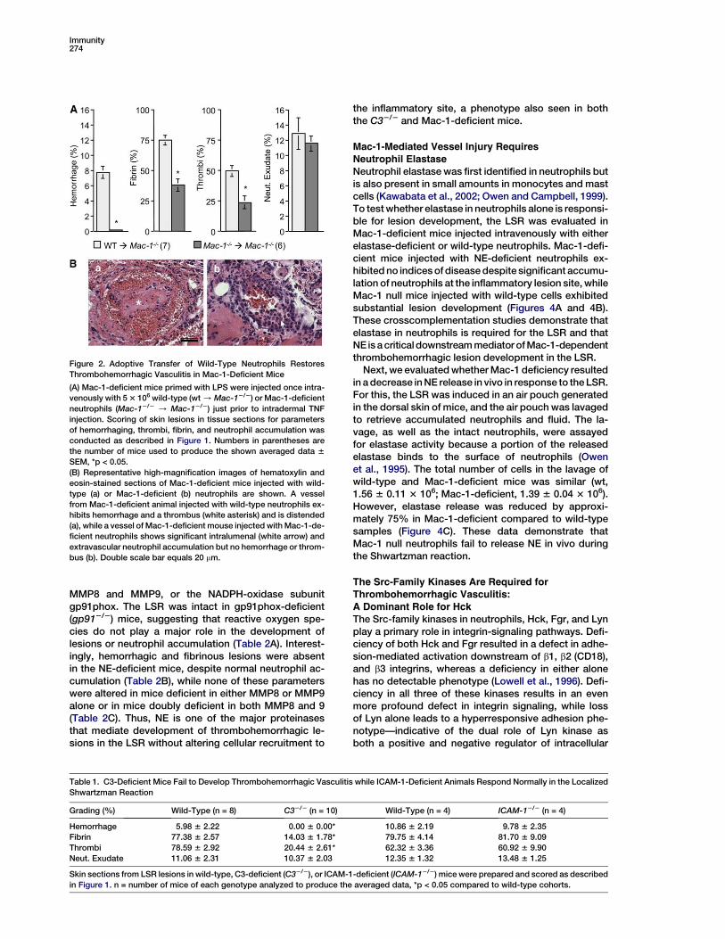
Figure 2. Adoptive Transfer of Wild-Type Neutrophils Restores
Thrombohemorrhagic Vasculitis in Mac-1-Deficient Mice
(A) Mac-1-deficient mice primed with LPS were injected once intra-
venously with 5 3 106 wild-type (wt / Mac-12/2) or Mac-1-deficient
neutrophils (Mac-12/2 / Mac-12/2) just prior to intradermal TNF
injection. Scoring of skin lesions in tissue sections for parameters
of hemorrhaging, thrombi, fibrin, and neutrophil accumulation was
conducted as described in Figure 1. Numbers in parentheses are
the number of mice used to produce the shown averaged data 6
SEM, *p < 0.05.
(B) Representative high-magnification images of hematoxylin and
eosin-stained sections of Mac-1-deficient mice injected with wild-
type (a) or Mac-1-deficient (b) neutrophils are shown. A vessel
from Mac-1-deficient animal injected with wild-type neutrophils ex-
hibits hemorrhage and a thrombus (white asterisk) and is distended
(a), while a vessel of Mac-1-deficient mouse injected with Mac-1-de-
ficient neutrophils shows significant intralumenal (white arrow) and
extravascular neutrophil accumulation but no hemorrhage or throm-
bus (b). Double scale bar equals 20 mm.
the inflammatory site, a phenotype also seen in boththe C32/2 and Mac-1-deficient mice.
Mac-1-Mediated Vessel Injury RequiresNeutrophil Elastase
Neutrophil elastase was first identified in neutrophils butis also present in small amounts in monocytes and mastcells (Kawabata et al., 2002; Owen and Campbell, 1999).To test whether elastase in neutrophils alone is responsi-ble for lesion development, the LSR was evaluated inMac-1-deficient mice injected intravenously with eitherelastase-deficient or wild-type neutrophils. Mac-1-defi-cient mice injected with NE-deficient neutrophils ex-hibited no indices of disease despite significant accumu-lation of neutrophils at the inflammatory lesion site, whileMac-1 null mice injected with wild-type cells exhibitedsubstantial lesion development (Figures 4A and 4B).These crosscomplementation studies demonstrate thatelastase in neutrophils is required for the LSR and thatNE is a critical downstream mediator of Mac-1-dependentthrombohemorrhagic lesion development in the LSR.
Next, we evaluated whether Mac-1 deficiency resultedin a decrease in NE release in vivo in response to the LSR.For this, the LSR was induced in an air pouch generatedin the dorsal skin of mice, and the air pouch was lavagedto retrieve accumulated neutrophils and fluid. The la-vage, as well as the intact neutrophils, were assayedfor elastase activity because a portion of the releasedelastase binds to the surface of neutrophils (Owenet al., 1995). The total number of cells in the lavage ofwild-type and Mac-1-deficient mice was similar (wt,1.56 6 0.11 3 106; Mac-1-deficient, 1.39 6 0.04 3 106).However, elastase release was reduced by approxi-mately 75% in Mac-1-deficient compared to wild-typesamples (Figure 4C). These data demonstrate thatMac-1 null neutrophils fail to release NE in vivo duringthe Shwartzman reaction.
The Src-Family Kinases Are Required forThrombohemorrhagic Vasculitis:
A Dominant Role for HckThe Src-family kinases in neutrophils, Hck, Fgr, and Lynplay a primary role in integrin-signaling pathways. Defi-ciency of both Hck and Fgr resulted in a defect in adhe-sion-mediated activation downstream of b1, b2 (CD18),and b3 integrins, whereas a deficiency in either alonehas no detectable phenotype (Lowell et al., 1996). Defi-ciency in all three of these kinases results in an evenmore profound defect in integrin signaling, while lossof Lyn alone leads to a hyperresponsive adhesion phe-notype—indicative of the dual role of Lyn kinase asboth a positive and negative regulator of intracellular
Table 1. C3-Deficient Mice Fail to Develop Thrombohemorrhagic Vasculitis while ICAM-1-Deficient Animals Respond Normally in the Localized
Shwartzman Reaction
Grading (%) Wild-Type (n = 8) C32/2 (n = 10) Wild-Type (n = 4) ICAM-12/2 (n = 4)
Hemorrhage 5.98 6 2.22 0.00 6 0.00* 10.86 6 2.19 9.78 6 2.35
Fibrin 77.38 6 2.57 14.03 6 1.78* 79.75 6 4.14 81.70 6 9.09
Thrombi 78.59 6 2.92 20.44 6 2.61* 62.32 6 3.36 60.92 6 9.90
Neut. Exudate 11.06 6 2.31 10.37 6 2.03 12.35 6 1.32 13.48 6 1.25
Skin sections from LSR lesions in wild-type, C3-deficient (C32/2), or ICAM-1-deficient (ICAM-12/2) mice were prepared and scored as described
in Figure 1. n = number of mice of each genotype analyzed to produce the averaged data, *p < 0.05 compared to wild-type cohorts.

Mac-1 Signaling in Elastase-Dependent Vasculitis275
signaling (Mocsai et al., 2002; Pereira and Lowell, 2003).To examine whether Src-family kinases are critical forMac-1-mediated signaling in vivo, we analyzed mice de-ficient in Hck, Fgr, or Lyn alone as well as mice lacking allthree kinases in the LSR. Fgr2/2 mice developed hemor-rhagic lesions that were partially reduced compared towild-type animals while Lyn deficiency resulted in a fur-ther modest reduction in disease indices (Figure 5A). Inboth these kinase mutant strains, all animals showedclear evidence of some hemorrhage after TNF injection.In contrast, 73% of Hck2/2 mice were completely free ofextravascular hemorrhage and fibrin deposition, withthe remaining animals displaying variable amounts ofdisease formation. Triple src-deficient mice exhibitedno hemorrhage and significantly reduced fibrin deposi-tion in response to the LSR (Figure 5A). These data indi-cate that the Src-family kinases in myeloid leukocyteshave partially redundant roles in initiating thrombohe-morrhagic vasculitis, with the Hck kinase playing themost important function. Loss of Hck alone or deficiencyof all three kinases produced a profound impairment inhemorrhage and fibrin formation in the LSR model, with-out affecting neutrophil migration (Figure 5A), a pheno-type very similar to that of the C3-, Mac-1-, or neutrophilelastase-deficient mice.
Figure 3. Complement C3 Is Deposited along Vessel Walls during
Experimental Thrombohemorrhagic Vasculitis
Skin-tissue sections from wt (top) or Mac-1-deficient (middle) mice
subjected to the LSR and wt mice injected with PBS alone (bottom)
were stained with polyclonal anti-C3 antibody. C3 staining is ob-
served along a vessel (arrow) from a wild-type (top) and Mac-1-de-
ficient (middle) animal subjected to LSR, while no C3 staining is ob-
served in a vessel (arrowhead) of a wild-type mouse injected with
PBS alone (bottom). Complement C3-deficient mice subjected to
LSR showed no staining (not shown), demonstrating the specificity
of the C3 antibody.
To evaluate whether the phenotypes observed in thesingle and triple src mutant mice could be attributed tothe role of Src-family proteins specifically in neutrophils,Mac-1-deficient mice were injected with neutrophilsfrom either Hck2/2, Fgr2/2, or triple src mutant miceand examined in the LSR. Mac-1-deficient mice injectedwith Hck2/2 neutrophils prior to initiation of the LSRshowed significantly reduced hemorrhage and modestlyreduced fibrin deposition compared to injection of wild-type cells (Figures 5B and 5C). Adoptive transfer of triplesrc mutant neutrophils into Mac-1-deficient mice failedto reconstitute disease formation; the Mac-1-deficientrecipients in these experiments showed no evidence ofextravascular hemorrhage or fibrin deposition. Thus, re-constitution of Mac-1 mutant mice with either Hck2/2 ortriple src-deficient neutrophils produces the same phe-notypic response in the LSR as seen in the original kinasemutant animals, indicating that loss of these kinases inneutrophils alone is responsible for the amelioration ofinflammatory disease in this model. In contrast, Mac-1-deficient mice that were injected with Fgr2/2 neutrophilsshowed nearly normal levels of hemorrhage (Hemor-rhage %: wt neutrophils, 12.58 6 0.39; Fgr2/2 neutro-phils, 9.81 6 1.11, p = 0.40), which differed from the mod-erate reduction in hemorrhage seen in the Fgr2/2 micealone. Thus, the loss of Fgr from cells other than neutro-phils likely contributed to the partially reduced responseto the LSR observed in Fgr2/2 mice (Figure 5A).
To further validate that loss of Src-family kinases inneutrophils alone is capable of blocking disease activityin the LSR and that Mac-1 is upstream of these kinases,we attempted to restore disease to triple src mutantmice by injection of wild-type or Mac-1-deficient neutro-phils. As expected, triple src mutant mice injected withwild-type neutrophils displayed normal levels of hemor-rhage and fibrin deposition after initiation of the LSR,while triple src mutant mice injected with Mac-1-defi-cient cells remained free of disease (Figures 5B and5C). These crosscomplementation experiments confirmthat Src-family kinases in neutrophils are required forthrombohemorrhagic vasculitis.
Syk Is Required for Mac-1-Dependent Vasculopathy
The described role for the Syk in outside-in integrin sig-naling in neutrophils in vitro (Mocsai et al., 2002) promp-ted us to determine whether this kinase is also requiredfor Mac-1-dependent thrombohemorrhagic vasculitisin vivo. To examine this, chimeric mice were generatedby injecting fetal liver cells from Syk2/2 mice into lethallyirradiated wild-type recipients, allowing us to circum-vent the perinatal lethality caused by the Syk mutation(Turner et al., 2000). Controls for these animals were irra-diated wild-type mice injected with fetal liver cells fromwild-type or Syk+/2 animals. Surprisingly, even wild-type mice reconstituted with wild-type cells after lethalirradiation failed to develop reproducible evidence ofthrombohemorrhagic vasculitis in the LSR (data notshown), precluding the examination of intact chimerasin these experiments. Therefore, the role of Syk kinasein neutrophils in the LSR was examined by attemptingto restore disease in Mac-1-deficient mice with neutro-phils from wild-type and Syk2/2 fetal liver chimericmice. Mac-1-deficient mice injected with neutrophilsfrom wild-type or Syk+/2 fetal liver chimeric mice

Immunity276
Table 2. Neutrophil Elastase-Deficient Mice Fail to Develop Thrombohemorrhagic Vasculitis in the LSR, while Mice Deficient in NADPH Oxidase
Function or Lacking MMP8 and MMP9 Respond Normally
(A) Mice Lacking the gp91phox Subunit of the NADPH Oxidase (gp912/2)
Grading (%) Wild-Type (n = 10) gp912/2 (n = 10)
Hemorrhage 6.28 6 0.74 4.64 6 1.87
Fibrin 73.03 6 6.13 61.13 6 20.50
Thrombi 57.62 6 5.56 52.35 6 8.93
Neut. Exudate 9.84 6 1.13 7.15 6 1.29
(B) Mice Lacking the Primary Granule Proteinase Neutrophil Elastase (NE2/2)
Grading (%) Wild-Type (n = 6) NE2/2 (n = 5)
Hemorrhage 8.91 6 1.03 0.02 6 0.02*
Fibrin 83.40 6 0.32 34.50 6 12.18*
Thrombi 54.31 6 9.96 14.34 6 1.52*
Neut. Exudate 14.78 6 2.23 10.91 6 1.39
(C) Mice Lacking the Primary Granule Proteinases MMP8 and/or MMP9 (Single MMP82/2 or MMP92/2 Mutants or Double MMP8/MMP92/2
Mutants)
Grading (%) Wild-Type (n = 5) MMP92/2 (n = 3) Wild-Type (n = 8) MMP82/2 (n = 6)
Hemorrhage 12.44 6 3.54 14.47 6 6.91 9.66 6 1.04 10.59 6 2.34
Fibrin 75.30 6 3.36 74.90 6 14.13 81.47 6 2.41 76.65 6 3.35
Thrombi 55.50 6 9.18 61.30 6 21.55 71.17 6 2.84 68.25 6 4.80
Neut. Exudate 13.63 6 1.78 17.06 6 0.33 11.33 6 1.38 9.42 6 2.22
Grading (%) Wild-Type (n = 4) MMP8/MMP92/2 (n = 3)
Hemorrhage 9.46 6 1.66 12.12 6 1.71
Fibrin 77.65 6 3.38 77.56 6 4.52
Thrombi 66.01 6 4.25 74.14 6 5.96
Neut. Exudate 10.96 6 2.40 9.55 6 1.65
LSR lesions in wild-type and indicated deficient mice were assessed for hemorrhage, thrombi, fibrin deposition, and neutrophil accumulation as
described in Figure 1. n = number of mice of each genotype analyzed to produce the averaged data, *p < 0.05 compared to wild-type cohorts.
developed hemorrhage and fibrin deposition in the LSR,while Mac-1-deficient mice receiving Syk2/2 cells failedto show evidence of disease (Table 3). As seen in theC32/2, Mac-1-deficient, NE-deficient, and the Hck,Fgr,Lyn2/2 triple knockout mice, Syk2/2 neutrophils wererecruited normally into the inflammatory site, indicatingthat this kinase is not involved in neutrophil migration, aspreviously reported in a thioglycollate-induced peritoni-tis model (Mocsai et al., 2002). These results directlyimplicate Syk kinase in neutrophils in the developmentof Mac-1-mediated thrombohemorrhagic vasculitis ata step involving neutrophil activation and elastase re-lease, without altering neutrophil recruitment.
Mac-1 Adhesion-Dependent Release of Elastase In
Vitro Requires Complement C3 and Hck KinaseOur in vivo results suggest that Mac-1-mediated adhe-sion to complement results in elastase release that islargely dependent on the Src-family kinase Hck. Previ-ous reports have shown that adhesion of activated neu-trophils to surfaces coated with integrin ligands (e.g.,fibrinogen or collagen) results in the exocytosis of pri-mary (azurophilic) and secondary (specific) granules(Mocsai et al., 1999, 2002). We designed an in vitro assayto assess the role of Src kinases in Mac-1-mediated ad-hesion and release of elastase (present in primary gran-ules) to complement-coated surfaces after TNF stimula-tion. Since biomaterials such as tissue-culture plates arewell described to activate complement C3 resulting indeposition of C3b and iC3b (Andersson et al., 2002; Che-noweth et al., 1981), this assay presents high-densityMac-1 ligands to the leukocyte, similar to the situation
of vascular deposition of C3 components in the LSR.The adhesion of TNF-stimulated wild-type neutrophilsto plates coated with freshly prepared mouse serumwas dependent on Mac-1 and complement, as shownby the fact that both Mac-1-deficient neutrophils andC32/2 serum failed to support significant neutrophil ad-hesion (Figure 6A). To measure the adhesion-dependentrelease of neutrophil elastase, we used a continuousspectroscopic assay containing a fluorogenic substratespecific for neutrophil elastase (Sklar et al., 1982). Adhe-sion of wild-type neutrophils to complement-coatedplates resulted in significant elastase release, whereasMac-1-deficient neutrophils or wild-type neutrophils ad-herent to plates coated with C32/2 serum exhibited sig-nificantly less elastase release (Figure 6B). Similar to theadhesion assay, primary granule release was very lowin Mac-1-deficient cells plated on C32/2 serum. Thus,TNF-induced Mac-1 interaction with complement iC3bis responsible for the majority (greater than 70%) of elas-tase release observed in this assay.
The role of Src-family kinases in Mac-1 and C3 adhe-sion-mediated elastase release was evaluated by as-sessing degranulation in neutrophils deficient in theHck, Fgr, or Lyn kinases alone or all three src kinases.As shown in Figure 6C, both the Hck2/2 and triple srcmutant cells manifested a large reduction in degranula-tion after adhesion to wild-type serum-coated surfaces.In contrast, Fgr2/2 and Lyn2/2 cells showed no signifi-cant decrease in elastase release in this assay. Thesein vitro elastase results correlate well with the in vivoobservations in the LSR and suggest that the Hck kinaseis the dominant neutrophil Src-family kinase involved

Mac-1 Signaling in Elastase-Dependent Vasculitis277
in signaling primary granule release after adhesion tosurface bound complement.
Discussion
Our studies establish a key role for Mac-1 in the destruc-tion of vascular integrity associated with a model ofthrombohemorrhagic vasculitis. These data are consis-tent with previous reports showing inhibition of LSRlesions after treatment with CD11b blocking antibody(Argenbright and Barton, 1992; Chang et al., 1993). The
Figure 4. Adoptive Transfer of Elastase-Deficient Neutrophils Fails
to Restore Thrombohemorrhagic Vasculitis to Mac-1-Deficient Mice
(A) Mac-1-deficient (Mac-12/2) mice primed with LPS were injected
once intravenously with 5 3 106 wild-type (wt / Mac-12/2) or neu-
trophil elastase-deficient (NE2/2 / Mac-12/2) cells just prior to
intradermal TNF injection, and lesions were scored as outlined in
Figure 1, Numbers in parentheses are the number of mice used to
produce the shown averaged data 6 SEM, *p < 0.05.
(B) Representative sections of Mac-1-deficient mice injected with
wild-type (a) or NE2/2 (b) neutrophils are shown. Mac-1-deficient
mice injected with wt cells exhibited significant hemorrhage, throm-
bus formation, and neutrophil infiltration (a), while those injected
with NE2/2 cells (b) displayed intense neutrophil infiltration at the
TNF injection site (white asterisk) without evidence of hemorrhage
or thrombus formation.
(C) Skin air pouches were generated in Mac-1-deficient (Mac-12/2)
and wild-type (wt) mice and the LSR was induced in this site. The
pouches were lavaged, and intact neutrophils and lavage fluid
were assayed for neutrophil elastase activity. The percent degranu-
lation was calculated by dividing the elastase activity by the total
cellular elastase activity measured after lysis of cells in the sample.
n = 4 for each genotype. Data shown are average 6 SD, *p < 0.05.
near absence of tissue injury in Mac-1-deficient micewas not the result of changes in cytokines induced dur-ing the LSR (IL-6, IL-1b, IL-1ra), nor was it attributable toreduced neutrophil accumulation in the skin. Thus theLSR model allows us to separate the function of integrin
Figure 5. Lack of Src-Family Kinases in Neutrophils Renders Mice
Resistant to Experimental Thrombohemorrhagic Vasculitis
(A) Wild-type, Fgr2/2, Lyn2/2, Hck2/2, or triple src mutant (H,F,L2/2)
mice were subjected to the LSR, and scoring of tissue sections for
percent hemorrhage, extravascular fibrin deposition, and neutrophil
exudates was performed as in Figure 1.
(B) Mac-1-deficient mice primed with LPS were injected once intra-
venously with 5 3 106 wild-type (wt / Mac-12/2), single mutant
hck2/2 (Hck2/2 / Mac12/2), or triple src mutant (H,F,L2/2 /
Mac-12/2) neutrophils just prior to intradermal TNF injection, and
lesions were scored as outlined in Figure 1. In addition, H,F,L2/2
mutant mice served as recipients for injection of either wild-type
(wt / H,F,L2/2) or Mac-1-deficient (Mac-12/2 / H,F,L2/2) cells,
after which mice were analyzed in the LSR as above. Numbers in
parentheses indicate the number of animals of each genotype
analyzed to produce the shown averaged data 6 SEM. *p < 0.005,
**p < 0.05 in comparison to wild-type.
(C) Photomicrographs show representative hematoxylin and eosin
(hemorrhage) or fibrin-stained skin sections from Mac-1-deficient
mice injected with either wild-type cells (wt / Mac-12/2) or triple
knockout cells (H,F,L2/2 / Mac-12/2).

Immunity278
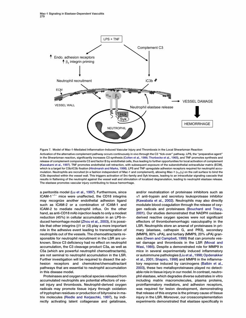
(i.e., Mac-1)-dependent inflammatory cell recruitmentfrom integrin-mediated cellular activation in an in vivo in-flammatory disease. In the vast majority of experimentalinflammation models, the presence of tissue injury usu-ally correlates with inflammatory (neutrophil) cell infil-trate into the lesion sites. However, in the LSR model,this is not the case—all of the mutant mice we have ex-amined displayed normal neutrophil recruitment into thedisease site independent of vascular injury and hemor-rhage. We have taken advantage of these aspects ofthe LSR model to study the upstream ligands, down-stream signaling molecules, and effector systems ofneutrophil Mac-1, thereby allowing us to analyze thecontribution of this integrin to cellular activation andresulting inflammatory tissue injury in vivo. Our studiesindicate that interaction of Mac-1 on neutrophils withcomplement iC3b deposits within the vessel wall leadsto an intracellular signaling pathway involving Src-familyand Syk kinases, causing degranulation and vascularinjury that is dependent on release of neutrophil elastase(Figure 7).
Mice deficient in complement C3, which therefore areunable to form the Mac-1 ligand iC3b, phenocopiedMac-1-deficient mice in all aspects of the LSR, consis-tent with a previous report suggesting suppression ofthe LSR after complement depletion (Fong and Good,1971). This suggests that the interaction of Mac-1 onneutrophils with complement C3 that is deposited alongthe vascular walls at the site of TNF injection promotesneutrophil activation leading to tissue damage. Comple-ment may be on the endothelium (Marks et al., 1989)and/or in the subendothelial matrix, which can fix iC3bupon its exposure after cytokine-induced endothelialcell retraction (Hindmarsh and Marks, 1998). Our findingthat restoration of disease in Mac-1-deficient mice re-quired the intravenous delivery of wild-type neutrophilssuggests that the interaction of Mac-1 on circulatingneutrophils with fixed complement must occur withinthe vascular lumen for significant vessel damage tooccur (Figure 7). Previous reports have suggested thatcomplement deposition in the vasculature is consis-tently associated with vasculitides (Chen et al., 2002;Danning et al., 1998; Prins et al., 1996). Mac-1 interactionwith complement C3bi could drive neutrophil degranula-tion at the vessel wall via a process often referred to as
Table 3. Adoptive Transfer of Syk-Deficient Neutrophils Fails to
Restore Normal Disease Activity in Mac-1-Deficient Mice
Undergoing LSR
Recipient Mac-12/2 (n = 8) Mac-12/2 (n = 8)
Donor Wild-Type or Syk+/2 Syk2/2
Hemorrhage (%) 12.55 6 0.24 0.38 6 0.10*
Fibrin (%) 92.40 6 3.16 6.87 6 0.49*
Neut. Exudate (%) 12.97 6 0.55 9.06 6 0.40
Mac-1-deficient mice (Mac-12/2) primed with LPS were injected
once intravenously with 5 3 106 wild-type/Syk+/2 or Syk-deficient
(Syk2/2) neutrophils just prior to intradermal TNF injection. Neutro-
phils were prepared from fetal liver chimeric mice that had previ-
ously been established with wild-type/Syk+/2 or Syk2/2 cells. Scor-
ing of skin lesions in the adoptively transferred Mac-12/2 tissue
sections for parameters of hemorrhaging, fibrin deposition, and neu-
trophil accumulation was conducted as described in Figure 1. n =
number of mice analyzed to produce the averaged data, *p < 0.05
compared to wild-type/Syk+/2 cohorts.
‘‘frustrated phagocytosis,’’ where phagocytes may forma tight seal around the complement deposits, creating acompartment into which secreted proteinases can beconcentrated and protected from serum proteinase in-hibitors, thus maximizing endothelial cell injury (Heipleet al., 1990; Wright and Silverstein, 1984).
Previous work has shown that treatment with CD18antibody results in the reduction in LSR-inducedhemorrhagic legions in rabbits (Argenbright and Barton,1992), although the effect of CD18 blockade on neutro-phil accumulation was not reported. Neutrophil recruit-ment in the LSR was essentially normal in Mac-1-defi-cient, LFA-1-deficient, and ICAM-1-deficient mice.Thus, it is possible that a combination of LFA-1 andMac-1 contributes to neutrophil recruitment in the LSRmodel. Indeed, only an ablation of both Mac-1 andLFA-1 significantly reduced neutrophil accumulation in
Figure 6. Adhesion of Neutrophils to Complement-Coated Surfaces
Leads to Elastase Release in Wild-Type but Not Mac-1- or Hck
Kinase-Deficient Cells
(A) Analysis of adhesion of TNF-stimulated wild-type or Mac-1-defi-
cient (Mac-12/2) neutrophils to wild-type or C3-deficient (C32/2)
fresh mouse serum-coated plates. TNF-stimulated neutrophils
were incubated with plates for 40 min, plates were washed, and
the number of attached neutrophils in three high-power fields was
counted microscopically. Data shown are the average 6 SD of two
independent experiments.
(B) The release of the azurophilic granule marker neutrophil elastase
was assessed after plating of TNF-activated wild-type or Mac-12/2
neutrophils on fresh mouse serum-coated dishes. The data are plot-
ted as % degranulation, which represents the amount of neutrophil
elastase released from 2.5 3 103 neutrophils per well, divided by the
total cellular elastase activity (determined by lysis of cells in 0.02%
Triton X-100) after 60 min of adhesion to the indicated serum-coated
surface. Data shown are the average 6 SD of two independent
experiments.
(C) Release of neutrophil elastase from wild-type, Mac-12/2, Hck2/2,
Fgr2/2, Lyn2/2, or triple src mutant (H,F,L2/2) neutrophils was con-
ducted as described above. Data shown are the average 6 SD. n = 4
to 8 independent experiments for each group except for the first (wt,
Fgr2/2, and Lyn2/2), which is n = 2.
*p < 0.005, **p < 0.05 in comparison to respective wt.
Mac-1 Signaling in Elastase-Dependent Vasculitis279
Figure 7. Model of Mac-1-Mediated Inflammation-Induced Vascular Injury and Thrombosis in the Local Shwartzman Reaction
Activation of the alternative complement pathway occurs continuously in vivo through the C3 ‘‘tick-over’’ pathway. LPS, the ‘‘preparative agent’’
in the Shwartzman reaction, significantly increases C3 synthesis (Colten et al., 1986; Thorbecke et al., 1965), and TNF promotes synthesis and
release of complement components C3 and factor B by endothelial cells, thus leading to further opportunities for local activation of complement
(Kawakami et al., 1997). TNF promotes endothelial cell retraction, with subsequent exposure of the subendothelial extracellular matrix (ECM),
which is a target for C3b/iC3b fixation (Hindmarsh and Marks, 1998). LPS and TNF upregulate adhesion receptors required for neutrophil accu-
mulation. Neutrophils are recruited (in a fashion independent of Mac-1 and complement), allowing Mac-1 (amb2) on the cell surface to bind the
iC3b deposited within the vessel wall. This triggers activation of Src-family and Syk kinases, leading to an intracellular signaling cascade that
results in flattening of the neutrophil against the vessel wall and stimulation of localized degranulation, leading to neutrophil elastase release.
The elastase promotes vascular injury contributing to tissue hemorrhage.
a peritonitis model (Lu et al., 1997). Furthermore, sinceICAM-12/2 mice were unaffected, the CD18 integrinsmay recognize another endothelial adhesion ligandsuch as ICAM-2 or a combination of ICAM-1 andICAM-2 to mediate neutrophil influx. On the otherhand, as anti-CD18 mAb injection leads to only a modestreduction (45%) in cellular accumulation in an LPS-in-duced hemorrhage model (Zhou et al., 2003), it is possi-ble that other integrins (b1 or b3) play a more importantrole in the adhesive event leading to transmigration ofneutrophils out of the vessels. The chemoattractants re-sponsible for neutrophil recruitment in the LSR are un-known. Since C3 deficiency had no effect on neutrophilaccumulation, the C3 cleavage product C3a, as well asC5a (which are powerful neutrophil chemoattractants),are not seminal to neutrophil accumulation in the LSR.Further investigation will be required to dissect the ad-hesion receptors and chemokine/chemoattractantpathways that are essential to neutrophil accumulationin this disease model.
Proteinases and oxygen radical species released fromaccumulated neutrophils are potential effectors of ves-sel injury and thrombosis. Neutrophil-derived oxygenradicals may promote tissue injury through oxidationof tryptophan residues or production of bityrosine in ma-trix molecules (Riedle and Kerjaschki, 1997), by indi-rectly activating latent collagenase and gelatinase,
and/or neutralization of proteinase inhibitors such asa1 anti-trypsin and secretory leukoprotease inhibitor(Kawabata et al., 2002). Neutrophils may also directlymodulate blood coagulation through the release of oxy-gen radicals and proteinases (Bouchard and Tracy,2001). Our studies demonstrated that NADPH oxidase-derived reactive oxygen species were not significanteffectors of thrombohemorrhagic vasculopathy in theLSR. Neutrophils store an arsenal of proteinases in pri-mary (elastase, cathepsin G, and PR3), secondary(MMP8, 80% uPA), and tertiary (MMP9, 20% uPA) gran-ules (Owen and Campbell, 1999) that can promote ves-sel damage and thrombosis in the LSR (Movat andWasi, 1985). Despite a demonstrated role for MMP9 inmice in several experimentally induced inflammatoryor autoimmune pathologies (Liu et al., 1998; Opdenakkeret al., 2001; Shapiro, 1998) and MMP8 in the inflamma-tory response induced by carcinogens (Balbin et al.,2003), these two metalloproteinases played no detect-able role in tissue injury in our model. In contrast, neutro-phil elastase, which degrades diverse substrates in vitroincluding matrix macromolecules, plasma proteins,proinflammatory mediators, and adhesion receptors,was required for lesion development, demonstratingthat release of this enzyme is the primary cause of tissueinjury in the LSR. Moreover, our crosscomplementationexperiments demonstrated that elastase specifically in

Immunity280
neutrophils (versus other leukocytes) was involved. Theimportance specifically of elastase versus other MMPsfor the development of vasculopathy suggests that de-spite the large number of proteinases that are frequentlycoexpressed and their broad substrate specificity in vi-tro, each enzyme may be restricted to a very specificfunction in vivo. Our in vivo analyses suggested thatMac-1-mediated release of elastase is important in thedevelopment of disease. While we believe that the de-velopment of hemorrhagic lesions is due mainly toMac-1-dependent signaling leading to elastase release,it is also possible that Mac-1 contributes to diseasepathogenesis by directly retaining elastase on the neu-trophil surface (Cai and Wright, 1996) and thereby con-centrating its proteolytic activity (Owen and Campbell,1999).
The downstream signaling reactions after leukocyteintegrin engagement have been well studied with cell-based assay methods (Berton et al., 2005; Lowell,2004). Engagement of Mac-1 leads to activation of Src-family and Syk kinases, which in turn signal to down-stream molecules such as SLP-76 and the Vav-familyexchange factors. Activation of this signaling pathwayleads to degranulation and respiratory burst in neutro-phils. The critical role of these signaling molecules hasbeen defined by examination of neutrophil functionalresponses in vitro by means of cells isolated from therespective knockout mice (Gakidis et al., 2004; Lowell,2004; Newbrough et al., 2003). However, there havebeen very few studies of the functional roles of these sig-naling molecules in in vivo disease models that reflectintegrin function. Mice lacking both Hck and Fgr have re-duced mortality and tissue damage in a systemic endo-toxin model, which correlates with reduced neutrophilaccumulation in the liver parenchyma (Lowell and Ber-ton, 1998). Similarly, Fgr2/2 mice have reduced eosino-phil accumulation during allergic pneumonitis (Vicentiniet al., 2002). There are no reports of the contribution ofSyk kinase in an in vivo disease model. Our results illumi-nate the central role of this kinase and the overlappingroles of Src-family kinases in a Mac-1-dependent in-flammatory injury model (Figure 7), thus validating thephysiologic significance of prior in vitro cell-basedexperiments.
Previous studies indicate a largely redundant role forthe Src-family kinases downstream of integrin ligation.This is especially true for Hck versus Fgr—no uniquefunctional role for these kinases has been defined incell-based experiments. Thus, the dominant role forthe Hck kinase in the primary granule release responseafter complement binding by neutrophils, which corre-lated with the reduced disease in the LSR observed inthe Hck2/2 mice, is one of the few examples availabledemonstrating the functional importance of a specificSrc-family kinase in leukocyte function. Hck has beenlocalized to primary granules and has been shown totranslocate to the phagosomal membrane after serum-opsonized zymosan phagocytosis (Mohn et al., 1995),consistent with a major functional role in primary granulemobilization in neutrophils.
By using a genetic approach and adoptive neutrophiltransfer methods, we have defined a signaling pathwaybeginning with Mac-1 recognition of fixed complement,leading to activation of Src-family and Syk kinases that
in turn stimulate neutrophil activation in which releaseof a specific proteinase (NE) is responsible for the hem-orrhagic vasculitis in the Shwartzmann reaction (Fig-ure 7). Integrin signaling leading to activation of neutro-phil effector function has been postulated to contributeto tissue injury during inflammation. However, in the ma-jority of disease models, it is difficult to validate a role forintegrins in leukocyte activation independent of theirrole in cellular recruitment. The mechanism of the path-ophysiology in the LSR has allowed us to separate theseintegrin functions and demonstrate a clear role for integ-rin signaling through nonreceptor tyrosine kinases toleukocyte activation in an in vivo disease model. The ob-vious implication of these studies is that inhibitors di-rected against the signaling molecules downstream ofMac-1 will provide novel therapeutic approaches tothe treatment of vasculitis and potentially other inflam-matory disease states.
Experimental Procedures
Reagents and Antibodies
Recombinant murine TNF was from R&D systems (Minneapolis,
MN). Escherichia coli LPS 055:B5 was from Sigma (St. Louis, MO).
These studies were conducted with LPS that had high endotoxin
activity (1,500,000 endotoxin units/mg) and low protein contamina-
tion (1.4%) according to manufacturer’s certificate of analysis. Rab-
bit anti-human fibrinogen and rabbit anti-human C3 complement
were from DAKO (Carpinteria, CA).
Mice
Gene-deleted mice backcrossed to C57Bl/6 are denoted as B6 with
a F# designating the number of generations the animals were back-
crossed to C57Bl/6. The gene nomenclature in mice is given in pa-
rentheses. gp91phox2/2 (Cybb) mice B6F12 and LFA-1-deficient
(Itgal) mice B6F12 were purchased from Jackson Laboratory (Bar
Harbor, ME). Mac-1-deficient (Itgam) mice (Coxon et al., 1996) are
B6F9 and were bred and maintained in the Viral Antigen Free facility
at the Longwood Medical Research Center (LMRC) animal housing
facility at Harvard Medical School. ICAM-1-deficient (Icam1) mice
(Xu et al., 1994) B6F7 were bred and maintained at the CBR Institute
of Biomedical Research, Harvard Medical School (Boston, MA).
Complement C3-deficient (C3) (Wessels et al., 1995) mice B6F10
were generously provided by Dr. Michael C. Carroll (CBR Institute
of Biomedical Research). Neutrophil elastase-deficient (Ela2) mice
(Belaaouaj et al., 1998) were B6F10. Hck2/2 (Hck), Fgr2/2 (Fgr),
Lyn2/2 (Lyn), and triple src-deficient Hck,Fgr,Lyn2/2 mice were all
B6F15 and maintained in the SPF facility at UCSF. Syk2/2 (Syk)
B6F6 heterozygous mice were used for production of Syk2/2 fetal
liver stem cells used for reconstitution of lethally irradiated B6 recip-
ients, to generate Syk2/2 fetal liver chimeras as described (Mocsai
et al., 2002). Age-matched wild-type C57Bl/6 mice were used for
all the aforementioned C57Bl/6 gene-deleted animals and were
bred in the LMRC facility or purchased from Jackson Laboratory.
For Syk2/2 fetal liver chimeras, sibling fetal liver cells from either
wild-type or Syk+/2 mice were used to generate control chimeras.
Syk2/2 versus wild-type or heterozygous fetal animals were deter-
mined by visual inspection (the Syk2/2 fetus has an obvious bleed-
ing/edema phenotype), and fetal liver was used immediately for
transfer to irradiated recipients without separately identifying wild-
type versus Syk+/2 cells. Neutrophil elastase-deficient mice and
mice deficient in MMP8 (Mmp8) (C57Bl/6/129Sv mixed strain) (Bal-
bin et al., 2003) or MMP9 (Mmp9) (pure 129Sv) (Vu et al., 1998) or
mice deficient in both MMP8 and MMP9 were bred in the Viral Anti-
gen Free facility at Harvard School of Public Health animal housing
facility and maintained in the LMRC facility. Age-matched wild-
type controls for MMP8- and MMP9-deficient mice were C57/129
strain (B6129F1, hybrid) and pure 129Sv/Ev strain (129S6/SvEv, in-
bred), respectively, and were purchased from Taconic. Experimental
procedures were approved by the Animal Care and Use Committee
of Harvard Medical School.

Mac-1 Signaling in Elastase-Dependent Vasculitis281
Local Shwartzman Reaction Induction
8- to 12-week-old age-matched male mice were primed on day 0 by
a subcutaneous injection of Escherichia coli LPS 055:B5 at 100 mg/
mouse in 0.1 ml of sterile PBS with a 30G 1/2 gauge needle (Becton
Dickinson). 24 hr later (day 1), recombinant murine TNF (R&D) at 0.3
mg/mouse was injected at the same site (Subramaniam et al., 1996).
On day 2, the skin was excised and evaluated for MPO activity and/
or paraffin sections were made. Hematoxylin and eosin (HE)-stained
paraffin sections were prepared, and the degree of inflammatory cell
infiltration, thrombus formation, and fibrin deposition as well as
hemorrhage were scored microscopically, as described below
(Grading of Lesions).
In some experiments, skin air pouches were generated prior to the
induction of the LSR. 4 ml of air was injected under the dorsal skin of
mice followed by an additional injection of air on day 3. After 7 days,
LPS was instilled in the air pouch, followed 24 hr later by injection of
TNF. After an additional 24 hr, the air pouch was lavaged with 3 ml
ice-cold PBS. Equal amount of lavage fluid containing the same
number of cells from mice were assayed for elastase release.
Grading of Lesions
Morphometric Analysis
The skin was excised and paraffin sections were made for micro-
scopic grading of lesions. The sections were hematoxylin and eosin
(HE) stained to assess hemorrhaging and the percentage of morpho-
logically intact vessels that were occluded by thrombi. A specific
esterase stain described below identified neutrophils, and fibrin de-
position was assessed by fibrin/fibrinogen immunostaining. Quanti-
fication of hemorrhaging and neutrophil accumulation were done as
follows and reported as the percent of total events. For a hemorrhage
score, four consecutive fields from the HE-stained sections were ob-
tained at 403 magnification and digitized as JPEG images with QI-
maging QCapture Software (Quantitative Imaging Corp., Burnaby,
BC, Canada). The stored digital images were analyzed by Image-
Pro Plus software (Media Cybernetics, Silver Spring, MD), in which
the specified color was extracted and total area was measured.
The red signal in H&E-stained sections indicated red blood cells
and the extracted signal inside vessels was excluded. The blue sig-
nal in esterase-stained sections indicated neutrophils. The darker
blue, large cells were mast cells and were excluded. The score for
each of these was given as a percentage and calculated as follows:
red or blue signal divided by the total measured area. Sections were
immunostained with antibody to fibrin to assess fibrin localization in
the sections, and the percentage of morphologically intact vessels
in the entire section with fibrin staining outside the vessel wall was
determined.
Neutrophil Enumeration
Chloroacetate Esterase Reaction. Paraffin sections from the
Shwartzman lesion site were deparaffinized and incubated in freshly
prepared chloroacetate solution containing 0.0125% Naphesol
AS-D (Sigma) and 0.0625% Fast Blue BB salt (Sigma) in phosphate
buffer (pH 7.3) for 1.5 hr in the dark. Neutrophils were enumerated
on digitized images as described in the previous section.
Myeloperoxidase Assay. Skin samples were excised, weighed,
and homogenized in 10% hexadecyltrimethylammonium bromide
in 50 mM phosphate buffer (pH 6.0). The samples were centrifuged
and the supernatant was assayed for MPO activity with a chromogen
(o-dianisidine HCl) as previously described (Bradley et al., 1982). The
relative MPO activity was converted to neutrophil content in tissues
via a standard curve generated with defined numbers of bone mar-
row-derived murine neutrophils.
Immunohistochemistry
For fibrin staining, paraffin sections from the Shwartzman lesion site
were deparaffinized, sequentially blocked with avidin D solution and
biotin blocking solution (Vector Laboratories), and stained with
a rabbit anti-human fibrinogen (1:1000 dilution; Dako), which cross-
reacts with mouse fibrin/fibrinogen. Sections were then treated with
a biotinylated goat anti-rabbit antibody (Vector Laboratories) and
developed with DAB substrate kit for horseradish peroxidase
(Vector Laboratories). Immunostaining for complement C3 was con-
ducted as previously reported with some modification (Sinclair et al.,
1981). In brief, 7 mm deparaffinized/hydrated paraffin sections were
treated with 0.1% trypsin (Sigma) for 30 min at 37�C to unmask
antigen. The sections were incubated with rabbit anti-human C3
complement at 1:100. Staining was visualized by microscopy at
403.
Ribonuclease Protection Assay
Snap-frozen skin samples from the Shwartzman lesion were homog-
enized in TRIzol reagent (Life Technologies, Gaithersburg, MD) with
a Dounce-type homogenizer. Total RNA was then subjected to a
multiprobe RPA system (BD Biosciences, San Diego, CA) with the
mCK-2b DNA template set according to the manufacturer’s instruc-
tions. Protected fragments were resolved in a 6% polyacrylamide-
urea gel. The dried gels were developed with a phosphorimager
(Molecular Dynamics, Sunnyvale, CA), and bands were quantitated
with NIH image. Within each sample, the density of each specific
mRNA transcript was divided by that of the glyceraldehyde phos-
phate dehydrogenase (GAPDH) band.
Neutrophil Adoptive Transfers
Murine neutrophils were collected from two femurs by washing out
the bone marrow with 1 ml of ice-cold RPMI with 5% FCS followed
by NH4Cl lysis. Neutrophils were isolated from the cell suspension
by density gradient centrifugation on Percoll (Sigma) with stepwise
gradients of 55%, 65%, and 75% Percoll. After centrifugation at
1600 rpm for 30 min at 4�C without the brake, the band between
65% and 75% of Percoll was collected. Cells (>95% neutrophils)
were then washed and suspended in HBSS without Ca2+/Mg2+ sup-
plemented with 0.25 mM HEPES (pH 7.4), at a concentration of 2.5 3
107 cells/ml. 5 3 106 cells in 200 ml HBSS without Ca2+/Mg2+ were in-
jected into mice via tail vein 2 hr prior to TNF injection. Assuming
a blood volume of 2.2 ml per mouse, the heterologous neutrophils
are present at a concentration of approximately 2.3 3 106 neutro-
phils per ml of blood. The circulating neutrophil count of a wild-
type C57Bl/6 mouse averaged 2.5 3 106 neutrophils per ml. Thus,
the reconstituted neutrophils are introduced into mice, on average,
at a 1:1 ratio with the endogenous circulating neutrophils.
Neutrophil Adhesion Assay
Fresh mouse serum mixed at equal volume with DPBS (PBS plus
Ca2+ and Mg2+) was placed in 24-well plates and incubated at
37�C for 1 hr. The plates were washed with PBS and blocked with
1% PVP (Polyvinylpyrrolidone, Sigma) at room temperature for 1
hr and washed extensively with PBS at 37�C for 40 min. 2.5 3 105
neutrophils in 500 ml of DPBS, pretreated with rmTNF (50 ng/ml at
37C for 15 min), were added to the wells for 40 min, and then the
plates were washed gently three times with prewarmed PBS. The re-
maining adherent cells in three microscopic fields at 403 magnifica-
tion were quantified.
Primary Granule Release Assay
Elastase release was analyzed with a real-time fluorimetric assay as
previously described (Sklar et al., 1982). In brief, 2.5 3 105 neutro-
phils in 500 ml DPBS pretreated with TNF were added to 24-well
plates coated with fresh mouse serum as described above in the
presence of 100 ml 20 mM elastase substrate peptide N-methoxysuc-
cinyl-Ala-Ala-Pro-Val-7-amido-4-methylcoumarin (Sigma). The fluo-
rescence of the samples was measured at 380 nm excitation and 460
nm emission with a Spectra Max spectrofluorimeter (Molecular De-
vices, Sunnyvale, CA) at 2 min intervals over a period of 60 min total.
Elastase activity was calculated as being proportional to the instan-
taneous slope of the increase in the fluorescence of the reaction
product 7 amino 4-methylcoumarin. The percent release of elastase
was calculated by dividing the elastase activity by the total cellular
elastase activity as measured by lysing cells in the well with 0.02%
Triton-X100 at the end of the run. For analysis of neutrophils and la-
vage fluid retrieved from the air pouch of mice, equal number of cells
in lavage fluid were evaluated.
Statistical Analysis
All data are expressed as means 6 standard error of the mean (SEM)
for data resulting from in vivo analyses of mice and 6 standard de-
viation (SD) for data derived from in vitro assays on isolated murine
neutrophils. In all cases, an unpaired t test was used to compare two
groups. Significance was set at p < 0.05.

Immunity282
Acknowledgments
We are grateful to Xavier Cullere (Brigham and Women’s Hospital)
for helpful suggestions and critical reading of the manuscript. We
would also like to thank the following investigators for generously
providing gene mutant mice: Dr. Caroline Owen (Brigham and
Women’s Hospital, Boston, MA) for mice deficient in both MMP8
and MMP9, and Dr. Michael Carroll (CBR Institute for Biomedical Re-
search, Boston, MA) for complement C32/2 mice. This work was
supported by an AHA Established Investigator Award (T.N.M.), the
Uehara Memorial Foundation, Japan (J.H.), the T32HL07627 (D.M.),
NIH RO1 AR050800 and DK51643 (T.N.M.), RO1 AI068150 and
AI065495 (C.L.), R37 HL41002 (D.D.W.), and R01 HL54853 (S.D.S.).
Received: October 2, 2005
Revised: May 5, 2006
Accepted: May 24, 2006
Published online: July 27, 2006
References
Anderson, D.C., and Springer, T.A. (1987). Leukocyte adhesion defi-
ciency: an inherited defect in the Mac-1, LFA-1, and p150,95 glyco-
proteins. Annu. Rev. Med. 38, 175–194.
Andersson, J., Ekdahl, K.N., Larsson, R., Nilsson, U.R., and Nilsson,
B. (2002). C3 adsorbed to a polymer surface can form an initiating
alternative pathway convertase. J. Immunol. 168, 5786–5791.
Andrew, D.P., Spellberg, J.P., Takimoto, H., Schmits, R., Mak, T.W.,
and Zukowski, M.M. (1998). Transendothelial migration and traffick-
ing of leukocytes in LFA-1-deficient mice. Eur. J. Immunol. 28, 1959–
1969.
Argenbright, L.W., and Barton, R.W. (1992). Interactions of leukocyte
integrins with intercellular adhesion molecule 1 in the production of
inflammatory vascular injury in vivo. The Shwartzman reaction revis-
ited. J. Clin. Invest. 89, 259–272.
Arnaout, M.A. (1990). Structure and function of the leukocyte adhe-
sion molecules CD11/CD18. Blood 75, 1037–1050.
Balbin, M., Fueyo, A., Tester, A.M., Pendas, A.M., Pitiot, A.S., Astu-
dillo, A., Overall, C.M., Shapiro, S.D., and Lopez-Otin, C. (2003). Loss
of collagenase-2 confers increased skin tumor susceptibility to male
mice. Nat. Genet. 35, 252–257.
Belaaouaj, A., McCarthy, R., Baumann, M., Gao, Z., Ley, T.J., Abra-
ham, S.N., and Shapiro, S.D. (1998). Mice lacking neutrophil elastase
reveal impaired host defense against gram negative bacterial sep-
sis. Nat. Med. 4, 615–618.
Berton, G., Mocsai, A., and Lowell, C.A. (2005). Src and Syk kinases:
key regulators of phagocytic cell activation. Trends Immunol. 26,
208–214.
Bouchard, B.A., and Tracy, P.B. (2001). Platelets, leukocytes, and
coagulation. Curr. Opin. Hematol. 8, 263–269.
Bradley, P.P., Priebat, D.A., Christensen, R.D., and Rothstein, G.
(1982). Measurement of cutaneous inflammation: estimation of neu-
trophil content with an enzyme marker. J. Invest. Dermatol. 78, 206–
209.
Brozna, J.P. (1990). Shwartzman reaction. Semin. Thromb. Hemost.
16, 326–332.
Cai, T.Q., and Wright, S.D. (1996). Human leukocyte elastase is an
endogenous ligand for the integrin CR3 (CD11b/CD18, Mac-1, alpha
M beta 2) and modulates polymorphonuclear leukocyte adhesion.
J. Exp. Med. 184, 1213–1223.
Caveggion, E., Continolo, S., Pixley, F.J., Stanley, E.R., Bowtell,
D.D., Lowell, C.A., and Berton, G. (2003). Expression and tyrosine
phosphorylation of Cbl regulates macrophage chemokinetic and
chemotactic movement. J. Cell. Physiol. 195, 276–289.
Chang, H.R., Vesin, C., Grau, G.E., Pointaire, P., Arsenijevic, D.,
Strath, M., Pechere, J.C., and Piguet, P.F. (1993). Respective role
of polymorphonuclear leukocytes and their integrins (CD-11/18) in
the local or systemic toxicity of lipopolysaccharide. J. Leukoc.
Biol. 53, 636–639.
Chen, K.R., Toyohara, A., Suzuki, A., and Miyakawa, S. (2002). Clin-
ical and histopathological spectrum of cutaneous vasculitis in rheu-
matoid arthritis. Br. J. Dermatol. 147, 905–913.
Chenoweth, D.E., Cooper, S.W., Hugli, T.E., Stewart, R.W., Black-
stone, E.H., and Kirklin, J.W. (1981). Complement activation during
cardiopulmonary bypass: evidence for generation of C3a and C5a
anaphylatoxins. N. Engl. J. Med. 304, 497–503.
Colten, H.R., Strunk, R.C., Perlmutter, D.H., and Cole, F.S. (1986).
Regulation of complement protein biosynthesis in mononuclear
phagocytes. Ciba Found. Symp. 118, 141–154.
Coxon, A., Rieu, P., Barkalow, F.J., Askari, S., von-Andrian, U.H.,
Arnaout, M.A., and Mayadas, T.N. (1996). A novel role for the b2 integ-
rin, CD11b/CD18, in neutrophil apoptosis: a homeostatic mecha-
nism in inflammation. Immunity 5, 653–666.
Danning, C.L., Illei, G.G., and Boumpas, D.T. (1998). Vasculitis asso-
ciated with primary rheumatologic diseases. Curr. Opin. Rheumatol.
10, 58–65.
Ding, Z.M., Babensee, J.E., Simon, S.I., Lu, H., Perrard, J.L., Bullard,
D.C., Dai, X.Y., Bromley, S.K., Dustin, M.L., Entman, M.L., et al.
(1999). Relative contribution of LFA-1 and Mac-1 to neutrophil adhe-
sion and migration. J. Immunol. 163, 5029–5038.
Dobrovolskaia, M.A., and Vogel, S.N. (2002). Toll receptors, CD14,
and macrophage activation and deactivation by LPS. Microbes
Infect. 4, 903–914.
Fong, J.S., and Good, R.A. (1971). Prevention of the localized and
generalized Shwartzman reactions by an anticomplementary agent,
cobra venom factor. J. Exp. Med. 134, 642–655.
Gakidis, M.A., Cullere, X., Olson, T., Wilsbacher, J.L., Zhang, B.,
Moores, S.L., Ley, K., Swat, W., Mayadas, T., and Brugge, J.S.
(2004). Vav GEFs are required for b2 integrin-dependent functions
of neutrophils. J. Cell Biol. 166, 273–282.
Gasser, O., and Schifferli, J.A. (2005). Microparticles released by hu-
man neutrophils adhere to erythrocytes in the presence of comple-
ment. Exp. Cell Res. 307, 381–387.
Harris, E.S., McIntyre, T.M., Prescott, S.M., and Zimmerman, G.A.
(2000). The leukocyte integrins. J. Biol. Chem. 275, 23409–23412.
Heiple, J.M., Wright, S.D., Allen, N.S., and Silverstein, S.C. (1990).
Macrophages form circular zones of very close apposition to IgG-
coated surfaces. Cell Motil. Cytoskeleton 15, 260–270.
Henderson, R.B., Hobbs, J.A., Mathies, M., and Hogg, N. (2003).
Rapid recruitment of inflammatory monocytes is independent of
neutrophil migration. Blood 102, 328–335.
Hindmarsh, E.J., and Marks, R.M. (1998). Complement activation
occurs on subendothelial extracellular matrix in vitro and is initiated
by retraction or removal of overlying endothelial cells. J. Immunol.
160, 6128–6136.
Kakkar, A.K., and Lefer, D.J. (2004). Leukocyte and endothelial ad-
hesion molecule studies in knockout mice. Curr. Opin. Pharmacol.
4, 154–158.
Kawabata, K., Hagio, T., and Matsuoka, S. (2002). The role of neutro-
phil elastase in acute lung injury. Eur. J. Pharmacol. 451, 1–10.
Kawakami, Y., Watanabe, Y., Yamaguchi, M., Sakaguchi, H., Kono,
I., and Ueki, A. (1997). TNF stimulates the biosynthesis of comple-
ment C3 and factor B by human umbilical vein endothelial cells.
Cancer Lett. 116, 21–26.
Liu, Z., Shipley, J.M., Vu, T.H., Zhou, X., Diaz, L.A., Werb, Z., and
Senior, R.M. (1998). Gelatinase B-deficient mice are resistant to
experimental bullous pemphigoid. J. Exp. Med. 188, 475–482.
Lowell, C.A. (2004). Src-family kinases: rheostats of immune cell sig-
naling. Mol. Immunol. 41, 631–643.
Lowell, C.A., and Berton, G. (1998). Resistance to endotoxic shock
and reduced neutrophil migration in mice deficient for the Src-family
kinases Hck and Fgr. Proc. Natl. Acad. Sci. USA 95, 7580–7584.
Lowell, C.A., and Berton, G. (1999). Integrin signal transduction in
myeloid leukocytes. J. Leukoc. Biol. 65, 313–320.
Lowell, C.A., Fumagalli, L., and Berton, G. (1996). Deficiency of Src
family kinases p59/61hck and p58c-fgr results in defective adhe-
sion-dependent neutrophil functions. J. Cell Biol. 133, 895–910.

Mac-1 Signaling in Elastase-Dependent Vasculitis283
Lu, H., Smith, C.W., Perrard, J., Bullard, D., Tang, L., Shappell, S.B.,
Entman, M.L., Beaudet, A.L., and Ballantyne, C.M. (1997). LFA-1 is
sufficient in mediating neutrophil emigration in Mac-1-deficient
mice. J. Clin. Invest. 99, 1340–1350.
Marks, R.M., Todd, R.F., 3rd, and Ward, P.A. (1989). Rapid induction
of neutrophil-endothelial adhesion by endothelial complement fixa-
tion. Nature 339, 314–317.
Mayadas, T.N., and Cullere, X. (2005). Neutrophil beta2 integrins:
moderators of life or death decisions. Trends Immunol. 26, 388–395.
Mocsai, A., Ligeti, E., Lowell, C.A., and Berton, G. (1999). Adhesion-
dependent degranulation of neutrophils requires the Src family ki-
nases Fgr and Hck. J. Immunol. 162, 1120–1126.
Mocsai, A., Zhou, M., Meng, F., Tybulewicz, V.L., and Lowell, C.A.
(2002). Syk is required for integrin signaling in neutrophils. Immunity
16, 547–558.
Mohn, H., Le Cabec, V., Fischer, S., and Maridonneau-Parini, I.
(1995). The src-family protein-tyrosine kinase p59hck is located on
the secretory granules in human neutrophils and translocates
towards the phagosome during cell activation. Biochem. J. 309,
657–665.
Movat, H.Z., and Wasi, S. (1985). Severe microvascular injury in-
duced by lysosomal releasates of human polymorphonuclear leuko-
cytes. Increase in vasopermeability, hemorrhage, and microthrom-
bosis due to degradation of subendothelial and perivascular
matrices. Am. J. Pathol. 121, 404–417.
Newbrough, S.A., Mocsai, A., Clemens, R.A., Wu, J.N., Silverman,
M.A., Singer, A.L., Lowell, C.A., and Koretzky, G.A. (2003). SLP-76
regulates Fcgamma receptor and integrin signaling in neutrophils.
Immunity 19, 761–769.
Opdenakker, G., Van den Steen, P.E., Dubois, B., Nelissen, I., Van
Coillie, E., Masure, S., Proost, P., and Van Damme, J. (2001). Gelat-
inase B functions as regulator and effector in leukocyte biology.
J. Leukoc. Biol. 69, 851–859.
Owen, C.A., and Campbell, E.J. (1999). The cell biology of leukocyte-
mediated proteolysis. J. Leukoc. Biol. 65, 137–150.
Owen, C.A., Campbell, M.A., Sannes, P.L., Boukedes, S.S., and
Campbell, E.J. (1995). Cell surface-bound elastase and cathepsin
G on human neutrophils: a novel, non-oxidative mechanism by
which neutrophils focus and preserve catalytic activity of serine pro-
teinases. J. Cell Biol. 131, 775–789.
Pereira, S., and Lowell, C. (2003). The Lyn tyrosine kinase negatively
regulates neutrophil integrin signaling. J. Immunol. 171, 1319–1327.
Philips, M.R., Abramson, S.B., and Weissman, G. (1989). Neutrophil
adhesion and autoimmune vascular injury. Clin. Aspects Autoim-
mun. 3, 6–15.
Prins, M., Veraart, J.C., Vermeulen, A.H., Hulsmans, R.F., and Neu-
mann, H.A. (1996). Leucocytoclastic vasculitis induced by pro-
longed exercise. Br. J. Dermatol. 134, 915–918.
Riedle, B., and Kerjaschki, D. (1997). Reactive oxygen species cause
direct damage of Engelbreth-Holm-Swarm matrix. Am. J. Pathol.
151, 215–231.
Riemekasten, G., Ziemer, S., Haupl, T., Melzer, C., Loddenkemper,
K., Hauptmann, S., Burmester, G.R., and Hiepe, F. (2002). Shwartz-
man phenomenon in a patient with active systemic lupus erythema-
tosus preceding fatal disseminated intravascular coagulation.
Lupus 11, 204–207.
Ross, G.D. (2000). Regulation of the adhesion versus cytotoxic
functions of the Mac-1/CR3/aMb2-integrin glycoprotein. Crit. Rev.
Immunol. 20, 197–222.
Shapiro, S.D. (1998). Matrix metalloproteinase degradation of extra-
cellular matrix: biological consequences. Curr. Opin. Cell Biol. 10,
602–608.
Sinclair, R.A., Burns, J., and Dunnill, M.S. (1981). Immunoperoxidase
staining of formalin-fixed, paraffin-embedded, human renal biopsies
with a comparison of the peroxidase-antiperoxidase (PAP) and
indirect methods. J. Clin. Pathol. 34, 859–865.
Sklar, L.A., McNeil, V.M., Jesaitis, A.J., Painter, R.G., and Cochrane,
C.G. (1982). A continuous, spectroscopic analysis of the kinetics of
elastase secretion by neutrophils. The dependence of secretion
upon receptor occupancy. J. Biol. Chem. 257, 5471–5475.
Stetson, C.A., and Good, R.A. (1951). Studies on the mechanism of
the Shwartzman-phenomenon; evidence for the participation of
polymorphonuclear leucocytes in the phenomenon. J. Exp. Med.
93, 49–63.
Subramaniam, M., Frenette, P.S., Saffaripour, S., Johnson, R.C.,
Hynes, R.O., and Wagner, D.D. (1996). Defects in hemostasis in
P-selectin-deficient mice. Blood 87, 1238–1242.
Tang, T., Rosenkranz, A., Assmann, K.J., Goodman, M.J., Gutierrez-
Ramos, J.C., Carroll, M.C., Cotran, R.S., and Mayadas, T.N. (1997). A
role for Mac-1 (CDIIb/CD18) in immune complex-stimulated neutro-
phil function in vivo: Mac-1 deficiency abrogates sustained
Fcgamma receptor-dependent neutrophil adhesion and comple-
ment-dependent proteinuria in acute glomerulonephritis. J. Exp.
Med. 186, 1853–1863.
Thorbecke, G.J., Hochwald, G.M., van Furth, R., Muller-Eberhard,
H.J., and Jacobson, E.B. (1965). Problems in determining the sites
of synthesis of complement components. In CIBA Symposium
Complement, G.E.W.W.a.J. Knight, ed. (London: Churchill Ltd.),
pp. 99–119.
Turner, M., Schweighoffer, E., Colucci, F., Di Santo, J.P., and Tybu-
lewicz, V.L. (2000). Tyrosine kinase SYK: essential functions for
immunoreceptor signalling. Immunol. Today 21, 148–154.
Vicentini, L., Mazzi, P., Caveggion, E., Continolo, S., Fumagalli, L.,
Lapinet-Vera, J.A., Lowell, C.A., and Berton, G. (2002). Fgr defi-
ciency results in defective eosinophil recruitment to the lung during
allergic airway inflammation. J. Immunol. 168, 6446–6454.
Vu, T.H., Shipley, J.M., Bergers, G., Berger, J.E., Helms, J.A., Hana-
han, D., Shapiro, S.D., Senior, R.M., and Werb, Z. (1998). MMP-9/
Gelatinase B is a key regulator of growth plate angiogenesis and
apoptosis of hypertrophic chondrocytes. Cell 93, 411–422.
Wessels, M.R., Butko, P., Ma, M., Warren, H.B., Lage, A.L., and Car-
roll, M.C. (1995). Studies of group B streptococcal infection in mice
deficient in complement component C3 or C4 demonstrate an
essential role for complement in both innate and acquired immunity.
Proc. Natl. Acad. Sci. USA 92, 11490–11494.
Wright, S.D., and Silverstein, S.C. (1984). Phagocytosing macro-
phages exclude proteins from the zones of contact with opsonized
targets. Nature 309, 359–361.
Xu, H., Gonzalo, J.A., St Pierre, Y., Williams, I.R., Kupper, T.S., Co-
tran, R.S., Springer, T.A., and Gutierrez-Ramos, J.C. (1994). Leuko-
cytosis and resistance to septic shock in intercellular adhesion
molecule 1-deficient mice. J. Exp. Med. 180, 95–109.
Zhou, J.S., Friend, D.S., Feldweg, A.M., Daheshia, M., Li, L., Austen,
K.F., and Katz, H.R. (2003). Prevention of lipopolysaccharide-in-
duced microangiopathy by gp49B1: evidence for an important role
for gp49B1 expression on neutrophils. J. Exp. Med. 198, 1243–1251.