Palmitoylation of huntingtin by HIP14is essential for its trafficking and function
Upload
independentCategory
view
3download
0
Life Without Huntingtin: Normal Differentiation intoFunctional Neurons
Martina Metzler, *Nansheng Chen, †Cheryl D. Helgason, Rona K. Graham, Kerrie Nichol,Krista McCutcheon, Jamal Nasir, †‡R. Keith Humphries, *Lynn A. Raymond, and
Michael R. Hayden
Centre for Molecular Medicine and Therapeutics, Department of Medical Genetics,*Department of Psychiatry, Division ofNeuroscience, and‡Department of Medicine, University of British Columbia, and†Terry Fox Laboratory, British Columbia
Cancer Agency, Vancouver, British Columbia, Canada
Abstract: Huntington disease (HD) is a neurodegenera-tive disorder associated with polyglutamine expansion ina recently identified protein, huntingtin. Huntingtin iswidely expressed and plays a crucial role in development,because gene-targeted HD2/2 mouse embryos die earlyin embryogenesis. To analyze the function of normal hun-tingtin, we have generated HD2/2 embryonic stem (ES)cells and used an in vitro model of ES cell differentiationto analyze their ability to develop into neuronal cells.Expression analysis of wild-type ES cells revealed thathuntingtin is expressed at all stages during ES cell differ-entiation with high expression in neurons. Expressionlevels increased with the maturation of differentiatingneurons, demonstrating that expression of huntingtin isdevelopmentally regulated in cell culture and resemblesthe pattern of expression observed in differentiating neu-rons in the mouse brain. It is interesting that HD2/2 EScells could differentiate into mature postmitotic neuronsthat expressed functional voltage- and neurotransmitter-gated ion channels. Moreover, both excitatory and inhib-itory spontaneous postsynaptic currents were observed,indicating the establishment of functional synapses in theabsence of huntingtin. These results demonstrate thathuntingtin is not required for the generation of functionalneurons with features characteristic of postmitotic neu-rons in the developing mouse brain. Key Words: Hun-tingtin—Knockout—Embryonic stem cells—Neurogen-esis—Ion channels—Synaptic transmission.J. Neurochem. 72, 1009–1018 (1999).
Huntington disease (HD) belongs to an emerginggroup of eight neurodegenerative diseases, associatedwith trinucleotide repeat expansion (Ross, 1995; Andrewet al., 1997; Nance, 1997). These CAG expansions en-code polyglutamine stretches of increasing length withinthe encoded protein and result in the manifestation of adisease phenotype, usually in midlife (Huntington’s Dis-ease Collaborative Research Group, 1993; Brandt et al.,1996). Each disease shows a unique pathology charac-terized by neuronal loss within specific areas of the brain,
even though the disease-associated proteins are widelyexpressed in the central nervous system (Ross, 1995;Nance, 1997). This suggests that polyglutamine expan-sion is not the only factor that determines which cellswithin the central nervous system undergo neurodegen-eration. The protein, its subcellular localization, andother factors, such as the distribution of interacting pro-teins, are likely to play a crucial role in the processleading to highly selective cell loss (Nasir et al., 1996).Several compelling findings suggest that activation ofcaspases and subsequent polyglutamine-dependent ag-gregation of smaller protein fragments are central stepsin the pathogenesis of these diseases (DiFiglia et al.,1997; Paulson et al., 1997; Skinner et al., 1997; Well-ington et al., 1998). The generation of polyglutamine-containing aggregates coincides with enhanced cytotox-icity in transgenic mice and in cell culture (Davies et al.,1997; Hackam et al., 1998; Martindale et al., 1998). Thisprocess is believed to be caused by a gain of functionmutation in HD that may relate to the normal or a newfunction of the disease-associated protein (Nasir et al.,1996; Ross et al., 1997; White et al., 1997).
The function of the disease-associated protein in HD,called huntingtin, has not been defined. Huntingtin is a340-kDa protein that is widely expressed, with highestlevels of expression in brain and testis of human, rat, and
Received August 28, 1998; revised manuscript received October 14,1998; accepted October 15, 1998.
Address correspondence and reprint requests to Dr. M. R. Hayden atCentre for Molecular Medicine and Therapeutics, Department of Med-ical Genetics, University of British Columbia, 980 West 28th Avenue,Vancouver, BC V5Z 4H4, Canada.
Abbreviations used:Ab, antibody; AMPA, a-amino-3-hydroxy-5-methyl-4-isoxazolepropionic acid; APV, aminophosphonovalerate;CNQX, 6-nitro-7-cyanoquinoxaline-2,3-dione; E, embryonic day; EB,embryoid body; ES, embryonic stem; GABA,g-aminobutyric acid;GFAP, glial fibrillary acidic protein; HD, Huntington disease; NMDA,N-methyl-D-aspartate; NSE, neuron-specific enolase; PBS, phosphate-buffered saline; PTX, picrotoxin; RT, room temperature; TTX, tetro-dotoxin.
1009
Journal of NeurochemistryLippincott Williams & Wilkins, Inc., Philadelphia© 1999 International Society for Neurochemistry
mouse tissues. Most studies reveal that huntingtin is acytosolic protein often found in association with vesiclesand microtubules (DiFiglia et al., 1995; Wood et al.,1996a,b). In addition to these observations, the interac-tion with HAP-1 (huntingtin-associated protein) (Li etal., 1995) and HIP-1 (huntingtin interacting protein)(Kalchman et al., 1997; Wanker et al., 1997) suggests apossible role of huntingtin in vesicle trafficking andcytoskeletal functions.
Huntingtin plays a crucial role in early embryogenesis,because mouse embryos, homozygous for a targetedmutation in the HD gene, die at approximately embry-onic day (E) 7.5 (Duyao et al., 1995; Nasir et al., 1995;Zeitlin et al., 1995). Chimeric analysis of wild-type andHD2/2 embryos that were injected with HD2/2 or wild-type embryonic stem (ES) cells, respectively, has shownthat embryonic lethality results from a primary defect inextraembryonic tissues (Dragatsis et al., 1998). Never-theless, these studies do not provide further insight as towhether huntingtin expression affects neuronal develop-ment and physiology. Some insight into a role of hun-tingtin in brain development has come from the analysisof gene-targeted mice that expressed,50% of normalhuntingtin. These mice show extensive mid- and hind-brain abnormalities at E18.5 and die at or shortly afterbirth (White et al., 1997).
In the present study, we have used an alternativeapproach to study the function of huntingtin during neu-ronal development. This approach applies knockout tech-nology to analyze the differentiation of ES cells intoneurons that lack huntingtin. Similar studies have pro-vided extensive information about gene function in he-matopoietic cells, such as homozygous deletion ofGATA-1 (Weiss et al., 1994) or SCL/Tal-1 (Porcher etal., 1996), which perturb primitive or definitive hemato-poiesis during mouse ontogeny. Moreover, the analysisof mutant ES cell lines is especially useful in cases wherea targeted mutation is embryonic lethal or the gene ofinterest has pleiotropic effects, which may obscure itsanalysis in nullizygous mice. The differentiation of EScells into functional neurons has been established in cellculture (Bain et al., 1995; Stru¨bing et al., 1995), and theircapacity to form excitatory and inhibitory synapses hasbeen described (Finley et al., 1996). Here, we haveanalyzed for the first time gene function in the context ofES cell-derived neuronal differentiation. Despite the highexpression of huntingtin in wild-type neurons, the gen-eration of neurotransmitter-responsive postmitotic neu-rons was not inhibited by lack of huntingtin. Moreover,HD2/2 neurons developed synaptic currents, demon-strating that signal transduction events at the pre- andpostsynaptic membrane can occur in the absence of hun-tingtin.
MATERIALS AND METHODS
Generation of ES cells that lack huntingtinR1 wild-type and mutant ES cells were cultured as described
previously (Metzler et al., 1994). To generate ES cells that lack
huntingtin, HD1/2 ES cells [clone 4.4 (Nasir et al., 1995)] werecultured in high concentrations of G418 (2, 2.5, and 3 mg/ml).One HD2/2 ES cell clone (clone 4.4.2) was identified by PCRand Southern blot, as described (Nasir et al., 1995), among 161G418-resistant clones. This clone was expanded further, andaliquots were prepared and stored frozen in liquid nitrogen. Allcell lines used in this study had a normal karyotype as deter-mined by standard techniques (Lee et al., 1990).
Western blot analysisEmbryoid bodies (EBs) were collected, and adherent cells
were scraped from tissue culture plates and washed twice withphosphate-buffered saline (PBS). Expression of huntingtin andactin was detected as described (Kalchman et al., 1997) usingthe polyclonal antibody (Ab) BKPI, directed against huntingtin,and anti-actin Abs (Sigma).
EB formation and differentiation of cells intoneurons and glia
ES cells that were grown on primary embryonic fibroblastswere transferred onto 0.1% gelatinized tissue culture plates forone passage to remove feeder cells. Subsequently, 1.53 106
cells were transferred as a single-cell suspension onto bacteri-ologic dishes and cultured in the presence of differentiationmedium containing Dulbecco’s modified Eagle medium(GIBCO, Burlington, Ontario, Canada), 8% fetal bovine serum(GIBCO), 2% newborn calf serum (GIBCO), 2 mM glutamine,1 mM sodium pyruvate, 0.1 mM nonessential amino acids, 0.1mM b-mercaptoethanol, and penicillin/streptomycin. Underthese conditions, EBs were formed and cultured for 4 days inthe absence of, then followed by 4 days in the presence orabsence of, 0.53 1026 M all-trans-retinoic acid (Sigma, St.Louis, MO, U.S.A.). Subsequently, these 8-day EBs were trans-ferred onto 0.1% gelatinized tissue culture plates and culturedfor as long as 41 days.
To perform double immunofluorescence and patch-clampanalysis, the neuronal cell culture system was optimized fur-ther. All-trans-retinoic acid-treated 8-day EBs were disruptedby trypsinization. Subsequently, 43 105 cells were plated ontopoly-D-lysine- (Sigma) and laminin- (Becton Dickinson, Mis-sissauga, Ontario, Canada) coated glass cover slides and cul-tured in the presence of differentiation medium containing 5ng/ml basic fibroblast growth factor (Becton Dickinson). Dur-ing the time of patch-clamp analysis (days 10–19 after platingof 8-day EBs), the differentiation medium was supplementedfurther with 1 mM cytosine b-D-arabinofuranoside, 10mM5-fluoro-29-deoxyuridine, and 10mM uridine (all from Sigma)to inhibit proliferation of glia cells.
Immunohistochemistry and doubleimmunofluorescence
Cells were washed twice with PBS and fixed in 4% (wt/vol)paraformaldehyde at room temperature (RT) for 15 min. Cellswere permeabilized in PBS, 1% paraformaldehyde, 0.3% Tri-ton X-100 for 5 min at RT. To detect neurons, cells wereincubated for 1 h at RT with an antiserum against neuron-specific enolase (anti-NSE; Polysciences, Inc., Washington,PA, U.S.A.) in PBS, 2% normal serum. Glia cells were detectedusing a monoclonal Ab against glial fibrillary acidic protein(anti-GFAP; Sigma). Subsequently, cells were washed in PBSand incubated with affinity-purified biotinylated anti-rabbit oranti-mouse IgG (H1L, Vector Laboratories, Burlingame, CA,U.S.A.). After extensive washing, cells were incubated withVectastain AB solution (Vector Laboratories) for 30 min at RT,washed again, and developed with 3,39-diaminobenzidine tet-
J. Neurochem., Vol. 72, No. 3, 1999
1010 M. METZLER ET AL.

rahydrochloride staining solution [0.05M Tris-Cl, pH 7.6, 0.05M imidazole, 0.025M Ni(NH4)2(SO4)2z6H2O, 0.25 mg/ml 3,39-diaminobenzidine tetrahydrochloride, 0.01% H2O2]. Positivecells became visible within 5 min, and the color reaction wasstopped by washing with PBS.
For double immunofluorescence, cells were washed, fixed,and permeabilized as described above. Huntingtin expressionwas detected using a monoclonal Ab 2166 (Chemicon, Te-mecula, CA, U.S.A.), which was applied overnight at 4°C.Subsequently, cells were washed and incubated with anti-NSEantiserum for 1 h at RT.Expression of huntingtin and NSE wasvisualized following incubation with Texas red- and fluoresceinisothiocyanate-conjugated secondary Abs for 1 h at RT.Glassslides were mounted in 10 mM Tris-Cl, pH 7.5, 50% glyceroland viewed with a Zeiss (Axioscope) microscope using a CCDcamera (Princeton Instrument Inc.).
ElectrophysiologyThe electrophysiological recording procedure was essen-
tially as described previously (Chen et al., 1997). In brief,coverslips with adherent differentiated neurons were trans-ferred to the stage of an inverted microscope (Axiovert 100,Zeiss, Thornbury, NY, U.S.A.), and patch-clamp recordings inthe whole-cell configuration were made at RT (20–22°C) undervoltage clamp. Currents were acquired and analyzed usingpCLAMP 6 software and the Axopatch 200A amplifier (AxonInstruments, Foster City, CA, U.S.A.).
Agonists and antagonists were applied rapidly by switchingflow through the two sides of au tube, using computer-trig-gered solenoid valves. Control solution was also continuouslygravity-fed directly into the recording chamber. Cells werebathed in standard external solution containing 145 mM NaCl,5.4 mM KCl, 1.8 mM CaCl2, 11 mM glucose, and 10 mMHEPES, titrated to pH 7.35 with NaOH.N-Methyl-D-aspartate(NMDA), glycine, kainate, andg-aminobutyric acid (GABA)were diluted from 100 mM stock solutions into the extracellularsolution just before recording. For NMDA-evoked current re-cordings, 50mM glycine was added to the extracellular record-ing solution. Recording electrodes were pulled from borosili-cate glass (Warner Instrument Corp., Hamden, CT, U.S.A.)using a vertical puller (Model PP-83, Narashige ScientificInstruments, Tokyo, Japan). Tip resistance was 4–6 MV whenfilled with solution that contained 145 mM CsCl, 5.5 mM1,2-bis(2-aminophenoxy)ethane-N,N,N9,N9-tetraacetic acid, 0.5mM CaCl2, 2 mM MgCl2, 2 mM tetraethylammonium chloride,4 mM MgATP, and 10 mM HEPES titrated to pH 7.2 withCsOH.
For continuous recording of spontaneous postsynaptic cur-rents, we used the Fetchex software from pCLAMP. Currentswere sampled at 10 kHz and filtered at 5 kHz by a low-passBessel filter within the Axopatch 200A amplifier. Currenttraces were stored directly on the hard disk and later analyzedusing CLAMPFIT. Individual events were identified manually,and current decay was fit to a single or double exponentialusing the Chebyshev method. Results are reported as means6 SEM unless otherwise noted.
RESULTS
Generation of ES cells that lack huntingtinMouse embryos homozygous for a targeted mutation
in the HD gene die between E7 and E8 (Duyao et al.,1995; Nasir et al., 1995; Zeitlin et al., 1995). To analyzethe developmental potential of cells that lack huntingtin,
we have generated ES cells that are homozygous for atargeted mutation in the HD gene by gene conversion[clone 4.4.2 (Mortensen et al., 1992)]. PCR and Southernblot analysis of clone 4.4.2 revealed that no remainingwild-type allele was present when compared with HD1/2
ES cell lines (Fig. 1). Further analysis of clone 4.4.2 bywestern blotting showed that no huntingtin expressionwas detectable in contrast to wild-type cells (Goldberg etal., 1996; Fig. 2). This provides evidence that in ES cellsthe targeted mutation in both alleles of the HD genefunctions as null allele. Karyotype analysis of wild-type,several HD1/2 ES cell lines [clone 4.4, 4.4.4, 35.8, and2B26 (Zeitlin et al., 1995)], and HD2/2 ES cells (clone4.4.2) revealed a normal karyotype (data not shown).
Expression of huntingtin in differentiating ES cellsExpression of huntingtin was assessed during the dif-
ferentiation of ES cells into EBs. EB formation occurredwith wild-type, HD1/2 (clones 4.4, 4.4.4, 35.8, and2B26), and HD2/2 (clone 4.4.2) ES cell lines. Examina-tion of tissue sections prepared from paraffin-embedded8-day EBs and stained with hematoxylin/eosin revealedno structural differences among EBs of all three geno-types using light microscopy (data not shown).
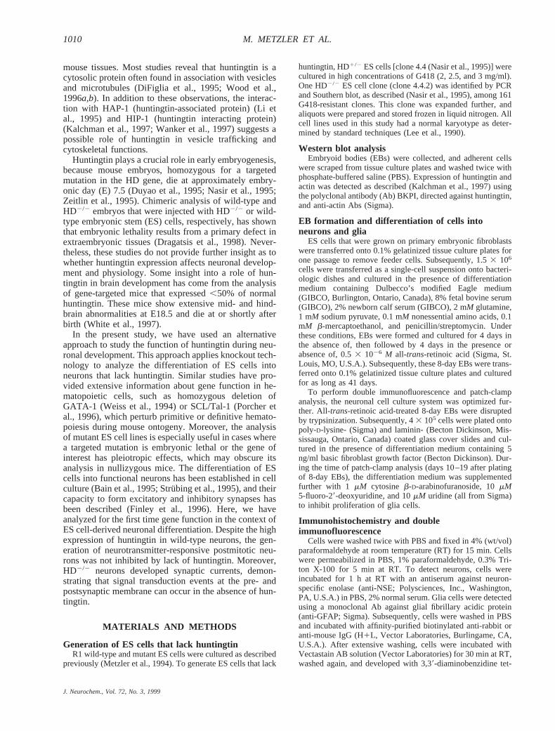
Huntingtin expression was analyzed in undifferenti-ated ES cells and 8-day EBs (Fig. 2A) and again after 6,17, and 41 days of EB differentiation on tissue cultureplates (Fig. 2B). Throughout differentiation, huntingtin isexpressed as a 340-kDa protein in wild-type cells. Asexpected, huntingtin expression was reduced by;50%in HD1/2 cells, and no huntingtin protein was detected inHD2/2 cells by 7.5% or 12.5% sodium dodecyl sulfate–polyacrylamide gel electrophoresis, followed by westernblot analysis (Fig. 2 and data not shown). This providesfurther support that no truncated products are detected inthis system and that the mutation in HD2/2 cells func-tions as null allele. The absence of any cross-reactivity in
FIG. 1. Southern blot analysis of ES cell lines heterozygous(1/2) or homozygous (2/2) for a targeted mutation in the HDgene. DNA was isolated from ES cell clone 35.8 (lanes 1 and 5),clone 4.4 (lanes 2 and 6), clone 4.4.4 (lanes 3 and 7), and clone4.4.2 (lanes 4 and 8) and digested with the restriction enzymesHindIII (lanes 1–4) or EcoRI (lanes 5–8). Southern blots werehybridized with a genomic XbaI-HindIII fragment isolated fromintron 5 of the mouse HD gene. In HindIII digests, two fragmentswere detected that correspond to the wild-type (2.3 kb) andmutant (3.3 kb) allele. In EcoRI digests, the wild-type and mutantallele correspond to a fragment of 5.4 and 6.4 kb in size, respec-tively. In clone 4.4.2, only the mutant allele was detected (lanes4 and 8).
J. Neurochem., Vol. 72, No. 3, 1999
1011DEVELOPMENT OF NEURONS THAT LACK HUNTINGTIN
HD2/2 cell lysates clearly shows that the Ab BKPIspecifically detects huntingtin. Furthermore, it suggeststhat the smaller protein fragments of 180, 160, and 90kDa, which are present in wild-type and HD1/2 cellextracts, are huntingtin-derived proteolytic fragments.These fragments were detected at different intensitiesdepending on the cell culture type analyzed and weremost pronounced in 8-day EBs (Fig. 2A).
ES cells can differentiate into neurons and glia inthe absence of huntingtin
Huntingtin is already expressed during neurogenesisin developing mouse embryos (Bhide et al., 1996).Therefore, we determined whether the absence of hun-tingtin influences the generation of neurons and gliaduring ES cell differentiation by comparing wild-type,HD1/2, and HD2/2 ES cell cultures.
Neurons were detected in HD2/2 cell cultures and inthe parental controls by immunohistochemical stainingwith anti-NSE Abs 5 and 10 days after transfer of 8-day
EBs onto tissue culture plates (Fig. 3). No obvious dif-ferences were observed in morphology of uni-, bi-, andmultipolar neurons between the different genotypes. Thenumber of neurons that developed in each culture couldbe increased dramatically in the presence of 0.53 1026
M all-trans-retinoic acid as reported earlier (Stru¨bing etal., 1995; Bain et al., 1996). At day 5 after plating,;20%of cells were neurons. Similarly, the development of gliawas observed in HD2/2 cell cultures and the parentalcontrols using anti-GFAP Abs (Fig. 3). Approximately5% of cells were glia at day 5 after plating.
Our results indicate that early neurogenesis occurs inthe absence of huntingtin and that huntingtin expressionis not crucial for the development of neurons and gliafrom ES cells. Moreover, HD2/2 cells respond to thepresence of all-trans-retinoic acid, indicating that reti-noic acid-responsive signal transduction events occur.
Huntingtin expression during neuronal developmentin differentiating ES cells
During the development of mouse embryos and inpostnatal life, the pattern of huntingtin expression fol-lows the gradient of neurogenesis with highest expres-sion in mature postmitotic neurons (Bhide et al., 1996).To determine directly whether huntingtin is expressed indeveloping neurons in cell culture, double-label immu-nofluorescence studies have been performed using Absdirected against NSE and huntingtin (Fig. 4). The ex-pression of huntingtin was first detected in neurons de-rived from wild-type cells at day 5 after plating. At latertime points, an overall increase in the expression ofhuntingtin was observed in postmitotic neurons wherehuntingtin localized to cell bodies, dendrites, and axonterminals (Fig. 4A and C and data not shown). At day 10after plating, expression of huntingtin was observed in allneurons analyzed and the level of expression of hunting-tin was significantly higher than that in nonneuronalcells. In contrast, no huntingtin was observed in neuronsderived from HD2/2 ES cells (Fig. 4B and D).
This double-label immunofluorescence analysis ofhuntingtin expression provides conclusive evidence thathuntingtin is not crucial for lineage commitment of pro-genitor cells that differentiate into neurons. Moreover,the absence of huntingtin does not seem to down-regu-late neurogenesis, because the density of neurons thatdeveloped in HD2/2 cell cultures was similar whencompared with that in control cell lines.
Neurons express functional voltage- and ligand-gated ion channels in the absence of huntingtin
The development of voltage- and neurotransmitter-gated currents and the presence of functional synapses indifferentiating neurons derived from ES cells have beenreported previously (Stru¨bing et al., 1995; Finley et al.,1996). These electrophysiological studies allow the as-sessment of functional neuronal properties, such as ex-citability and synaptic transmission. Therefore, to deter-mine whether huntingtin modulates these properties,we analyzed voltage-dependent and neurotransmitter-
FIG. 2. Western blot analysis of huntingtin expression during EScell differentiation in wild-type (1/1), heterozygous (1/2), andhomozygous (2/2) ES cells. A: Huntingtin expression was ana-lyzed in undifferentiated ES cells (lanes 1–3) and 8-day EBs(lanes 4–6). B: Huntingtin expression was analyzed in 8-day EBsafter 6 (lanes 1 and 2), 17 (lanes 3 and 4), and 41 (lanes 5 and 6)days of differentiation on tissue culture plates. Huntingtin ex-pression was visualized using the Ab BKPI, which immunoreactswith the N-terminal portion of huntingtin. The expression of actinwas determined by probing each blot with anti-actin Abs toconfirm equal loading of protein in lysates derived from wild-type, heterozygous, and homozygous cells. The smaller proteinfragments seen in A and B are most likely huntingtin-derivedproteolytic fragments.
J. Neurochem., Vol. 72, No. 3, 1999
1012 M. METZLER ET AL.

evoked currents in wild-type and HD2/2 neurons bypatch-clamp recording in the whole-cell configuration.The results are shown in Figs. 5–8 and represent record-ings made in parallel from a single batch each of wild-type and HD2/2 ES cells that were differentiated inparallel to form neurons. These batches of wild-type andHD2/2 cells were well matched with respect to neuronal
density, size, and morphology. These results are repre-sentative of recordings made from an additional fivebatches of wild-type and HD2/2 cells that were differ-entiated in parallel to form neurons.
To ensure that recordings were made from neurons,we used two main criteria. First, we chose cells forrecording that were morphologically similar to those
FIG. 3. Neurons and glia develop in the absence ofhuntingtin. Neurons (A–D) and glia (E and F) weredetected by immunohistochemistry 10 days aftertransfer of 8-day EBs onto tissue culture plates.Wild-type cells (1/1) are shown in the left panels,and cells homozygous for targeted disruption inthe HD gene (2/2) are shown in the right panels.Neurons and glia were detected following incuba-tion with anti-NSE and anti-GFAP Abs, respec-tively. Scale bars 5 10 mm.
FIG. 4. Huntingtin is expressed in differentiating neurons in culture. Double-label immunofluorescence studies were performed 10 days after plating.Neurons were identified with anti-NSE Abs followed by incubation withfluorescein isothiocyanate-conjugated secondary Abs (A and B). Huntingtinexpression was visualized using monoclonal Ab 2166 followed by incuba-tion with Texas red-conjugated secondary Abs (C and D). Wild-type cells(1/1) are shown in the left panels, and homozygous cells (2/2) are shownin the right panels. Differentiated neurons are indicated by arrows. The lowlevel of fluorescence seen in HD2/2 cells with monoclonal Ab 2166 isbackground staining; similar levels of fluorescence were observed in theabsence of monoclonal Ab 2166.
J. Neurochem., Vol. 72, No. 3, 1999
1013DEVELOPMENT OF NEURONS THAT LACK HUNTINGTIN

staining positively for NSE. These cells had two or moreprocesses and formed networks with processes project-ing from other cells; as well, cell bodies had a roundedcontour under Hoffman optics, whereas all other cellsappeared flat. Second, we tested for the presence ofvoltage-gated Na1 current (INa; see Fig. 5A1). Of thosecells that morphologically resembled NSE-positive cells,95% had large amplitudeINa and the large majority alsoexhibited spontaneous action potentials, features uniqueto neurons. As well, inhibitors of glial cell proliferationwere added to all cultures used for patch-clamp analysis.A detailed analysis of the development ofINa betweendays 12 and 19 after plating of wild-type and HD2/2
cells revealed that current densities developed similarlyover time and were strongly inhibited by administrationof 200 nM tetrodotoxin (TTX; Fig. 5A). No significantdifferences were observed between wild-type andHD2/2 neurons, and the recorded Na1 current densitieswere similar in amplitude when compared with previousresults from ES cell-derived neurons (Stru¨bing et al.,1995).
In addition to voltage-gated current, we analyzed re-sponses to the inhibitory and excitatory neurotransmit-ters GABA and glutamate, respectively. In these exper-iments, current was recorded under voltage clamp at aholding potential of260 mV. Current responses to ap-plication of 1 mM GABA were similar in kinetics andamplitude for HD2/2 and wild-type neurons (Fig. 5B).GABA-evoked currents could be specifically antago-nized by 500mM picrotoxin (PTX), a noncompetitive
antagonist of GABAA receptors (Tanelian et al., 1993).As well, current densities recorded in our study werewithin the range previously reported for GABA-gatedreceptors (Stru¨bing et al., 1995).
The analysis of glutamate receptor channels was ofparticular importance, because it has been suggested thatthese receptors play a role in neurodegeneration in HD(Choi, 1988; DiFiglia, 1990; Rubinsztein et al., 1997).Glutamate receptor channels are found throughout themammalian brain and constitute the major excitatoryneurotransmitter system (Hollmann and Heinemann,1994). Three subclasses of ionotropic glutamate recep-tors have been distinguished by their pharmacologicaland electrophysiological properties, and these are namedafter their preferred agonists: NMDA,a-amino-3-hy-droxy-5-methyl-4-isoxazolepropionic acid (AMPA), andkainate (Monaghan et al., 1989).
Our results showed that in both wild-type and HD2/2
neurons, rapid application of 1 mM kainate evoked ap-parently nondesensitizing currents that were completelyantagonized by 40mM 6-nitro-7-cyanoquinoxaline-2,3-
FIG. 6. Expression of AMPA- and NMDA-type glutamate recep-tors in neurons that lack huntingtin. A1 and B1: Representativecurrent traces from HD2/2 neurons are shown. A2 and B2:Current densities are plotted for wild-type (■) and HD2/2 (o)neurons as described in Fig. 5. A 1 mM kainate-activated, non-desensitizing current was reversibly blocked by 40 mM CNQX(A1). Columns represent data for peak kainate-evoked currentsrecorded from n 5 12–17 different neurons (A2). The current–voltage curve was generated by recording current response to 1mM kainate after stepping voltage from 260 mV to test poten-tials ranging from 2100 to 1100 mV in 20-mV increments (A3).Points represent means 6 SEM from three different HD2/2 neu-rons. A 100 mM NMDA-evoked current was reversibly blockedby 100 mM APV (B1). Columns represent data for peak NMDA-evoked currents recorded from n 5 9–16 different neurons (B2).*Significant difference (p , 0.05) between wild-type and HD2/2
current densities by one-way ANOVA.
FIG. 5. Expression of functional voltage-gated and GABA-oper-ated ion channels in neurons that lack huntingtin. A1 and B1:Representative current traces from HD2/2 neurons are shown.A2 and B2: Current densities (pA/pF) are plotted against the timein culture following plating. Columns represent means 6 SEM.■, wild-type; o, HD2/2 neurons. Voltage-gated Na1 currents(INa) were activated by command voltage pulses from a holdingpotential of 290 mV to a test potential of 220 mV and blockedby administration of 200 nM TTX (A1). INa was recorded from n5 12–18 each of wild-type and HD2/2 neurons per day (A2).GABA-evoked currents were reversibly blocked by 500 mM PTX;[GABA] 5 1 mM (B1). Each column represents data for peakGABA-evoked currents recorded from n 5 14–18 different neu-rons (B2).
J. Neurochem., Vol. 72, No. 3, 1999
1014 M. METZLER ET AL.
dione (CNQX; Fig. 6A1), features characteristic ofAMPA receptors (Hollmann and Heinemann, 1994).Kainate-evoked current densities were not significantlydifferent for the two genotypes (Fig. 6A2). As well,kainate-evoked currents recorded from both wild-typeand HD2/2 neurons exhibited a linear current–voltagerelationship (Fig. 6A3; n5 3 for each genotype), aswould be expected for AMPA receptors containing aGluR2 subunit (Hollmann and Heinemann, 1994). Incontrast, NMDA-evoked currents were characterized byfast onset and slow desensitization, as well as a revers-ible, voltage-independent block by 100mM aminophos-phonovalerate (APV; Fig. 6B1). Between days 11 and 14after plating, no significant differences were observed inNMDA-evoked current density between the two geno-types (Fig. 6B2). However, NMDA-evoked current den-
sities recorded on day 17 were significantly larger forHD2/2 ES cell-derived neurons (12.596 1.61 pA/pF, n5 11) than for wild-type ES cell-derived neurons (8.246 0.72 pA/pF, n5 12; p , 0.05 byone-way ANOVA).Current densities for both NMDA- and kainate-evokedresponses were within the range of those previouslyreported (Stru¨bing et al., 1995).
Postsynaptic currents develop in the absence ofhuntingtin
As further evidence for intact neuronal function, spon-taneous postsynaptic currents were observed duringwhole-cell recordings from both wild-type and HD2/2
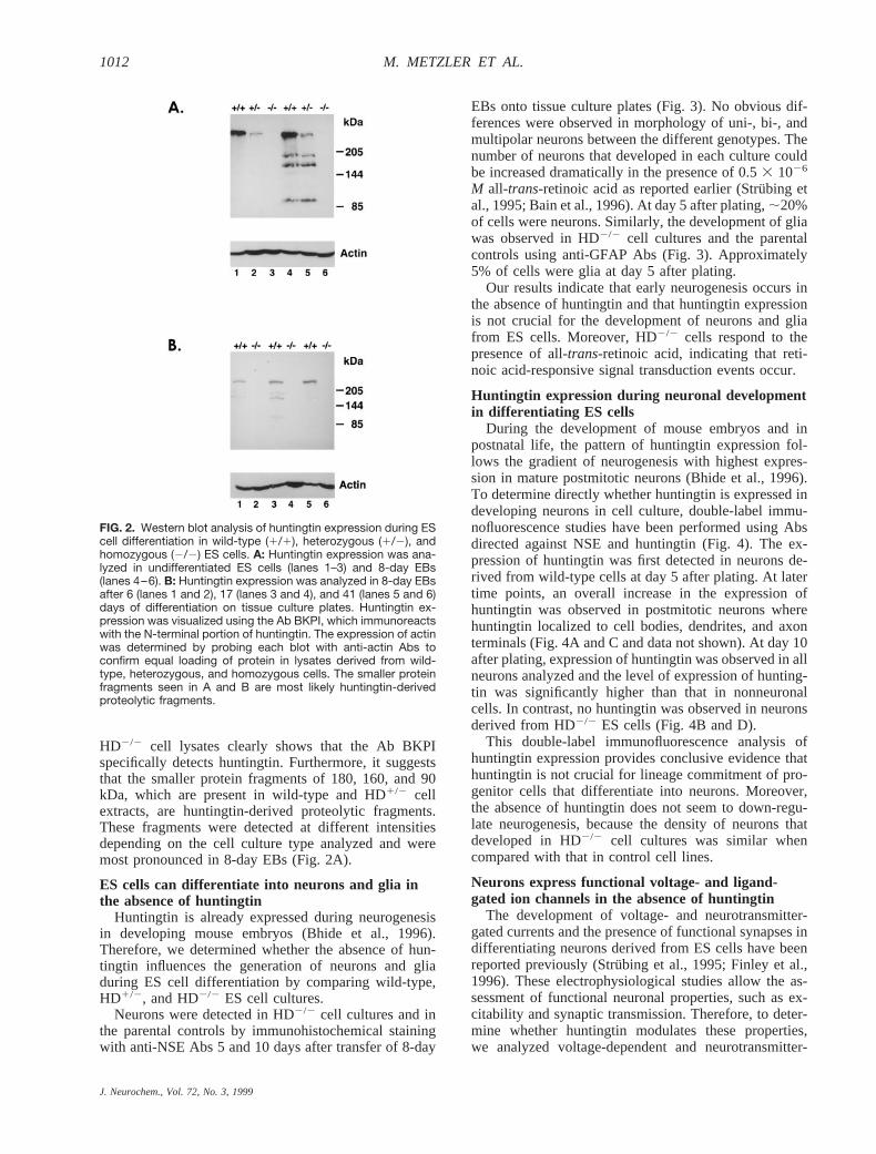
neurons at a holding potential of260 mV (Fig. 7).Spontaneous postsynaptic currents were recorded from24% of neurons differentiated from either wild-type orHD2/2 ES cells (31 of 130 for wild-type and 30 of 124for HD2/2 neurons). Prolonged recordings ($300 s) ofspontaneous postsynaptic currents were made from atotal of 10 neurons (four wild-type and six HD2/2).Representative traces recorded from the two differentgenotypes are shown in Fig. 7A and B. Based on the timecourse of current decay, the spontaneous events could bedivided into at least two groups (Fig. 7E–J). “Fast”spontaneous synaptic currents, defined as those exhibit-ing decay time constants of,8 ms (see Fig. 7E and F),were generally small in amplitude, and the mean decaytime constants for recordings from wild-type and HD2/2
neurons were 2.96 0.2 ms (n5 4 cells) and 3.56 0.15ms (n 5 6 cells), respectively. These rapidly decayingcurrents were reversibly blocked by CNQX (Fig. 7C andD) and therefore likely represent AMPA receptor-medi-ated responses as previously reported (Stru¨bing et al.,1995; Finley et al., 1996). Such currents represented.80% of the spontaneous events recorded from one offour wild-type and one of six HD2/2 neurons. Includedwithin this group were currents that exhibited a decaytime course best fit by two exponentials, with the fastertime constant in the range of 1.8–7.4 ms and the slowertime constant in the range of 36–276 ms (Fig. 7G and H).The majority of these currents showed a slow componentwith a time constant markedly lower than that of theinhibitory currents (see below). These ‘‘compound’’spontaneous postsynaptic currents suggest coactivationof AMPA and NMDA receptors (Stru¨bing et al., 1995),
FIG. 7. Spontaneous postsynaptic currents recorded from neu-rons derived from wild-type (A, C, E, G, and I) and HD2/2 (B, D,F, H, and J) ES cells. Representative current traces were re-corded under whole-cell voltage-clamp (VH 5 260 mV) (A andB). Representative current traces were recorded from the samecells shown in A and B in the presence of 50 mM CNQX (C andD). Note the absence of events with rapidly decaying currents(E–J). Sample spontaneous synaptic currents from A and B areshown at higher gain to illustrate the following: monoexponentialdecay with fast time constant (E and F); biexponential decay,with fast time constant similar to currents shown in E and F anda second component with time constant ;100 times slower (Gand H); monoexponential decay with relatively slow time con-stant (I and J). Currents were recorded in the absence of TTXbetween days 11 and 19 following plating of the 8-day wild-typeor HD2/2 EBs.
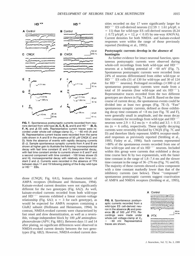
FIG. 8. Spontaneous postsyn-aptic currents recorded from awild-type ES cell-derived neu-ron in the absence (A) and pres-ence (B) of 500 mM PTX. Re-cordings were made underwhole-cell voltage-clamp at VH5 260 mV. Representativetraces are shown.
J. Neurochem., Vol. 72, No. 3, 1999
1015DEVELOPMENT OF NEURONS THAT LACK HUNTINGTIN

but we did not test for block of the slow component byAPV. Finally, in the majority of neurons (three of fourwild-type and five of six HD2/2), spontaneous synapticcurrents showed predominantly ‘‘slow’’ decay kineticsfit by a single exponential with a time constant in therange of 12–60 ms (e.g., Fig. 7I and J). These currentswere largely eliminated by treatment with 500mM PTX(Fig. 8) and were resistant to treatment with CNQX (Fig.7C and D), consistent with inhibitory postsynaptic cur-rents mediated by either GABAA or glycine receptors(Tanelian et al., 1993; Tapia and Aguayo, 1998). Takentogether, these results indicate that both wild-type andHD2/2 neurons form functional excitatory and inhibitorysynapses.
DISCUSSION
Here we report findings from studies of HD2/2 EScells that directly address the role of huntingtin in neu-ronal differentiation. HD2/2 ES cells contain a targetedmutation in both alleles of the HD gene and exhibit thecomplete absence of huntingtin expression. During dif-ferentiation of wild-type ES cells, huntingtin is expressedin both undifferentiated and differentiated cells, with ahigh abundance in neurons. It is surprising, however, thatlineage commitment and differentiation of precursorcells into neurons and glia proceeded in HD2/2 cells inthe absence of huntingtin. Moreover, fundamental neu-rophysiological properties were normal in HD2/2 neu-rons, as demonstrated by the expression of functionalvoltage-gated and receptor-operated ion channels and thedevelopment of spontaneous postsynaptic currents.
Expression of huntingtin in the wild-type culture sys-tem was observed at all stages during ES cell differen-tiation and throughout neuronal development. Highestexpression levels were found in neuronal cells, whichincreased in parallel with neuronal maturation and anincrease in excitability of ES cell-derived neurons(Strubing et al., 1995). In mature postmitotic neurons,huntingtin localized to cell bodies, dendrites, and someaxon terminals. This pattern of expression demonstratescorrect developmental regulation of huntingtin expres-sion in ES cell-derived neurons and makes this cellculture system very attractive for gene function analysisof huntingtin.
In adult mouse and human brain, huntingtin immuno-reactivity varies among different groups of neurons(Bhide et al., 1996; Ferrante et al., 1997). Such variationin the level of huntingtin expression has not been ob-served in neurons derived from wild-type ES cells.Therefore, it is rather unlikely that a group of neuronshas been selected in HD2/2 cell cultures that normallywould express no or low levels of huntingtin. Moreover,neurons developed at similar density in HD2/2 cell cul-tures when compared with control cell cultures. Thisfinding suggests that no selection against a particulargroup of neurons occurred in the absence of huntingtin.
In previous studies, huntingtin has been found to in-teract with HIP1, a homologue of the yeast protein Sla2p,
which is essential for the assembly and function of thecortical cytoskeleton (Kalchman et al., 1997; Wanker etal., 1997). In light of this fact, it is of interest thatneuronal processes and axons were formed apparentlynormally, indicating that the interaction of huntingtinwith HIP1 is not required for these morphogenic pro-cesses.
To assess later stages of neuronal development, aswell as neuronal function, patch-clamp analysis was per-formed in wild-type and HD2/2 neurons. The majorfinding of these studies is that neurons derived fromHD2/2 ES cells showed features characteristic of post-mitotic neurons with neurophysiological properties thatwere nearly indistinguishable from those of wild-typeneurons. Both HD2/2 and wild-type neurons developedvoltage-dependent Na1 channels and responded simi-larly to stimulation with the excitatory agonists NMDAand kainate and the inhibitory agonist GABA. The in-duced currents revealed the expected pharmacologicalprofile of each channel type, and current densities re-corded in our study were typical for ES cell-derivedneurons (Stru¨bing et al., 1995). Moreover, in neuronsfrom both genotypes, application of 1 mM kainateevoked apparently nondesensitizing currents with linearcurrent–voltage curves, characteristic of GluR2-contain-ing AMPA receptors (Hollmann and Heinemann, 1994).These results are consistent with previous studies in EScell-derived neurons (Finley et al., 1996) and also withAMPA receptor-mediated currents recorded from themajority of mammalian neurons (for review, see Holl-mann and Heinemann, 1994). The only difference foundbetween these two cell lines was a small, but statisticallysignificant, increase in mean amplitude of NMDA-evoked currents at day 17 in HD2/2 compared withwild-type neurons. The physiological significance of thisdifference is unclear, as it occurred at only one timepoint, but the result bears further investigation in futurestudies. However, our results do not rule out the possi-bility that huntingtin modulates the time course of ion-channel expression, because patch-clamp recordingswere performed 10–19 days after plating, when near-saturating levels of ion-channel expression have beenestablished (Stru¨bing et al., 1995).
In addition to recordings of current responses to theexogenous application of GABA, kainate, and NMDA, itwas possible to record spontaneous postsynaptic currentsfrom 24% of either wild-type or HD2/2 ES cells. Thisactivity was characterized in part by fast kinetics andCNQX inhibition, features of AMPA receptor-mediatedcurrents (Stru¨bing et al., 1995; Finley et al., 1996). Themajority of postsynaptic activity resulted from inhibitorypostsynaptic currents mediated by either GABAA or gly-cine receptors, which is in good agreement with a pre-vious study by Stru¨bing et al. (1995) showing a prepon-derance of inhibitory postsynaptic activity in ES cell-derived neurons.
Surface expression and synaptic targeting of neuro-transmitter receptors are required to obtain measurablepostsynaptic activity. Synaptic targeting of glutamate
J. Neurochem., Vol. 72, No. 3, 1999
1016 M. METZLER ET AL.

receptors is thought to involve direct interaction of thesereceptors with cytoskeletal proteins (for review, seeEhlers et al., 1996). Obviously, these cellular and neu-ron-specific processes occur in the absence of huntingtinand seem unaffected by the lack of interaction betweenhuntingtin and proteins involved in cytoskeletal and ves-icle transport functions (Li et al., 1995, 1998; Kalchmanet al., 1997; Wanker et al., 1997). As well, the absence ofthese interactions appears to have no impact upon syn-aptic vesicle delivery to and release from axon terminals,because spontaneous vesicle release at excitatory andinhibitory synapses was similar in neurons of both ge-notypes. However, we cannot conclude that all vesiclefunction and transport processes were normal in HD2/2
neurons, because the results of our electrophysiologicalanalysis reflect only a relatively crude assessment ofsynaptic function. For example, we did not assess theactivity of G protein-coupled neurotransmitter receptorsor the modulation of ionotropic receptor function. Asboth may be highly dependent on vesicle turnover and/orinteractions with cytoskeletal proteins (Bohm et al.,1997; Nusser et al., 1998; O’Brien et al., 1998; Sprengelet al., 1998), we cannot rule out a role for huntingtin inthese processes.
In summary, we have provided evidence that differ-entiation of postmitotic neurons, expression of functionalvoltage-gated and receptor-operated ion channels, andsynapse formation can occur in neurons that lack hun-tingtin. Moreover, this study provides direct evidencethat loss of function of huntingtin is unlikely the crucialmechanism resulting in neurodegeneration in HD. TheES cell-derived culture system has been an essential toolto decipher various aspects of neuronal development inthe absence of huntingtin. Nevertheless, this study doesnot rule out the possibility that in a more complexenvironment, like the developing mouse embryo, lack ofhuntingtin expression affects other cellular processes thathave not been assessed in our cell culture system. Forexample, reduced expression of huntingtin leads to manyorganizational abnormalities in the brains of E18.5 gene-targeted mouse embryos. The cause of these abnormali-ties has not been defined but may involve aberrant cellproliferation or effects on developmental programmedcell death, because ectopic cell masses of differentiatedpostmitotic neurons have been observed (White et al.,1997). Further analyses are now required to determinemore quantitative aspects of neuronal development andhow huntingtin may affect proliferation of neuronal pro-genitor cells and cell viability through different develop-mental stages.
Acknowledgment: The authors thank S. Zeitlin for provid-ing the 2B26 ES cell line and F. R. Jirik for the R1 ES cell line.We are grateful to G. J. Shaw for karyotype analysis, J. Holmesfor his support in preparation of illustrations, and T. H. Murphyfor useful discussions and advice. This work is supported by theCanadian Networks of Centres of Excellence (NCE-Genetics)to MRC (Canada) operating grants to M.R.H., funding from theSwiss National Research Foundation and the Huntington’s Dis-
ease Society of America (HDSA) to M.M., and an MRC(Canada) operating grant to L.A.R. N.C. is supported by theHDSA; L.A.R. is an MRC (Canada) Scholar. Further supportwas provided by the National Cancer Institute of Canada withfunds from the Canadian Cancer Society and Terry Fox Run toR.K.H. M.R.H. is an established investigator of the B.C. Chil-dren’s Hospital.
REFERENCES
Andrew S. E., Goldberg Y. P., and Hayden M. R. (1997) Rethinkinggenotype and phenotype correlations in polyglutamine expansiondisorders.Hum. Mol. Genet.6, 2005–2010.
Bain G., Kitchens D., Yao M., Huettner J. E., and Gottlieb D. I. (1995)Embryonic stem cells express neuronal properties in vitro.Dev.Biol. 168,342–357.
Bain G., Ray W. J., Yao M., and Gottlieb D. I. (1996) Retinoic acidpromotes neural and represses mesodermal gene expression inmouse embryonic stem cells in culture.Biochem. Biophys. Res.Commun.223,691–694.
Bhide P. G., Day M., Sapp E., Schwarz C., Sheth A., Kim J., YoungA. B., Penney J., Golden J., Aronin N., and DiFiglia M. (1996)Expression of normal and mutant huntingtin in the developingbrain.J. Neurosci.16, 5523–5535.
Bohm S. K., Grady E. F., and Bunnett N. W. (1997) Regulatorymechanisms that modulate signalling by G-protein-coupled recep-tors.Biochem. J.332,1–18.
Brandt J., Bylsma F. W., Gross R., Stine O. C., Ranen N. G., and RossC. A. (1996) Trinucleotide repeat length and clinical progressionin Huntington’s disease.Neurology46, 527–531.
Chen N., Moshaver A., and Raymond L. A. (1997) Differential sensi-tivity of recombinantN-methyl-D-aspartate receptor subtypes tozinc inhibition.Mol. Pharmacol.51, 1015–1023.
Choi D. W. (1988) Glutamate neurotoxicity and diseases of the nervoussystem.Neuron1, 623–634.
Davies S. W., Turmaine M., Cozens B. A., DiFiglia M., Sharp A. H.,Ross C. A., Scherzinger E., Wanker E. E., Mangiarini L., andBates G. P. (1997) Formation of neuronal intranuclear inclusionsunderlies the neurological dysfunction in mice transgenic for theHD mutation.Cell 90, 537–548.
DiFiglia M. (1990) Excitotoxic injury of the neostriatum: a model forHuntington’s disease.Trends Neurosci.13, 286–289.
DiFiglia M., Sapp E., Chase K., Schwarz C., Meloni A., Young C.,Martin E., Vonsattel J. P., Carraway R., Reeves S. A., BoyceF. M., and Aronin N. (1995) Huntingtin is a cytoplasmic proteinassociated with vesicles in human and rat brain neurons.Neuron14, 1075–1081.
DiFiglia M., Sapp E., Chase K. O., Davies S. W., Bates G. P., VonsattelJ. P., and Aronin N. (1997) Aggregation of huntingtin in neuronalintranuclear inclusions and dystrophic neurites in brain.Science277,1990–1993.
Dragatsis I., Efstratiadis A., and Zeitlin S. (1998) Mouse mutantembryos lacking huntingtin are rescued from lethality by wild-type extraembryonic tissues.Development125,1529–1539.
Duyao M. P., Auerbach A. B., Ryan A., Persichetti F., Barnes G. T.,McNeil S. M., Ge P., Vonsattel J.-P., Gusella J. F., Joyner A. L.,and MacDonald M. E. (1995) Inactivation of the mouse Hunting-ton’s disease gene homologHdh. Science269,407–410.
Ehlers M. D., Mammen A. L., Lau L.-F., and Huganir R. L. (1996)Synaptic targeting of glutamate receptors.Curr. Opin. Cell Biol.8,484–489.
Ferrante R. J., Gutekunst C.-A., Persichetti F., McNeil S. M., KowallN. W., Gusella J. F., MacDonald M. E., Beal M. F., and HerschS. M. (1997) Heterogeneous topographic and cellular distributionof huntingtin expression in the normal human neostriatum.J. Neu-rosci. 17, 3052–3063.
Finley M. F. A., Kulbarni N., and Huettner E. (1996) Synapse forma-tion and establishment of neuronal polarity by P19 embryoniccarcinoma cells and embryonic stem cells.J. Neurosci.16,1056–1065.
J. Neurochem., Vol. 72, No. 3, 1999
1017DEVELOPMENT OF NEURONS THAT LACK HUNTINGTIN
Goldberg Y. P., Nicholson D. W., Rasper D. M., Kalchman M. A.,Koide H. B., Graham R. K., Bromm M., Kazemi-Esfarjani P.,Thornberry N. A., Vaillancourt J. P., and Hayden M. R. (1996)Cleavage of huntingtin by apopain, a proapoptotic cysteine pro-tease, is modulated by the polyglutamine tract.Nat. Genet.13,442–449.
Hackam A. S., Singaraja R., Wellington C. L., Metzler M., McCutch-eon K., Zhang T., Kalchman M., and Hayden M. R. (1998) Theinfluence of huntingtin protein size on nuclear localization andcellular toxicity.J. Cell Biol. 141,1097–1105.
Hollmann M. and Heinemann S. (1994) Cloned glutamate receptors.Annu. Rev. Neurosci.17, 31–108.
Huntington’s Disease Collaborative Research Group (1993) A novelgene containing a trinucleotide repeat that is expanded and unsta-ble on Huntington’s disease chromosomes.Cell 72, 971–983.
Kalchman M. A., Koide H. B., McCutcheon K., Graham R. K., NicholK., Nishiyama K., Lynn F. C., Kazemi-Esfarjani P., WellingtonC. L., Metzler M., Goldberg Y. P., Kanazawa I., Gietz R. D., andHayden M. R. (1997) HIP1, a human homolog ofS. cerevisiaeSla2p, interacts with membrane-associated huntingtin in the brain.Nat. Genet.16, 44–53.
Lee J. J., Warburton D., and Robertson E. J. (1990) Cytogeneticmethods for the mouse: preparation of chromosomes, karyotyping,in situ hybridization.Anal. Biochem.189,1–17.
Li S.-H., Gutekunst C.-A., Hersch S. M., and Li X.-J. (1998) Interac-tion of huntingtin-associated protein with dynactin P150Glued.J. Neurosci.18, 1261–1269.
Li X. J., Li S. H., Sharp A. H., Nucifora F. C. Jr., Schilling G., LanahanA., Worley P., Snyder S. H., and Ross C. A. (1995) A huntingtin-associated protein enriched in brain with implications for pathol-ogy. Nature378,398–402.
Martindale D., Hackam A. S., Wieczorek A., Ellerby L., WellingtonC. L., McCutcheon K., Singaraja R., Kazemi-Esfarjani P., DevonR., Bredesen D. E., Tufaro F., and Hayden M. R. (1998) Lengthof huntingtin and its polyglutamine tract influences localizationand frequency of intracellular aggregates.Nat. Genet.18, 150–154.
Metzler M., Gertz A., Sarkar M., Schachter H., Schrader J. W., andMarth J. D. (1994) Complex asparagine-linked oligosaccharidesare required for morphogenic events during post-implantationdevelopment.EMBO J.13, 2056–2065.
Monaghan D. T., Bridges R. J., and Cotman C. W. (1989) The exci-tatory amino acid receptors: their classes, pharmacology, anddistinct properties in the function of the nervous system.Annu.Rev. Pharmacol. Toxicol.29, 365–402.
Mortensen R. M., Conner D. A., Chao S., Geisterfer-LowranceA. A. T., and Seidman J. G. (1992) Production of homozygousmutant ES cells with a single targeting construct.Mol. Cell. Biol.12, 2391–2395.
Nance M. A. (1997) Clinical aspects of CAG repeat diseases.BrainPathol.7, 881–900.
Nasir J., Floresco S. B., O’Kusky J. R., Diewert V. M., Richman J. M.,Zeisler J., Borowski A., Marth J. D., Phillips A. G., and HaydenM. R. (1995) Targeted disruption of the Huntington’s disease generesults in embryonic lethality and behavioral and morphologicalchanges in heterozygotes.Cell 81, 811–823.
Nasir J., Goldberg Y. P., and Hayden M. R. (1996) Huntington disease:new insights into the relationship between CAG expansion anddisease.Hum. Mol. Genet.5, 1431–1435.
Nusser Z., Hajos N., Somogyi P., and Mody I. (1998) Increased numberof synaptic GABAA receptors underlies potentiation at hippocam-pal inhibitory synapses.Nature395,172–177.
O’Brien R. J., Lau L.-F., and Huganir R. L. (1998) Molecular mech-anisms of glutamate receptor clustering at excitatory synapses.Curr. Opin. Neurobiol.8, 364–369.
Paulson H. L., Perez M. K., Trottier Y., Trojanowski J. Q., SubramonyS. H., Das S. S., Vig P., Mandel J.-L., Fischbeck K. H., and
Pittman R. N. (1997) Intranuclear inclusions of expanded poly-glutamine protein in spinocerebellar ataxia type 3.Neuron 19,333–344.
Porcher C., Swat W., Rockwell K., Fujiwara Y., Alt F. W., and OrkinS. H. (1996) The T cell leukemia oncoprotein SCL/tal-1 is essen-tial for development of all hematopoietic lineages.Cell 86,47–57.
Ross C. A. (1995) When more is less: pathogenesis of glutamine repeatneurodegenerative diseases.Neuron15, 493–496.
Ross C. A., Becher M. W., Colomer V., Engelender S., Wood J. D., andSharp A. H. (1997) Huntington’s disease and dentatorubral-pal-lidoluysian atrophy: proteins, pathogenesis and pathology.BrainPathol.7, 1003–1016.
Rubinsztein D. C., Leggo J., Chiano M., Dodge A., Norbury G., RosserE., and Craufurd D. (1997) Genotypes at the GluR6 kainatereceptor locus are associated with variation in the age of onset ofHuntington disease.Proc. Natl. Acad. Sci. USA94, 3872–3876.
Skinner P. J., Koshy B. T., Cummings C. J., Klement I. A., Helin K.,Servadio A., Zoghbi H. Y., and Orr H. T. (1997) Ataxin-1 with anexpanded glutamine tract alters nuclear matrix-associated struc-tures.Nature389,971–974.
Sprengel R., Suchanek B., Amico C., Brusa R., Burnashev N., RozovA., Hvalby O., Jensen V., Paulsen O., Andersen P., Kim J. J.,Thompson R. F., Sun W., Webster L. C., Grant S. G. N., Eilers J.,Konnerth A., Li J., McNamara J. O., and Seeburg P. H. (1998)Importance of the intracellular domain of NR2 subunits forNMDA receptor function in vivo.Cell 92, 279–289.
Strubing C., Ahnert-Hilger G., Shan J., Wiedenmann B., Hescheler J.,and Wobus A. M. (1995) Differentiation of pluripotent embryonicstem cells into the neuronal lineage in vitro gives rise to matureinhibitory and excitatory neurons.Mech. Dev.53, 275–287.
Tanelian D. L., Kosek P., Mody I., and MacIver M. B. (1993) The roleof the GABAA receptor/chloride channel complex in anesthesia.Anesthesiology78, 757–776.
Tapia J. C. and Aguayo L. G. (1998) Changes in the properties ofdeveloping glycine receptors in cultured mouse spinal neurons.Synapse28, 185–194.
Wanker E. E., Rovira C., Scherzinger E., Hasenbank R., Walter S., TaitD., Colicelli J., and Lehrach H. (1997) HIP1: a huntingtin inter-acting protein isolated by the yeast two-hybrid system.Hum. Mol.Genet.6, 487–495.
Weiss M. J., Keller G., and Orkin S. H. (1994) Novel insights intoerythroid development revealed through in vitro differentiation ofGATA-1-embryonic stem cells.Genes Dev.8, 1184–1196.
Wellington C. L., Ellerby L. M., Hackam A. S., Margolis R. L., TrifiroM. A., Singaraja R., McCutcheon K., Salvesen G. S., Propp S. S.,Bromm M., Rowland K. J., Zhang T., Rasper D., Roy S., Thorn-berry N., Pinsky L., Kakizuka A., Ross C. A., Nicholson D. W.,Bredesen D. E., and Hayden M. R. (1998) Caspase cleavage ofgene products associated with triplet expansion disorders gener-ates truncated fragments containing the polyglutamine tract.J. Biol. Chem.273,9158–9167.
White J. K., Auerbach W., Duyao M. P., Vonsattel J.-P., Gusella J. F.,Joyner A. L., and MacDonald M. E. (1997) Huntingtin is requiredfor neurogenesis and is not impaired by the Huntington’s diseaseCAG expansion.Nat. Genet.17, 404–410.
Wood J. D., Harper P. S., and Jones A. L. (1996a) Characteristics ofcytosolic and membrane bound forms of huntingtin.Am. J. Hum.Genet.59, A295.
Wood J. D., MacMillan J. C., Harper P. S., Lowenstein P. R., and JonesA. L. (1996b) Partial characterisation of murine huntingtin andapparent variations in the subcellular localisation of huntingtin inhuman, mouse and rat brain.Hum. Mol. Genet.5, 481–487.
Zeitlin S., Liu J.-P., Chapman D. L., Papaioannou V. E., and Efstra-tiadis A. (1995) Increased apoptosis and early embryonic lethalityin mice nullizygous for the Huntington’s disease gene homologue.Nat. Genet.11, 155–162.
J. Neurochem., Vol. 72, No. 3, 1999
1018 M. METZLER ET AL.
Copyright © 2022 FDOKUMEN