Label-free proteomic analysis to confirm the predicted proteome of Corynebacterium...
15
RESEARCH ARTICLE Open Access Label-free proteomic analysis to confirm the predicted proteome of Corynebacterium pseudotuberculosis under nitrosative stress mediated by nitric oxide Wanderson M Silva 1,4,5 , Rodrigo D Carvalho 1 , Siomar C Soares 1 , Isabela FS Bastos 1 , Edson L Folador 1 , Gustavo HMF Souza 3 , Yves Le Loir 4,5 , Anderson Miyoshi 1 , Artur Silva 2 and Vasco Azevedo 1* Abstract Background: Corynebacterium pseudotuberculosis biovar ovis is a facultative intracellular pathogen, and the etiological agent of caseous lymphadenitis in small ruminants. During the infection process, the bacterium is subjected to several stress conditions, including nitrosative stress, which is caused by nitric oxide (NO). In silico analysis of the genome of C. pseudotuberculosis ovis 1002 predicted several genes that could influence the resistance of this pathogen to nitrosative stress. Here, we applied high-throughput proteomics using high definition mass spectrometry to characterize the functional genome of C. pseudotuberculosis ovis 1002 in the presence of NO-donor Diethylenetriamine/ nitric oxide adduct (DETA/NO), with the aim of identifying proteins involved in nitrosative stress resistance. Results: We characterized 835 proteins, representing approximately 41% of the predicted proteome of C. pseudotuberculosis ovis 1002, following exposure to nitrosative stress. In total, 102 proteins were exclusive to the proteome of DETA/NO-induced cells, and a further 58 proteins were differentially regulated between the DETA/NO and control conditions. An interactomic analysis of the differential proteome of C. pseudotuberculosis in response to nitrosative stress was also performed. Our proteomic data set suggested the activation of both a general stress response and a specific nitrosative stress response, as well as changes in proteins involved in cellular metabolism, detoxification, transcriptional regulation, and DNA synthesis and repair. Conclusions: Our proteomic analysis validated previously-determined in silico data for C. pseudotuberculosis ovis 1002. In addition, proteomic screening performed in the presence of NO enabled the identification of a set of factors that can influence the resistance and survival of C. pseudotuberculosis during exposure to nitrosative stress. Keywords: Corynebacterium pseudotuberculosis, Caseous lymphadenitis, Proteomics, Label-free proteomics, Nitrosative stress, Nitric oxide Background Corynebacterium pseudotuberculosis is a Gram-positive, facultative, intracellular pathogen belonging to the Coryne- bacterium, Mycobacterium, Nocardia, or CMN, group. This group belongs to the phylum Actinobacteria. The defining characteristics of the CMN group are a specific cell wall organization, consisting of peptidoglycan, arabinogalactan, and mycolic acids, and a high chromosomal G + C content [1]. C. pseudotuberculosis ovis is the etiological agent of the chronic infectious disease caseous lymphadenitis, which af- fects small ruminants worldwide. As a result, C. pseudotu- berculosis ovis is responsible for significant economic losses in the goat and sheep industries, mainly stemming from de- creased meat, wool, and milk production, reproductive dis- orders, and carcass contamination [1,2]. Bacterial factors that contribute to the virulence of C. pseudotuberculosis in- clude phospholipase D [3], toxic cell wall lipids [4], and the iron transporter fagABC complex [5]. * Correspondence: [email protected] 1 Depto de Biologia Geral, Instituto de Ciências Biológicas, Universidade Federal de Minas Gerais, Belo Horizonte, Brazil Full list of author information is available at the end of the article © 2014 Silva et al.; licensee BioMed Central. This is an Open Access article distributed under the terms of the Creative Commons Attribution License (http://creativecommons.org/licenses/by/4.0), which permits unrestricted use, distribution, and reproduction in any medium, provided the original work is properly credited. The Creative Commons Public Domain Dedication waiver (http://creativecommons.org/publicdomain/zero/1.0/) applies to the data made available in this article, unless otherwise stated. Silva et al. BMC Genomics 2014, 15:1065 http://www.biomedcentral.com/1471-2164/15/1065
-
Upload
independent -
Category
Documents
-
view
3 -
download
0
Transcript of Label-free proteomic analysis to confirm the predicted proteome of Corynebacterium...

Silva et al. BMC Genomics 2014, 15:1065http://www.biomedcentral.com/1471-2164/15/1065
RESEARCH ARTICLE Open Access
Label-free proteomic analysis to confirm thepredicted proteome of Corynebacteriumpseudotuberculosis under nitrosative stressmediated by nitric oxideWanderson M Silva1,4,5, Rodrigo D Carvalho1, Siomar C Soares1, Isabela FS Bastos1, Edson L Folador1,Gustavo HMF Souza3, Yves Le Loir4,5, Anderson Miyoshi1, Artur Silva2 and Vasco Azevedo1*
Abstract
Background: Corynebacterium pseudotuberculosis biovar ovis is a facultative intracellular pathogen, and theetiological agent of caseous lymphadenitis in small ruminants. During the infection process, the bacterium issubjected to several stress conditions, including nitrosative stress, which is caused by nitric oxide (NO). In silico analysis ofthe genome of C. pseudotuberculosis ovis 1002 predicted several genes that could influence the resistance of thispathogen to nitrosative stress. Here, we applied high-throughput proteomics using high definition mass spectrometryto characterize the functional genome of C. pseudotuberculosis ovis 1002 in the presence of NO-donor Diethylenetriamine/nitric oxide adduct (DETA/NO), with the aim of identifying proteins involved in nitrosative stress resistance.
Results: We characterized 835 proteins, representing approximately 41% of the predicted proteome of C.pseudotuberculosis ovis 1002, following exposure to nitrosative stress. In total, 102 proteins were exclusive to the proteomeof DETA/NO-induced cells, and a further 58 proteins were differentially regulated between the DETA/NO and controlconditions. An interactomic analysis of the differential proteome of C. pseudotuberculosis in response to nitrosative stresswas also performed. Our proteomic data set suggested the activation of both a general stress response and a specificnitrosative stress response, as well as changes in proteins involved in cellular metabolism, detoxification, transcriptionalregulation, and DNA synthesis and repair.
Conclusions: Our proteomic analysis validated previously-determined in silico data for C. pseudotuberculosis ovis 1002. Inaddition, proteomic screening performed in the presence of NO enabled the identification of a set of factors that caninfluence the resistance and survival of C. pseudotuberculosis during exposure to nitrosative stress.
Keywords: Corynebacterium pseudotuberculosis, Caseous lymphadenitis, Proteomics, Label-free proteomics, Nitrosativestress, Nitric oxide
BackgroundCorynebacterium pseudotuberculosis is a Gram-positive,facultative, intracellular pathogen belonging to the Coryne-bacterium, Mycobacterium, Nocardia, or CMN, group. Thisgroup belongs to the phylum Actinobacteria. The definingcharacteristics of the CMN group are a specific cell wallorganization, consisting of peptidoglycan, arabinogalactan,
* Correspondence: [email protected] de Biologia Geral, Instituto de Ciências Biológicas, UniversidadeFederal de Minas Gerais, Belo Horizonte, BrazilFull list of author information is available at the end of the article
© 2014 Silva et al.; licensee BioMed Central. ThCommons Attribution License (http://creativecreproduction in any medium, provided the orDedication waiver (http://creativecommons.orunless otherwise stated.
and mycolic acids, and a high chromosomal G +C content[1]. C. pseudotuberculosis ovis is the etiological agent of thechronic infectious disease caseous lymphadenitis, which af-fects small ruminants worldwide. As a result, C. pseudotu-berculosis ovis is responsible for significant economic lossesin the goat and sheep industries, mainly stemming from de-creased meat, wool, and milk production, reproductive dis-orders, and carcass contamination [1,2]. Bacterial factorsthat contribute to the virulence of C. pseudotuberculosis in-clude phospholipase D [3], toxic cell wall lipids [4], and theiron transporter fagABC complex [5].
is is an Open Access article distributed under the terms of the Creativeommons.org/licenses/by/4.0), which permits unrestricted use, distribution, andiginal work is properly credited. The Creative Commons Public Domaing/publicdomain/zero/1.0/) applies to the data made available in this article,

Silva et al. BMC Genomics 2014, 15:1065 Page 2 of 15http://www.biomedcentral.com/1471-2164/15/1065
In silico analysis of the genome of C. pseudotuberculo-sis ovis 1002 [6], as well as the pan-genome analysis of15 other strains of C. pseudotuberculosis [7], identifiedgenes involved in the response of this pathogen to differ-ent types of stress. Recently, the functional genome of C.pseudotuberculosis ovis 1002 was evaluated at the tran-scriptional level following exposure to different types ofabiotic stress, including heat, osmotic, and acid stresses[8]. This allowed the characterization of several genes in-volved in distinct biological processes that favor the sur-vival of the pathogen under the given stress condition.However, during the infection process, C. pseudotubercu-
losis encounters nitrosative stress, caused by nitric oxide(NO), in the macrophage intracellular environment. A re-active nitrogen species (RNS) found in mammalian sys-tems, NO is produced from L-arginine by NO synthases(NOS), and is present in three isoforms: endothelial NOS,neuronal NOS, involved in blood pressure control andneural signaling, and inducible NOS, associated with hostdefenses [9,10]. The NO produced during bacterial infec-tion has antimicrobial properties, killing pathogens by caus-ing damage to DNA, RNA, and proteins [11]. However,several pathogens contain pathways that allow bacterial sur-vival under nitrosative stress conditions, including NO-sensitive transcriptional regulators [12], DNA and proteinrepair systems [13], and antioxidant systems [14].Currently, little is known about the factors involved in
the resistance of C. pseudotuberculosis to nitrosative stress.Pacheco et al. [15] showed that the alternative sigma (σ)factor, σE, plays a role in the survival of C. pseudotuberculo-sis in the presence of RNS. A σE null strain showed in-creased susceptibility to nitric oxide compared with thewild-type, and, in an in vivo assay, was unable to persist inmice. However, in iNOS-deficient mice, the mutant strainmaintained its virulence [15]. In the same study, the extra-cellular proteome of C. pseudotuberculosis was analyzed inresponse to nitrosative stress, allowing the characterizationof proteins that contribute to the adaptive processes of thepathogen in this environment [15].To complement the results obtained in previous stud-
ies, and to identify factors involved in the survival of C.pseudotuberculosis under nitrosative stress conditions,we applied high-throughput proteomics using an liquidchromatograph high definition mass spectrometry (LC-HDMSE) (data-independent acquisition, in ion mobilitymode) approach to evaluate the global expression of thefunctional genome of C. pseudotuberculosis ovis 1002 atthe protein level under nitrosative stress conditions.
MethodsBacterial strain and growth conditionsC. pseudotuberculosis biovar ovis strain 1002, isolatedfrom a goat, was maintained in brain heart infusion broth(BHI; HiMedia Laboratories Pvt. Ltd., Mumbai, India) at
37°C. For stress-resistance assays, strain 1002 was culti-vated in a chemically-defined medium (CDM), containingNa2HPO4.7H2O (12.93 g/l), KH2PO4 (2.55 g/l), NH4Cl(1 g/l), MgSO4.7H2O (0.20 g/l), CaCl2 (0.02 g/l), 0.05% (v/v) Tween 80, 4% (v/v) MEM vitamin solution (Invitrogen,Gaithersburg, MD, USA), 1% (v/v) MEM amino acid solu-tion (Invitrogen), 1% (v/v) MEM non-essential amino acidsolution (Invitrogen), and 1.2% (w/v) glucose, at 37°C [16].
Nitric oxide assay and preparation of whole bacterial lysatesDiethylenetriamine/nitric oxide adduct (DETA/NO) re-sistance of C. pseudotuberculosis was characterized aspreviously described [15]. When strain 1002 reached ex-ponential growth phase (OD600 = 0.6) in the chemically-defined medium, the culture was divided into two ali-quots (control condition, strain 1002_Ct; NO exposure,strain 1002_DETA/NO), and DETA/NO was added tothe appropriate aliquot to a final concentration of0.5 mM. The growth of strain 1002 in the presence ofDETA/NO was then evaluated for 10 h. For proteomicanalysis, protein was extracted after 1 h of exposure toDETA/NO. Both the control and DETA/NO cultureswere centrifuged at 4,000 × g for 10 min at 4°C. The cellpellets were washed in phosphate buffered saline andthen resuspended in 1 ml of lysis buffer (7 M urea, 2 Mthiourea, 4% (w/v) CHAPS, and 1 M dithiothreitol(DTT)). The cells were then sonicated using five 1-mincycles on ice. The resulting lysates were centrifuged at14,000 × g for 30 min at 4°C. The extracted proteinswere then submitted to centrifugation at 13,000 × g for10 min using a spin column with a threshold of 10 kDa(Millipore, Billerica, USA). Proteins were denatured with(0.1% (w/v) RapiGEST SF surfactant at 60°C for 15 min(Waters, Milford, CA, USA), reduced using 10 mMDTT for 30 min at 60°C, and alkylated with 10 mMiodoacetamide in a dark chamber at 25°C for 30 min.Next, the proteins were enzymatically digested with 1:50(w/w) trypsin at 37°C for 16 hours (sequencing grade modi-fied trypsin; Promega, Madison, WI, USA). The digestionprocess was stopped by adding 10 μl of 5% (v/v) Trifluoroa-cetic acid (TFA) (Fluka, Buchs, Germany). Glycogen phos-phorylase was added to the digests to a final concentrationof 20 fmol/μl as an internal standard for normalizationprior to each replicate injection. Analysis was carried outusing a two-dimensional reversed phase (2D RP-RP)nanoUPLC-MS (Nano Ultra Performance Liquid Chroma-tography) approach, using multiplexed HDMSE label-freequantitation as described previously [17].
LC-HDMSE analysis and data processingQualitative and quantitative by 2D nanoUPLC tandemnanoESI-HDMSE (Nano Electrospray High DefinitionMass Spectrometry) experiments were conducted usinga 1-h reversed phase (RP) acetonitrile (0.1% v/v formic

Silva et al. BMC Genomics 2014, 15:1065 Page 3 of 15http://www.biomedcentral.com/1471-2164/15/1065
acid) gradient (7–40% (v/v)) at 500 nl/min on ananoACQUITY UPLC 2D RP × RP Technology system[18]. A nanoACQUITY UPLC High Strength Silica(HSS) T3 1.8 μm 75 μm× 15 cm column (pH 3) wasused in conjunction with a RP XBridge BEH130 C185 μm 300 μm× 50 mm nanoflow column (pH 10). Typ-ical on-column sample loads were 250 ng of the totalprotein digests for each of the five fractions (250 ng/fraction/load). For all measurements, the mass spec-trometer was operated in resolution mode, with a typicaleffective m/z conjoined ion-mobility resolving power ofat least 1.5 M FWHM, an ion mobility cell filled with ni-trogen gas, and a cross-section resolving power at least40 Ω/ΔΩ. All analyses were performed using nano-electrospray ionization in the positive ion mode nanoESI(+), and a NanoLockSpray (Waters) ionization source.The lock mass channel was sampled every 30 s. Themass spectrometer was calibrated with a MS/MSspectrum of [Glu1]-fibrinopeptide B (Glu-Fib) humansolution (100 fmol/μl) delivered though the referencesprayer of the NanoLockSpray source. The double-charged ion ([M + 2H]2+ = 785.8426) was used for initialsingle-point calibration, and MS/MS fragment ions ofGlu-Fib were used to obtain the final instrument calibra-tion. Multiplexed data-independent scanning with addedspecificity and selectivity of a non-linear “T-wave” ionmobility (HDMSE) experiments were performed using aSynapt G2-S HDMS mass spectrometer (Waters). Themass spectrometer was set to switch automatically be-tween standard MS (3 eV) and elevated collision ener-gies HDMSE (19–45 eV) applied to the transfer “T-wave”collision-induced dissociation cell with argon gas. Thetrap collision cell was adjusted for 1 eV using a millisec-ond scan time adjusted based on the linear velocity ofthe chromatography peak delivered though nanoAC-QUITY UPLC, to obtain a minimum of 20 scan pointsfor each single peak at both low-energy and high-energytransmission, followed by an orthogonal accelerationtime-of-flight from 50–2000m/z. The radio frequency(RF) offset (MS profile) was adjusted so that thenanoUPLC-HDMSE data were effectively acquired froman m/z range of 400–2000, which ensured that anymasses observed in the high energy spectra of less than400m/z arose from dissociations in the collision cell.
Data processingProtein identification and quantitative data packagingwere generated using dedicated algorithms [19,20], andby searching against a C. pseudotuberculosis databasewith default parameters for ion accounting [21]. The da-tabases were reversed “on-the fly” during the databasequery searches, and appended to the original database toassess the false positive rate of identification. For properprocessing of spectra and database searching conditions,
ProteinLynxGlobalServer v.2.5.2 (PLGS) with IdentityE
and ExpressionE informatics v.2.5.2 (Waters) were used.UniProtKB (release 2013_01) with manually-reviewedannotations was also used, and the search conditions werebased on taxonomy (C. pseudotuberculosis), maximummissed cleavages by trypsin allowed up to one, and vari-able carbamidomethyl, acetyl N-terminal, phosphoryl, andoxidation (M) modifications [21,22]. The IdentityE algo-rithm with Hi3 methodology was used for protein quanti-tation. The search threshold for accepting each individualspectrum was set to the default value, with a false-positivevalue of 4%. Biological variability was addressed by analyz-ing each culture three times. Normalization was per-formed using the ExpressionE tool with a housekeepingprotein that showed no significant difference in abundanceacross all injections. The proteins obtained were organizedby the PLGS ExpressionE tool algorithm into a statisticallysignificant list corresponding to increased and decreasedregulation ratios among the different groups. The quanti-tation values were averaged over all of the samples, andthe quoted standard deviations at p ≤ 0.05 in the Expres-sionE software refer to the differences between biologicalreplicates. Only proteins with a differential expression log2ratio between the two conditions greater than or equal to1.2 were considered [23].
Bioinformatics analysisThe identified proteins were analyzed using the predic-tion tools SurfG+ v1.0 [24], to predict sub-cellularlocalization, and Blast2GO, to predict gene ontologyfunctional annotations [25]. The PIPS software predictedproteins present in pathogenicity islands [26]. Theprotein-protein interaction network was constructedusing interolog mapping methodology and metrics ac-cording to Rezende et al. [27]. A preview of the inter-action network was generated using Cytoscape version2.8.3 [28], with a spring-embedded layout. CMRegNetwas used to predict gene regulatory networks [29].
ResultsEffects of nitric oxide on the growth of C.pseudotuberculosisIn this study, we examined the exponential growth of C.pseudotuberculosis strain 1002 under nitrosative stress.The growth and cell viability of strain 1002 was moni-tored for 10 h with and without DETA/NO supplemen-tation (Figure 1). The control culture reached stationaryphase by 5 h post-inoculation, while the culture contain-ing DETA/NO did not reach stationary phase until ap-proximately 10 h post-inoculation. However, theseresults showed that although DETA/NO (0.5 mM) af-fected the growth rate, C. pseudotuberculosis likelycontains factors that promote survival in the presenceof RNS.

Figure 1 Growth and survival profile of C. pseudotuberculosis during NO exposure. (A) Growth of C. pseudotuberculosis after 10 h exposureto 0.5 mM DETA/NO. (B) Survival of C. pseudotuberculosis evaluated by colony forming units. The results shown in A and B represent an averageof three independent experiments.
Silva et al. BMC Genomics 2014, 15:1065 Page 4 of 15http://www.biomedcentral.com/1471-2164/15/1065
Label-free proteomic analysis of C. pseudotuberculosisgrown under nitrosative stress conditionsTotal proteome digests from three biological replicatesof each individual condition were subjected to LC/MSE.In total, we identified more than 31,000 peptides, with anormal distribution of 10 ppm error of the total identi-fied peptides. Peptides as source fragments, peptideswith a charge state of at least [M + 2H]2+, and the ab-sence of decoys were factors considered to increase dataquality. A combined total of 2,063 proteins were presentin at least two of the three biological replicates for thetwo conditions tested, with an average of 15 peptides perprotein, and a false discovery rate (FDR) of 0% whendecoy detection was set at agreement of two out of threereplicates. The proteins referred to as exclusive to onecondition or another was only identified in one condi-tion within the detection limits of the experiment(LOD). The dynamic range of the quantified proteins is
about 3 logs, and proteins unique to one condition or an-other were only observed above the LOD of the experi-ment, which was determined by the sample normalizationprior to injection. Therefore, in our study, all samples werenormalized using “scouting runs” taking into account thestoichiometry between the intensity and molarity propor-tion prior to the replicate runs per condition. The dynamicrange was similar for each sample, and the total amountof sample used in fmol was nearly the same. We generatea graph of protein amounts of the identified proteins fromall samples against protein ranks (Figure 2A).After, analysis by PLGS v2.5.2 software, the 2,063 pro-
teins originally identified in two out of three replicateswere narrowed down to 699 proteins with p ≤ 0.05.Among these proteins, 44 were up-regulated in the pres-ence of DETA/NO, while 14 proteins were down-regulated (Table 1, Figure 2B and C). The remaining 641proteins with p ≤ 0.05 and log2 < 1.2 that were common to
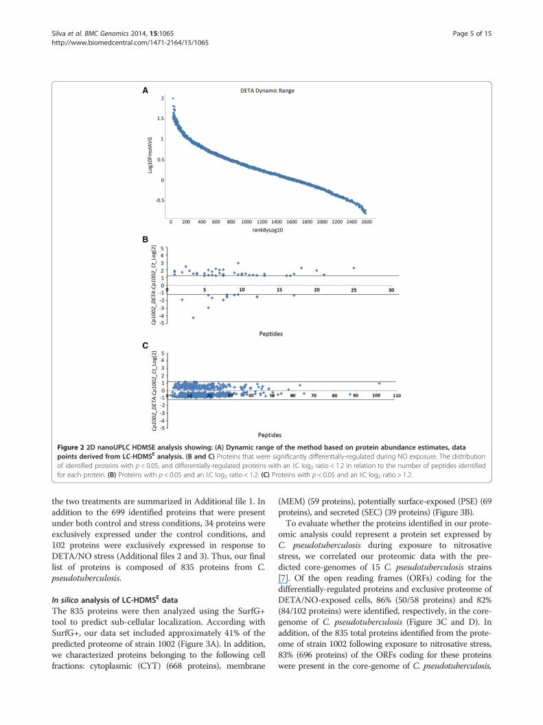
Figure 2 2D nanoUPLC HDMSE analysis showing: (A) Dynamic range of the method based on protein abundance estimates, datapoints derived from LC-HDMSE analysis. (B and C) Proteins that were significantly differentially-regulated during NO exposure. The distributionof identified proteins with p < 0.05, and differentially-regulated proteins with an I:C log2 ratio < 1.2 in relation to the number of peptides identifiedfor each protein. (B) Proteins with p < 0.05 and an I:C log2 ratio < 1.2. (C) Proteins with p < 0.05 and an I:C log2 ratio > 1.2.
Silva et al. BMC Genomics 2014, 15:1065 Page 5 of 15http://www.biomedcentral.com/1471-2164/15/1065
the two treatments are summarized in Additional file 1. Inaddition to the 699 identified proteins that were presentunder both control and stress conditions, 34 proteins wereexclusively expressed under the control conditions, and102 proteins were exclusively expressed in response toDETA/NO stress (Additional files 2 and 3). Thus, our finallist of proteins is composed of 835 proteins from C.pseudotuberculosis.
In silico analysis of LC-HDMSE dataThe 835 proteins were then analyzed using the SurfG+tool to predict sub-cellular localization. According withSurfG+, our data set included approximately 41% of thepredicted proteome of strain 1002 (Figure 3A). In addition,we characterized proteins belonging to the following cellfractions: cytoplasmic (CYT) (668 proteins), membrane
(MEM) (59 proteins), potentially surface-exposed (PSE) (69proteins), and secreted (SEC) (39 proteins) (Figure 3B).To evaluate whether the proteins identified in our prote-
omic analysis could represent a protein set expressed byC. pseudotuberculosis during exposure to nitrosativestress, we correlated our proteomic data with the pre-dicted core-genomes of 15 C. pseudotuberculosis strains[7]. Of the open reading frames (ORFs) coding for thedifferentially-regulated proteins and exclusive proteome ofDETA/NO-exposed cells, 86% (50/58 proteins) and 82%(84/102 proteins) were identified, respectively, in the core-genome of C. pseudotuberculosis (Figure 3C and D). Inaddition, of the 835 total proteins identified from the prote-ome of strain 1002 following exposure to nitrosative stress,83% (696 proteins) of the ORFs coding for these proteinswere present in the core-genome of C. pseudotuberculosis,

Silva et al. BMC Genomics 2014, 15:1065 Page 6 of 15http://www.biomedcentral.com/1471-2164/15/1065
this result correspond approximately 46% of the predictedcore-genome of C. pseudotuberculosis (Figure 3E).
Functional classification of the proteome ofC. pseudotuberculosis expressed under exposure tonitrosative stressThe strain 1002 proteome was functionally classified usingthe Blast2Go tool [24]. A large proportion of thedifferentially-regulated proteins and those exclusive to onecondition were identified as hypothetical proteins. Accord-ing to the biological function prediction, 18 biological pro-cesses were classified as differentially regulated (Figure 4A).In addition, the analysis of the exclusive proteome of eachcondition revealed 12 common processes between the con-trol and stress conditions (Figure 4B). However, seven bio-logical processes were identified only in stress-exposed cells.These processes were antibiotic metabolism (six proteins),nucleotide metabolism (five proteins), oxidative phosphoryl-ation (three proteins), translation (three proteins), glycolysispathways (one protein), iron-sulfur clusters (one protein),and starch and sucrose metabolism (one protein). Amongall processes identified, DNA synthesis and repair proteins(14 proteins) were most common. An overview of the C.pseudotuberculosis response to nitrosative stress accordingwith the proteins identified is shown in Figure 5.The proteins that were grouped into of transcriptional
process were evaluated by CMRegNet and among regu-lators identified; we identified the GntR- family regula-tory protein (D9Q5B7_CORP1), genes regulated byGntR-type regulators are usually involved in carbohy-drate metabolism. The CMRegNet analysis showed thatof the four genes under the control of this regulator, theN-acetylglucosamine kinase (D9Q5B6_CORP1) proteinwas highly expressed by C. pseudotuberculosis in responseto DETA/NO. We identified other regulator the LexA re-pressor (D9Q8W2_CORP1) that was down regulated inthe DETA/NO condition. According with CMRegNet, twoproteins regulated by this repressor were detected in theDETA/NO proteome specific, pyridoxal biosynthesis lyase(PdxS; D9Q5T9_CORP1) and DNA translocase (D9Q8Z6_CORP1). Others proteins under the control of this re-pressor was detected, however not presented significantdifferential regulation like RecA protein
Protein-protein interaction networkTo investigate the interactions among the proteins iden-tified as exclusive and differentially regulated in cells ex-posed to DETA/NO, we generated a protein interactionnetwork using Cytoscape. The interactome analysis re-vealed 67 protein-protein interactions (Figure 6). DnaB/DNA helicase (D9Q578_CORP1), identified in the ex-clusive proteome for strain 1002_DETA/NO, and PyrE/orotate phosphoribosyltransferase (D9Q4S2_CORP1),which was down-regulated in strain 1002_DETA/NO,
showed the greatest number of interactions with otherproteins (eight interactions each). Moreover, both ofthese proteins interact with proteins that are involved inmetabolic processes, DNA processes, antibiotic metab-olism, cell cycling, and translation.
DiscussionC. pseudotuberculosis is exposed to different forms ofoxidative and nitrosative stress during the infectionprocess. A previous study showed that C. pseudotubercu-losis resists nitrosative stress generated by the NO-donorDETA/NO, and that a low concentration of DETA/NO(100 μM) induces a change in the extracellular proteomethis pathogen [15]. To better understand the physiologyof C. pseudotuberculosis in response to nitrosative stress,we analyzed the proteome of whole bacterial lysates ofC. pseudotuberculosis in response to exposure to DETA/NO (0.5 mM).
The strain 1002 proteome under nitrosative stress revealsproteins involved in bacterial defense against DNA damageProteomic analysis identified proteins involved in DNArepair systems in both the exclusive proteome of DETA/NO-exposed cells and in the differentially-regulated prote-ome. We detected the proteins formamidopyrimidine-DNA glycosylase (Fpg) (D9Q598_CORP1), RecB (D9Q8C9_CORP1), and methylated-DNA-protein-cysteine meth-yltransferase (Ada) (D9Q923_CORP1), the genes for whichwere previously identified in a transcriptome analysis ofstrain 1002 in response to different abiotic stresses [8].Activation of these proteins in response to nitrosativestress confirms that they belong a group of generalstress-response proteins in C. pseudotuberculosis.The expression of Fpg was up-regulated in response to
acid stress [8]. We also identified endonuclease III (EndoIII) (D9Q615_CORP1), which, in addition to Fpg, is in-volved in the base excision repair (BER) system of variousbacteria. This system cleaves N-glycosidic bonds fromdamaged bases, allowing their excision and replacement. InSalmonella enterica serovar Typhimurium, the BER systemrepairs DNA damaged by exposure to NO. In addition, anS. Typhimurium strain defective in Fpg demonstrated re-duced virulence in a murine model [30]. Our interactomeanalysis showed that Endo III had one of the highest num-bers of interactions with other proteins, including interac-tions with proteins involved in DNA replication such zincmetalloprotease (D9Q378_CORP1) and DNA translocase(D9Q8Z6_CORP1), suggesting that this protein could playan important role in the defense pathway against RNS.The Ada and RecB protein were up-regulated in re-
sponse to osmotic stress [8]. Ada is involved in the re-pair of DNA-methylation damage, this protein haveplays important in the pathway DNA damage [31]. RecBis a component of the RecBC system, which is part of

Table 1 Proteins identified as differentially-expressed following exposure to nitrosative stress
Uniprot access Proteins Score Peptides log2 DETA:CT(a)
p-value(a) Subcellularlocalization(c)
Gene name Genome(b)
Transport
F9Y2Z3_CORP1 Cell wall channel 5321.88 4 1.42 1 CYT porH Shared
Cell division
D9Q7G2_CORP1 Hypothetical protein 2417.8 21 1.34 1 CYT Cp1002_0716 Core
DNA synthesisand repair
D9Q5V6_CORP1 Nucleoid-associated protein 2327.08 5 1.52 1 CYT ybaB Core
D9Q923_CORP1 Methylated-DNA-protein-cysteinemethyltransferase
6332.83 8 1.22 1 CYT ada Core
D9Q4P0_CORP1 7,8-dihydro-8-oxoguanine-triphosphatase
1640.23 8 −1.97 0 CYT mutT Core
Transcription
D9Q8W2_CORP1 LexA repressor 800.31 6 −1.37 0.04 CYT lexA Shared
D9Q5L4_CORP1 ECF family sigma factor k 364.82 8 −1.58 0 CYT sigK Core
Translation
D9Q753_CORP1 Fkbp-type peptidyl-prolylcis-trans isomerase
7113.34 3 2.43 1 CYT fkbP Core
D9Q830_CORP1 50S ribosomal protein L35 2271.66 1 1.36 1 CYT rpmI Core
D9Q7W1_CORP1 Aspartyl glutamyl-tRNAamidotransferase subunit C
3100.8 7 1.24 0.99 CYT gatC Core
D9Q582_CORP1 50S ribosomal protein L9 41082.46 10 −1.25 0 CYT rplI
D9Q6H6_CORP1 30S ribosomal protein S8 45333.23 9 −1.34 0 CYT rpsH Core
Cell communication
D9Q559_CORP1 Hypothetical protein 1402.27 6 1.99 1 PSE Cp1002_2005 Core
D9Q5U9_CORP1 Thermosensitive gluconokinase 2068.35 7 1.96 0.99 CYT gntK Core
D9Q668_CORP1 Sensory transduction proteinRegX3
2540.92 13 1.45 1 CYT regX3 Core
Detoxification
D9Q7U6_CORP1 Thioredoxin 1835.7 11 1.50 1 CYT trxA Core
D9Q4E5_CORP1 Glutathione peroxidase 1426.27 10 1.47 1 CYT Cp1002_1731 Core
D9Q5T5_CORP1 Glyoxalase bleomycinresistance proteindihydroxybiphenyl dioxygenase
2417.77 11 1.28 1 CYT Cp1002_0124 Shared
D9Q5N2_CORP1 NADH dehydrogenase 7030.94 12 1.25 1 CYT noxC Shared
D9Q680_CORP1 Glutaredoxin-like domain protein 292.69 2 −1.91 0 CYT Cp1002_0272 Core
Glycolysispathways
D9Q5B6_CORP1 N-Acetylglucosamine kinase 228.69 6 1.74 0.98 CYT nanK Core
D9Q4U9_CORP1 Alcohol dehydrogenase 236.02 17 1.22 1 CYT adhA Shared
Iron-sulfur clusters
D9Q7L6_CORP1 Ferredoxin 36927.57 7 2.10 1 CYT fdxA Core
Antibioticresistance
D9Q827_CORP1 Metallo-beta-lactamasesuperfamily protein
657.33 6 −2.95 0 CYT Cp1002_0937 Core
Silva et al. BMC Genomics 2014, 15:1065 Page 7 of 15http://www.biomedcentral.com/1471-2164/15/1065

Table 1 Proteins identified as differentially-expressed following exposure to nitrosative stress (Continued)
Amino acidmetabolism
D9Q622_CORP1 Phosphoserine phosphatase 949.15 9 1.58 0.99 PSE serB Core
D9Q4N1_CORP1 Carboxylate-amine ligase 205.54 16 1.24 1 CYT Cp1002_1819 Core
D9Q6H4_CORP1 L-serine dehydratase I 284.11 17 −1.37 0 MEM sdaA Core
Lipid metabolism
D9Q520_CORP1 Glycerophosphoryldiester phosphodiesterase
2417.8 21 1.34 1 PSE glpQ Core
Oxidativephosphorylation
D9Q8I5_CORP1 Cytochrome aa3 controllingprotein
676.2 6 1.28 1 MEM Cp1002_1095 Core
Specific metabolicpathways
D9Q5M9_CORP1 Inositol-3-phosphate synthase 7473.38 18 2.25 1 CYT ino1 Core
D9Q721_CORP1 Hypothetical protein 4602.9 17 1.44 1 SEC Cp1002_0573 Core
D9Q689_CORP1 3-Hydroxyisobutyratedehydrogenase
2137.24 12 1.34 1 CYT mmsB Core
D9Q4X1_CORP1 Urease accessory protein UreG 1532.39 12 −1.6 0 CYT ureG Core
Nucleotidemetabolism
D9Q4S2_CORP1 Orotate phosphoribosyltransferase 2618.52 8 −1.26 0 CYT pyrE Core
Unknown function
D9Q6Y9_CORP1 Hypothetical protein 491.89 10 2.87 1 CYT Cp1002_0540 Core
D9Q6C7_CORP1 Hypothetical protein 689.6 25 2.25 1 PSE Cp1002_0320 Core
D9Q3P3_CORP1 Hypothetical protein 5703.38 3 1.87 1 CYT Cp1002_1474 Core
D9Q5V4_CORP1 Hypothetical protein 994.52 1 1.7 1 CYT Cp1002_0143 Core
D9Q610_CORP1 Hypothetical protein 27217.36 2 1.67 1 CYT Cp1002_0202 Core
D9Q8D8_CORP1 Hypothetical protein 2324.12 7 1.57 0.98 CYT Cp1002_1048 Shared
D9Q6W1_CORP1 Hypothetical protein 9303.91 4 1.54 1 CYT Cp1002_0512 Core
D9Q6V5_CORP1 Hypothetical protein 1346.2 4 1.5 0.99 CYT Cp1002_0506 Core
D9Q5R7_CORP1 Hypothetical protein 2090.7 8 1.42 1 CYT Cp1002_0105 Core
D9Q917_CORP1 Hypothetical protein 555.89 10 1.37 1 PSE Cp1002_1281 Core
D9Q3P5_CORP1 Hypothetical protein 1121.7 6 1.29 1 SEC Cp1002_1476 Core
D9Q7U5_CORP1 Hypothetical protein 517.06 8 1.28 1 CYT Cp1002_0852 Core
D9Q7L1_CORP1 Hypothetical protein 15693.97 6 1.28 1 SEC Cp1002_0766 Core
D9Q3P6_CORP1 Hypothetical protein 1729.59 5 1.22 1 CYT Cp1002_1477 Core
D9Q6Z7_CORP1 Hypothetical protein 1835.7 13 1.22 1 CYT Cp1002_0548 Core
D9Q8V8_CORP1 Hypothetical protein 293.23 8 −1.48 0 Cp1002_1221 Core
D9Q6C8_CORP1 Hypothetical protein 413.31 12 −1.52 0 PSE Cp1002_0321 Core
D9Q5H0_CORP1 Hypothetical protein 12376.2 6 −1.71 0 CYT Cp1002_0007 Core
D9Q4D5_CORP1 Hypothetical protein 10161.64 4 −4.29 0 CYT Cp1002_1721 Shared
Others
D9Q5N5_CORP1 Iron-regulated MEM protein 992.54 8 2.01 0 PSE piuB Core
D9Q922_CORP1 CobW/HypB/UreG,nucleotide-binding
1771.22 20 1.88 1 CYT Cp1002_1286 Core
Silva et al. BMC Genomics 2014, 15:1065 Page 8 of 15http://www.biomedcentral.com/1471-2164/15/1065

Table 1 Proteins identified as differentially-expressed following exposure to nitrosative stress (Continued)
D9Q8C4_CORP1 Prokaryotic ubiquitin-likeprotein Pup
2194.86 1 1.84 1 CYT pup Core
D9Q7B8_CORP1 Ribosomal-protein-alaninen-acetyltransferase
2791.1 10 1.34 1 CYT rimJ Shared
D9Q7K9_CORP1 Arsenate reductase 5147.54 8 1.32 1 CYT arsC Core
(a) Ratio values to: strain 1002_DETA/NO:strain 1002_Ct, Log(2) Ratio > 1.5, p > 0.95 = up-regulation, p < 0.05 = down-regulation.(b) Core-genome analysis of 15 strains of C. pseudotuberculosis: shared = present in two or more strains; core = present in 15 strains of C. pseudotuberculosis.(c) CYT =cytoplasmic, MEM = membrane, PSE = potentially surface-exposed, SEC = secreted.
Silva et al. BMC Genomics 2014, 15:1065 Page 9 of 15http://www.biomedcentral.com/1471-2164/15/1065
the SOS response the more regulatory network encodedby prokaryotic involved in DNA repair [32]. The RecBCsystem acts in the recombination or degradative repairof arrested DNA replication forks. Studies in S. Typhi-murium showed that recBC mutant strains are more at-tenuated than recA mutants in a murine model ofinfection [33]. In addition, unlike recA mutants, recBCmutants were susceptible to RNS [34], indicating thatRecBC is highly important in the bacterial response tonitrosative stress. The LexA repressor (D9Q8W2_CORP1),
Figure 3 Correlation of in silico predicted data with proteome resultsproteome. (B) Prediction of the subcellular localization of the proteins idencells exposed to DETA/NO in relation to the core genome of C. pseudotubepresent in 15 strains of C. pseudotuberculosis). (D) Analysis of the exclusiveof C. pseudotuberculosis (shared genome: present in only two strains; core gcoverage of the core-genome of C. pseudotuberculosis in relation to the ch
which forms part of the general SOS system along withRecA [35], was down-regulated in C. pseudotuberculosiscells exposed to DETA/NO. We also detected the RecAprotein (D9Q8Y3_CORP1); however, despite having a p-value <0.05, the fold-change of −0.50 showed that this pro-tein was not activated under the experimental conditions.Studies performed in Mycobacterium tuberculosis showedthat recA was not induced until cells had been exposed toDETA/NO (0.5 mM) for 4 h, but that hydrogen peroxideinduced the immediate expression of recA [36], suggesting
. (A) Percentage of coverage of the C. pseudotuberculosis 1002 in silicotified by LC/MS. (C) Analysis of the differentially-regulated proteins ofrculosis (shared genome: present in only two strains; core genome:proteome of cells exposed to DEA/NO in relation to the core-genomeenome: present in 15 strains of C. pseudotuberculosis). (E) Percentaracterized proteome in vitro.

Figure 4 Comparison of biological processes between control and DETA/NO conditions. A representation of the biological processes inrelation to a set list of proteins identified as (A) differentially-regulated in DETA/NO-stressed cells and (B) comparison of exclusive biologicalprocess between the two test conditions.
Silva et al. BMC Genomics 2014, 15:1065 Page 10 of 15http://www.biomedcentral.com/1471-2164/15/1065
that RecA is involved in the later stages of the nitrosativestress response. Nevertheless, CMRegNet analysis identifiedother proteins that are regulated by LexA in the DETA/NO-specific proteome, including pyridoxal biosynthesislyase (PdxS; D9Q5T9_CORP1) and DNA translocase(D9Q8Z6_CORP1).
NO-sensitive transcriptional regulators are activated inthe presence of NOTo activate these DNA repair systems, it is essential thatbacteria can detect ROS and RNS, and concomitantly acti-vate the transcriptional regulators needed for the expressionof genes involved in protection against these compounds. Inthe DETA/NO-specific proteome, we detected the transcrip-tion factor WhiB (D9Q6Y2_CORP1). The WhiB
transcriptional family is composed of iron-sulfur (Fe-S) clus-ter proteins. These proteins are O2- and NO-sensitive, andallow the sensing of both external environmental signalsand the redox state for intracellular bacteria [37,38]. In M.tuberculosis, the reaction of the iron-sulfur cluster of WhiB3with NO generates a dinitrosyl iron complex (DNIC), whichactivates a sensing mechanism in response to the NO, con-sequently activating a system of defense against nitrosativestress [12]. In addition, other in vivo and in vitro studieshave also demonstrated that WhiB regulators play a role inthe adaptation and survival of M. tuberculosis during expos-ure to redox environments [12,39-41].We identified other regulators that are activated in re-
sponse to environmental stimuli, such as a MerR-familytranscriptional regulator (D9Q889_CORP1) and a LysR-

Figure 5 Overview of C. pseudotuberculosis response to nitrosative stress. All proteins detected by proteomic analysis are marked in red(differentially-regulated proteins or exclusive to the proteome of DETA/NO-stressed cells).
Silva et al. BMC Genomics 2014, 15:1065 Page 11 of 15http://www.biomedcentral.com/1471-2164/15/1065
type transcriptional regulator (LTTR) (D9Q7H8_CORP1).This regulator was also highly expressed in the transcrip-tional response of C. pseudotuberculosis 1002 to acidstress [8]. MerR-type regulators have been described inthe detoxification of toxic metal in several pathogenic andnon-pathogenic bacteria [42]. Other studies have shownthat this class of regulator plays a role in bacterial resist-ance to oxidative and nitrosative stress [43,44]. LTTRs areassociated with the regulation of several biological pro-cesses, as well as in the adaptive response of bacteria todifferent types of stress [45]. In Vibrio cholerae, LTTRs areassociated with efflux pump regulation, which contributeto antimicrobial resistance, and are involved incolonization of the human host [46]. In pathogens like E.coli [47], Enterococcus faecalis [48], S. enterica [49], andPseudomonas aeruginosa [50], LTTRs are involved in re-sistance to oxidative stress.
The detoxification pathways of C. pseudotuberculosisfollowing NO exposureOur proteomic analysis identified proteins specificallyexpressed by cells exposed to DETA/NO that are involved
in the detoxification process. Two of these proteins werethioredoxin (trxA) (D9Q7U6_CORP1) and glutathione per-oxidase (D9Q4E5_CORP1). The thioredoxin and glutathi-one systems play major roles in thiol and disulfide balance,respectively [14]. In pathogens such as Helicobacter pylori,Streptococcus pyogenes, and M. tuberculosis, this system is ofgreat importance in combating the presence of ROS/RNS[36,51,52]. A glyoxalase/dioxygenase (D9Q5T5_CORP1)was identified in the differential proteome of cells exposedto DETA/NO. This protein was previously detected in theproteome of C. pseudotuberculosis strain 1002 in responseto 0.1 mM DETA/NO [15]. The presence of this proteinsuggests that glyoxalase/dioxygenase plays a role in the re-sistance of this pathogen to nitrosative stress.Nevertheless, unlike P. aeruginosa, which contains a
complete denitrification pathway [53], the predicted genomeof C. pseudotuberculosis ovis 1002 revealed a truncated de-nitrification pathway. However, we detected the nitric-oxidereductase cytochrome b (NorB) (D9Q5T6_CORP1) inthe exclusive proteome of DETA/NO-stressed cells.norB, which codes for this nitric-oxide reductase, is or-ganized into the norCBQDEF operon in Paracoccus

Figure 6 Protein-protein interactions. Protein-protein interactions of the proteins identified in DETA/NO-exposed cells. Exclusive proteome,circle; up-regulated, square; and down-regulated, rhombus. The sizes of the nodes represent the degree of interaction for each gene/protein; themajor nodes demonstrate greater interactions. The colors of nodes and lines are in an increasing gradient scale from yellow to green to blue. Thenetworks were visualized using Cytoscape.
Silva et al. BMC Genomics 2014, 15:1065 Page 12 of 15http://www.biomedcentral.com/1471-2164/15/1065
denitrificans [54], and into the norCBD operon in P. aer-uginosa [55]. The C. pseudotuberculosis genome waspredicted to only contain norB. Moreover, norB is lo-cated in the Cp1002PiCp12 pathogenicity island, sug-gesting horizontal acquisition of the gene by thispathogen. Nitric-oxide reductase is an important proteinin the denitrification process of some bacteria [56]. In P.aeruginosa, NorB plays a role in both the growth of thepathogen in the presence of NO, and in its survival inmacrophages [55]. The flavohemoglobin Hmp is involvedin the NO detoxification pathway in S. Typhimurium, andlevels of Hmp are increased approximately two-fold in
macrophages [57]. Interestingly, in N. meningitidis, NorBlevels are increased ten-fold in macrophages [58], demon-strating the great power of this protein in the detoxifica-tion process.
Metabolic profile of C. pseudotuberculosis in response tonitrosative stressIn addition to the presence of proteins involved in bac-terial defense and detoxification pathways, strain 1002needs to undergo metabolic adaptation to favor bacterialsurvival. We observed a metabolic readjustment in thispathogen in the proteomic analysis. Of the proteins

Silva et al. BMC Genomics 2014, 15:1065 Page 13 of 15http://www.biomedcentral.com/1471-2164/15/1065
involved in central carbohydrate metabolism, we de-tected only phosphoglycerate mutase (D9Q533_CORP1)and N-acetylglucosamine kinase (D9Q5B6_CORP1) inthe proteome of DETA/NO-exposed cells. Other essen-tial proteins involved in glycolysis (the Embdem-Meyerhof pathway), the pentose phosphate pathway, andthe citric acid cycle were not detected. Similar resultswere found in a metabolomic study of V. cholerae in re-sponse to nitrosative stress [59].However, we hypothesized that C. pseudotuberculosis
uses oxidative phosphorylation to obtain energy. This issupported by the presence of cytochrome C oxidasepolypeptide I (D9Q486_CORP1), succinate dehydrogen-ase cytochrome b556 subunit (D9Q650_CORP1), andubiquinol-cytochrome C reductase cytochrome C subunit(D9Q3J7_CORP1) in the exclusive proteome of DETA/NO-stressed cells, and by the up-regulation of the cyto-chrome oxidase assembly protein (D9Q8I5_CORP1)under the same conditions. However, this oxidative phos-phorylation may be associated with the bacterial cultureconditions used in this work, in which C. pseudotuberculo-sis was cultivated in the presence of DETA/NO under aer-obic conditions. Studies have shown that growing M.tuberculosis in a low concentration of NO with low levelsof O2 can induce anaerobic respiration as a result ofthe inhibition of the respiratory proteins cytochrome coxidase and NADH reductase by irreversible ligationof NO. The ligation of NO to the respiratory proteinsis an effect that may be both short-term reversible andlong-term irreversible [60]. Thus, we suggest that acti-vation of the oxidative phosphorylation system may bea more effective pathway for this pathogen to obtainenergy [61].Another metabolic adjustment was observed in rela-
tion to amino acid biosynthesis. Transporters and en-zymes involved in the synthesis of methionine,tryptophan, and serine were identified. However, thepresence of these proteins can be associated with thebioavailability of these amino acids during exposure toNO. In addition, we detected two oligopeptide trans-port ATP-binding proteins (OppD) (D9Q6G5_CORP1/D9Q3X0_CORP1) that compose the oligopeptide permeasesystem (Opp). This complex is associated with the internal-ization of peptides from the extracellular environment tobe used as a source of carbon and nitrogen in bacterial nu-trition [62]. We also identified proteins that are cofactorsof metabolism, such as CoaBC (D9Q8L2_CORP1), phos-phopantetheine adenylyltransferase (D9Q809_CORP1),and 2-dehydropantoate 2-reductase (D9Q7J9_CORP1).The presence of these proteins demonstrates activity inpantothenic acid metabolism and the biosynthesis of coen-zyme A (CoA). Studies performed in species such as Cor-ynebacterium diphtheriae [63], Streptococcus haemolyticus[64], and M. tuberculosis [65] showed that pantothenic
acid and CoA could have an important role in the growthand viability of these pathogens.
ConclusionsIn this work, we applied high-throughput proteomics tocharacterize the proteome of C. pseudotuberculosis ovis1002 following exposure to NO. Our proteomic analysisgenerated two profiles, which together validated findingsfrom previous in silico analyses of C. pseudotuberculosisovis 1002. The proteomic profile generated after theaddition of the NO-donor, DETA/NO (0.5 mM), re-vealed a set of proteins that are involved in distinct bio-logical process. We detected proteins related to both thegeneral stress response and to a more specific nitrosativestress response, which together form a network of factorsthat promote the survival of this pathogen under stressconditions. However, more detailed studies are needed toassess the true role of these proteins in response to nitro-sative stress in C. pseudotuberculosis. In conclusion, thisfunctional analysis of the genome of C. pseudotuberculosisshows the versatility of this pathogen in the presence ofNO. Moreover, the results presented in this study provideinsights into the processes of resistance of C. pseudotuber-culosis during exposure to nitrosative stress.
Additional files
Additional file 1: Table S1. Complete list of proteins identified assignificantly altered (p < 0.05).
Additional file 2: Table S2. Unique proteins identified in strain1002_DETA/NO.
Additional file 3: Table S3. Unique proteins identified in strain 1002control condition.
AbbreviationsG: Guanine; C: Citosine; NO: Nitric oxide; RNS: Reactive nitrogen species;NOS: Nitric oxide synthases; LC-HDMSE: Liquid chromatograph high definitionmass spectrometry; LC/MS: Liquid chromatograph mass spectrometry;CDM: Chemically-defined médium; DETA/NO: Diethylenetriamine/nitricoxide adduct; DTT: Dithiothreitol; 2D-RP: Two-dimensional reversedphase; nanoUPLC: Nano Ultra performance liquid chromatography;nanoESI-HDMS: Nano electrospray high definition mass spectrometry;HSS: High strength silica; PLGS: Protein lynx global server; FDR: Falsediscovery rate.
Competing interestsThe authors declare that they have no competing interests.
Authors’ contributionsWMS, RDC, and IFSB performed microbiological analyses and samplepreparation for proteomic analysis. GHMFS and WMS conducted theproteomic analysis. SCS and ELF performed bioinformatics analysis of thedata. YLL and AM contributed substantially to data interpretation andrevisions. VA and AS participated in all steps of the project as coordinators,and critically reviewed the manuscript. All authors read and approved thefinal manuscript.
AcknowledgementsThis work was supported by the Genomics and Proteomics Network of theState of Pará of the Federal University of Pará, the Amazon ResearchFoundation (FAPESPA), the National Council for Scientific and Technological

Silva et al. BMC Genomics 2014, 15:1065 Page 14 of 15http://www.biomedcentral.com/1471-2164/15/1065
Development (CNPq), the Brazilian Federal Agency for the Support andEvaluation of Graduate Education (CAPES), the Minas Gerais ResearchFoundation (FAPEMIG), and Waters Corporation, Brazil. Yves Le Loir is therecipient of a PVE grant (71/2013) from Programa Ciências sem Fronteiras.
Author details1Depto de Biologia Geral, Instituto de Ciências Biológicas, UniversidadeFederal de Minas Gerais, Belo Horizonte, Brazil. 2Instituto de CiênciasBiológicas, Universidade Federal do Pará, Belém, Pará, Brazil. 3WatersCorporation, MS Applications and Development Laboratory, São Paulo, Brazil.4Institut National de la Recherche Agronomique - INRA, UMR1253 STLO,Rennes 35042, France. 5Agrocampus Ouest, UMR1253 STLO, Rennes 35042,France.
Received: 4 September 2014 Accepted: 24 November 2014Published: 4 December 2014
References1. Dorella FA, Pacheco LG, Oliveira SC, Miyoshi A, Azevedo V: Corynebacterium
pseudotuberculosis: microbiology, biochemical properties, pathogenesisand molecular studies of virulence. Vet Res 2006, 37:201–218.
2. Baird GJ, Fontaine MC: Corynebacterium pseudotuberculosis and its role inovine caseous lymphadenitis. J Comp Pathol 2007, 137:179–210.
3. Hodgson AL, Bird P, Nisbet IT: Cloning, nucleotide sequence, andexpression in Escherichia coli of the phospholipase D gene fromCorynebacterium pseudotuberculosis. J Bacteriol 1990, 172:1256–1261.
4. Hard GC: Comparative toxic effect of the surface lipid of Corynebacteriumovis on peritoneal macrophages. Infect Immun 1975, 12:1439–1449.
5. Billington SJ, Esmay PA, Songer JG, Jost BH: Identification and role invirulence of putative iron acquisition genes from Corynebacteriumpseudotuberculosis. J Bacteriol 2002, 180:3233–3236.
6. Ruiz JC, D'Afonseca V, Silva A, Ali A, Pinto AC, Santos AR, Rocha AA, LopesDO, Dorella FA, Pacheco LG, Costa MP, Turk MZ, Seyffert N, Moraes PM,Soares SC, Almeida SS, Castro TL, Abreu VA, Trost E, Baumbach J, Tauch A,Schneider MP, McCulloch J, Cerdeira LT, Ramos RT, Zerlotini A, Dominitini A,Resende DM, Coser EM, Oliveira LM, et al: Evidence for reductive genomeevolution and lateral acquisition of virulence functions in twoCorynebacterium pseudotuberculosis strains. PLoS One 2011, 18:e18551.
7. Soares SC, Silva A, Trost E, Blom J, Ramos R, Carneiro A, Ali A, Santos AR, Pinto AC,Diniz C, Barbosa EG, Dorella FA, Aburjaile F, Rocha FS, Nascimento KK, GuimarãesLC, Almeida S, Hassan SS, Bakhtiar SM, Pereira UP, Abreu VA, Schneider MP,Miyoshi A, Tauch A, Azevedo V: The pan-genome of the animal pathogenCorynebacterium pseudotuberculosis reveals differences in genome plasticitybetween the biovar ovis and equi strains. PLoS One 2013, 8:e53818.
8. Pinto AC, de Sá PH, Ramos RT, Barbosa S, Barbosa HP, Ribeiro AC, Silva WM,Rocha FS, Santana MP, de Paula Castro TL, Miyoshi A, Schneider MP, Silva A,Azevedo V: Differential transcriptional profile of Corynebacteriumpseudotuberculosis in response to abiotic stresses. BMC Genomics 2014, 9:14.
9. Marletta MA: Nitric oxide synthase: aspects concerning structure andcatalysis. Cell 1994, 23:927–930.
10. Griffith OW, Stueh DJ: Nitric oxide synthases: properties and catalyticmechanism. Annu Rev Physiol 1995, 57:707–736.
11. Nathan C, Shiloh MU: Reactive oxygen and nitrogen intermediates in therelationship between mammalian hosts and microbial pathogens.Proc Natl Acad Sci USA 2000, 1:8841–8848.
12. Singh A, Guidry L, Narasimhulu KV, Mai D, Trombley J, Redding KE, Giles GI,Lancaster JR Jr, Steyn AJ: Mycobacterium tuberculosis WhiB3 responds toO2 and nitric oxide via its [4Fe-4S] cluster and is essential for nutrientstarvation survival. Proc Natl Acad Sci USA 2007, 10:11562–11567.
13. Ehrt S, Schnappinger D: Mycobacterial survival strategies in the phagosome:defence against host stresses. Cell Microbiol 2009, 11:1170–1178.
14. Lu J, Holmgren A: The thioredoxin antioxidant system. Free Radic Biol Med2013, 8:75–87.
15. Pacheco LG, Castro TL, Carvalho RD, Moraes PM, Dorella FA, Carvalho NB,Slade SE, Scrivens JH, Feelisch M, Meyer R, Miyoshi A, Oliveira SC, DowsonCG, Azevedo V: A role for sigma factor σ(E) in Corynebacteriumpseudotuberculosis resistance to nitric oxide/peroxide stress. FrontMicrobiol 2012, 3:126.
16. Moura-Costa LF, Paule BJA, Freire SM, Nascimento I, Schaer R, Regis LF, ValeVLC, Matos DP, Bahia RC, Carminati R, Meyer R: Chemically defined
synthetic medium for Corynebacterium pseudotuberculosis culture.Rev Bras Saúde Prod An 2002, 3:1–9.
17. Silva JC, Gorenstein MV, Li GZ, Vissers JP, Geromanos SJ: Absolutequantification of proteins by LC/MSE: a virtue of parallel MS acquisition.Mol Cell Proteomics 2006, 5:144–156.
18. Gilar M, Olivova P, Daly AE, Gebler JC: Two-dimensional separation ofpeptides using RP-RP-HPLC system with different pH in first and secondseparation dimensions. J Sep Sci 2005, 28:1694–1703.
19. Silva JC, Denny R, Dorschel CA, Gorenstein M, Kass IJ, Li GZ, McKenna T, NoldMJ, Richardson K, Young P, Geromanos S: Quantitative proteomic analysis byaccurate mass retention time pairs. Anal Chem 2005, 77:2187–2000.
20. Geromanos SJ, Vissers JP, Silva JC, Dorschel CA, Li GZ, Gorenstein MV,Bateman RH, Langridge JI: The detection, correlation, and comparison ofpeptide precursor and product ions from data independent LC-MS withdata dependant LC-MS/MS. Proteomics 2009, 9:1683–1695.
21. Li GZ, Vissers JP, Silva JC, Golick D, Gorenstein MV, Geromanos SJ: Databasesearching and accounting of multiplexed precursor and product ionspectra from the data independent analysis of simple and complexpeptide mixtures. Proteomics 2009, 9:1696–1719.
22. Curty N, Kubitschek-Barreira PH, Neves GW, Gomes D, Pizzatti L, Abdelhay E,Souza GH, Lopes-Bezerra LM: Discovering the infectome of humanendothelial cells challenged with Aspergillus fumigatus applying a massspectrometry label-free approach. J Proteomics 2014, 31:126–140.
23. Levin Y, Hadetzky E, Bahn S: Quantification of proteins using data-independent analysis (MSE) in simple and complex samples: a systematicevaluation. Proteomics 2011, 11:3273–3287.
24. Barinov A, Loux V, Hammani A, Nicolas P, Langella P, Ehrlich D, Maguin E,van de Guchte M: Prediction of surface exposed proteins in Streptococcuspyogenes, with a potential application to other Gram-positive bacteria.Proteomics 2009, 9:61–73.
25. Conesa A, Gotz S, García-Gómez JM, Terol J, Talón M, Robles M: Blast2GO: auniversal tool for annotation, visualization and analysis in functionalgenomics research. Bioinformatics 2005, 15:3674–3676.
26. Soares SC, Abreu VA, Ramos RT, Cerdeira L, Silva A, Baumbach J, Trost E,Tauch A, Hirata R Jr, Mattos-Guaraldi AL, Miyoshi A, Azevedo V: PIPs:pathogenicity island prediction software. PLoS One 2012, 7:e30848.
27. Rezende AM, Folador EL, Resende Dde M, Ruiz JC: Computationalprediction of protein-protein interactions in Leishmania predictedproteomes. PLoS One 2012, 7:e51304.
28. Shannon P, Markiel A, Ozier O, Baliga NS, Wang JT, Ramage D, Amin N,Schwikowski B, Ideker T: Cytoscape: a software environment for integratedmodels of biomolecular interaction networks. Genome Res 2003, 13:2498–2504.
29. Pauling J, Röttger R, Tauch A, Azevedo V, Baumbach J: CoryneRegNet 6.0-Updated database content, new analysis methods and novel featuresfocusing on community demands. Nucleic Acids Res 2012, 40:D610–614.
30. Richardson AR, Soliven KC, Castor ME, Barnes PD, Libby SJ, Fang FC: The baseexcision repair system of Salmonella enterica serovar typhimurium counteractsDNA damage by host nitric oxide. PLoS Pathog 2009, 5:e1000451.
31. Sedgwick B: Repairing DNA-methylation damage. Nat Rev Mol Cell Biol2004, 5:148–157.
32. Baharoglu Z, Mazel D: SOS, the formidable strategy of bacteria againstaggressions. FEMS Microbiol Rev 2014, 38:1126–1145.
33. Cano DA, Pucciarelli MG, García-del Portillo F, Casadesús J: Role of the recBCDrecombination pathway in Salmonella virulence. J Bacteriol 2002, 184:592–595.
34. Koskiniemi S, Andersson DI: Translesion DNA polymerases are required forspontaneous deletion formation in Salmonella typhimurium. Proc NatlAcad Sci U S A. 2009, 23:10248–10253.
35. Butala M, Zgur-Bertok D, Busby SJ: The bacterial LexA transcriptionalrepressor. Cell Mol Life Sci 2009, 66:82–93.
36. Voskuil MI, Bartek IL, Visconti K, Schoolnik GK: The response ofMycobacterium tuberculosis to reactive oxygen and nitrogen species.Front Microbiol 2011, 13:105.
37. Green J, Paget MS: Bacterial redox sensors. Nat Rev Microbiol 2004, 2:954–966.38. Green J, Rolfe MD, Smith LJ: Transcriptional regulation of bacterial virulence
gene expression by molecular oxygen and nitric oxide. Virulence 2014, 4:5(4).39. Singh A, Crossman DK, Mai D, Guidry L, Voskuil MI, Renfrow MB, Steyn AJ:
Mycobacterium tuberculosis WhiB3 maintains redox homeostasis byregulating virulence lipid anabolism to modulate macrophage response.PLoS Pathog 2009, 5:e1000545.
40. Chawla M, Parikh P, Saxena A, Munshi M, Mehta M, Mai D, Srivastava AK,Narasimhulu KV, Redding KE, Vashi N, Kumar D, Steyn AJ, Singh A:

Silva et al. BMC Genomics 2014, 15:1065 Page 15 of 15http://www.biomedcentral.com/1471-2164/15/1065
Mycobacterium tuberculosisWhiB4 regulates oxidative stress response tomodulate survival and dissemination in vivo. Mol Microbiol 2012, 85:1148–1165.
41. Larsson C, Luna B, Ammerman NC, Maiga M, Agarwal N, Bishai WR: Geneexpression of Mycobacterium tuberculosis putative transcription factorsWhiB1-7 in redox environments. PLoS One 2012, 7:e37516.
42. Hobman JL: MerR family transcription activators: similar designs, differentspecificities. Mol Microbiol 2007, 63:1275–1278.
43. McEwan AG, Djoko KY, Chen NH, Couñago RL, Kidd SP, Potter AJ, JenningsMP: Novel bacterial MerR-like regulators their role in the response tocarbonyl and nitrosative stress. Adv Microb Physiol 2011, 58:1–22.
44. Brown NL, Stoyanov JV, Kidd SP, Hobman JL: The MerR family oftranscriptional regulators. FEMS Microbiol Rev 2003, 27:145–163.
45. Maddocks SE, Oyston PC: Structure and function of the LysR-typetranscriptional regulator (LTTR) family proteins. Microbiology 2008,154:3609–3623.
46. Chen S, Wang H, Katzianer DS, Zhong Z, Zhu J: LysR family activator-regulated major facilitator superfamily transporters are involved in Vibriocholerae antimicrobial compound resistance and intestinal colonisation.Int J Antimicrob Agents 2013, 41:188–192.
47. Gonzalez-Flecha B, Demple B: Role for the oxyS gene in regulation ofintracellular hydrogen peroxide in Escherichia coli. J Bacteriol 1999,181:3833–3836.
48. Verneuil N, Rincé A, Sanguinetti M, Posteraro B, Fadda G, Auffray Y, Hartke A,Giard JC: Contribution of a PerR-like regulator to the oxidative-stress responseand virulence of Enterococcus faecalis. Microbiology 2005, 151:3997–4004.
49. Lahiri A, Das P, Chakravortty D: The LysR-type transcriptional regulator Hrgcounteracts phagocyte oxidative burst and imparts survival advantage toSalmonella enterica serovar Typhimurium. Microbiology 2008, 154:2837–2846.
50. Reen FJ, Haynes JM, Mooij MJ, O'Gara F: A non-classical LysR-type tran-scriptional regulator PA2206 is required for an effective oxidative stressresponse in Pseudomonas aeruginosa. PLoS One 2013, 8:e54479.
51. Comtois SL, Gidley MD, Kelly DJ: Role of the thioredoxin system and thethiol-peroxidases Tpx and Bcp in mediating resistance to oxidative andnitrosative stress in Helicobacter pylori. Microbiology 2003, 149:121–129.
52. Brenot A, King KY, Janowiak B, Griffith O, Caparon MG: Contribution ofglutathione peroxidase to the virulence of Streptococcus pyogenes. InfectImmun 2004, 72:408–413.
53. Kalkowski I, Conrad R: Metabolism of nitric oxide in denitrifyingPseudomonas aeruginosa and nitrate-respiring Bacillus cereus. FEMSMicrobiol Lett. 1991, 15:107–111.
54. de Boer AP, van der Oost J, Reijnders WN, Westerhoff HV, Stouthamer AH,van Spanning RJ: Mutational analysis of the nor gene cluster whichencodes nitric-oxide reductase from Paracoccus denitrificans. Eur JBiochem 1996, 15:592–600.
55. Kakishima K, Shiratsuchi A, Taoka A, Nakanishi Y, Fukumori Y: Participation ofnitric oxide reductase in survival of Pseudomonas aeruginosa in LPS-activatedmacrophages. Biochem Biophys Res Commun 2007, 6:587–591.
56. Hendriks J, Oubrie A, Castresana J, Urbani A, Gemeinhardt S, Saraste M:Nitric oxide reductases in bacteria. Biochim Biophys Acta 2000, 15:266–273.
57. Stevanin TM, Poole RK, Demoncheaux EA, Read RC: Flavohemoglobin Hmpprotects Salmonella enterica serovar Typhimurium from nitric oxide-related killing by human macrophages. Infect Immun 2002, 70:4399–4405.
58. Stevanin TM, Moir JW, Read RC: Nitric oxide detoxification systemsenhance survival of Neisseria meningitidis in human macrophages and innasopharyngeal mucosa. Infect Immun 2005, 73:3322–3329.
59. Stern AM, Liu B, Bakken LR, Shapleigh JP, Zhu J: A novel protein protectsbacterial iron-dependent metabolism from nitric oxide. J Bacteriol 2013,195:4702–4708.
60. Brown GC: Regulation of mitochondrial respiration by nitric oxideinhibition of cytochrome C oxidase. Biochim Biophys Acta 2001, 1:46–57.
61. Kadenbach B: Intrinsic and extrinsic uncoupling of oxidativephosphorylation. Biochim Biophys Acta 2003, 5:77–94.
62. Payne JW, Smith MW: Peptide transport by micro-organisms. Adv MicrobPhysiol 1994, 36:1–80.
63. Mueller JH, Klotz AW: Pantothenic acid as a growth factor for thediphtheria bacillus. J Am Chem Soc 1938, 60:3086–3087.
64. Mcllwain H: Pantothenic acid and the growth of Streptococcushaemolyticus. Br J Exp Pathol 1939, 20:330–333.
65. Sassetti CM, Boyd DH, Rubin EJ: Genes required for mycobacterial growthdefined by high density mutagenesis. Mol Microbiol 2003, 48:77–84.
doi:10.1186/1471-2164-15-1065Cite this article as: Silva et al.: Label-free proteomic analysis to confirmthe predicted proteome of Corynebacterium pseudotuberculosis undernitrosative stress mediated by nitric oxide. BMC Genomics 2014 15:1065.
Submit your next manuscript to BioMed Centraland take full advantage of:
• Convenient online submission
• Thorough peer review
• No space constraints or color figure charges
• Immediate publication on acceptance
• Inclusion in PubMed, CAS, Scopus and Google Scholar
• Research which is freely available for redistribution
Submit your manuscript at www.biomedcentral.com/submit