KDM6B/JMJD3 histone demethylase is induced by vitamin D and modulates its effects in colon cancer...
11
KDM6B/JMJD3 histone demethylase is induced by vitamin D and modulates its effects in colon cancer cells Fa ´bio Pereira 1 , Antonio Barba ´chano 1 , Javier Silva 2 , Fe ´lix Bonilla 2 , Moray J. Campbell 3 , Alberto Mun ˜oz 1, ∗ and Marı ´a Jesu ´ s Larriba 1, ∗ 1 Department of Cancer Biology, Instituto de Investigaciones Biome ´dicas ‘Alberto Sols’, Consejo Superior de Investigaciones Cientı ´ficas-Universidad Auto ´noma de Madrid, E-28029 Madrid, Spain, 2 Department of Medical Oncology, Hospital Universitario Puerta de Hierro, E-28223 Majadahonda, Spain and 3 Department of Pharmacology and Therapeutics, Roswell Park Cancer Institute, Buffalo, NY 14263, USA Received July 21, 2011; Revised and Accepted August 30, 2011 KDM6B/JMJD3 is a histone H3 lysine demethylase with an important gene regulatory role in development and physiology. Here, we show that human JMJD3 expression is induced by the active vitamin D metabolite 1a,25- dihydroxyvitamin D 3 (1,25(OH) 2 D 3 ) and that JMJD3 modulates the gene regulatory action of this hormone. 1,25(OH) 2 D 3 activates the JMJD3 gene promoter and increases the level of JMJD3 RNA in human cancer cells. JMJD3 upregulation was strictly dependent on vitamin D receptor (VDR) expression and was abolished by cycloheximide. In SW480-ADH colon cancer cells, JMJD3 knockdown or expression of an inactive mutant JMJD3 fragment decreased the induction by 1,25(OH) 2 D 3 of several target genes and of an epithelial adhesive phenotype. Moreover, JMJD3 knockdown upregulated the epithelial-to-mesenchymal transition inducers SNAIL1 and ZEB1 and the mesenchymal markers fibronectin and LEF1, while it downregulated the epithelial proteins E-cadherin, Claudin-1 and Claudin-7. Additionally, JMJD3 knockdown abolished the nuclear export of b-catenin and the inhibition of b-catenin transcriptional activity caused by 1,25(OH) 2 D 3 . Importantly, the ex- pression of JMJD3 correlated directly with that of VDR and inversely with that of SNAI1 in a series of 96 human colon tumours. Our results indicate for the first time that an epigenetic gene coding for a histone demethylase such as JMJD3 is a VDR co-target that partially mediates the effects of 1,25(OH) 2 D 3 on human colon. INTRODUCTION Histone methylation on specific lysine residues is a crucial point for gene expression in eukaryotic cells that is controlled by the opposite action of histone lysine methyltransferases (KMTs), such as Polycomb group proteins, and demethylases (KDMs), such as LSD1 and the family of Jumonji (Jmj) C domain-containing enzymes (1). Several KDMs have been implicated in development, differentiation or stem cell renewal, and their mutation or deregulation have been linked to cancer and other diseases (2 – 7). JMJD3 (KDM6B) and UTX (KDM6A) specifically demethylates di- and tri-methyl-lysine 27 on histone H3 (H3K27me2/3) (8 – 10). The presence of H3K27me3 at transcriptional sites usually correlates with gene repression (7,11,12), and Polycomb-mediated H3K27 methylation pre- marks genes for DNA methylation and silencing in cancer (13). Thus, JMJD3 is expected to enable the activation of genes, which indeed occurs for those involved in animal body patterning and the inflammatory response (8,10,14,15). Moreover, JMJD3 is induced upon activation of the RAS-RAF signalling pathway and contributes to the transcrip- tional activation of the p16 INK4A tumour suppressor in diploid fibroblasts (16,17), and it is aberrantly overexpressed and seems to be involved in Epstein–Barr virus-associated Hodgkin’s lymphoma (18). In addition, JMJD3 appears to have transcriptional effects unrelated to the level of histone methylation at least in lipopolysaccharide-stimulated mouse ∗ To whom correspondence should be addressed at: Instituto de Investigaciones Biome ´dicas ‘Alberto Sols’, Arturo Duperier 4, E-28029 Madrid, Spain. Tel: +34 915854451; Fax: +34 915854401; Email: [email protected] (A.M.), [email protected] (M.J.L.) # The Author 2011. Published by Oxford University Press. All rights reserved. For Permissions, please email: [email protected] Human Molecular Genetics, 2011, Vol. 20, No. 23 4655–4665 doi:10.1093/hmg/ddr399 Advance Access published on September 2, 2011 by guest on July 4, 2016 http://hmg.oxfordjournals.org/ Downloaded from
-
Upload
independent -
Category
Documents
-
view
2 -
download
0
Transcript of KDM6B/JMJD3 histone demethylase is induced by vitamin D and modulates its effects in colon cancer...
KDM6B/JMJD3 histone demethylase is inducedby vitamin D and modulates its effects in coloncancer cells
Fabio Pereira1, Antonio Barbachano1, Javier Silva2, Felix Bonilla2, Moray J. Campbell3,
Alberto Munoz1,∗ and Marıa Jesus Larriba1,∗
1Department of Cancer Biology, Instituto de Investigaciones Biomedicas ‘Alberto Sols’, Consejo Superior de
Investigaciones Cientıficas-Universidad Autonoma de Madrid, E-28029 Madrid, Spain, 2Department of Medical
Oncology, Hospital Universitario Puerta de Hierro, E-28223 Majadahonda, Spain and 3Department of Pharmacology
and Therapeutics, Roswell Park Cancer Institute, Buffalo, NY 14263, USA
Received July 21, 2011; Revised and Accepted August 30, 2011
KDM6B/JMJD3 is a histone H3 lysine demethylase with an important gene regulatory role in development andphysiology. Here, we show that human JMJD3 expression is induced by the active vitamin D metabolite 1a,25-dihydroxyvitamin D3 (1,25(OH)2D3) and that JMJD3 modulates the gene regulatory action of this hormone.1,25(OH)2D3 activates the JMJD3 gene promoter and increases the level of JMJD3 RNA in human cancercells. JMJD3 upregulation was strictly dependent on vitamin D receptor (VDR) expression and was abolishedby cycloheximide. In SW480-ADH colon cancer cells, JMJD3 knockdown or expression of an inactive mutantJMJD3 fragment decreased the induction by 1,25(OH)2D3 of several target genes and of an epithelial adhesivephenotype. Moreover, JMJD3 knockdown upregulated the epithelial-to-mesenchymal transition inducersSNAIL1 and ZEB1 and the mesenchymal markers fibronectin and LEF1, while it downregulated the epithelialproteins E-cadherin, Claudin-1 and Claudin-7. Additionally, JMJD3 knockdown abolished the nuclear exportof b-catenin and the inhibition of b-catenin transcriptional activity caused by 1,25(OH)2D3. Importantly, the ex-pression of JMJD3 correlated directly with that of VDR and inversely with that of SNAI1 in a series of 96 humancolon tumours. Our results indicate for the first time that an epigenetic gene coding for a histone demethylasesuch as JMJD3 is a VDR co-target that partially mediates the effects of 1,25(OH)2D3 on human colon.
INTRODUCTION
Histone methylation on specific lysine residues is a crucialpoint for gene expression in eukaryotic cells that is controlledby the opposite action of histone lysine methyltransferases(KMTs), such as Polycomb group proteins, and demethylases(KDMs), such as LSD1 and the family of Jumonji (Jmj) Cdomain-containing enzymes (1). Several KDMs have beenimplicated in development, differentiation or stem cellrenewal, and their mutation or deregulation have been linkedto cancer and other diseases (2–7).
JMJD3 (KDM6B) and UTX (KDM6A) specificallydemethylates di- and tri-methyl-lysine 27 on histone H3(H3K27me2/3) (8–10). The presence of H3K27me3 at
transcriptional sites usually correlates with gene repression(7,11,12), and Polycomb-mediated H3K27 methylation pre-marks genes for DNA methylation and silencing in cancer(13). Thus, JMJD3 is expected to enable the activation ofgenes, which indeed occurs for those involved in animalbody patterning and the inflammatory response (8,10,14,15).Moreover, JMJD3 is induced upon activation of theRAS-RAF signalling pathway and contributes to the transcrip-tional activation of the p16INK4A tumour suppressor in diploidfibroblasts (16,17), and it is aberrantly overexpressed andseems to be involved in Epstein–Barr virus-associatedHodgkin’s lymphoma (18). In addition, JMJD3 appears tohave transcriptional effects unrelated to the level of histonemethylation at least in lipopolysaccharide-stimulated mouse
∗To whom correspondence should be addressed at: Instituto de Investigaciones Biomedicas ‘Alberto Sols’, Arturo Duperier 4, E-28029 Madrid, Spain.Tel: +34 915854451; Fax: +34 915854401; Email: [email protected] (A.M.), [email protected] (M.J.L.)
# The Author 2011. Published by Oxford University Press. All rights reserved.For Permissions, please email: [email protected]
Human Molecular Genetics, 2011, Vol. 20, No. 23 4655–4665doi:10.1093/hmg/ddr399Advance Access published on September 2, 2011
by guest on July 4, 2016http://hm
g.oxfordjournals.org/D
ownloaded from
macrophages, and the possibility of non-histone substratesand/or other activities of KDMs is emerging (19,20).
Many epidemiological and preclinical studies suggest a pro-tective effect of vitamin D against several neoplasias, particu-larly colon cancer, whose confirmation is pending of adequateclinical trials (21–23). The active vitamin D metabolite1a,25-dihydroxyvitamin D3 (1,25(OH)2D3) is a hormonewith wide gene regulatory effects in higher organisms(24,25), which is in line with the expression of the vitaminD receptor (VDR) in over 30 different cell types (26). VDRis a member of the nuclear hormone receptor superfamily oftranscription factors modulated by ligand, phosphorylationand perhaps other modifications (27,28). Although part ofVDR is extranuclear and upon ligand binding can activate sig-nalling pathways, these are thought to converge with the regu-lation of gene transcription as the major effect of DNA-boundVDR/1,25(OH)2D3 complexes (29,30).
1,25(OH)2D3 is believed to regulate the expression of hun-dreds of genes but only a subset of them seem to containVDR-binding sites (VDRE, vitamin D regulatory elements).In addition, very few genes respond to 1,25(OH)2D3 withdrastic changes in their transcription rate; rather thehormone has a weak, moderate effect that is compatible witha permissive action. We hypothesized that this could berelated to epigenetic changes: hormone-induced alterations atthe chromatin level that could facilitate gene transcriptionregulation by other agents. Supporting this, transcriptomicstudies by us and others have revealed JARID2/JMJ,JARID1A/KDM5A and JMJD2B/KDM4B as candidate1,25(OH)2D3 targets in human SW480-ADH colon orSCC25 squamous cell carcinoma cells (31 and our own unpub-lished data). All this led us to investigate the possibility that1,25(OH)2D3 affects the expression of genes controlling epi-genetic processes such as those regulating histone modifica-tions. As the majority of 1,25(OH)2D3 target genes areinduced while only around one-third are repressed, we chosehistone H3 lysine demethylases to study.
Our results show that 1,25(OH)2D3 induces the expression ofJMJD3 in several types of human cancer cells. The regulation istranscriptional and probably mediated by one or more short-lived proteins. Importantly, a knockdown approach has shownthat JMJD3 mediates in part the induction by 1,25(OH)2D3 ofseveral target genes such as CDH1/E-cadherin and CST5/cystatin D and modulates the expression of the epithelial-to-mesenchymal transition (EMT) inducers SNAIL1 andZEB1. Probably as a consequence of these actions, JMJD3 con-trols the phenotype of human colon cancer cells. Importantly,the level of expression of VDR and JMJD3 directly correlatein human colon tumours, suggesting that the regulation ofJMJD3 by 1,25(OH)2D3 may take place in vivo during coloncancer progression and mediates at least in part the protectiveaction of 1,25(OH)2D3 against this neoplasia.
RESULTS
The JMJD3 gene is induced by 1,25(OH)2D3
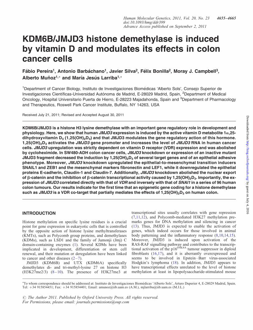
To study whether 1,25(OH)2D3 could affect the expression ofhistone H3 lysine demethylases, we analysed the RNA levelof 10 JmjC-domain proteins by quantitative reverse
transcriptionpolymerase chain reaction (qRT–PCR) inSW480-ADH colon cancer cells treated for 4 or 48 h with thehormone. As control, we studied CYP24A1 and CDH1/E-cadherin, known to be induced, and CYP27B1, which isrepressed by 1,25(OH)2D3. We found the strongest change(2–3-fold increase at 48 h treatment) for JMJD3, whileJMJD4 showed only a 25% decrease and the remaining geneswere unaffected (Supplementary Material, Fig. S1). To investi-gate in depth the observed effect, we first performed time-course and dose-curve experiments. A progressive increase inthe JMJD3 RNA level was detected following hormone treat-ment (1027
M) (Fig. 1A) that was dose-dependent at 48 h(Fig. 1B). The general validity and specificity of this findingwere examined using a panel of human cancer cell lines,investigating the requirement of VDR, and analysing the puta-tive effect of other nuclear receptors ligands. A common upre-gulation (2–3-fold) by 1,25(OH)2D3 was found in severalhuman colon (SW1417, SW48, KM12C), breast (MCF7,MDA-MB-453, MDA-MB-231) and skin (UACC-257) cancercell lines (Supplementary Material, Fig. S2A). No effect of1,25(OH)2D3 was detected in other cancer cell lines, such asHT29 or Caco-2 (colon), SK-MEL-28 (skin) or AGS (gastric)(data not shown). Importantly, no regulation was detected inSW620 and SW480-R cells that do not express VDR (32) (Sup-plementary Material, Fig. S2A). Consistently, VDR knockdownby shRNA in SW480-ADH cells abolished the increase inJMJD3 RNA expression caused by 1,25(OH)2D3 (Fig. 1C).Finally, neither estradiol, progesterone, dexamethasone, triiodo-thyronine, all-trans- or cis-retinoic acid or docosahexaenoicacid (ligands for oestrogen, progesterone, glucocorticoid,thyroid, retinoic or rexinoid receptors, respectively) changedbasal JMJD3 RNA expression nor affected the induction by1,25(OH)2D3 (Supplementary Material, Fig. S2B).
Next we studied the mechanism of JMJD3 upregulation. Toinvestigate whether the effect on JMJD3 RNA level was dueto the activation of the gene promoter, we studied the effectof 1,25(OH)2D3 in SW480-ADH cells that were transfectedwith a series of plasmids containing distinct sequences ofthe promoter upstream of the luciferase reporter gene (17).Promoter constructs P1–P5 containing 23422, 22439,22314, 22224 and 22173 bp immediately upstream of theJMJD3 transcription start site active in macrophages(MF-TSS) were activated by the hormone (Fig. 1D). On theother hand, the activity of P6 and P7 constructs spanning22102 and 21428 bp upstream of this site, or P8 containing22928 bp upstream of the JMJD3 start site active in embryon-ic stem cells (ESC-TSS), were not affected by 1,25(OH)2D3
(Fig. 1D). This suggests that 1,25(OH)2D3 induces JMJD3transcription via sequences located from 22173 to22102 bp upstream of the MF-TSS. This region of the pro-moter contains sites for RAP1, Sp1, Krox-20, ETF, C/EBPaand AP-1 transcription factors and is crucial for the inductionof JMJD3 promoter by BRAF (17). However, the involvementof other sequences not contained in the constructs studiedcannot be ruled out. The protein synthesis inhibitor cyclohex-imide (CHX) blocked the increase in JMJD3 RNA by1,25(OH)2D3 (Fig. 1E). The long treatment with CHX that isneeded because of the slow kinetics of JMJD3 inductionmakes the requirement of intermediary short-lived protein(s)suggestive, but not definitive. In addition, no statistically
4656 Human Molecular Genetics, 2011, Vol. 20, No. 23
by guest on July 4, 2016http://hm
g.oxfordjournals.org/D
ownloaded from
significant VDR binding to three putative VDREs revealed byin silico analysis of the JMJD3 gene (228697-AGGCCATTTAGTTCA; 21073-TGACCTCTACCACCT; +9183-TGACCCAGCCGACCC respect to the ESC-TSS) was found bymeans of chromatin immunoprecipitation assays (Supplemen-tary Material, Fig. S3A and B). This negative result furthersupports an indirect mechanism of regulation.
JMJD3 modulates the gene regulatory effectsof 1,25(OH)2D3
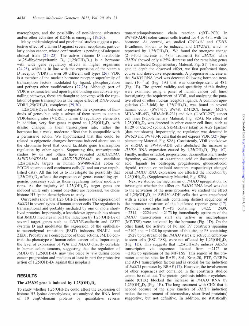
To study the importance of JMJD3 in 1,25(OH)2D3 action, wegenerated SW480-ADH cells stably expressing JMJD3shRNA (shJMJD3) or a non-targeting shRNA (shControl).First, we confirmed a stable downregulation of JMJD3 expres-sion, which did not affect VDR RNA expression (Fig. 2A).
One of the most important effects of 1,25(OH)2D3 in humancolon and breast carcinoma cells is the induction of E-cadherinprotein, which causes a morphological change to an adhesiveepithelial phenotype (32–34). Thus, we analysed these
biological parameters in shJMJD3 cells. The increase in bothintercellular adhesion and E-cadherin protein expression by1,25(OH)2D3 was partially inhibited in shJMJD3 cells(Fig. 2B), and was co-incident with changes in the tubulincytoskeleton and cell morphology (Fig. 2B). In line withthis, the induction of CDH1/E-cadherin RNA and also thatof CYP24A1 and CST5/cystatin D, two other 1,25(OH)2D3
targets (35,36), was inhibited in shJMJD3 cells (Fig. 2C).JMJD3 knockdown decreased also the basal expression ofCDH1/E-cadherin and CST5/cystatin D (Fig. 2C). Theseeffects were reproduced at the promoter and protein levels: theactivity of the CDH1/E-cadherin gene promoter was lower inshJMJD3 than in shControl cells (Fig. 2D); likewise, the basaland induced expression of E-cadherin and cystatin D proteinswas lower in shJMJD3 cells (Fig. 2E). In contrast, however,JMJD3 knockdown did not affect the expression of the VDRprotein (Fig. 2E), indicating that JMJD3 is not equally requiredfor the activation of all genes and that the inhibition of the generegulatory action of 1,25(OH)2D3 in shJMJD3 cells is not aconsequence of diminished VDR expression.
Figure 1. 1,25(OH)2D3 induces JMJD3 expression in colon cancer cells. (A) qRT–PCR analysis of JMJD3 RNA levels in SW480-ADH cells treated with1027
M 1,25(OH)2D3 for the indicated times. (B) JMJD3 RNA expression in SW480-ADH cells treated with the indicated doses of 1,25(OH)2D3 for 48 h.(C) JMJD3 RNA levels in SW480-ADH cells expressing shVDR or shControl that were treated with 1027
M 1,25(OH)2D3 or vehicle for 48 h. (D) Activityof a series of JMJD3 promoter constructs (P1–P8) (17) in SW480-ADH cells. Cells transfected with the indicated construct were incubated with 1027
M
1,25(OH)2D3 or vehicle for 48 h and the expression of luciferase in cell extracts was estimated. Inset: scheme of the JMJD3 promoter constructs used. Constructsare represented as grey boxes and arrows indicate the position of the transcription start site active in embryonic stem cells (ESC-TSS) and in macrophages(MF-TSS). (E) JMJD3 RNA levels in SW480-ADH cells pretreated 30 min with 8 mg/ml CHX as indicated and then with 1027
M 1,25(OH)2D3 or vehiclefor 24 or 36 h. In (A–C) and (E), 18S rRNA expression was used for normalization. Data are expressed as the mean+SEM of three independent experiments(A–D) or as mean+SD of a representative experiment performed in triplicate (E).
Human Molecular Genetics, 2011, Vol. 20, No. 23 4657
by guest on July 4, 2016http://hm
g.oxfordjournals.org/D
ownloaded from
Figure 2. JMJD3 knockdown inhibits the induction of an adhesive epithelial phenotype and target genes by 1,25(OH)2D3 without affecting VDR expression.(A) qRT–PCR analysis of the RNA levels of JMJD3 and VDR in SW480-ADH shControl and shJMJD3 cells. SDHA was used for normalization. Mean+SEM(n ¼ 3). (B) Upper panels, representative phase-contrast images of SW480-ADH shControl and shJMJD3 cells treated with 1027
M 1,25(OH)2D3 or vehicle for48 h. Bar: 10 mm. Lower panels, representative immunofluorescence and confocal microscopy images of E-cadherin and b-tubulin expression in SW480-ADHshControl and shJMJD3 cells treated with 1027
M 1,25(OH)2D3 or vehicle for 48 h. Bar: 10 mm. (C) qRT–PCR analysis of the RNA levels of CDH1/E-cadherin,CYP24A1 and CST5/cystatin D in SW480-ADH shControl and shJMJD3 cells treated with 1027
M 1,25(OH)2D3 or vehicle for 8 h. 18S was used for normalization.Mean+SEM (n ¼ 3). (D) Activity of the CDH1/E-cadherin promoter (2987 to +92 bp) in SW480-ADH shControl and shJMJD3 cells. The cells were transfectedwith the CDH1/E-cadherin promoter construct and the expression of luciferase in cell extracts was estimated 48 h later. Mean+SEM (n ¼ 4). (E) Western blotanalysis of E-cadherin, cystatin D and VDR protein expression in SW480-ADH shControl and shJMJD3 cells treated with 1027
M 1,25(OH)2D3 or vehicle for 8or 48 h. b-Actin was used as control. Numbers refer to the fold-change with respect to untreated shControl cells. The western blot shown correspond to a represen-tative experiment of the three performed.
4658 Human Molecular Genetics, 2011, Vol. 20, No. 23
by guest on July 4, 2016http://hm
g.oxfordjournals.org/D
ownloaded from
To exclude off-target effects of the knockdown approach andconfirm the role of JMJD3 mediating 1,25(OH)2D3 action, wegenerated cells expressing ectopically a hemagglutinin(HA)-tagged inactive JMJD3 C-terminal region harbouring aHis to Ala1388 mutation in the iron-binding centre (MUT1141-1641) (10). As a control, we generated also cells expres-sing the same wild-type JMJD3 polypeptide (WT 1141–1614)or an empty vector (Mock). Expression of the correspondingexogenous polypeptides was confirmed by qRT–PCR andwestern blot (Supplementary Material, Fig. S4A and B). Ashappened for JMJD3 knockdown, neither MUT 1141-1614nor WT 1141-1641 JMJD3 polypeptides altered the expressionof VDR RNA or protein (Supplementary Material, Fig. S4Aand B). Importantly, MUT 1141-1641 cells behaved asshJMJD3 cells in terms of 1,25(OH)2D3-induced phenotypechange, basal and 1,25(OH)2D3-induced CDH1/E-cadherin,CST5/cystatin D and CYP24A1 RNA and protein expression,
and CDH1/E-cadherin promoter activity (SupplementaryMaterial, Fig. S4C–F). These effects were specific, as theydid not take place in cells expressing the WT 1141-1641JMJD3 polypeptide (Supplementary Material, Fig. S4C–F).
JMJD3 knockdown enhances the expressionof EMT inducers
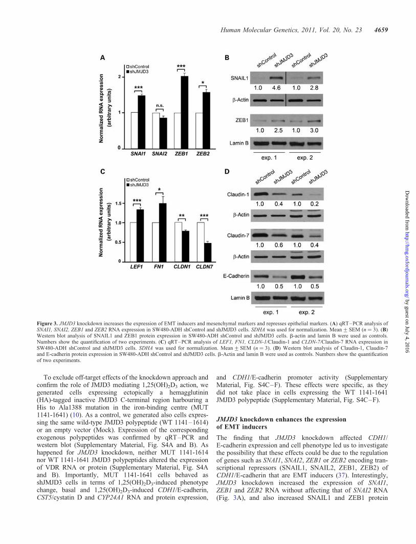
The finding that JMJD3 knockdown affected CDH1/E-cadherin expression and cell phenotype led us to investigatethe possibility that these effects could be due to the regulationof genes such as SNAI1, SNAI2, ZEB1 or ZEB2 encoding tran-scriptional repressors (SNAIL1, SNAIL2, ZEB1, ZEB2) ofCDH1/E-cadherin that are EMT inducers (37). Interestingly,JMJD3 knockdown increased the expression of SNAI1,ZEB1 and ZEB2 RNA without affecting that of SNAI2 RNA(Fig. 3A), and also increased SNAIL1 and ZEB1 protein
Figure 3. JMJD3 knockdown increases the expression of EMT inducers and mesenchymal markers and represses epithelial markers. (A) qRT–PCR analysis ofSNAI1, SNAI2, ZEB1 and ZEB2 RNA expression in SW480-ADH shControl and shJMJD3 cells. SDHA was used for normalization. Mean+SEM (n ¼ 3). (B)Western blot analysis of SNAIL1 and ZEB1 protein expression in SW480-ADH shControl and shJMJD3 cells. b-actin and lamin B were used as controls.Numbers show the quantification of two experiments. (C) qRT–PCR analysis of LEF1, FN1, CLDN-1/Claudin-1 and CLDN-7/Claudin-7 RNA expression inSW480-ADH shControl and shJMJD3 cells. SDHA was used for normalization. Mean+SEM (n ¼ 3). (D) Western blot analysis of Claudin-1, Claudin-7and E-cadherin protein expression in SW480-ADH shControl and shJMJD3 cells. b-Actin and lamin B were used as controls. Numbers show the quantificationof two experiments.
Human Molecular Genetics, 2011, Vol. 20, No. 23 4659
by guest on July 4, 2016http://hm
g.oxfordjournals.org/D
ownloaded from
levels (Fig. 3B). Consistent also with a role of JMJD3 prevent-ing EMT, shJMJD3 cells had higher RNA levels of the mesen-chymal genes lymphoid enhancer-binding factor 1 (LEF1) andfibronectin (FN1), and lower RNA and protein levels of theepithelial genes CLDN1/Claudin-1, CLDN7/Claudin-7 andCDH1/E-cadherin (Fig. 3C and D).
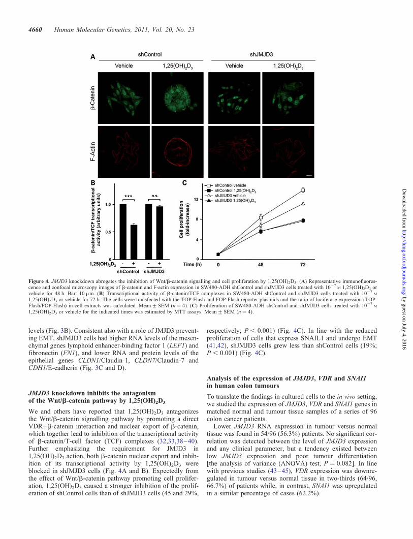
JMJD3 knockdown inhibits the antagonismof the Wnt/b-catenin pathway by 1,25(OH)2D3
We and others have reported that 1,25(OH)2D3 antagonizesthe Wnt/b-catenin signalling pathway by promoting a directVDR–b-catenin interaction and nuclear export of b-catenin,which together lead to inhibition of the transcriptional activityof b-catenin/T-cell factor (TCF) complexes (32,33,38–40).Further emphasizing the requirement for JMJD3 in1,25(OH)2D3 action, both b-catenin nuclear export and inhib-ition of its transcriptional activity by 1,25(OH)2D3 wereblocked in shJMJD3 cells (Fig. 4A and B). Expectedly fromthe effect of Wnt/b-catenin pathway promoting cell prolifer-ation, 1,25(OH)2D3 caused a stronger inhibition of the prolif-eration of shControl cells than of shJMJD3 cells (45 and 29%,
respectively; P , 0.001) (Fig. 4C). In line with the reducedproliferation of cells that express SNAIL1 and undergo EMT(41,42), shJMJD3 cells grew less than shControl cells (19%;P , 0.001) (Fig. 4C).
Analysis of the expression of JMJD3, VDR and SNAI1in human colon tumours
To translate the findings in cultured cells to the in vivo setting,we studied the expression of JMJD3, VDR and SNAI1 genes inmatched normal and tumour tissue samples of a series of 96colon cancer patients.
Lower JMJD3 RNA expression in tumour versus normaltissue was found in 54/96 (56.3%) patients. No significant cor-relation was detected between the level of JMJD3 expressionand any clinical parameter, but a tendency existed betweenlow JMJD3 expression and poor tumour differentiation[the analysis of variance (ANOVA) test, P ¼ 0.082]. In linewith previous studies (43–45), VDR expression was downre-gulated in tumour versus normal tissue in two-thirds (64/96,66.7%) of patients while, in contrast, SNAI1 was upregulatedin a similar percentage of cases (62.2%).
Figure 4. JMJD3 knockdown abrogates the inhibition of Wnt/b-catenin signalling and cell proliferation by 1,25(OH)2D3. (A) Representative immunofluores-cence and confocal microscopy images of b-catenin and F-actin expression in SW480-ADH shControl and shJMJD3 cells treated with 1027
M 1,25(OH)2D3 orvehicle for 48 h. Bar: 10 mm. (B) Transcriptional activity of b-catenin/TCF complexes in SW480-ADH shControl and shJMJD3 cells treated with 1027
M
1,25(OH)2D3 or vehicle for 72 h. The cells were transfected with the TOP-Flash and FOP-Flash reporter plasmids and the ratio of luciferase expression (TOP-Flash/FOP-Flash) in cell extracts was calculated. Mean+SEM (n ¼ 4). (C) Proliferation of SW480-ADH shControl and shJMJD3 cells treated with 1027
M
1,25(OH)2D3 or vehicle for the indicated times was estimated by MTT assays. Mean+SEM (n ¼ 4).
4660 Human Molecular Genetics, 2011, Vol. 20, No. 23
by guest on July 4, 2016http://hm
g.oxfordjournals.org/D
ownloaded from

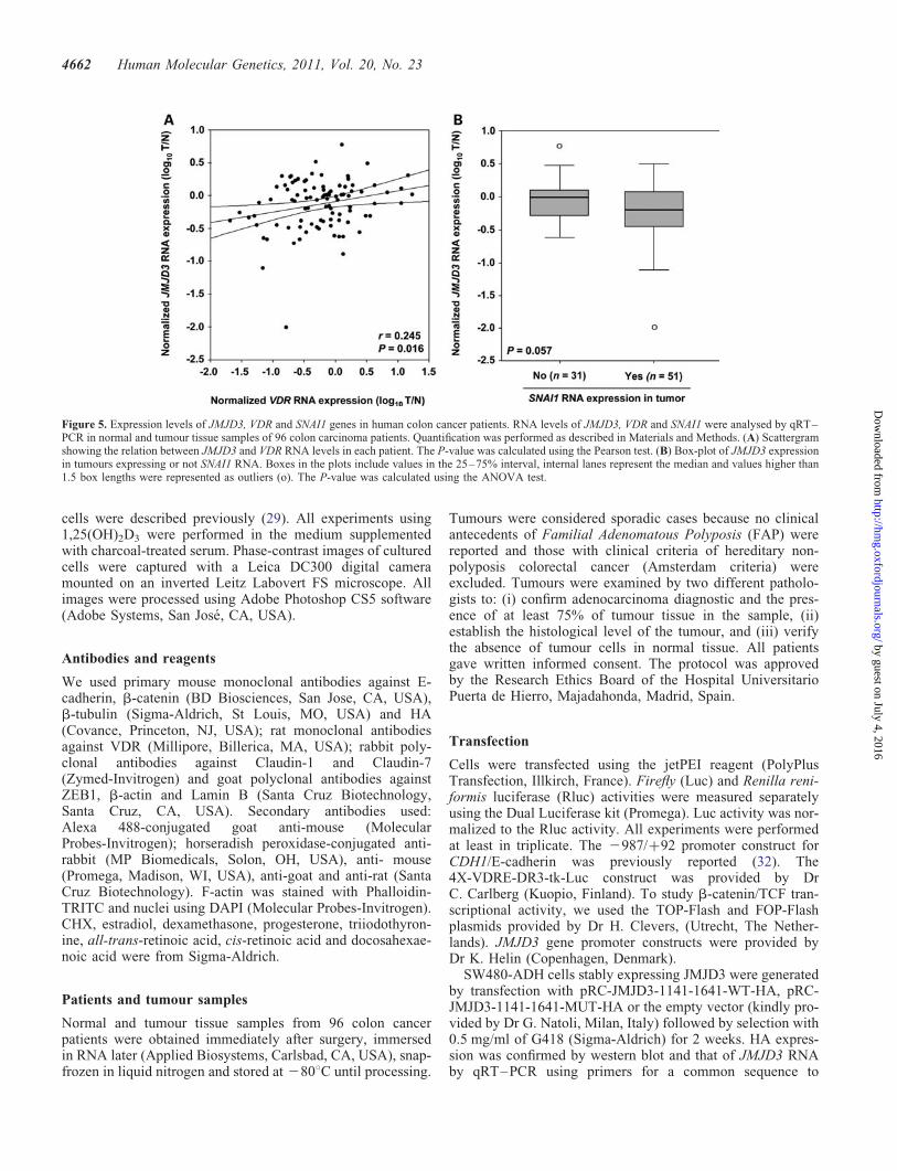
Interestingly, a significant direct correlation was foundbetween the RNA levels of JMJD3 and VDR (Pearson correl-ation coefficient r ¼ 0.245, P ¼ 0.016) (Fig. 5A). In contrast,the expression of JMJD3 and SNAI1 RNA showed a stronginverse trend with borderline significance (the ANOVA test,P ¼ 0.057) in 82 informative cases (Fig. 5B).
DISCUSSION
In this study, we demonstrate that the human histone H3 lysinedemethylase JMJD3 gene is induced by 1,25(OH)2D3 in apanel of cancer cell lines. Data obtained using a series ofJMJD3 promoter constructs suggest that 1,25(OH)2D3
induces JMJD3 transcription via sequences located from22173 to 22102 bp upstream the MF-TSS. The blockadeof JMJD3 induction using CHX and the absence of VDRbinding to three putative VDREs present in the JMJD3 genesuggest an indirect mechanism of regulation. However, thelong CHX treatments needed due to the slow kinetics ofJMJD3 regulation, could affect VDR expression and thereforethe possibility of a direct regulation of JMJD3 by1,25(OH)2D3 cannot be excluded. Also, other putativeVDREs may exist. We show that JMJD3 is implicated inthe regulation of several 1,25(OH)2D3 target genes such asCDH1/E-cadherin and CST5/cystatin D. Moreover, knock-down experiments indicate that JMJD3 contributes to maintainthe adhesive epithelial cell phenotype, as its downregulationaugments the expression of SNAIL1 and ZEB1 transcriptionfactors. The study of JMJD3 expression in normal andtumour tissue of colon cancer patients strongly supports itsrole during the progression of this neoplasia. Furthermore,the direct correlation between the expression levels ofJMJD3 and VDR suggests that 1,25(OH)2D3 regulatesJMJD3 in the human colon.
Recent studies have shown the importance of KMTs andKDMs in the gene regulatory effects of several nuclear recep-tors (28). Transcriptional activation of nuclear receptor targetgenes requires changes in the local chromatin environmentthat are at least partially consequence of the direct or indirectrecruitment by the receptors of histone methylasesand demethylases. In addition, several histone-modifyingenzymes target oestrogen and retinoic acid nuclear receptorsand/or their coactivators and corepressors (SRC3, CBP/p300. . . ) (28). Altogether, these findings strongly suggest thatKMTs and KDMs mediate the ligand-dependent regulationof gene expression by some nuclear receptors (46). ForVDR, dynamic modification of H3K27me3 has recentlybeen reported in the regulation of CDKN1A by 1,25(OH)2D3
in normal prostate cells (47). In line with this and in acancer context, our results demonstrate for the first time thata KDM modulates ligand-activated VDR effects on gene tran-scription. From the screening of a large number ofJmjC-containing proteins (Supplementary Material, Fig. S1),it is conceivable that KDMs other than JMJD3 can be alsoinvolved and thus, that the gene regulatory activity of VDR/1,25(OH)2D3 can be fine-tuned by the combinatorial actionof several histone-modifying enzymes, probably in acell-type-dependent manner.
Mechanistically, the induction of JMJD3 may have severaldifferent effects on 1,25(OH)2D3 action that are not mutuallyexclusive. First, to contribute to a local chromatin environ-ment that facilitates the expression of target genes viaH3K27me2/3 demethylation. CDH1/E-cadherin and CST5/cystatin D may be examples. Secondly, to demethylate VDRand/or any of its coregulators, although no data supportingthis exist. Thirdly, to alter chromatin of genes other thanthose transcriptionally regulated by 1,25(OH)2D3 that may,however, act additively or synergistically with this hormone.Finally, the proposed existence of JMJD3 effects unrelatedto histone demethylation (20) opens the possibility to distinct,unknown mechanism(s) of modulation of 1,25(OH)2D3 action.
Interestingly, the first genome-wide studies performed toidentify VDR-binding sites by ChIP-Seq have revealed ahuge number of them in the human and mouse genomes(48,49). As it happens with other transcription factors, onlya subset of VDR-binding sites are expected to be functional,and this is thought to depend on the epigenetic status of thecell (50). Conceivably, JMJD3 may contribute to generate achromatin structure favourable to gene activation by1,25(OH)2D3 and so, its induction by 1,25(OH)2D3 would bea pre-requisite to ensure an optimal gene regulatory actionof the hormone.
Studies by Kato’s group have proposed DNA methylation aspart of the mechanism of gene repression by 1,25(OH)2D3
(51), while Gadd45a, a well-known 1,25(OH)2D3 targetgene, promotes epigenetic gene activation by repair-mediatedDNA demethylation in Xenopus laevis (52). Our work,however, links a histone demethylase to gene activation bythis hormone.
Moreover, the analysis of global transcriptomic studiesavailable in the Oncomine database (http://www.oncomine.org) has previously evidenced reduced JMJD3 expression inseveral human cancers relative to normal tissues includingseveral hematopoietic malignancies and lung and liver carcin-omas (16,17). Our study, however, is the first one to measureJMJD3 expression individually in a series of human cancerpatients under well-controlled conditions, and together withthe results generated in cultured colon cancer cells, it strength-ens the previously suggested nature of JMJD3 as a tumoursuppressor (16,17).
In conclusion, our study uncovers the epigenetic regulatorJMJD3 as a new 1,25(OH)2D3 target gene that in turn inte-grates with the gene regulatory actions of this hormone. Dis-ruption of this link in human cancer cells may play a roleduring colon tumourigenesis and potentially offer new thera-peutic targets to delay tumour progression.
MATERIALS AND METHODS
Cells and cell culture
Human cancer cell lines SW480-ADH and SW480-R (32),SW620, SW1417, HCT116, HT29, KM12C, Caco-2, MCF7,MDA-MB-231, MDA-MB-453, UACC-257, SK-MEL-28and AGS were cultured in Dulbecco’s modified Eagle’smedium (DMEM) plus 10% fetal bovine serum (Invitrogen,Paisley, UK), except MDA-MB-453 that was cultured inDMEM:F12 (Invitrogen). SW480-ADH shVDR and shControl
Human Molecular Genetics, 2011, Vol. 20, No. 23 4661
by guest on July 4, 2016http://hm
g.oxfordjournals.org/D
ownloaded from
cells were described previously (29). All experiments using1,25(OH)2D3 were performed in the medium supplementedwith charcoal-treated serum. Phase-contrast images of culturedcells were captured with a Leica DC300 digital cameramounted on an inverted Leitz Labovert FS microscope. Allimages were processed using Adobe Photoshop CS5 software(Adobe Systems, San Jose, CA, USA).
Antibodies and reagents
We used primary mouse monoclonal antibodies against E-cadherin, b-catenin (BD Biosciences, San Jose, CA, USA),b-tubulin (Sigma-Aldrich, St Louis, MO, USA) and HA(Covance, Princeton, NJ, USA); rat monoclonal antibodiesagainst VDR (Millipore, Billerica, MA, USA); rabbit poly-clonal antibodies against Claudin-1 and Claudin-7(Zymed-Invitrogen) and goat polyclonal antibodies againstZEB1, b-actin and Lamin B (Santa Cruz Biotechnology,Santa Cruz, CA, USA). Secondary antibodies used:Alexa 488-conjugated goat anti-mouse (MolecularProbes-Invitrogen); horseradish peroxidase-conjugated anti-rabbit (MP Biomedicals, Solon, OH, USA), anti- mouse(Promega, Madison, WI, USA), anti-goat and anti-rat (SantaCruz Biotechnology). F-actin was stained with Phalloidin-TRITC and nuclei using DAPI (Molecular Probes-Invitrogen).CHX, estradiol, dexamethasone, progesterone, triiodothyron-ine, all-trans-retinoic acid, cis-retinoic acid and docosahexae-noic acid were from Sigma-Aldrich.
Patients and tumour samples
Normal and tumour tissue samples from 96 colon cancerpatients were obtained immediately after surgery, immersedin RNA later (Applied Biosystems, Carlsbad, CA, USA), snap-frozen in liquid nitrogen and stored at 2808C until processing.
Tumours were considered sporadic cases because no clinicalantecedents of Familial Adenomatous Polyposis (FAP) werereported and those with clinical criteria of hereditary non-polyposis colorectal cancer (Amsterdam criteria) wereexcluded. Tumours were examined by two different patholo-gists to: (i) confirm adenocarcinoma diagnostic and the pres-ence of at least 75% of tumour tissue in the sample, (ii)establish the histological level of the tumour, and (iii) verifythe absence of tumour cells in normal tissue. All patientsgave written informed consent. The protocol was approvedby the Research Ethics Board of the Hospital UniversitarioPuerta de Hierro, Majadahonda, Madrid, Spain.
Transfection
Cells were transfected using the jetPEI reagent (PolyPlusTransfection, Illkirch, France). Firefly (Luc) and Renilla reni-formis luciferase (Rluc) activities were measured separatelyusing the Dual Luciferase kit (Promega). Luc activity was nor-malized to the Rluc activity. All experiments were performedat least in triplicate. The 2987/+92 promoter construct forCDH1/E-cadherin was previously reported (32). The4X-VDRE-DR3-tk-Luc construct was provided by DrC. Carlberg (Kuopio, Finland). To study b-catenin/TCF tran-scriptional activity, we used the TOP-Flash and FOP-Flashplasmids provided by Dr H. Clevers, (Utrecht, The Nether-lands). JMJD3 gene promoter constructs were provided byDr K. Helin (Copenhagen, Denmark).
SW480-ADH cells stably expressing JMJD3 were generatedby transfection with pRC-JMJD3-1141-1641-WT-HA, pRC-JMJD3-1141-1641-MUT-HA or the empty vector (kindly pro-vided by Dr G. Natoli, Milan, Italy) followed by selection with0.5 mg/ml of G418 (Sigma-Aldrich) for 2 weeks. HA expres-sion was confirmed by western blot and that of JMJD3 RNAby qRT–PCR using primers for a common sequence to
Figure 5. Expression levels of JMJD3, VDR and SNAI1 genes in human colon cancer patients. RNA levels of JMJD3, VDR and SNAI1 were analysed by qRT–PCR in normal and tumour tissue samples of 96 colon carcinoma patients. Quantification was performed as described in Materials and Methods. (A) Scattergramshowing the relation between JMJD3 and VDR RNA levels in each patient. The P-value was calculated using the Pearson test. (B) Box-plot of JMJD3 expressionin tumours expressing or not SNAI1 RNA. Boxes in the plots include values in the 25–75% interval, internal lanes represent the median and values higher than1.5 box lengths were represented as outliers (o). The P-value was calculated using the ANOVA test.
4662 Human Molecular Genetics, 2011, Vol. 20, No. 23
by guest on July 4, 2016http://hm
g.oxfordjournals.org/D
ownloaded from
JMJD3 1141-1641 WT and MUT (forward: GGAGACCTTTATCGCCTCTG; reverse: TCCCTTTCACCTTGGCATT).
Gene silencing
To knockdown JMJD3 expression, SW480-ADH cells wereseeded in 150 mm dishes and infected with lentiviral particlescontaining a U6 promoter driving an short hairpin (shRNA)targeting JMJD3 RNA. Control cells were infected with lenti-virus bearing a non-targeting shRNA that activates the RISCcomplex and the RNA interference pathway but that containsat least five mismatched nucleotides compared with anyhuman gene. Mission shRNA lentiviral particles againsthuman JMJD3 and scramble negative control (Sigma-Aldrich)were used. After infection, cells were selected with 1 mg/mlpuromycin (Sigma-Aldrich) for 1 week.
Quantitative RT–PCR
RNA was extracted from �30 mg of tumour or normal tissueand from cultured cells using RNeasy mini kit (Qiagen,Hilden, Germany). RNA was retrotranscribed using the HighCapacity cDNA Archive Kit (Applied Biosystems). Primersused for qRT–PCR are listed in Supplementary Material,Table S1. We also used the following TaqMan probes:JMJD3 (Hs00389749_m1), CYP24A1 (Hs00167999_m1),VDR (Hs01045840_m1), SNAI1 (Hs00195591_m1), CST5(Hs00983867_m1), UTX (Hs00958902_m1) and 18S(4310893E) (Applied Biosystems). RNA expression valueswere normalized versus the housekeeping gene succinate de-hydrogenase complex subunit A (SDHA) or with the 18S ribo-somal RNA (18S). In experiments with cell lines, we used the7500 StepOne Plus Real-Time PCR System using the TaqManor SYBR Gene Expression Master Mix (Applied Biosystems).Thermal cycling was initiated with a denaturation step of 958Cfor 10 min and consisted of 40 cycles (denaturation at 958C10 s, annealing and elongation at 608C for 60 s). RNA expres-sion analysis for human colon samples was performed in aLightCycler apparatus using the LightCycler-FastStart DNAMasterPLUS SYBR Green I Kit (Roche Diagnostics, Basel,Switzerland). Thermal cycling was initiated with a denatur-ation step of 958C for 10 min and consisted of 40 cycles of de-naturing at 948C 0 s; annealing at 598C (JMJD3), 608C (VDR),688C (SNAI1) or 598C (SDHA) for 5 s and elongation at 728Cfor 5 s. All experiments were performed in triplicate.
Western blot
Proteins were separated by sodium dodecyl sulfate–polyacryl-amide gel electrophoresis. After blotting onto a polyvinylidenedifluoride membrane, proteins were revealed following theECL technique (GE Healthcare, Chalfont St. Gills, UK). Dif-ferent exposure times of the films were used to ensure thatbands were not saturated. Quantification of the films wasdone by densitometry using ImageJ software.
Immunofluorescence and confocal microscopy
Cells were fixed in methanol at 2208C for 1 min (E-cadherin,b-tubulin and b-catenin staining) or in P-formaldehyde
(F-actin staining) for 15 min and permeabilized in 0.2%Triton X-100, and then washed four times in phosphate-buffered saline (PBS). Cells were incubated with theprimary antibodies diluted in PBS for 1 h at 378C. Afterfour washes in PBS, cells were incubated with secondary anti-bodies for 45 min at room temperature and washed three timesin PBS. For F-actin staining, cells were incubated withPhalloidin-TRITC for 15 min at 378C and washed in PBS.Finally, cover slips containing the cells were mounted usingProlong Gold antifade reagent (Molecular Probes-Invitrogen).Cell imaging was performed on a Leica TCS SP5 DMI6000microscope using argon ion (488 nm), HeNe (543 nm) andviolet diode (405 nm) lasers. Images were acquired sequential-ly by direct register using Leica Confocal Software (LAS AF).
Cell proliferation assay (MTT)
This assay is based on the cleavage of the yellow [3-(4,5-dimethythiazol-2-yl)-2,5-diphenyl] tetrazolium bromide salt(MTT) to purple formazan crystals by metabolic active cells.Ten thousand cells were seeded in 24-well plates and treatedwith 1,25(OH)2D3 or vehicle. At indicated times, cells wereincubated with the MTT solution (final concentration of0.5 mg/ml, Roche Diagnostics) for 4 h at 378C. After this incu-bation period, a water-insoluble formazan dye is formed. Aftersolubilization with 500 ml of 0.04 M HCl isopropanol during30 min at R/T, the formazan dye was quantified using a scanningmicroplate spectrophotometer (VersaMax, Molecular Devices,Sunnyvale, CA, USA). The absorbance was measured as 570–630 nm. All experiments were performed using triplicates.
In silico screening of VDREs and chromatinimmunoprecipitation assays
The sequence surrounding the TSS of the human JMJD3 genewas obtained from the ENSEMBL database. This TSS wascoincident with the ESC-TSS described by Agger et al. (17).A region spanning 50 kb upstream and 50 kb downstream ofthe TSS was analysed for putative VDREs as previouslydescribed (53,54). The effect of single nucleotide variationson the classical VDRE sequence (AGTTCAnnnAGTTCA)suggested RGKTCA (R ¼ A/G and K ¼ G/T) as the consen-sus hexameric core sequence for VDREs. We screened forhexamers pairs in DR3, DR4 and ER6 to ER9 orientationfitting the RGKTCA consensus sequence or having at mosttwo deviations from it. We selected the three theoreticallybest VDREs (228 697, 21073 and +9183) based on theirpredicted binding strength, location relative to the TSS andconservation across multiple species.
X-ChIP was used to measure the association of VDR andRNA polymerase II to the three VDREs and the TSS of theJMJD3 gene as previously described (55). Rabbit polyclonalanti-VDR and anti-RNA polymerase II antibodies (both fromAbcam, Cambridge, UK) were used for immunoprecipitation.qPCR was performed in the 7900 HT Sequence DetectionSystem (Applied Biosystems) using SensiMix (dT) Kit andSYBR Green (both from Quantace, London, UK). Primersused were as follows: 228 697 (forward: AGGGCAACTA-TATCCCTAAGGT; reverse: TGGAGCGTTTTAATGCTGTC), 21073 (forward: CTAGCCTGAAGCTCCCTTCC;
Human Molecular Genetics, 2011, Vol. 20, No. 23 4663
by guest on July 4, 2016http://hm
g.oxfordjournals.org/D
ownloaded from
reverse: ACCAAGGCCTCCTTCCTAAG), +9183 (forward:CTTGCGAGACCCTTGTGG; reverse: AGTCGCTCAGTCCCACTGTC) and TSS (forward: CCCCACCTCTAACTGACTTTTT; reverse: TGGGGTACAACAGACCACAG).Thermal cycling was initiated with a denaturation step at958C for 10 min and consisted of 40 cycles of denaturing at958C 15 s; annealing at 588C (228 697, TSS) or 628C(21073, +9183) for 30 s and elongation at 728C for 30 s.All experiments were performed in triplicate.
Statistical analysis
Results are expressed as mean+SEM unless otherwise speci-fied. Statistical significance was assessed by two-tailed un-paired Student’s t-test using the GraphPad Instat3 program.Differences were considered significant when P , 0.05. Thesingle asterisk indicates P , 0.05, the double asterisk P ,0.01, and the triple asterisk P , 0.001.
The correlation between the tumour versus normal ratio(T/N) of JMJD3 RNA expression and that of VDR insamples from colon cancer patients was studied using thePearson correlation coefficient. Since SNAI1 RNA was notdetected in any normal tissue, the T/N cannot be calculatedand its expression was evaluated as the presence or absencein tumour tissue. Therefore, correlation between SNAI1tumour expression and T/N of JMJD3 expression wasstudied using the ANOVA test. Associations between clinico-pathological parameters and JMJD3 gene expression werestudied using the ANOVA test. Statistical analysis of geneexpression data from colon cancer patients was performedusing SPSS software (SPSS, Chicago, IL, USA). Differenceswere considered statistically significant when P , 0.05.
SUPPLEMENTARY MATERIAL
Supplementary Material is available at HMG online.
ACKNOWLEDGEMENTS
We thank Drs H. Clevers, C. Carlberg, G. Natoli and K. Helinfor kindly providing us reagents, and T. Martınez for technicalassistance. We also thank Drs J.L. Thorne, L.P. O’Neill andB.M. Turner for their help with X-ChIP experiments andDr M. Heinaniemi for VDRE in silico analysis.
Conflict of Interest statement. None declared.
FUNDING
This work was supported by grants of the Ministerio deCiencia e Innovacion of Spain (SAF2010-18302 to A.M.,SAF2010-20750 to F.B.); and the Fondo Europeo de Desar-rollo Regional-Instituto de Salud Carlos III (RD06/0020/0009 to A.M., RD06/0020/0020 to F.B.).
REFERENCES
1. Nottke, A., Colaiacovo, M.P. and Shi, Y. (2009) Developmental roles ofthe histone lysine demethylases. Development, 136, 879–889.
2. Yamane, K., Tateishi, K., Klose, R.J., Fang, J., Fabrizio, L.A.,Erdjument-Bromage, H., Taylor-Papadimitriou, J., Tempst, P. and Zhang,Y. (2007) PLU-1 is an H3K4 demethylase involved in transcriptionalrepression and breast cancer cell proliferation. Mol. Cell, 25, 801–812.
3. Peng, J.C. and Karpen, G.H. (2009) Heterochromatic genome stabilityrequires regulators of histone H3 K9 methylation. PLoS Genet., 5,e1000435.
4. van Haaften, G., Dalgliesh, G.L., Davies, H., Chen, L., Bignell, G.,Greenman, C., Edkins, S., Hardy, C., O’Meara, S., Teague, J. et al. (2009)Somatic mutations of the histone H3K27 demethylase gene UTX inhuman cancer. Nat. Genet., 41, 521–523.
5. Dalgliesh, G.L., Furge, K., Greenman, C., Chen, L., Bignell, G., Butler,A., Davies, H., Edkins, S., Hardy, C., Latimer, C. et al. (2010) Systematicsequencing of renal carcinoma reveals inactivation of histone modifyinggenes. Nature, 463, 360–363.
6. Qi, H.H., Sarkissian, M., Hu, G.Q., Wang, Z., Bhattacharjee, A., Gordon,D.B., Gonzales, M., Lan, F., Ongusaha, P.P., Huarte, M. et al. (2010)Histone H4K20/H3K9 demethylase PHF8 regulates zebrafish brain andcraniofacial development. Nature, 466, 503–507.
7. Chi, P., Allis, C.D. and Wang, G.G. (2010) Covalent histonemodifications—miswritten, misinterpreted and mis-erased in humancancers. Nat. Rev. Cancer, 10, 457–469.
8. Agger, K., Cloos, P.A., Christensen, J., Pasini, D., Rose, S., Rappsilber, J.,Issaeva, I., Canaani, E., Salcini, A.E. and Helin, K. (2007) UTX andJMJD3 are histone H3K27 demethylases involved in HOX generegulation and development. Nature, 449, 731–734.
9. Hong, S., Cho, Y.W., Yu, L.R., Yu, H., Veenstra, T.D. and Ge, K. (2007)Identification of JmjC domain-containing UTX and JMJD3 as histone H3lysine 27 demethylases. Proc. Natl Acad. Sci. USA, 104, 18439–18444.
10. De Santa, F., Totaro, M.G., Prosperini, E., Notarbartolo, S., Testa, G. andNatoli, G. (2007) The histone H3 lysine-27 demethylase Jmjd3 linksinflammation to inhibition of polycomb-mediated gene silencing. Cell,130, 1083–1094.
11. Swigut, T. and Wysocka, J. (2007) H3K27 demethylases, at long last.Cell, 131, 29–32.
12. Li, B., Carey, M. and Workman, J.L. (2007) The role of chromatin duringtranscription. Cell, 128, 707–719.
13. Schlesinger, Y., Straussman, R., Keshet, I., Farkash, S., Hecht, M.,Zimmerman, J., Eden, E., Yakhini, Z., Ben-Shushan, E., Reubinoff, B.E.et al. (2007) Polycomb-mediated methylation on Lys27 of histoneH3 pre-marks genes for de novo methylation in cancer. Nat. Genet., 39,232–236.
14. Lan, F., Bayliss, P.E., Rinn, J.L., Whetstine, J.R., Wang, J.K., Chen, S.,Iwase, S., Alpatov, R., Issaeva, I., Canaani, E. et al. (2007) A histone H3lysine 27 demethylase regulates animal posterior development. Nature,449, 689–694.
15. Lee, M.G., Villa, R., Trojer, P., Norman, J., Yan, K.P., Reinberg, D., DiCroce, L. and Shiekhattar, R. (2007) Demethylation of H3K27 regulatespolycomb recruitment and H2A ubiquitination. Science, 318, 447–450.
16. Barradas, M., Anderton, E., Acosta, J.C., Li, S., Banito, A.,Rodriguez-Niedenfuhr, M., Maertens, G., Banck, M., Zhou, M.M., Walsh,M.J. et al. (2009) Histone demethylase JMJD3 contributes to epigeneticcontrol of INK4a/ARF by oncogenic RAS. Genes Dev., 23, 1177–1182.
17. Agger, K., Cloos, P.A., Rudkjaer, L., Williams, K., Andersen, G.,Christensen, J. and Helin, K. (2009) The H3K27me3 demethylase JMJD3contributes to the activation of the INK4A-ARF locus in response tooncogene- and stress-induced senescence. Genes Dev., 23, 1171–1176.
18. Anderton, J.A., Bose, S., Vockerodt, M., Vrzalikova, K., Wei, W., Kuo,M., Helin, K., Christensen, J., Rowe, M., Murray, P.G. et al. (2011) TheH3K27me3 demethylase, KDM6B, is induced by Epstein-Barr virus andover-expressed in Hodgkin’s Lymphoma. Oncogene, 30, 2037–2043.
19. Huang, J. and Berger, S.L. (2008) The emerging field of dynamiclysine methylation of non-histone proteins. Curr. Opin. Genet. Dev., 18,152–158.
20. De Santa, F., Narang, V., Yap, Z.H., Tusi, B.K., Burgold, T., Austenaa, L.,Bucci, G., Caganova, M., Notarbartolo, S., Casola, S. et al. (2009) Jmjd3contributes to the control of gene expression in LPS-activatedmacrophages. EMBO J., 28, 3341–3352.
21. Deeb, K.K., Trump, D.L. and Johnson, C.S. (2007) Vitamin D signallingpathways in cancer: potential for anticancer therapeutics. Nat. Rev.
Cancer, 7, 684–700.
4664 Human Molecular Genetics, 2011, Vol. 20, No. 23
by guest on July 4, 2016http://hm
g.oxfordjournals.org/D
ownloaded from
22. Giovannucci, E. (2011) The epidemiology of vitamin D and cancer risk. InFeldman, D., Pike, J.W. and Adams, J.S. (eds.), Vitamin D, 3rd edn, Vol.2. Elsevier - Academic Press, San Diego, CA, USA, pp. 1569–1590.
23. IARC Working Group Reports. (2008) Vitamin D and Cancer, Vol. 5.WHO Press, Lyon, France.
24. Carlberg, C. and Seuter, S. (2009) A genomic perspective on vitamin Dsignaling. Anticancer Res., 29, 3485–3493.
25. Krishnan, A.V., Trump, D.L., Johnson, C.S. and Feldman, D. (2010) Therole of vitamin D in cancer prevention and treatment. Endocrinol. Metab.
Clin. North Am., 39, 401–418, table of contents.26. Norman, A.W. (2008) From vitamin D to hormone D: fundamentals of the
vitamin D endocrine system essential for good health. Am. J. Clin. Nutr.,88, 491S–499S.
27. Thorne, J. and Campbell, M.J. (2008) The vitamin D receptor in cancer.Proc. Nutr. Soc., 67, 115–127.
28. Wu, S.C. and Zhang, Y. (2009) Minireview: role of protein methylationand demethylation in nuclear hormone signaling. Mol. Endocrinol., 23,1323–1334.
29. Ordonez-Moran, P., Larriba, M.J., Palmer, H.G., Valero, R.A.,Barbachano, A., Dunach, M., Garcıa de Herreros, A., Villalobos, C.,Berciano, M.T., Lafarga, M. et al. (2008) RhoA-ROCK andp38MAPK-MSK1 mediate vitamin D effects on gene expression,phenotype, and Wnt pathway in colon cancer cells. J. Cell Biol., 183,697–710.
30. Ordonez-Moran, P. and Munoz, A. (2009) Nuclear receptors: genomic andnon-genomic effects converge. Cell Cycle, 8, 1675–1680.
31. Wang, T.T., Tavera-Mendoza, L.E., Laperriere, D., Libby, E., MacLeod,N.B., Nagai, Y., Bourdeau, V., Konstorum, A., Lallemant, B., Zhang, R.et al. (2005) Large-scale in silico and microarray-based identification ofdirect 1,25-dihydroxyvitamin D3 target genes. Mol. Endocrinol., 19,2685–2695.
32. Palmer, H.G., Gonzalez-Sancho, J.M., Espada, J., Berciano, M.T., Puig, I.,Baulida, J., Quintanilla, M., Cano, A., Garcıa de Herreros, A., Lafarga, M.et al. (2001) Vitamin D3 promotes the differentiation of colon carcinomacells by the induction of E-cadherin and the inhibition of beta-cateninsignaling. J. Cell Biol., 154, 369–387.
33. Xu, H., McCann, M., Zhang, Z., Posner, G.H., Bingham, V., El-Tanani,M. and Campbell, F.C. (2009) Vitamin D receptor modulates theneoplastic phenotype through antagonistic growth regulatory signals. Mol.
Carcinog., 48, 758–772.34. Pendas-Franco, N., Gonzalez-Sancho, J.M., Suarez, Y., Aguilera, O.,
Steinmeyer, A., Gamallo, C., Berciano, M.T., Lafarga, M. and Munoz, A.(2007) Vitamin D regulates the phenotype of human breast cancer cells.Differentiation, 75, 193–207.
35. Vaisanen, S., Dunlop, T.W., Sinkkonen, L., Frank, C. and Carlberg, C.(2005) Spatio-temporal activation of chromatin on the human CYP24gene promoter in the presence of 1alpha,25-Dihydroxyvitamin D3. J. Mol.
Biol., 350, 65–77.36. Alvarez-Dıaz, S., Valle, N., Garcıa, J.M., Pena, C., Freije, J.M., Quesada,
V., Astudillo, A., Bonilla, F., Lopez-Otın, C. and Munoz, A. (2009)Cystatin D is a candidate tumor suppressor gene induced by vitamin D inhuman colon cancer cells. J. Clin. Invest., 119, 2343–2358.
37. Peinado, H., Olmeda, D. and Cano, A. (2007) Snail, Zeb and bHLHfactors in tumour progression: an alliance against the epithelialphenotype? Nat. Rev. Cancer, 7, 415–428.
38. Shah, S., Islam, M.N., Dakshanamurthy, S., Rizvi, I., Rao, M., Herrell, R.,Zinser, G., Valrance, M., Aranda, A., Moras, D. et al. (2006) Themolecular basis of vitamin D receptor and beta-catenin crossregulation.Mol. Cell, 21, 799–809.
39. Egan, J.B., Thompson, P.A., Vitanov, M.V., Bartik, L., Jacobs, E.T.,Haussler, M.R., Gerner, E.W. and Jurutka, P.W. (2010) Vitamin Dreceptor ligands, adenomatous polyposis coli, and the vitamin D receptor
FokI polymorphism collectively modulate beta-catenin activity in coloncancer cells. Mol. Carcinog., 49, 337–352.
40. He, W., Kang, Y.S., Dai, C. and Liu, Y. (2011) Blockade of Wnt/beta-catenin signaling by paricalcitol ameliorates proteinuria and kidneyinjury. J. Am. Soc. Nephrol., 22, 90–103.
41. Vega, S., Morales, A.V., Ocana, O.H., Valdes, F., Fabregat, I. and Nieto,M.A. (2004) Snail blocks the cell cycle and confers resistance to celldeath. Genes Dev., 18, 1131–1143.
42. Larriba, M.J., Valle, N., Palmer, H.G., Ordonez-Moran, P., Alvarez-Dıaz,S., Becker, K.F., Gamallo, C., Garcıa de Herreros, A., Gonzalez-Sancho,J.M. and Munoz, A. (2007) The inhibition of Wnt/b-catenin signalling by1a,25-dihydroxyvitamin D3 is abrogated by Snail1 in human colon cancercells. Endocr. Relat. Cancer, 14, 141–151.
43. Pena, C., Garcıa, J.M., Silva, J., Garcıa, V., Rodrıguez, R., Alonso, I.,Millan, I., Salas, C., Garcıa de Herreros, A., Munoz, A. et al. (2005)E-cadherin and vitamin D receptor regulation by SNAIL and ZEB1 incolon cancer: clinicopathological correlations. Hum. Mol. Genet., 14,3361–3370.
44. Larriba, M.J., Martın-Villar, E., Garcıa, J.M., Pereira, F., Pena, C., Garcıade Herreros, A., Bonilla, F. and Munoz, A. (2009) Snail2 cooperates withSnail1 in the repression of vitamin D receptor in colon cancer.Carcinogenesis, 30, 1459–1468.
45. Palmer, H.G., Larriba, M.J., Garcıa, J.M., Ordonez-Moran, P., Pena, C.,Peiro, S., Puig, I., Rodrıguez, R., de la Fuente, R., Bernad, A. et al. (2004)The transcription factor SNAIL represses vitamin D receptor expressionand responsiveness in human colon cancer. Nat. Med., 10, 917–919.
46. Garcia-Bassets, I., Kwon, Y.S., Telese, F., Prefontaine, G.G., Hutt, K.R.,Cheng, C.S., Ju, B.G., Ohgi, K.A., Wang, J., Escoubet-Lozach, L. et al.(2007) Histone methylation-dependent mechanisms impose liganddependency for gene activation by nuclear receptors. Cell, 128, 505–518.
47. Thorne, J.L., Maguire, O., Doig, C.L., Battaglia, S., Fehr, L., Sucheston,L.E., Heinaniemi, M., O’Neill, L.P., McCabe, C.J., Turner, B.M. et al.(2011) Epigenetic control of a VDR-governed feed-forward loop thatregulates p21(waf1/cip1) expression and function in non-malignantprostate cells. Nucleic Acids Res., 39, 2045–2056.
48. Ramagopalan, S.V., Heger, A., Berlanga, A.J., Maugeri, N.J., Lincoln,M.R., Burrell, A., Handunnetthi, L., Handel, A.E., Disanto, G., Orton,S.M. et al. (2010) A ChIP-seq defined genome-wide map of vitamin Dreceptor binding: associations with disease and evolution. Genome Res.,20, 1352–1360.
49. Pike, J.W. (2011) Genome-wide principles of gene regulation by thevitamin D receptor and its activating ligand. Mol. Cell Endocrinol., inpress, doi:10.1016/j.mce.2011.05.012.
50. Farnham, P.J. (2009) Insights from genomic profiling of transcriptionfactors. Nat. Rev. Genet., 10, 605–616.
51. Kim, M.S., Kondo, T., Takada, I., Youn, M.Y., Yamamoto, Y., Takahashi,S., Matsumoto, T., Fujiyama, S., Shirode, Y., Yamaoka, I. et al. (2009)DNA demethylation in hormone-induced transcriptional derepression.Nature, 461, 1007–1012.
52. Barreto, G., Schafer, A., Marhold, J., Stach, D., Swaminathan, S.K.,Handa, V., Doderlein, G., Maltry, N., Wu, W., Lyko, F. et al. (2007)Gadd45a promotes epigenetic gene activation by repair-mediated DNAdemethylation. Nature, 445, 671–675.
53. Matilainen, M., Malinen, M., Saavalainen, K. and Carlberg, C. (2005)Regulation of multiple insulin-like growth factor binding protein genes by1alpha,25-dihydroxyvitamin D3. Nucleic Acids Res., 33, 5521–5532.
54. Heinaniemi, M., Uski, J.O., Degenhardt, T. and Carlberg, C. (2007)Meta-analysis of primary target genes of peroxisomeproliferator-activated receptors. Genome Biol., 8, R147.
55. Saramaki, A., Banwell, C.M., Campbell, M.J. and Carlberg, C. (2006)Regulation of the human p21(waf1/cip1) gene promoter via multiplebinding sites for p53 and the vitamin D3 receptor. Nucleic Acids Res., 34,543–554.
Human Molecular Genetics, 2011, Vol. 20, No. 23 4665
by guest on July 4, 2016http://hm
g.oxfordjournals.org/D
ownloaded from