Targeting Histone Deacetylases: Opportunities for Cancer ...
Upload
khangminh22Category
view
3download
0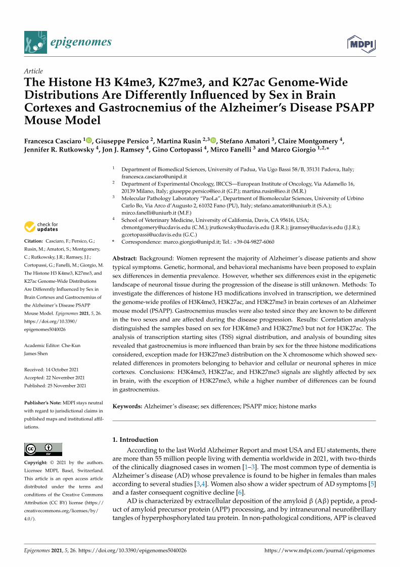
epigenomes
Article
The Histone H3 K4me3, K27me3, and K27ac Genome-WideDistributions Are Differently Influenced by Sex in BrainCortexes and Gastrocnemius of the Alzheimer’s Disease PSAPPMouse Model
Francesca Casciaro 1 , Giuseppe Persico 2, Martina Rusin 2,3 , Stefano Amatori 3, Claire Montgomery 4,Jennifer R. Rutkowsky 4, Jon J. Ramsey 4, Gino Cortopassi 4, Mirco Fanelli 3 and Marco Giorgio 1,2,*
�����������������
Citation: Casciaro, F.; Persico, G.;
Rusin, M.; Amatori, S.; Montgomery,
C.; Rutkowsky, J.R.; Ramsey, J.J.;
Cortopassi, G.; Fanelli, M.; Giorgio, M.
The Histone H3 K4me3, K27me3, and
K27ac Genome-Wide Distributions
Are Differently Influenced by Sex in
Brain Cortexes and Gastrocnemius of
the Alzheimer’s Disease PSAPP
Mouse Model. Epigenomes 2021, 5, 26.
https://doi.org/10.3390/
epigenomes5040026
Academic Editor: Che-Kun
James Shen
Received: 14 October 2021
Accepted: 22 November 2021
Published: 25 November 2021
Publisher’s Note: MDPI stays neutral
with regard to jurisdictional claims in
published maps and institutional affil-
iations.
Copyright: © 2021 by the authors.
Licensee MDPI, Basel, Switzerland.
This article is an open access article
distributed under the terms and
conditions of the Creative Commons
Attribution (CC BY) license (https://
creativecommons.org/licenses/by/
4.0/).
1 Department of Biomedical Sciences, University of Padua, Via Ugo Bassi 58/B, 35131 Padova, Italy;[email protected]
2 Department of Experimental Oncology, IRCCS—European Institute of Oncology, Via Adamello 16,20139 Milano, Italy; [email protected] (G.P.); [email protected] (M.R.)
3 Molecular Pathology Laboratory “PaoLa”, Department of Biomolecular Sciences, University of UrbinoCarlo Bo, Via Arco d’Augusto 2, 61032 Fano (PU), Italy; [email protected] (S.A.);[email protected] (M.F.)
4 School of Veterinary Medicine, University of California, Davis, CA 95616, USA;[email protected] (C.M.); [email protected] (J.R.R.); [email protected] (J.J.R.);[email protected] (G.C.)
* Correspondence: [email protected]; Tel.: +39-04-9827-6060
Abstract: Background: Women represent the majority of Alzheimer’s disease patients and showtypical symptoms. Genetic, hormonal, and behavioral mechanisms have been proposed to explainsex differences in dementia prevalence. However, whether sex differences exist in the epigeneticlandscape of neuronal tissue during the progression of the disease is still unknown. Methods: Toinvestigate the differences of histone H3 modifications involved in transcription, we determinedthe genome-wide profiles of H3K4me3, H3K27ac, and H3K27me3 in brain cortexes of an Alzheimermouse model (PSAPP). Gastrocnemius muscles were also tested since they are known to be differentin the two sexes and are affected during the disease progression. Results: Correlation analysisdistinguished the samples based on sex for H3K4me3 and H3K27me3 but not for H3K27ac. Theanalysis of transcription starting sites (TSS) signal distribution, and analysis of bounding sitesrevealed that gastrocnemius is more influenced than brain by sex for the three histone modificationsconsidered, exception made for H3K27me3 distribution on the X chromosome which showed sex-related differences in promoters belonging to behavior and cellular or neuronal spheres in micecortexes. Conclusions: H3K4me3, H3K27ac, and H3K27me3 signals are slightly affected by sexin brain, with the exception of H3K27me3, while a higher number of differences can be foundin gastrocnemius.
Keywords: Alzheimer’s disease; sex differences; PSAPP mice; histone marks
1. Introduction
According to the last World Alzheimer Report and most USA and EU statements, thereare more than 55 million people living with dementia worldwide in 2021, with two-thirdsof the clinically diagnosed cases in women [1–3]. The most common type of dementia isAlzheimer’s disease (AD) whose prevalence is found to be higher in females than malesaccording to several studies [3,4]. Women also show a wider spectrum of AD symptoms [5]and a faster consequent cognitive decline [6].
AD is characterized by extracellular deposition of the amyloid β (Aβ) peptide, a prod-uct of amyloid precursor protein (APP) processing, and by intraneuronal neurofibrillarytangles of hyperphosphorylated tau protein. In non-pathological conditions, APP is cleaved
Epigenomes 2021, 5, 26. https://doi.org/10.3390/epigenomes5040026 https://www.mdpi.com/journal/epigenomes

Epigenomes 2021, 5, 26 2 of 15
by α-secretase and consequently by the γ-secretases (PSEN), while in the amyloidogenicpathway, the β-secretase enzyme cleaves APP to produce a soluble fragment which isthen cleaved by γ-secretase to release the Aβ peptide [7]. Consistently, mice expressingthe mutated human γ-secretases 1 and the mutant chimeric mouse/human mutant APP(Mo/HuAPP695swe) (PSAPP mice) accumulate Aβ toxic plaques in the brain cortex anddevelop behavioral hallmarks of AD patients [8].
AD is a complex disease with a multiorgan involvement. Slow gait speed and de-creased grip strength are associated with cognitive decline [9], and muscle is known to beone of the tissues with the most sex-differentially expressed genes in humans [10].
In a 3xTg-AD mouse model, which contains three gene mutations, namely APPSwe,PS1M146V, and tauP301L, it has been reported that disfunctions in skeletal muscle occurat different levels. For example, they showed that skeletal muscle functionality is alreadyaffected in 3-month-old 3xTg-AD mice with an age-dependent accumulation of amyloid-β1-40 peptide [11]. Furthermore, in a double transgenic mouse model expressing a chimericmouse/human amyloid precursor protein (APP) with the Swedish mutation (APPswe)and a mutant human presenilin 1 (PS1) with the delta E9 (PS1∆E9), it has been shownthat skeletal muscle cells have a significantly decreased maximal mitochondrial oxygenconsumption capacity compared to non-transgenic, age-matched mice, with similar deficitsto those previously described in brain [12].
In the last years, multiple studies have shown the involvement of epigenetic regulationin the progression of AD. For example, it was found that some cytosines, particularlythose at -207 to approximately -182 in the promoter region of the APP gene, are mostlymethylated and their demethylation with age may lead to Aβ deposition in the agedbrain [13]. Moreover, different studies in mouse models of AD have shown an involvementof histone deacetylases in speeding up the progression of the disease [13].
Emerging evidence from animals and humans suggests that epigenetic mechanisms orenvironmental factors are likely to also play a role in the different incidence and progressionof the disease in the two sexes [14].
A good example of sex-specific epigenetic regulation is X chromosome inactivationin females, a process which occurs largely due to a combination of DNA methylation andhistone modifications [15].
Recently, it has been proposed that histone modifications may be involved in thedifferent responses to stress between males and females [16]. For example, Ramzan et al.have shown that the expression of histone variant H2A.Z has context-specific effects onthe regulation of fear memory and related disorders, so higher levels of H2A.Z in femalemice may represent a risk factor for PTSD and associated increased pain sensitivity [17].Furthermore, analysis of the bed nucleus of the stria terminalis and preoptic area in adultmale and female mice revealed 248 regions differently enriched in H3K4me3 in the twogroups [18].
Here, we explore the landscape of three well characterized transcription-associatedhistone modifications, both in brain cortexes and gastrocnemius muscle in a PSAPP mousemodel of AD. In particular, H3K4me3 and HK27ac are associated with gene activation,while H3K27me3 is correlated with repression and especially with the inactivation ofone copy of the X chromosome in females [19]. Multiple studies correlate alterations inthe signal of those histone marks with the aging process. For example, high levels ofH3K27me3 were observed in the brain of mice with an accelerated aging phenotype [20],while global histone acetylation was found regulated in aged mice brain [21]. Alterationsin H3K4me3 signal was observed in different aged tissues of mouse models [22,23]. Inhumans, H3K4me3 distribution in prefrontal neurons from 11 individuals resulted todecrease in 600 loci in early life and to increase in other 100 loci of aged adults [24].
Chromatin immunoprecipitations, followed by Illumina sequencing, were conductedfor these modifications and bioinformatics analyses results are here presented.

Epigenomes 2021, 5, 26 3 of 15
2. Materials and Methods2.1. Mice
Five female and five male PSAPP mice were generated at the mouse facility of theUniversity of California (Davis, CA, USA) and housed in polycarbonate cages on racksin a room with controlled temperature (22–24 ◦C) and humidity (40–60%). Mice wereindividually housed in a HEPA filtered room maintained on a 12-hr light–dark cycle.Health checks were conducted on all mice at least once a day. Sentinel mice were housedin the same room and exposed to bedding from the study mice on a weekly basis. Healthscreens were completed on sentinel mice every three months. Serology tests included MHV,Sendai, PVM, MPV, MVM, M.pul and arth, TMEV (GDVII), Ectro, EDIM, MAD1 and 2,LCM, Reo-3, and MNV. All tests were negative throughout the study. All animal protocolswere approved by the UC Davis Institutional Animal Care and Use Committee and werein accordance with the NIH guidelines for the Care and Use of Laboratory Animals.
Mice were multi-housed and provided ad libitum access to a chow diet LabDiet 5001(LabDiet, Saint Louis, MO, USA) prior to the start of the study. At 6 months of age, micewere singly housed and placed on a modified AIN-93 diet (Table S12). Food intake was setat 11.2 kcal/day, which was fed ~1 h prior to lights out, and water was provided ad lib.
Brain cortexes and gastroectonemius muscles were collected from sacrificed micefollowing standard procedures [25,26].
2.2. ChIPSeq
Brain cortexes and gastrocnemius muscle chromatins were obtained from five male(n = 4 for male cortex) and five female PSAPP-mice sacrificed at the age of 13 months whenthese animals begin to show symptoms of cognitive decline and immediately snap frozenin liquid nitrogen.
Chromatin extraction was performed on 10-mg tissues following the chromatin im-munoprecipitation (ChIP) standard procedure [27].
Extracted chromatin was immunoselected with anti-H3K4me3 (#39159, Lot 22119006,Active Motif, Carlsbad, CA, USA), anti-H3K27ac (ab4729, Lot. GR3231887-1; Abcam,Cambridge, UK) and H3K27me3 (07-449, Lot. 3091919; Millipore, Temecula, CA, USA) anti-bodies following the procedure already described without limited reversal of crosslinking(LRC) step [28].
The bound fractions were de-crosslinked, and purified DNA was used for librarypreparation. Libraries were then sequenced in 51 bp paired-end read mode on a NovaSeq2000 sequencer (Illumina, San Diego, CA, USA).
Sample c05 was excluded from the analysis due to problems during the preparation.
2.3. Computational Pipeline
Reads were aligned to mm10 using “bwa” (v0.7.17), a software package for mappinglow-divergent sequences against a large reference genome [29]. Unmapped reads, readswith a mapping quality (MAPQ) value smaller than 1, duplicate reads, and those thatmapped outside of chr 1–19 and Chr X were removed using SAMtools. Resulting alignmentreads (stored in a standard BAM format, which is the compressed binary version of a SAMfile that is used to represent aligned sequences up to 128 Mb) were converted into a bedpeformat (browser extensible data paired-end format, which helps to concisely describedisjoint genome features, such as structural variations or paired-end sequence alignments)using the “bamTobed” script of bedtools (v2.30.0) [30]. Peak detection was performed withepic2 software using the following parameters: fragment size = 200; window size = 200;g = 2 for H3K4me3 and H3K27ac; g = 3 for H3K27me3; FDR < 0.05; and e = 100 [31].Bedtools was also used to merge the peak files of each group prior to annotating them.
Differential binding analysis was performed by the Diffbind R package (v3.2.6) anddifferentially bound sites were identified among different conditions. Stringency in theanalysis was obtained by creating a consensus dataset for each condition, including peaks

Epigenomes 2021, 5, 26 4 of 15
that were present in at least three samples of the considered group. Only different bound(DB) sites with an FDR (false discovery rate) of <0.1 were considered [32].
The ChIPseeker R package (v1.28.3) was applied to annotate peak files and DB sitesusing the curated RefSeq set version 130306 [33].
Pathway analysis was conducted using the ingenuity pathway analysis (IPA) softwarefrom QIAGEN (version September 2021). Pathways with an absolute z-score of >2 wereconsidered significant.
For some analyses, BAM files of replicates from each group were merged usingBAMtools and indexed using SAMtools. Merged bam files were then used to generatea bigwig (a file format for display of dense, continuous data in a genome browser track)using deepTools bamCoverage (v3.5.1) with a bin size of 10 bp (size of the bins, in bases),bins per million (BPM) mapped reads, normalization (ChrX was ignored for normalization),and reads extended to 200 bp.
The signal around the TSSs was calculated for 23,359 genes. The signal, calculatedusing deepTools computeMatrix, was reported as a mean signal in bins of 10 bp, with arange of ±3 kB around the TSSs. Missing data were treated as zero. The output was thenplotted using plotHeatmap and plotProfile (deepTools) [34].
3. Results3.1. H3K4 and H3K27 me3 Analysis on X Chromosome Distinguishes Males and Females
We first checked the quality of sequencing results and no significant variability in thenumber of reads related to sex was revealed (Figure S1).
To estimate differences in the distribution of the analyzed histone’s marks betweenfemales and males, we performed a correlation analysis, including all the aligned readsin autosomes (chromosomes, chr 1-19) and X. According to Spearman correlation anal-ysis, males and females formed distinguishable clusters for H3K4me3 (Figure 1a,b) andfor H3K27me3 (Figure 1e,f), which is known to be involved in the X chromosome inac-tivation [35]. In contrast, H3K27ac showed higher correlation coefficients (above 0.9),indicating a greater similarity among all the analyzed samples (Figure 1c,d). As expected,the H3K4me3 and H3K27me3 sex-related correlation was lost if only autosome chromo-somes are considered, underlining the contribution of the X chromosome in generatingthis phenomenon (Figure S2).
3.2. Brain Cortex Shows a Signal around the Transcription Starting Sites (TSS) That Is MoreHomogenous Than Gastrocnemius
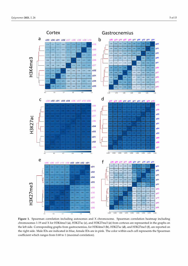
Since histone modifications play a critical role in transcription control, we investigatedthe signal around (−2.5 kb + 2.5 kb) the TSS present in all the chromosomes (n = 23,359 sites).The comparison of the average signal from all the TSS revealed a substantial differencebetween the two sexes only for H3K27me3 (Figure 2m,p), and not for H3K4me3 andH3K27ac (Figure 2a,d,g,j).
Focusing on the analysis of the average signals on TSS separating autosomes andthe X chromosome, the intensity of the H3K27me3 signal on the X chromosome scoredalmost double in females than males in both cortex and gastrocnemius (Figure 2o,r). Inthe gastrocnemius, a slight difference in the H3K27me3 signal on TSS was also found ifconsidering only the autosomes. Differences in the H3K27ac signal on TSS also appearedon the X chromosome of gastrocnemius.
These results indicate that sex differences of histone modifications distribution in thecortex of PSAPP mice occur to a lesser extent in comparison to the gastrocnemius, whosefiber composition and gene expression is influenced by sex [36,37].
Epigenomes 2021, 5, 26 5 of 15
Epigenomes 2021, 5, x FOR PEER REVIEW 5 of 16
Figure 1. Spearman correlation including autosomes and X chromosome. Spearman correlation heatmap including chro-mosomes 1-19 and X for H3K4me3 (a), H3K27ac (c), and H3K27me3 (e) from cortexes are represented in the graphs on the left side. Corresponding graphs from gastrocnemius, for H3K4me3 (b), H3K27ac (d), and H3K27me3 (f), are reported on the right side. Male IDs are indicated in blue, female IDs are in pink. The color within each cell represents the Spearman coefficient which ranges from 0.68 to 1 (maximal correlation).
Cortex Gastrocnemiusa b
e
c
f
d
H3K4
me3
H3K2
7ac
H3K2
7me3
Figure 1. Spearman correlation including autosomes and X chromosome. Spearman correlation heatmap includingchromosomes 1-19 and X for H3K4me3 (a), H3K27ac (c), and H3K27me3 (e) from cortexes are represented in the graphs onthe left side. Corresponding graphs from gastrocnemius, for H3K4me3 (b), H3K27ac (d), and H3K27me3 (f), are reported onthe right side. Male IDs are indicated in blue, female IDs are in pink. The color within each cell represents the Spearmancoefficient which ranges from 0.68 to 1 (maximal correlation).
Epigenomes 2021, 5, 26 6 of 15
Epigenomes 2021, 5, x FOR PEER REVIEW 6 of 16
3.2. Brain Cortex Shows a Signal around the Transcription Starting Sites (TSS) That Is More Homogenous Than Gastrocnemius
Since histone modifications play a critical role in transcription control, we investi-gated the signal around (−2.5 kb + 2.5 kb) the TSS present in all the chromosomes (n = 23,359 sites). The comparison of the average signal from all the TSS revealed a substantial difference between the two sexes only for H3K27me3 (Figure 2m,p), and not for H3K4me3 and H3K27ac (Figure 2a,d,g,j).
H3K4
me3
All chromosomes Autosomes X chromosomeCortex
Gastrocnemius
Cortex
Cortex
Gastrocnemius
Gastrocnemius
H3K2
7ac
H3K2
7me3
a b
e
c
fd
g h i
j k l
m n o
p q r
Figure 2. Signal around TSS of H3K4me3, H3K27ac, and H3K27me3 in cortex and gastrocnemius tissues. H3K4me3 (a–f),H3K27ac (g–l), and H3K27me3 (m–r) average density signal around TSS (±2.5 kb) is plotted for cortex and gastrocnemiusas indicated, for chromosomes 1–19 and X on the left, only autosomes in the middle and only X on the right. Males areindicated in blue, females in pink.

Epigenomes 2021, 5, 26 7 of 15
3.3. Binding Sites Are Differently Affected by Sex in Cortex and Gastrocnemius
The position of the marked H3 histones was investigated genome-wide using the epic2tool to identify enriched peaks. Overall, the number of peaks detected in the two sexesappeared to be comparable (Figure S3), except for the H3K4me3 in the female cortex, whosenumber was higher in three out of five samples. The analysis of peak distribution withinthe different genes features revealed that most H3K4me3 peaks were found in promoters(Figure 3a,b), although distal intergenic regions were more represented in female cortexes(Figure 3a). H3K27ac peaks were more uniformly distributed among the different features(Figure 3c,d), whereas the majority of the H3K27me3-enriched regions were identified indistal intergenic areas (Figure 3e,f).
Epigenomes 2021, 5, x FOR PEER REVIEW 8 of 16
Figure 3. Feature distribution of called peaks for H3K4me3, H3K27ac, and H3K27me3 in cortex and gastrocnemius tissues. Feature distribution of the identified peaks for H3K4me3 (a), H3K27ac (c), and H3K27me3 (e) in cortexes are reported on the left. Feature distribution of H3K4me3 (b), H3K27ac (d), and H3K27me3 (f) peaks found in the gastrocnemius are re-ported in the plots on the right. Males are in blue, females in pink. The color code indicating each genomic feature is shown.
Then, we identified the sites that showed different intensity of histone marks signals between the sexes (Diffbind analysis). To avoid an excess of false positive results due to the unspecific binding of the antibody during the ChIP, we generated a consensus dataset of peaks for each group (Table S1), as reported in the methods section, and we separately analyzed autosomes and X chromosome.
To investigate the heterogeneity in these sets of peaks among samples, principal com-ponent analyses (PCA) were performed. Results revealed that females and males separate only if considering the X chromosome (Figure 4d–f,j–l), with the principal component 1 showing the maximum variance for H3K27me3 (96% for both tissues) (Figure 4f,l) and the minimum for H3K27ac (around 55/59%) (Figure 4e,k). PCA for the H3K4me3 X chromo-some resulted in a more variable component 1 between the two sexes, with a higher vari-ance shown by cortexes (78%) (Figure 4d,j). Taking in account autosomes, male and female H3K4me3 and H3K27ac differentially marked peaks appeared and merged for both tis-sues (Figure 4a,b,g,h).
GastrocnemiusCortex
H3K4
me3
H3K2
7ac
a b
c
f
d
H3K2
7me3
e
Figure 3. Feature distribution of called peaks for H3K4me3, H3K27ac, and H3K27me3 in cortex and gastrocnemius tissues.Feature distribution of the identified peaks for H3K4me3 (a), H3K27ac (c), and H3K27me3 (e) in cortexes are reported on theleft. Feature distribution of H3K4me3 (b), H3K27ac (d), and H3K27me3 (f) peaks found in the gastrocnemius are reportedin the plots on the right. Males are in blue, females in pink. The color code indicating each genomic feature is shown.
Then, we identified the sites that showed different intensity of histone marks signalsbetween the sexes (Diffbind analysis). To avoid an excess of false positive results due tothe unspecific binding of the antibody during the ChIP, we generated a consensus datasetof peaks for each group (Table S1), as reported in the methods section, and we separatelyanalyzed autosomes and X chromosome.
To investigate the heterogeneity in these sets of peaks among samples, principal com-ponent analyses (PCA) were performed. Results revealed that females and males separate

Epigenomes 2021, 5, 26 8 of 15
only if considering the X chromosome (Figure 4d–f,j–l), with the principal component 1showing the maximum variance for H3K27me3 (96% for both tissues) (Figure 4f,l) andthe minimum for H3K27ac (around 55/59%) (Figure 4e,k). PCA for the H3K4me3 X chro-mosome resulted in a more variable component 1 between the two sexes, with a highervariance shown by cortexes (78%) (Figure 4d,j). Taking in account autosomes, male andfemale H3K4me3 and H3K27ac differentially marked peaks appeared and merged for bothtissues (Figure 4a,b,g,h).
Epigenomes 2021, 5, x FOR PEER REVIEW 9 of 16
Figure 4. Principal component analysis of Diffbind consensus peak sets. Principal component anal-ysis of the signals from regions selected after differential binding analysis are graphed. The left col-umn reports information on H3K4me3, the middle one reports on H3K27ac, while H327me3 is ana-lyzed on the right part of the figure.
The figure can be divided in two parts: the upper part presents the results related to the cortex for autosomes (a–c) and the X chromosome (d–f) for all the histone marks ana-lyzed, while the lower part shows the principal component analyses of autosomal (g–i) and X (j–l) chromosomes in muscle.
Considering autosomes, the Diffbinding analysis between males and females identi-fied only two sites for H3K4Me3 and H3K27ac and five for H3K27me3 in the cortex, whereas 139 H3K4me3, 206 H3K27me3, and 781 H3K27ac sites were discovered in the gastrocnemius (Table 1).
Table 1. Differentially bound sites of H3K4me3, H3K27ac, and H3K27me3 in cortex and gas-trocnemius tissues. The number of the differentially bound sites (FDR < 0.1) found in each compar-ison is reported.
Histone Modification Chromosomes N° of db Sites
in Cortex N° db Sites in
Gastrocnemius
Figure 4. Principal component analysis of Diffbind consensus peak sets. Principal componentanalysis of the signals from regions selected after differential binding analysis are graphed. The leftcolumn reports information on H3K4me3, the middle one reports on H3K27ac, while H327me3 isanalyzed on the right part of the figure.

Epigenomes 2021, 5, 26 9 of 15
The figure can be divided in two parts: the upper part presents the results relatedto the cortex for autosomes (a–c) and the X chromosome (d–f) for all the histone marksanalyzed, while the lower part shows the principal component analyses of autosomal (g–i)and X (j–l) chromosomes in muscle.
Considering autosomes, the Diffbinding analysis between males and females iden-tified only two sites for H3K4Me3 and H3K27ac and five for H3K27me3 in the cortex,whereas 139 H3K4me3, 206 H3K27me3, and 781 H3K27ac sites were discovered in thegastrocnemius (Table 1).
Table 1. Differentially bound sites of H3K4me3, H3K27ac, and H3K27me3 in cortex and gastrocnemius tissues. The numberof the differentially bound sites (FDR < 0.1) found in each comparison is reported.
Histone Modification Chromosomes N◦ of db Sites in Cortex N◦ db Sites in Gastrocnemius
Autosomes 2 139H3K4me3 X chromosome 65 102
Autosomes 2 781H3K27ac X chromosome 56 69
H3K27me3 Autosomes 5 206X chromosome 2783 855
Regarding the X chromosome, Diffbind identified 65 H3K4me3, 56 H3K27ac, and2782 H3K27me3 sex differential sites in the cortex, while 102 H3K4me3, 69 H3K27ac,and H3K27me3 sex differential sites in the gastrocnemius (Table 1). The investigation ofthe location of these sites (Tables S2–S7) revealed that a larger number of sex differentialpromoters involves the H3K27me3 in the X chromosome and the H3K27ac in the autosomes.
3.4. Genes Involved in Cognitive Functions Show Different H3K27me3 Signal between Sex
To disclose the function of the genes, whose promoters were differently histone-marked by sex, ingenuity pathway analysis (IPA) was performed on the different lists ofthe identified genes. IPA is a web-based software application for the analysis, integration,and interpretation of data derived from OMICS experiments, and it is able not only tocategorize genes in pathways and biological functions, but it can give an idea of thedirection of the regulation (expressed by a positive or negative z-score) [38]. Due to thelow number of genes differentially marked between the sexes in the cases of H3K4me3and H3K27ac, few terms for biological processes were recognized (Tables S8–S11). For IPAanalysis on the genes whose promoters showed different sex-related H3K27me3 peaks(n = 488), the majority of these genes were classified as involved in processes linked toneuronal cell functions and several to behavioral functions (Table 2). Notably, among thegenes assigned with these pathways, eight (HSD17B10, GATA1, HTR2C, OGT, AGTR2,CYBB, GRIA3, MAOA) were reported to be associated to AD according to the GeneCardsdatabase (https://www.genecards.org/, accessed on 27 September 2021) with a relevancescore superior to two [39–46].
In the gastrocnemius, the sex-dependent H3K27ac-marked promoters (n = 123) showedhigher signals in females on genes related to inflammation and lower signals on thoserelated to apoptosis (Table S10). Genes assigned with these terms are the brain-derivedneurotrophic factor (BDNF), the histamine N-methyltransferase (HNMT), and the insulinreceptor factor 1 (IRS1), which are all associated with AD (with a relevance score of 14.34,76, and 3.65, respectively) [47–49].
Finally, the cortex shares with gastrocnemius sex differences in H3K27me3 promotersof genes involved in cytoskeleton organization (Tables 2 and 3).

Epigenomes 2021, 5, 26 10 of 15
Table 2. Biological signatures associated to H3K27me3-regulated promoters on the X chromosome in cortex. Results of an ingenuity pathway analysis for promoters differentially markedby H3K27me3 in X chromosome from PSAPP cortex are reported. Only terms with an activation z-score above |2| are considered. All the results are presented as increased/decreased infemales with respect to males.
Cortex Diseases or Functions(H3K27me3 X) p-Value Predicted Activation State Activation Z-Score Molecules N◦ of Molecules
Spatial learning 2.70 × 10⁻⁵ Increased 2.646 AP1S2, ARHGEF9, CYBB, DLG3, Gprasp2, GRIPAP1, HTR2C,KDM5C, MECP2, OPHN1, PHF8, SLC6A8, UBE2A, ZDHHC9 14
Growth of neurites 4.98 × 10⁻³ Increased 2.138AR, ARX, CCDC120, CDKL5, DCX, EFNB1, ELK1, FRMD7, GJB1,MAO, MID1, OGT, PLXNA3, RAB33A, SLC25A5, SNX12, SYN1,
TLR7, TRPC519
Organization of cytoskeleton 1.17 × 10⁻⁷ Increased 2.074
AGTR2, AMOT, AR, ARHGAP4, ARHGAP6, ARHGEF9, ATP7A,BRWD3, CAPN6, CDK16, CDKL5, CETN2, CUL4B, CXCR3, CYBB,
DCX, DGKK, DLG3, DOCK11, EFNB1, ELK1, F8A1 (includesothers), FGD1, FLNA, FRMD7, GATA1, GDI1, GJB1, GPM6B,
Gprasp2, HDAC6, HDAC8, HPRT1, IL1RAPL1, KDM5C, MAOA,MECP2, MID1, MID1IP1, mir-384, MPP1, MTM1, NR0B1, OFD1,OGT, OPHN1, PAK3, PCYT1B, PLS3, PLXNA3, PLXNB3, POF1B,
PQBP1, RAB33A, RPGR, RPS6KA3, SH3KBP1, SHROOM2,SHROOM4, SLITRK2, SYN1, TLR7, Tmsb4x (includes others),
TRPC5, USP9X
65
Organization of cytoplasm 8.76 × 10⁻⁷ Increased 2.074
AGTR2, AMOT, AR, ARHGAP4, ARHGAP6, ARHGEF9, ATP7A,BRWD3, CAPN6, CDK16, CDKL5, CETN2, CUL4B, CXCR3, CYBB,
DCX, DGKK, DLG3, DOCK11, EFNB1, ELK1, F8A1(includesothers), FGD1, FLNA, FRMD7, GATA1, GDI1, GJB1, GPM6B,
Gprasp2, HCFC1, HDAC6, HDAC8, HPRT1, HSD17B10, IL1RAPL1,KDM5C, MAOA, MECP2, MID1, MID1IP1, mir-384, MPP1, MTM1,
NR0B1, OFD1, OGT, OPHN1, PAK3, PCYT1B, PLS3, PLXNA3,PLXNB3, POF1B, PQBP1, RAB33A, RPGR, RPS6KA3, SH3KBP1,
SHROOM2, SHROOM4, SLITRK, SYN1, TLR7, Tmsb4x (includesothers), TRPC5, USP9X
67
Tremor 1.14 × 10⁻³ Decreased −2.011 ARAF, CA5B, GABRQ, GJB1, GPM6B, GRIA3, IKBKG, MECP2,PLP1, TIMP1 10
Differentiation of Th2 cells 1.09 × 10⁻² Decreased −2.236 FOXP3, GATA1, IL13RA2, let-7, TLR7 5
Movement Disorders 2.29 × 10⁻³ Decreased −2.242
ABCB7, AIFM1, AMER1, AP1S2, AR, ARAF, ARHGEF9, ARMCX2,AR, ATP6AP2, BCAP31, CA5B, CDKL5, CETN2, CXCR3, F8A1
(includes others), GABRQ, GJB1, GPM6B, GRIA3, GRPR, HPRT1,HTR2C, IDS, IGSF1, IKBKG, KCND1, MAOA, MECP2, OGT, PDK3,PGK1, PGRMC1, PLP1, PRKX, PTCHD1, RGN, RS1, SRPX, SRPX2,
SYN1, SYTL4, TIMP1, Tmsb4x (includes others), XIAP
45
Motor dysfunction or movementdisorder 1.72 × 10⁻³ Decreased −2.666
ABCB7, AIFM1, AMER1, AP1S2, AR, ARAF, ARHGEF9, ARMCX2,AR, ATP6AP2, BCAP31, CA5B, CDKL5, CETN2, CXCR3, F8A1
(includes others), GABRQ, GJB1, GPM6B, GRIA3, GRPR, HPRT1,HTR2C, IDS, IGSF1, IKBKG, KCND1, MAOA, MECP2, MTM1,
OGT, PDK3, PGK1, PGRMC1, PLP1, PRKX, PTCHD1, RGN, RS1,SRPX, SRPX2, SYN1, SYTL4, TIMP1, Tmsb4x (includes others),
XIAP
46

Epigenomes 2021, 5, 26 11 of 15
Table 3. Biological signatures associated to H3K27me3-regulated promoters on gastrocnemius X chromosome. Results of an ingenuity pathway analysis for promoters differentiallymarked by H3K27me3 in X chromosome from PSAPP gastrocnemius are reported. Only terms with an activation z-score above |2| are taken in consideration. All the results are presentedas regulated (increased/decreased) in females with respect to males.
Gastrocnemius Diseases orFunctions (H3K27me3 X) p-Value Predicted Activation State Activation Z-Score Molecules N◦ of Molecules
Organization of actin cytoskeleton 27.23 × 10⁻³ Increased 2.000 ARHGAP4, EFNB1, FGD1, GPM6B, MSN, OPHN1, PAK3, PLS3,Tmsb4x (includes others) 9
Abdominal cancer 1.41 × 10⁻⁵ Decreased −2.000
ABCB7, ACE2, AIFM1, AMER1, ARHGAP36, ARHGAP4,ARHGEF9, ARMCX1, ARMCX2, ARMCX3, ARMCX4, ARMCX5,
ARX, ASB9, ATP2B3, AVPR2, AWAT2, BCOR, BCORL1, BEX1, BGN,BMX, BRS3, BTK, CA5B, CACNA1F, CCDC160, CD40LG, CDX4,
CHRDL1, CLDN2, CNGA2, CNKSR2, COL4A5, DACH2,DCAF12L2, DGAT2L6, DOCK11, DUSP9, EFNB1, EGFL6, ELF4,ERCC6L, FAM155B, FGD1, FOXO4, FOXR2, FRMD7, FRMPD3,GABRA3, GABRQ, GATA1, GDPD2, GJB1, GPC3, GPC4, GPM6,
GPR101, GPR143, GPR50, GRIA3, GRPR, GSPT2, GUCY2F, HCFC1,HDAC8, HPRT1, HSD17B1, HTR2C, IDH3G, IGSF1, IL13RA1,
IL1RAPL1, IL1RAPL2, IQSEC2, IRAK1, KCND1, KCNE5, KLF8,KLHL15, L1CAM, LANCL3, LHFPL1, LONRF3, MAGEA10,
MAGEA11, MAGED1, MAGEE1, MAGEE2, MAP7D2, MBNL3,mir-452, MSN, NAA10, NAP1L2, NEXMIF, NONO, NRK, NYX,OGT, OPHN1, OPN1LW, OTUD6A, PAK3, PCDH11X, PCDH19,
PDK3, PDZD4, PHF6, PHKA1, PLAC1, PLS3, PLXNA3, PLXNB3,PNMA3, PNMA5, PRKX, PRPS1, PRPS2, PTCHD1, RAB33A, RAI2,RAP2C, RBM41, RNF128, RPS4Y1, RRAGB, SLC16A2, SLC25A43,SLC38A5, SLC6A8, SLC7A3, SLITRK4, SOWAHD, SOX3, SRPX,STARD8, SYN1, SYTL4, TAB3, TAF1, TBX22, TCEANC, THOC2,
TMEM164, TMEM255A, TMEM47, TREX2, TRPC5, TSC22D3,TSPYL2, UPF3B, USP51, UTP14A, VGLL1, YIPF6, ZC4H2,
ZCCHC12, ZDHHC9, ZFX, ZIC3, ZMYM3
162

Epigenomes 2021, 5, 26 12 of 15
4. Discussion
As observed in patients and in animal models of AD, the disease progresses differentlyin the two sexes. The brains of PSAPP female mice accumulate significantly more amyloidplaques than male mice [50] and show more severe angiopathy and inflammation [51] from6 months of age. Consistently, PSAPP female mice show lower cognitive abilities thanPSAPP male mice [52]. Indeed, neurodegeneration and cognitive decline are worse amongfemales from different models of mice with dementia [53]. Because of this difference insusceptibility to the disease, the question arises as to whether epigenetic signal may beinvolved in the different prevalence of the diseases between sexes.
In the present study, we determined the H3K4me3, H3K27ac, and H3K27me3 genome-wide profiles of cortexes from 13-month-old PSAPP male and female mice. The comparisonof the global distribution of H3K4 and H3K27me3 signals revealed differences in the Xchromosome capable to distinguish males and females, whereas the H3K27ac distributionwas more homogenous between sexes. As expected, this sex-dependent regulation wasparticularly evident for the H3K27me3 signal around the TSS of genes localized on theX chromosome, since the inactivation process of genome portions of one of the two Xchromosomes present in females.
Searching for differently bound sites also confirmed that the epigenetic landscapefor the three considered histone modifications in the PSAPP mice is less influenced bysex in the brain than in muscle, whose transcriptomic profile is known to be particularlydifferent between sexes [54]. However, a significantly different level of H3K27me3 on the Xchromosome was observed in the cortex of male and female PSAPP mice. Interestingly,several of these X chromosome sites are involved in the regulation of neuronal functions,such as spatial learning, cellular organization, and development, and some of them arereported to be associated with AD [39–46].
Autosomes and the sex chromosomes differ in their evolutionary origins and in theirinvolvement in cognitive functions. Despite many similarities between females and males,sex differences are present in learning and memory [55]. Notably, in humans, 3.75% ofall genes are located on the X chromosome [56] and almost one third of these genes areinvolved in cognitive functions [57]. In females, one of the copies of the X chromosome issilenced. This process of X-chromosome inactivation evolved as a mechanism to regulategene dosage. However, it does not affect all genes equally and those genes that aredifferently regulated, particularly under stress conditions, such as the appearance ofamyloid plaques in the brain, may impact on the progression of neuronal disease.
Histone modifications are reported to be involved in the genome remodeling duringlearning and memory [58]. In particular, an increase in H3K4me3 in the mouse hip-pocampus has been related to long-term memory [59]. Numerous studies indicate thathippocampus-dependent memory and synaptic plasticity may rely on histone acetyltrans-ferases and histone deacetylases activity [16]. However, similar to histone acetylation, themajority of studies on histone methylation have exclusively used males [16]. Thus, theimpact of histone methylation in mediating sex- dependent memory processes are not wellunderstood. Some evidence suggests that the activity of histone methyltransferases anddemethylases may be influenced by sex. For example, the histone demethylase KDM5Cand UTX are coded by X-linked genes and escape X-inactivation in females, and maymediate sex differences in brain development, memory, and behavior [60,61].
In conclusion, this study reveals that the chromatin of brain cortex from PSAPP miceshows a sex-dependent signature of histone modifications distinct from other tissues,such as the gastrocnemius. Epigenetic signals have been suggested to be involved insex-dependent cognitive decline even if, so far, no epigenomes of male and female cortexesof AD models are available. The results presented here show that important sex differencesexist in the distribution of histone modifications in transcription control regions of severalgenes involved in neuronal functions that may be involved in the cognitive decline inAD patients.

Epigenomes 2021, 5, 26 13 of 15
Supplementary Materials: The following are available online at https://www.mdpi.com/article/10.3390/epigenomes5040026/s1, Figure S1: Number of aligned reads, Figure S2: Spearman correlationincluding only autosomes, Figure S3: Number of peaks, Table S1: Number of regions investigatedduring the differential bounding analyses, Table S2: H3K4me3 differentially bound sites and re-lated annotations in cortex, Table S3: H3K4me3 differentially bound sites and related annotationsin gastrocnemius, Table S4: H3K27ac differentially bound sites and related annotations in cortex,Table S5: H3K27ac differentially bound sites and related annotations in gastrocnemius, Table S6:H3K27me3 differentially bound sites and related annotations in cortex, Table S7: H3K27me3 differen-tially bound sites and related annotations in gastrocnemius, Table S8: Biological signatures associatedto H3K4me3-regulated promoters on cortex X chromosome, Table S9: Biological signatures associatedto H3K4me3-regulated promoters on gastrocnemius X chromosome, Table S10: Biological signaturesassociated to H3K27ac-regulated promoters on gastrocnemius autosomes, Table S11: Biologicalsignatures associated to H3K27ac-regulated promoters on gastrocnemius X chromosome, Table S12:Diet composition.
Author Contributions: Conceptualization, F.C., G.P., J.R.R., J.J.R., G.C., M.F. and M.G.; methodology,S.A., C.M.; data analysis, F.C. and G.P.; investigation, F.C. and M.R.; resources, G.C. and M.G.; datacuration, F.C.; writing F.C. and M.G.; project coordination, M.G. All authors have read and agreed tothe published version of the manuscript.
Funding: National Institute on Aging—NIH U.S. grant #1R56AG057163-01A1.
Institutional Review Board Statement: All experiments were conducted in accordance with theNational Institutes of Health Guidelines for the Care and Use of Laboratory Animals. All proce-dures were approved by the Institutional Animal Care and Use Committee of the University ofCalifornia, Davis.
Informed Consent Statement: Not applicable.
Data Availability Statement: ChIP-seq data are deposited on GEO repository and accessible withGSE189260 number (https://www.ncbi.nlm.nih.gov/geo (accessed on 4 November 2021)). All thedata that support the figures and the other findings are available from the authors upon request.
Conflicts of Interest: The authors declare no conflict of interest.
References1. Thies, W.; Bleiler, L. Alzheimer’s Association 2013 Alzheimer’s Disease Facts and Figures. Alzheimer’s Dement. 2013, 9, 208–245.
[CrossRef]2. Gauthier, S.; Rosa-Neto, P.; Morais, J.; Webster, C. World Alzheimer Report 2021: Journey through the Diagnosis of Dementia;
Alzheimer’s Disease International: London, UK.3. Beam, C.R.; Kaneshiro, C.; Jang, J.Y.; Reynolds, C.A.; Pedersen, N.L.; Gatz, M. Differences Between Women and Men in Incidence
Rates of Dementia and Alzheimer’s Disease. J. Alzheimer’s Dis. 2018, 64, 1077–1083. [CrossRef]4. Andersen, K.; Launer, L.; Dewey, M.; Letenneur, L.; Ott, A.; Copeland, J.; Dartigues, J.; Kragh-Sorensen, P.; Baldereschi, M.;
Brayne, C.; et al. Gender Differences in the Incidence of AD and Vascular Dementia: The EURODEM Studies. EURODEMIncidence Research Group. Neurology 1999, 53, 1992–1997. [CrossRef] [PubMed]
5. Schmidt, R.; Kienbacher, E.; Benke, T.; Dal-Bianco, P.; Delazer, M.; Ladurner, G.; Jellinger, K.; Marksteiner, J.; Ransmayr, G.;Schmidt, H.; et al. Sex Differences in Alzheimer’s Disease. Neuropsychiatry 2008, 22, 1–15.
6. Tschanz, J.T.; Corcoran, C.D.; Schwartz, S.; Treiber, K.; Green, R.C.; Norton, M.C.; Mielke, M.M.; Piercy, K.; Steinberg, M.;Rabins, P.V.; et al. Progression of Cognitive, Functional and Neuropsychiatric Symptom Domains in a Population Cohort withAlzheimer’s Dementia The Cache County Dementia Progression Study. Am. J. Geriatr. Psychiatry 2011, 19, 532–542. [CrossRef][PubMed]
7. Chen, X.-Q.; Mobley, W.C. Alzheimer Disease Pathogenesis: Insights From Molecular and Cellular Biology Studies of OligomericAβ and Tau Species. Front. Neurosci. 2019, 13, 659. [CrossRef]
8. Ordoñez-Gutierrez, L.; Fernandez-Perez, I.; Herrera, J.L.; Anton, M.; Benito-Cuesta, I.; Wandosell, F. AβPP/PS1 Transgenic MiceShow Sex Differences in the Cerebellum Associated with Aging. J. Alzheimer’s Dis. 2016, 54, 645–656. [CrossRef]
9. Ogawa, Y.; Kaneko, Y.; Sato, T.; Shimizu, S.; Kanetaka, H.; Hanyu, H. Sarcopenia and Muscle Functions at Various Stages ofAlzheimer Disease. Front. Neurol. 2018, 9, 710. [CrossRef] [PubMed]
10. Gershoni, M.; Pietrokovski, S. The Landscape of Sex-Differential Transcriptome and Its Consequent Selection in Human Adults.BMC Biol. 2017, 15, 7. [CrossRef]

Epigenomes 2021, 5, 26 14 of 15
11. Monteiro-Cardoso, V.; Castro, M.; Oliveira, M.M.; Moreira, P.; Peixoto, F.; Videira, R. Age-Dependent Biochemical Dysfunction inSkeletal Muscle of Triple-Transgenic Mouse Model of Alzheimer’s Disease. Curr. Alzheimer Res. 2015, 12, 100–115. [CrossRef][PubMed]
12. Schuh, R.A.; Jackson, K.C.; Schlappal, A.E.; Spangenburg, E.E.; Ward, C.W.; Park, J.H.; Dugger, N.; Shi, G.L.; Fishman, P.S.Mitochondrial Oxygen Consumption Deficits in Skeletal Muscle Isolated from an Alzheimer’s Disease-Relevant Murine Model.BMC Neurosci. 2014, 15, 1–12. [CrossRef] [PubMed]
13. Liu, X.; Jiao, B.; Shen, L. The Epigenetics of Alzheimer’s Disease: Factors and Therapeutic Implications. Front. Genet. 2018, 9, 579.[CrossRef]
14. Alagiakrishnan, K.; Gill, S.S.; Fagarasanu, A. Genetics and Epigenetics of Alzheimer’s Disease. Postgrad. Med. J. 2012, 88, 522–529.[CrossRef] [PubMed]
15. Avner, P.; Heard, E. X-Chromosome Inactivation: Counting, Choice and Initiation. Nat. Rev. Genet. 2001, 2, 59–67. [CrossRef][PubMed]
16. Keiser, A.A.; Wood, M.A. Examining the Contribution of Histone Modification to Sex Differences in Learning and Memory. Learn.Mem. 2019, 26, 318–331. [CrossRef] [PubMed]
17. Ramzan, F.; Creighton, S.; Hall, M.; Baumbach, J.; Wahdan, M.; Poulson, S.; Michailidis, V.; Stefanelli, G.; Narkaj, K.; Tao, C.; et al.Sex-Specific Effects of the Histone Variant H2A.Z on Fear Memory, Stress-Enhanced Fear Learning and Hypersensitivity to Pain.Sci. Rep. 2020, 10, 14331. [CrossRef] [PubMed]
18. Shen, E.; Ahern, T.; Cheung, I.; Straubhaar, J.; Dincer, A.; Houston, I.; de Vries, G.; Akbarian, S.; Forger, N. Epigenetics and SexDifferences in the Brain: A Genome-Wide Comparison of Histone-3 Lysine-4 Trimethylation (H3K4me3) in Male and FemaleMice. Exp. Neurol. 2015, 268, 21–29. [CrossRef]
19. Lawrence, M.; Daujat, S.; Schneider, R. Lateral Thinking: How Histone Modifications Regulate Gene Expression. Trends Genet.2016, 32, 42–56. [CrossRef]
20. Wang, C.M.; Tsai, S.N.; Yew, T.W.; Kwan, Y.W.; Ngai, S.M. Identification of Histone Methylation Multiplicities Patterns in theBrain of Senescence-Accelerated Prone Mouse. Biogerontology 2010, 11, 87–102. [CrossRef]
21. Ryu, S.H.; Kang, K.; Yoo, T.; Joe, C.O.; Chung, J.H. Transcriptional Repression of Repeat-Derived Transcripts Correlates withHistone Hypoacetylation at Repetitive DNA Elements in Aged Mice Brain. Exp. Gerontol. 2011, 46, 811–818. [CrossRef] [PubMed]
22. Sun, D.; Luo, M.; Jeong, M.; Rodriguez, B.; Xia, Z.; Hannah, R.; Wang, H.; Le, T.; Faull, K.F.; Chen, R.; et al. Epigenomic Profilingof Young and Aged HSCs Reveals Concerted Changes during Aging That Reinforce Self-Renewal. Cell Stem Cell 2014, 14, 673–688.[CrossRef] [PubMed]
23. Dhawan, S.; Tschen, S.-I.; Bhushan, A. Bmi-1 Regulates the Ink4a/Arf Locus to Control Pancreatic β-Cell Proliferation. Genes Dev.2009, 23, 906–911. [CrossRef]
24. Cheung, I.; Shulha, H.P.; Jiang, Y.; Matevossian, A.; Wang, J.; Weng, Z.; Akbarian, S. Developmental Regulation and IndividualDifferences of Neuronal H3K4me3 Epigenomes in the Prefrontal Cortex. Proc. Natl. Acad. Sci. USA 2010, 107, 8824–8829.[CrossRef] [PubMed]
25. Meyerhoff, J.; Muhie, S.; Chakraborty, N.; Naidu, L.; Sowe, B.; Hammamieh, R.; Jett, M.; Gautam, A. Microdissection of MouseBrain into Functionally and Anatomically Different Regions. J. Vis. Exp. 2021, 168, e61941. [CrossRef]
26. Cutler, A.; Corbett, A.; Pavlath, G. Biochemical Isolation of Myonuclei from Mouse Skeletal Muscle Tissue. Bio-Protocol 2017,7, e2654. [CrossRef] [PubMed]
27. Schmidt, D.; Wilson, M.; Spyrou, C.; Brown, G.; Hadfield, J.; Odom, D. ChIP-Seq: Using High-Throughput Sequencing to DiscoverProtein-DNA Interactions. Methods 2009, 48, 240–248. [CrossRef]
28. Amatori, S.; Persico, G.; Paolicelli, C.; Hillje, R.; Sahnane, N.; Corini, F.; Furlan, D.; Luzi, L.; Minucci, S.; Giorgio, M.; et al.Epigenomic Profiling of Archived FFPE Tissues by Enhanced PAT-ChIP (EPAT-ChIP) Technology. Clin. Epigenetics 2018, 10, 143.[CrossRef]
29. Li, H.; Durbin, R. Fast and Accurate Short Read Alignment with Burrows-Wheeler Transform. Bioinformatics 2009, 25, 1754–1760.[CrossRef]
30. Quinlan, A.; Hall, I. BEDTools: A Flexible Suite of Utilities for Comparing Genomic Features. Bioinformatics 2010, 26, 841–842.[CrossRef]
31. Stovner, E.; Sætrom, P. Epic2 Efficiently Finds Diffuse Domains in ChIP-Seq Data. Bioinformatics 2019, 35, 4392–4393. [CrossRef][PubMed]
32. Stark, R.; Brown, G. DiffBind: Differential Binding Analysis of ChIP-Seq Peak Data. Available online: https://bioconductor.org/packages/release/bioc/vignettes/DiffBind/inst/doc/DiffBind.pdf (accessed on 1 October 2021).
33. Yu, G.; Wang, L.-G.; He, Q.-Y. ChIPseeker: An R/Bioconductor Package for ChIP Peak Annotation, Comparison and Visualization.Bioinformatics 2015, 31, 2382–2383. [CrossRef] [PubMed]
34. Ramírez, F.; Ryan, D.P.; Grüning, B.; Bhardwaj, V.; Kilpert, F.; Richter, A.S.; Heyne, S.; Dündar, F.; Manke, T. DeepTools2: A nextGeneration Web Server for Deep-Sequencing Data Analysis. Nucleic Acids Res. 2016, 44, W160–W165. [CrossRef] [PubMed]
35. Rougeulle, C.; Chaumeil, J.; Sarma, K.; Allis, C.; Reinberg, D.; Avner, P.; Heard, E. Differential Histone H3 Lys-9 and Lys-27Methylation Profiles on the X Chromosome. Mol. Cell. Biol. 2004, 24, 5475–5484. [CrossRef]
36. Haizlip, K.M.; Harrison, B.C.; Leinwand, L.A. Sex-Based Differences in Skeletal Muscle Kinetics and Fiber-Type Composition.Physiology 2015, 30, 30–39. [CrossRef] [PubMed]

Epigenomes 2021, 5, 26 15 of 15
37. Welle, S.; Tawil, R.; Thornton, C.A. Sex-Related Differences in Gene Expression in Human Skeletal Muscle. PLoS ONE 2008,3, e1385. [CrossRef]
38. Ingenuity Downstream Effects Analysis in IPA®. Available online: http://pages.ingenuity.com/rs/ingenuity/images/0812%20downstream_effects_analysis_whitepaper.pdf (accessed on 4 November 2021).
39. Takehashi, M.; Tanaka, S.; Masliah, E.; Ueda, K. Association of Monoamine Oxidase A Gene Polymorphism with Alzheimer’sDisease and Lewy Body Variant. Neurosci. Lett. 2002, 327, 79–82. [CrossRef]
40. Bodily, P.M.; Fujimoto, M.S.; Page, J.T.; Clement, M.J.; Ebbert, M.T.W.; Ridge, P.G. A Novel Approach for Multi-SNP GWAS andIts Application in Alzheimer’s Disease. BMC Bioinform. 2016, 17, 455–463. [CrossRef]
41. Alsema, A.M.; Jiang, Q.; Kracht, L.; Gerrits, E.; Dubbelaar, M.L.; Miedema, A.; Brouwer, N.; Hol, E.M.; Middeldorp, J.;van Dijk, R.; et al. Profiling Microglia From Alzheimer’s Disease Donors and Non-Demented Elderly in Acute Human Post-mortem Cortical Tissue. Front. Mol. Neurosci. 2020, 13, 134. [CrossRef] [PubMed]
42. Quitterer, U.; AbdAlla, S. Improvements of Symptoms of Alzheimer’s Disease by Inhibition of the Angiotensin System. Pharmacol.Res. 2020, 154, 104230. [CrossRef]
43. Dos Santos, J.; Vizuete, A.; Hansen, F.; Biasibetti, R.; Gonçalves, C. Early and Persistent O-GlcNAc Protein Modification in theStreptozotocin Model of Alzheimer’s Disease. J. Alzheimer’s Dis. 2018, 61, 237–249. [CrossRef]
44. Holmes, C.; Arranz, M.; Collier, D.; Powell, J.; Lovestone, S. Depression in Alzheimer’s Disease: The Effect of Serotonin ReceptorGene Variation. Am. J. Med. Genet. 2003, 119B, 40–43. [CrossRef]
45. He, X.; Isaacs, C.; Yang, S. Roles of Mitochondrial 17β-Hydroxysteroid Dehydrogenase Type 10 in Alzheimer’s Disease. J.Alzheimer’s Dis. 2018, 62, 665–673. [CrossRef] [PubMed]
46. Chu, J.; Wisniewski, T.; Praticò, D. GATA1-Mediated Transcriptional Regulation of the γ-Secretase Activating Protein IncreasesAβ Formation in Down Syndrome. Ann. Neurol. 2016, 79, 138–143. [CrossRef]
47. Giuffrida, M.L.; Copani, A.; Rizzarelli, E. A Promising Connection between BDNF and Alzheimer’s Disease. Aging 2018, 10,1791–1792. [CrossRef] [PubMed]
48. Panula, P.; Rinne, J.; Kuokkanen, K.; Eriksson, K.; Sallmen, T.; Kalimo, H.; Relja, M. Neuronal Histamine Deficit in Alzheimer’sDisease. Neuroscience 1998, 82, 993–997. [CrossRef]
49. Wang, W.; Tanokashira, D.; Fukui, Y.; Maruyama, M.; Kuroiwa, C.; Saito, T.; Saido, T.; Taguchi, A. Serine Phosphorylation of IRS1Correlates with Aβ-Unrelated Memory Deficits and Elevation in Aβ Level Prior to the Onset of Memory Decline in AD. Nutrients2019, 11, 1942. [CrossRef]
50. Wang, J.; Tanila, H.; Puoliväli, J.; Kadish, I.; van Groen, T. Gender Differences in the Amount and Deposition of Amyloidbeta inAPPswe and PS1 Double Transgenic Mice. Neurobiol. Dis. 2003, 14, 318–327. [CrossRef]
51. Jiao, S.; Bu, X.; Liu, Y.; Zhu, C.; Wang, Q.; Shen, L.; Liu, C.; Wang, Y.; Yao, X.; Wang, Y. Sex Dimorphism Profile of Alzheimer’sDisease-Type Pathologies in an APP/PS1 Mouse Model. Neurotox. Res. 2016, 29, 256–266. [CrossRef]
52. Li, X.; Feng, Y.; Wu, W.; Zhao, J.; Fu, C.; Li, Y.; Ding, Y.; Wu, B.; Gong, Y.; Yang, G.; et al. Sex Differences between APPswePS1dE9Mice in A-Beta Accumulation and Pancreatic Islet Function during the Development of Alzheimer’s Disease. Lab. Anim. 2016, 50,275–285. [CrossRef]
53. Yang, J.-T.; Wang, Z.-J.; Cai, H.-Y.; Yuan, L.; Hu, M.-M.; Wu, M.-N.; Qi, J.-S. Sex Differences in Neuropathology and CognitiveBehavior in APP/PS1/Tau Triple-Transgenic Mouse Model of Alzheimer’s Disease. Neurosci. Bull. 2018, 34, 736–746. [CrossRef]
54. Lopes-Ramos, C.M.; Chen, C.Y.; Kuijjer, M.L.; Paulson, J.N.; Sonawane, A.R.; Fagny, M.; Platig, J.; Glass, K.; Quackenbush, J.;DeMeo, D.L. Sex Differences in Gene Expression and Regulatory Networks across 29 Human Tissues. Cell Rep. 2020, 31, 107795.[CrossRef]
55. Andreano, J.M.; Cahill, L. Sex Influences on the Neurobiology of Learning and Memory. Learn. Mem. 2009, 16, 248–266. [CrossRef]56. Skuse, D.H. X-Linked Genes and Mental Functioning. Hum. Mol. Genet. 2005, 14, 27–32. [CrossRef]57. Laumonnier, F.; Cuthbert, P.C.; Grant, S.G.N. The Role of Neuronal Complexes in Human X-Linked Brain Diseases. Am. J. Hum.
Genet. 2007, 80, 205–220. [CrossRef] [PubMed]58. Campbell, R.; Wood, M. How the Epigenome Integrates Information and Reshapes the Synapse. Nat. Rev. Neurosci. 2019, 20,
133–147. [CrossRef]59. Gupta, S.; Kim, S.Y.; Artis, S.; Molfese, D.L.; Schumacher, A.; Sweatt, J.D.; Paylor, R.E.; Lubin, F.D. Histone Methylation Regulates
Memory Formation. J. Neurosci. 2010, 30, 3589–3599. [CrossRef] [PubMed]60. Berletch, J.B.; Yang, F.; Xu, J.; Carrel, L.; Disteche, C.M. Genes That Escape from X Inactivation. Hum. Genet. 2011, 130, 237–245.
[CrossRef] [PubMed]61. Greenfield, A.; Carrel, L.; Pennisi, D.; Philippe, C.; Quaderi, N.; Siggers, P.; Steiner, K.; Tam, P.P.L.; Monaco, A.P.; Willard, H.F.; et al.
The UTX Gene Escapes X Inactivation in Mice and Humans. Hum. Mol. Genet. 1998, 7, 737–742. [CrossRef]
Copyright © 2022 FDOKUMEN