J-4EL Scientific Report 2001 - OSTI.GOV
175
PAUL SCHERRER INSTITUT ISSN 1423-7326 March 2002 J-4EL Scientific Report 2001 Volume III Condensed Matter Research with Neutrons ed. by: Jurg Schefer, Denise Castellazzi, Margit Braun-Shea CH-5232 Villigen PSI Switzerland Phone: 056/310 21 11 Telefax: 056/310 21 99 WWW-Version: FUN.WEB.PSI.CH
-
Upload
khangminh22 -
Category
Documents
-
view
2 -
download
0
Transcript of J-4EL Scientific Report 2001 - OSTI.GOV

PAUL SCHERRER INSTITUT ISSN 1423-7326 March 2002
J-4EL
Scientific Report 2001 Volume III
Condensed Matter Research with Neutrons
ed. by: Jurg Schefer, Denise Castellazzi, Margit Braun-Shea
CH-5232 Villigen PSI Switzerland
Phone: 056/310 21 11 Telefax: 056/310 21 99
WWW-Version: FUN.WEB.PSI.CH

Condensed Matter Research with Neutrons
Head: Dr. W.E. Fischer Secretary: R. Bercher Phone+41 (56)310.3402 FAX+41 (56)310.3131
FUN.WEB.PSI.CH
Laboratory for Neutron Scattering
ETHZ & PSI (LNS)
Head: Prof. Dr. A. Furrer
Secretaries: D. Castellazzi
M. Braun-Shea
Phone+41 (56)310.2087 FAX+41 (56)310.2939
Condensed Matter Theory Group
(CMT)
Head: Dr. R. Morf
Phone+41 (56)310.4459 FAX+41 (56)310.2939
Low Temperature Facilities
(LTF)
Head: Dr. B. van den Brandt
Phone+41 (56)310.4027 FAX+41 (56)310.3191
LNS.WEB.PSI.CH CMT.WEB.PSI.CH LTF.WEB.PSI.CH
WWW-Version: FUN.WEB.PSI.CH

CONTENTS
Editorials
Forschung und Neutronen (FuN Department) 1 Laboratory for Neutron Scattering ETHZ&PSI (LNS) 2 Condensed Matter Theory Group (CMT) 3 Low Temperature Facilities (LTF) 4
High-Temperature Superconductors 5
Copper isotope effect on the pseudogap in Lai.8iHoo.o4Sr0.i5Cu04 studied 7 by neutron crystal-field spectroscopy D. Rubio Temprano, K. Conder, A. Furrer, V. Trounov and K. A. Muller
Search for structural anomalies around the opening of the pseudogap in 8 HoBa2Cu408 D. Rubio Temprano, K. Conder, P. Fischer, A. Furrer, V. Trounov and D. Chernyshov
First direct evidence of square flux-line lattice in the slightly overdoped 9 high-Tc superconductor Lai.83Sr0.i7CuO4 R. Gilardi, J. Mesot, A. Drew, U. Divakar, S.L. Lee, E.M. Forgan, V.K. Aswal, N. Momono and M. Oda
Interplay between spin- and vortex-dynamics in the slightly overdoped 10 high-Tc superconductor Lai.83Sr0.i7CuO4 J. Mesot, R. Gilardi, M. Bohm, A. Hiess, N. Momono and M. Oda
Neutron diffraction study of infinite-layer high-Tc superconductors 11 Sri_xRxCu02 (R = Pr, La) A. Mirmelstein, A. Podlesnyak, V. Bobrovskii, E. Mitberg, N. Golosova and A. Furrer
Oxygen isotope exchange and effect on Tc in a new superconductor 12 AuBa2Ca3Cu40n K. Conder, E.M. Kopnin, R. Khasanov and S. Kazakov
Strongly Correlated Electron Systems 13
Chiral fluctuations in MnSi above Tc 15 B. Roessli, P. Boni and W.E. Fischer
Neutron scattering studies of the ErNi2B2C magnetic phase diagram 16 A. Jensen, K. Norgaard, A.B. Abrahamsen, N.H. Andersen, S.K. Klausen, P. Hedegard, J. Jensen, P.C. Canfield, M.R. Eskildsen and F. Altdorfer
CEF nature of the magnetic excitations in ordered HoNi2B2C 17 N. Cavadini, P. Allenspach, P.C. Canfield and Ph. Bourges
Neutron and X-ray study on the structural properties below the metal- 18 insulator transition in Ca2Ru04 U. Staub, B. Schmitt, F. Gozzo, T. Bortolamedi, K. Conder, C. Hormann, P. Pattison and D. Sheptyakov
High-intensity powder neutron diffraction investigation of 19 antiferromagnetic Ce ordering in CeB6 P. Fischer, O. Zaharko, A. Schenck, S. Kunii and T. Hansen
Chemical structure and antiferromagnetic Ce ordering in Ceo.75Lao.25Be 20 K. Iwasa, K. Kuwahara, M. Kohgi, A. Donni, P. Fischer, T. Hansen and S. Kunii
Incommensurate magnetic ordering and crystalline electric field splitting 21 in Er3Pd20Si6 T. Herrmannsdorfer, A. Donni, P. Fischer, L. Keller, E. Clementyev, A. Furrer, H. Kitazawa, J. A. Konter and B. van den Brandt
The barocaloric effect in the heavy fermion compound Ce3Pd20Ge6 22 Th. Strassle, A. Furrer, A. Donni and T. Komatsubara
Magnetic ordering in Ce3Cu4Ge4 and Ce3Cu4Sn4 23 O. Zaharko and L. Keller
Magnetic ordering in Er3Cu4Ge4 and Er3Cu4Sn4 24 O. Zaharko and L. Keller
Low-energy fluctuations in YbPd2Sn by INS and |LiSR spectroscopy 25 B. Roessli, A. Amato, C. Baines, N. Bernhoeft, A. Stunault, P. Fischer and A. Donni

Influence of oxygen content on the magnetic and structural properties of 26 the perovskite-type compound Hoo.iSr0.9Co03_x A. Podlesnyak, K. Conder, N. Golosova, E. Mitberg, A. Mirmelstein and S. Kazakov
Magnetic structure of the spin-chain compounds Ca2+xY2_xCu5Oio 27 A. Mirmelstein, D. Sheptyakov, A. Podlesnyak, K. Karpinski, S. Kazakov and P. Boni
Temperature dependence of the dynamical susceptibility in UPd2AI3 28 B. Roessli, N. Bernhoeft, G. Lander, A. Hiess, N. Aso and N. Sato
Temperature dependence of the uranium magnetism in the uranium 29 monochalcogenides UX (X = S, Se, Te) T. Herrmannsdorfer, P. Fischer, K. Mattenberger and O. Vogt
Low Dimensional Magnetism 31
Search for multiparticle states in the S=1/2 quantum magnet TICuCI3 33 J. Padiyath, Ch. Riiegg, N. Cavadini, J. Mesot, K. Kramer, H.-U. Gudel, T. Perring and H. Mutka
Lattice dynamics in the S=1/2 quantum magnet TICuCI3 34 J. Padiyath, Ch. Riiegg, N. Cavadini, J. Mesot, K. Kramer, H.-U. Gudel, T. Perring and H. Mutka
Temperature renormalization of the singlet-triplet excitations in the S=1/2 35 quantum magnet TICuCI3 Ch. Riiegg, N. Cavadini, J. Padiyath, A. Furrer, K. Kramer and H.-U. Gudel
Zeeman splitting of the singlet-triplet excitations in the S=1/2 quantum 36 magnet TICuCI3 Ch. Riiegg, N. Cavadini, A. Furrer, K. Kramer, H.-U. Gudel, H. Mutka and F. Thomas
Magnetic excitations in a quantum spin liquid across Hc - Part 1 37 N. Cavadini, Ch. Riiegg, A. Furrer, K. Kramer, H.-U. Gudel, K. Habicht and P. Vorderwisch

Magnetic excitations in a quantum spin liquid across Hc - Part 2 38 N. Cavadini, Ch. Riiegg, A. Furrer, K. Kramer, H.-U. Gudel, K. Habicht and P. Vorderwisch
Distance dependence of the dimer exchange in CsMn0.28Mg0.72Br3 under 39 change of temperature and pressure Th. Strassle, D. Rubio Temprano, F. Juranyi, S. Janssen, D. Sheptyakov, K. Kramer, H.-U. Gudel and A. Furrer
d-Electron Magnetism 41
Critical magnetic scattering in CuB204 43 M. Bohm, B. Roessli, J. Schefer, B. Ouladdiaf, U. Staub and G.A. Petrakovskii
Re-investigation of the magnetic structure in CuB204 44 M. Bohm, B. Roessli, J. Schefer, B. Ouladdiaf, U. Staub and G.A. Petrakovskii
Soliton lattice in coppermetaborate, CuB204, in the presence of an 45 external magnetic field J. Schefer, M. Bohm, B. Roessli, G. A. Petrakovskii, B. Ouladdiaf and U. Staub
Order by disorder in CsMnBr3 46 B. Roessli and P. Boni
Metamagnetic transition in Eri_xYxCo2 (x = 0, 0.4) single-crystals probed by 47 neutron scattering in magnetic fields A. Podlesnyak, Th. Strassle, J. Schefer and A. Mirmelstein
Neutron scattering studies of the pressure effect on the magnetic 48 transition in Er0.57Y0.43Co2 A. Podlesnyak, Th. Strassle, A. Mirmelstein and R. Sadykov
Non-collinear order in invar iron-nickel alloys 49 P. Boni, E. Clementjev and B. Roessli
Fincher-Burke excitations in single-q chromium 50 P. Boni, B. Roessli, E. Clementyev, Ch. Stadler, G.Shirane and S. A. Werner

f-Electron Magnetism 51
The magnetic structure of Eu4Ga8Ge16 53 M. Christensen, B. B. Iversen, D. Bryan, B. Lebech, P. Fischer and L. Keller
Crystal-electric field potential in the layered binary compound ErBr3 54 B. Roessli, Th. Strassle, K. Kramer and H.-U. Gudel
Magnetic ordering and magnetic excitations in HoCo03 single crystals 55 D. Khalyavin, S. Shiryaev, A. Podlesnyak and J. Mesot
Effect of oxygen content on the crystal field interaction in 56 Hoo.iSr0.9Co03_x perovskites A. Podlesnyak, K. Conder, A. Furrer, N. Golosova, A. Mirmelstein and S. Kazakov
Structural and magnetic phase transitions in DyB6 57 L. Keller, P. Fischer, A. Donni and S. Kunii
Magnetic ordering in terbium dodecaboride 58 A. Murasik, A. Czopnik, M. Zolliker, L. Keller, N. Shitsevalova and Y. Paderno
Unusual low-temperature magnetic properties of Tmln3 59 A. Murasik, A. Czopnik, L. Keller and T. Konter
Single ion anisotropy in Pro.07Lao.93Ni 60 E. Clementyev, P. Allenspach, P.A. Alekseev and G. Lapertot
Structure and Dynamics 61
Crystal and magnetic structures of new layered oxides Sr2GaMn05+y 63 D.V. Sheptyakov, A.M. Balagurov, V.Yu. Pomjakushin, P. Fischer, L. Keller, A.M. Abakumov, E.V. Antipov, M.V. Lobanov, B.Ph. Pavlyuk and M.G. Rozova
Crystal structure of the new cobaltite HoBaCo407 64 D.V. Sheptyakov, A. Podlesnyak, S.N. Barilo, S.V. Shiryaev, G.L. Bychkov, D.D. Khalyavin, D.Yu. Chernyshov, N.I. Leonyuk
Single-crystal neutron diffraction investigation on the ground state GS and 65 the metastable state SI of Na2[Fe(CN)5)NO]-2H20 D. Schaniel, J. Schefer, B. Delley, M. Imlau and Th. Woike

Polarized optical absorption spectroscopy on the metastable electronic 66 state SI in Na2 [Fe(CN)5NO]-2H20 D. Schaniel, J. Schefer, B. Delley, M. Imlau and Th. Woike
Incommensurately modulated structure of the holographic data storage 67 material Sr0.6iBa0.39Nb2O6 D. Schaniel, J. Schefer, V. Petricek, M. Imlau, T. Granzow and Th. Woike
Pressure dependence of the crystal-field excitations in NdAI3 measured at 68 FOCUS using an aluminium pressure cell Th. Strassle, R. Sadykov, F. Juranyi, S. Janssen and A. Furrer
Search for lattice distortions in CeB6 69 P. Fischer, O. Zaharko, A. Schenk and S. Kunii
Chemical structure of Cs2ErCI5 70 K. Kramer, P. Fischer and L. Keller
Order-disorder phase transition in NaBD4 71 P. Fischer and A. Ziittel
Improved experimental determination of the temperature dependence of 72 the K2Na[Ag(CN)2]3 structure P. Fischer, B. Lucas, C. L. Larochelle, H. H. Patterson and M. A. Omary
Neutron diffraction study up to 1475°C of 3:2 mullite 73 G. Brunauer, F. Frey, H. Boysen and P. Fischer
Phonon softening in Ni-Mn-Ga 75 P. Miillner, B. Schonfeld, F. Altorfer, G. Kostorz, and V. A. Chernenko
Ammonium reorientational dynamics in the unconventional S=1/2 76 quantum magnet NH4CuCI3 Ch. Riiegg, N. Cavadini, S. Janssen, A. Furrer, K. Kramer and H.-U. Gudel
Polarized neutron scattering from dynamically polarized protons close to 77 paramagnetic centers B. van den Brandt, H. Glattli, I. Grillo, P. Hautle, H. Jouve, J. Kohlbrecher, J.A. Konter, E. Leymarie, S. Mango, R. May, H.B. Stuhrmann and O. Zimmer

Condensed Matter Theory 79
Point defects, ferromagnetism and transport in calcium hexaboride 81 R. Monnier and B. Delley
Density of states for dirty d-wave superconductors: A unified and dual 82 approach for different types of disorder C. Chamon and C. Mudry
Transport properties and density of states of quantum wires with off- 83 diagonal disorder P.W. Brouwer, C. Mudry and A. Furusaki
Fokker-Planck equations and density of states in disordered quantum 84 wires M. Titov, P.W. Brouwer, A. Furusaki and C. Mudry
Isothermal molecular dynamics simulations with the DMol3 method 85 R. Windiks and B. Delley
Saddle point refinement: Finding transition states 86 N. Govind, J.W. Andzelm, G. Fitzgerald, R. Windiks and B. Delley
Intercalation and high temperature superconductivity in fullerides 87 A. Bill and V.Z. Kresin
Electronic properties of C6o-2CHX3 (X=CI,Br) 88 R. Windiks, A. Bill, B. Delley and V.Z. Kresin
Spectral weights, zero modes and neutron scattering in a 2D quantum 89 dipolar vortex lattice H.B. Braun, B. Roessli, K. Kramer, A. Wildes, P. Fischer and H.-U. Gudel
Fractional quantum hall gaps and effective mass of composite fermions 90 R.H. Morf and N. d'Ambrumenil
Fractional quantum hall gaps in tetracene 91 R.H. Morf and N. d'Ambrumenil
Chiral solitons & polarized neutrons in the Ising-chain compound CsCoBr3 92 H.B. Braun, J. Kulda, B. Roessli, P. Boni, K. Kramer and D. Visser

Multilayers and Interfaces
Improved remanent supermirror polarizers 95 J. Stahn, M. Christensen, P. Kailbauerand D. Clemens
Development of components for polarization analysis 96 H. Grimmer, O. Zaharko, M. Horisberger, H.-Ch. Mertins, D. Abramsohn, F. Schafers and Ch. Klemenz
Stress anisotropy in Fe0.87Co0.i3 / Si multilayers 97 D. Clemens, P. Kailbauer, J. Stahn, M. Horisberger and B. Schnyder
Molecular modeling of phosphate nucleation on silica glass 98 R. Windiks and B. Delley
Shockly type surface state on Cu(111) and vicinal Cu(111) 99 B. Delley, F. Baumberger, T. Greberand J. Osterwalder
Surface distribution of Cu adatoms on Xe-HOPG 100 M. Pivetta, F. Patthey, W.-D. Schneider and B. Delley
Exchange springs and hysteresis loops - A n analytical approach 101 A. Bill and H.B. Braun
Instrumental and Support Activities 103
RITA-II: Installation and first year of operation 105 F. Altorfer, S. Klausen, J. Holm, K. Lefmann, S. Bang, D. F. McMorrow, P. Keller, Ch. Kagi and R. Biirge
Simulations and experiments on RITA-II at PSI 106 S.N. Klausen, K. Lefmann, D. F. McMorrow, F. Altorfer, S. Janssen and M. Luthy
The cold neutron triple-axis spectrometer RITA-I 107 B. Roessli, A. Podlesnyak and K. Lefmann
A new MICA monochromator for the time-of-flight spectrometer FOCUS 108 S. Janssen, L. Holitzner, J. Mesot, R. Thut, Ch. Kagi, R. Biirge, M. Christensen and J. Stahn

Mechanism of electrostriction - a feasibility study 109 J. Stahn and R. Biirge
TASP upgrade to 4-circle option between 10K and 450 K 110 D. Schaniel, J. Schefer, Ch. Kagi, M. Zolliker, B. Roessli, B. Schoenfeld, M. Konnecke and D. Maden
A new single-crystal pressure cell for TriCS up to 3 GPa 111 R. Sadykov, D. Sheptyakov, O. Zaharko, Th. Strassle and J. Schefer
A pressure cell up to 1 GPa for the commercial quantum design PPMS 112 system Th. Strassle, T. Miihlebach, R. Thut and P. Allenspach
Progress and status of HRPT 113 P. Fischer, G. Frey, M. Konnecke, D. Sheptyakov, R. Thut, R. Biirge, U. Greuterand N. Schlumpf
Instrument control at SINQ 114 M. Konnecke
Extension of the NEXUS file format for HDF5 U. Filges and M. Konnecke
115

Publications 117
Internal Reports 124
Conference, Workshop and Seminar Contributions 125
Seminars at PSI 135
Lectures and Courses 139
Members of Scientific Committees 141
Higher Degrees Awarded 145
Awards Received 145
Guests 147
SINQ User Statistics 149
SINQ Scientific Committee 150
Staff 151
Photo Gallery 2001 155

1
Editorials
!
H j ^ . M SINQ produced first ^pSr"''^- I I neutrons a few days
before Christmas 1996. Since various technical tasks had to be finished off, a somewhat restricted operation with first experiments could be set up in 1997. Since 1998
full routine operation with continuously rising proton current was established and since maintained.
The facility stopped operation at the end of the year 2001 for the scheduled shut down. By that time the spallation target of the "Cannelloni"-type (D20-cooled steel pins filled with lead) had received a total charge of more then 10 Ah at an average proton current higher than 1 mA. Thereby nearly 4 mol's of neutrons had been released from this target. The two operational years with this target delivered the neutrons for about 300 experiments. During this operational period not one single interrupt caused by the spallation target has been recorded - indeed a convincing evidence for the reliability of this system. The probes inserted into the target and some of its parts will now soon be available to the materials scientists for careful investigation.
SINQ as a continuous spallation neutron source was considered to be a "high risk" project. Furthermore it was often accompanied with the suspicion to represent the "worst of two worlds" - meaning that this facility would suffer from the disadvantages but not benefit from the advantage of a spallation neutron source - the pulse structure. According to our operational experience these fears are not justified provided the various concerns have been properly taken into consideration during design and construction.
This report testifies what can be achieved at a continuous spallation neutron source. I believe that these research activities compare well with those from a beam-tube reactor of medium flux. It is true that for some experiments part of the data has been taken at ILL, ISIS and HMI-Berlin. ISIS is often favorable due to its abundance of high energy neutrons, ILL due to its high thermal flux and the lack of a triple axis
spectrometer at a thermal beam tube at SINQ - a shortcoming to be removed as soon as possible. HMI is often attractive for certain sample environments not yet available at SINQ. These kind of contacts and collaborations are however also a great significance for us, in order to check continuously our scientific standard.
On the other hand we have with the same motivation a strong interest in a broad international user community at PSI. At SINQ this is reflected by a nearly fifty percent share of the beam time at SINQ-spectrometers by users from outside Switzerland. In view of the EU-agreement on "Transnational Access to Research Infrastructure" and the bilateral agreement with Riso, mentioned in the editorial of A. Furrer in this report this "open house" principle at PSI ought to be further established. I am confident to be allowed to conclude that SINQ at PSI has by now established itself as a mature center for neutron scattering with some international significance.
A glance through the contributions of this report shows that our in house research emphasizes to some extent the topics "magnetism" and "superconductivity", - a fundamental question being their connection. Is it only a coexistence or a deep interplay? This question is in various ways addressed in the theoretical work and by the experimental investigations using, beside neutrons as probes also muons and synchrotron light. Hence we try to exploit the complementary nature of these probes.
Let me finally express my appreciation to all staff contributing to this successful operational year. Special thanks deserve the crews of the accelerator-and the spallation source operation. Finally, I have the pleasure to congratulate Christian Ruegg for his "Young Scientists Awards" given to him for innovative work presented at the International Conference on Neutron Scattering (2001, Munich)
5 r \AA^>o Walter E. Fischer

2
LABORATORY FOR NEUTRON SCATTERING
Looking back to the passed twelve months I think that the year 2001 was a year of consolidation for the Laboratory for Neutron Scattering LNS (a joint venture
between the PSI Villigen and the ETH Zurich). The spallation neutron source SINQ continued to provide neutrons rather reliably, and the pool of LNS instruments for neutron scattering were in continuous operation at the traditionally high level of technical performance, which attracted again a large number of external users to participate at the experiments. We also benefited from the unusually low fluctuation rate of the LNS personnel: In the year 2001 we had only three departures and six entries of new collaborators (mostly doctorate and post-doctorate students). Consolidation does not mean to relax and to merely enjoy what has been achieved in the past. In a period of consolidation it is important to prepare the ground for future improvements of the efficiency and impact of the laboratory's activities. I am pleased to realize that our efforts towards improving the future were rather successful as outlined below. Firstly, our proposal entitled "Access to the neutron scattering facility SINQ" has been favorably evaluated by an expert commission of the European Union. As a result our activities at SINQ will benefit from a substantial financial support for a 28 months' period (December 2001 - March 2004). Our proposal received the highest grades with respect to the quality of the infrastructure, the quality of the user support, and the quality of the research carried out at SINQ. For the latter I quote the experts' comments: "Recent scientific highlights listed in the proposal are impressive in quality and range of topics covered." This is indeed a highly gratifying statement! Secondly, our co-operation with the former neutron scattering group at Riso National Laboratory is well underway since January 2001 when a bilateral agreement was signed. The triple-axis spectrometer RITA-II was already operational at SINQ in May 2001 due to a well-concerted technical collaboration between Riso and LNS staff members. Moreover, a major fraction of Riso devices for sample environment (magnets, cryostats, dilution inserts, etc.) were moved to SINQ. Two further Riso instruments, SANS and RITA-I, will be installed at SINQ in early 2002 and 2003, respectively. I am confident that the tremendous
synergies achieved so far on the level of instruments and personnel will soon be complemented by an increased number of mutual scientific projects. In order to strengthen the scientific interactions, a joint "Swiss-Danish Workshop on Neutron Scattering" was held at PSI in November 2001 with almost 60 participants. Thirdly, and most importantly, I would like to comment on the laboratory's in-house research program. The activities of the staff members of the LNS covered again a broad range of topics in high-temperature superconductivity, strongly correlated electron systems, magnetism, structure and dynamics of materials, multilayers, and instrumental developments. As can be seen from the present annual report, the performed work resulted in several instrumental improvements and provided interesting insights into the structure and dynamics of condensed matter. Full use was made of the excellent possibilities to explore novel properties and phenomena by applying extreme conditions to the investigated samples such as very low temperatures, high magnetic fields, high pressure, laser irradiation, etc. I refrain from picking particular highlights out of the following reports and leave it to the reader to judge the relevance and impact of the results. Nevertheless, I mention with pleasure that a recent work performed at the LNS was identified by the International Science Index (ISI) as a so-called "Fast Breaking Paper" for the whole field of physics in the period October-November 2001, namely the work entitled "Large isotope effect on the pseudogap in the high-temperature superconductor HoBa2Cu408" by D. Rubio et al., Phys. Rev. Lett. 84, 1990 (2000). ISI lists the top 1% of highly cited papers in 22 broad fields of science, and those which have the largest percentage increase of citations in a bimonthly period are called "Fast Breaking Papers", because they are just beginning to attract the attention of the scientific community. This proves impressively the quality and impact of the research carried out at LNS and SINQ, and hopefully some of the highlights described in the present annual report will be able to achieve a top ranking by ISI in the future. Finally I wish to express my gratitude to all the scientific, technical and administrative staff members of the LNS for their competent, engaged and constructive collaboration, and I extend my thanks to all those inside and outside PSI for their support and co-operation in the year 2001.
Albert Furrer Head of the LNS
3
CONDENSED MATTER THEORY GROUP
The Condensed Matter Theory Group is engaged in four main areas of condensed matter research: (i) strongly correlated electron systems, (ii) magnetic systems in low dimensions, (iii) the calculation of electronic structure based on density functional theory and (iv)
disordered systems. These projects have been undertaken in collaboration with researchers from the Laboratory for Neutron Scattering and in national and international collaborations with various universities. This is exemplified by the contributions in the present annual report. These cover the following subjects:
ErBr3: A 2-dimensional quantum dipolar vortex lattice Chiral solitons in CsCoB^: Theory and experiment Theory of exchange springs and hysteresis loops in layered nanomagnets Excitation gaps of fractional quantum Hall states Superconductivity in intercalated systems Investigations of defect properties in hexaborides Modeling of phosphate nucleation on silica glass Surface states on Copper Copper adatoms on highly-ordered pyrolithic graphite Molecular-dynamics simulations based on density functional theory Chemical reactions: How to find the transition states Density of states of dirty d-wave superconductors Transport properties and density of states in disordered quantum wires
The Condensed Matter Theory Group has made intense efforts to help stimulate the scientific atmosphere at PSI. This was done by organizing different activities at PSI, such as a well-attended Mini-Symposium on High-Temperature Superconductivity with participants from various universities, and a series of 12 lectures on condensed matter theory from a field-theoretic perspective. These lectures, given by Christopher Mudry, were attended by theorists and also by a small group of very dedicated experimentalists from the Laboratory of Neutron Scattering (LNS).
As advertised in the last Annual Report, Christopher Mudry has started in the spring 2001 to give lectures also at the University of Zurich, again on the theory of condensed matter physics. Interest has been expressed by the Institute of Theoretical Physics at ETH for a similar course, which Christopher Mudry plans to give in the autumn of 2002. In the past two years, systems based on organic crystals have led to great surprises in condensed matter physics: Metal-lnsulator-Semiconductor (MIS) devices have been produced with extremely high quality interfaces which allowed for the first time the observation of the fractional quantum Hall effect in 'plastic' (cf. p. 91). Even more surprising results have been obtained with intercalated fullerides which are characterized by superconducting transition temperatures up to 117K. The physical mechanism leading to this extremely high transition temperature has not yet been established with certainty. The group which discovered this high transition temperature maintains that density of state effects alone could explain this unusual effect. However, work in our group points to the relevance of additional elastic coupling via the modes of the intercalated molecule (cf. p. 87), and at the present time, it appears doubtful that density of state effects alone can lead to the observed behavior.
As a highlight of the past year, I like to mention the excellent opportunities our group were given for establishing strong links to major research centers, by invitations to Dr. Braun for an extended visit to Courant Institute in New York and to Dr. Mudry for a three month stay at the Yukawa Institute in Kyoto. Please note that our contributions to this annual report are located in sections "Condensed Matter Theory", "Structure and Dynamics" and "Multilayers and Interfaces".
Rudolf H. Morf
Head of the Condensed Matter Theory Group

4
LOW TEMPERATURE FACILITIES GROUP
The year 2001 has brought a wide variety of activities and experiments, at PSI and in other laboratories. While the number of experiments in the field of particle scattering off polarized nuclei gradually diminished, we saw a steep increase of activity in the field of small angle polarized neutron scattering off polarized protons. In a larger collaboration (ILL - IBS Grenoble - CEA Saclay - TU
Munich - PSI Villigen) experiments were performed at the instrument SANS of SINQ in June and November, at D22 of ILL in March and May and at PAPOL of LLB in October on a number of interesting model systems. In partly deuterated substances with intentionally added paramagnetic centers we varied in the neighbourhood of these centers the spin contrast by polarizing the surrounding hydrogen nuclei by the method of dynamic nuclear polarization. Several other groups at PSI were provided this year with low temperature instruments made by our team: a special horizontal cryostat, suited for use under UHV conditions and with a voltage of 12 kV at the sample, was successfully operated in an experiment on a low energy
muon beam to study the penetration of a magnetic field at the surface of a type-l superconductor. The construction of a first version of a long vertical cryostat, suitable for UHV, to be used at a beamline of the SLS was completed. Again a series of experiments on powdered samples at temperatures down to 100 mK was supported at the DMC instrument at SINQ. Two PSI designed and constructed rapidly interchangeable dilution refrigerators were successfully used. These examples underline once more the importance of low temperature experiments in various fields of physics and in this sense of a group with expertise and a long tradition in working in this temperature range. Dr. Salvatore Mango who has lead this group for more than 30 years retired end of November. In a meeting, attended by many colleagues, guests and users from PSI and from abroad, greeting messages from around the world were included in a laudatio, in which his over 100 experimental runs were recalled. They can be considered as a demonstration of the wealth of experimental possibilities in this field of physics. We wish him a good health and all the best for the future.
Ben van den Brandt Head of Low Temperature Facilities Group (LTF)
High Temperature Superconductors
4 . 8 -
w
n
^ 2 , 4
m C 1.6
I 4)
- 0 . 8
r* ID r (I) r (1)
~®,s -o.e -#.4 -0.2 $ o»2 u*4 o,s §,$ Energy Transfer (meV)
5

6

7
COPPER ISOTOPE EFFECT ON THE PSEUDOGAP IN La1 STUDIED BY NEUTRON CRYSTAL-FIELD SPECTROSCOPY
8 1 H ° 0 0 4 S r 0 1 5 C u O 4
D. Rubio Temprano1, K. Conder1, A. Furrer1, V. Trounov2, K. A. Mullet3
1 Laboratory for Neutron Scattering, ETHZ& PSI, CH-5232, Vllllgen, Switzerland 2Petersburg Nuclear Physics Institute, Gatchlna 188350, Russia
3Physics Institute, University of Zurich, CH-8057 Zurich, Switzerland
The copper Isotope effect (?3Cu -*65Cu) on the pseudogap temperature T* In the optimally doped La181Ho004Sr015CuO4 compound has been Investigated by means of neutron crystal-field spectroscopy. The measurements give no evidence for an Isotope shift, which supports the Idea that the copper umbrella-type phonon mode Is responsible for the large copper Isotope effect found for HoBa2Cu408.
In order to shed light on the large copper isotope effect on the pseudogap found in HoBa2Cu408 [1], experiments on the single-layer cuprate La2.xSrxCu04 appear to be important. This compound does not present the copper umbrella-type phonon mode. Consequently, this mode cannot be responsible for a copper isotope effect on T* in La2. xSrxCu04J if there is any, as proposed for HoBa2Cu408.
The powder samples were prepared by a standard solid-state reaction, and the single-phase character was confirmed by neutron powder diffraction measurements at HRPT/SINQ. The shift in the critical temperature due to copper isotope substitution (63Cu ^65Cu) was found by magnetometry to be ATC=-0.40(4) K, giving rise to an isotope exponent of a=0.29(4). The inelastic neutron scattering experiments (see Fig. 1) were performed on the high-resolution time-of-flight spectrometer FOCUS/SINQ (X,=5.75 A, rE«46 |aeV). Due to the very small rare-earth solubility limit rather long counting times (~8 h) were required in order to obtain reasonable statistics.
o £ 2
-1 -0 5 0 0 5 1
Energy Transfer (meV)
Fig.1: Energy spectrum of neutrons scattered from La! 81 Ho0 04Sr015
63Cu04 at T=10 K.
Fig. 2 shows the temperature depedence of the intrinsic linewidth corresponding to the r1
(1)^r3(1)
crystal-field transition in the 63Cu and 63Cu compounds. For both compounds, r(T) is constant at the lowest temperatures with a residual width (r0~0.12 meV) due to the chemical disorder around the Ho3+
La, 6 iHon S r ' ' C u O , 1 81 0 04 0 15 "
0 2 0 4 0 6 0 8 0 100 120 140 160
Temperature (K)
Fig.2: Temperature dependen ce of the intrinsic linewidth (FWHM) corresponding to the r1
(1)^r3(1) ground state crystal-fieldtransition.
ions. With increasing the temperature, the linewidth raises rapidly up to 60 K, and from there on the increase is almost linear as expected for the normal state. We therefore set T*«60 K for both compounds, i.e. there is no evidence for a copper isotope effect on the pseudogap in La181Hcb04Sr015CuO4[2].
This result is in strong contrast to that found for HoBa2Cu408 [1], and supports the idea that the copper umbrella-type phonon mode may be responsible for the large copper isotope effect on the pseudogap found in HoBa2Cu408.
[1] D. Rubio Temprano et al, Eur. Phys. J. B Rapid Note 19, 5 (2001)
[2] D. Rubio Temprano et al, Applied Physics A (2002), in print.

8
SEARCH FOR STRUCTURAL ANOMALIES AROUND THE OPENING OF THE PSEUDOGAP IN HoBa2Cu408
D. Rubio Temprano1, K. Conder
1, P. Fischer
1, A. Furrer
1, V. Trounov
2, D. Chernyshov
2
laboratory for Neutron Scattering, ETHZ & PSI, CH5232 Vllllgen PSI, Switzerland 2Petersburg Nuclear Physics Institute, Gatchlna 188350, Russia
The temperature dependence of the structural parameters of HoBa263
Cu408 has been studied by neutronpowder diffraction in the vicinity of the pseudogap temperature T*~160 K. The data provided no evidence for the presence of structural anomalies, within the experimental uncertainties in the temperature range considered.
In order to search for structural anomalies due to the formation of the normalstate pseudogap, neutron
powder diffraction measurements were performed in the HoBa2
63Cu408 highTc cuprate. Previous
measurements in Y1.xCaxBa2Cu408 [1,2] showed an abrupt contraction of the 0Y(Ca)Y sandwich at T«160 K, which was correlated to the opening of the pseudogap. The experiment was performed on the highresolution powderdiffractometer HRPT at SINQ. The incoming wavelength was set to ^,=1.495 A, in combination with a pyrolytic graphite filter in order to avoid higher order contamination. The Rietveltd refinements (see Fig. 1) were performed according to the space group AM MM, with which all the experimentally observed reflections can be indexed. The space group holds in the temperature range considered (130 < T < 190 K).
2Theta (°)
Fig.1: Neutrondiffraction pattern measured for HoBa2
63Cu4O8atT=130K.
Fig. 2 (upper panel) shows the temperature dependence of the cell parameters a, b and c as well as of the cell volume V. The data are reduced with respect to the corresponding values at T=130 K (a=3.84259(7) A, b=3.86869(8) A, c=27.20310(57) A and V=404.396(24) A
3). All the parameters present a
more or less linear increase with temperature, free of anomalies. The same behavior was found for other structural parameters characteristic of the Cu02 planes, like the zcoordinates of the planar Cu2 and 03 atoms, the distance between them or the bond angle 03Cu203.
? i
educed
Lattic
e p
ara
mete
rs
(r
| 025
E
0 15
o b
" V
■ ! '
(A) (A) (A)
(A3) ,
I [
11 i i ■• ■ ■ ■
Temperature (K)
• 0
T { 1
i *
11
BC U 2 (
A 1)
B03<
A1>
Fig.2: Up: Temperature dependence of the structural parameters a, b, c and of the cell volume V. Down: Temperature dependence of the Bfactors for the Cu2 and 03 atoms.
Fig. 2 (lower panel) shows the temperature dependence of the DebyeWaller factors of the Cu2 and 03 atoms, which give an idea of the average atomic displacements. As expected, they found to increase with temperature in a monotonic way. A similar dependence was found for the remaining atoms in the unit cell. In conclusion, the structural parameters of HoBa2
63Cu408 showed no anomaly in the vicinity of
T*. This is an indication that the pseudogap opening is not likely to be caused or accompanied by static lattice distortions.
[1] H. Schwer et al, Physica C 235240, 801 (1994) [2] V. A. Trounov et al, Physica C 227, 285 (1994)

9
FIRST DIRECT EVIDENCE OF SQUARE FLUX-LINE LATTICE IN THE SLIGHTLY OVERDOPED HIGH-TC SUPERCONDUCTOR La, 83Sr017CuO4
R. Gilardi1, J. Mesot1, A. Drew2, U. Divakar2, S.L. Le<#, E.M. Forgan3, V.K. Aswal4, N. Momono5, M. Oda5
1 Laboratory for Neutron Scattering, ETHZ& PSI, Switzerland 2School of Physics and Astronomy, University of St. Andrew, UK 3School of Physics and Astronomy, University of Birmingham, UK
4Spallation Neutron Source Division, PSI, Switzerland 5Department of Physics, Hokkaido University, Japan
We report the first direct evidence of flux-line lattice (FLL) in the La2.xSrxCu04 compound. The data have been taken on the Small Angle Neutron Scattering (SANS) instrument at PSI. At the highest magnetic field measured (B=0.8 T) the FLL exhibits an intrinsic four-fold symmetry. At lower fields (B=0.2 T) the structure of the FLL is more difficult to resolve, but a field dependent transition to a more conventional hexagonal structure can not be excluded. Furthermore, our results on the temperature dependence of the FLL show that the intensity decreases steadily with increasing temperature and vanishes slightly below Tc.
Despite belonging to the family of the first high-temperature cuprate superconductors (HTSC) to be discovered, the microscopic observation of flux vortices in La2.xSrxCu04 has to date remained remarkably elusive. Here we report the first such observations on a slightly overdoped compound [1]. In our SANS experiment, the La183Sr017CuO4 single crystal (Tc=37 K) was mounted in a cryomagnet with the field (up to 0.8 T) applied parallel to the incident neutron beam (X=8 A). The Cu02 planes were oriented perpendicular to the field direction and the (110) orthorhombic axis was aligned at 32 degrees to the horizontal axis. At the lowest field measured (B=0.2 T) the intensity lies on a ring, representing diffraction from a FLL which is essentially polycrystalline (see Fig.1a). However the ring contains a significant amount of structure, and many Bragg spots can be identified, reflecting a finite number of domains. A careful analysis of the spot positions indicates that the pattern could arise from the superposition of diffraction from four domain orientations of hexagonal symmetry, in analogy to what has been seen in untwinned YBa2Cu3Ox crystals [2].
20 40 60 80 100 20 40 60 80 100 detector pixels detector pixels
Fig 1: Smoothed SANS diffraction patterns taken at 5 K after field cooling in a) B=0.2 T and b) 0.8 T. The zero-field data has been subtracted, in order to remove the large background signal.
As the field is increased to 0.8 T a completely different pattern emerges, with all the magnetic scattering concentrated in four intense spots appearing along the (110) directions, forming a perfect square (see Fig.1b).
Fig.2a shows the tangential average of the neutron signal, as a function of the modulus of the wavevector q. As expected the position of the peak maximum changes with field, thus establishing the magnetic origin of the neutron signal. The field dependence of the peak position of the Bragg spots is shown in Fig.2b. At high magnetic fields the positions are as expected for a square lattice (dashed line in Fig.2b), whereas at low field the difference between hexagonal and square symmetry is difficult to resolve.
q (1/A) B(T)
Fig 2: a) Tangential average of the neutron signal at different fields, b) Square of the peak position as a function of field.
When the c-axis is rotated 30 degrees away from the field direction, the pattern retains the four-fold symmetry and the scattered intensity remains unchanged. Thus it is unlikely that this pattern arises from pinning distortions due to the presence of twin boundaries and our data represent the first observation of an intrinsically square FLL in a HTSC cuprate. Such a square lattice is expected for d-wave superconductors [3] but could as well originate from other sources of anisotropy. In addition to the field dependence, we have also investigated the temperature dependence of the FLL. Our preliminary results show that the intensity of the FLL decreases steadily with increasing temperature and vanishes at a temperature slightly below Tc.
[1] R. Gilardi et al., submitted to Nature. [2] ST. Johnson et al., PRL 82, 2792 (1999) [3] A.J. Berlinsky et al., PRL 75, 2200 (1995)

10
INTERPLAY BETWEEN SPIN- AND VORTEX-DYNAMICS IN THE SLIGHTLY OVERDOPED HIGH-Tc SUPERCONDUCTOR Lai.83Sro.i7Cu04
J. Mesot1, R. Gilardi
1, M. Bohm
12, A. Hiess
2, N. Momono
3, M. Oda
3
laboratory for Neutron Scattering, ETH Zurich & PSI Villigen, CH-5232 Villigen PSI, Switzerland 2Institute Laue-Langevin, BP 156, F-38042 Grenoble, Cedex, France
3Department of Physics, Hokkaido University, Sapporo 060-0810, Japan
The magnetic-field dependence of the incommensurate spin fluctuations in the slightly overdoped high-
temperature superconductor La2-xSrxCu04 (x=0.17) has been measured by means of inelastic neutron scattering. At low temperatures the spectrum of these excitations doesn't change significantly under the application of a magnetic field of 5 Tesla along the c-axis. As a function of temperature we observe that the opening of the spin gap seems to track the irreversibility line (or vortex-melting line) rather than the superconducting transition temperature Tc.
From the magnetic point of view the doped La2_ xSrxCu04 compounds are characterized by the presence of incommensurate spin excitations located in the vicinity of the antiferromagnetic wavevector (1/2,1/2) of the undoped parent compound [1]. While such excitations could originate from coherence effects in the superconducting state [2], it has also been proposed that they could indicate the presence of dynamical stripes [3]. In order to investigate the interplay between magnetic fluctuations and high-Tc superconductivity we have performed field-dependent inelastic neutron scattering experiments at the Institute Laue-Langevin (IN22) on a slightly overdoped single crystal (x=0.17, Tc=37 K).
a ) 3 0 0 -
E LO 2 5 0 -
2 0 0
1 5 0
▲ B=0T T=40K • B=0T T=5K O B=5T T=5K
I
B!*Mto * ■ i - 0 . 2 0 . 0 0 . 2
h i i Q = ( l / 2 + f a - h ) / 2 , l / 2 + $ - h ) / 2 )
0 2 4 6 8 10 12 14 16
E n e r g y t r a n s f e r (meV)
Fig 1: a) Q-scans at various temperatures and various magnetic fields (AE=4 meV). b) Energy scans with and without field at T=5 K and Q=(1/2+0.13,1/2).
At sufficiently high energy transfer (AE>6 meV), excitations centered at Q=(1/2+8,1/2) and Q=(1/2,1/2+8) with 8=0.13 can be observed at all temperatures. At lower energy transfer these excitations can be observed only above Tc, whereas at temperatures well below Tc no magnetic signal can be detected (see Fig. 1a for AE=4 meV), thus indicating the opening of a spin gap. Low temperature energy scans at Q=(1/2+0.13,1/2) confirm the presence of a spin gap of about 5 meV (see Fig. 1 b). As a function of magnetic field we do not observe significant changes of the spectral weight distribution. This result differs from recent reports on optimally doped La2_xSrxCu04 [4], where additional weight appeared in the gap under the application of a magnetic field of 7.5 Tesla.
4 0 0 ■
A E = 2 . 5 meV
10 20 30 40 50 T ( K )
Fig 2: Temperature dependence of the spin fluctuations at AE=2.5meV and Q=(1/2+0.13,1/2), B=0T and B=5T.
More interesting is the observation that, under a magnetic field, the opening of the spin gap appears to be related to the irreversibility (or vortex-melting) line rather than Tc (see Fig. 2). A similar result is presented in ref. [4] and indicates a very subtle interplay between the spin- and vortex-lattice dynamics in this material.
[1] T.E. Mason et al., Phys. Rev. Lett. 68,1414 (1992) [2] N. Bulut et al., Phys. Rev. Lett. 64, 2723 (1990) [3] J.M. Tranquada et al., Nature 375, 561 (1995) [4] B. Lake et al., Science 291, 1759 (2001)

11
NEUTRON DIFFRACTION STUDY OF INFINITE-LAYER HIGH-TC SUPERCONDUCTORS Sr 1 x R x Cu0 2 (R = Pr, La)
A. Mirmelstein 1,£, A. Podlesnyak 3, V. Bobrovskii2, E. Mitberg 2,N. Golosova2 and A. Furrer3
1 Physics Department E21, TUM, James-Frank-str., D-85748, Garching, Germany 2Institute for Metal Physics, Russian Academy of Sciences, 620219 Ekaterinburg GSP-170, Russia
3Laboratory for Neutron Scattering, ETHZ & PSI, CH-5232 Villigen PSI, Switzerland
The crystal structure of the infinite-layer high-Tc superconductors Sr1.xRxCu02 (R = Pr, La) as a function of x (0.07 <x<0.15) was measured on the SINQ instrument HRPT
The aim of the present study is a detailed structural characterization of the infinite-layer copper-oxide superconductors Sr1.xRxCu02 (R = Pr, La; 0.07 < x < 0.15). These experiments constitute an integral part of the research project "NMR and neutron spectroscopic study of electron-hole asymmetry in high-Tc superconductors" undertaken by the cooperation between the LNS, ETHZ & PSI, Institute for Metal Physics (Ekaterinburg, Russia), Vereshchagin High Pressure Physics Institute (Troitsk, Moscow reg.), and Rutherford-Appleton Laboratory (Didcot, UK).
Phenomenon of electron-hole asymmetry is directly related to a general problem of a formation of metallic states in doped antiferromagnet with strong correlations giving thus strong constraint on the possible mechanisms for high-Tc superconductivity in cuprates. For instance, absence of the pseudogap behavior in the electron-doped (ED) cuprates either implies different mechanisms of superconductivity in these and the hole-doped materials or disregards a direct relation between the normal-state pseudogap and the superconducting gap. Therefore, detail experimental investigation of the electronic states in the ED cuprates is of crucial importance.
Infinite-layer (IL) copper oxides Sr1.xRxCu02 have the simplest crystal structure among the family of copper-oxide high-Tc superconductors and offer therefore an ideal system towards an improved understanding of the mechanisms of high-Tc superconductivity. The ceramic samples of Sr1.xRxCu02 (R = Pr, La; 0.07 < x< 0.15) studied in the present work have been prepared as described in Ref. [1] and using the magneto-impulse pressing of the precursors before the high pressure (90 kbar) synthesis at 1270 K.
The structural parameters of the samples under study were determined at HRPT in high intensity mode using ?i=1.197A. Neutron powder diffraction patterns were recorded at T = 1 . 5 K . Some additional measurements were performed at elevated temperatures. A typical example of the neutron powder diffraction pattern for the samples under consideration is shown in Fig. 1. The results obtained can be summarized as follows.
- "
; LUiUJIi/ ikl UUJUAA/W I I I I I II I 1 I II II I I II II II Mil II MM 1 IIII II l l l l II III 1 I II II 1 I I I II -
0 20 40 60 80 100 120 140 160 2-Theta (deg.)
F i g . 1 : Observed neutron diffraction pattern, calculated profile and difference curve for the Sr093Pr007CuO2 sample at T = 1.5 K
First, neutron scattering experiments revealed almost single-phase character of the samples. As expected, this phase is well described by the tetragonal P4/mmm space group (the Bragg R-factor is about 1.7 for all the samples). Only small amount of other phase contamination was detected (~ 2 + 2.5% of Sr4Cu6O10 [2]). Second, crystal lattice parameters exhibit systematic variation as a function of rare-earth concentration, namely, the lattice parameter a increases with x while c-parameter decreases. Such a behavior reflects the electron doping of the Cu02 planes induced by the R3+ substitution for Sr24, sites. Measurements of AC-susceptibility and Meissner effect show rather good superconducting properties of the samples under study (40 to 70 percent of the superconducting volume fraction depending on x which is a good result for the large sample mass - 15 g). We conclude therefore, that these samples can be used for further experiments such as inelastic neutron scattering study of the crystal-field interaction and NMR measurements to probe spin dynamics in the normal state.
This work was partially supported by the INTAS (grant No. 99-0256).
[1] A. Podlesnyak et al., J. of Superconductivity, 13 145 (2000)
[2] S. Kazakov et al., Physica C, 276 139 (1997)

12
OXYGEN ISOTOPE EXCHANGE AND EFFECT ON Tc IN A NEW SUPERCONDUCTOR AuBa2Ca3Cu40ii
K. Conder1, E.M. Kopnin
2, R. Khasanov
3, S. Kazakov
4
1 Laboratory for Neutron Scattering, ETHZ & PSI, CH5232 Villigen PSI, Switzerland 2National Institute for Research in Inorganic Materials, 11 Namiki, Tsukuba, Ibaraki 3050044, Japan
3Physiklnstitut der Universitat Zurich, 8057 Zurich, Switzerland and
Paul Scherrer Institute, CH5232 Villigen PSI, Switzerland 4Laboratory for Solid State Physics, ETH 8093 Zurich, Switzerland
Oxygen isotope was substituted into the recently discovered superconductor AuBa2Ca3Cu4011. Isotope effect exponent (Tc^m'
a) aM).07 was obtained in SQUID measurements.
AuBa^asC^On (Au1234) was recently synthesised under high pressure (6 GPa) at 12501300°C in a belt
type apparatus [1]. Oxygen isotope exchange was performed in a closed apparatus [2] under oxygen 18
02 pressure slightly above 1 bar. A reference sample was also annealed at the same conditions in natural oxygen. As the exchange could only be performed at low pressure (cost of the
1802 gas!) we
had to keep the exchange temperature as low as possible in order to avoid decomposition of the compound. Therefore, insitu estimation of the progress of the isotope exchange was crucial for this work. This was achieved analysing the composition of the gas phase during the exchange, with a mass spectrometry. Fig. 1 shows the result of the thermogravimetric investigations of the
1sOsubstituted sample. The
sample was heated in a stream of 16
02 with a heating rate 0.2°C/min. From the weight change between 500
650°C, we could estimate the 18
0 content in the sample to be 62+5%. Further decrease of the weight above 620°C is due to a decomposition of the sample. In the XRD pattern of the decomposed sample, the strongest lines are characteristic for the metallic gold.
,—""^^
■
■
1.3% \ decompc
— , — I
>sitioh
200 400 600 800
Tem perature [°C]
Fig. 1: Thermogravimetric curves obtained for the 1s
Osubstituted AuBa^asC^On heated in a stream of natural oxygen.
Additionally, we have performed hydrogen reduction (gas mixture He+10%H2) of the
1sOsubstituted
sample on thermobalance. The ratio of the spectroscopic signals measured for H2
18Q and H2
160
during reduction indicate that the 1s
O content in the sample was 55±5%. The results of the SQUID measurements are shown in Fig.2. The measurements have been performed using a zerofieldcooling (ZFC) and a fieldcooling (FC) regime with a field of 10 Oe. As the magnetisation curves are not completely parallel (see bottom picture at Fig. 2) the isotope shift was determined as a difference of onsets for both curves. The ATC=0.4K was found in this way. Assuming that the
1sO content
in the sample was 58+5% and TC=92K the isotope effect exponent ( T ^ n O a=0.07 was obtained. This value is slightly higher than a=0.02 obtained for Y123. The values of Tc for both investigated samples were about 7K lower than reported previously [1]. This is probably caused by the oxygenloss during the annealing of the sample at low oxygen pressure. As both
160 and
180samples were annealed at the
same conditions, we are convinced that both samples had the same oxygen stoichiometry.
.
F C_ — . * * * ? ?
O^0T SS8»*ZFC
.
•
' '
16o .
180 •
Fig.2: SQUID measurements of the
180
substituted and the reference sample
T(K)
E.M. Kopnin, S.M. Loureiro, T. Asaka, Y. [1] Anan, Y. Matsui and E. Takayama
Muromachi, Chem. of Materials, (2001) accepted.
[2] K. Conder, Mater. Sci. Eng., R32 (2001) 41
102.
13
Strongly Correlated Electron Systems
ft aft T [K)
30i

14

15
CHIRAL FLUCTUATIONS IN MnSi ABOVE Tc
B. Roessli1, P. Boni
2 and W. E. Fischer1
1 Laboratory for Neutron Scattering, ETH Zurich & PSI, CH5232 Villigen PSI, Switzerland 2 PhysikDepartment E21, Technische Universitat Munchen, D85747 Garching, Germany
Magnetic fluctuations in the noncentrosymmetric intermetallic compound MnSi are found to depend on the polarization direction of the neutron beam with respect to the scattering vector. This shows that due to the DzyaloshinskiiMoriya interaction the antisymmetric part of the scattering function has a finite value and describes fluctuations with chiral character.
Ordered states with helical arrangement of the magnetic moments are described by a chiral order parameter C = Si x S2, which yields the left or righthanded rotation of neighboring spins along the pitch of the helix. The detection of chiral fluctuations is however a difficult task and it is only recently that chiral fluctuations could be observed in triangular antiferromagnets with polarizedneutron scattering when an external magnetic field is applied [1,2]. The metallic compound MnSi crystallizes in the cubic space group P2i3 that lacks a center of symmetry. The Curie temperature is Tc = 29.5 K. Below Tc the magnetic moments build a ferromagnetic spiral along the [1 1 1] direction with a period of approximately 180 A. The spontaneous magnetic moment of Mn is \i ~ 0.4/i^ that is strongly reduced from the free ion value of 2.5/i^. The lattice parameter of the unit cell is a = 4.558 A. The four Mn atoms are placed at the positions (u, u, u), (\u, ^+u,u), (^+uJuJ^u). Being a prototype of a weak itinerant ferromagnet, the magnetic fluctuations in MnSi have been investigated in detail by means of polarized [3] and unpolarized neutron scattering [4]. We investigated the paramagnetic fluctuations in a single crystal of MnSi with dimensions 2x2x4 cm
3 on the tripleaxis spectrometer TASP at the neutron spallation source SINQ. The spectrometer was operated in the constant final energy mode with a neutron wave vector £/=1.97 A
1. In order to suppress contamination
by higher order neutrons a pyroliticgraphite filter was installed in the scattered beam. The polarization of the neutron beam was maintained along the neutron path by a guide field BP=10G that defines the polarization of the neutrons P0 with respect to the scattering vector Q. In contrast to previous experiments, where the spin state of the neutrons was also measured after scattering in order to distinguish between longitudinal and transverse fluctuations, we did not analyze the polarization of the scattered neutrons during the course of these new experiments. A typical constantenergy scan at huo=0.5 meV measured in the paramagnetic phase using a polarized beam is shown in Fig.1. In a first step we chose the polarization of the neutron beam along the scattering vector Q and repeated the measurements with the polarization aligned along Q. It is obvious from Fig. 1 that the inelastic scattering is polarization dependent. Of particular importance, we find that the neutron peaks appear at positions incommensurate with respect to the chemical lattice, namely at Q = T ± S(f is a reciprocal lattice vector). Because the crystal structure of MnSi is noncentrosymmetric and
the magnetic groundstate forms a helix, we interpret the transverse part of the dynamical susceptibility as a DzyaloshinskiiMoriya interaction with a uniform D
vector. In that case, the neutron crosssection depend
s on the polarization of the neutron beam [5] through (^t)p ~ 0 • <J)«J • po)9fa(^^)x(^»), Re. markably, the DMvector D induces a polarization
dependent term in the crosssection through the prod
uct (<5?.P0).
'§ C:
a u K £
400
350
300
250
200
150
100
50
0
-
■ Polar, along Q
□ Polar, along -Q
r
Vym/
m a
i}»V
-
MnSi
T=31K
■
0.7 0.8 0.9 1 1.1 1.2 1.3 (0,q,q) (rlu)
Fig.1: TASP inelasticspectra in MnSi (hu = 0.5 meV) at T=31K for neutron spin parallel and anti
parallel to the scattering vector Q, respectively.
Hence the magnetic fluctuations in the paramagnetic phase for metallic compounds with noncentrosymmetric crystal symmetry like MnSi can have a chiral nature. The chiral fluctuations are accessible by polarized inelastic neutron scattering. It will be interesting to pursue such investigations in magnetic insulators with DMinteractions, highTc superconductors (e.g. La2Cu04), nickelates, quasione dimensional antiferromagnets or metallic compounds like FeGe where antisymmetric interactions play a significant role in forming the magnetic groundstate.
[1] S. V. Maleyev, Phys. Rev. Lett. 75, 4682 (1995). [2] V. P. Plakhty et al., Europhys. Lett. 48,215 (1999). [3] S. Tixier et al., Physica B 241243, 613, (1998). [4] Y. Ishikawa et al., Phys. Rev. B, 31, 5884 (1985). [5] D.N. Aristov and S.V. Maleyev Phys. Rev. B 62
(2000) R751.

16
NEUTRON SCATTERING STUDIES OF THE ErNi2B2C MAGNETIC PHASE DIAGRAM
A. Jensen \ K Norgaard Toft1, A. B. Abrahamsen \ N. H. Andersen \ S. K Klausen \ P. Hedegard2, J. Jensen2, P. C. Canfield3, M. R. Eskildsen4 andF Altorfer5
1 Materials Research Department, Riso National Laboratory, DK-4000 Roskilde, Denmark. 2 Orsted Laboratory, Niels Bohr Institute fAPG, DK-2100 Copenhagen, Denmark
3 Ames Laboratory and Department of Physics and Astronomy, Iowa State University, Ames, Iowa 50011 4 Universite de Geneve, France
5 Laboratory for Neutron Scattering, ETHZ and PSI, CH-5232 Villigen-PSI, Switzerland
The superconductor ErNi2B2C (14/mmm) (Tc = 11 K) belongs to a series of equivalent superconducting materials. In ErNi2B2 C the magnetic Er moments orders antiferromagnetically at TN = 6 K in a transversely polarized spin-density wave with a modulation wave vector along the a axis. We have studied the modulation wave vector as function of temperature and magnetic field along the c axis, and found a temperature variation that changes little in fields up to 1.8 T
The magnetic Er ions in the superconductor ErNi2B2C orders antiferromagnetically in a transversely polarized spin-density wave with a modulation wave vector q « (0.55,0,0) and magnetic moments along [010] at TN = 6 K. At temperatures below 6 K, the crystal symmetry is lowered from tetragonal to orthorhombic (a/b - 1 = 0.016%) [1]. At low temperatures the magnetic diffraction pattern shows higher-order harmonics indicating a squaring-up of the magnetic structure, as shown in figure 1. In this experiment we have done longitudinal as well as transverse scans of the first and third order magnetic peaks. From the positions of these peaks, the spin-density modulation wave vector q has been calculated. The insert in figure 1 shows the variation of q with temperature in zero magnetic field as well as B = 1.6 T and B = 1.8 T. At approximately 4 K there is a jump in the size of the wave vector from q « (0.55,0,0) to q « (0.554,0,0). The errorbars, which are omitted for clarity, are of the same size as the datapoints. There appears to be no sign of magnetic field dependence in the size of q. The exact spin structure associated with the spin-density modulation wave vector is not known yet. Calculations considering spin structures compatible with q found in this experiment
* - 1 0 u
c O P
°10~
Q.
§10" o
10"
0 556
0 554 [0.55,0,0]
^ o-O 552
[ 2 ' 0 ' 0 j 3 \ 0 55
0 548
' 1
/I 2
, [-2,0,0]+%
4 6 T(K)
0.3 0.4 0.5 0.6 H (rlu)
0.7 0.8
Fig.1: The magnetic diffraction pattern at T=1.7 K. The first order peak q « (0.55,0,0) is shown along with the third [2,0,0]^ and 5th order [2,0,0];}" peaks. The insert shows the temperature dependence of the spin-density modulation wave vector q. Legends are: B = 0 T (circles), B = 1.6 T (stars), B = 1.8 T (squares)
has been done by Jens Jensen [2]. In figure 2 is shown the integrated intensity from transverse scan of the first order magnetic peak and the [200] Bragg peak in an external magnetic field B = 1.6 T along the crystallography c-axis. As the temperature approaches T^ the integrated intensity of the magnetic peak vanishes. The jump in integrated intensity at approximately 4 K can very well be connected to the change in spin-density modulation wave vector. The vanishing of the magnetic peak is at temperatures greater than 4.3 K accompanied by a lowering of the integrated intensity of the Bragg peak. The transverse width of the Bragg peak is FWHM « 0.008 A - 1 . The lowering of the integrated intensity can therefore be explained by the orthorhombic to tetragonal transition since the change in lattice parameters corresponds to approximately 0 004 A - in reciprocal space [3].
0.3
£0.25 0
£0.15 c
0.1 O h=0.553 o h=2
0.05 Z CJ 4 5 t> /
T(K)
Fig.2: The integrated intensity from transverse scan of the first order magnetic peak and the [200] Bragg peak in an external magnetic field B = 1.6 T along the crystallographic c-axis.
[1] C. Detlefs, D. L. Abernathy, G. Grubel, P. C. Can-field, Europhys. Lett. 47, 352 (1999)
[2] J. Jensen, SISSA cond-mat/0201133 [3] C. Detlefs, A. H. M. Z. Islam, T. Gu, C. Stassis,
P. C. Canfield, J. P. Hill, T. Vogt, Phys. Rev. B 56, 7843(1997)

17
CEF NATURE OF THE MAGNETIC EXCITATIONS IN ORDERED HoNi2B2C
N. Cavadini1, P. Allenspach1, P.C. Canfield2, Ph. Bourges* 1 Laboratory for Neutron Scattering, ETH Zurich & Paul Scherrer Institute, CH-5232 Villigen PSI 2AMES Laboratory, Dept. of Physics and Astronomy, Iowa State University, USA-Iowa 50011
3Laboratoire Leon Brillouin, CEA/Saclay, F-91191 Gif-sur-Yvette
The energy dependence of the low-lying magnetic excitations in HoNi2B2C was studied by inelastic neutron scattering at fixed T=2K. Intrinsic splitting of the singlet-doublet r4—r5* CEF transition from the antiferromagnetic ordered ground state is reported throughout the reciprocal space. The experimental results are commented on the basis of CEF model expectations.
Below T=5K, HoNi2B2C is antiferromagnetic (AF) and superconducting (SC) at the same time. The interplay between the AF and SC state is at the origin of a rich phase diagram detailed elsewhere [1]. Inelastic neutron scattering (INS) investigations in 11B substituted HoNi211B2C single crystals were recently accomplished (DruchaL, SINQ PSI, fixed Ef=3.5meV). The determination of the low-lying excitations in the ordered antiferromagnetic phase reveals the emergence of two nearly degenerate transitions, which at T=2K are centred around E=1.6meV and E=1.8meV energy transfer, respectively (see top of Figure 1). The relevant crystal electric field (CEF) levels of the J=8 Ho3+ ion in the energy range of interest are the singlet ground-state r4, an excited doublet state r5* and an excited singlet state Tv From a calculation based on the CEF parameters determined in the paramagnetic phase, the experimental observations at T=2K are consistently identified as renormalized, nearly degenerate r4—r5* CEF transitions [2]. A quantitative estimate of the staggered internal mean field based on the observed energy renormalization yields Hmf~3.0T. The clear advantage of neutron scattering on single crystals relies however on the selective exploration of the E(q) energy dependence of the CEF transitions, thus isolating the nature and strength of the individual exchange couplings contributing to the mean field. The energy of the observed r4—r5* transition remained almost unchanged upon varying the wave vector, in agreement with the simple dispersion relation E(q)=A+MJ(q) valid for weakly interacting CEF states. Here M denotes the calculated squared transition matrix element | ( r 4 | J | r 5 } | 2 «1 and the remaining notation has the usual meaning. Interestingly, the apparently frozen energy splitting of the r5* doublet quantitatively compares to the internal mean field. Theories predict a strong suppression of the dispersive behavior E(q) of the excited states in antiferromagnetic superconductors, which can be restored upon crossing the upper critical field Hc [3]. For this reason, investigations in an external field H || [1,1,0] were additionally performed at selected Q points, fixed T=2K. The direct study of the field-dependence of the r4—r5*transi t ion revealed substantial retention of the energy splitting, supporting the CEF nature of the experimental observations (4F2, LLB, fixed Ef=5.0meV). Whereas within experimental
accuracy the relative doublet splitting did not vary as function of the applied field, an overall energy renormalization was unambiguously reported with pronounced inflection at the boundary of the upper metamagnetic phase (Figure 1). Further investigations are planned in order to complete the dynamic survey in the different metamagnetic phases [4].
50 -
°s
o AMP
, i
SINQ PSI
ZF
W o VOJD . y~To . . . . . .
c o o c o
CD
c CO c CD
1 1.5 2 2.5 3 energy transfer [meV]
Fig 1: Neutron spectra of the r4-T5*CEF transition observed in HoNi211B2C at Q=(0,0,3) r.l.u., fixed T=2K. Continuous lines consist of double peak refinements on top of a common background. Instrumental settings are introduced in the text, the horizontal bars denote the calculated energy resolution.
[1] P.C. Canfield etal., Physica C 230, 397 (1994). [2] U. Gasser etal., Z. Phys. B 101, 345 (1996). [3] A.I. Budzin, JETP Lett. 40, 956 (1984). [4] P.C. Canfield etal., Phys. Rev. B 55, 970 (1997).

18
NEUTRON AND X-RAY STUDY ON THE STRUCTURAL PROPERTIES BELOW THE METAL-INSULATOR TRANSITION IN Ca2Ru04.
U. Staub1, B. Schmitt1, F. Gozzo1, T Bortolamedi1, K. Conder2, C. Hormann3, P. Pattison4, and D. Sheptyakov2. 1 Swiss Light Source, Paul Scherrer Institute, CH-5232 Villigen PSI, Switzerland
2Laboratory for Neutron Scattering, ETHZ & PSI, CH-5232 Villigen PSI, Switzerland 3Institute of Physics, University of Erlangen, D-91052 Erlangen, Germany
4lnstitutde Crystallogaphie, Universite de Lausanne, CH-1015 Lausanne, Switzerland
Ca2Ru04 was investigated by high-resolution neutron and X-ray powder diffraction and magnetization measurements. An unusual type of h,k,l dependent asymmetric broadening is observed below the metal-insulator transition at 340K. Magnetization measurements show that the exchange of the oxygen isotope O16 - O18 may have a small effect on the magnetic properties, even though TM^TN.
Layered perovskite ruthenates have attracted considerable interest since the discovery of superconductivity in Sr2Ru04, the only isostructural analog to the high-Tc materials and proposed p-wave superconductor. The substitution of Sr by Ca leads to rather different physical properties. Ca2Ru04 is a Mott insulator with a thermal driven metal-insulator (Ml) transition at TMi=340 K, strongly dependent on the exact oxygen stochiometry. Associated with TMi is a first-order structural phase transition with significant changes in the lattice constants.
2 Theta (in degrees)
Fig 1: Neutron diffraction pattern of Ca2Ru04 taken at HRPT at ambient temperature.
Fig. 2 shows a section of the high-resolution pattern taken at the Swiss Norwegian beam line at the ESRF. A reasonable model to describe the peak shape is to introduce uncorrelated strain with a triangular distribution of d-spacings, which is a rather unusual case. Such a simple model can qualitatively describe the peak shapes and broadenings observed in the high resolution diffraction pattern. The observed strain is likely to be associated to a small oxygen non-stoichiometry, which affects "locally" the electronic structure and leads to a distribution of TMi and correspondingly, to a distribution of lattice distortions.
The high-resolution neutron diffraction patterns taken at HRPT at 295 K (see Fig. 1) and 400K show a clear isostructural phase transition. The reflections at high temperatures, in the metallic state, are sharp and symmetric. At low temperatures, they exhibit a significant broadening depending on the miller indices h,k,l. High-resolution synchrotron X-ray powder diffraction experiments show that this broadening is asymmetric and additionally, its shape is a function of h,k,l (see Fig. 2). The peaks shape can be modeled by a non Gaussian distribution of d-spacings probably caused by some oxygen non-stochiometry. Unfortunately such a complicated model is very difficult to be included in the computer code for the Rietveld refinement, which leads to significant deviations when describing the neutron diffraction pattern with the micro-strain based on a Gaussian distribution of d-spacings.
Fig 2: X-ray diffraction pattern of Ca2Ru04 taken at ambient temperature. The lines correspond to the model based on triangular distributions of the interplanar distances.
Our magnetization measurements performed on the samples with oxygen 160 exchanged for 180 indicate that there is a minor effect on the magnetization depending on the oxygen isotope, which could indicate a coupling between lattice and magnetic properties (magneto-elastic interaction) even though TN^TM|.

19
HIGH-INTENSITY POWDER NEUTRON DIFFRACTION INVESTIGATION OF ANTIFERROMAGNETIC Ce ORDERING IN CeB6
P. Fischer1, O. Zaharko1, A. Schenck2, S. KuniP, and T. Hansen4
1 Laboratory for Neutron Scattering, ETHZ & PSI, CH-5232 Villigen PSI, Switzerland 2IPP, ETHZ, CH-5232 Villigen PSI, Switzerland
3 Physics Department, Tohoku University, Aramaki, Sendai 980-8578, Japan 4 Institut Laue-Langevin, F-38042 Grenoble Cedex 9, France
On D20 at ILL, Grenoble a powder sample of Ce11B6 was investigated at low temperatures down to 60 mK In agreement with previous neutron diffraction investigations both ka1 = [1/4,1/4,0] and kb1 = [1/4,1/4,1/2] coexist in the antiferromagnetic state of this Kondo compound. The corresponding ordered magnetic Ce moments amount at the lowest temperature to 0.41(1) /LLB and 0.52(1) /LLB, respectively. They are oriented perpendicular to ka1.
With decreasing temperature the Kondo system CeB6 first shows antiferroquadrupolar ordering with kQ = [1/2,1/2,1/2] below TQ = 3.2 K. Antiferromagnetic ordering occurs at temperatures below TN = 2.4 K, characterized by ka1 = [1/4,1/4,0] and kb1 = [1/4,1/4,1/2] [1]. Because of severe discrepancies between neutron and more recent JLISR investigations [2], we performed (experiment 5-31-1233) on D20 at ILL, Grenoble new neutron diffraction measurements with wavelength X = 2.421 A and presently highest intensity on a powder sample of Ce11B6. By means of a 3He/4He dilution refrigerator including condensation of liquid He into the sample space, temperatures down to 60 mK were reached with a cylindrical Al sample container of inner diameter 6 mm. Based on group theory considerations with the simplest assumption of combining ±ka1, ±kb1, commensurate modulations such as jiasin(ka1tn + n/2) + jibsin(kb1tn) with basic translations tn are possible with amplitudes \i parallel to directions [1,1,0], [0,0,1] and [1,-1,0] for the one-dimensional irreducible representations x2, x3 and x4, respectively. Presently best agreement (Fig. 1) was obtained for 60 mK with program FullProf for x4 with ordered magnetic Ce moments [ia = 0.41(1) JLLB and jLLb = 0.52(1) JLLB, respectively (Fig. 2). Previously published models of the magnetic structure of CeB6 yield worse agreements. However, we cannot exclude more complicated multi-k configurations or the recently observed possibility [3] of partial magnetization density also on the B sites in external applied magnetic fields. An experimental problem were 'ferromagnetic' intensities on the percent level and residual Al intensities in the monitor based D20 difference pattern. As the former ones resemble the nuclear diffraction pattern, we think that they are rather due to extinction than of magnetic origin and thus were excluded from the refinement. This is confirmed by the absence of significant changes of the chemical structure in the quadrupolar and magnetically ordered states of CeB6, in the later HRPT investigations, cf. the corresponding contribution to this report.
24 32 40
2e n Fig. 1 : Observed (D20, renormalized) magnetic difference pattern [l(60 mK) - 1(3.8 K)] +5, calculated and difference pattern of Ce11B6, %2 = 2.55, F^ = 13.7 %.
Fig. 2: Corresponding simplest magnetic structure of CeB6.
[1] W. A. C. Erkelens et al., J. Magn. Magn. Mater. 63&64, 61 (1987).
[2] A. Schenck, Muon Science (Eds. S. L. Lee, S. H. Kilcoyne, R. Cywinski and P. Osborne, Scottish Univ. Summer School in Physics, 1999) p. 39.
[3] M. Saitoh et al., Activity report on neutron scattering research, Univ. of Tokyo, ISSN 1343-0297 (2001) p. 156.

20
CHEMICAL STRUCTURE AND ANTIFERROMAGNETIC Ce ORDERING IN Ce075La025B6
K Iwasa1, K Kuwahara
1, M. Kohgi
1, A. DonnF, P. Fischer
3, T Hansen
4 and S. KuniF
1 Department of Physics, Tokyo Metropolitan University, Tokyo 192-0397, Japan 2 Department of Physics, Niigata University, Niigata 950-2181, Japan
3 Laboratory for Neutron Scattering, ETHZ & PSI, CH-5232 Villigen PSI, Switzerland 4 Institut Laue-Langevin, F-38042 Grenoble Cedex 9, France
5 Physics Department, Tohoku University, Aramaki, Sendai 980-8578, Japan
On HRPT at SINQ and on D20 at ILL, Grenoble a powder sample of Ce0 75La02511
B6 was investigated at low temperatures down to 74 mK At this temperature the magnetic ordering is similar to the one of CeB6 with ordered magnetic Ce moments of 0.32(1) /LLB and 0.50(1) /LLB, respectively.
With respect to the research on strongly correlated electron systems the Kondo compound CeB6 and its La diluted systems C e ^ L a ^ are of particular interest due to the evidence for the importance of multipoles of electron orbitals for their magnetic properties. For x = 0.25 both a magnetic phase III at low temperatures as well as in the temperature range from TN = 1.6 K to 1.2 K a magnetic phase IV exist for zero external magnetic field. A previous neutron powder study of Ce075La025
11B6 on DMC at SINQ did not yield
significant magnetic Bragg peaks at 250 mK, presumably because of small ordered magnetic Ce moments estimated to be of the order of 0.1 |LtB [1]. Motivated by the considerably higher neutron intensity of D20 at ILL, we performed on this instrument (experiment 5-31-1284) additional neutron^ diffraction measurements with wavelength X = 2.421 A on a new powder sample of nominal composit ion Ce075La025
11B6. By means of a
3He/
4He dilution
refrigerator including condensation of liquid He into the sample space, temperatures down to 74 mK were reached with a cylindrical Al sample container of inner diameter 6 mm.
Ce La 11
B f t,20K, 1.197 A, HI 0.75 0.25 6'
g 16000 O
H 12000
LU h- 8000 -
O 4000 h H D LU
i i i i i i 11 11 111 1111 i i i i 1 1 i i i i i i i i i i
obs -cal -dif
JILL 0 - — — j — i i ■•—>—H~*— - *+ j - "+ i—
_LL_11 luJ 111
' hkl
iluLJjU-jJyL,
Fig. 1: Observed (HRPT), calculated and difference neutron diffraction pattern of Ce075La025
11B6 at 20 K,
illustrating good sample quality.
transmission measurements the rather large value JLLT
= 0.952 was determined for X = 1.8856 A and r = 2.85 mm. Analysing the magnetic difference pattern for 74 mK with a commensurate magnetic structure as used for CeB6 (see corresponding contribution to this progress report), one obtains (assuming the nominal composition) also similar ordered magnetic Ce moments [ia = 0.32(1) JLLB and jLLb = 0.50(1) JLLB. The corresponding fit is illustrated in Fig. 2. Thus the magnetic ground state configurations of C e ^ L a ^ appear to be similar for x = 0 and 0.25 in zero external magnetic field.
o cr H 0 300 Lllg Z O LU ° O
1" 200 -
z z £° t £ 100
QCO
LUZ ° (3 <
i i i i i i i i i i i i i 111 111 I I i 111 111 111 i
obs
-dif
r^K^!\
^ ^ # ^
-i i i i i i i i i i i i i i i i i i i i i i i i i i i i i i i-
8 16 24 32 40 48 56 64 29 [°]
Fig. 2: Observed (D20, renormalized) magnetic difference pattern [l(74 mK) - 1(3.5 K)] + 200, calculated and difference pattern of Ce075La025
11B6, %
2
= 0.95, Rm = 19.2 %. The excluded regions have similar causes as in the case of CeB6.
Unfortunately measurements in phase IV were rather limited because of about 30 % loss of beam time (allocated only two days) due to a sudden stop of the reactor, caused by safety problems. Apart from an unidentified, rather diffuse peak around scattering angle 26 = 16.3 degrees, no significant magnetic Bragg peaks were detected in the difference pattern 1(1.25 K)-1(3.5 K).
Later the sample was also investigated on HRPT at SINQ (Fig. 1) with respect to the chemical structure at temperatures down to 1.5 K. La is found to be statistically distributed on the same sites as Ce. By
[1] K. Iwasa, K. Kuwahara, M. Kohgi, A. Donni, L. Keller and S. Kunii, SINQ Exp. Rep. II/00S-5.

21
INCOMMENSURATE MAGNETIC ORDERING AND CRYSTALLINE ELECTRIC FIELD SPLITTING IN Er3Pd2oSi6
T Herrmannsdorfer \ A. Donni2, P. Fischer1, L. Keller1, E. Clementyev \ A. Furrer \ H. Kitazawa 3, J. A. Konter4, B. van den Brandt4
laboratory for Neutron Scattering, ETHZ & PSI, CH-5232 Villigen PSI, Switzerland 2Department of Physics, Niigata University, Niigata 950-2181, Japan
3National Research Institute for Metals, Tsukuba 305-0047, Ibaraki, Japan 4Low Temperature Facilities, Paul Scherrer Institute, CH-5232 Villigen PSI, Switzerland
For the intermetallic compound Er3Pd2oSi6, we found in low-temperature neutron diffraction experiments two successive magnetic phase transitions with additional incommensurate components. The crystalline electric field ground states r8
(3) and r6 of the Er*+ multiplet 4l15/2 were determined by inelastic neutron scattering for the 8c and 4a sites, respectively.
From macroscopic specific heat and magnetic susceptibility measurements [1], it was reported that the intermetallic compound Erd3Pd2oSi6 undergoes only one magnetic phase transition around 0.35 K. Within the frame of our neutron studies on the R3Pd2oX6 (R = rare earth, X = Si, Ge) system, we have found that similarly to the other members of the rare-earth series [2], Er3Pd2oSi6 actually shows two successive magnetic phase transitions at approx. 0.95 K and 0.75 K, which are originating from ordering of the 8c and 4a sites with the propagation vectors [1,1,1] and [0,0,1], respectively. Furthermore, the magnetic neutron powder diffraction pattern (Fig. 1) shows additional satellite reflections that can all be indexed by an incommensurate propagation vector k1 = [0,0,q] with q «0.32, but appear in two groups at different temperatures of approx. 0.95 K. and 0.35 K, respectively. The first group accompanies the commensurate 8c site ordering, while the additional onset of the second group is attributed to the 4a sites and can be clearly seen by the additional growing of the very intense (0,0,0*) satellite. We have also determined the crystalline electric field (CEF) splitting of the Er3+ multiplet 4 l 1 5 / 2 by inelastic neutron scattering (Fig. 2). The observed transitions
Fig.1: DMC magnetic neutron powder diffraction patterns of Er3Pd20Si6.
; J , — < N W - A ? » ^ * ^ *
w Qi
-3 - 2 - 1 0 1 Neutron energy loss / meV
Fig. 2: Observed and calculated neutron energy spectra of Er3Pd20Si6, measured on FOCUS using an incoming energy of Ej = 2.3 meV.
can nicely be explained on the basis of the level schemes given in Fig. 3, with the CEF ground states r8
(3) and T6 for the 8c and 4a sites, respectively. From these, the ordered magnetic moments are expected to be 6.34 | j B for the 8c sites and 3.00 | j B for the 4a sites, which are in accordance with the results obtained from a magnetic structure analysis.
5.60 meV T8(1)
2.26 m e V — Ts(
1.24 meV Tb
8c site 4a site
Fig. 3: The proposed CEF level schemes for the two Er sites of Er3Pd20Si6.
[1] J. Kitagawa, N. Takeda, M. Ishikawa, J. Alloys Comp. 256, 48(1997).
[2] A. Donni, T. Herrmannsdorfer, P. Fischer, L. Keller, F. Fauth, K.A. McEwen, T. Goto, T. Komatsubara, J. Phys.: Condens. Matter 12 9441 (2000) and references therein.

22
THE BAROCALORIC EFFECT IN THE HEAVY FERMION COMPOUND Ce3Pd2oGe6
Th.Strassle1, A.Furrer1, A.Donni2, TKomatsubara3
laboratory for Neutron Scattering, ETH Zurich & PSI Villigen, CH-5232 Villigen PSI, Switzerland 2Department of Physics, Niigata University, Niigata 950-2181, Japan
3Centre of Low Temperature Physics, Tohoku University, Sendai 980-8578, Japan
We report on the first direct observation of the barocaloric effect in a heavy fermion compound, namely Ce3Pd2oGe6. The cooling of the system occurs upon adiabatic release of uniaxial pressure. The corresponding change in magnetic entropy is associated to an increase of the 4f-conduction electron hybridization and to a trigonal distortion under pressure.
Among the family of Kondo lattice compounds Ce3Pd20Ge6 features one of the largest y=c/T factors (Y~ 8 J /mol K2). The compound crystallizes in the cubic Fd3m structure with two distinct cubic sites for the Ce3+ ion and shows magnetic order at TQ=1.25 K and TN=0.75 K. The Kondo temperature TK is reported a few Kelvin. The crystal field (CEF) realizes a quartet ground-state r8 for both Ce3+ sites and an excited doublet state at 4.0 and 5.2 meV, respectively [1]. The seldom case of a quartet ground-state for Ce3+ makes this compound an interesting candidate for the barocaloric efffect (BCE).
sample
/
(1)
/ /
l o
/ /
1 2 —o (2)
(0)
/ / / /
and trigonal symmetry, one finds: (i) p>0 leads to an increase of the 4f-conduction electron hybridization and hence an increase of the Kondo temperature (ATK/TK«20%) and (ii) p>0 leads to a trigonal distortion splitting the quartet ground-state into two doublet states by AD « 0.4 K.
(flWa^jTYfr
h- 2 7 -
T— 29 0
~~I 29 5
~~l 30 0
t f 1
30 5
Time t [s]
O T2 meas T2 fit V i t
— I — 31 0
~~I 31 5
~~r 32 0
Fig 1: Experimental setup and thermal conduction scheme for the measurement of the BCE.
We have studied the BCE on a cylindrical single crystal of 3 mm side length [2]. Uniaxial pressure was applied along the <111> direction. The sample temperature was measured by a Au/Fe-chromel thermocouple glued with thermal varnish on the sample surface (Fig. 1). The temperature was logged at a rate of 20s"1. Fig.2 shows the temperature evolution of the sensor after a pressure release of 0.3 GPa. Accounting the dynamic nature of this measurement, the heat flow out of the sample and the heat load of the sensor must be considered (r|12, r|10). The actual temperature of the sample (1) may be retrieved from the measured temperature of the sensor (2) by a simple system of heat flow equations (details in [2]). The intensive BCE AT may now be modeled obeying the adiabatic equation
Fig 2: Measured temperature evolution at a pressure release of 0.3 GPa with fits for the sensor (dashed) and sample temperature (solid).
Temperature T0 [K]
Fig 3: Observed and calculated temperature dependence of the BCE.
Spo(T + AT)=Spi(T) (1)
where S=Smag+S|att+Se- denotes the total entropy of the system with magnetic, phononic and electronic contributions. Assuming no pressure dependence in Siatt and Se. and modeling Smag by the resonance-level model [3] for an effective S=3/2 ground-state in cubic
[1] L.Keller, A.Donni, M.Zolliker, T.Komatsubara, Physica B 259-261, 336 (1999)
[2] Th.Strassle, A.Furrer, A.Donni, T.Komatsubara, J.Appl.Phys., in press
[3] K.Schotte, U.Schotte, Phys.Lett.A 55, 38 (1975)

23
MAGNETIC ORDERING IN Ce3Cu4Ge4 and Ce3Cu4Sn4
O. Zaharko1, L. Keller1
1 Laboratory for Neutron Scattering, ETH Zurich & PSI Villigen, CH-5232 Villigen PSI, Switzerland
The Ce3Cu4Ge4 and Ce3Cu4Sn4 compounds order antiferromagnetically at 10.3 K They undergo multiple magnetic transitions with temperature and applied magnetic field indicative for a competition between the RKKY exchange interactions and Kondo interaction.
Magnetic ordering of Ce3Cu4X4 (X=Ge, Sn) (str. type Gd3Cu4Ge4, sp. gr. Immm, 2 Ce sites) was studied by magnetization and heat capacity measurements and neutron powder diffraction. Both compounds order antiferromagnetically below TN=10.3 K and undergo multiple magnetic transitions with T and under applied magnetic field. Figure 1 presents the low temperature demagnetization at H=0.1 T and heat capacity at H=0 T. The electronic specific heat coefficient, determined by subtracting the renormalized lattice contribution of Y3Cu4Ge4, is Y=225 mJ/(molCe)K2 for germanide and Y^300 mJ/(molCe)K2 for stannide.
1 0 T[K] 2 ° 30
Fig 1: Magnetization and specific heat of Ce3Cu4X4 measured with a ppms device. Results for X=Sn are consistent with [1].
3000-
2000-I
|iooo-|
t o £1000-
© ©
©
M,| 1 1
©
^+1 +1 +1
+I^H © +1 CJ^H
AL © i -H
+1 T t
L u Ce3Cu3Ge4 1.5 -15 K
UwjkiiUWW^A^^ I I I I II I I I Mil I I I I II II HI I I I I I IB I I I I I IBII l l l l I I H i l l I I I I I I
40 60 26 [deg]
Fig 2: Observed, calculated and difference 1.5-15 K pattern of Ce3Cu4Ge4 (DMC, X = 2.54 A).
favourable in the TN-T2 interval. Due to the weakness of the magnetic intensity, the origin of transition at T, cannot be established. At temperatures below T2 the propagation vector becomes commensurate, k=(000). In the T2-T3 interval the Ce magnetic moments order along x. At T3 the (100) peak appears due to the reorientation of the magnetic moments into the xy-plane. The 1.5 K ordered moment values of Ce1 and Ce2 (Table 1) are slightly reduced compared to the full moment value (gJjaB =2.14 JLIB) of the Ce3+ ion.
Table 1: Current knowledge about the magnetic
TN=10.3K T,=9K T2=7.3 K T,=2.6 K TN=10.3K T1=7.8 K T2=5K T3=2.6 K
TN — T2 T2-T3 <T3
M\|— T 2
<T3
Ce3Cu4Ge4 ICM
(0 0 0), mCe1,mCe2||x (0 0 0), mCe1, mCe2inxy
mCfi1=1.90(4)|LiB, mCfi2=1.07(3)|LiB
Ce3Cu4Sn4 (0 0 0)
(0 0 0), mCe1 inxy, mCe2||x mCe1=1.89(3)|LiB, mCe2=1.05(2)|LiB
For stannide the wave vector is k=(000) below TN. The temperature variation of the (001) and (111) intensities reveals transitions at T^ and T2 (Figure 3), their origin is under investigation. At T3 an additional (100) magnetic peak appears. The 1.6 K refinement converges for a model with the Ce1 magnetic moments confined to the xy-plane and Ce2||x.
CeCuSn 3 4 4
Fig 3: Temperature variation of (001) and (111) integrated intensities of Ce3Cu4Sn4 (DMC, X = 2.54 A).
Observed behaviour suggests a competition between the RKKY interactions and Kondo effect.
The DMC neutron powder diffraction revealed the main steps of the magnetic ordering. For germanide an incommensurate (ICM) propagation vector is
[1] S. Singh, S. K. Dhar, Palenzona 298 68 (2000).
P. Manfrinetti, A.

24
MAGNETIC ORDERING IN Er3Cu4Ge4and Er3Cu4Sn4
O. Zaharko1, L. Keller1
1 Laboratory for Neutron Scattering, ETH Zurich & PSI Villigen, CH-5232 Villigen PSI, Switzerland
The Er3Cu4Ge4 and Er3Cu4Sn4 compounds exhibit complex antiferromagnetic ordering of two Er sites. Incommensurate structures appear in a narrow temperature interval below TN. At intermediate temperatures short-range commensurate magnetic structures are favoured. At low temperatures in addition to commensurate magnetic ordering, an incommensurate ordering sets in.
The Er3Cu4Ge4 and Er3Cu4Sn4 intermetallic phases have simillar structures with 2 different Er sites (germanide: str. type Gd3Cu4Ge4 [1], sp. gr. Immm, a=13.750(4), b=6.586(2), c=4.142(1) A, stannide: str. type Tm3Cu4Sn4 [2], sp. gr. 12/m, a=14.484(3), b=6.876(1), c=4.3879(8) A, Y=90 .51 (1 ) ) . Antiferromagnetic order sets in at TN=7.7 K for germanide and 5.8 K for stannide. As follows from the magnetization and heat capacity data shown in Figure 1 two successive magnetic transitions occur at T^ and T2 for both compounds.
10 , ,
2 I i i i i i 0 10 2 0 3 0
Fig 1: Magnetization and specific heat of Er3Cu4Ge4 and Er3Cu4Sn4 (ppms device, Quantum Design).
Fig 2: DMC spectra of Er3Cu4Ge4 (X = 2.54 A).
The DMC neutron powder diffraction data reveal similar features of magnetic ordering for the two
compounds. However, the wave vectors and the resulting magnetic structures are different (Table 1). In a narrow temperature interval, just below TN, incommensurate magnetic phases (ICM) occur. The corresponding k-vectors are (0 1/2-5 0) for germanide (Figure 2) and (1/2-/ 1/2-/ 0) for stannide. The temperature lowering favours short-period commensurate (CM) structures, which exist in the intermediate T-T2 regime. The ICM-CM transition at T^ is rather typical for systems with axial anisotropy [3]. It is induced by a competition of long-range exchange interactions of the 4(e) Er3+ ions in the presence of magneto-crystalline anisotropy.
Table 1: Various stages of the magnetic ordering and associated wave vectors k.
Er3Cu4X4
X=Ge TN=7.7 K T1=7.2 K T2=4.5 K
X=Sn TN=5.8 K T1=5.6 K T2=1.9 K
T range
T N - T , T I - T 2
<T2
T N - T , T I - T 2
<T2
k-vectors
(0 1/2-50), 5j26K =0.0187(2) (0 1/2 0)
(0 1/2 0), others
(1/2-71/2-yO), y567K =0.0650(4) (1/2 1/2 0)
(1/2 1/2 0), (e 0 0), e183K=0.0722(6)
Below T2 diffraction patterns become more complex. For stannide one additional ICM k-vector (e 0 0) is required to index new magnetic peaks, several ICM k-vectors for germanide. The transition at T2, presumably, is due to the magnetic ordering of the 2(d) Er3+ ions. Different local symmetry of the two sites leads to a frustrated magnetic state and to a coexistence of the ICM and CM magnetic ordering of the two sublattices. Detailed study is in progress.
[1] W. Rieger, Mon. fur Chemie, 101 449 (1970). [2] F. Thirion, Steinmetz, B. Malaman Mat. Res.
Bull, 18 1537(1983). [3] D. Gignoux, D. Schmitt, Phys. Rev. B, 48 12682
(1993).

25
LOW-ENERGY FLUCTUATIONS IN YbPd2Sn BY INS AND //SR SPECTROSCOPY
B. Roessli1, A. Amato2 C Baines2, N. Bernhoeft3, A. Stunault4, P. Fischer1 and A. Donni5
1 Laboratory for Neutron Scattering, ETH Zurich & PSI, CH-5232 Villigen PSI, Switzerland 2 Laboratory for Muon-Spin Spectroscopy, Paul-Scherrer Institute, CH-5232 Villigen, Switzerland
3CEA-Grenoble, Av. des Martyrs, F-38054 Grenoble 4lnstitut Laue-Langevin, Av. des Martyrs, F-38054 Grenoble
bDept. of Physics, Niigata Univ., Japan
By means of high-resolution inelastic scattering we found quasi-elastic (QE) scattering in the new Heuler compound YbPd2Sn in a broad range of momentum transfers. We ascribe this QE scattering to fluctuations within the electronic ground-state the degeneracy of which is lifted by internal magnetic fields. In addition, fiSR-Spectroscopy investigation of the new Heuler compound YbPd2Sn has revealed an increase of the muon depolarisation rate upon entering in the superconducting state.
The search for novel pairing mechanisms has received a large stimulation from the discovery of materials exhibiting a coexistence of superconductivity and magnetism. For example, whereas static magnetic order disappears upon doping in the sc phase of High - Tc materials, it pertains in heavy fermions compounds like UPd2Al3, UNi2Al3, URu2Si2, and UGe2. To clarify the interplay between magnetism and superconductivity, it is essential to caracterise the magnetic response in such compounds. In particular, a key experimental issue is to show if, in contrast with the Chevrel phases, the same electronic states which become magnetically ordered also participate in superconductivity.
-1 -0 8 - 0 6 - 0 4 - 0 2 0 02 04 06 08
E (meV)
Fig.1: Quasi-elastic scattering in YbPd2Sn as measured on TASP.
Recently, coexistence of superconductivity and magnetic order has been reported for the cubic heusler compounds ErPd2Sn [1] and YbPd2Sn [2]. YbPd2Sn is a special case as antiferromagnetim sets in only at very low temperatures deep in the superconducting phase. Namely, whereas YbPd2Sn become superconducting at Tc ~ 2.3K, a commensurate magnetic structure with propagation vector [001] and magnetic moments aligned along the [1,1,1] direction is found by neutron diffraction only below TN ~ 0.22K. At saturation the static magnetic moments of the Yb ions have a value of fiB ~ 1.4//B [3]. The upper critical field Bc2 is low (0.04 T at T -t 0 K) and in a small magnetic field of H = 0.05T reentrant behavior has been observed which might imply competition between the superconductivity and antiferromagnetic correlations in YbPd2Sn [4].
An experiment on the triple-axis spectrometers TASP (SINQ) and IN14 (ILL) has shown that quasi-elastic magnetic fluctuations are observable up to ~150K in YbPd2Sn. The characteristic width is of the order of 30fieV below T=1 OK and rapidly broadens with increasing temperature. At all temperatures the neutron intensity was found to be Q-independent. Also the charac-tersitical temperature dependence of the signal allows us to ascribe this QE-scattering to fluctuations within the electronic ground-state of the Yb-ions, the degeneracy of which is lifted by internal magnetic fields.
10u 10 Temperature ( K )
Fig.2: //SR depolarisation rate in YbPd2Sn measured on GPS and LTF. Note the increase below Tc.
In parallel we performed a study of the fiSR response of this material at PSI using the general purpose spectrometer GPS and the low-temperature facility LTF. Interestingly, the muon depolarisation signal is found to increase upon passing the superconducting temperature Tc which suggest a possible link between magnetism and superconductivity in this material. A detailed study of the neutron and fiSR signals under applied magnetic field is in progress.
[1] H.B. Stanley, J.W. Lynn, R.N. Shelton and P. Klavin-s, J. Appl. Phys. 61, 3371 (1987).
[2] H.A. Kierstead etal., Phys. Rev. B 32, 135 (1985). [3] A. Donni, P. Fischer, F. Fauth, P. Convert, Y Aoki,
H. Sugawara, and H. Sato, Physica B 259-261, 705 (1999).
[4] Y Aoki etal., J.M.M.M. 177-181, 559 (1998).

26
INFLUENCE OF OXYGEN CONTENT ON THE MAGNETIC A N D STRUCTURAL PROPERTIES OF THE PEROVSKITE-TYPE C O M P O U N D Hoo.iSro.9Co03-x
A. Podlesnyak \ K. Conder \ N. Golosova 2, E. Mitberg 2, A. Mirmelstein 2, S. Kazakov 3
1 Laboratory for Neutron Scattering, ETHZ & PSI, CH-5232 Villigen PSI, Switzerland 2 Institute for Metal Physics RAS, 620219 Ekaterinburg GSP-170, Russia
3 Laboratory for Solid State Physics, ETH Honggerberg, CH-8097 Zurich, Switzerland
The magnetization, crystal and magnetic structure are studied in Ho0 iSr0 9Co03.x as a function of oxygen content. It is suggested that as the oxygen content is decreased, this compound undergoes a transition from a ferromagnetic (FM) to an antiferromagnetic (AFM) state. The oxygen vacancies in the strongly reduced samples are long-range ordered.
Perovskite-type compounds R^ySryCoOs^ (R = rare earth element) attract a great interest due to their unique magnetic and transport properties (high oxygen ion diffusivity and electron conductivity). With a view to study the crystal structure and the nature of the magnetic interaction in such materials we have carried out systematic magnetic and neutron diffraction measurements on the series HooiSr09Co03_x as a function of oxygen nonstoichiometryx. The samples HooiSr09Co03_x (x = 0.15, 0.20, 0.27, 0.31, 0.42, 0.49) were obtained via a standard solid-state reaction method. The magnetic measurements were performed on a Quantum Design PPMS system in the temperature range from 2 to 300 K. Neutron diffraction data were collected using the powder diffractometer HRPT installed at the spallation source SINQ under the experimental conditions: angular range 5°< 26>« 165°, wavelength X = 1.494 A, temperature range 12 to 300 K. Structure refinements were done using the FullProf program. According to magnetic measurements compounds with x = 0.15, 0.20, 0.27 show FM ordering at Tc = 181 K, 170 K and 98 K, respectively (Fig. 1).
T 1 1 1 1 1 1 1 1 1 r
0 50 100 150 200 250 300
Temperature (K)
Fig.1: Temperature dependence of the magnetic susceptibility (real part) of HooiSr09Co03_x samples registered with f = 1000 Hz. The inset shows data for x = 0.27 and 0.31 in detail.
2-Theta (deg)
Fig. 2: Low temperature (circles) and room temperature (lines) neutron powder diffraction patterns for HooiSr09Co03_x. Arrows denote the AFM peaks.
From preliminary analysis of the neutron diffraction data (Fig. 2) we suppose: (i) as the oxygen content is decreased, Ho0iSr09CoO3_x undergoes a transition from the FM (x < 0.27) to the AFM state (x > 0.2); (ii) reduced samples (0.2 < x < 0.27) exhibit either the coexistence of FM and AFM regions or canted antiferromagnetism. (iii) strongly reduced samples (x = 0.42, 0.49) have Neel temperatures TN above room temperature in agreement with studies of the parent compound SrCo025 [1]. The oxygen vacancies in these compounds are long-range ordered. A detailed analysis of the crystal and magnetic structure is in progress.
[1] T. Takeda, Y. Yamaguchi, H. Watanabe, J. Phys. Soc. Jap. 33 970 (1972)

27
MAGNETIC STRUCTURE OF THE SPIN-CHAIN COMPOUNDS Ca2+xY2XCu5O10
A. Mirmelstein h2
, D. Sheptyakov 3
, A. Podlesnyak 3
, K. Karpinski4, S. Kazakov
4, P. Boni
1
1 Physics Department E21, TUM, James-Frank-str., D-85748, Garching, Germany 2lnstitute for Metal Physics, Russian Academy of Sciences, 620219 Ekaterinburg GSP-170, Russia
laboratory for Neutron Scattering, ETHZ & PSI, CH-5232 Villigen PSI, Switzerland 4Laboratoty for Solid State Physics, ETH Honggerberg, CH-8093 Zurich, Switzerland
The magnetic structure of the spin-chain compounds Ca2+xY2.xCu5O10 as a function of x(0 <x< 1.2) was measured on the SINQ instrument DMC.
Since the discovery of high Tc superconductivity in cuprate materials, low-dimensional copper-oxide systems have attracted a great deal of attention. In particular, intriguing are the magnetic properties of spin-chain compounds due to the enhancement of quantum fluctuations in one dimension. Recently, a new system Ca2+xY2.xCu5O10 has been synthesized which consists only of linear chains and allows a variable doping level ranging from x= 0 to x= 2 (formal copper valences from +2 to +2.4) [1]. This wide range of hole doping makes this compounds particularly interesting for the detailed study of doping-induced dimensional crossover between 3D and 1D. In fact, the undoped compound exhibits long-range AF order with TN = 29 K [2], but as holes are doped into the chains, the magnetic susceptibility and the specific heat data indicate a change from 3D long-range order to 1D chain behavior [3]. Therefore, this compound series is an excellent model for linking experimental and theoretical studies of the spin and charge dynamics in doped low-dimensional copper oxides. The aim of the present study is to characterize the magnetic state of Ca2+xY2.xCu5O10 as a function of xby neutron powder diffraction measurements. The ceramic samples of Ca2+xY2.xCu5O10 (0 < x < 1.2) were prepared at high oxygen pressure (~ 200 bar) as described in [1]. Concentration dependence of the magnetic structure for the compound series was studied at DMC using X=2.56 A. Neutron powder diffraction patterns were recorded in the temperature interval 1.5 < T < 40 K. In agreement with the previous results [2], the undoped Ca^CUgO^ displays an AF ordering at TN = 29.5 K. The Cu ions form an orthorhombic sublattice within the orthorhombic Fmmm structure. The magnetic peaks could be indexed within this chemical unit cell with k = [0,0,1]. The magnetic moments are aligned along the b direction of the unit cell, i.e. perpendicular to the chain direction [100]. Since the low temperature ordered moment (0.95±0.03)JLIB is close to the free ion value for Cu
2+, spin fluctuations
are negligible in the undoped material. The hole doping induced by increase in x leads to the lowering of the Neel temperature and to the uniform decrease of intensities of all the magnetic reflections relatively to the almost x-independent nuclear peaks (Fig. 1). We thus conclude that in a first approximation the magnetic interactions in the system do not vary
with increasing x. In order to understand whether the decrease of magnetic peaks intensities occurs due to the decrease of the number of spins involved into the long-range order or due to reduction of the effective Cu moment, we have measured the magnetic susceptibility, the high-temperature values of which as a function of x correspond to the spin cancellation of exactly one spin by one extra Ca (Fig. 1). Assuming all the residual (magnetic) Cu ions to be involved into the long-range order, the ratio (magnetic intensity/Curie constant)
172 gives the upper limit for doping-induced reduction of the Cu magnetic moments (Fig. 1). Therefore, we conclude that the hole doping results in essential reduction of the Cu magnetic moments, most probably due to enhancement of spin fluctuations near the 3D -> 1D crossover. Further analysis of the results obtained is in progress. We thank A. Erb for his help in sample synthesis.
# l(magn)/l(nuclear) ^ expected Curie constant
zi ' ^ w ^ - _ A Curie constant atT=300K • X "" "V
":"^-A. A effective moment per Cu ion
5 ? 0.8 . \ W ^ A '
o I . . "-■- ■ v- I 0 0.5 x 1 1.5 2
F i g . 1 : Relative variation of intensities of magnetic reflections (normalized first to the intensity of the nearest nuclear reflection), Curie constant at 300 K and effective Cu magnetic moment as a function of x for Ca2+xY2.xCu5O10. Dashed line shows the behavior of Ccurie correspondent to the spin cancellation of exactly one spin by one extra Ca.
[1] A. Hayashi, B. Batlogg, R. Cava, Phys. Rev. B 58 2678 (1998)
[2] H.F. Fong et al., Phys. Rev. B 59 6873 (1999)
[3] M.D. Chabot and J.T. Markert, Phys. Rev. Lett. 86 163 (2001)

28
TEMPERATURE DEPENDENCE OF THE DYNAMICAL SUSCEPTIBILITY IN UPd2AI3.
B. Roessli1, N. Bernhoeft
2, G. Lander
3, A. Hies^, N. Aso andN. Sato
5
1 Laboratory for Neutron Scattering, ETH Zurich & PSI, CH5232 Villigen PSI, Switzerland 2CEAGrenoble, Av. des Martyrs, F38054 Grenoble, France
3 European Commission, JRC, ITE, D76125 Karlsruhe, Germany 4 Institut LaueLangevin, Av. des Martyrs, F38054 Grenoble, France
5 Physics Dept, Tohoku University, Sendai 98077, Japan
We investigated the temperature dependence of the dynamical susceptibility in UPd2AI3 for energy transfers huo < 6meV. We find that both in the ordered antiferromagnetic and in the paramagnetic states, the data show two significant excitations: 1.Inelastic (T< TN) or diffuse (T> TN) scattering around the magnetic zone center (0,0,0.5); 2.Broad inelastic intensity localized at the zone boundary (0.5,0,0.5) which persists in the paramagnetic state.
The heavy fermion superconductor UPd2AI3 exhibits the unusual combination of an antiferromagnetic phase transition, at TTV=14.3 K, followed by a superconducting phase transition below 2 K without destruction of the ordered magnetic moment. Previous polarized inelastic neutron scattering reveals the presence of two coupled modes, both transverse to the sublattice magnetization. On passing into the superconducting phase an abrupt change was observed in the magnetic inelastic response. It was shown that it is reasonable to consider the superconducting state as arising out of interactions between quasiparticles which are strongly renormalized by the lowfrequency exchange field ([1,2]). We investigated the temperature dependence of the dynamical susceptibility for energy transfers huo < 6meV using the coldneutron tripleaxis spectrometers IN14 (ILL) and TASP (SINQ). The measurements performed on TASP were operated with a fixed final wavevector k/=1.5A
_l. A Befilter was installed in the scattered
beam to filter out contamination by higher neutron wavelengths. Due to the relatively low scattering intensity it was necessary to use an horizontally focusing analyzer and to remove the collimation from the beam. With that setup the energy resolution at elastic position is about 0.17 meV. The background was determined by rotating the analyzer away from the Bragg angle by 10 degrees and amounts to about 1 neutron count per minute independent of the scattering angle and incident neutron energy. The neutron data was taken in the antiferromagnetically ordered state at T=2.5K and T=12K and a neutron set was measured above T^ at T=20K. At all temperatures the data show two significant features:
• inelastic or diffuse scattering around the magnetic zone centers;
• broad inelastic intensity at the zone boundary.
Of particular interest is that the inelastic scattering around the zone boundary is not dispersive and has a pole at about 4mev at T=2K which renormalises when the temperature is increased. We think that this excitation mirrors fluctuations of spins coupled on a short length scale (typically a few cells), i.e. it is a clusterlike excitation.
UPd_2AI_3 T=2 5K all open
+
\ + -k +
\ +
'2mev obs' u 2 9 +
'2mev_sim calc' u 2 3 ■
/ +\
+ + + + X +
N*-+ + + + +
(q 0 0 5) (rlu)
UPd_2AI_3 T=2 5K all open
'3mev obs' u 2 9 +
'3mev_sim calc' u 2 3 ■
>±L+ +
.++ + + + +.
(q 0 0 5) (rlu)
UPd_2AI_3 T=2 5K all open
'4mev obs' u 1 5 +
'4mev_sim calc' u 2 3 ■
+ + + + +
+ \ t+++ ++
+ +
(q 0 0 5) (rlu)
Fig.1: Selected constant energyscans from TASP in UPd2AI3 at T=2.5K (top:2 meV; middle:3 meV; bottom:4 meV).
[1] N. Bernhoeft et al., Phys.Rev.Lett. 81, (1998) 4244. N. Metoki et al., Phys.Rev.Lett. 80, (1998) 5417

29
TEMPERATURE DEPENDENCE OF THE URANIUM MAGNETISM IN THE URANIUM MONOCHALCOGENIDES UX (X = S, Se, Te)
T Herrmannsdorfer \ P. Fischer \ K. Mattenberger 2
, O. Vogt2
laboratory for Neutron Scattering, ETHZ & PSI, CH-5232 Villigen PSI, Switzerland 2Laboratory for Solid State Physics, ETH Zurich, CH-8093 Zurich, Switzerland
We have studied the temperature dependence of the lattice parameters and of the ordered magnetic Uranium moment in the Uranium monochalcogenides UX. A strong rhombohedral distortion of the NaCI type lattice occurs together with magnetic ordering below the Curie temperatures. The ordered magnetic moment values were generally found to be higher than in earlier neutron studies.
The question whether the 5f electrons in the Uranium monochalcogenides UX (X= S, Se, Te) are to be viewed in a localised moment or in a band like picture has been a challenging field of research. Within our studies on the temperature and pressure dependence of the Uranium magnetism in the UX system [1], we measured by high-resolution neutron powder diffraction the temperature dependence of the lattice parameters and of the ordered magnetic Uranium moment for US, USe and UTe in order to complement the latest available neutron data from Ref. [2]. Apart from very minor traces of impurity peaks, the neutron powder diffraction data (Fig. 1) show that our samples are of excellent quality, which is reflected in relatively high ordering temperatures and also in significantly higher saturation values for the U moment (Table 1) when compared to the literature values. As the moment values amount to about 66%, 78% and 75% of the free U
4+ ion values for US, USe and UTe, respectively, this suggests that for the description of the magnetism in the Uranium monochalcogenides, a higher degree of localisation than previously assumed should be considered.
° 4 -
c 2
2
— " * * -+4
lilUL I " ' ■> t -
H • 1 • 1 • 1 • 1 • 1 • 1 • 1-
Uu JUU-JL ±=± 100 120 140 160
Fig.1: HRPT Neutron powder diffraction patterns of US above and below the Curie temperature.
Table 1: The Curie temperatures, ordered magnetic Uranium saturation moments and lattice parameters of the Uranium monochalcogenides.
US USe UTe
T c / K 179(2) 176(2) 104(2)
M U / M E
2.1(1) 2.5(1) 2.4(1)
a /A(10K) 5.4820(15) 5.7303(15) 6.1238(15)
At low temperatures, the most striking structural feature is a rhombohedral distortion of the cubic NaCI type unit cell, which especially at higher scattering angles can be nicely seen from the splitting of the equivalent cubic Bragg peaks. While for USe and UTe the lattice is cubic within error limits for T > Tc (Fig. 2), for US a residual cell distortion remains even above the Curie temperature. This means, that in addition to magnetostriction there is another mechanism present in US that causes a minor lattice distortion at temperatures above the Curie point, i.e. in the absence of magnetoelastic coupling.
5 742 -
5 740 -
5 738 -
5 736 -
5 734 -
5 732 -
5 730 -
90 0
- 89 9
-898 ^
- 89 7 8
- 89 6
0 20 40 60 80 100 120 140 160 180 200 220 Temperature / K
89 5
Fig. 2: Lattice parameter a and unit cell angle a of USe in the pseudocubic setting as a function of temperature.
[1] T. Herrmannsdorfer, P. Fischer, T. Strassle, I. N. Goncharenko, K. Mattenberger, O. Vogt, Appl. Phys. A (2002) at press.
[2] F. A. Wedgwood, M. Kuznietz, J. Phys. C: Solid State Phys. 5 3012(1972).

30

31
Low Dimensional Magnetism
CMh.0,0) Q»(-h,0,1+2h) Momentum transfer (r.l.u,]

32

33
SEARCH FOR MULTIPARTICLE STATES IN THE S=1/2 QUANTUM MAGNET TICuCI3
J. Padiyath1, Ch. Ruegg1, N. Cavadini1, J. Mesot1, K. Kramer2, H.-U. Gudel2, T Perring3, H .Mutka4
1 Laboratory for Neutron Scattering, ETHZ & PSI, CH-5232 Villigen PSI, Switzerland 2Department for Chemistry and Biochemistry, University of Berne, CH-3000 Bern 9, Switzerland
3ISIS Facility, Rutherford Appleton Laboratory, Chilton Didcot, Oxon 0X11 OQX, United Kingdom 4 InstitutLaue Langevin, B.P. 156, F-38042 Grenoble Cedex 9, France
The search for a continuum of magnetic excitations in S-1/2 TICuCI3 was performed by single crystal time-of-flight spectroscopy on FOCUS (SINQ), MAPS (ISIS) and IN4 (ILL). A weak signal from 10 to 12 meV was found at low Q values. This could be a sign for the multiparticle state above the singlet-triplet excitation.
The compound S=1/2 TICuCI3 is a truly three dimensional quantum spin magnet with a finite spin energy gap from the singlet ground state to excited triplet states [1]. In this experiment a multiplarticle continuum above the singlet-triplet excitation is studied, which is theoretically expected on the basis of the parameters reported in [1,2]. The measurements were performed on MAPS (ISIS) by single crystal time-of-flight spectroscopy at T=1.5 K with an incident energy of 25 meV. The scattering plane is the a V -plane. The incident beam is parallel to the (1 0 -2) Bragg reflection. The multiparticle continuum fills the dynamic range corresponding to two elementary singlet-triplet excitations [1]. The range of scattering vector Q is indicated in Fig. 1.
OS 0 OS 1
LO»,.0.0]m 1.610A' Fig. 1: Covered Q range of the MAPS experiment, Ei=25 meV, T=1.5 K. The energy transfer is given by the arrow on the top of the plot. Visible is the singlet-triplet excitation below 8 meV [1-3]. The broad line is the elastic line. The intensity is given in arbitrary units according to the scale on the right.
A projection of the energy versus Q from Fig. 1 is presented in Fig. 2. Well visible is the singlet-triplet excitation (as in the FOCUS experiments [3]), which shows a characteristic intensity modulation from the dimer structure factor.
T=1.5K, Ei - 2 5 m e V
[ -0.08 Qh 0.-2 Q ] in J.429A*
Fig. 2: A projection of the energy versus Q from the same data as in Fig. 1. The intensity of the singlet-triplet excitation is modulated by the dimer structure factor. The intensity registered in the energy range between 10 and 12 meV is compatible with the multiparticle states [1].
In the range between 10 and 12 meV intensity is observed, which is compatible with a possible continuum. This energy range of the multiparticle states is discussed in [1]. Further measurements are planned on MAPS in summer 2002 to get additional data and to check the Q-dependence of the signal between 10 and 12 meV These new experiments should allow to conclusively determine the nature of the signal.
[1] N.Cavadini, G.Heigold, W.Henggeler et al., Phys. Rev. B 63, 172414 (2001).
[2] G.Heigold, Diploma Thesis (ETHZ 2000).
[3] Ch. Riiegg, Diploma Thesis (ETHZ 2001).

34
LATTICE DYNAMICS IN THE S=1/2 QUANTUM MAGNET TICuCI3
J. Padiyath1, Ch. Ruegg1, N. Cavadini1, J. Mesot1, K Kramer2, H.-U. Gudel2, T Perring3, H .Mutka4
1 Laboratory for Neutron Scattering, ETHZ & PSI, CH-5232 Villigen PSI, Switzerland 2Department for Chemistry and Biochemistry, University of Berne, CH-3000 Bern 9, Switzerland
3ISIS Facility, Rutherford Appleton Laboratory, Chilton Didcot, Oxon 0X11 OQX, United Kingdom 4 InstitutLaue Langevin, B.P. 156, F-38042 Grenoble Cedex 9, France
Time-of-flight experiments on FOCUS (SINQ), MAPS (ISIS) and IN4 (ILL) were performed to study the magnetic and lattice contributions to the excitation spectrum of TICuCI3. On IN4 phonons were found in two different energy ranges, between 11 and 14 meV and above 17 meV. The former overlaps with the expected magnetic multiparticle continuum.
The compound S=1/2 TICuCI3 is a truly three dimensional quantum spin magnet with a finite spin energy gap from the singlet ground state to excited triplet states [1]. In the present experiment the dispersion of the low-lying phonon branches were studied. The task was the investigation of a disturbance to a possible multiparticle magnetic state above the magnetic singlet-triplet excitation [1, 2]. The measurement was done on IN4 (ILL) with an incident energy of 35 meV and at 2 different temperatures (T=1.5 K and 100 K). IN4 is a time-of-flight spectrometer with 3 detector banks. The single crystal is oriented with the (1 0 -2) Bragg reflection parallel to the incident beam. Here we comment on the results collected in the a V scattering plane. Dispersive phonons are found. Flat phonons between 11 and 14 meV and more dispersive phonons above 17 meV were reported.
Ei=35 ieU, M . 5 t
[Q h , 0 ,0 ] in 1 610A"1
Fig. 1: Survey of the IN4 experiment. The covered Q range is indicated. The energy transfer is plotted from 1 to 25 meV, intensity features are addressed in the text.
The dispersion of the phonons is shown in Fig. 1. The intensity gains with increasing |Q| and temperature (T=1.5 K and 100 K). The most dispersive phonon of this measurement covers the energy range above 17 meV Constant Q cuts at low |Q| values for T=1.5 K
and 100 Kare presented in Fig. 2. Between 11 and 14 meV a very clear double peak in the intensity is observed (A, B). Additional intensity at higher energy above 17 meV is equally visible (C). The flank of the elastic line unfortunately prevents a complete evaluation of the signal at lower energies.
A & squares T = 1 5 K ™ - ^ ^ x aretes T = IQQK
} \ , \ 2000- n ^ > - ~
& ** * % 15GQ v " * ^ x .
500- > % ;
5 10 15 20 25
Fig. 2: Constant Q cuts at low |Q|. Phonons between 11 and 14 meV as a double peak (A,B) and above 17 meV (C) are reported. Squares and circles are cuts at T=1.5 K and T=100 K, respectively. The corresponding Q integration runs over Qh for -0.1 < Qk
< 0.1 r.l.u. and 0.4 < Q, < 0.6 r.l.u.
The overall results are in agreement with former three-axis energy scans at selected high Q values. In order to separate out the phonons from the possible magnetic continuum in the energy range between 11 and 14 meV, we have also performed experiments at ISIS on MAPS (see previous report).
[1] N.Cavadini, G.Heigold, W.Henggeler et al., Phys. Rev. B 63, 172414 (2001).
[2] G.Heigold, Diploma Thesis (ETHZ 2000).
[3] Ch. Ruegg, Diploma Thesis (ETHZ 2001).

35
TEMPERATURE RENORMALIZATION OF THE SINGLET-TRIPLET EXCITATIONS IN THE S=1/2 QUANTUM MAGNET TICuCI3
Ch. Ruegg1, N. Cavadini1, J. Padiyath1, A. Furred, K. Kramer2, H.U. Gudel2
1 Laboratory for Neutron Scattering, ETHZ& PSI, CH-5232 Villigen PSI, Switzerland 2Department for Chemistry and Biochemistry, University of Berne, Freiestrasse 3, CH-3000 Bern 9, Switzerland
The S=1/2 quantum system TICuCI3 has a nonmagnetic singlet ground state and an energy gap A^0.8 meV to triplet excited states. The temperature renormalization of the elementary singlet-triplet excitations has been investigated by inelastic neutron scattering on TASP (SINQ PSI) for T=1.4 K up to T=13.9 K The sharp resolution limited peaks at T=1.5 K show strong temperature dependence of the excitation energies and damping. A comparison with similar observed effects in the parent compound KCuCI3 is proposed.
The title compound S=1/2 TICuCI3 is a three-dimensional quantum antiferromagnet with a nonmagnetic singlet ground state and a finite spin energy gap A«0.8 meV to triplet excited states. The elementary singlet-triplet excitations of dimer origin are dispersive in all directions of reciprocal space with energies around the dominant antiferromagnetic coupling J=-5.4 meV and a bandwidth up to 7.1 meV [1]. For the related compound KCuCI3 the temperature renormalization of the elementary triplet waves has been reported [2]. Here J=-4.3 meV and the bandwidth corresponds to the energy range between 2.7 meV and 5.1 meV [3]. A mean field RPA dimer model, including the suppression of the interdimer correlations by temperature, well explains the observed excitation energy shifts towards the value of the dominant coupling, but overestimates the effect at larger T. Inelastic neutron scattering on TICuCI3 gives the possibility to investigate the microscopic mechanisms and energy scales of the observed renormalization of the excitations in a complementary parameter range. Especially the relevance of energy corrections from higher order diagrammatic correlation expansions is of interest. At temperatures up to T=13.9 K inelastic neutron scattering measurements have been performed on the cold three-axis spectrometer TASP (SINQ PSI, Villigen) with fixed final energy Ef=4.7 meV and a large single crystal oriented for scattering in the aV-plane.
200
160
120
Energy [meV]
Fig 1: Observed spectra from the minimum of the dispersion at Q=(0,0,1) r.l.u. and different temperatures. Solid lines are fits to the data as explained in the text.
The observed neutron intensity profiles from constant Q-scans along the low lying Q=[-x,0,1+2x] r.l.u. direction, including the minimum point of the dispersion, show strong upward energy shifts and increasing damping of the excitations with temperature, Fig. 1. The solid lines represent fits to the data after full four-dimensional convolution of a Lorentzian type intrinsic peak with the highly asymmetric instrumental line shape. Compared to the results obtained for KCuCI3, in TICuCI3 the expectations from a simplified mean field RPA model clearly underestimate the observed temperature renormalization, Fig. 2. A detailed analysis of the data including higher order energy corrections and additional measurements along other reciprocal directions with higher energy singlet-triplet excitations will help to conclusively elucidate the microscopic mechanisms behind the observed effects.
28
24
2
08
04
( -0502 )
(-0 25 0 1 5 )
• • • ( 0 0 1 )
4 6 8 10 Temperature [K]
12 14
Fig 2: Temperature renormalization of the singlet-triplet excitations along the Q=[-x,0,1+2x] r.l.u. direction. Solid lines represent expectations from a mean field RPA dimer model [3].
[1] N. Cavadini, G. Heigold, W. Henggeler, A. Furrer, H.-U. Gudel, K. Kramer, H. Mutka, Phys. Rev. B, 63, 172414(2001).
[2] N. Cavadini, Ch. Ruegg, W. Henggeler, A. Furrer, H.-U. Gudel, K. Kramer, H. Mutka, Eur. Phys. J. B, 18,565(2000).
[3] N. Cavadini, G. Heigold, W. Henggeler, A. Furrer, H.-U. Gudel, K. Kramer, H. Mutka, J. Phys.: Condens. Matter, 12, 5463 (2000).

36
ZEEMAN SPLITTING OF THE SINGLETTRIPLET EXCITATIONS IN THE S=1/2 QUANTUM MAGNET TIC11CI3
Ch. Ruegg1, N. Cavadini
1, A. Furrer*, K Kramer
2, H.U. Gudef, H. Mutka
3, F. Thomas
3
1 Laboratory for Neutron Scattering, ETHZ& PSI, CH5232 Villigen 2Department for Chemistry and Biochemistry, Universitat Bern, Freiestrasse 3, CH3000 Bern 9
3lnstitut LaueLangevin, BP 156, F38042 Grenoble Cedex 9
The S=1/2 quantum antiferromagnet TICuCl3 shows a spin energy gap A^0.8 meV of dimer origin between the singlet ground state and triplet excited states. The progressive Zeeman splitting of the singlettriplet excitation has been investigated by inelastic neutron scattering on IN14 (ILL) from zerofield up to 11=5.5 T The H=0 T dimer Heisenberg model with an additional Zeeman term explains the observed spectra, which stresses the robustness of the dimer limit approach describing the title compound.
Much interest is devoted to the occurrence of quantum phase transitions in strongly coupled spin systems. An external magnetic field suppresses the spin energy gap in dimerized singlet ground state S=1/2 TICuCI3 [12]. The system becomes quantum critical at Hc«6 T, where the energy of the lowest Zeeman split triplet excitation crosses the nonmagnetic ground state. Antiferromagnetic ordering of the truly three
dimensional system is reported above Hc and has been explained by BoseEinstein condensation of dilute magnons [3]. A substantial study of the elementary singlettriplet excitations in the low field phase H<HC of TICuC5 at T=1.5 K is presented in the following. Inelastic neutron scattering measurements have been performed on the cold threeaxis spectrometer IN14 (ILL, Grenoble) with fixed final energy Ef=4.7 meV. The orientation of the large single crystal corresponded to the aVscattering plane.
H = 5.5T
0.0 0.5 0.5 0.0 Q=(h,0,0) Q=(h,0, 1+2h) Momentum transfer [r.l.u.]
Fig 1: Qdependence of the Zeeman split triplet modes along two direction of reciprocal space at T=1.5 K. Solid lines are fits to the data using a Heisenberg Hamiltonian in perturbative dimer limit including an additional Zeeman term and the Lande factor g as the only free parameter, see [4].
In an external magnetic field H=5.5 T constant Q
scans along Q=[h,0,0] r.l.u. and Q=[h,0,1+2h] r.l.u.
directions show homogeneous Zeeman splitting of the degenerate triplet waves, Fig. 1. A Heisenberg model, H=0 T parameters from Ref. [1], with an additional Zeeman interaction term convincingly explains the observed split energy dispersion, which underlines the validity of the dimer approach for the description of the title compound. Moreover the intensity distribution between the split S
z={1,0,1} modes is reproduced by
{1/4,1/2,1/4} with respect to the zerofield value, as expected by sum rules. The linearity of the observed splitting has been investigated at selected reciprocal points from H=0 T up to H=5.5T, Fig. 2. A detailed analysis of the data, including a comparison with results for the sister compound KCuCI3 (HL>23 T), is presented elsewhere [4].
T CD
N LU
wx
CD c
LU
1.0
0.5
0.0
0.5
1.0
• • • • +
BS
i *
^
O " □
D a 5
e
& * a
D a .
0.0 1.0 0.2 0.4 0.6 0.8
External field (H/ Hc) Fig 2: Progressive Zeeman splitting of the triplet modes observed at T=1.5K in TICuCI3 (squares, (g/2)Hc=6 T) and KCuCI3 (circles, (g/2)Hc=23 T) up to H=5.5 T and H=14 T, respectively.
[1] N. Cavadini, G. Heigold, W. Henggeler, A. Furrer, H.U. Gudel, K. Kramer, H. Mutka, Phys. Rev. B, 63, 172414(2001).
[2] Ch. Ruegg, N. Cavadini, A. Furrer, K. Kramer, H.U. Gudel, P. Vorderwisch, H. Mutka, Appl. Phys. A, (in press)
[3] H. Tanaka, A. Oosawa, T. Kato, H. Uekusa, Y. Ohashi, K. Kakurai, A. Hoser, J. Phys. Soc. Jpn., 70, 939(2001).
[4] N. Cavadini, Ch. Ruegg, A. Furrer, H.U. Gudel, K. Kramer, H. Mutka, A. Wildes, P. Vorderwisch, Phys. Rev. B, (in press)

37
MAGNETIC EXCITATIONS IN A QUANTUM SPIN LIQUID ACROSS Hc - PART 1
N. Cavadini1, Ch. Ruegg
1, A. Furred, K Kramer
2, H.U. Gudef, K Hablchf, P. Vorderwisch
3
1 Laboratory for Neutron Scattering, ETH Zurich & Paul Scherrer Institute, CH-5232 Villigen PSI 2Department for Chemistry and Biochemistry, University of Berne, CH-3000 Bern 9
3BENSC, Hahn-Meltner-lnstltut, D-14109 Berlin Wannsee
Field-driven magnetic ordering occurs In three-dimensional quantum spin liquids when the Zeeman energy giiBH overcomes the slnglet-trlplet energy gap of the elementary excitations. Virtually nothing is known about the excitations In this novel state. The dynamic framework of the field-driven magnetic ordering In S=1/2 spin liquid TlCuCl3 Is Illustrated for the first time by means of Inelastic neutron scattering.
Increasing interest is devoted to the occurrence of quantum phase transitions in solid state physics. Quantum magnetism is the ideal testing ground for novel predictions, but appropriate scientific cases in neutron scattering often suffers from intrinsic sample limitations. S=1/2 spin liquid TICuCI3 is an exception of recent interest [1-3]. The modest energy gap A/gjiB=6T allows in TICuCI3 experimental access to a quantum phase transition, which at "T=0" separates quantum disorder from field-driven order. By means of neutron diffraction the value of the staggered magnetization M(H)=0.26jiB per Cu
2 + at H=12T was recently determined [2]. Extending the above investigations to the excited states, a study of the magnetic spectrum in the field range 0<H<12T is presented in the following [3]. Measurements were performed on the cold neutron spectrometer FLEX (HMI, Berlin), operated at fixed final energy Ef=4.7meV with a Be filter in front of the analyzer. A large TICuCI3 single crystal oriented in the scattering plane spanned by the (0,2,0) and (1,0,4) Bragg reflections was mounted in the VM-1 cryomagnet and kept at T=1.5K. Above HC=6T, the field dependent profiles observed at the excitation minimum Q=(0,4,0) r.l.u. consist of two sharp transitions, corresponding to the renormalized |1-1) and |10) Zeeman states (Figure 1). A simple picture based on dimer states in an effective internal field captures the experimental observations, under consideration of
H = HD + HZ + HMF (1) where HD denotes the zero-field dimer interaction term, /-/zthe external Zeeman term, HMF\\\e staggered internal mean-field term [3]. The latter accounts for the field-induced transverse antiferromagnetic order and is responsible for the mixing of the elementary dimer states, justifying the observed spectral transitions. From Eq. (1), an additional low-lying transition corresponding to the renormalized |11) Zeeman state is expected. The flank of the incoherent line prevented the complete investigation of this issue due to the finite energy resolution. Nevertheless, evidence for additional low-lying intensity is reported from the measurements performed at H=12T (Figure 2, arrow). The broad nature of the additional intensity casts doubts on the validity of the Zeeman description for the soft mode. Further experimental efforts conclusively elucidate its nature as presented in Part 2.
>̂ : ! / : E ?
1 5 ■ ^r . - '#
~ CD
: -- #""' ^ - " ^ " uj 1 j- ,-■#'""'*" I # -
#^ :
Ob : " - ^ ; ^ :
0 2 4 6 8 10 12 Field strength [T]
Fig 1: Energies of the Zeeman transitions observed in TICuCI3 at fixed T=1.5K, from [3]. The hatched area denotes the additional intensity evidenced at H=12T.
Fig 2: Representative neutron spectra observed in TICuCI3at fixed T=1.5K, from [3]. Instrumental set-up and experimental outcomes are commented in the text.
[1] N. Cavadini etal., Phys. Rev. B 63, 172414 (2001). [2] H. Tanaka etal., J. Phys. Soc. Jpn. 70, 939 (2001). [3] Ch. Ruegg etal., Appl. Phys. A (at press).

38
MAGNETIC EXCITATIONS IN A QUANTUM SPIN LIQUID ACROSS Hc - PART 2
N. Cavadini1, Ch. Ruegg1, A. Furrer1, K. Kramer2, H.U. Gudel2, K. Habicht3, P. Vorderwisch3
laboratory for Neutron Scattering, ETH Zurich & Paul Scherrer Institute, CH-5232 Villigen PSI 2Department for Chemistry and Biochemistry, University of Berne, CH-3000 Bern 9
3BENSC, Hahn-Meitner-lnstitut, D-14109 Berlin Wannsee
The low-lying magnetic excitations in the ordered phase of S=1/2 spin liquid TICuCI3 are explored by inelastic neutron scattering. Evidence for a novel gapless mode emerging at the three-dimensional antiferromagnetic zone centre is reported. The experimental results suggest a separation of the spectrum in classic-like and quantum-like modes.
Recent investigations in S=1/2 TICuCI3 are apt to demonstrate a quantum phase transition, whose parameters are completely accessible by neutron scattering [1]. In a previous experiment, the dynamic framework of the ordered high-field phase in TICuCI3 was investigated for the first time. Above HC=6T, the spectrum features main transitions corresponding to renormalized Zeeman modes. The field dependence of these transitions is well reproduced by a pertubative model based on dimers in a staggered internal field [1]. Efforts to identify the counterpart of the soft Zeeman mode led to the evidence of additional low-lying scattering at H > Hc, which however could not be described by the above approach. The present experiment fully characterizes the nature of the low-lying magnetic excitations as follows. Measurements were performed on the cold neutron spectrometer FLEX (HMI, Berlin) equipped with the VM-1 cryomagnet. A major improvement against former investigations was a dilution inset operated for the present purpose at fixed T=50mK. This ensured better background suppression and closer quantum critical conditions. Sample mounting and instrumental settings correspond to those already adopted in the previous experiment. Reducing the temperature from T=1.5K to T=50mK, the mentioned low-lying spectral features is observed to sharpen in (Q,co) space. Q scans at constant energy transfer reveal the presence of well-defined modes emerging from the field-induced Bragg reflections (Figure 1). Their magnetic origin is certified by the comparison of the neutron profiles at H=14T and H=6T (Figure 1, bottom) under the same instrumental conditions. Above results are interpreted as the evidence for harmonic spin waves originating from the field-induced commensurate Neel points. While consistent with the three-dimensional (3D) nature of the magnetic interactions in TICuCI3, two facts render this interpretation highly nontrivial. On the one hand, the progressively ordered ground state is manifestly tuned at "T=0" by the applied external field only. On the other hand, the "classical" spin waves coexist in the spectrum with the remains of the "quantum" dimer modes (see Part 1). These novel experimental insights reinforce the claims on the effective quantum-classical separation of the spectrum realized in the ordered phase of spin liquids, see for example [2-3]. In S=1/2 TICuCI3 a rare parametric range is accessible by the applied field. Therefore excellent prospects are experimentally given for the deeper understanding of the ordered high-field phase
of 3D spin liquids (Figure 2). In this respect the severe lack of theoretical expectations is stressed.
_ H=14T E=1.25meV
to c 0
0.75meV
0.45meV
H=6T 0.45meV
o 0 . 0 0 . 1 0 . 2 0 . 3
Aq=(0,k,0) [r.l.u.]
Fig 1: Constant energy profiles observed in TICuCI3 at fixed T=50mK, Q=(0,4,0)+Aq as indicated [r.l.u.]. Data are vertically displaced for convenience.
Fig 2: Sketch of the described low-lying spectrum at the field-induced Bragg reflection Q (black circle). The dashed arrows qualitatively illustrate the constant energy scans in Figure 1.
[1] Ch. Ruegg etal., Appl. Phys. A (at press). [2] A. Zheludev et al., Phys. Rev. Lett. 85, 4799
(2000). [3] M. Kenzelmann et al., Phys. Rev. B 64, 054422
(2001).

39
DISTANCE DEPENDENCE OF THE DIMER EXCHANGE IN CsMn0.28Mgo.72Br3 UNDER CHANGE OF TEMPERATURE AND PRESSURE
Th.Strassle1, D.Rubio
1, F.Juranyi
1, S.Janssen
1, D.Sheptyakov
1, K.Kramer
2, H.U.Gudel
2,A.Furrer
1
laboratory for Neutron Scattering, ETH Zurich & PSI Villigen, CH5232 Villigen PSI, Switzerland 2Department of Chemistry, Universitat Bern, Freiestrasse 3, Bern, CH3000 Bern, Switzerland
In order to study the distance dependence of the Mn dimer exchange in CsMn0.28Mg0.72Br3 we have carried out inelastic neutron measurements on FOCUS. The first dimer excitation was measured for dilated and compressed MnMn distances by means of increased temperatures and by applying hydrostatic pressure, respectively. The results are expected to give information on the origin of the biquadratic exchange found in this compound.
CsMnBr3 (space group P63/mmc) forms chains of facesharing MnBr3 octahedra parallel to the caxis (Fig. 1). Upon dilution with nonmagnetic Mg
2+ ions the Mn chains get split into small clusters of exchange coupled Mn
2+ ions. Here we focus on a Mn concentration of x=0.28 for which Mn dimers are most probable. Earlier inelastic neutron scattering (INS) studies on CsMn028Mgo72Br3 have shown dimer excitations deviating considerably from the Lande interval rule (En+1 En oc n). This deviation was accounted to biquadratic exchange K Biquadratic exchange is known to play an important role in magnetostriction and may be reflected in the distance dependence of the bilinear exchange J. Hence we have carried out INS measurements on FOCUS for polycrystalline samples of CsMn028Mg072Br3 to determine J for dilated (high T) and compressed (high p) MnMn distances. Fig. 2 shows selected INS spectra of the 1
st dimer excitation at ambient pressure p for 7=4.2 K, 200 K and at p=0.73(2) GPa for 7=4.2 K. The spectra for 7>4.2 were carried out with an incoming neutron wavelength of ^,=4.8 A in a standard Al sample holder. For the measurements under pressure an Al pressure cell [2] was used with X,,=4.3 A. Elastic constants of the sample and the pressure calibrant (NaCI) were determined on the powder diffractometer HRPT. Fig. 3 shows the resulting distance dependence of J. Hydrostatic pressure seems to yield in larger dJIddMn.Mn values than the effect of thermal expansion. A detailed analysis of the results taking into account the nature of the exchange and the anisotropy of both the thermal and the elastic effect is in progress. Results will be communicated separately.
0 *\ ^ 0 * • ©
Fig 1: Nondiluted CsMnBr3 forms chains of Mn2+ ions.
i ' — i — ■ — i ■ — r 1.2 1.6 2.0 2.4
Energy Eloss [meV]
Fig 2: 1s dimer excitation for various 7 and p.
1.00 •
0.95
0.85 df) o
0.80 O h 1 1 1 1 r 3.20 3.21 3.22 3.23 3.24 3.25
d(MnMn) [A]
Fig 3: Dependence of the bilinear exchange J in function of MnMn distance (filled markers: 7=4.2K, p=0, p=0.73GPa, open markers: p=0, 7=15, 50, 100, 150, 200K).
[1] U.Falk, A.Furrer, N.Furer, H.U.Gudel, J.K.Kjems, Phys.Rev. B 35, 4893 (1987)
[2] see report on pressure dependence of CEF in NdAI3, this volume

40

41
d-Electron Magnetism
03
0,4
H 0.3
I 0.2
0.1
0.0 0 5 10 15 20 25
- n r r r n n r i r r i i i r — i i r t i r
0 TASP • mzz
TM°iza.m±aMW, z
T -(S4tfclUH)lt
* I I, I t, i ,1 I, ,i„ 1 I, ,1 i L I I, 1 ,1 L_J I, I t, I I,
T«Sip» te IK]

42

43
CRITICAL MAGNETIC SCATTERING IN CuB O 2 ^ 4
M. Boehm 1 2 , B. Roessli1, J. Schefer1^. Ouladdiaf2, UStaub3, G. A. Petrakovskii4
1 Laboratory for Neutron Scattering, ETH Zurich & PSI, CH-5232 Villigen PSI, Switzerland 2 Institut Laue Langevin, 6 rue Jules Horowitz, 38042 Grenoble, Cedex 9
3 Swiss Light Source, Paul Scherrer Institute, CH-5232 Villigen, Switzerland 4 Institute of Physics SB RAS, 660036 Krasnoyarsk, Russia
CuB204 undergoes two magnetic phase transitions. A first one at TN=20K from a paramagnatic into a commensurate aniferromagnetlcally ordered state and a second one at T=9.5K Into an Incommensurate state. The ordered magnetic structures are accompanied by a large contribution of diffuse magnetic Intensity.
The measurements of the magnetic susceptibility and the specific heat have shown two phase transitions of magnetic origin in CuB204. As reported earlier [1], later neutron investigations revealed the existence of an antiferromagnetic phase with propagation vector k = (0,0,0) in the temperature range 20>T>10K, and an incommensurate phase below T*=9.5K with continuously increasing propagation vector to a value of k = (0,0,0.15) at T=2K. The two magnetic ordered phases are accompanied by a considerable amount of diffuse magnetic scattering. This diffuse scattering has been observed in the whole temperature range from the lowest measured temperature of T=2K up to temperatures higher then the Neel-temperature TTV=20K. Though we could not measure at lower temperatures due to technical restrictions, it seems that a small fraction of the diffuse intensity remains even approaching T=0K. Diffuse intensity at temperatures above TTV=20K clearly indicates that short range correlations remain between the magnetic Cu2+ above the three dimensional ordering temperature TN. As reported earlier [1], we assume that CuB204 is the only example for the sponteanous formation of a magnetic soliton lattice, i. e. without application of an external field. This peculiar spin arrangement happens near the phase transition T*, as kind of compromise between the commensurate magnetic structure at temperatures larger T* and a regular helix below T*. Theory predicts in such a case diffuse scattering near the central peak [2], though an explicit expression for complex magnetic structures like in CuB204 is missing. In spite of this fact we decided to study the diffuse magnetic intensity experimentally. Previous data from measurements at the triple axis spectrometer IN22 at ILL (France) turned out to be not sufficient for a quantitative treatment. Therefore, we collected additional data at the triple axis spectrometer TASP at SINQ over the temperature range 2<T<25K. We were cutting the whole Brillouin zone along the reciprocal [0,0,q] direction through the magnetical zone center (1,1,0). We used a two axis mode by turning the analyser crystal vertical to the beam. The absorbtion of the analyser crystal was negligible. The integration over the final energy of the scattered neutrons was limited by the opening angle of the detector tube. It is interesting to note that the measured intensity at specific temperatures corresponds, after normalization, with the intensities measured at IN22 (see Fig.1), though that time the analyser was used. The energy resolution at IN22 with k/=2.662 A - 1 was about 1 meV, so that any criti
cal fluctuations in energy in CuB204 must be below that value. Fig.1 shows a typical scan along the (0,0,q) direction through the magnetic zone center (1,1,0). We approximated the diffuse intensity by a Lorentzian, which turned out to be valid only near the transition temperature TN. Fig.2 shows the intensity integrated along (0,0,q) as a function of temperature. The line is a guide to the eye. A detailed analysis is in progress.
77
o o
1 .1? d o
M
Z3UU
2000
1500
1000
500
-' 1 '
o
1 1 1 1
TASP i l i i
a ' ' ' 02
° c£« ^ j
:T=19.8K . . I . . . . I . . .
i 1 i i i i 1 i i
CuB204
. I . . . . I . .
1 '-
-
-0 3 -0 2 -0 1 0 0 0 1
q [r.l.u.] 02 03
Fig.1: Diffuse magnetic scattering around the magnetical zone center (1,1,0) at T=19.8K
0.5
O TASP 0.4 P • IN22
T TN=(20.03±0.01)K:
T =(9.25±0.01)K
10 15
Temperature [K\
25
Fig.2: Diffuse magnetic intensity integrated along the direction (1,1,q)
[1] B. Roessli, J. Schefer, G. Petrakovskii, B. Ouladdiaf, M. Boehm, U. Staub, A. Vorotynov, L Bezmatenikh, Phys. Rev. Lett. 85, 1885 (2001).
[2] Yu. A. Izyumov, Physica B 174 (1991) 9-17. [3] G. Petrakovskii, D. Velikanov, A. Vorotynov, A. Bal-
aev, K. Sablina, A. Amato, B. Roessli, J. Schefer, U. Staub, J. Mag. Mag. Mat. 205, 105-109 (1999).

44
REINVESTIGATION OF THE MAGNETIC STRUCTURE IN CuB20 2 ^ 4
M. Boehm 1 2 , B. Roessli1, J. Schefer1^. Ouladdiaf2, UStaub3, G. A. Petrakovskii4
1 Laboratory for Neutron Scattering, ETH Zurich & PSI, CH-5232 Villigen PSI, Switzerland 2 Institut Laue Langevin, 6 rue Jules Horowitz, 38042 Grenoble, Cedex 9 3 Swiss Light Source, Paul Scherrer
Institute, CH-5232 Villigen, Switzerland 4 Institute of Physics SB RAS, 660036 Krasnoyarsk, Russia
We reinvestigated the magnetic structure in CuBz 04 in its commensurate, antiferromagnetic phase with the help of representational analysis. We find a slightly different structure with respect to the one published earlier. Spins on one sublattlce show a small canting which gives rise to a weak ferromagnetic moment, In agreement with magnetization measurements.
CuB204 has been synthesized long time ago [1], but it is only recently that the magnetic properties of this compound have been investigated by means of susceptibility, specific heat and /iSR measurements. In that paper [2], it was shown that CuB204 undergoes a magnetic phase transition to a weak-ferromagnetic state at TTV=21K, followed by a second magnetic transition at T*=10K. Later neutron diffraction experiments revealed that in the temperature range T* <T<T^ the magnetic structure of CuB204 is commensurate associated to the wave-vector k = (0,0,0), while below T* the system changes into an incommensurate state. CuB204 crystallizes in the tetragonal space-group I42d [1]. The chemical cell contains 12 copper atoms in the oxidation state Cu2+. Four of them occupy lattice site 4b with local symmetry (4 . .), in the following labeled site 'A, and the other eight ions are located on lattice site Sd with local symmetry (. 2 .), labeled site 'B'. Previous analysis of the magnetic structure postulated a 90 degree canting of spins at site A and B [3] in the tetragonal plane. As Cu(A) and Cu(B) magnetic moments did not compensate a spontaneous ferromagnetic moment equal to 0.1/i^ per formula unit was found in the tetragonal plane [3]. However, this value is about two times larger compared to the value of 0.045/i^//.^. obtained by magnetization measurements [2]. Canted magnetic structures on the same symmetry site must be represented in a basis of two dimensional order parameters. As the magnetic propagation vector in the commensurate phase is k = (0,0,0), the structure of CuB204 arises from one of the representations of point group 42m. Four of them are one dimensional and therefore neglected in this analysis, the fifth one is two dimensional. Using the projection operator technique [4] one gets for site A two two-dimensional basis-vectors. One corresponds to an antiferromagnetic alignement in the tetragonal basal plane between the two Cu2+ spins on site A , the second one to a ferromagnetic alignement, also in the basal plane but perpendicular to the antiferromagnetic arrangement. According to group theory any linear combination of these two basis-vectors is allowed by symmetry. As a result, any tilting angle between the two Cu spins from 0 degree (antiferromagnetic alignement) to 90 degrees is possible. We were analyzing a set of 25 pure magnetic peaks measured at the four-circle diffractometer D10 at ILL with the help of the fitting program FULLPROF [5]. Best values for the goodness-of-fit parameter x2 a r e
restrained to a canting angle up to ± 10 degrees away
from an antiferromagnetic alignement. We have chosen an angle of 3 degrees, which fits best the magnetization data. A similar analysis for lattice site B gives an antiferromagnetic alignement along the tetragonal axis (see Fig. 1), with small components in the x and y direction. As the moments on site B are strictly antiferromagnetic, no net ferromagnetic moment arises from this subsystem. The value of the magnetic moments at T=12K are about 0.9/x# for atoms at lattice site A, and 0.25/i£ at site B.
Fig.1: Antiferromagnetic structure of CuB204 in the commensurate phase. Cu(A) and Cu(B) positions are represented by black and open symbols, respectively. The tetragonal c axis is pointing out of the plane.
[1] M. Martinez-Ripoll, S. Martinez-Carrera, S. Garcia-Blanco, Acta Cryst. B 27, 677 (1971).
[2] G. Petrakovskii, D. Velikanov, A. Vorontinov, A. Bal-aev, K. Sablina, A. Amato, B. Roessli, J. Schefer, U. Staub, J. Mag. Mag. Mat. 205, 105-109 (1999).
[3] B. Roessli, J. Schefer, G. Petrakovskii, B. Ouladdiaf, M. Boehm, U. Staub, A. Vorotinov, L. Bezmartenikh, Phys. Rev. Lett. 85, 1885 (2001).
[4] FA. Cotton, Chemical Applications of Group Theory, John Wiley & Sons, New York.
[5] J. Rodriguez-Carvajal, "FULLPROF: A Program for Rietveld Refinement and Pattern Matching Analysis", Abstracts of the Satellite Meeting on Powder Diffraction of the XV Congress of the lUCr, p. 127, Toulouse, France (1990).

45
SOLITON LATTICE IN COPPERMETABORATE, CuB204, IN THE PRESENCE OF AN EXTERNAL MAGNETIC FIELD
J. Schefer1, M. Boehm2, B. Roessli1, G. A. Petrakovskii3, B. Ouladdiaf and U. Staub4
1 Laboratory for Neutron Scattering, ETHZ & PSI, CH-5232 Villigen PSI, Switzerland 2Institute Laue-Langevin, 6, rue Jules Horowitz, BP 156, F-38042 Grenoble Cedex, France
3Institute of Physics, Siberian Branch, Russian Academy of Science, RU-660036 Krasnoyarsk, Russia 4 Swiss Light Source, SLS Paul Scherrer Institute, CH-5232 Villigen PSI, Switzerland
The spontanous formation of magnetic soliton lattices in coppermetaborate, CuB204, below 10 K can be supressed by applying an external magnetic field. We present here experimental results at 4.2K with a magnetic field applied along the tetragonal [110] direction showing the existence of a soliton lattice up to 1.3 Tesla. Increasing the external magnetic field induces a phase transition from the incommensurate to the commensurate structure similar to increasing temperature at zero field as reported earlier. The formation of domain walls (solitons) can therefore be associated to combined effects of Dzyaloshinskii interaction and anisotropy. The T/H-phase diagrams shows a coexistence region of commensurate and incommensurae phases in agreement with the theory of a solition lattice at fields up to 1.3 Tesla.
Coppermetaborate forms a non-centric tetragonal lattice with spacegroup l-42d [1]. The predicted spontaneous formation of a soliton behavior was observed in CuB204 [2] using neutron diffraction. At T=2K the propagation vector of the magnetic structure is k0 ~ (0,0,0.14) (r.l.u.). For simple cases, theory predicts, that this helix gets interrupted by regions yielding a phase shifts of 27i/n in the helix modulation, where n is the order of anisotropy (e.g. n=4 as in CuB204). These domain walls are the solitons. The incommensurate magnetic structure phase is described as a spin-density wave with constant spin amplitudes and phase modulations. The two magnetic sublattices order differently: Whereas the Cu(A) spins are saturated below 10K, Cu(B) steadily increases and reaches -0.7 JLIB at a temperature T=2K. The magnetic moments are confined close to the basal ab-plane. The presence of higher harmonics of the magnetic satellites and the temperature dependence of the diffuse scattering around the phase transition are consistent with the existence of a magnetic soliton in this three-dimensional lattice, as shown by Dzyaloshinskii [3]. It is only allowed in materials with non-centric Cu-O bonds.
Applying a magnetic field along the [110] crystallographic direction, the propagation vector k0 (H=0) of the magnetic structure decreases as a function of increasing magnetic field with a quadratic law. The incommensurability of the soliton spirals disappears completely above the critical field H* = 1.3 Tesla at the temperature T=4.2K [4]. This suggests, that coppermetaborate accepts the former commensurate structure as observed by neutron diffraction [2] between 10 K and 20K at H=0. However, diffraction measurements have to be performed therefore at eg. 4.2K and a magnetic field of 1 Tesla along [110]. We do not observe k=3kG satellites. However, for H>0, we observe supplementary higher harmonics
proportional to k=2k0 (cf. Fig.1), which may be related to a deformation of the helix yielding additional terms in the fourier expansion. At a field H> 1.3 Tesla, this second order modulation is not observed anymore.
10000
(75 1000
100
T "
140
120 -
100 ^ $
—i ' H=1 Tesla (1-10)
T=4 2 K
1.7 1.8 1.9 q [r.l.u.]
2.0
Fig.1: Elastic neutron diffraction pattern of CuB204 performed on TASP at SINQ at T=4.2K with a magnetic field H of 1 Tesla applied along [1,-1,0]. The insert shows the 2k-satellite of the (002) reflection at a field of 0.8 Tesla. The phase transition to the commensurate phase appears at a field of 1.3 Tesla [4].
[1] M. Martinez-Ripol et al, Acta Cryst. B27, 677(1971)
[2] B. Roessli et al., PRL , 86, 1885 (2001) [3] I. E. Dzyaloshinskii,
Sov. Phys. JETP 19, 960 (1964) [4] J. Schefer et. al, J. Appl. Physics, accepted
for publication (2002)
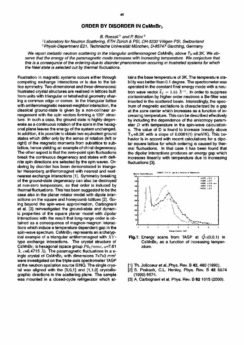
46
ORDER BY DISORDER IN CsMnBr
B. Roessli1 and P. Boni
2
1 Laboratory for Neutron Scattering, ETH Zurich & PSI, CH5232 Villigen PSI, Switzerland 2 PhysikDepartment E21, Technische Universitat Munchen, D85747 Garchlng, Germany
We report Inelastic neutron scattering In the triangular antlferromagnet CsMnBr3 above TN=8.3K We observe that the energy of the paramagnetic mode Increases with Increasing temperature. We conjecture that this Is a consequnce of the orderlngdueto disorder phenonmenon occurlng In frustrated systems for which the Neel state Is selected out by thermal fluctuations.
Frustration in magnetic systems occurs either through competing exchange interactions or is due to the lattice symmetry. Twodimensional and threedimensional frustrated crystal structures are realised in lattices built from units with triangular or tetrahedral geometry sharing a common edge or corner. In the triangular lattice with antiferromagnetic nearestneighbor interaction, the classical groundstate is given by a noncollinear arrangement with the spin vectors forming a 120° structure. In such a case, the ground state is highly degenerate as a continuous rotation of the spins in the hexagonal plane leaves the energy of the system unchanged. In addition, it is possible to obtain two equivalent ground states which differ only by the sense of rotation (left or right) of the magnetic moments from sublattice to sublattice, hence yielding an example of chiral degeneracy. The other aspect is that the zeropoint spin fluctuations break the continuous degeneracy and states with definite spin directions are selected by the spin waves. Ordering by disorder has been demonstrated in triangular Heisenberg antiferromagnet with nearest and nextnearest exchange interactions [1]. Symmetry breaking of the groundstate degeneracy can also be destroyed at nonzero temperature, so that order is induced by thermal fluctuations. This has been suggested to be the case also in the planar rotator model with dipole interactions on the square and honeycomb lattices [2]. Going beyond the spinwave approximation, Carbognani et al. [3] reinvestigated the groundstate and dynamic properties of the square planar model with dipolar interactions with the result that longrange order is obtained as a consequence of magnonmagnon interactions which induce a temperature dependent gap in the spinwave spectrum. CsMnBr3 represents an archetypical example of a triangular antiferromagnet with XYtype exchange interactions. The crystal structure of CsMnBr3 is hexagonal (space group P63/rarac, a=7.61 °A, c=6.4715 °A). The paramagnetic fluctuations in a single crystal of CsMnBr3 with dimensions 7x7x3 mm
3
were investigated on the tripleaxis spectrometer TASP at the neutron spallation source SINQ. The single crystal was aligned with the [0,0,1] and [1,1,0] crystallography directions in the scattering plane. The sample was mounted in a closedcycle refrigerator which at
tains the base temperature of 3K. The temperature stability was better than 0.1 degree. The spectrometer was operated in the constant final energy mode with a neutron wave vector kf = 1.55 A
1. In order to suppress
contamination by higher order neutrons a Befilter was inserted in the scattered beam. Interestingly, the spectrum of magnetic excitations is characterized by a gap at the zone center which increases as a function of increasing temperature. This can be described effectively by including the dependence of the anisotropy parameter D with temperature in the spinwave calculations. The value of D is found to increase linearly above T7v=8.3K with a slope of 0.0061(1) (meV/K). This behavior is in accord with recent calculations for a dipolar square lattice for which ordering is caused by thermal fluctuations. In that case it has been found that the dipolar interactions produces an energy gap which increases linearily with temperature due to increasing fluctuations [3].
!
1
\ \
' S01272
^ t s01301 ■/ 's01312
y\
u"5:9
u 5 : 9
1 0 1 2 3 4 5 6 7
Energy transfer (meV)
Fig.1: Energy scans from TASP at Q=(0,0,1) in CsMnBr3 as a function of increasing temperature.
[1] Th. Jolicoeur et al.,Phys. Rev. B 42, 480 (1990). [2] S. Prakash, C.L. Henley, Phys. Rev. B 42 6574
(1990)6574. [3] A. Carbognani et al, Phys. Rev. B 62 1015 (2000).

47
METAMAGNETIC TRANSITION IN Eri_xYxCo2 (x = 0, 0.4) SINGLE-CRYSTALS PROBED BY NEUTRON SCATTERING IN MAGNETIC FIELDS
A. Podlesnyak \ Th. Strassle \ J. Schefer \ A. Mirmelstein 2
1 Laboratory for Neutron Scattering, ETHZ & PSI, CH-5232 Villigen PSI, Switzerland 2 Institute for Metal Physics RAS, 620219 Ekaterinburg GSP-170, Russia
We present results of single-crystal neutron diffraction experiments focused on the behavior of the metamagnetic transition in the Er1.xYxCo2 (x = 0, 0.4) pseudobinary Laves-phase compounds.
Despite the consensus on the general features of the itinerant electron metamagnetism of the d subsystem in RCo2 cubic Laves phases even some basic properties remain controversial. We report on temperature-dependent single-crystal neutron diffraction measurements of the £ ^ . ^ 0 0 2 (x = 0,0.4) compounds under external magnetic fields up to 4 T. The experiments were performed at the TriCS 4-circle diffractometer using a wavelength of X = 1.179 A. An Oxford cryostat with a superconducting magnet was
10 20 30 40 50
Temperature (K)
60
Fig.1: The temperature dependence of the square root of the normalized integrated intensities of the (220) (circles) and (222) (triangles) magnetic Bragg reflections in the ErCo2 compound. Open and filled symbols represent the data for 0 and 4 T external magnetic field, respectively. The lines are a guide to the eye.
H
1 0
0 8
0 6
04
02
00
'
A
■
A
2TW ' A
-a—6-A
■
> A 6
-
-
10 15
HoH CO
Fig. 2: The magnetic field dependence of the square root of the normalized integrated intensities of the (220) (circles) and (222) (triangles) magnetic Bragg reflections in the ErCo2 compound at T = 35 K.
used for the measurements. We have measured the temperature and magnetic field dependences of several Bragg reflections. It is noteworthy that some structure factors in these compounds are characteristic for Er atoms only (/? or k or l^4n and h+k+l = 4n) or for Co atoms only (/?, k and / = 4n+2). Figs. 1-4 show the square root of the integrated intensities for the (220) and (222) reflections, which directly monitor the ordered moments |uEr and jLtC0J respectively. We found that both the localized 4f Er and itinerant 3d Co sublattice orders at the same temperature. Further analysis is in progress.
10 15 20 25 Temperature (K)
30
Fig. 3: The temperature dependence of the square root of the normalized integrated intensity of the (220) (circles) and (222) (triangles) magnetic Bragg reflection in the Er06Y04Co2 compound.
0 05 10 15 20 25 30 35 40
l̂ oH (T)
Fig. 4: The magnetic field dependence of the square root of the normalized integrated intensities of the (220) (circles) and (222) (triangles) magnetic Bragg reflections in the Er06Y04Co2 compound at T = 17 K.
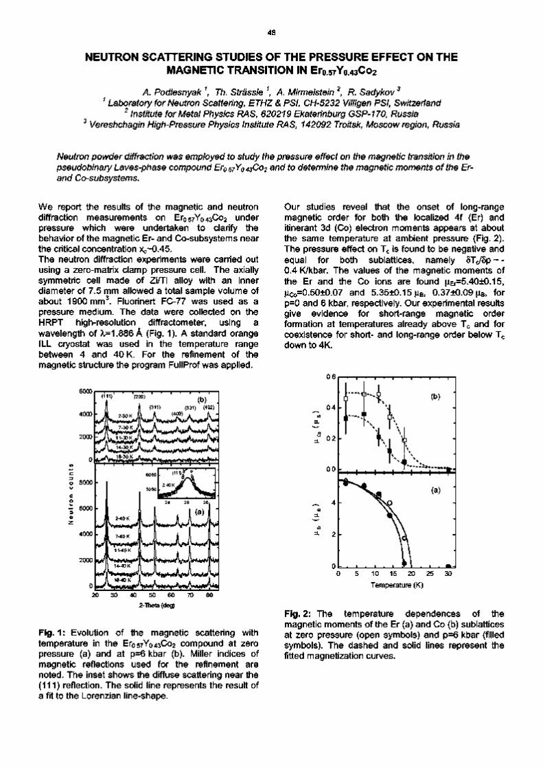
48
NEUTRON SCATTERING STUDIES OF THE PRESSURE EFFECT ON THE MAGNETIC TRANSITION IN Er0.57Yo.43Co2
A. Podlesnyak \ Th. Strassle \ A. Mirmelstein 2, R. Sadykov 3
1 Laboratory for Neutron Scattering, ETHZ & PSI, CH-5232 Villigen PSI, Switzerland 2 Institute for Metal Physics RAS, 620219 Ekaterinburg GSP-170, Russia
3 Vereshchagin High-Pressure Physics Institute RAS, 142092 Troitsk, Moscow region, Russia
Neutron powder diffraction was employed to study the pressure effect on the magnetic transition in the pseudobinary Laves-phase compound Er057Y043Co2 and to determine the magnetic moments of the Er-and Co-subsystems.
We report the results of the magnetic and neutron diffraction measurements on Er057Yo43Co2 under pressure which were undertaken to clarify the behavior of the magnetic Er- and Co-subsystems near the critical concentration xc~0.45. The neutron diffraction experiments were carried out using a zero-matrix clamp pressure cell. The axially symmetric cell made of Zi/Ti alloy with an inner diameter of 7.5 mm allowed a total sample volume of about 1900 mm3. Fluorinert FC-77 was used as a pressure medium. The data were collected on the HRPT high-resolution diffractometer, using a wavelength of ?i=1.886 A (Fig. 1). A standard orange ILL cryostat was used in the temperature range between 4 and 40 K. For the refinement of the magnetic structure the program FullProf was applied.
6000
4000 -
2000 -
4 ^ } ^ ^
o 8000
- 6000
4000
2000
(111) (220)
(311) (400)
^Kf^lH^^^ 20 30 40 50 60 70 80
2-Theta (deg)
Fig.1: Evolution of the magnetic scattering with temperature in the Er057Yo43Co2 compound at zero pressure (a) and at p=6 kbar (b). Miller indices of magnetic reflections used for the refinement are noted. The inset shows the diffuse scattering near the (111) reflection. The solid line represents the result of a fit to the Lorenzian line-shape.
Our studies reveal that the onset of long-range magnetic order for both the localized 4f (Er) and itinerant 3d (Co) electron moments appears at about the same temperature at ambient pressure (Fig. 2). The pressure effect on Tc is found to be negative and equal for both sublattices, namely 8Tc/8p ~ -0.4 K/kbar. The values of the magnetic moments of the Er and the Co ions are found juEr=5.40±0.15, jLiCo=0.50±0.07 and 5.35±0.15 JLIB, 0.37±0.09 JLIB, for p=0 and 6 kbar, respectively. Our experimental results give evidence for short-range magnetic order formation at temperatures already above Tc and for coexistence for short- and long-range order below Tc down to 4K.
10 15 20 25
Temperature (K)
Fig. 2: The temperature dependences of the magnetic moments of the Er (a) and Co (b) sublattices at zero pressure (open symbols) and p=6 kbar (filled symbols). The dashed and solid lines represent the fitted magnetization curves.

49
NON-COLLINEAR ORDER IN INVAR IRON-NICKEL ALLOYS
P. Boni1, E. Clementyev1, and B. Roessli2
1 Technical University of Munich, Physics Department E21, D-85747 Garching, Germany 2Laboratory for Neutron Scattering, ETHZ & PSI, CH-5232 Villigen PSI, Switzerland
Invar alloys like Fe65Ni35 show almost zero thermal expansion (invar effect) over a wide range of temperature T The most popular model to explain this effect assumes that the magnitude of the magnetic moments changes with increasing T such that magnetostriction compensates for thermal expansion. Recently it has been proposed that in addition the magnetic moments assume a non-collinear arrangement. In order to prove this model we have performed polarized beam experiments to detect a transverse magnetisation component. (Boni_01_FeNi.doc, 11/01 S-23)
Invar alloys have been the subject of numerous investigations using transport measurements as well as neutron scattering. One of the first models to explain the invar effect goes back to Weiss [1], who suggested that there are two possible states for the fee structure of Fe, namely a state with
• high volume, ferromagnetic • low volume, anti-ferromagnetic.
Transitions between this two states are suposed to lead to magneto-strictive effects that compensate the thermal expansion. More recent band structure calculations [2] indicate the existence of two states, namely
• high volume, high spin • low volume, low spin.
Close to the invar concentration, the curves for the respective binding energies differ by a small amount of energy and transitions between them can occur. However, this model cannot be correct because it would lead to a first order phase transition [3].
On the experimental side, the situation is also not understood. The magnetization M as calculated from the measured spin wave dispersion curves disagrees strongly with bulk measurements and has lead to the suggestion that a "forbidden" magnetic mode contributes to the strong decrease of M with increasing 7 [4]. Recently, a forbidden mode has indeed been observed by means of polarized neutron scattering [5], however, its role with respect to the invar property is not clear.
In general, all theoretical approaches have assumed that the magnetic moments in invar are collin-ear. Recently, van Schilfgaarde et al. [6] have allowed for the possibility that the moments may exhibit non-collinear spin arrangements. They find, that the transition from the high-spin ferromagnetic configuration to the increasingly disordered non-collinear state occurs as the volume decreases. This model is in good agreement with various experiments, like i) the smooth decrease of the magnetic moment \i with increasing T, ii) the equilibrium volume is correct, and the iii) the strange behavior of M may be understood.
In order to prove the new model we performed neutron scattering experiments with polarization analysis to search for transverse components (spin-flip scattering) of the magnetic moment \i and to measure \i versus T We reduced demagnetisation effects in the sample by cutting a plate with a thickness of 1 mm from the large single crystal (8x8x18
mm ). A vertical field B = 30 mT was sufficent to saturate the sample even at room temperature. The neutrons were polarized using a remanent bender before and after the sample. The idea of the experiment was to prove that the transverse component of the magnetic moments increases with increasing T
In order to correct for the 7-dependence of the flipping ratio determined R by measuring phonons and used this number to correct for the elastic. Unfortunately, after submission of the proposal, the experimental space at TASP was dramatically reduced by concrete blocks so that it was not possible to measure the proper phonons. Instead, we investigated the diffuse magnetic scattering from the sample in the forward direction to study a possible influence of the canting on the magnetic correlations. Fig. 1 shows the difference of measurements performed at 655 K and 300 K. Two features are noteworthy, namely the strong peak at £ = -0.04 and the dip near £ = -0.15. We plan to investigate the polarisation dependence of this scattering in a future experiment.
1200
1000
800
600
400
200
0
-200
' 1 '
1 _ 4000 '_ in 3000 • J 2000 • § 1000 : 8 o
-1
52
' 1 '
F e 6 5 N i 3 5
1
J .0
s
-0.5 0. 0 k -1.0 -0.8 -0.6 -0.4 -0.2
(C00) 0.0
2/3/02 Fi leDiff_655_300opj
Fig. 2: Difference of diffuse scattering
[1] R. Weiss, Proc. R. Soc. London A82 (1963) 281. [2] for a review see E. F. Wasserman, in Ferromag
netic Materials 5, Ed. K. H. Buschow and E. P. Wohlfarth, North-Holl., Amsterdam (1990) 237.
[3] M. Schroter, Phys. Rev. B 52, 188 (1995). [4] Y. Ishikawa et al., J. Magn. Magn. Mat.10 (1979)
183. [5] P. J. Brown et al. J. Phys.: Cond. Matter 8 (1996)
1527. [6] M. van Schilfgaarde et al., Nature 400 (1999) 46.

50
FINCHERBURKE EXCITATIONS IN SINGLEQ CHROMIUM
P. Boni1, B. Roessli
2, E. Clementyev
1, Ch. Stadler
1, G.Shirane
3, and S. A. Werner
4
1 Technical University of Munich, Physics Department E21, D85748 Garching, Germany 2Laboratory for Neutron Scattering, ETHZ & PSI, CH5232 Villigen PSI, Switzerland 3Brookhaven National Laboratory, Physics Department, Upton, NY 11974, U.S.A.
4Department of Physics, University of Missouri, Columbia, Missouri 65211 U.S.A.
For quite some time it has been speculated that the longitudinally polarized FincherBurke (FB) modes in Cr follow a linear dispersion curve with a slope that resembles the velocity of sound of the longitudinal acoustic phonon along (£ 0 0. We have investigated the cross section fo the FB modes with high
resolution neutron scattering and show that wether the dispersion nor the intensity of these modes are compatible with excitations having a linear dispersion curve. (Boni_01 _Chrom.doc, 11/98 L23)
Cr is one of the most interesting itinerant antiferromagnets [1]. At the Neel temperature TN = 311 K, it undergoes a second order phase transition to a transversely polarized spin density wave phase (TSDW) characterized by incommensurate wave vectors Q
± = (1+8 0 0). As a result of the cubic symmetry of paramagnetic Cr, three types of domains are present along the three [100] (Fig. 1).
4 ^ /n 1 rVkO JWF ©allowed
(0 0 0)
1$ nuclear
™ © forbidden
(1-8 0 0) (10 0)
Fig. 1: Reciprocal lattice of bcc Cr. The filled circles indicate the allowed satellites in the singleQ state.
The magnetic excitations exhibit many unusual features. In particular, the magnetic modes that originate from Q* exhibit such a steep dispersion that the creation and annihilation peaks cannot be resolved. In addition, low energy excitations with a linear dispersion (FincherBurke modes) that is the same as that of the [£00] longitudinal acoustic phonon have been reported [2]. Recent high resolution experiments indicate that this result may have to be revised [3].
200 c o E o o § 120
160
£ 80 CO c CD
■£ 40
i 1 1 1 r
• (0.986 0 0) Gaussian fit
• (0.946 0 0)
H . , ...ft'1,
1/16/02 File Const_Q_230 opj
2 0 2 4 6 Energy (meV)
10
Fig. 2: ConstantQ scans measured at T = 230 K. The scan at (0.946 0 0) provides the background.
Using the cold tripleaxis spectrometer TASP we followed the dispersion of the excitations using constantCD scans. A typical result Q = (0.986 0 0) is shown in Fig. 2 together with the background as measured at (0.946 0 0). Near 6.5 meV a well defined peak is observed. In addition, magnetic scattering is observed that is essentially independent of E. If the FBmodes would follow a linear dispersion we would expect peaks near 2 meV and 5.5 meV. The former peak should be much more intense than the latter because of the thermal population factor { EMBED Equation.3 }. We definitely do not observe the lowenergy peak. Our measurements confirm also the localized, commensurate excitation at E = 8 meV
In Fig. 3 we show the energyposition of the peaks in constantQ scans. Clearly, the dispersion of the modes does not extrapolate towards the incommensurate positions Q
±. The crossed solid lines indicate
the expected dispersion of the FBmodes. We conclude that the FBmodes do not follow a linear dispersion.
CD
E ^ 4 E> CD
iS 2
0.96 0.98 1.00 1.02 1.04 (C00)
Fig. 3: The circles indicate the measured data points. Their "dispersion" does not extrapolated to the pre
dicted incommensurate Oppositions.
[1] E. Fawcett, Rev. Mod. Phys. 60, 209 (1988). [2] S. K. Burke, W. G. Stirling, K. R. A. Ziebeck, and
J. G. Booth, Phys. Rev. Lett. 51, 494 (1983). [3] P. Boni, B. Roessli, E. Clementyev, Ch. Stadler,
G. Shirane, and S. A. Werner, Applied Physics A (accepted for publication).

51
f-Electron Magnetism
I jp., J,,,, |—..f-,|,-,,,r,,,,,p, p.-, y-, r „ „ |~, r,„, |„-, r„„, | - , r-..|...

52

53
THE MAGNETIC STRUCTURE OF Eu4Ga8Gei6
M. Christensen1, B. B. Iversen1, D. Bryan2, B. Lebech3, P. Fischer4 and L. Keller4
1 Department of Chemistry, University of Aarhus, DK-8000 Aarhus, Denmark 2 Department of Chemistry, University of California, Santa Barbara, California 93106, USA
3 Riso National Laboratory, DK-4000 Roskilde, Denmark 4Laboratory for Neutron Scattering, ETHZ&PSI, CH-5232 Villigen PSI, Switzerland
Complementary powder diffraction data measured at the DMC and HRPT beam lines have been used to determine the magnetic structure of Eu4Ga8Ge16. Due to difficult synthesis conditions only a small amount of 153Eu isotope enriched sample was available (250 mg). The rare-earth
containing nanocage material has an anti-ferromagnetic phase transition at 8 K
The new material Eu4Ga8Ge16 was recently synthesized as by-product in an attempt to prepare novel thermoelectric type I clathrate structures [1]. The material has the type IV clathrate structure, and is one of the first magnetic clathrates. Magnetic susceptibility measurements have shown that Eu4Ga8Ge16 has an antiferromagnetic phase transition at 8 K, figure 1.
Refinement of the high temperature data gave an accurate nuclear structure (a = 4.1231(1) A, b = 11.2532(4) A and c = 13.1993(4) A), and this was fixed in refinement of the magnetic structure. The magnetic moment is refined to be along the a-axis - except for a 17° inclination in c direction, (Ra = 6.22+0.05 jLiB, Rb = 0, Rc = 1.94+0.15 JLIB, giving a total magnetic moment of 6.52+0.10 JLIB).
Red iiite :s paraftet to the 100 axis
I ' I 3D 40 50 60
Fig. 1: Magnetic susceptibility versus temperature.
Heat capacity measurements carried out at PSI clearly support that it is an order-disorder transition, figure 2.
cheu60_lp8b.dat
Fig. 2: Heat capacity versus temperature
In order to get an in-depth understanding of the magnetic properties we have measured well resolved low order powder diffraction data at DMC (k = 4.2 A), and high-resolution data at HRPT (k = 1.9 A). Data were measured above and below the phase transition (15 K and 1.5 K), figure 3.
Fig. 3
40 60 80 100 120 SCATTERING ANGLE
Low order data above and below the transition
From temperature dependent measurements it is seen that saturation has not been reached at 1.5 K. The distance between the Eu atoms along the a-axis chain is 4.123 A. These chains have an almost intra ferromagnetic ordering, which suggest a predominant dipole-dipole interaction. The non-perfect alignment hints that other interactions also contribute.
Fig. 4: A bc-plane projection of the structure.
[1] J. D. Bryan et al, Chem. Mat. 13, 253 (2001).

54
CRYSTAL-ELECTRICAL FIELD POTENTIAL IN THE LAYERED BINARY COMPOUND ErBr3
B. Roessli1, Th. Strassle1, K Kramer2 and H.U. Gudel2
1 Laboratory for Neutron Scattering, ETH Zurich & PSI, CH-5232 Villigen PSI, Switzerland 2 Department for Chemistry and Biochemistry, University of Bern, Freierstrasse 3, CH-3000 Bern 9, Switzerland
The crystalllne-electrlc-fleld (CEF) splitting of the rare-earth Ions within the lowest J multiplet of Er3+ In the quasi-2Dimensional layered compound ErBr3 is measured by inelastic neutron scattering. The magnetic susceptiblity calculated from the reconstructed CEF energy-levels is found to be strongly anisotropic in accord with experiment.
Recently, unusual magnetic ordering has been observed in the binary compounds ErX3 with X=Br, I [1] and described by the propagation vector k=[1/3,1/3,0]. The Er3+ magnetic moments build a two sublattice triangular magnetic ordering with the nearest-neighbor spins forming an angle of 60° between each other. The magnetic moments lie in the basal plane of the honeycomb lattice. We show the results of inelastic neutron-scattering in polycrystalline ErBr3 with the aim to determine the crystalline-electrical field acting on the 4 / electrons at the rare-earth site. The inelastic neutron measurements were performed both at the neutron spallation source SINQ at the Paul-Scherrer Institut with the cold-neutron triple-axis spectrometer TASP The analyser energy was set at fe/=1.64 A - 1 which resulted in an energy resolution at the elastic position of about 0.2meV.
3
O U o
6000 5000 4000 3000 2000 1000
0
ErBr3, T=10K
$L 'measured' AT 'calculated'
^Z ^ l ^k«<#
O
-
jp\ -
-4 0 4 Energy Transfer (meV)
Fig.1: Energy spectrum of ErBr3 at T=10K. The line denotes the calculated spectra according to the crystal-field parameters obtained from least-square fitting the data with the inelastic neutron cross-section taken on TASP
Fig. 1 shows the results of an energy scan measured in ErBr3 at T=10K and |Q|=1.5 A"1 . Apart from the incoherent elastic line centered at huo = 0 meV, one recognizes four peaks at huo = 1.52, 3.0, 4.75 and 6.21 meV which correspond to neutron energy-loss s-cattering and for the negative energy transfers at TIUJ =-1.44 and -2.94 meV, respectively. The magnetic origin of these peaks is established by the dependence of their intensity which is found to decrease as a function of increasing momentum transfer Q, and hence
varies like the magnetic form factor f(Q). For the trigonal symmetry of the Er site in ErX3 (X=Br,l), the data was analysed on the basis of the CEF operator HCEF = B§0§ + B202 + B|Ol+B§0§ + B6
30|+B660§.
We find from a least-square fit procedure of the data the CEF-parameters £§=-5.9 meV, £$=-4.9 meV, £f=-10.9 meV and B%=0.7 meV, respectively. A comparison of the calculated paramagnetic single-ion susceptibilities with the experimental DC susceptibility measured in ErBr3 is shown in Fig. 2. The agreemen-t with the data is reasonably good, although for both compounds the calculated in-plane susceptiblity is lower than actually measured. Both the calculated and observed single-ion susceptiblities Xo (a = x, y, z) are extremely anisotropic with xl « Xo = Xo a t ' o w temperatures. At T=0K, the magnetic moments are therefore confined by the crystalline-electric field in the hexagonal plane of the crystal, in agreement with the magnetic structure determined by neutron diffraction [1].
x(emu/mole)
14
ErBrq
60 80 100
Temperature (K) Fig.2: The symbols refer to the observed magnetic
susceptiblity of ErBr3, whereas the lines are the calculations using the CEF parameters.
[1] K.W. Kramer, H.U. Gudel, B. Roessli, P. Fischer, A. Donni, N. Wada, F. Fauth, M.T. Fernandez-Diaz, and T. Hauss, Phys. Rev. B 60, R3724 (1999); K.W. Kramer, H.U. Gudel, P. Fischer, F Fauth, M.T. Fernandez-Diaz, and T. Hauss, Euro. Phys. J. B 38, 39 (2000).

55
MAGNETIC ORDERING AND MAGNETIC EXCITATIONS IN H0C0O3 SINGLE CRYSTALS
D. Khalyavin 1
, S. Shiryaev1, A. Podlesnyak
2, J. Mesot
2
11nstitute of Solid State & Semiconductors Physics, P. Brovka str. 17, Minsk, Belarus
2 Laboratory for Neutron Scattering, ETHZ & PSI, CH5232 Villigen PSI, Switzerland
The temperature dependence of the magnetic excitations in orthocobaltite H0C0O3 single crystals was measured by inelastic neutron scattering. Furthermore, measurements of magnetic Bragg peaks allowed us to determine the Curie temperature Tc at which the Ho ions order.
Due to the unusual magnetic behavior of the cobalt ions, the RC0O3 (R = rare earth ion), with perovskite structure have attracted great attentions. The splitting of the Co 3d levels by the ligand crystal field in these compounds is comparable in magnitude to the interatomic exchange energy. As a consequence, the compounds are diamagnetic at low and paramagnetic at hightemperatures [1,2]. Much less is known about the magnetism related to the rare earth ions in these compounds (type of the magnetic ordering, parameters of crystalfield splitting).
Inelastic neutron scattering (INS) measurements have been performed at different values of the scattering vector Q with fixed final energies Ef=3.0 and 3.9 meV. Fig. 2 shows typical INS spectra in the temperature range 1.3<T<7K. At the lowest temperature two magnetic peaks can be observed at energies of about 0.23 and 0.45 meV, respectively. Above Tc the INS spectra exhibit a complicated structure consisting of broad features. Clearly more work is needed in order to establish the temperature dependence of both the magnetic structure and magnetic excitations in these compounds. Further analysis and investigations are in progress.
~ 6000
O o c 4000 o
CD
c 2000
- 1 1 1 1 1 1 1 1 1 1 -
1.2
e — 1 0 0 1 up # — 1 0 0 1 down
1.4 1.6 1.8
Temperature (K)
2.0 2.2
Fig.1: Temperature dependence of the (001) magnetic Bragg reflection. Open and filled circles represent the data for heating and cooling, respectively. The solid lines are a guide to the eye.
250
V*«wi
0.50 0.25 0.00 0.25 0.50 0.75 1.00
Energy transfer (meV)
Fig. 2: Energy spectra of neutrons scattered from H0C0O3 at the scattering vector Q=(0,0,1.2) and temperatures 1.3 K (•), 5 K (V) and 7 K (♦). The lines are the result of a fit to the spectrum at T=1.3K assuming resolutionlimited Gaussian lineshapes.
For that reason we have undertaken series of both elastic and inelastic neutron scattering studies on a H0C0O3 single crystal. The measurements were performed on the triple axis spectrometer TASP at the spallation source SINQ. A standard orange He cryostat was used in the temperature range between 1.3 and 10 K. The temperature dependence of the (001) magnetic Bragg reflection is shown in Fig. 1. The determined magnetic ordering temperature is equal to TC=1.8K, that is significantly lower than the Tc=2.4 K obtained in earlier powder neutron diffraction studies of H0C0O3 [3].
[1] M.A. SenarisRodriguez and J.B. Goodeno
ugh, J. Sol. St. Chem. 118, 323 (1995)
[2] K. Asai, O. Yokokura and N. Nishimori, Phys. Rev. B 50, 3027(1994)
[3] A. Kappatsch, S. QuezelAmbruanz, J. Siva
rdiere, J. de Physique 31, 369 (1970)

56
EFFECT OF OXYGEN CONTENT ON THE CRYSTAL FIELD INTERACTION IN Hoo.iSro.9Co03-x PEROVSKITES
A. Podlesnyak1, K Conder1, A. Furrer1, N. Golosova 2, A. Mirmelstein 2, S. Kazakov 3
1 Laboratory for Neutron Scattering, ETHZ & PSI, CH-5232 Villigen PSI, Switzerland 2 Institute for Metal Physics RAS, 620219 Ekaterinburg GSP-170, Russia
3 Laboratory for Solid State Physics, ETH Honggerberg, CH-8097 Zurich, Switzerland
The inelastic neutron scattering (INS) has been employed to study the crystalline-electric-field (CEF) interaction in the perovskite-type compounds Ho0 iSr0gCoO3.x with x = 0.15, 0.27, 0.49. It is shown that the CEF interaction is strongly sensitive to the oxygen content.
It is well known that the crystal structure, magnetic and transport properties of the nonstoichiometric cobalt perovskites depend strongly on the oxygen content (see e.g. [1]). However, the detailed mechanism of the magnetic and structural transitions is not well understood. The INS studies of the CEF interaction on perovskite-type structures, namely, RMeOs, (R = rare earth, Me = Al, Cr, Ga, Ni, Co) reveal important information about the peculiarities of the electronic structure and play a key role in determining the magnetic ground state. Our previous INS experiments on HooiSr09Co03 (under I/99-S17 short term proposal) allowed to reproduce the CEF splitting based on cubic symmetry at the R3+ site [2,3]. In order to determine the CEF peculiarities in the reduced cobalt perovskites Ho0iSro9Co03.x (x = 0.15, 0.27, 0.49) we focus here on the low energy part of the spectra. The triple-axis spectrometer TASP at the spallation source SINQ was used to determine the temperature (Fig. 1) and oxygen content (Fig. 2) dependencies of the CEF transitions.
600
Z 400
200
- 1 0 1 2 3 4 5 Energy transfer (meV)
Fig.1: Low-energy spectra of neutrons scattered from HooiSr09Co02 85 at T = 3K (circles), 50 K (squares) and 100 K (triangles).
Energy scans up to an energy transfer AE = 5.6 meV were recorded at temperatures 3 < T < 1 0 0 K , modulus of the scattering vector Q = 1.8 A"1 and scattered energy Ef = 4.95 meV. A Be-filter was used to reduce high-order contamination. We found that the oxygen reduction in the perovskite-type compounds Ho0iSro9Co03.x results in drastic changes of the CEF splitting (Fig. 2). Since we can neglect a molecular field associated with the Co sublattice, these changes most probably connected with a lowering of the crystal symmetry and the ordering of oxygen vacancies.
600
400
200
1 2 3 4 Energy transfer (meV)
Fig. 2: Low-energy spectra of neutrons scattered from Ho0iSro9Co03.x at T = 3 K. Circles, triangles and squares represent the data for x = 0.15,0.27 and 0.49, respectively.
[1] A. Podlesnyak, K. Conder, N. Golosova et al., FUN Progress Report (2001).
[2] A. Podlesnyak, A. Mirmelstein, N. Golosova et al., to be published in Applied Physics A.
[3] A. Podlesnyak, A. Mirmelstein, N. Golosova et al., FUN Progress Report (1999), p. 31.

57
STRUCTURAL AND MAGNETIC PHASE TRANSITIONS IN DyB6
L. Keller \ P. Fischer \ A. Donni2, S. Kunll
3
1 Laboratory for Neutron Scattering, ETHZ& PSI, CH5232 Villigen PSI, Switzerland 2 Department of Physics, Niigata University, Niigata 9502181, Japan 3 Department of Physics, Tohoku University, Sendai 9808578, Japan
The magnetism of polycrystalllne Dy11
B6 has been Investigated at SINQ on the powder dlffractometers HRPT and DMC. Below the quadrupolar ordering temperature TQ= 32 K we succeeded to determine the distortion of the crystal structure from cubic to trigonal. Below TN = 26 K longrange dipolar order leads to magnetic Bragg peaks that can be Indexed with the commensurate propagation vector k = (1/4, 1/4, 1/2).
The rareearth hexaboride DyB6 undergoes two phase transitions at low temperatures. At the quadrupolar ordering temperature TQ = 32 K the quadrupole inter
actions prevail over the conventional dipolar magnetic interactions and the quadruples form a phase transi
tion on their own in coexistence with still disordered dipole moments. At lower temperatures a second phase transition is observed and the dipole moment lattice orders in a longrange magnetic structure below TN = 26 K. Neutron scattering is the method of choice for the investigation of phase transitions in solids. While the quadrupoleorder cannot be directly ob
served, highresolution neutron diffraction is a power
ful tool to observe the structural distortion associated with this quadrupolar order. Recently, first neutron diffraction experiments on DyB6 were reported [1]. Now we performed new measure
ments of a polycrystalline Dy11
B6 sample on the two complementary diffractometers HRPT and DMC at SINQ, in order to investigate the relations of chemical structures, quadrupolar and magnetic ordering.
25000
20000
>> 15000
| 10000
5000
DyB f i HRPT A, = 1.886 A
T = 40K
T = 28K
T = 20 K s
T=1 .5K
_i i i i i i i i i i i i i i i
\J
i i i i i i i
135 140 145 150 20 [°]
155 160
Fig. 1: HRPT data of DyB§ at T = 1.5, 20, 28 and 40K, measured at X = 1.886 A The data sets have been vertically shifted for better comparison. The tempera
ture independent peak at 20 = 150° is due to the DyB12 phase also found in our sample.
Fig. 1 illustrates the temperature dependence of the highscattering angle part of neutron diffraction pat
terns measured on HRPT. According to profile refine
ments, based on the cubic space group Pm3m for T > TQ and the trigonal space group R3m for T < TQ, the trigonal angle a = p = y changes from the cubic 90° at 40 K to 90.535° at 1.5 K (Fig. 2). The angle a > 90° corresponds to a shrinking of the cubic crystal struc
ture along a [1,1,1] direction. A similar effect has been observed in HoB6 [2]. At TN = 26 K DyB6 undergoes a magnetic phase tran
sition to a longrange dipolar order. Antiferromagnetic Bragg peaks appear in the lowtemperature diffraction patterns which may be indexed with the magnetic propagation vector k = (1/4, 1/4, 1/2). The temperature dependence of the integrated magnetic peak intensity measured on DMC is also shown in Fig. 2.
90 5
90 4
90 3
90 2
90 1
90 0
I I I I I I I I I I I I I I
I t | I I I I | I I I I | i I # I | I I I I |
b)
I f I I * I I ^ \
I I i i i I i I ■ 0 5 10 15 20 25 30 35 40
Temperature [K]
Fig. 2: a) Temperature dependence of the integrated intensity of magnetic Bragg peak (dipolar ordering); b) Temperature dependence of the trigonal angle a, indi
cating the quadrupolar ordering.
[1] K. Takahashi et al., J. Magn. Magn. Mat. 177
181, 1097 (1998); Physica B 241243, 696 (1998).
[2] A. Donni et al., J. Phys. Soc. Jpn. 70, Suppl. A, 448(2001).

58
MAGNETIC ORDERING IN TERBIUM DODECABORIDE
A. Murasik1, A. Czopnik
2, M. Zolliker^, L. Keller^, N. Shitsevalova
4, Y. Paderno
4
institute of Atomic Energy, Swierk, Poland 2Institute for Low Temperature, Wroclaw, Poland
3Laboratory for Neutron Scattering, ETHZ & PSI, CH5232 Villigen PSI, Switzerland
4Institute for Problems of Material Science, Kiev, Ukraine
Neutron diffraction experiments carried out on powder sample of the TbB12 enriched with 11
B isotope show that this compound exhibits antiferromagnetic ordering described by a propagation vector k = CA±T, VI±% VI±T), (T = 0.022) with the small admixture of the phase with r = 0.063. The measurements performed in several temperatures in the ordered state did not reveal detectable features which could confirm the exis
tence of two phase transitions deduced from earlier specific heat measurements.
Rare earth dodecaborides are known to exhibit a variety of physical properties resulting from the unfilled 4fshell of the rare earth ions. For example LuB12 below 0.4 K is superconducting, YB12 is an intermediate valence system while Dy, Ho, Er, Tm and Tbdodecaborides appear to order antiferromagnetically at low temperatures. They crystallize in a cubic FCC structure of the UB12type. Recently we have studied by means of neutron diffraction the magnetic structural properties of TmB12 [1]. We have shown that the magnetic ordering occurring below 3.2 K conforms to an incommensurate, sinusoidally modulated magnetisation wave propagating through the crystal, in all three [100]type directions.
As a continuation of structural studies of rare earth dodecaborides we have undertaken recently the study of TbB12. The terbium dodecaboride appeared to display very interesting magnetic features. Its Neel temperature is relatively high 22 K. The specific heat measurements show two ^points at T1 = 16.6 K and T2 = 18.2 K [2]. Therefore it was of interest to examine its magnetic properties by neutron scattering technique. The measurements have been done at the Swiss Spallation Neutron Source SINQ of the Paul Scherrer Institute at Villigen, Switzerland.
Measurements were carried out within the temperature range 4.2 25 K, i.e. below and above anticipated magnetic ordering. Fig. 1 displays the neutron diffraction diagram taken in a wide range of scattering angles including in particular the region of low angles where the magnetic contribution usually is the strongest. It is seen that at 4.2 K, in addition to nuclear intensities (see vertical bars indicating positions of nuclear peaks) strong magnetic satellite peaks appear around the reciprocal lattice points where the superstructure lines would occur if the magnetic unit cell, in comparison to the nuclear one, is doubled in three directions. In addition to these peaks the small magnetic satellites were also noticed, which due to the greater line widths appeared as the "wings" around the strong magnetic satellites. Using the FULLPROF fitting procedure, we were able to preliminary determine the individual propagation vectors k = (1/̂ ±x, VZ±T, V*±i), (with ^ = 0.022 and x2 = 0.063) corresponding to each magnetic phase and to fit the data with acceptable accuracy.
=: o
2 T = 4 2 K lobs
Icalc Diff
TbB6
^={1/2+^,1 /2+^,1 /2+^} k2 = {1/2+T2,1/2+x2,1/2+x2}
S J L J I L J U O ^ ~ihiv"/u-
I III ifln nil ii infill i i
" i ' ■ i II I n i 111 i i Jim i Jlii
20 40
2Theta [deg] 60 80
Fig 1: Neutron diffraction patterns of TbB12 taken at 4.2 K. The open circles denote experimental data, the thick solide line the FULLPROF fit, and the thin solid line is the difference between lobs and Icalc. The vertical bars indicate successively positions of nuclear reflections, TbB6 impurity, and positions of magnetic satellite reflections arising from magnetic moment modulated structures with propagation vectors ki, k2.
As no higher order magnetic satellites were detected, only the sinusoidal moment arrangements were considered. The structure can be described as a modulation of magnetic moments (stacked parallel in (100)) sheets that propagate along the three crystallography edges of the chemical cubic unit cell. Surprisingly, no detectable magnetic phase transition was seen around the T1 and T2 temperature. This observation is in apparent conflict with the earlier specific heat study [2], so the careful examining of present data as well as additional measurements are called for.
[1] Czopnik, A. Murasik, L. Keller, N. Shitsevalova, Y. Paderno, phys. stat. sol.(b), 221 R7 (2000)
[2] N. Shitsevalova (Ph.D thesis unpublished)

59
UNUSUAL LOWTEMPERATURE MAGNETIC PROPERTIES OF Tmln3
A. Murasik1, A. Czopnik
2, L. Kellef, T. Konter
4
institute of Atomic Energy, Swierk, Poland 2Institute for Low Temperature, Wroclaw, Poland
3Laboratory for Neutron Scattering, ETHZ & PSI, CH5232 Villigen PSI, Switzerland
4Paul Scherrer Institute, CH5232 Villigen PSI, Switzerland
Neutron diffraction experiments performed on powder samples of Tmln3 show that this compound, depending on sample preparation, exhibits different magnetic properties visualized through the magnitude of the Neel temperature and the form of magnetic ordering.
We have shown in the previous [1] and present work [2], that the low temperature magnetic behavior of Tmln3 strongly depends on the method of sample preparation, in particular on releasing the strains. The most pronounced effect of unreleased strains has been visualized on non annealed sample which exhibited a coexistence of two magnetic structures possessing pseudotetragonal unit cell enlarged either along one axis or having the basal plane cube edges twice that of the chemical cell. (Fig 1).
Tmln3 Phase 1 k = {0, 0 1/2} Phase 2 k = {1/2,1/2,0}
Magn Moment
Fig 1: Coexistence of two different magnetic structures in nonannealed polycrystalline sample of Tmln3[1].
Tmln3
Schematic sketch of the incommensurate and magnetic moment modulated structure with a propagation vector: k = {0, 0, 1/2+T}
Mu=2 Skj exp{27ii k R,}
j numbering of atoms in the unit cell I numbering of unit cell
or i ■ <sC««s®. O e£™«* e ^ * o
Fig 2: Transformation of magnetic phase 1 into incommensurate structure found in Tmln3 (present work). Note the reorientation of magnetic moment's alignment in comparison to Fig. 1.
The outstanding difference between the previous and present results obtained on a carefully annealed sample is that the phase 1, described in a more general formalism as a phase with k = (0, 0, V2) persists in a new sample either as a single phase within 0.2 < T < 1.4 K or as the dominant phase for T >1.4 K. The small inclusion of additional phase at T >
1.4 K, representing a sinusoidally modulated magnetisaton wave propagating along the [0,0,1] axis, with the moments also oriented in this direction (cf: Fig. 2) seems to prove that the annealing process did not release yet the strains completely. Note that the transition from commensurate to incommensurate structure involves also a change of the moment alignment. The determined Neel temperature TN = 1.75 K, in comparison to that found in previous sample, is apparently lower. The ordered moment JLI at T = 0.2 K was found to be 4.53 juB. Its value for 0.2 < T < 1.35 K was determined in the customary manner. Some questions however arose at 1.4 K when the additional phase has appeared. The correct calculation of magnetic moments for each phase needs a scale factor describing its contribution in the whole scattering process. The simplest hypothesis introduced to the fitting procedure was that the magnitude of magnetic moments in the commensurate phase and the amplitudes of magnetic moments of incommensurate phases are equal and depend on temperature in a similar way.
Under this assumption we were able to estimate the relative proportion between commensurate and incommensurate phases. At 1.4 K their ratios were as 90:10 %. Due to expansion of incommensurate phase with rising of temperature, the latter phase was yet seen at T ~ 1.7 K. Similar effect i.e. the temperature variation of the phase 1 to phase 2 ratio was observed also previously.
The appearance of incommensurate phases is accompanied by an abrupt change of the magnetic moment. By examining the neutron diagram the shape of the background in the vicinity of 30° 45° we concluded that the part of the longrange ordered moments decay at (1.35 1.4) K into short range clusters. Summarizing, the unusual properties of Tmln3 seem to result from weak and competing magnetic interactions, which in high symmetry of the magnetic ion environment and in the presence of coupling via conduction electrons, may produce the frustrated system that can easily be perturbed by external forces.
[1] A. Murasik, A. Czopnik and L. Keller Phys. Stat. Sol. (a) 186, R1 R3 (2001).
[2] A. Murasik, A. Czopnik, L. Keller and T. Konter, Phys. Stat. Sol. (a) (submitted for publication).

60
SINGLE ION ANISOTROPY IN Pro.07Lao.93Ni
E.Clementyev1, P.AIIenspach
2, P.A.AIekseev
3 and G.Lapertot4
1 Physik-Department E21, Technische Universitat Munchen, D-85748 Garching, Germany 2Laboratory for Neutron Scattering, ETHZ and PSI, CH-5232 Villigen PSI, Switzerland
3Russian Research Center "Kurchatov Institute", RU-123182 Moscow, Russia
4DRFMC/SPSMS, CEA-Grenoble, F-38054 Grenoble Cedex, France
The paramagnetic spectral response has been measured in Pr0 o7La0 93Ni single crystal along the main crystallographic directions. Two crystal field transitions out of the ground state are clearly seen in the energy transfer range up to 6 meV. The observed anisotropy of these single-ion transitions along with the Q-dependence of the magnetic modes in pure PrNi play a crucial role in understanding of the soft mode-
driven magnetic ordering in this compound.
Magnetic ordering mechanism in singlet ground state systems fundamentally differs from one in conventional magnets (see [1] and references therein). The so-called soft mode-driven phase transition is considered as a plausible physical scenario in such systems. A dramatic softening of one of the magnetic modes has been found at the magnetic Bragg point in PrNi [2]. The crystal field (CF) transitions in this system propagate through the crystal due to the exchange coupling between magnetic ions. These dispersive modes are strongly anisotropic due to the exchange interaction between magnetic ions and due to the single-ion anisotropy inherent to Pr ions in low-symmetry crystal surrounding. Discrimination of these two sources of anisotropy is indispensable for getting a quantitative physical picture of the magnetic ordering in PrNi. The main purpose of the present experiment was to determine the single-ion anisotropy of the magnetic excitations in PrNi. The only way to investigate pure single ion effects is to suppress the exchange coupling between Pr ions. That is why a single crystal of diluted compound Pr007La093Ni was used.
600
500
3 CO
400
% 300 c 0) 200
1 1
; *
; 9
■ 1
0 rP ° ° A A O OO
A ^ A Q $ ° ° O ft,&k A *
W O
100
- 1 0 1 2 3 4 5 6 Energy (meV)
Fig 1: Neutron spectrum of Pr007La093Ni measured on RITA-II at T=9K along the directions: [001] - filled circles, [010] - open circles and [100] - triangles.
fixed final energy 5.57 meV. A PG graphite filter was placed after the sample to suppress higher order contaminations. The measurements were performed in two scattering planes to access the main crystallographic directions in orthorhombic Pro 071-30 93NL Two well-defined CF transitions out of the ground state were observed along the directions [100], [010] and [001] (see Fig.1). The energies of the peaks are in a good agreement with the high-temperature energy positions of the magnetic modes in pure PrNi [2]. CF transitions indeed appeared to be strongly anisotropic. Using the integrated intensity of the peaks one can estimate the matrix elements of the CF transitions due to the existence of the polarization factor of the magnetic scattering function:
S(Q,E)~Y, X 1,j o^x,y,z
\<i\Ja\M XhoH-Ej-E,)
where E, and E} are the energies of the CF states, Ja-projection of the total angular momentum operator, Q-neutron momentum transfer. The values of the matrix elements along with the parameters of the anisotropic exchange coupling between Pr ions play a crucial role in understanding of the unconventional magnetic ordering in PrNi.
[1] B.R.Cooper in: Magnetic Properties of Rare Earth Metals, ed. R.J.Elliot, Plenum Press (1972), Ch.2, 17.
[2] E.S.Clementyev et al., PSI Scientific Report vol. 111(1999)37.
Inelastic neutron scattering experiments have been performed on the triple-axis spectrometer RITA-II (single neutron detector configuration). Constant-Q scans were measured at temperatures 9K to 20K for

61
Structure and Dynamics

62

63
CRYSTAL AND MAGNETIC STRUCTURES OF NEW LAYERED OXIDES Sr2GaMn05+v
D.V. Sheptyakov1, A.M. Balagurov*, V.Yu. Pomjakushin2, P.Fischer1, L.Keller1, A.M. Abakumov3, E.V. Antipov3, M.V. Lobanov3, B.Ph. Pavlyuk, M.G. Rozova3
1 Laboratory for Neutron Scattering, ETHZ& PSI, CH-5232 Villigen PSI, Switzerland 2 Frank Laboratory of Neutron Physics, JINR, 141980 Dubna, Russia
3 Department of Chemistry, Moscow State University, Moscow 119899, Russia
In continuation of the project aimed at precise structural studies of the novel complex manganese oxides with the brownmlllerlte-type structures, the crystal and magnetic structures of the series of Sr2GaMnOs+y compounds were studied with HRPT and DMC powder dlffractometers at SINQ, yielding an essential dependence of magnetic Mn ordering on oxygen concentraion.
The recently discovered family of complex manganese oxides A2GaMn05+y (A=Ca, Sr) [1-2] has attracted the attention due to the possibility of obtaining the compounds with new electronic and magnetic properties. Being perovskite type derivatives, their crystal structures belong to a brownmillerite type and consist of alternating (AO), (Mn02), (AO) and (GaO) or (Ga01+x) layers. Incomplete oxygen anions sublattice (presence of vacancies in GaO layers) provides a possibility of varying the formal Mn oxidation state in a very wide range, which gives rise to different low-temperature physical properties.
The Sr-containing representatives of the family, a series of Sr2GaMn05+y oxides, were studied by neutron powder diffraction with HRPT and DMC diffractometers at SINQ. The crystal structure (orthorhombic, sp.gr. I m a 2) of the reduced compound Sr2GaMn0497 is shown in the Fig. 1. It consists of a well-ordered sequence of layers of MnOe octahedra and Ga04 tetrahedra, separated by buffer SrO layers.
Fig 1: Chemical and low temperature magnetic (TN^190 K) structures of Sr2GaMn0497. Magnetic and chemical unit cells are identical.
The ultimately doped compound, Sr2GaMn0552, has a tetragonal perovskite-like structure (sp.gr. P 4/m m m), with a distinct disorder of the Ga and O atoms in the anion-deficient Ga0152 layer (Fig. 2).
The Jahn-Teller deformation of the MnOe octahedra in it is strongly suppressed due to the oxidation.
Fig 2: Chemical and low temperature magnetic (TN^110 K) structures of Sr2GaMn0552. The magnetic unit cell parameters am, cm are related to the chemical unit cell parameters a, c as follows: a m =aV2 , cm =c. Oxygen positions in the disordered Ga0152
layer are only partially filled.
At temperatures between 100 and 200 K, all the studied compounds undergo transitions into antiferromagnetic states. It was found that the magnetic ordering type in them depends essentially on the oxygen content. The Mn magnetic moments are aligned at opposite directions within the Mn02 layer, and the coupling between magnetic moments lying in the adjacent Mn02 layers is antiferromagnetic for the reduced, and ferromagnetic for the oxidized Sr2GaMn0497 and Sr2GaMn0552 compounds, respectively. The material with an intermediate oxygen content Sr2GaMn0513 displays the same ordering type as Sr2GaMn0497, while in the composition Sr2GaMn0541, a combination of these two magnetic ordering types was observed. In view of the 2-D character of these systems, the 3-D magnetic Mn ordering is remarkable.
[1] A.M. Abakumov, M.G. Rozova, B.Ph. Pavlyuk, et. al.: J. Solid. State. Chem. 158, 100 (2001).
[2] A.M. Abakumov, M.G. Rozova, B.Ph. Pavlyuk, et. al.: J. Solid. State. Chem. 160, 353 (2001).

64
CRYSTAL STRUCTURE OF THE NEW COBALTITE HoBaCo407
D.V. Sheptyakov1, A. Podlesnyak
1, S.N. Barllcf, S.V Shiryaev
2,
G.L. Bychkov2, D.D. Khalyavlrf, D.Yu. Chernyshov
3, N.I. Leonyuk?
1 Laboratory for Neutron Scattering, ETHZ& PSI, CH-5232 Villigen PSI, Switzerland 2 Institute of Solid State & Semiconductor Physics, Minsk 220072, Belarus
3 Laboratory for Chem. and Mineral. Crystallography, University of Berne, Frelestr. 3, CH-3012 Bern, Switzerland 4 Geology Department, Moscow State University, Moscow, Russia
The crystal structure of new mixed cobalt oxide HoBaCo4Oy has been solved from X-ray and neutron powder diffraction data taken with the HRPT dlffractometer at SINQ, and was confirmed by single crystal X-ray diffraction.
The motivation of the research which is being done under the on-going SINQ approved project 11/01 L-19 is to study the features of charge/orbital/spin ordering on the verge of Co
2+—>Co
3+ spin-state transition in the doped rare earth cobaltites. Single crystals of a new cobaltite phase were grown from melt of superstoichiometric mixture of Ho203, BaO and CoO. About 5g of small single crystals were crushed to prepare a powder sample.
Powder diffraction measurements have been carried out with a PSI laboratory X-ray diffractometer and HRPT diffractometer at SINQ with neutron wavelength 1.494 A. The powder patterns were indexed in a hexagonal unit cell with parameters a=6.298 A, c=10.225 A. A solution of the structure has been found in the space group P 63m c. The new crystal structure is illustrated in Fig. 1.
Fig 1: Schematic view of the crystal structure of HoBaCo407.
This crystal structure, which is probably the first obtained representative of the new family of cobaltites, is structurally similar to that of Ba2Er2Zn8013 [1] and Baln2Zn307 [2]. It has a layered character. One of the layers is formed by a repeated sequence of three Co04 tetrahedra (the average Co-O distance is 1.932 A), another one contains one Co04 tetrahedron (average Co-O bond distance being 1.876 A) and one HoOe octahedron; the vast cavity in this layer is filled by the 10-fold coordinated Ba cation. All polyhedra are interconnected via their corners, thus forming a three-
dimensional network.
The fact that the two different types of Co04 tetrahedra in this structure are characterized by different bond lengths to the oxygen ligand anions, provides strong indication for actual ordering of the Co cations in different oxidation states. The structural parameters refined from the HRPT powder data (see the refinement in Fig. 2) are given in Table 1.
5~~-&Mj| j f i mi i miIIIIIi II
JidUul^^ 20 40 60 80 100 120 140 160
TwoTheta, deg.
Fig 2: Neutron powder diffraction pattern of HoBaCo407 measured at HRPT. Observed points, calculated profile and difference curve are shown. Peak positions are marked with ticks.
Table 1: The structural parameters of the HoBaCo407 as refined from the HRPT powder neutron diffraction data at room temperature.
Atom x y z Ho 2/3 1/3 0 Ba 2/3 1/3 0.6272(4) Co1 0.1709(4) 0.8291(4) 0.8137(7) Co2 0 0 0.564(1) O 0.5004 (6) 0.4996 (6) 0.8743 (6) O 0 0 0.3834(6)
_ 0 -0.1631 (6) 0.1631 (6) 0.1249 (5)
Recently this new chemical structure has been confirmed by single crystal X-ray diffraction (done at the University of Berne). The structure model is also in good agreement with the recent findings of the EPMA analysis carried out at Moscow State University on a single crystal specimen, which revealed an average COmpOSlt lOn H O T (+0.03)1^1.04(±0.02)^O3.61(±0.07)O6.96(±0.19) ■
[1] Mueller-Buschbaum Hk., Rabbow C, Zeitschrift fur Naturforschung B 51, (1996), 343-347.
[2] Sfreddo O., Mueller-Buschbaum Hk., Zeitschrift fur Naturforschung B 53, (1998), 517-520.

65
SINGLE-CRYSTAL NEUTRON DIFFRACTION INVESTIGATION ON THE GROUND STATE GS AND THE METASTABLE STATE SI OF Na2 [Fe(CN)5 NO]-2H20
D. Schaniel1, J. Schefer1, B. Delley2, M. Imlau3, Th. Woike4
laboratory for Neutron Scattering, ETHZ & PSI, CH-5232 Villigen PSI, Switzerland 2Condensed Matter Theory Group, FUN Department, CH-5232 Villigen PSI, Switzerland
3Fachbereich Physik, Universitat Osnabruck, D-49069 Osnabruck, Germany 4lnstitut fur Mineralogie und Geochemie, Universitat zu Koln, D-50674 Koln, Germany
We performed neutron diffraction measurements on single crystals of Na2[Fe(CN)5NO] 2H20 (Sodiumnitroprusside) using the four-circle neutron diffractometer TriCS at SINQ. The same set of reflections was measured for the ground state and the light-induced metastable state SI. The differences indicate that the main structural changes are located at the Fe-N-0 bond of the nitrosyl-molecule.
In Na2[Fe(CN)5NO]2H20 (SNP) two metastable electronic states SI and Sll can be excited by exposure to light in the blue-green spectral range below temperatures of 200 K and 140 K, respectively [1]. The extremely long lifetime of more than 109s offers many applications such as holographic data storage as well as the possibility of a series of optical and other structural investigations on the two excited states. The structure of these two excited states is still a matter of debate. For the metastable state SI one model is claiming an inversion of the N-O bond in the nitrosyl-molecule [2] (Fig.1) whereas the second uses a harmonic potential to describe a N-O relaxation [3]. In order to find an answer to this open question, we performed neutron diffraction experiments on deuterated single crystals of SNP (diameter 10 mm, thickness 0.8 mm). The metastable state SI was excited by illuminating the single crystal with light of wavelength 476.5 nm and a polarization parallel to the crystallographic c-axis. The crystal was illuminated during 100 h with an average power of 100 mW/cm2, which yields an exposure of about 36000 Ws/cm2. This guarantees a population of the metastable state SI of over 20% for the whole crystal. The change of intensity of some selected Bragg reflections, which are sensitive to the population of SI [4], during the illumination showed that the population was successful. After collecting a full data set (1100 reflections, 844 independent) at T= 40K, the crystal was heated to 21 OK in order to depopulate SI and reestablish the ground state. Then the same data set was collected in the ground state of the crystal at T=40K. The analysis of the ground state data set was straightforward and shows that the quality of the crystal was not influenced by the illumination process. The refinement of the data was performed with the program package JANA2000 [5]. Agreement factors for the final refinement are R(obs)=3.67 and Rw(obs)=4.43. In Table 1 the distances resulting from this refinement are compared to those from References [2] and [6], showing a good agreement. The refinement of the data set containing contributions from SI and the ground state is not so straightforward. Preliminary analysis of the differences between the two data set shows clearly, that the main structural changes occur at the N-O bond of the nitrosyl-
molecule, in agreement with earlier measurements [2,3].
d[A] N4-01 Fe-N4 Fe-C1 Fe-C2 Fe-C3 N1-C1 N2-C2 N3-C3
this work 1.132(3) 1.672(2) 1.926(2) 1.931(2) 1.944(2) 1.164(2) 1.162(2) 1.162(2)
[2] 1.1331(10) 1.6656(7) 1.9257(9) 1.9310(6) 1.9403(6) 1.1591(12) 1.1603(8) 1.1622(8)
[6] 1.1169(23) 1.6677(14) 1.9228(18) 1.9287(14) 1.9397(14) 1.1543(19) 1.1567(16) 1.1557(16)
Table 1: Distances in the ground state of SNP.
O N2
Fig 1 Nitrosyl-molecule in the ground state of SNP.
[1] Th. Woike, W. Krasser, P.S. Bechthold, S. Haussuhl, Phys. Rev. Lett. 53 (18), 1767 (1984)
[2] M.D. Carducci, M.R. Pressprich, P. Coppens, J. Chem. Soc, 119 (11), 2669 (1998)
[3] J. Schefer, Th. Woike, M. Imlau, B. Delley, Eur. Phys. J. B. 3, 349(1998)
[4] http://fun.web.psi.ch/reports/2000/g02.pdf [5] V. Petricek, M. Dusek, The crystallographic
computing system JANA2000, Institute of Physics, Praha 2000
[6] J. Schefer, Th. Woike, S. Haussuhl, M.T. Fernandez Diaz, Z. f. Krist. 212, 29 (1997)

66
POLARIZED OPTICAL ABSORPTION SPECTROSCOPY ON THE METASTABLE ELECTRONIC STATE SI IN Na2 [Fe(CN)5 NO]-2H20
D. Schaniel1, J. Schefer1, B. Delley2, M. Imlau3, Th. Woike4
laboratory for Neutron Scattering, ETHZ & PSI, CH-5232 Villigen PSI, Switzerland 2Condensed Matter Theory Group, FUN Department, CH-5232 Villigen PSI, Switzerland
3Fachbereich Physik, Universitat Osnabruck, D-49069 Osnabruck, Germany 4lnstitut fur Mineralogie und Geochemie, Universitat zu Koln, D-50674 Koln, Germany
The metastable state SI in single crystals of orthorhombic Na2[Fe(CN)5NO] 2H20 (Sodiumnitroprusside) was investigated by polarized absorption spectroscopy. The development of the spectra with increasing population of SI was monitored and the found transitions were ascribed to SI.
In Sodiumnitroprusside (SNP) a metastable electronic state SI can be excited by irradiation with light below a characteristic decay temperature of 198 K. About 50% of the [Fe(CN)5NO]2" anions can be transferred into SI with a light polarization perpendicular to the quasi fourfold axis N-C-Fe-N-O using a wavelength of 440-470nm. De-excitation into the ground state (GS) takes place by irradiation with light in the spectral range of 600-1200nm or by thermal heating over the decay temperature. We measured polarized absorption spectra on single crystals of SNP in order to establish an orbital level diagram for GS and to observe the changes concomitant with the excitation of SI. Fig.1 shows the absorption spectra of the ground state in SNP at room temperature and at 95K for two different polarization directions of the probing light: E||c-axis, E||a-axis. Also indicated in Fig.1 is the deconvolution of the spectra with four Gaussian curves and a horizontal baseline. The fit parameters position vmax, area A and FWHM r of the Gaussian bands are summarized in Table 1.
15 20 25 30 35x10 15 20 25 30 35x10 wavenumber [cm ] wavenumber [cm ]
Fig 1: Polarized absorption spectra of GS of SNP.
By irradiating the single crystals with laser light, anions are transferred from the ground state into the metastable state, i.e. the absorption caused by the ground state decreases whereas new absorption bands from the new state appear. Fig.2 shows this process during the population of SI. We measured the absorption spectrum for several different total exposures Q [mW/cm2]. For the measurement with E||c we find two isosbestic points at 17800 cm"1 and
26700 cm"1, i.e. at these wavenumbers the absorption coefficient a is independent of Q. Inside this interval only the ground state decreases whereas at lower and higher wavenumbers the new bands of SI appear. For a detailed description and assignment of the transitions of the state SI and the origin of the narrow extinction band, shown in Fig.2, see Ref. [1].
E||c 2b2^7e 6e^7e 2b2^3b1 Ella 2b2^7e 6e->7e 2b2H>3b1
Vmax [cm"1] 20190 25770 31630 Vmax [cm"1] 19790 25960 31720
A [cm"2] 0.32x10⁶
1.12x10⁶
1.56x10⁶
A [cm"2] 0.16x10⁶
1.30x10⁶
1.13x10⁶
r [cm"1] 3750 5420 3330 r [cm"1] 3750 5060 3360
Table 1: Electronic transitions of GS in SNP.
wavenumber [cm ] wavenumber [cm *]
Fig 2: Absorption spectra for different populations of SI showing the two characteristic isosbestic points at 17800 cm"1 and 26700 cm"1 for E||c (a) and the increase of the baseline and the very narrow band at 20000cm"1 for E ||a (b).
[1] D. Schaniel, J. Schefer, B. Delley, M. Imlau, Th. Woike, Mat. Res. Soc. Symp. Proc. 674, V2.5.1-V2.5.6(2001).

67
INCOMMENSURATELY MODULATED STRUCTURE OF THE HOLOGRAPHIC DATA STORAGE MATERIAL Sr061Ba039Nb2O6
D. Schaniel1, J. Schefer1, V. Petricek2, M. Imlau3, T. Granzow4, Th. Woike4
1 Laboratory for Neutron Scattering, ETHZ& PSI, CH-5232 Villigen PSI, Switzerland 2Institute of Physics, Academy of Sciences, CZ-18221 Praha 8, Czech Republic
3Fachbereich Physik, Universitat Osnabruck, D-49069 Osnabruck, Germany 4Institut fur Mineralogie und Geochemie, Universitat zu Koln, D-50674 Koln, Germany
We studied the incommensurately modulated structure of Sr0 61Ba0 3gNb206 (SBN) by means of single-crystal neutron diffraction. Structural refinement of the oxygen atoms using the super-space approach yields the largest amplitude for positional modulation for those oxygen atoms lying in the same plane (z-const.) as the Sr and Ba atoms.
We investigated the origin of the incommensurate modulation of the structure of SBN. For this purpose we measured 1157 (260 independent) main and 620 (360 independent) satellite reflections on the 4-circle single-crystal neutron diffractometer TriCS at SINQ. The average structure (see Fig.1 in [1]) determined from the main reflections is in agreement with previous X-ray measurements [2]. The modulation vectors Q12=(0.3075,±0.3075,0.5) found in X-ray experiments [3] could be verified using the positions of the satellites. As our data sets are not large enough to perform a complete structural refinement, we concentrated our analysis on the behaviour of the oxygen atoms, as neutrons are much more sensitive on these than X-rays. We used the description with two modulation vectors Q12=(0.3075,±0.3075,0) in 5-dimensional super-space (space-group X4bm with centering vectors (0,0,0,0,0) and (0,0,0.5,0.5,0.5)). The third component of the modulation vectors was taken into account by doubling the cell along the c-axis (a=12.52A, c=7.87A). Because two modulation vectors are present, the modulation of position, occupation and temperature factors are described with two harmonic waves. The whole refinement was done within the program package JANA2000. For the analysis we used the parameters found by Woike et al. [3] as starting values. Subsequently we refined the positional parameters of the single oxygen atoms. The oxygen atoms 0(4) and 0(5) show the largest amplitude of modulation in the tetragonal plane. These are the two oxygen atoms lying in the same plane (z=const.) as the strontium and barium atoms. 0(5) belongs to the Nb(1)06-octahedron and 0(4) to the Nb(2)06-octahedron. The refinement of the 0(4)-parameters had the largest effect on lowering the revalues from 0.095 and 0.181 to 0.085 and 0.165 for main and satellite reflections, respectively. As shown in Fig.1 this refinement resulted in a slightly larger modulation amplitude than found with X-rays. The average position of 0(4) is (0.076,0.206,0.233) and the amplitude of the modulation is about 0.2 A. The refinement of the 0(5)-parameters yielded only very small changes compared to X-ray refinement. The oxygen atoms 0(1), 0(2) and 0(3), which are lying in the same plane as the Nb-atoms, have a much smaller modulation-amplitude in the tetragonal plane. They show the largest modulation amplitudes along the z-direction. Fig.2 shows the modulation for the oxygen atoms 0(5) and 0(3) of the Nb(1)06-octahedron along the y- and z-direction, respectively. The plots are given in fractional coordinates and as a
function of x4 (x5=0). x4 and x5 are related to the modulation vectors by x4=(r+n)Q1 and x5=(r+n)Q2. The refinement of the positional parameters of all oxygen atoms resulted in final R-values of 0.063 and 0.153 for main and satellite reflections, respectively. A more detailed analysis will be published in [4].
Fig 1: Modulation of 0(4).
x5=0 000,x=0 000,z=0 233 x5=0 000 x= 0 006 y=0 344
Fig 2: Modulation of 0(5) and 0(3).
[1 ] http://fun.web.psi.ch/reports/2000/g01 .pdf [2] T.S. Chernaya, B.A. Maksimov, I.V. Verin, L.I.
Ivleva, V.I. Simonov, Cryst. Reports 42, 375 (1997).
[3] Th. Woike, V. Petricek, M. Dusek, N.K. Hansen, P. Fertey, C. Lecomte, G. Chapuis, M. Wohlecke, R. Pankrath, Acta Cryst. B, submitted
[4] D. Schaniel, J. Schefer, V. Petricek, M. Imlau, T. Granzow, Th. Woike, Appl. Phys. A, accepted for publication (2002)

68
PRESSURE DEPENDENCE OF THE CRYSTAL-FIELD EXCITATIONS IN NdAI3 MEASURED AT FOCUS USING AN ALUMINIUM PRESSURE CELL
Th.Strassle1, R.Sadykov2, F.Juranyi1, S.Janssen1, A.Furrer1
laboratory for Neutron Scattering, ETH Zurich & PSI Villigen, CH-5232 2Vereshchagin High-Pressure Physics Institute RAS, 142092 Troitsk, Moscow Region, Russia
The crystal-field excitations of the hexagonal rare-earth compound NdAI3 have been studied at ambient pressure p and at p=0.84 GPa. Pressure is found to shift the crystal-field excitations to higher energies, which at first glance cannot be explained by the change in lattice parameters solely. Structure-induced changes of the electronic properties of the system must be considered too.
In many cases chemical substitution fails to address the primary question about local effects on altered atomic distances in a compound, as the substituting ion brings in its intrinsic electronic properties and is implanted discretely into the host lattice. The neutron being a local probe in inelastic neutron spectroscopy makes the use of a pressure cell a rewarding technique. We have built an aluminum pressure cell allowing sample volumes of 2500 mm3 at pressures up to 1 GPa. The axially symmetric clamp cell consists of a solid cylinder of hardened aluminum with an outer diameter of 70 mm and an inner core tube of 1.5 mm thickness and 7 mm inner diameter made of steel (or zero-matrix alloy). The consequent use of Al for the body of the cell and the large sample volume allow good intensity to background ratios. The pressure of the cell is measured on a neutron diffractometer by the change in the lattice constant of a suitable calibrant (eg. NaCI or CsCI) at base temperature.
The title compound NdAI3 crystallizes in the hexagonal space group P63/mmc. The high symmetry as well as the low magnetic ordering temperature 7"N=4.0(1)K make this compound an ideal candidate to study the influence of pressure on the crystal field (CEF), as there are only 4 CEF parameters and sharp, well split excitations. For all the measurements an incident neutron wavelength of 2.09 A was chosen. The spectrum at ambient pressure was measured at 7=10 K in a standard Al can (0 8mm) and in the pressure cell (Fig. 1, lower spectra). The use of the pressure cell yielded no significant loss in quality, however it increased the measurement time by a factor of 6 for the same statistics (and same sample volume). The spectra were fitted by a least-squares procedure resulting in CEF parameters similar to values found in literature [1], which though are based on less accurate thermal neutron TOF measurements. The spectrum at p=0.84(5) GPa is also shown in Fig. 1. The whole inelastic spectrum shifts to higher energy. Based on the p=0 data the four CEF parameters for p>0 can readily be fitted (table 1). With the exception of B4° (with largest uncertainty) all parameters increase in magnitude. B6° dominates the splitting scheme and increases by as much as 10 %. Before releasing pressure the lattice constants of the compound and the pressure calibrant (CsCI) have been determined on the powder diffractometer HRPT at 10 K. The lattice constants a, c were measured 6.455(1), 4.589(4) A at p=0 and 6.438(4), 4.56(1) A at
p>0, respectively. Preliminary estimations on base of the extended point-charge model lead to the conclusion that the large change in the CEF parameters cannot be explained by mere geometric arguments. Rather effects like a suppressed screening due to conduction electrons leading to increased effective charges of the ligands and hence to an increased CEF must be considered (note that the density of states at EF is expected to decrease with pressure). In order to rule out any anomalous pressure dependence of the ordering temperature we have carried out additional DC magnetization measurements under pressure, which resulted in d7N/dp=0.51(6)K/GPa.
700x10 -
600-
500-
400 -
300-
200 -
1 0 0 -
0 -"I T 8 10
Energy E [meV]
T 14
Fig 1: INS spectra of NdAI3 at p=0.84(5)GPa and p=0 (7=10 K). Solid lines are fits with CEF parameters listed in table 1.
P [GPa] 0
0.84(5)
B2° 0.095(2)
0.105(11)
B4U-103
-0.61(5) -0.56(11)
B6°-104
-0.71(1) -0.78(2)
B6b-103
0.82(1) 0.87(5)
Table 1: Best-fit CEF parameters (Stevens notation, values in [meV]).
[1] P.A.AIekseev, E.A.Goremychkin, B.Lippold, E.Muhle, I.P.Sadikov, Phys.Stat.Sol.(b) 119, 651 (1983)

69
SEARCH FOR LATTICE DISTORTIONS IN CeB 6
P. Fischer1, O. Zaharko
1, A. Schenck, and S. KuniP
1 Laboratory for Neutron Scattering, ETHZ & PSI, CH-5232 Villigen PSI, Switzerland 2IPP, ETHZ, CH-5232 Villigen PSI, Switzerland
3 Physics Department, Tohoku University, Aramaki, Sendai 980-8578, Japan
In contrast to ferroquadrupolar HoB6, the high-resolution neutron diffraction investigations performed on HRPT did not reveal significant lattice distortions or structural changes in the antiferroquadrupolar and antiferromagnetic states of CeB6, but indicate small changes of the lattice parameter associated with these phases.
The Kondo system CeB6 crystallizes with cubic CaB6 type chemical structure. Below TQ = 3.2 K this compound shows antiferroquadrupolar ordering with k = [1/2,1/2,1/2], and antiferromagnetic ordering occurs at temperatures below TN = 2.4 K [1] (see also our corresponding contribution to this progress report). From HRPT measurements HoB6 is known to exhibit substantial trigonal lattice distortions in the ferroquadrupolar and antiferromagnetic states [2]. This motivated us to search within SINQ proposal 1/01S-10 by means of HRPT for similar distortions in CeB6. For this purpose a Ce
11B6 powder sample had
been filled under He gas atmosphere into a V container of 8 mm diameter and was mounted in an ILL type cryostat.
CeB^ 4 13725
4 13720
4 13715 °<
4 13710
4 13705
4 13700
Fig. 1: Temperature dependence of the lattice parameter of CeB6 as measured with increasing temperature on HRPT with X = 1.197 A neutrons.
neutrons did not reveal significant deviations from cubic symmetry, see Fig. 2. Apparently there are within the present resolution also no other significant changes of the chemical structure due to quadrupolar or antiferromagnetic ordering.
Ce11
B6, 2.6 K, 1.8856 A, 12', 24'
(D z D o o > H 10 z LU H Z
Z o DC H => LU Z
100000
80000
60000
40000
20000
-20000
-
-
-
0 20
' '
i l 40
, , ,
l.l 60
I - - f , - , ■
80 100 29 f
'
-
120
'
I. 140
' obs cal dif
hkl
160
Fig. 2: Observed (HRPT), calculated (P m -3 m) and difference neutron diffraction pattern of Ce
11B6 at 2.6
K.
[1] W. A. C. Erkelens, L. P. Regnault, P. Burlet, J. Rossat-Mignod, S. Kunii and T. Kasuya, J. Magn. Magn. Mater. 63&64, 61 (1987).
[2] A. Donni, P. Fischer, L. Keller, V. Pomjakushin, Y. Nemoto, T. Goto and S. Kunii, J. Phys. Soc. Japan. 70, Suppl. A, 448 (2001).
[3] R. Schefzyk, M. Peschke, F. Steglich and K. Winzer, J. Magn. Magn. Mater. 63&64, 67 (1987).
As illustrated in Fig. 1, the lattice parameter of CeB6 shows a maximum around TQ and a slight increase in the antiferromagnetic phase compared to the almost constant value in the paramagnetic state for temperatures up to 8 K. Similarly maxima of the thermal expansion coefficient both around TQ and TN were previously published in ref. [3]. However, the high-resolution meaurements (primary collimation 12' and secondary collimation 24', angular step 0.05°) made at 1.5 K and at 2.6 K with 1.8856 A

70
CHEMICAL STRUCTURE OF Cs2ErCI5
K. Kramer1, P. Fischer2 and L. Keller2
department fur Chemie und Biochemie, Universitat Bern, Freiestrasse 3, CH-3000 Bern 9, Switzerland 2 Laboratory for Neutron Scattering, ETHZ & PSI, CH-5232 Villigen PSI, Switzerland
By means of neutron diffraction investigations the chemical structure of Cs2ErCI5 was shown to be isostructural to Cs2DyCI5. Essential preferred orientation [0,1,0] was observed.
Compared to the 5f-Urananium compounds such as K2UX5 (X = CI, Br, I) [1], the magnetic ordering of Cs2ErCI5 is of interest and was recently investigated on DMC. As the chemical structure of Cs2DyCI5 is of a different type [2], we measured Cs2ErCI5 on HRPT at low temperature and at room temperature (X = 1.8856 A, collimations a1 = 12', a2 = 24'). By transmission \xr was determined as 0.419 for this neutron wavelength.
atom Cs(1) Cs(2)
Er Cl(1) Cl(2) Cl(3) Cl(4)
x 0.4734(8) 0.1649(8) 0.1773(5)
0 0.3357(2) 0.2920(4) 0.0178(5)
y 0.25 0.25 0.25
0 0.5030(5)
0.25 0.25
z 0.7139(5) 0.4169(5) 0.0657(3)
0 0.1202(2) 0.9084(3) 0.2005(3)
B[A2] 2.8(1) 2.8
1.60(8) 2.60(3) 2.60 2.60 2.60
Table 1: Structure parameters of Cs2ErCI5 at room temperature. Lattice parameters a = 9.492 A, b = 7.435 A, c= 15.188 A, space group P n m a.
As illustrated in Fig. 1, Cs2ErCI5 is found to be isostructural to Cs2DyCI5 both at 295 K and at low temperatures. Corresponding structural parameters for room temperature are summarized in Table 1. However, major preferred orientation [0,1,0] was observed. Even by sample rotation at room temperature this effect was still important and had been corrected in the profile fits in the March approximation [3].
[1] L. Keller, A. Furrer, P. Fischer, P. Allenspach, K. Kramer, H. U. Gudel, A. Donni and T. Suzuki, Phys. Rev. B 51, 2881-2890 (1995).
[2] G. Meyer, Z. Anorg. Allg. Chem. 469, 149 (1980).
[3] J. Rodriguez-Carvajal, Physica B 192, 55 (1993).
Z) O o
CO
z LU H Z Z O oc H Z> LU
Z) O o
z> LU
Fig. 1: Observed, calculated (727 contributing Bragg reflections h,k,l; %2 = 3.54, RBragg = 0.086) and difference neutron diffraction pattern of Cs2ErCI5 (rotating sample in V container of 8 mm diameter) at room temperature.

71
ORDERDISORDER PHASE TRANSITION IN NaBD4
P. Fischer1 and A. ZutteP
1 Laboratory for Neutron Scattering, ETHZ & PSI, CH5232 Villigen PSI, Switzerland 2 Physics Department, University of Fribourg, Perolles, CH1700 Fribourg, Switzerland
For the first time the lowtemperature structure of NaBD4 has been determined by means of neutron diffraction. The deuterium ions are tetrahedrally coordinated to B3+ according to space group P 4 21 c.
With respect to hydrogenstorage for mobile applications, the compounds LiBH4 and NaBH4 may become interesting [1]. Since a long time it is known from e. g. specific heat measurements that NaBH4 exhibits a secondorder phase transition at 189.9 K [2], which was interpreted to be of orderdisorder type in ref. [3]. As the lowtemperature structure could not be determined from Xray data, we performed with the
NaBD , 295 K, 1.197 A, F 4 3 m
20000
^16000 -
O O £12000 h
z LU
b: 8000 h
O DC H Z> LU
4000
obs. cal. dif.
hkl
JJUUJW !^\»^y^ rw'^*>^»' '«t.W«V«jV
,*#<^
I I ) M i M i i i M i M i M 11 i M i i _i I i I i I i I i I i I i I i L
0 20 40 60 80 100 120 140 160 20 f]
NaBD , 1 0 K , 1.197 A, P4 2 c
20000
^16000 -
O O £12000 h C0
8000
4000
obs. cal. dif.
hkl
iy^^ MI i HI i MI 11 IB I I i ii in 11 ■mm III i in IIIIII II mi ■■■■milium ■ ■ mini ■■
_i I i I i I i I i I i I i I i L 0 2 0 4 0 6 0 8 0 100 120 140 160
20 f]
Fig. 1: Observed (HRPT), calculated and difference neutron diffraction patterns of NaBD4 above and below the orderdisorder phase transition temperature.
aim of locating deuterium precisely on NaBD4 neutron diffraction investigations on HRPT in the high intensity mode with neutron wavelength X = 1.197 A both at room temperature and at 10 K. Because of the difficult synthesis a commercial sample with natural B was used (enclosed in a V cylinder of 5 mm diameter). As illustrated in Fig. 1, there is clear evidence for the phase transition. At room temperature the structure model of Davis and Kennard with disordered deuterium distributed over two sites [4] has been confirmed. Based on the observed extinction rules, the lowtemperature structure was determined to be tetragonal, corresponding to space group P 4 21 c. It is illustrated in Fig. 2. By profile fits deuterium is found to be ordered on tetrahedral sites (8e): [0.0070(8), 0.7695(2), 0.3809(1)] around B. The lattice parameters (a « c/4l) are similar to the Xray values published in ref. [5]. The thermal parameters were refined to BNaB = 0.51 (2) A2
, BD = 1.61 (1) A2.
c = 5.869 A
a = 4.333 A
Fig. 2: Chemical structure of NaBD4 at 10 K.
[1] L. Schlapbach and A. Zuttel, Nature 414, 353 (2001).
[2] H. L. Johnston and N. C. Hallett, J. Am. Chem. Soc. 75, 1467(1953).
[3] W. H. Stockmeyer and C. C. Stephenson, J. Chem. Phys. 21, 1311 (1953).
[4] R. L. Davis and C. H. L. Kennard, J. Solid State Chem. 59, 393(1985).
[5] S. C. Abrahams and J. Kalnajs, J. Chem. Phys. 22,434(1954).

72
IMPROVED EXPERIMENTAL DETERMINATION OF THE TEMPERATURE DEPENDENCE OF THE K2Na[Ag(CN)2]3 STRUCTURE
P. Fischer1, B. Lucas
2, C. L. Larochelle
3, H. H. Patterson
4 and M. A. Omary5
1 Laboratory for Neutron Scattering, ETHZ& PSI, CH5232 Villigen PSI, Switzerland 2 Department of Physics, University of Queensland, Brisbane, Queensland 4072, Australia
3 Department of Physics, University of Maine, Orono, ME 04469, USA 4 Department of Chemistry, University of Maine, Orono, ME 04469, USA
5 Department of Chemistry, University of North Texas, Denton, TX 76203, USA
Particularly at low temperatures essential differences in the lattice parameters of K2Na[Ag(CN)2]3 as previously measured on HRPT with a cooling machine, compared to cryostat Investigations at SNBL, were found to be mainly caused by thermal contact problems of the neutron sample.
7
^
1 ■ i
^ ? i
. . . . . . . . .
o X
^ ^ ^rf . . . . . . . . .
, . . . . ,
s*
. . . . . .
1 '
/ ' '/
1
' 0 . :_
;
■
:
150 T[K]
7 06
7 04
7 02
o< 7 00
K Na[Ag(CN)2]3, C 2/m
^ <
^ <
— A — C A Cx
^^^\ ^ S°rf
j r
V^N^ — • — a n [ A ] — ■ — b /sqrt(3)
O a"x [A] D bx/sqrt(3)
'/£. /**
N\^" ^A
150 T[K]
-
* * D
I I I .
° i
i
— n
o X
■ n
. I . .
,
. I
u ■ -
. . . I .
■
:
:
-
J °l-
"
. . . I 1 5 0
T[K]
Fig. 1: Unit cell volume (upper figure) and lattice parameters for space group C 2/m of K2Na[Ag(CN)2]3 as a function of temperature. The lines are polynomial fits of order < two. The neutron and synchrotron Xray data (reevaluated from data of ref. [3]) are from HRPT and SNBL, respectively.
Concerning luminescence thermochromism the com
pound K2Na[Ag(CN)2]3 is of particular interest. To understand these optical properties as a function of temperature, precise neutron diffraction studies are needed, as linear Ag(CN)2 units are important. Preliminary highresolution neutron diffraction (HRPT) results on the temperature dependence of the chemical structure of K2Na[Ag(CN)2]3 in the temperature range from 9 K to 300 K will be published in near future [1], see also [2]. The measurements were performed on HRPT at SINQ with a CTI closed
cycle helium refrigerator mounted in an evacuated Al pot, and the sample was enclosed in a cylindrical V container filled under He gas atmosphere. Apart from a V radiation shield at an intermediate temperature, the sample can was cooled only from above. To reduce preferred orientation effects, sample/CTI oscillation was used. As essential differences were found compared to published neutron and synchrotron Xray diffraction investigations based on cryostat cooling [3], additional neutron diffraction measurements were performed with an oscillating ILL type cryostat on HRPT at SINQ (X = 1.8856 A) in the temperature range from 1.5 K to 200 K. Corresponding results concerning the lattice parameterss are shown in Fig. 1. Presumably due to better thermal contact and smaller temperature gradient over the sample, the agreement with ref. [3] is now much better. Space group C 2/m is confirmed to hold for K2Na[Ag(CN)2]3 in the whole temperature range. Further details will be published elsewhere.
[1] P. Fischer, B. Lucas, H. H. Patterson and C. L. Larochelle, to be publ. In Appl. Phys. A (2002).
[2] P. Fischer, B. Lucas and H. H. Patterson, PSI Scientific Report 2000/Vol. Ill, ISSN 1423
7326,71 (2001). [3] C. L. Larochelle, M. A. Omary, H. H. Patterson,
P. Fischer, F. Fauth, P. Allenspach, B. Lucas and P. Pattison, Solid State Commun. 114, 155(2000).

73
NEUTRON DIFFRACTION STUDY UP TO 1475°C OF 3:2 MULLITE
G. Brunauer\ F. Frey\ H. Boy serf and P. Fischer2
1lnstltut fur Krlstallographle und Angewandte Mlneralogle, LMU, D-80333 Munchen, Germany laboratory for Neutron Scattering, ETHZ and PSI, CH-5232 Villigen PSI, Switzerland
To understand the high temperature behaviour of mullite, experiments have been performed on pure and Cr-doped 3:2 mullite at HRPT. The data gave a rather uniform picture of the thermal expansion coefficients: a linear behaviour below 1000°C and significantly increased (mean) expansion between 1000°C and 1475°C. There is an expansion of the Al(1)06 octahedra, whereas the crosslinking AI(2)-Si04-tetrahedra stay more or less rigid, but may slightly rotate. Thus a major expansion along the b-direction may be qualitatively understood. A structure discussion, is affected by disorder in mullite, in particular in the Cr-doped mullite, reflected by the presence of a modulated diffuse background.
Mullite is a major component of many traditional ceramics. Because of its favourable thermo-mechanical properties (like small thermal expansion, small heat-conductivity and high chemical resistance) mullite is also interesting for technical application in high performance ceramics at high temperatures [1]. In order to understand the high temperature behaviour of mullite, neutron scattering experiments have been performed at HRPT. Mullite is a nesosilicate (stoichiometry AI4+2xSi2-2xOio-x) (0.15<x<0.60), where x indicates the number of oxygen vacancies per unit cell. There are two stable compositions of mullite, the 2:1- (x=0.40) and the 3:2-mullite (x=0.25), indicating the ratio of AI203:Si02. Mullite is orthorhombic with the space group Pbam and has the following lattice parameters: a=7.58A, b=7.68A und c=2.89A. Its structure is similar to that of sillimanite AI2Si05 (Pbmn) and is composed out of edge sharing chains of [AI06]-octahedra parallel to the c axis that are linked by [(AI,Si)04]-tetrahedra (Fig.1). There are three different Al and four different O sites, but only one possible site for the Si. All atoms lie on special sites (mirror planes), Al(1) and 0(3) on centrosymmetric sites (2/m). Mullite shows disorder, which is related to the alternative occupancy of Al(2)/Si and 0(3), and Al(*) and Q(*) (Fig.1).
Specimens from two different samples of a 3:2-mullite (x=0.25) were investigated: (a) undoped 3:2 sinter-mullite (71.4wt% A I 2 0 3 J 28.6wt% Si02) and (b) Cr-doped sinter-mullite with composition: 52wt% Al203, 38wt% Si02, 10wt% Cr203. Neutron diffraction experiments of both samples were carried out at instrument HRPT using a wavelength of 1.49A. The measurements were performed at temperatures of r.t. and 1000°C on the pure mullite sample, and at r.t., 1000°C and 1475°C on the Cr-doped sample. The powder patterns were analysed by the Rietveld technique using the program FULLPROF and pseudo-Voigt profile functions for the shape of the reflections. The structural parameters are refined with anisotropic atomic displacement parameters (a.d.p.'s). %2-values were generally in the range between 2.0 and 3.5, the Rwp's are around 13%. A mirror furnace was used for the high temperature measurements. The sample was contained in a Pt
can, the powder patterns were therefore affected by Pt-reflections which could be used for an internal temperature calibration.
Fig.1: Structure of mullite, here Cr-doped mullite at 1000°C
a) Thermal expansion Results concerning the thermal expansion are published in [2]: below 1000°C the mullites display a linear and only small increase of the lattice constants. The expansion along the b-direction is always largest, the values of the Cr-doped mullite are slightly smaller than in undoped sinter-mullite. The anisotropy ratio A(b)/A(a) is larger in the doped material. Above 1000°C there is a significant change towards larger values of the expansion coefficients along a, b, and c. Again the expansion along b is largest. At high temperatures the b-expansion in the Cr-mullite even exceeds that of the undoped material. Tentatively this change may be treated as a "discontinuity" in the thermal expansion. Therefore it was a major goal to understand this property on the basis of changes in the atomic structure.
(b) Structure In the mullite structure there are 10 positional parameters and one parameter related to the occupancy of the Al(*)-site. All the other occupancies

74
have been given constraints to this parameter [3]. From the temperature dependence of the atomic positions and the lattice constants the behaviour of mean bond lengths and also bond angles can be derived. Generally the values of the Cr-doped mullite are larger. A main result is the expansion of the Al(1)06 octahedra as function of temperature. Whereas the (longer) Al(1)-0(1) distance (1.97A) increases from the r.t.-value by an amount of 0.03-0.04A (1500°C), the (shorter) AI(1)-0(2) distance (1.91 A) increases in the same temperature interval by 0.02-0.03A. In case of the Cr doped mullite the T04 -tetrahedra exhibit only small displacements of the central T-atom and changes of the T-0(2) and T-0(3) bond lengths by -0.05 and +0.04, respectively. The mean T-O distance stays more or less constant during heating (Fig.2). In case of the undoped mullite the equivalent T-O distances show a significant increase above 1000°C. There is a clear indication of a depopulation of the T-site in favour of the Al* site with rising temperature. This site is commonly assumed to be occupied by Al-atoms only. In undoped and Cr-doped mullite there is a decrease from approximately 5%. Under the aspect of an expanding lattice this result seems to be plausible because the Al(*)04 tetrahedra are significantly larger as compared to the (regular) T04 tetrahedra. The distances AI(*)-0/0(*) are 1.65-1.70A, AI(*)-0(2) around 1.80A, and Al(*)-0(1) around 1.85A depending on the sample material and on the particular measurement. That means, only the AI(*)-0(*) distance has an approximately regular value, whereas the remaining Al(*)-0(1) and AI(*)-0(2) bonds are significantly larger. Therefore, the Al(*)04 tetrahedron seems to be considerably distorted. However, we have to keep in mind that we consider only crystal-chemical aspects of the average structure. The local structures of the disordered real structure may be significantly different. As it was mentioned in the introduction, the mullite structure is remarkably disordered under different aspects. The present work on Cr-doped mullite shows a new disorder feature which manifests itself in a significant modulated diffuse scattering contribution in the diffraction pattern. This contribution increases with temperature (Fig.3). The origin of this additional scattering is not clear, but definitely not due to "normal" TDS. One speculative explanation is due to Cr203 precipitates. In that case it would be highly interesting to learn about the influence of these precipitates, if any, on the material properties. A final remark refers to the observation of additional Bragg reflections in high temperature powder patterns of Cr-doped mullite which increase with rising temperature. We failed to explain these reflections by trial fittings with an Al203 phase or a second mullite phase as proposed elsewhere [4].
A basic structural change is due to the expansion of the AI06 octahedra irrespective of the amount and kind of doping. The effect of the two octahedra per unit cell add up.
average bond lengths of the AI(2)-tetrahedron
1.71
£ 1-705 H
jj 1.7 i C 5 1.695
1.69
Sinter (PSI)
Sinter (D2B)
Cr (PSI)
Cr(E9)
500 1000 temperature [°C]
1500
Fig.2: Mean bond lengths of the T04 tetrahedra as determined from the HRPT experiment in comparison with other experiments at D2B (ILL) and E9 (HMI)
Fig.3: Comparison of the modulated diffuse background of Cr-doped mullite at r.t. (blue line) and 1000°C (red line)
The octahedra linkage is provided by a rotation of the T04-tetrahedra as can be deduced from the atomic shifts during heating: wheras 0(1) moves with a main component along the positive a-direction. 0(2) mainly moves along the negative b-direction. In total we have an enhancement of an expansion along the b-lattice constant. Concerning the larger expansion coefficients at higher temperatures, which are rather unusual for silicate structures, one might speculate about a dominant influence of the T-rotation below 1000°C and of the octahedra expansion above 1000°C. The octahedra expansion might be enhanced by Cr-doping because the Cr ions are exclusively included at the Al(1)-site. Most likely this discussion is oversimplified with respect to the inherent disorder related to the interchange of occupied T and Al(*)-sites. Therefore a more detailed structural discussion should be based on the structure of disordered mullite, by analysis of diffuse scattering.
[1] H. Schneider, K. Okada, J. A. Pask, Mullite and Mullite Ceramics, J. Wiley & Sons, UK, 1994
[2] G. Brunauer, F. Frey et al., J. Eur. Ceram. Soc, in press
[3] G. Brunauer, H. Boysen et al., Z. Kristallogr. 216, 284(2001)
[4] R. X. Fischer, H. Schneider, Z. Kristallogr. 16, 92 (1999)

75
PHONON SOFTENING IN Ni-Mn-Ga
P. Mullner1, B. Schonfeld1, F. Altorfer2, G. Kostort, and V. A. Chernenko3
1ETH Zurich, Instltut fur Angewandte Physlk, CH-8093 Zurich, Switzerland 2Laboratory for Neutron Scattering, ETHZ& PSI, CH-5232 Villigen PSI, Switzerland
3Institute of Magnetism, 03142 Kiev, Ukraine
The TA2 phonon branch of Nl-26.6 at.% Mn-20.3 at.% Ga was Investigated at (2-^ £, 0) In the temperature range 370 K to 575 K No soft mode was found for £ = 0.33.
Ni-Mn-Ga alloys exhibit a strong magneto-mechanical coupling resulting from a martensitic and a magnetic phase transformation. Of special interest is the case where the magnetic transformation takes place in the temperature range of the martensitic transformation. For technological applications, transformation temperatures above room temperature are desirable. Both transformation temperatures can be varied in a wide range by changing the alloy composition. These considerations resulted in the choice of Ni-26.6 at.% Mn-20.3 at.% Ga with a martensite start temperature of 366 K and a Curie temperatue of 373 K.
A single crystal of about 2 cm3 was grown by the Bridgman method. It was mounted in a furnace suitable for performing experiments between 370 and 575 K at a temperature stability of ±1 K. The measurements at RITA-2 were done with the (100)-plane in the scattering plane of the spectrometer, and neutrons with a final energy of 5.6 meV were registered. Scans in the constant q-mode at energy transfers between 0.7 and 4.0 meV were done at q = (2-£, £, 0) (£ = 0.2, 0.33, 0.4) at 575 K, 470 K, 420 K and 370 K starting at the highest temperature. For the 13 positions of a single scan and still limited counting statistics, about 13 h were required. The result for £ = 0.33 is shown in Fig. 1. The phonon branch shifts from 2.8 meV at 575 K to 1.9 meV at 420 K. The peak energy for all scans is given in Table 1. At 370 K no peaks could be detected.
\ / / A\\_
_5 K_575K
p 470 K
—1—420 K ^ ^ 3 7 0 K
\ /A / \ \ v̂ V^X „̂ = -̂
Y " — - s ^
0 1 2 3 4 energy [meV]
Fig. 1: Constant q scans for £ = 0.33. No phonon peak is found at 370 K.
575 K 470 K 420 K
£ = 0.2 1.7 ±0.1 1.3 ±0.1
-
£, = 0.33 2.7 ±0.1 2.0 ±0.1 1.9 ±0.1
£ = 0.4 --
2.8 ±0.1
Table 1: Energies in meV for the TA2 phonons.
The following conclusions can be drawn:
There is no indication of a soft mode between 370 K and 575 K as no large difference in the phonon energy with temperature is seen for £ = 0.2 and £ = 0.33. Whether some particular phonon softening takes place for £ = 0.33 cannot be decided as the intensity is too low and the step width in energy transfer is too large. In a similar investigation for an alloy with nominally identical composition, Stuhr et al. [1] found phonon peaks down to 354 K, a temperature below the martensite start temperature of the alloy of the present study. The different behavior might be traced back to the significance of any compositional variation for the martensite start temperature. Both results are in contrast to the well-developed dip in the TA2 phonon branch seen by Zheludev et al. [2] for stoichiometric Ni2MnGa.
[1] U. Stuhr, P. Vorderwisch and V. V. Kokorin, J. Phys: Condens. Matter 12, 7541 (2000).
[2] A. Zheludev, S.M. Shapiro, P. Wochner, A. Schwartz, M. Wall and L.E. Tanner, J. Physique IV, Coll. C8, 1139(1995).
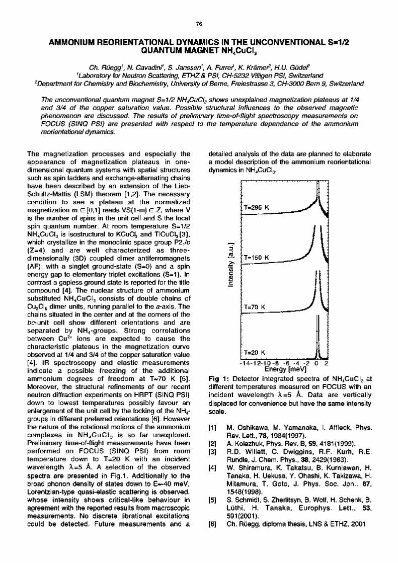
76
AMMONIUM REORIENTATIONAL DYNAMICS IN THE UNCONVENTIONAL S=1/2 QUANTUM MAGNET NH4CuCI3
Ch. Ruegg1, N. Cavadini1, S. Janssen1, A. Furrer*, K Kramer2, H.U. Gudef 1 Laboratory for Neutron Scattering, ETHZ& PSI, CH-5232 Villigen PSI, Switzerland
2Department for Chemistry and Biochemistry, University of Berne, Freiestrasse 3, CH-3000 Bern 9, Switzerland
The unconventional quantum magnet S=1/2 NH4CuCl3 shows unexplained magnetization plateaus at 1/4 and 3/4 of the copper saturation value. Possible structural influences to the observed magnetic phenomenon are discussed. The results of preliminary time-of-flight spectroscopy measurements on FOCUS (SINQ PSI) are presented with respect to the temperature dependence of the ammonium reorientational dynamics.
The magnetization processes and especially the appearance of magnetization plateaus in one-dimensional quantum systems with spatial structures such as spin ladders and exchange-alternating chains have been described by an extension of the Lieb-Schultz-Mattis (LSM) theorem [1,2]. The necessary condition to see a plateau at the normalized magnetization m e [0,1] reads VS(1-m) e Z, where V is the number of spins in the unit cell and S the local spin quantum number. At room temperature S=1/2 NH4CuCI3 is isostructural to KCuC3 and TICuCI3[3], which crystallize in the monoclinic space group P2/C (Z=4) and are well characterized as three-dimensionally (3D) coupled dimer antiferromagnets (AF): with a singlet ground-state (S=0) and a spin energy gap to elementary triplet excitations (S=1). In contrast a gapless ground state is reported for the title compound [4]. The nuclear structure of ammonium substituted NH4CuCI3 consists of double chains of Cu2CI6 dimer units, running parallel to the a-axis. The chains situated in the center and at the corners of the bc-unit cell show different orientations and are separated by NH4-groups. Strong correlations between Cu2+ ions are expected to cause the characteristic plateaus in the magnetization curve observed at 1/4 and 3/4 of the copper saturation value [4]. IR spectroscopy and elastic measurements indicate a possible freezing of the additional ammonium degrees of freedom at T«70 K [5]. Moreover, the structural refinements of our recent neutron diffraction experiments on HRPT (SINQ PSI) down to lowest temperatures possibly favour an enlargement of the unit cell by the locking of the NH4-groups in different preferred orientations [6]. However the nature of the rotational motions of the ammonium complexes in NH4CuCI3 is so far unexplored. Preliminary time-of-flight measurements have been performed on FOCUS (SINQ PSI) from room temperature down to T=20 K with an incident wavelength X=5 A. A selection of the observed spectra are presented in Fig.1. Additionally to the broad phonon density of states down to E«-40 meV, Lorentzian-type quasi-elastic scattering is observed, whose intensity shows critical-like behaviour in agreement with the reported results from macroscopic measurements. No discrete librational excitations could be detected. Future measurements and a
detailed analysis of the data are planned to elaborate a model description of the ammonium reorientational dynamics in NH4CuCI3.
c 0
-14-12-10-8 -6 -4 -2 Energy [meV]
Fig 1: Detector integrated spectra of NH4CuCI3 at different temperatures measured on FOCUS with an incident wavelength X=5 A. Data are vertically displaced for convenience but have the same intensity scale.
[1]
[2] [3]
[4]
[5]
[6]
M. Oshikawa, M. Yamanaka, I. Affleck, Phys. Rev. Lett., 78, 1984(1997). A. Kolezhuk, Phys. Rev. B, 59, 4181(1999). R.D. Willett, C. Dwiggins, R.F. Kurh, R.E. Rundle, J. Chem. Phys., 38, 2429(1963). W. Shiramura, K. Takatsu, B. Kurniawan, H. Tanaka, H. Uekusa, Y. Ohashi, K. Takizawa, H. Mitamura, T. Goto, J. Phys. Soc. Jpn., 67, 1548(1998). S. Schmidt, S. Zherlitsyn, B. Wolf, H. Schenk, B. Luthi, H. Tanaka, Europhys. Lett., 53, 591(2001). Ch. Ruegg, diploma thesis, LNS & ETHZ, 2001

77
POLARIZED NEUTRON SCATTERING FROM DYNAMICALLY POLARIZED PROTONS CLOSE TO PARAMAGNETIC CENTERS
B. van den Brandt \ H. Glattli2,1. Grillo 3, P. Hautle \ H. Jouve 4, J. Kohlbrecher \ J.A. Konter \ E. Leymarie 2, S. Mango 1, R. May3, H.B. Stuhrmann 4'5 and O. Zimmer6
1Paul Scherrer Insltute, CH - 5232 Villigen PSI, Switzerland 2CEA Saclayl SPEC, F- 91191 Gif-sur-Yvette, France
3lnstitut Laue Langevin, BP 156, F- 38042 Grenoble Cedex 9, France 4lnstitut de Biologie Structurale Jean-Pierre Ebel, F - 38027 Grenoble Cedex 1, France
5GKSS Forschungszentrum, D - 21502 Geesthacht, Germany 6Technische Universitat Munchen, James-Franck-Strasse, D - 85748 Garching, Germany
Domains of polarized protons in organic samples created by dynamic nuclear polarization (DNP) around paramagnetic centers have been investigated with small angle polarized neutron scattering. A time dependent scattering amplitude has been observed in the process of DNP and the possibility to maintain polarization gradients by combined rf and microwave irradiation has been explored.
As a complementary tool to isotopic substitution, dynamic nuclear polarization (DNP) has been used for some time to create contrast in SANS on hydrogenous samples exploiting the strong spin dependence of neutrons scattering on protons. Taking advantage of the electron-nuclear dipolar interaction the high thermal equilibrium polarization of isolated paramagnetic centers can be transferred to the nearby nuclei by microwave irradiation close to the EPR line. This localized polarization then spreads out into the bulk of the sample through spin diffusion. The DNP mechanism does not a priori lead to a homogeneous polarization, as the two processes might proceed with different time constants. Situations can be imagined in which domains of highly polarized protons around the paramagnetic centers survive long enough to produce a contrast that can be detected by neutron scattering [1,2]. Thus the study of radicals, playing a key role in the biological activity of certain molecules [3], inaccessible by magnetic neutron scattering, becomes feasible. In a series of experiments at D22 at the ILL, time dependent small angle scattering was observed in samples of glassy glycerol-water mixtures with different deuteration containing a small concentration of the paramagnetic EHBA-Cr(V) complex [4]. We reversed the polarization direction periodically (every 10 s) by switching the microwave frequency and measured the change in the scattering intensity in time bins of 0.1 s length. The Q-dependence could be fitted at all times with a simple but realistic model for the EHBA complex and the polarization of the protons close to the Cr(V) could be obtained from the amplitude of the coherent scattering [5]. Similar measurements on a diluted solution of catalase containing tyrosyl radicals have shown the first evidence of a time-dependent shape of the polarization domains, which could be extracted from the large SANS signal of the catalase as a small time-(i.e. polarization-) dependent contribution [5]. A complementary experimental approach that aims at
producing and maintaining a steady state spin contrast situation has been investigated at the SANS apparatus at SINQ [6]. Through a fine tuned interplay between rf and microwave irradiation the process of polarization buildup could be restricted to the protons surrounding the paramagnetic center. Selective saturation of those protons through which the bulk protons communicate with the protons close to the electronic spins may influence spin diffusion [7]. Furthermore the density distribution of these "intermediate" protons, which is a Lorentzian function of their distance to the normal Larmor frequency, i.e. distance to the electron, could provide a handle for the variation of the size of the polarized protons domain. In fact, first experiments at SINQ seem to support these hypotheses. Permanently driving the DNP, we observe a change in contrast of a factor of almost two when scanning the rf irradiation frequency across the proton magnetic resonance. This can be explained by a drastic change of the scattering density of the protons of the EHBA-Cr(V) complex. This technique may open the way to structural and dynamical studies not yet accessible to magnetic neutron scattering: It provides sensitivity enhancement for very dilute paramagnets and in addition EPR (microwave frequency induced) selectivity could be obtained in samples with several distinct paramagne-tic centers.
[1] J.B. Hayter et al., Phys. Rev. Lett. 33 (1974) 696. [2] H.B. Stuhrmann etal., J. Appl. Cryst. 30 (1997)
839. [3] D.C. Bicout, M.J. Field, P. Gouet, H.M. Jouve,
Biochemica et Biophysica Acta 1252 (1995) 172. [4] B. van den Brandt et al., PSI Scientific report 3
(2000) 78 & 94. [5] to be published [6] P. Hautle et al., SINQ Proposal 1/01-S45. [7] S.F.J Cox, S.F.J. Read and W.Th. Wenckebach,
J. Phys. C10(1977) 2917.

78

79
Condensed Matter Theory

80

81
POINT DEFECTS, FERROMAGNETISM AND TRANSPORT IN CALCIUM HEXABORIDE
R. Monnier1 and B. Delley1
1 Laboratory for Solid-State Physics, ETHZ, Switzerland 2 Condensed Matter Theory, PSI, CH-5232 Villigen PSI, Switzerland
The formation energy and local magnetic moment of hypothetical point defects in CaB^ are computed using a super-cell approach. It Is found that the substitution of Ca by La does not lead to the formation of a local moment. However, a neutral B6 vacancy carries a moment of 2.4 Bohr magnetons.
Recent experiments [1] on alkaline earth hexaborides have revealed an extreme sensitivity of their physical properties to stoichiometry and impurity content. A big surprise has been the observation of high temperature weak ferromagnetism in CaB6, SrB6 and BaB6 lightly doped with lanthanum and thorium, as well as in the isovalent substitutional alloy Cao995Ba0oo5B6 , soon followed by the discovery of the same phenomenon in nominally pure CaB6 , SrB6 . It was further found that when CaB6 is grown from a calcium-rich mixture of the elements, it becomes very weakly paramagnetic, while its low temperature (T<80 K) resistivity is increased by more than two orders of magnitude. The former observation suggests that the ferromagnetism of the nominally pure systems is related to the presence of vacancies on the metal sub-lattice and/or to the resulting intrinsic (hole) doping. We have investigated the possibility that the observed spontaneous magnetization is localized at imperfections in the CaB6 lattice and qualitatively discuss the effect of these imperfections on the electrical conductivity [2].
Table: Formation energy and local magnetic moment of point defects in CaB6, (a from gas phase, b from elements dissolved in a liquid Al flux,
c from borothermal reduction of CaO)
Defect Ca vacancy La (Ca) Al (Ca) B(Ca) B vacancy B6 vacancy Ca (B6) La (B6) Al (B6)
E Form [eV] 6.63a,4.966,4.6c
-1.94a,-0.14'' 3.73a, S.O6
4.69a, S-S6
11.05a,5.66'c
50.7a,18.36-c
44.2a, 13.56
40.6a, 11.7'' 42.5a, 13.16
Moment [/xB] <0.001 <0.001 <0.001 <0.001 <0.001
2.36 1.32
<0.001 <0.001
We first consider the defects on the metal sub-lattice. When a neutral Ca atom is removed from the crystal, it takes along two valence electrons, and the vacancy left behind acts as an acceptor. As seen in the Table, the substitution of Ca by La increases the binding energy of the compound. This remains the case even after correcting by the difference in heats of vaporization of La and Ca (-1.81 eV/atom), and all the lanthanum in the flux should therefore be incorporated. According to our
calculation, there is no magnetic moment associated with the La impurity.
1 . B4
> I - o ""* - ii£ii„ m. t A $ A Li ^
-1 .
-6 -4 -2 EF 2 Energy [eV]
Fig.1: Partial s,p,(d) density of states for a dangling bond B atom in CaB6. magnetic moment per atom is 0.22/iB-
The loss of complete boron octahedra by the structure has been invoked by Noack and Verhoeven [3] in order to explain their gravimetric data on zone refined LaB6. According to our calculations, this process is energetically more favorable than the creation of an equivalent number of well separated, single boron vacancies, but the computed formation energy of 18.3 eV implies that, unless the resulting void is stabilized by another factor, like eg a large impurity cation or a Ca vacancy acting as an acceptor and taking away electrons from inter octahedra bonding orbitals, it will not be formed under thermal equilibrium conditions. What makes it interesting, however, is the large magnetization it carries. The moment is mainly localized on the six neighboring boron octahedra, most of it (6 x 0.24 JJLB) in the dangling bonds from the atoms immediately adjacent to the void.
[1] DP. Young et al, Nature 397, 412, (1999) [2] R. Monnier, B. Delley, Phys. Rev. Lett. 87, 157204,
(2001) [3] M. A. Noack, J. D. Verhoeven, J. Crystal Growth 49,
595(1980)

82
DENSITY OF STATES FOR DIRTY d-WAVE SUPERCONDUTORS: A UNIFIED AND DUAL APPROACH FOR DIFFERENT TYPES OF DISORDER
C Chamon x and C Mudry2
1 Department of physics, Boston University, Boston, MA 02215, USA 2 Paul Scherrer Instltut, CH-5232, Villigen PSI, Switzerland
A two-parameter field theoretical representation Is given of a 2-dlmenslonal dirty d-wave superconductor that Interpolates between the Gaussian limit of uncorrelated weak disorder and the unitary limit of a dilute concentration of resonant scatterers. It is argued that a duality holds between these two regimes from which follows that a linearly vanishing density of states in the Gaussian limit transforms into a diverging one in the unitary limit arbitrarily close to the Fermi energy.
Extrinsic defects in high-Tc materials have observable consequences for the local and bulk density of states (DOS) of quasiparticles. This is seen by scanning tunnelling microscopy, nuclear magnetic resonance, and neutron scattering. Popular wisdom distinguishes "weak" extrinsic disorder introduced by defects above or below the copper-oxide planes through oxygen doping, from "strong" extrinsic disorder caused by substitutions of in-plane copper atoms with ions such as zinc. Given that the symmetry of the superconducting state is predominantly of the d-wave type in high-Tc materials, it is thus not surprising that a considerable theoretical effort has been devoted to the effect of quenched disorder on a d-wave order parameter by reducing the problem to two models. The first one is a model of Anderson localization for a single "nodal" quasiparticle described by a two-dimensional (2D) non-magnetic random Bogoliubov-de-Gennes (BdG) Hamiltonian. The second one is a Kondo model in which a single magnetic moment couples to a bath of 2D "nodal" quasiparticles. At this level of approximation no attempt is made to describe either the existence of quasiparticle interactions or the self-consistent effect of defects on the superconducting order parameter, although these effects might be important. In spite of the simplicity of either model many important theoretical issues remain unresolved to this date. Even in the simpler case of Anderson localization, the effect of disorder on the quasiparticle DOS of a d-wave superconducting state is poorly understood. The physics of localization is expected to dominate at long distances and small energies in 2D. This fact together with the existence of particle-hole symmetry below Tc makes it essential to account for the level repulsion upon approaching the Fermi energy eF. However, the physics of level repulsion and localization are nonper-turbative with respect to standard perturbative techniques [expansions in (hpi)'1 < 1, kF the Fermi momentum and £ the mean free path in the Born approximation]. The existence of four isolated nodes of the superconducting pairing function A k in the 2D Brillouin zone also invalidates, upon approaching eF, expansions in powers of {kFt)~l < 1- Moreover, there are some theoretical arguments and numerical evidences that have been attributed to the existence of nodes of
A k suggesting that the dependence on energy of the DOS depends in a dramatic fashion on the microscopic details of the disorder. On the one hand, for a high density of very weak s-wave scatterers [the Gaussian limit (GL)] level repulsion dominates and causes the DOS to vanish linearly upon approaching eF. On the other hand, a dilute density of uncorrelated and resonant s-wave scatterers [the unitary limit (UL)] is argued to result in a diverging DOS. The purpose of this paper [1] is to present a unified picture of these two limits. Starting from a BdG Hamiltonian describing a dirty d-wave superconductor with white-noise correlated disorder that depends on two parameters, the impurity concentration p and the variance g of the disorder, we construct an effective field theory that generates disorder averaged Green functions. We show how this effective action reduces to the nonlinear sigma model (NLSM) describing a random BdG Hamiltonian with time reversal symmetry and spin rotation symmetry in the limit of p -)► oo with g finite. The effective action is treated by performing an expansion in powers of p (virial expansion) in the opposite limit of g finetuned to its resonant value g ^ gr with p finite. Carrying out the expansion up to first (second) order in p allows to identify this limit with the UL (the expansion parameter). As was the case with the (hpi)'
1 expansion, the virial expansion converges only for not too small energies. In order to better understand whether there can or cannot be a crossover from the UL to the GL, we use a variational method and show that the form of the selfconsistent equations is controlled by how the chemical potential is introduced in the problem. This suggests that the energy alone cannot connect the two limits if the chemical potential is chosen at the "resonant" condition for the UL. By expanding perturbatively for large energies, we identify a duality relation between the GL and UL series that would imply, at low energies, that the diverging DOS l/\e\ In
2 \l/e\ is connected by duality to the vanishing DOS |e| in the GL arbitrarily close to eF. This nonperturbative argument relies in an essential way on the nodes of A k .
[1] C. Chamon and C. Mudry, Phys. Rev. B 63, 100503 (2001).

83
TRANSPORT PROPERTIES AND DENSITY OF STATES OF QUANTUM WIRES WITH OFF-DIAGONAL DISORDER
P. W. Brouwer1, C Mudry2 and A. Furusaki3
1 Lyman Laboratory of Physics, Harvard University, Cambridge, MA 02138, USA 2 Paul Scherrer Instltut, CH-5232, Villigen PSI, Switzerland
3 Yukawa Institute for Theoretical Physics, Kyoto University Kyoto 606-8502, Japan
We review recent work on the random hopping problem In a quasl-one-dlmenslonal geometry of N coupled chains (quantum wire with off-diagonal disorder). Both density of states and conductance show a remarkable dependence on the parity of N. The theory is compared to numerical simulations.
With the realization that the problem of Anderson localization is amenable to a RG analysis came the understanding that some transport and spectral properties of a quantum particle subjected to a weak random potential depend qualitatively only on dimensionality and the symmetries of the random potential [1]. For essentially all types of randomness the dimensionality two of space plays the role of the lower critical dimension: Below two dimensions and for arbitrary weak disorder a quantum particle is always localized (i.e., the wavefunc-tion is insensitive to a change in boundary conditions, or, equivalently, exponential decay of the conductance with system size), whereas a finite amount of disorder is needed to localize a quantum particle in three dimensions. A notorious exception to this rule is the case of a particle on a single chain with random nearest neighbor hopping which appears in many reincarnations, e.g., random XY spin chains [2] and diffusion in random environments [3]. In all these cases, the cause underlying the different behavior of random systems with off-diagonal disorder is the existence of a sublattice, or chiral, symmetry, which is absent for diagonal disorder [4]. The purpose of this contribution [5] is to review our recent work (together with Simons and Altland [6]) on the random hopping problem in a quasi-one-dimensional "wire" geometry [6-9]. We consider the density of states (DoS) and conductance, and focus on the differences between the cases of off-diagonal and diagonal disorder in the localized regime. (For off-diagonal disorder, we assume absence of diagonal disorder.) The quantum wire is the logical intermediate between one and two dimensions [10]. The quasi-one-dimensional
geometry allows us to find exact solutions for the conductance at the band center e = 0 and the DoS near e = 0, while, in contrast to purely one-dimensional counterparts, it still shows a crossover from a diffusive to a localized regime, as is the case in two dimensions.
[1] For a review, see P. A. Lee and T. V Ramakrishnan, Rev. Mod. Phys. 57, 287 (1985).
[2] B. M. McCoy and T T Wu, Phys. Rev. 176, 631 (1968).
[3] D. S. Fisher, Phys. Rev. A 30, 960 (1984). [4] A. Altland, B. D. Simons, J. Phys. A32, L353-L359
(1999). [5] P. W. Brouwer, C. Mudry, and A. Furusaki, Physica
E 9, 333(2001). [6] P. W. Brouwer, C. Mudry, B. D. Simons, A. Altland,
Phys. Rev. Lett. 81, 862 (1998). [7] C. Mudry, P. W. Brouwer, A. Furusaki, Phys. Rev. B
59, 13221 (1999). [8] P. W. Brouwer, C. Mudry, A. Furusaki, Nucl. Phys.
B565, 653 (2000). [9] P. W. Brouwer, C. Mudry, A. Furusaki, Phys. Rev.
Lett. 84, 2913(2000). [10] Two dimensional models with off-diagonal disorder
are also studied in the context of strongly interacting electron systems [11]. We refer the reader to Ref. [12] for a review of work done on the two dimensional case.
[11] N. Nagaosa and P. A. Lee, Phys. Rev. Lett. 64, 2450 (1990). V Kalmeyer and S. C. Zhang, Phys. Rev. B 46 (1992) 9889; B. I. Halperin, P. A. Lee, and N. Read, Phys. Rev. B 47, 7312 (1993).
[12] A. Furusaki, Phys. Rev. Lett. 82, 604 (1999).

84
FOKKERPLANCK EQUATIONS AND DENSITY OF STATES IN DISORDERED QUANTUM WIRES
M. Titov1, P. W. Brouwer
2, A. Furusaki
3 and C Mudry4
1 InstltuutLorentz, Unlversltelt Leiden, P. O. Box 9506, 2300 RA Leiden, The Netherlands 2 Laboratory of Atomic and Solid State Physics, Cornell University Ithaca, NY 148532501
3 Yukawa Institute for Theoretical Physics, Kyoto University Kyoto 6068502, Japan 4 Paul Scherrer Institut, CH5232 Villigen PSI, Switzerland
We propose a general scheme to construct scaling equations for the density of states in disordered quantum wires for all ten pure Cartan symmetry classes. The anomalous behavior of the density of states near the Fermi level for the three chiral and four Bogollubovde Gennes universality classes Is analysed In detail by means of a mapping to a scaling equation for the reflection from a quantum wire In the presence of an imaginary potential.
Statistical properties of energy levels and wavefunctions in disordered electron systems are believed to be determined by, first of all, the fundamental symmetries of the Hamiltonian. Such a connection is best established in the case of "zerodimension", i.e., for finite size systems with a large dimensionless conductance g, where a description in terms of random matrix theory is valid. Using the link to Cartan's classification of symmetric spaces, it has been have pointed out that there exist only ten possible random matrix theories, whose form follows directly from the geometrical characteristics ("roots") of the corresponding symmetric space in Cartan's table. These ten random matrix theories are divided into three standard classes, three chiral classes, and four Bogoliubovde Gennes (BdG) classes, the subdivision in each class depending on the presence or absence of timereversal symmetry (TR) and spinrotation invariance (SR). The rational for the Cartan classification is believed to transcend "zerodimension" and has been applied to the construction of effective theories (e.g., nonlinearsigmamodels) for higher dimensional disordered systems. The standard classes are thus believed to be appropriate to the problem of an electron moving in a random potential, without further symmetries, irrespective of dimensionality. The chiral classes are appropriate to the case when the disorder is purely "offdiagonal", as is the case, e.g., for the lattice random flux model in quasione and twodimensions, the random hopping model, and for random XY spin chains. The BdG classes refer to systems with superconducting correlations, and were argued to be valid for vortices in superconductors, dirty unconventional superconductors, and (in the case of broken timereversal symmetry) normal metals in proximity to a superconductor. The BdG classes have also been argued to be of some relevance to problems in statistical mechanics such as random bond Ising and network models. In this paper, we show how the same structure also determines scaling equations for the density of states (DOS) v(e) in a quantum wire of infinite length (L ► oo)
at energy e. Our work builds on previous work for the chiral classes, where such a scaling equation was derived using a different method. [1] In addition, we present an exact solution for the DOS, something that could not be done in Ref. [1]. While v[e) is a nonsingular function of energy for the three standard classes, singular behavior is expected near e = 0 for the remaining seven symmetry classes, as e = 0 is a special point there. (The energy e = 0 corresponds to a point of particlehole symmetry in the chiral and BdG symmetry classes; it is the band center for lattice models with random hopping, or the Fermi energy in the case of the BdG classes.) Indeed, for the chiral classes, v{e) was found to depend sensitively on the parity of the channel number N for e close to zero, showing the v(e) oc l/|eln
3e| divergence char
acteristic of pure onedimensional systems with chiral symmetry for odd N, while v{s) oc |em / _ 1 lne| for even N. [1] For the BdG classes in the presence of spinrotation invariance (classes C and CI; we refer to the symmetry classes by Cartan's symbol for the symmetric space corresponding to their Hamiltonians), a suppression v(e) oc em / _ 1 is expected. In both cases, the characteristic energy scale for the DOS singularity is ~ hvF/N
2£, vF being the Fermi velocity. This distin
guishes the singularity in v{s) for quantum wires from its counterpart in zerodimension, where the characteristic energy scale is the mean level spacing, which goes to zero as the system size is increased. No exact results are known for the multichannel quantum wires from BdG symmetry classes in the absence of spinrotation invariance (classes D and DIM; for asymptotic results in one and twodimensions). The general scheme that we present in Ref. [2] fills this gap, and provides a unified framework for all ten symmetry classes.
[1] P. W. Brouwer, C. Mudry, and A. Furusaki, Phys. Rev. Lett. 84, 2913 (2000).
[2] M. Titov, P. W. Brouwer, A. Furusaki, and C. Mudry, Phys. Rev. B 63, 235318 (2001).
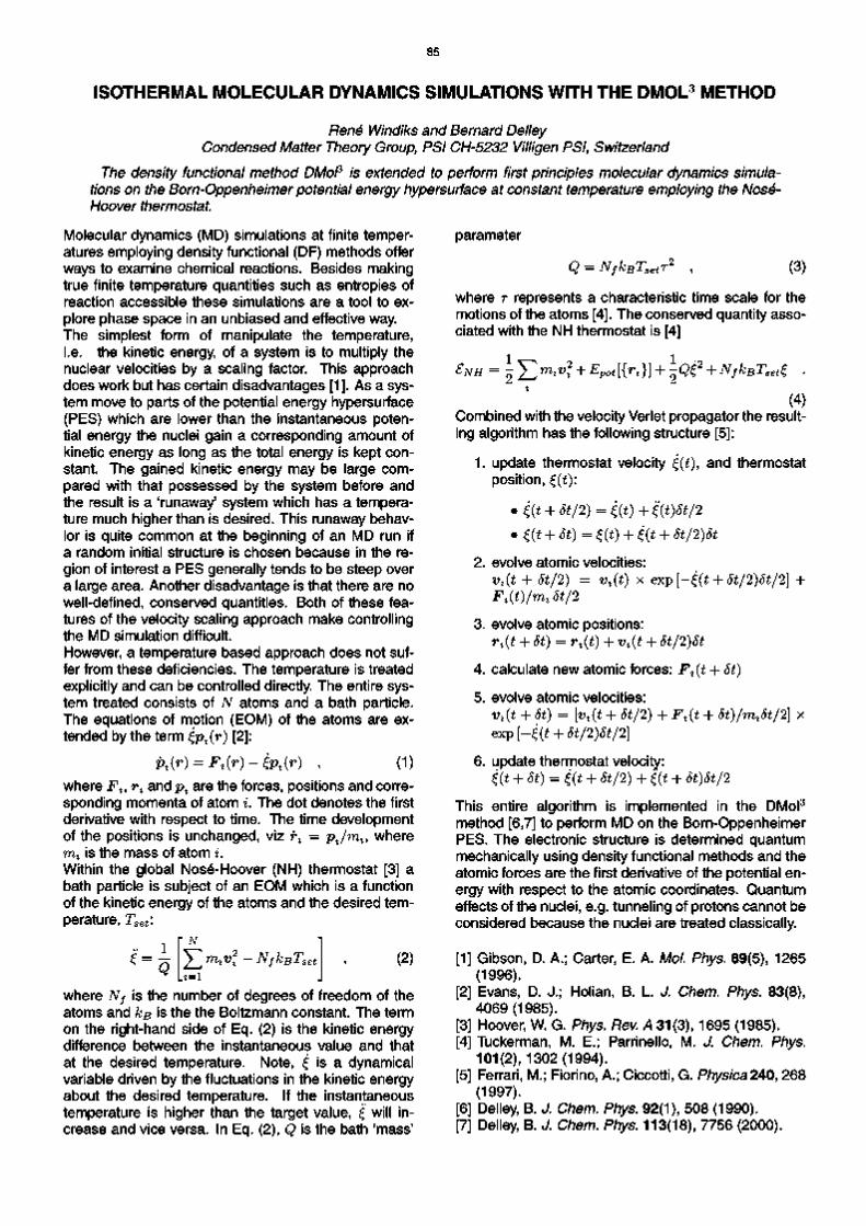
85
ISOTHERMAL MOLECULAR DYNAMICS SIMULATIONS WITH THE DMOL3 METHOD
Rene Windiks and Bernard Delley Condensed Matter Theory Group, PSI CH-5232 Villigen PSI, Switzerland
The density functional method DMol3 is extended to perform first principles molecular dynamics simulations on the Born-Oppenheimer potential energy hypersurface at constant temperature employing the Nose-Hoover thermostat.
Molecular dynamics (MD) simulations at finite temperatures employing density functional (DF) methods offer ways to examine chemical reactions. Besides making true finite temperature quantities such as entropies of reaction accessible these simulations are a tool to explore phase space in an unbiased and effective way. The simplest form of manipulate the temperature, i.e. the kinetic energy, of a system is to multiply the nuclear velocities by a scaling factor. This approach does work but has certain disadvantages [1]. As a system move to parts of the potential energy hypersurface (PES) which are lower than the instantaneous potential energy the nuclei gain a corresponding amount of kinetic energy as long as the total energy is kept constant. The gained kinetic energy may be large compared with that possessed by the system before and the result is a 'runaway' system which has a temperature much higher than is desired. This runaway behavior is quite common at the beginning of an MD run if a random initial structure is chosen because in the region of interest a PES generally tends to be steep over a large area. Another disadvantage is that there are no well-defined, conserved quantities. Both of these features of the velocity scaling approach make controlling the MD simulation difficult. However, a temperature based approach does not suffer from these deficiencies. The temperature is treated explicitly and can be controlled directly. The entire system treated consists of TV atoms and a bath particle. The equations of motion (EOM) of the atoms are extended by the term £pz(r) [2]:
pt(r) = Ft(r) - £Pl(r) , (1) where F%, r% and pz are the forces, positions and corresponding momenta of atom i. The dot denotes the first derivative with respect to time. The time development of the positions is unchanged, viz r% = pjmt, where mt is the mass of atom i. Within the global Nose-Hoover (NH) thermostat [3] a bath particle is subject of an EOM which is a function of the kinetic energy of the atoms and the desired temperature, Tset\
£: 1 Q
' N
rnlv2% - NfkBTset (2)
where Nf is the number of degrees of freedom of the atoms and kB is the the Boltzmann constant. The term on the right-hand side of Eq. (2) is the kinetic energy difference between the instantaneous value and that at the desired temperature. Note, £ is a dynamical variable driven by the fluctuations in the kinetic energy about the desired temperature. If the instantaneous temperature is higher than the target value, £ will increase and vice versa. In Eq. (2), Q is the bath 'mass'
parameter
Q = NfkBTsetr2 (3)
where r represents a characteristic time scale for the motions of the atoms [4]. The conserved quantity associated with the NH thermostat is [4]
SNH = l^m*v* + Epot[{n}} + lQ£2 + NfkBTsett .
(4) Combined with the velocity Verlet propagator the resulting algorithm has the following structure [5]:
1. update thermostat velocity £(t), and thermostat position, £(£):
• £(t + St/2) = £(t) + '£{t)St/2 • t(t + 5t)=£(t) + £(t + 5t/2)5t
2. evolve atomic velocities: vt(t + St/2) = v%(t) x exp [-£(t + St/2)St/2] + Fl(t)/mlSt/2
3. evolve atomic positions:
rt(t + St) = rt(t) + vt(t + St/2)St
4. calculate new atomic forces: Ft(t + St)
5. evolve atomic velocities: vt(t + St) = [vt(t + St/2) + F%(t + St)/mlSt/2] x exp [-£(t + St/2)St/2]
6. update thermostat velocity: {(t + St) = £(t + St/2) + £(t + St)St/2
This entire algorithm is implemented in the DMol3
method [6,7] to perform MD on the Born-Oppenheimer PES. The electronic structure is determined quantum mechanically using density functional methods and the atomic forces are the first derivative of the potential energy with respect to the atomic coordinates. Quantum effects of the nuclei, e.g. tunneling of protons cannot be considered because the nuclei are treated classically.
[1] Gibson, D. A.; Carter, E. A. Mol. Phys. 89(5), 1265 (1996).
[2] Evans, D. J.; Holian, B. L. J. Chem. Phys. 83(8), 4069(1985).
[3] Hoover, W. G. Phys. Rev. A 31(3), 1695 (1985). [4] Tuckerman, M. E.; Parrinello, M. J. Chem. Phys.
101(2), 1302(1994). [5] Ferrari, M.; Fiorino, A.; Ciccotti, G. Physica240, 268
(1997). [6] Delley, B. J. Chem. Phys. 92(1), 508 (1990). [7] Delley, B. J. Chem. Phys. 113(18), 7756 (2000).

86
SADDLE POINT REFINEMENT: FINDING TRANSITION STATES
N. Govind, J.W. Andzelm, G.Fitzgerald1, R. Windiks and B. Delley2
1Accelrys, San Diego CA 92121, USA 2 Condensed Matter Theory, PSI, CH-5232 Villigen PSI, Switzerland
Saddle points are key to the understanding of the topology of energy surfaces. We have studied a set of well characterized molecular transition states with the DMoP method to determine the capabilities and strength of density functional theory. A significant step forward locating saddle points theoretically has been made recently.
The first step in exploring the topology of the high dimensional energy surface of a complex molecular or solid system is to search for local minima that correspond to stable or metastable conformations. The second step is to locate the saddle points that separate the minima and to get an idea about the reaction paths. The determination of saddle points remains a difficult task, significantly more difficult than to find the minima for which a number of good algorithms exist. Another difficulty comes from the fact that the saddle point structure usually involves broken bonds and unusual coordination , which makes accurate determination of electronic energies significantly more difficult than for ground states. In order to address the second difficulty we have compared the energetics of a set of fairly well established transition states [1] with the energetics of a test set for the enthalpy of formation for the ground state as proposed by the Pople group. The test set for reactions consists mainly of hydrogen transfer reactions. The set of well known formation enthalpies involves a variety of small molecules. As the table shows, the rms errors for enthalpies and for barrier heights are very similar. The mean error shows that barrier heights are systematically underestimated on average. A problem is that for several of these hydrogen transfer reactions there is no (forward) energy barrier found with the majority of todays functionals. Fortunately, it is not true that density functional approximations typically miss low energy barriers. In the test shown in the table, transition state geometries of Lynch [1] are used.
Table: Errors of calculated enthalpies of formation and of energy barriers [kcal/mol]
test set of mean avg.abs. rms
Eform 148
+ 0.9 5.8 7.7
Ebarr 44
-6.3 7.1 8.4
Ereac 22
-0.5 2.2 2.9
Some time ago Halgren and Lipscomb [2] have defined a simple path connecting two structures (possibly via an intermediate structure), these paths are called synchronous transit and represent a rough approximation to the reaction path. This approach was improved recently by Govind combining these ideas with an efficient optimization approach [3] to arrive at a scheme
which seamlessly works for both gas phase molecular and solid state systems. Fig.1 illustrates the linear synchronous transit for the nitroprusside anion as well as the position of the exact saddle point. The energy barrier S2 -> GS is 0.68 eV in this case and state S2 is 1.40 eV above the ground state of the free anion when calculated with the Becke-Perdew91 functional.
0.0 S2
0.4 0.6 0.8 path parameter
1.0 GS
Fig.1: Energies along the linear synchronous transit between the metastable state S2 and the ground state of the nitroprusside anion. The black point represents the precise saddle point. Also shown are reactant, product and transition state structure.
[1] B.J.Lynch and D.G.Truhlar, J.Phys.Chem. A 105, 2936(2001).
[2] T.A. Halgren and W.N. Lipscomb, Chem. Phys. Lett. 49,225(1977).
[3] J. Baker, A. Kessi and B. Delley, J. Chem Phys. 105, 192(1996).

87
INTERCALATION AND HIGH TEMPERATURE SUPERCONDUCTIVITY IN FULLERIDES
A Bill1 and V.Z. Kresin2
1 Condensed Matter Theory, Paul Scherrer Institute, CH-5232 Villigen PSI, Switzerland 2 Lawrence Berkeley Laboratory, University of California, Berkeley, CA 94720, USA
Properties of a superconductor can be drastically affected by intercalating polyatomic molecules into the compound. A mechanism responsible for a large increase in Tc is proposed for such systems. It explains the recent remarkable observation of high Tc superconductivity In the hole-doped C60 • 2CHX3 (X=Cl,Br) compounds and the large shift in their Tc upon CI^Br substitution. We suggest two experiments to test the theory.
This work is concerned with the impact of intercalation by polyatomic molecules on the properties of a superconductor [1]. The study has been motivated by the recent remarkable observation of high Tc caused by intercalation of CHBr3 (bromoform) and CHCI3 (chloroform) molecules into hole-doped fullerides [2]. Usual superconducting fullerides [3] are chemically electron-doped compounds (e.g. Rb3C60) and it is believed that superconductivity is due to the coupling of electrons to intramolecular vibrational modes of C60. The new results obtained in Ref. [2] use an alternative doping technique by implementing the superconducting material into a FET device which allows also to dope holes into the compound. Hole-doping of pristine C60 has resulted in a Tc as high as 52K. Intercalating CHBr3 or CHCI3 into the crystal and doping with the above technique [2] has resulted in another dramatic jump to Tc ~ 117K (for hole-doping). We propose a mechanism that allows for a quantitative explanation of the drastic increase in Tc observed in [2]. It is essential to note first that there is not only a difference in the observed Tc between C60 and intercalated C60, but that there is also a large difference when intercalating CHCI3 (Tc = 80K) or CHBr3 (Tc = 117K), despite similar molecular structure. The picture we propose is that the intercalated molecules themselves actively, and importantly, participate in the pairing. The internal vibrational modes of the molecules provide for an additional attraction which leads to higher Tc. To show this, we start with the parent hole-doped ful-leride (Tc = 52K). We describe the superconducting state with Eliashberg's equations in which the following parameters are to be determined: Ai describes the electron-phonon coupling constant to the relevant C60 intramolecular vibrations of average frequency fix and p* is the Coulomb pseudopotential. Following [3] we estimate fix ~ O.leV and / i * = 0.15. Then, the value of Tc = 52K observed for hole-doped C60 implies Ai = 0.75. Intercalating polyatomic molecules into C60 results in an additional electron-phonon coupling term (~ A2) in the above mentioned equations. This term describes the coupling of holes with the vibrational manifold of the intercalated molecules (average frequency tt2). The increase in Tc induced by this additional coupling depends on the specific values of A2 and Q2. The coupling constants are given by McMillan's relation
A " M < n 2 > ' (1)
where v is the density of states at the Fermi level,
I the matrix element, and M and Q describe the vibrational spectrum. In essence, the denominator is the force constant of the vibrational mode coupling to the holes. This is the quantity changing for different haloform molecules. The vibrational modes that differ between chloroform and bromoform involve C and X=CI,Br ions. According to Eq. (1) and since M « Mc (the modes involving motion of H are too high in energy to be relevant for superconductivity [1]) the difference between A^1 and Afr is due to their different average vibrational frequencies. This frequency, fl2, can be extracted from the measured vibrational spectra of CHX3 (X=CI,Br). Let us start with X=CI for which £l$l = 85meV To obtain the observed value Tc = 80K [2] implies the value A^1 = 0.2 which is perfectly realistic. We now substitute CI^Br and compare C60 • 2CHCI3 and C60 • 2CHBr3. Given Eq. (1) and the vibrational spectrum of the molecules we calculate Afr ~ 2.65A2
J1. Thus, the "softening" of the modes lead to a noticeable increase in the coupling constant and thus of Tc. Indeed, knowing A^1 from above we obtain Afr « 0.55. Moreover, taking the frequency n f r ~ 70meV from literature we obtain Tc ~ 110K which is close to the experimentally observed value of Tc ~ 117K. We stress here that the last step of the calculation (bromoform case) is made without any adjustable parameter! The above calculations show that the drastic increase of Tc observed experimentally can indeed be described by the coupling of holes to the vibrational manifold of the intercalated molecules. The theory also reproduces the results obtained for electron doping. We further make two predictions [1]. One is the site selective isotope effect which consists in substituting 1 2C^1 3C on the haloform molecules only. Because of the coupling to the molecules, a small shift of Tc should be observed upon substitution. Furthermore, the same calculation as for bromoform can be performed for iodoform (CHI3) and leads to Tc ~ 140K! Both predictions can be tested experimentally.
[1] A. Bill, V.Z. Kresin, cond-mat/0109553,0110327; submitted.
[2] J.H. Schon, Ch. Kloc, B. Batlogg, Science 293, 2432(2001).
[3] O. Gunnarsson, Rev. Mod. Phys. 69, 575 (1997); W.E. Pickett, Sol. State Phys. 48, 225 (1994).
[4] R. Windiks, A. Bill, B. Delley, and V.Z. Kresin, Int. J. Mod. Phys. in press; see also report in this issue.

88
ELECTRONIC PROPERTIES OF C60 2CHX3 (X=CI,Br)
R. Windiksx, A. Billx, B. Delleyx, and V.Z. Kresin2
1 Condensed Matter Theory, Paul Scherrer Institute, CH-5232 Villigen PSI, Switzerland 2 Lawrence Berkeley Laboratory, University of California, Berkeley, CA 94720, USA
C60 • 2CHX3 with X=CI and Br has been shown recently to become superconducting below 80 and 117K, respectively. Using the density functional method DMoP we determine the crystal structure of this compound, and calculate the band structure as well as the density of states.
It has been shown recently [1] that solid C60 intercalated with haloform molecules CHX3 (X=CI,Br) becomes superconducting below Tc ~ 80K for X=CI and Tc ~ 117K for X=Br when doping the material with holes. Doping has been achieved by implementing the material into a Field Effect Transistor (FET). We have proposed a model that describes quantitatively the drastic increase of Tc upon intercalation and explains the large difference in Tc when substituting CI^Br [2,3]. On the other hand, little is known on the structure of these materials, and in particular on the exact orientation of the intercalated haloform molecules. Moreover, it would be of interest to determine the band structure and density of states (DoS) of the compounds and compare them with those of A3C60 (A is an alkali-ion) among which are found the highest Tcs for chemically doped C60 materials. Ultimately we aim at studying the question whether the change in DoS could be the cause for the observed increase in Tc as suggested in Ref. [1]. To achieve these goals, we started density functional calculations using the DMol3 method [5]. The average structure of neutral C60 • 2CHX3 has been determined by X-ray diffraction [4] and reveals a hexagonal lattice (unlike A3C60 which has an fee cell). Using this result we have optimized the structure with the above-mentioned method. Many configurations turn out to be close in energy. We find that different configurations not only correspond to different orientations of the C60 molecules, but also to orientational disorder of the haloform molecules. Fig. 1 displays one such configuration. As can be seen, haloform molecules are located away from the axis connecting two nearest neighbour C60 molecules. This contrasts with the location of some of the alkali ions in A3C60. The haloform molecules are also centered on points of lower symmetry than the alkali ions.
Fig.1: Calculated hexagonal crystal structure of neutral C60 • 2CHCI3 in one specific configuration optimized with a density functional method.
-
-
* y
/C&
!V V \£
y%
*%»*»
fm
-
-
M L A H K M T A I m r k h l wavevector DoS
Fig.2: Electronic band structure and DoS for the configuration of Fig. 1. Only the valence and conduction bands are shown.
Fig. 2 shows the band structure and DoS corresponding to the configuration of Fig. 1 for X=CI. Observe that the amplitude of the DoS at given energy \E\ is generally larger in the valence band than in the conduction band, confirming the expectation that hole-doping leads to a higher Tc than electron-doping. One further notes that the sizes of the bandwidths (~ 0.5eV) and the gap (~ 1.2eV) are similar to those found in A3C60 despite different crystal structures. The case X=Br shows similar features. These results were obtained for neutral C60 • 2CHX3. To analyze the question whether the DoS increases for the series C60, X=CI and X=Br critically depends on the behaviour of the band structure and the location of the Fermi energy when doping the compounds. These calculations are under way.
[1] J.H. Schon, Ch. Kloc, B. Batlogg, Science 293, 2432(2001).
[2] A. Bill, V.Z. Kresin, cond-mat/0109553,0110327; see also report in this issue.
[3] R. Windiks, A. Bill, B. Delley, and V.Z. Kresin, Int. J. Mod. Phys. In press.
[4] M.Jansen and G.Waidmann, Z anorg. allg. Chem. 621, 14(1995).
[5] B. Delley, J. Chem. Phys. 92, 508 (1990); 113, 7756 (2000).

89
SPECTRAL WEIGHTS, ZERO MODES AND NEUTRON SCATTERING IN A 2D QUANTUM DIPOLAR VORTEX LATTICE
H.B. Braun \ B. Roessli2, K. Kramer4, A. Wildes 3, P. Fischer2, H.U. Gudel 4
1 Condensed Matter Theory, PSI, CH-5232 Villigen PSI, Switzerland 2 Laboratory of Neutron Scattering, PSI & ETHZ, CH-5232 Villigen PSI, Switzerland
3 Institut Laue-Langevin, Grenoble, France 4 Department of Chemistry, University of Bern, Switzerland
The inelastic neutron scattering cross section of a system of easy-plane dipoles on a 2D honeycomb lattice is computed. The classical ground state is shown to be a vortex lattice. The dynamical structure factor is evaluated exactly and shown to be in excellent agreement with the measured inelastic neutron scattering data on the quasi 2D honeycomb magnet ErBr3. This uniquely characterizes this compound as the first example of a 2D system where dipolar interactions determine both magnetic order and the nature of the excitations.
Frustration in magnetic systems arises from the inability to satisfy all bonds simultaneously. This may occur for geometric reasons, the prototype example being a Heisenberg or Ising model on a triangular lattice. Further examples of such geometric frustration are spins on a 2D Kagome lattice or the 3D pyrochlores, so called "spin-ice" with degeneracies resembling those of the proton ordering in real ice [1]. Dipolar interactions necessarily fall into that category since their long-range nature makes it in general impossible to satisfy all bonds simultaneously. In the case of a simple cubic lattice and classical dipoles it is, however, known that regular order may result as shown by Luttinger and Tiszas calculation. Experimental realizations of 3D dipolar ordering are the compounds LiTbF4 or LiHoF4. Here we consider the magnetic structure and the spin excitations in the rare-earth trihalide ErBr3. We are able to demonstrate that the excitations can be entirely explained by dipolar interactions. This characterizes ErBr3 as the first magnet with 2D dipolar ordering. The crystal field levels of the Er ions with spin s = 15/2 consist of Kramers doublets, the lowest two being separated by 1.5meV. Earlier experimental studies have demonstrated 2D magnetic ordering below 400mK [2]. In order to determine the nature of the coupling between the Er spins, we have measured the inelastic excitation spectrum with neutrons. There are two characteristic features of the energy scans at T = O.IK performed on IN 14 at ILL: (i) no excitations between 0.3 meV and 1 meV, and (ii) for most ^-values in general only a few of the expected six spin-wave branches have been identified at a particular <?-value, cf. Fig. 1. The measured intensities have been compared with a model that includes only dipolar interactions between the lowest Kramers doublets of the Er spins. The neutron response is proportional to
^2 H ^ ' dte~ R,R' a,a' ^
q ( R - R / ) /eia4>R e~ia'<f>R, (t) > (D
where a, a' = ±, and <fiR = ^ + < R̂ is the sum of the static configuration and (dipolar) spin-waves, respectively. faa, = \\{q2
y - q2x)5a {2-ql- ql))5^]
is the polarization factor. The resulting intensities show excellent agreement with the data measured in the q-range covered by experiments, in particular, features (i) and (ii) are recovered.
i) ii)
\<t>)- ( +e*|6» V2 spin-orientation
C3^ easy-plane
% q
Fig.1:
300
250
| 200
^ 1 5 0
° 1 0 0
50
Fig.2:
(i) Crystal field levels of a single Er ion, allowing for an easy-plane spin description, (ii) Ground state of interacting dipoles on a honeycomb lattice. It is infinitely degenerate with respect to opposite rotation of the magnetization on the two sublattices, giving rise to an "optical" zero-mode, (iii) Dispersion of spin-wave type excitations of the vortex lattice. The dispersion but not the intensity has the periodicity of the magnetic Brillouin zone.
q&{ 0 , -15 ,d )
t)2» 0 4 0 6 energy /meV 04 06 08
energy /meV
Measured intensities at two points which are nearly equivalent with respect to the magnetic Brillouin zone. The curves represent the theoretical predictions with the overall energy scale as the only free parameter. Note that along high-symmetry directions the intensity of most branches is strictly zero! The energy of the invisible branches is indicated by vertical bars.
[1] A.P Ramirez, A. Hayashi, R.J. Cava, R. Sid-dharthan, and B.S. Shastry, Nature 399, 335 (2001).
[2] K. Kramer et al. Phys. Rev. B, 60 R3724, (1999).

90
FRACTIONAL QUANTUM HALL GAPS AND EFFECTIVE MASS OF COMPOSITE FERMIONS
R.H. Morf1 and N. d'Ambrumenil
2
1 Condensed Matter Theory, PSI, CH5232 Villigen PSI, Switzerland 2 Department of Physics, University of Warwick, England
The determination of energy gaps of fractional quantum Hall (FQH) states from exact diagonalizations of the Hamiltonian for small systems is complicated by the presence of finite size effects. These make the calculation of thermodynamic limit values of energy gaps difficult. Recent advances in understanding of finite size effects have for the first time made it possible to compute reliable values of gaps for higher order FHQ states atv = 3/7 and4/9. It turns out that our new gap values are consistent with theoretical results based on fieldtheoretic methods of Halperin, Lee and Read [1], which show that the effective mass of Composite Fermions diverges logarithmically when the filling factorv = 1/2 is approached.
One of the crucial tests of any theory is how accurately it predicts experimentally measurable quantities. In the frac
tional quantum Hall (FQH) effect, the energy gap A of FQH states can be measured rather well by determination of the activation energy of the longitudinal resistance of the quan
tum Hall device at a magnetic field B corresponding to the middle of a FQH plateau of the Hall resistance through pxx « px%xp(-A/(2kBT)). In this work, exact numerical diagonalizations have been car
ried out to determine energy gaps valid for macroscopic sys
tems. The energy gap determined in experiment corresponds to neutral excitations. These consist of a widely separated quasiparticlequasihole pair. Since exact diagonalizations can only be carried out for rather small systems, finite size effects become quite strong and have to be analyzed in detail to allow reliable extrapolation to the thermodynamic limit. The present limits for system sizes due to the dimension of the Hilbert space correspond to N = 13 for v = 1/3, or N = 16 at v = 2/5. These require the diagonalization of matrices of rank of order 100 million.
system size 1/N
Fig. 1: Energies of quasiparticles and quasiholes at v = 3/7,4/9. Note the smooth dependence on the num
ber of electrons N. A linear fit in 1/N accurately pre
dicts the energies in the thermodynamic limit N = <*>.
In our detailed analysis, we have found that the energy of neutral excitations should not be computed directly, but rather by computing the energies of single quasiparticle or quasihole
excitation separately. Indeed, finite size corrections on neutral excitations (excitons, consisting of a quasiparticlequasihole pair) are very large and show nonsystematic variations with system size N. At v = 2/5, it turns out that for accurate esti
mates of the energy gap on the basis of exciton energies, sys
tems with sizes less than N=12 cannot be used for accurate extrapolations. By contrast, excitation energies of individual quasiparticles and quasiholes for sizes N = 5,7,9 allow an accurate determination of the energy gap in the thermody
namic limit at v = 2/5. Our analysis has shown that reliable calculation of energy gas at v = 3/7 and 4/9 becomes possible if finite size effects present also in individual quasiparticle and quasihole excita
tions are properly accounted for. In Figure 1, we illustrate the size dependence of quasiparticle and quasihole energies at these filling factors. Our new results [3] are consistent with the theory of Halperin, Lee and Read [1] and its extension by Stern and Halperin [2] which predicted the presence of a logarithmic divergence of the effective mass of the quasiparticles involved in the FQH effect due to the Coulomb interaction.
oo" ■ ' 1 1 2 3 4
order p of fractional state v= p/(2p+1)
Fig. 2: The energy gaps at v = ^ + 1 are compared with two different theoretical calculations: If the effec
tive mass of composite fermions is vindependent, A^ ~ 1/(2/7+ 1), upper curve, while according to reference [1,2] due to gauge fluctuations it acquires a logarithmic divergence, lower curve. Both theories involve a single parameter C or C'. Clearly, the lower curve is a much better fit to our new results.
[1] B.I. Halperin, P.A. Lee and N. Read, Phys. Rev. B47, 7312 (1993).
[2] A. Stern and B.I. Halperin, Phys. Rev. B52, 5890 (1995). [3] R.H. Morf, N. d'Ambrumenil and S. DasSarma, Phys. Rev.
B (2002) to be published.
quasihole
0 07
^ 0 06 ^ D
I5
§ 0 04
% 0 03
0 0 2
0 01
0 07
: (a)
rCF
(b)
V
is
=
—
3/7
^
4/9
gap ^ ^
quasiparticle
quasihole
: ^^^^^:
■i
_ _ _ ^
'.
012
a, 0 08
S 0 06
B 0 04
0 02
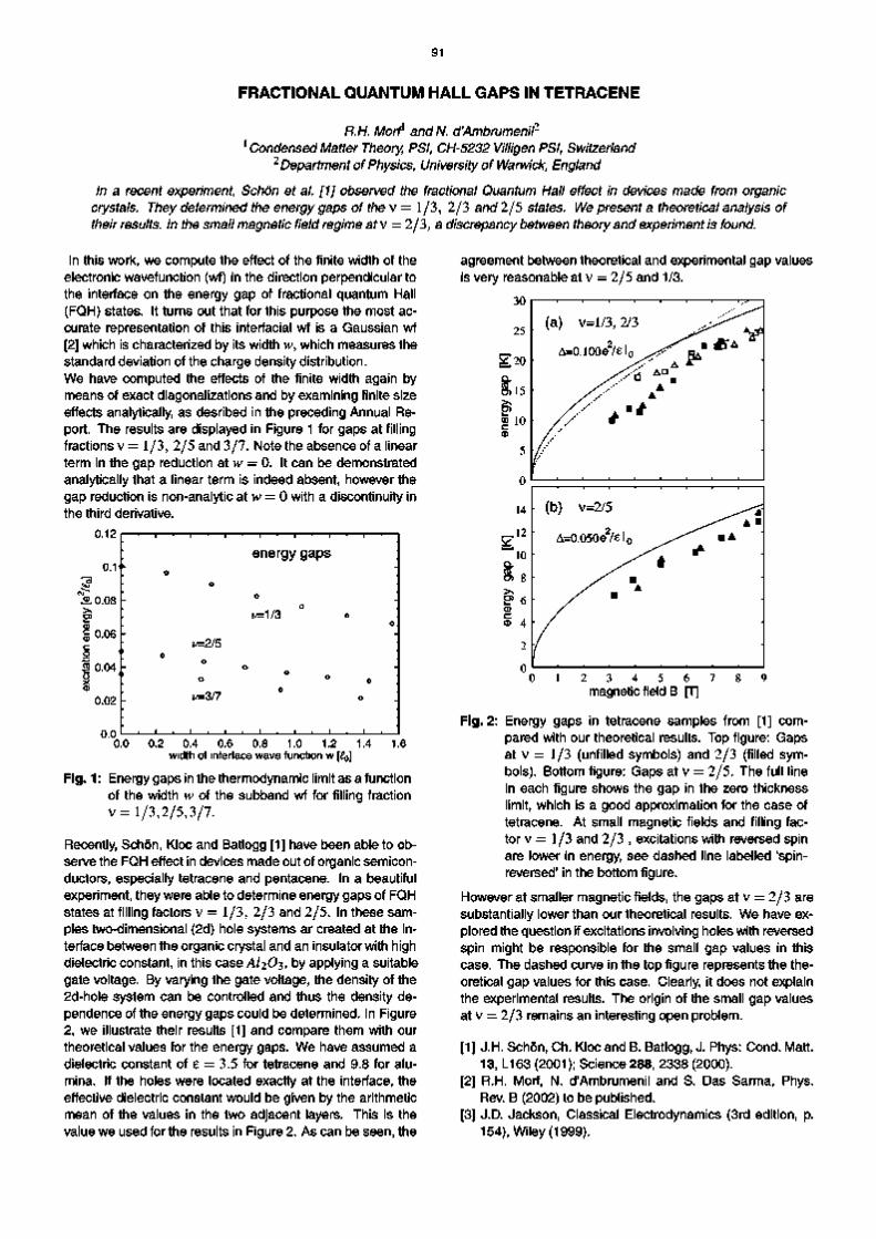
91
FRACTIONAL QUANTUM HALL GAPS IN TETRACENE
R.H. Morf1 and N. d'Ambrumenil
2
1 Condensed Matter Theory, PSI, CH5232 Villigen PSI, Switzerland 2 Department of Physics, University of Warwick, England
In a recent experiment, Schon et al. [1] observed the fractional Quantum Hall effect in devices made from organic crystals. They determined the energy gaps of thev = 1/3, 2/3 and 2/5 states. We present a theoretical analysis of their results. In the small magnetic field regime atv = 2/3, a discrepancy between theory and experiment is found.
In this work, we compute the effect of the finite width of the electronic wavefunction (wf) in the direction perpendicular to the interface on the energy gap of fractional quantum Hall (FQH) states. It turns out that for this purpose the most ac
curate representation of this interfacial wf is a Gaussian wf [2] which is characterized by its width w, which measures the standard deviation of the charge density distribution. We have computed the effects of the finite width again by means of exact diagonalizations and by examining finite size effects analytically, as desribed in the preceding Annual Re
port. The results are displayed in Figure 1 for gaps at filling fractions v = 1/3, 2/5 and 3/7. Note the absence of a linear term in the gap reduction at w = 0. It can be demonstrated analytically that a linear term is indeed absent, however the gap reduction is nonanalytic at w = 0 with a discontinuity in the third derivative.
0.12
0.1
agreement between theoretical and experimental gap values is very reasonable at v = 2/5 and 1/3.
«L0.08 >̂ CD
o § 0.04 o X CD
0.02
0.0
1
o
' o
'
1 ' 1
o
z/=2/5
° z/=3/7
i . | . |
energy gaps
o
v=M3 o
°
i
■
°■
©
■
0.0 0.2 0.4 0.6 0.8 1.0 1.2 1.4 width of interface wave function w [£0]
1.6
Fig. 1: Energy gaps in the thermodynamic limit as a function of the width w of the subband wf for filling fraction v = 1/3,2/5,3/7.
Recently, Schon, Kloc and Batlogg [1] have been able to ob
serve the FQH effect in devices made out of organic semicon
ductors, especially tetracene and pentacene. In a beautiful experiment, they were able to determine energy gaps of FQH states at filling factors v = 1/3, 2/3 and 2/5. In these sam
ples twodimensional (2d) hole systems ar created at the in
terface between the organic crystal and an insulator with high dielectric constant, in this case AI2O3, by applying a suitable gate voltage. By varying the gate voltage, the density of the 2dhole system can be controlled and thus the density de
pendence of the energy gaps could be determined. In Figure 2, we illustrate their results [1] and compare them with our theoretical values for the energy gaps. We have assumed a dielectric constant of 8 = 3.5 for tetracene and 9.8 for alu
mina. If the holes were located exactly at the interface, the effective dielectric constant would be given by the arithmetic mean of the values in the two adjacent layers. This is the value we used for the results in Figure 2. As can be seen, the
30
25
§ 2 0 Q .
o> 15 >> <D 10 c 0
5
0
14
^ 1 2
10 Q. 10 « o> 8 >> E> 6 0 c A 0 4
2
0
(a) v=l/3,2/3
A=0.100e2/el0 ^ ^
/ V D ■ /.'- A
■ / / * ' *
(b) v=2/5
A=0.050e 2 / e l 0 ^
^ ^ # / ■ / A
/ m
u
+
-_ v * A
■
^ A ■ ■ A
-
-
0 1 2 3 4 5 6 7 8 9 magnetic field B [T]
Fig.2: Energy gaps in tetracene samples from [1] com
pared with our theoretical results. Top figure: Gaps at v = 1/3 (unfilled symbols) and 2/3 (filled sym
bols). Bottom figure: Gaps at v = 2/5. The full line in each figure shows the gap in the zero thickness limit, which is a good approximation for the case of tetracene. At small magnetic fields and filling fac
tor v = 1/3 and 2/3 , excitations with reversed spin are lower in energy, see dashed line labelled 'spin
reversed' in the bottom figure.
However at smaller magnetic fields, the gaps at v = 2/3 are substantially lower than our theoretical results. We have ex
plored the question if excitations involving holes with reversed spin might be responsible for the small gap values in this case. The dashed curve in the top figure represents the the
oretical gap values for this case. Clearly, it does not explain the experimental results. The origin of the small gap values at v = 2/3 remains an interesting open problem.
[1] J.H. Schon, Ch. Kloc and B. Batlogg, J. Phys: Cond. Matt. 13, L163 (2001); Science 288, 2338 (2000).
[2] R.H. Morf, N. d'Ambrumenil and S. Das Sarma, Phys. Rev. B (2002) to be published.
[3] J.D. Jackson, Classical Electrodynamics (3rd edition, p. 154), Wiley (1999).
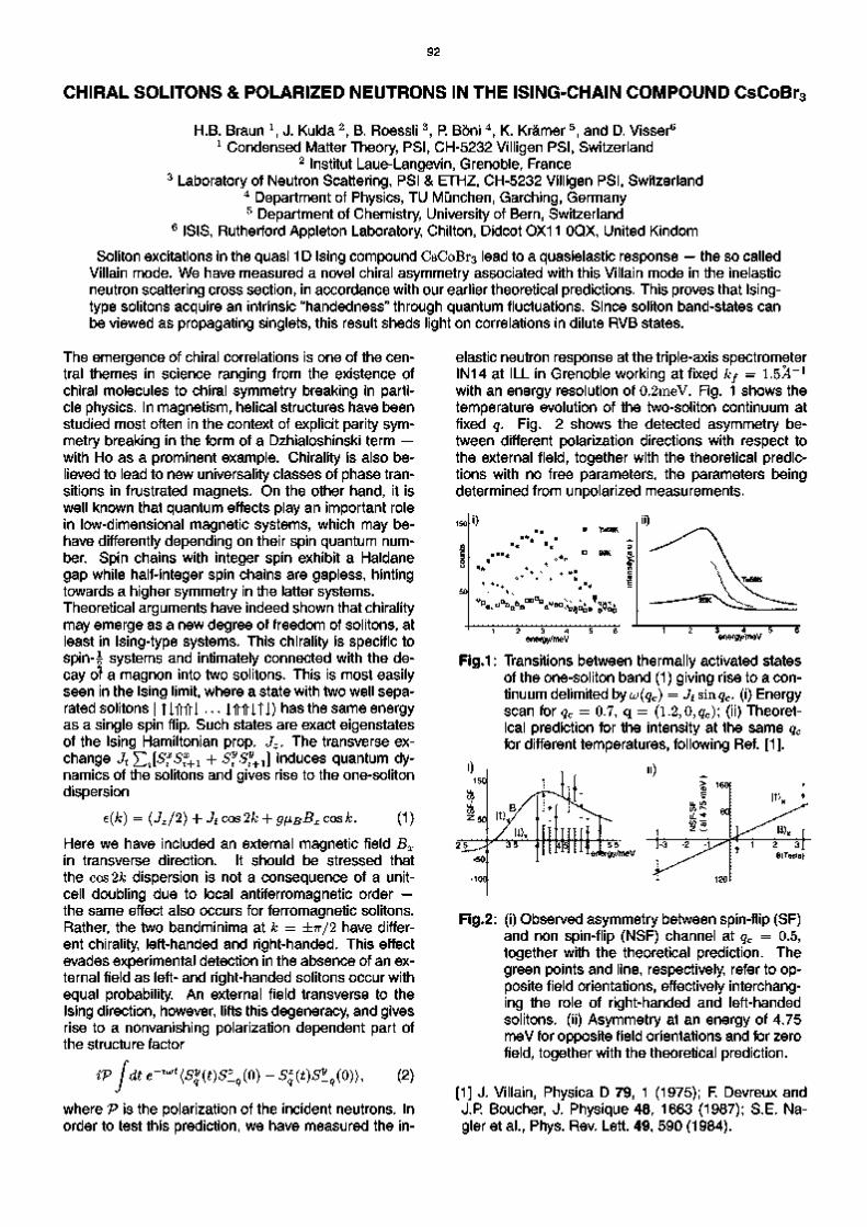
92
CHIRAL SOLITONS & POLARIZED NEUTRONS IN THE ISINGCHAIN COMPOUND CsCoBr3
H.B. Braun \ J. Kulda 2
, B. Roessli 3
, P. Boni 4
, K. Kramer 5
, and D. Visser6
1 Condensed Matter Theory, PSI, CH5232 Villigen PSI, Switzerland 2 Institut LaueLangevin, Grenoble, France
3 Laboratory of Neutron Scattering, PSI & ETHZ, CH5232 Villigen PSI, Switzerland 4 Department of Physics, TL) Munchen, Garching, Germany 5 Department of Chemistry, University of Bern, Switzerland
6 ISIS, Rutherford Appleton Laboratory, Chilton, Didcot 0X11 OQX, United Kindom
Soliton excitations in the quasi 1D Ising compound CsCoBr3 lead to a quasielastic response — the so called Villain mode. We have measured a novel chiral asymmetry associated with this Villain mode in the inelastic neutron scattering cross section, in accordance with our earlier theoretical predictions. This proves that Isingtype solitons acquire an intrinsic "handedness" through quantum fluctuations. Since soliton bandstates can be viewed as propagating singlets, this result sheds light on correlations in dilute RVB states.
The emergence of chiral correlations is one of the central themes in science ranging from the existence of chiral molecules to chiral symmetry breaking in particle physics. In magnetism, helical structures have been studied most often in the context of explicit parity symmetry breaking in the form of a Dzhialoshinski term — with Ho as a prominent example. Chirality is also believed to lead to new universality classes of phase transitions in frustrated magnets. On the other hand, it is well known that quantum effects play an important role in lowdimensional magnetic systems, which may behave differently depending on their spin quantum number. Spin chains with integer spin exhibit a Haldane gap while halfinteger spin chains are gapless, hinting towards a higher symmetry in the latter systems. Theoretical arguments have indeed shown that chirality may emerge as a new degree of freedom of solitons, at least in Isingtype systems. This chirality is specific to spin^ systems and intimately connected with the decay of a magnon into two solitons. This is most easily seen in the Ising limit, where a state with two well separated solitons | TI1T1TI • • • I1T1TITI) has the same energy as a single spin flip. Such states are exact eigenstates of the Ising Hamiltonian prop. Jz. The transverse exchange Jt^2zi
sz
sz+i + ^ ^ + 1 ] induces quantum dy
namics of the solitons and gives rise to the onesoliton dispersion
e(k) = (Jz/2) + Jt cos 2k + g^B^x c o s k. 0) Here we have included an external magnetic field Bx in transverse direction. It should be stressed that the cos2& dispersion is not a consequence of a unitcell doubling due to local antiferromagnetic order — the same effect also occurs for ferromagnetic solitons. Rather, the two bandminima at k = ±7T/2 have different chirality, lefthanded and righthanded. This effect evades experimental detection in the absence of an external field as left and righthanded solitons occur with equal probability. An external field transverse to the Ising direction, however, lifts this degeneracy, and gives rise to a nonvanishing polarization dependent part of the structure factor
ivjdt e-^(Syq(t)Sz_q(0) - Sz
q(t)Sy_q(0)), (2)
where V is the polarization of the incident neutrons. In order to test this prediction, we have measured the in
elastic neutron response at the tripleaxis spectrometer IN14 at ILL in Grenoble working at fixed kf = 1.5i
_ 1
with an energy resolution of 0.2meV. Fig. 1 shows the temperature evolution of the twosoliton continuum at fixed q. Fig. 2 shows the detected asymmetry between different polarization directions with respect to the external field, together with the theoretical predictions with no free parameters, the parameters being determined from unpolarized measurements.
Fig.1: Transitions between thermally activated states of the onesoliton band (1) giving rise to a continuum delimited by uo(qc) = Jtsmqc. (i) Energy scan for qc = 0.7, q = (l.2,0,gc); (ii) Theoretical prediction for the intensity at the same qc for different temperatures, following Ref. [1].
150
op en z 50
50.
100
, B/ i
/ U ) x • ^ 3 5 V
Y rl
5l [i i ' mj ■ 55
ergy/m
NS
FS
F
( at 4
75
meV
13 2 A ^
^ 120
!T)X *
* 1 2 3} B(Tesla)
Fig.2: (i) Observed asymmetry between spinflip (SF) and non spinflip (NSF) channel at qc = 0.5, together with the theoretical prediction. The green points and line, respectively, refer to opposite field orientations, effectively interchanging the role of righthanded and lefthanded solitons. (ii) Asymmetry at an energy of 4.75 meV for opposite field orientations and for zero field, together with the theoretical prediction.
[1] J. Villain, Physica D 79, 1 (1975); F. Devreux and J.R Boucher, J. Physique 48, 1663 (1987); S.E. Nagler et al., Phys. Rev. Lett. 49, 590 (1984).

93
Multilayers and Interfaces
.6 0,8 f 1.2 ml1

94

95
IMPROVED REMANENT SUPERMIRROR POLARIZERS
J. Stahn, M. Christensen, P. Kallbauer and D. Clemens Laboratory for Neutron Scattering, ETH Zurich & PSI, CH-5232 Villigen PSI, Switzerland
Neutron spin polarizers based on Fe/Si and Fe.89 Co.u/Si supermirrors were prepared by magnetron sputtering. For these polarizing efficiencies of 96 % to 98% could be reached in transmission when operated in a guide field of < 20 G.
3T We are working on the optimization of magnetically remanent supermirror neutron polarizers. The aim is to build compact neutron transmission polarizers [1] and polarizers to be operated in an antiparallel magnetic field. A possible application is shown in Fig. 1.
i&**~^
^ S S r ^
Fig.1: Spin filter based on a remanent supermirror polarizer. If the coercitive field strength of the polarizer is larger than the guide field, it is possible to have it polarized antiparallel (upper sketch) and parallel (lower sketch) to the guide field. This allows to select a spin state without using a spin flipper just by magnetizing the polarizer in a certain direction.
The supermirrors are prepared by magnetron sputtering using the low absorbing materials Fe or Fe.89Co.n and Si. The sputter parameters like power, gas pressures and addition of the reactive gases 02 and N2 were optimized by producing multilayers on glass.
co/° Fig.2: Transmission of spin up (7"t) and spin down (7;) neutrons through a Fe/Si:N,0, m = 2 supermirror with 149 layers in total. After saturating the polarizer in a magnetic field of 300 G it was measured in a guide field of < 20 G. The neutron wavelength was A = 4.74 A. The line denoted P gives the polarizing efficiency.
The main problem is the tensile stress in the Fe or Fe.89Co.ii layers. This can be partly compensated for by introducing compressive stress in the Si layers. But on the other side anisotropic stress is a condition for the wanted easy axis of magnetization. [2] Neutron transmission (T) and reflectivity (/?) curves of the supermirrors were measured on the 2 axes spectrometer TOPSI at SINQ. From these the polarizing efficiency was calculated without any further corrections:
n-Tt fit ~ Rj R± + fit Tl + T t
An example is given in Fig. 2. If no remanence is required, it is easier to reduce stress and prepare polarizers with more layers, polarizing up to higher angles of incidence. Fig. 3 shows transmission and polarizing efficiency for an non-remanent Fe.89Co.ii / Si supermirror. Polarizers of this type (with m = 2.5 [3]) will be used on the SANS at SINQ.
96% 93%
0.6 0.8 co/°
Fig.3: Transmission of spin up (7"t) and spin down (7;) neutrons through a Fe.89Co.n /Si, m = 2.3 supermirror with 299 layers in total. The polarizer was measured in a magnetic field of 300 G. u gives the angle of incidence, the neutron wavelength was A = 4.74 A. The line denoted P gives the polarizing efficiency.
The presented results were obtained within the project TECHNI of the EU program IHP/Networks (HRPI-1999-CT-50005) with financial support of the BBW Switzerland (Nr. 99.0593).
[1] CF. Majkrzak et al. SPIE 1738, 90 (1992) [2] M. Senthil Kumar, P. Boni, M. Horisberger, IEEE
Transactions on Magnetics 35, 3067 (1999) [3] m = 1 corresponds to 4?r sin UJ/X = 0.022 A - 1

96
DEVELOPMENT OF COMPONENTS FOR POLARIZATION ANALYSIS
H. Grimmer \ O. Zaharko \ M. Horisberger \ HCh. Mertins 2
, D. Abramsohn 2
, F. Schafers 2,
Ch. Klemenz 3
laboratory for Neutron Scattering, ETHZ & PSI, CH5232 Villigen PSI, Switzerland 2BESSY GmbH, AlbertElnstelnStrasse 15, D12489 Berlin, Germany 3AMP AC, University of Central Florida, Orlando, Fl 328162455, USA
CrIC transmission multilayers with large phase shift at the Carbon K edge were produced at PSI. reflectance of YBaCuO epltactlcal layers was found close to the Barium M5 edge.
High
The polarization state of a soft Xray beam, as pro
duced e.g. at the SLS, can be determined using a transmission multilayer as phase shifter and a reflec
tion multilayer at a grazing angle 0 = 45° (Brewster angle) as analyser for photon energies E < 600 eV; for higher energies either magnetooptic effects or single crystals may be used [1]. A transmission multilayer with much larger phase shift than available previously and a corresponding reflection multilayer, both con
sisting of 100 Cr/C bilayers, have been developed at PSI and tested at BESSY. The analyser has a period d = 3.13 nm and a reflectance for spolarized radiation Rs >19% for E in the range 268281 eV. Its reflec
tance ratio for s and p polarized radiation is Rs/Rp > 400 in the range 274281 eV with a peak value 1400 at 278 eV, where the Bragg angle 0B = 45° [2]. Cal
culations gave a period d = 3.96 nm for a Cr/C phase shifter with maximum figure of merit sin AVT, where A denotes the phase shift and T the transmission. The following figure shows that the phase shift is about three times larger than obtained previously [3].
16 -
£1 12 V)
\8
1.3 j j o i . 2
^ ■ Q . 1 . 1
H 1.0
CO 0
0
< -10
CO 0 CO 03
20
0 Q
Transmission Multilayers
Cr /C 2
0 0B [degrees]
Fig. 1: Transmission Ts, ratio TP/TS, and phase shift A as functions of the deviation between grazing angle and Bragg angle. Results at 277 eV for our sample (filled squares) and at 275 eV from ref. [3] (open cir
cles).
These multilayers can be used for unambiguous polarization determination at the Carbon K edge.
New reflecting optics for linear polarization analysis above 700 eV has also been investigated. YBa2Cu3Ox epitactical layers with caxis perpendicular to the surface, i.e. a period of 1.174 nm, were grown at EPFL by liquid phase epitaxy on (110) NdGa03 substrates. Measurements at the BESSY undulator beam line UE56/1PGM gave Rs = 10%, RS/RP = 200 at the Ba M5 edge (780 eV) and Rs = 3.6%, RS/RP = 3400 at 750 eV, where 0B is equal to the Brewster angle.
10 r'
CO CD 1 L
D) CO
m 0.1 fc
03 0.01 d CD
E
o c CO o 1E3 b0
"CD 01
3000
01
01 2000
1000 h
0
i i i i i i i i i i i i i i i i i i i i i i i i i i i i i i i i i i i i i i i i i i i
YBa2Cu3Ox
0 = 48.3° /
B
^ o . 0 = 4 5 . 0 " .
1 *i
T8 ■ ■ ■ ■ I ■ ■ ■ ■ I ■ ■ ■ ■ I ■ ■ ■ ■ I ■ ■ ■ ■ I ■ ■ ■ ■ I ■ ■ ■
710 720 730 740 750 760 770 780 790
Photon Energy E [eV]
Fig. 2: Bragg peak reflectances Rs and RP in % and their ratio Rs / RP. 0B is the grazing angle at which the Bragg peak appears.
A reflectance maximum at the Cu L3 edge of 932 eV was also observed. Further investigations have to show the applicability of this sample for polarization analysis in this energy range.
[1] F. Schafers etal., Appl. Optics, 384074 (1999)
[2] H. Grimmer etal., Surface Rev. and Letters, (in press)
[3] S. Di Fonzoetal., Rev. Sci. Instrum. 66 1513 (1995)

97
STRESS ANISOTROPY IN Feo.87Coo.13/Si MULTILAYERS
D. Clemens1, P. Kailbauer
1, J. Stahn
1, M. Horisberger
1, B. Schnydef
^ Laboratory for Neutron Scattering ETHZ & PSI, CH-5232 Villigen PSI, Switzerland 2Paul Scherrer Institut, CH-5232 Villigen PSI, Switzerland
Similar to the FeColT'r.N system that we developed to be applied in numerous neutron polarization devices and instruments we investigated Feo.37Coo.13/SI In order to use this material combination for remanent transmission polarizers. A systematic study of the Influence of partial gas pressures - for the working gas Ar as well as for reactive additions of air or N2 - has been performed on multilayers consisting of 20 bllayers having D-10 nm. Profilometry, reflectometry with X-rays and neutrons, and vibrating sample magnetometry were used to reveal the dependence of stress and magnetic behavior on the deposition parameters and to determine the multilayer structure.
In the past we have developed multiplayer coatings from FeCoV/Ti:N that worked as neutron polarizers in reflection. Because of the in-plane stress anisotropy that via magnetostriction translates to a magnetic anisotropy these mirrors exhibit remanence along the easy axis of magnetization and only coercive field of some a few mT can destroy the net magnetization. Thus the mirrors can be used in weak magnetic fields to polarize neutron beams. Especially for the analysis of the neutron's spin state a similar mirror, that can work in both transmission and reflection when coated, e.g. on a Si wafer, would be beneficial. The former system doesn't match with the refractive index of Si for neither spin state resulting in significant reflection at low q and is strongly absorbing transient neutrons because of almost 50% Co in the magnetic layers. We investigated Fe087Co0i3/Si as an alternative as for spin down neutrons this system has the same refractive index as Si. As a drawback Fe/Si is known for interfacial reactions and non-magnetic layers and in both cases the matching is destroyed. We tried to maintain distinct structures by reactively sputtering with small partial pressures of air and N2 at varied mixtures.
"T fc 1 ■ 0
? o w"
1 CO
7.0-
6.5-
6.0-
5.5-
5.0-
4.5-
4.0-
3.5-
3.0-
2.5-
2.0-
1.5-
1.0-5 10 15 20 25 30 35 40 45 50
Ar flow for FeCo [seem]
Fig. 1: Relation of total stress and sputter gas flow during deposition represented according to the transport direction in the PSI Z600 sputter plant.
uses Si wafers for which the surface curvature has already been characterized on which the coating is deposited and consecutively the curvature resulting from the total stress which was always larger than for FeCoV/Ti:N is measured in a second run. The easy magnetization axis is along substrate transport.
Fig. 2: Total stress related to the sputter gas flow for the deposition of the individual materials, always with an addition of 2.5 seem air + 2.5 seem N2
The elevation of Ar pressure and the addition of gases in reactive sputtering leads to increased stress, but always with a constant difference for the two directions with the peculiarity that 35 seem into FeCo gives another reduction for the total stress. Si produced with low/no gas addition has compressive stress and therefore reduces the total stress.
[1] D. Clemens etal., Physica Scripta 50, 195 (1994) [2] D.J. Mulleref al., Physica B 234-236, 1050 (1997) [3] D. Clemens et al., JMMM 177-181, 1237 (1998) [4] P. Boni et al., Physica B 267-268, 320 (1999)
Fig. 1 shows the dependence of the total stress in the film as measured with a profilometer. This method

98
MOLECULAR MODELING OF PHOSPHATE NUCLEATION ON SILICA GLASS
Rene Windiks and Bernard Delley Condensed Matter Theory Group, PSI CH-5232 Villigen PSI, Switzerland
Silica glasses are used for, e.g., prosthetic bones because such materials induce the formation of calcium phosphate layers on their surface and, hence, of interfacial bonds with body tissue. Density functional calculations performed on silica glass model surfaces covered with calcium phosphates revealed five-fold coordinated surface Si atoms which are very atypical.
Mammalian bone is a composite material that mainly consists of carbonate-rich hydroxy apatite (HA) [1]. Pure HA has the stoichiometric composition Ca5(P04)3OH and is used as a model compound for calcified tissues. Silica glass is a disordered network of corner- and edge-sharing Si02 tetrahedra which contains many defects. One type of highly reactive "defect" sites on silica surfaces are rings that consist of three corner-sharing Si02 tetrahedra (3R) [5].
Fig.1: (a) Slab model for a silica glass surface which interacts with CaHP04. The slab unit cell is represented by the long thin lines and is translated perpendicular to the c vector. Both slab sides perpendicular to the c vector are surface images and are covered with CaHP04. On the lower surface image HPO2" ions are bound by H-bonds only, (b) The upper image reveals five-fold coordinated Si atoms where one O atom belongs to HPO2". The small empty circles are H atoms, thick dashes lines represent H-bonds and thin dashed lines the coordination of Ca to O atoms.
Bioactive silica glasses and glass-ceramics initiate the formation of new HA layers and, hence, interfacial bonds with the existing bone. The new HA phase is equivalent chemically and structurally to the mineral phase in bone. Therefore, these materials are used
for prosthetic bones and dental implants [2]. There has been little research on elucidating this type of biomineralization because probably several mechanisms are involved in this process which is, therefore, very complex. One key step is the nucleation of calcium phosphates where the 3R are apparently the preferred absorption sites [3,4]. The predominant phosphate species in human blood (pH 7.4) is HPO2-. Our contributions are density functional (DF) calculations performed on model surfaces of silica glass to examine interactions of CaHP04 with 3R. The Si02 glass surface is represented by a periodically extended, i.e. crystalline surface that is described by a slab model [Fig. 1(a)]. The slab model based on a hypothetical bulk structure of linked 3R [6]. In order to find several stable configurations of CaHP04 on the silica surface we have generated different structures in a molecular dynamics simulated annealing approach employing classical force fields for interatomic interactions. The atomic positions of these initial structures are fully optimized in a given unit cell using DF methods. Surprisingly, one stable structure shows penta-coordinated surface Si atoms, Fig. 1(b), where the O atoms form a trigonal-bipyramid. The axial and equatorial Si-O distances are 179 pm and in the range between 167 and 174 pm, respectively. Such a coordination is atypical for surface Si atoms and usually appears in the bulk of silica glasses and high density silicates. Here this configuration is stabilized by Ca2+ ions which try to increase its coordination number up to six. There are four Ca- • • O distances in the range between 230 and 290 pm, typical for interactions between Ca2+ ions and 0^~ species. Penta-coordinated Si atoms can be recognized by electron spectroscopy (e.g. EXAFS) and NMR spectroscopy.
[1] Bertini, I.; Gray, H. B.; Lippard, S. J. Bioinorganic Chemistry. University Science Books, Mill Valley, (1994).
[2] Ratner, B. D.; Hoffman, A. S.; Schoen, F. J.; Lemons, J. E. Biomaterials Science: An Introduction to Materials in Medicine. Academic Press, San Diego, (1996).
[3] Coreno, J.; Martfnez, A.; Bolarfn, A. J. Biomed. Mater. Res. 57(1), 119, (2001).
[4] Sahai, N.; Tossell, J. A. J. Phys. Chem. B 104(18), 4322 (2000).
[5] West, J. K.; Wallace, S. J. Non-Cryst. Solids 152, 109(1993).
[6] Hamann, D. R. Phys. Rev. B 55(22), 14784 (1997).

99
SHOCKLEY TYPE SURFACE STATE ON CU(111) AND VICINAL CU(111)
B. Delley1 F Baumberger, T Greber and J. Osterwaldei2
1 Condensed Matter Theory, PSI, CH-5232 Villigen PSI, Switzerland 2 Physik-lnstitut der Universitat Zurich, Switzerland
Shockley surface states have been observed by electron spectroscopy on Cu(111) and the vicinal (332) and (221) surfaces. We have calculated the band-structure of Cu(111) and (332) (443) and (554) multilayer slabs using density functional theory. On Cu(111) the surface state is obtained in excellent agreement with experiment. On the vicinal slab models the surface state mixes with bulk states.
Cu metal exhibits 'neck' of the Fermi surface near the L point of the first Brillouin zone. In bulk Cu there are no states along the line from the r to the L point above the d-bands up to well beyond the Fermi energy. On the Cu(111) surface (which is normal to the r - L line) a two dimensional nearly free electron surface state exists.
K r M K wavevector DoS
Fig.1: Band-structure for a 13 layer Cu(111) slab. The highest two occupied bands near r are the front and back side surface states.
Using density functional theory calculations [2] using DMol3, we find that slab models with more than 9 layers clearly exhibit this Shockley surface state. Fig. 1 shows a band-structure for a 13 layer Cu(111) slab. The surface states are the highest two occupied bands near r. The front and back surface states interact by tunneling through the forbidden bulk-like central part of the slab, which produces a small bonding anti-bonding splitting of the states. Position and dispersion of the Shockley surface state are in excellent agreement with electron spectroscopic results [1]. This surface state is also seen in scanning tunneling microscopy and spectroscopy. As a note on the side, the upper end of the
Fig is at 'vacuum' zero which permits to read off the calculated work function. A contour plot of the surface state at the surface r point is shown in Fig. 2. The exponential decay towards the center of the slab is intuitively obvious from the Fig. The highest contours, except for the spikes near the nuclei, occur at the slab-vacuum interface. The picture also suggests that this surface state can be characterized as a wave packet with with normal component of wave-vectors near kF. In the case of vicinal surfaces investigated, the surface state overlaps in k-space and mixes with the bulk like bands of nearly free electrons of the neck part of the Fermi surface.
o @ @ ® © ® © ©
^ 5 © © © © © © © C O © © ® © ® ® ® £ 8 8 * 8 8 8 8 c 4& H J l 4k 4k Mb <H JS t-—-™Jt~—-™Jt-——Jfe-—~»wk-—~A<-——A-—™-4fc co 5 ^ 7 ^ o^T o ^ T o T o
in plane direction (-211)
Fig.2: Shockley surface state in Cu(111).
[1] F. Baumberger, T. Greber, J. Osterwalder, Phys. Rev. B 62, 15431 (2000)
[2] B. Delley, J. Chem. Phys. 113, 7756, (2000)

100
SURFACE DISTRIBUTION OF CU ADATOMS ON XE-HOPG
M. Pivetta, F Patthey W.-D. Schneider1 and B. Dellef 1 Institut de Physique de la Matlere Condensee, Unlverslte de Lausanne, Switzerland
2 Condensed Matter Theory, PSI, CH-5232 Villigen PSI, Switzerland
Work function changes due to Cu impurities on one monolayer Xe adsorbed on highly oriented pyrolytic graphite (HOPG) have been investigated by normal emission ultraviolet photoelectron spectroscopy and by density functional theory with DMoP calculations. The results suggest that Cu atoms adsorb as isolated impurities.
Recently, the observation of a Fano resonance in ultraviolet photoelectron spectroscopy (UPS) has been reported for the system Cu/Xe/HOPG (highly oriented pyrolytic graphite). [1] Below 50 K, one monolayer (ML) of Xe forms a commensurate overlayer on the HOPG surface leading to a folding of the graphite 7r-band by coupling the if-point of the graphite Brillouin zone with the r-point of the Xe layer one. This projection induces considerable spectral density between the Fermi level EF and 2 eV binding energy in normal emission UPS spectra. After subsequent deposition of 0.1 ML Cu a pronounced Fano resonance profile is observed in the photoelectron spectra. The resonance has been attributed to the interference between the graphite TT-band continuum and the discrete Cu 4s atomic-like level. In this interpretation the Cu atoms are considered as isolated impurities in the Xe layer. In order to determine the microscopic structure from a theoretical point of view, we have investigated the system Cu on Xe/HOPG with DMol3 density functional calculations. The starting system is a five layer graphite slab at experimental bulk geometry, covered with 1 ML Xe (no Cu adatoms) (line 1). The use of adatoms placed symmetrically on both sides eliminates long range electric fields from the back of the slab. The carbon atoms are kept fixed at the bulk graphite positions during optimization of the adsorbate. The second system we have studied consists of 0.25 ML of Cu substituting Xe atoms (line 2 in Table). The position of the Fermi energy with respect to 'vacuum' zero suggests a work function (WF) lowering of 0.77 eV with respect to the starting system. The third for 0.25 ML of Cu on 1 ML Xe (line 3) was started with Cu in a top layer in a hollow site. The optimized final position is sub-Xe surface, as indicated by the value of hCu, which represents the optimized distance of the Cu adatom from the center of the slab. The result of these simulations strongly suggests that the Cu adatom is more stable than the Xe-substitutional Cu atom, as indicated by the values for the total energy Etot for the systems of line 2 and line 3. These values for Etot suggest also that it is energetically favorable for the system
to keep the Xe layer on top. Interestingly the adatom Cu ends up in a sub-Xe surface position (line 3), which was somewhat unexpected.
4 6
3 8
O C u / X e / H O P G
- 0 5 (random) -0 43
0 (super lattice)
-O,-' < O Q. Q
0 0 0 2 0 4 0 6 0 8 Cu concentration (RG ML)
1 0
Fig.1: Measured work function change as a function of Cu coverage, and curves calculated from Topping model.
The 1 ML Cu sub 1 ML Xe (line 4) is less bound than the 0.25 ML Cu, suggesting a tendency against the formation of Cu islands. Calculations for all Cu on Xe/HOPG systems yield a very significantly lowered WF as compared to Xe on HOPG, and suggest a much larger WF lowering from the sub-Xe Cu atoms than for the substitutional case. The WF lowering that we find numerically are comparable to the one expected for alkali adatoms, confirming the results of our WF change measurements.
[1] F. Patthey, M.-H. Schaffner, W.-D. Schneider, B. Del-ley, Phys. Rev. Lett. 82, 2971, (1999)
Table: Density functional results for Cu on Xe/HOPG system
System eA0 (eV) Etot(eV) ECu(eV) hXe (au) hCu (au) 1 1MLXe 2 (0.25 ML Cu)(0.75 ML Xe) 3 (0.25MLCu)(1 MLXe) 4 (1 MLCu)(1 MLXe)
-0.77 -1.91 -1.80
1066.44 1066.93 1068.67 1073.44
0.49 2.23 1.75
19.47 19.37
19.34x3, 19.41 19.54
16.33 16.32 16.34

101
EXCHANGE SPRINGS AND HYSTERESIS LOOPS — AN ANALYTICAL APPROACH Andreas Bill, Hans-Benjamin Braun
Condensed Matter Theory, PSI, CH-5232 Villigen PSI, Switzerland
Magnetic bilayers are central parts of spin valves in GMR reading heads and basic storage elements in the most recent generation of hard-disks. We have obtained exact analytical solutions for the magnetization profile in such an exchange spring structure for arbitrary layer thickness, material parameters and external field. We also identify the existence of a crossover from an exponential to a power-law behaviour of the coercivity as a function of film thickness. In addition, we determine the energy barrier between states of opposite magnetization which determines the thermal stability of a nanoscale device.
Recent years have seen increasing experimental abilities to taylor nanoscale magnetic structures. In particular, it became possible to grow multilayer structures with variable thicknesses made of (either single crystals or polycristalline) ferro- (FM), antiferro- (AFM) or paramagnetic materials. Among systems of great technological interest are exchange springs (coupled soft FM/hard FM) as they are used in magnetic or magneto-optical recording and spin-valve devices. A better understanding of the physical processes occurring in exchange springs under an applied field is highly desirable as experiments reveal non-universal behaviour of the bilayers (e.g. regarding the thickness dependence of the coercivity [1]). This raises the question whether the variety of results is related to the formation of domains or grains in the ferromagnets, to the sensitivity on material parameters, to interface composition and roughness, etc. In order to separate such disorder related effects from the properties of an ideal sample, it is important to study the properties of an ideal bilayer. Motivated by the successful description of recent experiments on nanoscale magnets, we have therefore considered the magnetization in the soft-FM / hard-FM bilayer as a classical continuum field. We have solved the corresponding Euler-Lagrange equations exactly for a rather general model of a bilayer with arbitrary material parameters, interface coupling and external magnetic field. The configurations minimizing the energy can be expressed explicitly in terms of Jacobi's elliptic functions [2]. Our results explain both the experimentally observed shape of the hysteresis curve and an unusually large magnetization twist in the hard layer detected in recent neutron experiments [3].
external field H b) 1000
> 10
o 1
0.1
Ka/K! = 0.01; 0.05 0.1 0.01;
H c
A,
A,
L"2
= 14 \ = 7
0 1 L/82 ! 4
Fig.1: (a) Magnetic bilayer consisting of soft and hard magnetic material, (b) Coercivity in units of K 2 =M s as a function of thickness of the soft FM for two values of the interlayer exchange A i [-2 denotes the domain-wall width and M S the saturation magnetization]. Note the crossover from power-law to exponential decay.
Fig. 1b) shows the coercivity as a function of the thickness of the soft FM layer for different material parameters. For soft layers that are thicker than the bulk domain-wall width, the coercivity decays exponentially with film thickness and is rather insensitive to material parameters. Films thinner than the domain-wall width show a power law of the coercivity which depends sensitively on the material parameters. Fig. 2 shows a typical hysteresis loop and the magnetization profile for a few points on the loop as obtained from the analytical solutions. We point out three important features of our calculation: i) Because of interlayer coupling, one observes a significant twist of the magnetization (solid lines) in the hard layer (left side of Fig. 2b) even for applied fields well below the irreversibility field; ii) there is a finite phase slip at the interface between the hard (left) and soft (right) FMs, also for strong interface coupling; iii) the barrier (dashed lines) shows a response to the external field which is reverse to that of the stable configuration (solid lines). The barrier configuration and the magnetization configuration coalesce at the value of the critical field. The energy difference C E between the barrier and the stable configuration determines the thermal switching probability into the field-aligned state via the Arrhenius factor expf \ $ E =]% Tg.
Fig.2: (a) Characteristic hysteresis loop and (b) corresponding magnetization profile of the stable configuration (solid) and the barrier (dashed). h = H M S = K 2 is the reduced external field, and " (z) is the angle between the magnetization and the direction of the applied field, cf. Fig.1 a.
[1] E.E. Fullerton, J.S. Jiang, S.D. Bader, J. Magn. Magn. Mater. 200, 392 (1999).
[2] A. Bill, H.B. Braun, preprint. [3] K. V. O'Donovan, J. A. Borchers, C. F. Majkrzak,
O. Hellwig, and E. E. Fullerton, Phys. Rev. Lett. 88, 067201 (2002).

102
103
Instrumental and Support Activities

104

105
RITA-II: INSTALLATION AND FIRST YEAR OF OPERATION
F. Altorfer1, S. Klauserf, J.Holm2, K. Lefmann2, S. Bang1, D. McMorrow*, P. Keller1, C. Kagi1, R. Burge1
1 Laboratory for Neutron Scattering, ETHZ& PSI, CH-5232 Villigen PSI, Switzerland 2Materials Research Department, Rise National Laboratory, DK-4000 Roskilde, Denmark
In April 2001 the Re-Invented Triple Axis Spectrometer RITA-II was installed at SINQ neutron guide RNR-13 to replace TAS DruchaL. After the completion of testing & commissioning in May and June the instrument was ready for normal external user operations. During this report's period, 12 experiments were carried out by user groups from 7 institutions
In the course of the PSI-Ris0 collaboration it was decided to replace the cold triple axis spectrometer DruchaL situated at SINQ neutron guide RNR-13 by the newly developed spectrometer RITA-II (see figure 1). After final mechanical and electrical tests with RITA-II at Ris0 National Laboratory the instrument was shipped at the end of April 2001 to the PSI.
Fig 1: End of April 2001: experts from Ris0 National Laboratory and PSI make sure that the new cold neutron triple axis spectrometer RITA-II is installed on time in the SINQ guide hall. Note the large analyser shielding which houses the analyser and the 2-D detector (rectangular box, visible at centre).
RITA-II (RITA stands for Re-Invented Triple Axis) has several novel technical features:
• A two-dimensional wire detector replaces the usual single detector.
• The analyser consists of seven blades, each of them can be separately moved. This allows for simultaneous scans (energy, Q, etc).
• The detector and the analyser are mounted inside a joint shielded chamber that can be flooded with argon, thus reducing neutron background contributions.
• The vertical dimension of the detector allows for true background measurements out of the scattered beam.
Thanks to excellent preparation works from both sides the installation of RITA-II was well within schedule. After thorough testing of all components the focus was shifted to ensure the reliability of the spectrometer's operations. User operation of RITA-II started in July. A total of 12 experiments on RITA-II by user groups from 7 institutions covering investigations in the fields such as magnetic excitations, soft modes in intermetallic alloys were carried out. Figure 2 shows the results of a magnetic excitations investigation of K2V203 Transfer of Ris0 sample environment devices such as cryostats, closed cycle refrigerators etc. provides necessary equipment for a wide range of experiments. In addition RITA-2 can be run with:
• Vertical Magnet 6 Tesla • Euler cradle operational • Adapted PSI furnace for high temperature
experiments (T < 1400 K) • BeO and Be Filter available
More information on the instrument can be found under http://rita2.psi.ch.
Fig 2: Activies around RITA-II during 2001. Left: The first user experiments were performed in July 2001 with a K2V308 sample by D. F. McMorrow (Ris0) and S. Nagler (Oak Ridge Nat Lab). The figure comprises a combination of energy scans. Right: Stine Nyborg Klausen (Ris0) and Raffaele Gilardi (PSI) are taking care of Ris0's 8T magnet.

106
SIMULATIONS AND EXPERIMENTS ON RITA2 AT PSI
S. N. Klausen x, K Lefmann
x ,D.F. McMorrowx, F Altorfer
2, S. Janssen
2 and M. Luthy2
1 Materials Research Department, Riso National Laboratory, Dk4000 Roskllde, Denmark 2 Laboratory for Neutron Scattering, ETH Zurich & PSI, CH5232 Villigen PSI, Switzerland
The coldneutron triple axis spectrometer RITA2 designed and built at Rise National Laboratory was installed at the neutron source SINQ at Paul Scherrer Institute in April/May 2001. The spectrometer is placed at a cold neutron guide at the former DruChaL position. In connection with the installation of RITA2 computer simulations were performed using the neutron raytracing package McStas. The agreement between simulation and experiments is convincing both with respect to the absolute intensities, the energy variation of the intensity at the sample position, and to the energy resolution of the monochromatic pointtopoint focusing mode.
The RITA (ReInvented Triple Axis) concept is an improvement over the conventional tripleaxis design by having, e.g., a large analyserdetector tank to reduce background, and by an array of analyser crystals and a large positionsensitive detector allowing a large variety of data taking modes [1]. The RITA2 spectrometer [2] is controlled by the Riso software TASCOM, which is interfaced to the Monte Carlo raytracing package McStas, in which very detailed models of both spectrometers have been built [3]. It is thus possible to perform complete virtual experiments using the same control programs as in the real experiments.
Monochromator £
SINQ target
Sample
Fig.1: Schematic diagram of the RITA2 spectrometer. The monochromator curvature is variable, and each of the nine analyser blades can be rotated independently.
In the simulations the primary spectrometer essentially consists of a realistic model of the RITA2 spectrometer, see fig. 1. The model of the SINQ cold source is based on measurements of spectral intensities including absolute intensities. The agreement between simulated values and beam intensities measured at the real spectrometer by the Aufoil activation technique is convincing; for further details see ref. [2]. The energy resolution is simulated by a realistic model of the secondary spectrometer. The analyser is in the constant energy pointtopoint focusing mode where each analyzer blade is oriented independently. Taking into account the exact position of each blade they are set to monochromaticly diffract the neutrons from a point like sample to a point on the detector as described in ref. [1]. The simulated and measured energy widths of the resolution function in the described configuration are shown in fig. 2. At low neutron energy the agreement is fine. However, at higher neutron energies the simulated values are slightly higher then the measured ones.
0.7
0.6
^0 .5
^0.4h
:0.3
"0.2
0.1 ■
o Measured values • Simulated values
o® iP
!
3 4 5 6 7 8 9 10 Neutron energy (meV)
Fig.2: The energy resolution (FWHM) at huo = 0 in the monochromatic pointtopoint focusing analyser mode, shown vs. incoming neutron energy.
The energy dependence of the volume of the resolution function is valuable for inelastic experiments.The simulated and measured values are shown in fig. 3 along with the simulated values. The agreement is excellent, except that the simulations does not include the intensity dip at approximately 4.7 meV.
< i o
© 8
«b 6
4
2
o Measured values • Simulated values
Q # o o
i P!
Fig.3:
3 4 5 6 7 8 9 10 Neutron energy (meV)
The volume of the resolution function at TIUJ = 0 in the monochromatic pointtopoint focusing analyser mode, shown vs. incoming neutron energy.
[1] K. Lefmann etal. Physica B 283, 343 (2000); K. N. Clausen etal. Physica B 241243, 506 (1998).
[2] S. N. Klausen et al., to be published in Applied Physics A (2002); see the RITA2 home page, h t t p : / / r i t a 2 . p s i . c h
[3] K. Nielsen and K. Lefmann, Physica B 283, 426 (2000); P.O. Astrand et al. ICNS2001 proceedings; see the McStas home page, h t t p : / / n e u t r o n . r i s o e . d k

107
THE COLD NEUTRON TRIPLEAXIS SPECTROMETER RITA1
B. Roesslix, A. Podlesnyak
x and K Lefmann2
1 Laboratory for Neutron Scattering, ETH Zurich & PSI, CH5232 Villigen PSI, Switzerland 2 Department of Condensed Matter Physics and Chemistry, RiseNational Laboratory DK4000 Roskilde, Denmark
We present a MonteCarlo simulation for the primary spectrometer of the TripleAxis Spectrometer (TAS) RITA 1 which will be installed at the cold port 52 at SINQ. We show that with the combined use of supermirrors and horizontally focusing monochromator, it is expected that the neutron flux at sample position will be a factor ~3 higher compared to the coldneutron tripleaxis spectrometers located in the guide hall, at the cost, however, of a relaxed resolution.
The RITA1 (Reinvented Triple Axis) spectrometer is an instrument dedicated to inelastic neutron scattering which is located at the TAS6 position of the DR3 reactor in Riso National Laboratory in Denmark. The joint PSIRiso project is aimed at rebuilding the Rital tripleaxis spectrometer on the coldsource at sector 52 at the continuous neutron spallation source SINQ. With help of MonteCarlo simulations [1] based on the software package Mcstas, we found that simultaneous use of supermirrors with m=3 and of a doubly focusing monochromator leads to an averaged gain of 3 at the sample position when compared with the TAS coldneutron spectrometers installed in the guide hall.
Bender 9m long 3 segments r=650m PSD at l J jm
40.00 44C0 4300 JljOO SCO 6000
Fig.1: Simulated beamprofile at the exit of a curved bender.
As the distance from the cold source to the end of the biological shielding of SINQ is about 6 meters, building a neutron guide between the source and the monochromator position increases the neutron flux especially for neutrons in the range of interest 2meV <huo< 20meV here. To maximise the neutron flux, it has been decided to build a neutron guide with m=3 (which is nowadays technically possible) with large dimensions, i.e. width=6 cm and height=15 cm. We point out, however, that as the instrument is not located in the guide hall, special care has to be taken to remove the fast neutrons (and 7) which will produce unwanted background. MonteCarlo simulations have shown that building a bender with a length of 9 meter from the exit of the cold source to the monochromator position, although cutting off highenergy neutrons (about a factor 3 for E,=1 OOmeV when using a guide divided by 3 segments and with a radius of 650 meters), would produce too a collimated beam, as shown in Fig. 1. In that case, it will not be possible to take advantage of the horizontal focusing option of the graphite monochromator which is planned to be available at RITA1. Hence, it is foreseen to build a s
traight guide with m=3 and with an entrance width of 6 cm. To reduce the highenergy background a filter (either cold Si or Bi) will be put at the entrance of the guide. Combined with a velocity selector installed before the monochromator, we expect good background conditions. The velocity selector will also be used to prevent higherorder neutron wavelengths from being scattered by the monochromator. As shown in Fig. 2, using a guide with an entrance width of 6 cm will result in a flux gain of about 70%. However, in that case, the energy resolution is then worse than what is obtained for a flat monochromator like for the TAS installed in the neutron guide (see Fig. 3).
Expected gain for a curved monochromator 2 |
16
I 14
0 2 4 6 8 10 12 14 Energy (meV)
Fig.2: Expected neutron flux gain at sample position for the RITA1 spectrometer for a horizontally curved monochromator as compared to a flat one, as a function of energy.
X * I J
J >
: * *
* $
I El El'El El B D m ' —
4] i f
*
X
■
,X,
B J
— n n ^ El El' El El 55 56 57 58 59 6 61 6 2 63 64 65
Neutron Energy (meV)
Fig.3: Comparison of energy profile at sample position for a guide width=3 cm (squares) and 6 cm (crosses), respectively.
[1] The McStas software package has been developed by K. Nielsen and K. Lefmann, Riso Nat. Lab.

108
A NEW MICA MONOCHROMATOR FOR THE TIMEOFFLIGHT SPECTROMETER FOCUS
S. Janssen1, L. Holitzner
2'1, J. Mesot
1, R. Thut \ C. Kagi
1, R. Burge
1, M. Christensen
1, J. Stahn
1
1 Laboratory fur Neutron Scattering, ETH Zurich & PSI Villigen, CH5232 Villigen PSI, Switzerland 2Physical Chemistry, University of Saarbrucken, D66123 Saarbrucken, Gerrmany
A new monochromator for the timeofflight spectrometer FOCUS at SINQ was built, implemented and successfully tested with neutrons. Due to a dspacing of the MICAcrystals of almost 10A the monochromator gives access to initial wavelengths between 6 and 15A. The energy resolution of the instrument could be improved to values below 10peV (FWHM).
FOCUS the timeofflight spectrometer for cold neutrons at SINQ [1] is in user operation since almost three years. Up to now the range of incident energies was restricted to values between 2.3 and 20 meV. This is due to the accessible takeoff angles in combination with the dspacing (3.355A) of the FOCUS graphite (PG) monochromator. This double and variable focussing crystal monochromator has now been complemented by a second one which is mechanically identical but is equipped with MICAcrystals. With a dspacing of almost 10A these monochromator crystals allow for incident energies below 2.3meV down to values of 0.3meV.
E [meV]
1 0 0 0 0 c — i — i — i — i — i — i — i — i — i — i — i — i — i — i — i — i — i — i — i — :
\
1000 r *
100 r
10 r
energy resolution at the elastic line
PG 002 PG 004 MICA 002
• experiment
133 |xeV
. 26 |xeV 16|xeV
— 7 |xeV:
10 15 20
MA] Fig 1: Energy resolution at zero energy transfer as a function of incident wavelength. The dashed and solid lines indicate the results of a Monte Carlo simulation for the various crystal reflections.
The monochromator consists of 63 individual crystal pieces. After the mechanical and electrical completion those crystal pieces were adjusted on TOPSI. After that first tests of the monochromator on FOCUS were performed. The relative energy resolution AE/E, on FOCUS is in the order of 25%. Hence the absolute value of the resolution AE decreases with the incident energy. Fig. 1 shows the measured values of AE (FWHM) obtained with the MICA monochromator (MICA 002 reflection) together with the results of a Monte Carlo simulation of the resolution for the reflections PG002/004 and MICA002.
E [meV]
" 10
o
o
I c3
I i
o.i
0.01
0.8 — i — • -
0.4 ' — i — •
• PG 002 ♦ PG004 O MICA 002
RNR14 m e a s u r e m e n t
15 20 0 5 10
A, [A] Fig 2: Incident neutron flux at the FOCUS sample position as a function of incident wavelength. The solid line indicates the result of a timeofflight measurement of the intensity in guide number RNR 14.
As one can see resolutions below 10|jeV could be obtained at an initial wavelength of 16A (0.32meV). In Fig. 2 the obtained neutron flux at the sample position is plotted vs the initial wavelength. The flux to obtain resolutions of about 10|jeV is in the order of 10
3
n/cm2/s, which is a factor 1020 less than the values
obtained in the flux maximum at 4A. Hence this option will mostly be used for strongly scattering samples like studies on soft condensed matter or hydrogen containing samples. The new monochromator significantly inceases the dynamic range of FOCUS. In combination with the existing options of time or monochromatic focussing [2] the instrument will be even more flexible to serve the users' demands.
[1] J. Mesot, S. Janssen, L. Holitzner, R. Hempelmann, J. Neutr. Res., 3, 293 (1996)
[2] S, Janssen, D. RubioTemprano, A. Furrer, Physica B 283, 355 (2000)
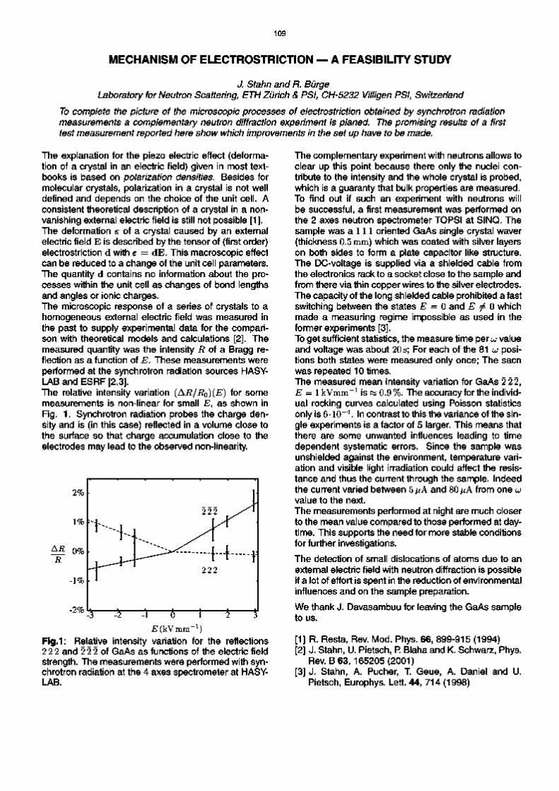
109
MECHANISM OF ELECTROSTRICTION — A FEASIBILITY STUDY
J. Stahn and R. Burge Laboratory for Neutron Scattering, ETH Zurich & PSI, CH-5232 Villigen PSI, Switzerland
To complete the picture of the microscopic processes of electrostriction obtained by synchrotron radiation measurements a complementary neutron diffraction experiment is planed. The promising results of a first test measurement reported here show which improvements in the set up have to be made.
The explanation for the piezo electric effect (deformation of a crystal in an electric field) given in most textbooks is based on polarization densities. Besides for molecular crystals, polarization in a crystal is not well defined and depends on the choice of the unit cell. A consistent theoretical description of a crystal in a non-vanishing external electric field is still not possible [1]. The deformation s o f a crystal caused by an external electric field E is described by the tensor of (first order) electrostriction d with e = dE. This macroscopic effect can be reduced to a change of the unit cell parameters. The quantity d contains no information about the processes within the unit cell as changes of bond lengths and angles or ionic charges. The microscopic response of a series of crystals to a homogeneous external electric field was measured in the past to supply experimental data for the comparison with theoretical models and calculations [2]. The measured quantity was the intensity R of a Bragg reflection as a function of E. These measurements were performed at the synchrotron radiation sources HASY-LAB and ESRF [2,3]. The relative intensity variation (AR/RQ)(E) for some measurements is non-linear for small E, as shown in Fig. 1. Synchrotron radiation probes the charge density and is (in this case) reflected in a volume close to the surface so that charge accumulation close to the electrodes may lead to the observed non-linearity.
^ ( k V m m - 1 ) Fig.1: Relative intensity variation for the reflections 2 2 2 and 2 2 2 of GaAs as functions of the electric field strength. The measurements were performed with synchrotron radiation at the 4 axes spectrometer at HASY-LAB.
The complementary experiment with neutrons allows to clear up this point because there only the nuclei contribute to the intensity and the whole crystal is probed, which is a guaranty that bulk properties are measured. To find out if such an experiment with neutrons will be successful, a first measurement was performed on the 2 axes neutron spectrometer TOPSI at SINQ. The sample was a 111 oriented GaAs single crystal waver (thickness 0.5 mm) which was coated with silver layers on both sides to form a plate capacitor like structure. The DC-voltage is supplied via a shielded cable from the electronics rack to a socket close to the sample and from there via thin copper wires to the silver electrodes. The capacity of the long shielded cable prohibited a fast switching between the states E = 0 and E ^ 0 which made a measuring regime impossible as used in the former experiments [3]. To get sufficient statistics, the measure time per UJ value and voltage was about 20 s; For each of the 81 UJ positions both states were measured only once; The sacn was repeated 10 times. The measured mean intensity variation for GaAs 2 2 2, E = 1 kVmm - 1 is « 0.9 %. The accuracy for the individual rocking curves calculated using Poisson statistics only is 6• 10~4. In contrast to this the variance of the single experiments is a factor of 5 larger. This means that there are some unwanted influences leading to time dependent systematic errors. Since the sample was unshielded against the environment, temperature variation and visible light irradiation could affect the resistance and thus the current through the sample. Indeed the current varied between 5/iA and 80/iA from one UJ value to the next. The measurements performed at night are much closer to the mean value compared to those performed at daytime. This supports the need for more stable conditions for further investigations.
The detection of small dislocations of atoms due to an external electric field with neutron diffraction is possible if a lot of effort is spent in the reduction of environmental influences and on the sample preparation.
We thank J. Davasambuu for leaving the GaAs sample to us.
[1] R. Resta, Rev. Mod. Phys. 66, 899-915 (1994) [2] J. Stahn, U. Pietsch, P. Blaha and K. Schwarz, Phys.
Rev. B 63, 165205(2001) [3] J. Stahn, A. Pucher, T. Geue, A. Daniel and U.
Pietsch, Europhys. Lett. 44, 714 (1998)

110
TASP UPGRADE TO 4-CIRCLE OPTION BETWEEN 10K AND 450 K
D. Schaniel1, J. Schefer
1, Ch. Kagi
1, M. Zolliker
1, B. Roessli \ B. Schoenfeld
2,
M. Konnecke1 and D. Maden
3
1 Laboratory for Neutron Scattering, ETHZ & PSI, CH-5232 Villigen PSI, Switzerland 2lnsitute for Applied Physics, ETH Honggerberg, CH-8093 Zurich, Switzerland
3Swiss Light Source, SLS, CH-5232 Villigen PSI, Switzerland
The Huber 512 Eulerian cradle has been successfully used on TASP. Programs RAFIN and HKLGEN have been adapted to calculate UB-matrix and generate q-scans in any direction. The MAD software has been modified in order to process this files.
The TASP spectrometer is equipped with a double goniometer for heavy equipment in its standard mode. This allows to tilt the sample +15° in two directions. Q-
scans were only possible in the scattering plane by aligning two reflections within this plane within the limitations of the goniometer.
In the last cycle we installed therefore a Huber Eulerian Cradle on TASP, giving free access to most reflections in more than one sector.
Advantages are obvious: A UB-matrix and all zero points can be refined from a set of 10 to 20 centered reflections This is done with the ILL program RAFIN, also used on TriCS. Based on this UB-matrix, we can calculate q-scans in any direction, lists of nuclear/magnetic reflections and its satellites with the program HKLGEN. Presently, we are processing these files in a batch mode ("special scans"). Depending on the demand of this option, it also could be integrated in the spectrometer software MAD. The cradle can be installed within two hours. Therefore, this it is offered in the future as a TASP option. The APD closed cycle refrigerator is available as a further option to be mounted on the cradle to extend the temperature range to 10 K up to 450K.
Fig. 1: Eulerian cradle on TASP. The instrument is shown in diffraction mode reducing background close to zero.
Fig. 2: Support engineered by the workshops of the Applied Physics department of ETH allowing exact and easy centering of the cradle in x,y and height.
Fig. 3 above shows a first scan using this option. The science is discussed in a follow-up SINQ-report.
■j. . . i . . . . i . . . . i . . . . i . . . . i . . . .•
- 1 0 -0 5 0 0 0 5 1 0
Fig. 3: Projection of a q-scan from (0.3075,5.3075.-.5) to (0.3075,5.3075,+.5) in Sra61Ba0.39Nb2O6 to (00L).
Together with the polarization option, TASP is a unique triple axis spectrometer.

111
A NEW SINGLE-CRYSTAL PRESSURE CELL FOR TriCS UP TO 3 GPa
R.Sadykov1, D.Sheptyakov2, O.Zaharko2, Th.Strassle2, J.Schefef
Wereshchagln High-Pressure Physics Institute RAS, 142190 Troitsk, Moscow region, Russia 2Laboratory for Neutron Scattering, ETH Zurich & PSI Villigen, CH-5232, Switzerland
A new hydrostatic pressure-cell for single-crystal neutron diffraction studies has been successfully tested at the single crystal diffractometer TrlCS up to 3 GPa at a temperature of 17 K.
In order to expand the current family of pressure cells at SINQ preliminary test measurements on a pressure cell for single-crystal neutron diffraction have been carried out. A schematic drawing of the cell is shown in Figure 1. The clamp cell consists of a body (1) made of Ni57Cr395Al3 5 alloy, an inner piston (2) and and a screw (3). For loading the force is applied via an auxiliary piston (4) and clamped by the screw. The design of the cell allows a maximal pressure of 3.5 GPa. Quasi-hydrostatic pressure conditions are obtained by placing the sample (5) into a capsule made of lead and filled with fluorinert FC-77 (6). The inner diameter of the cell accounts to 4 mm with a maximal height of about 8mm(for sample). In order to suit the 4-circle geometry of the diffractometer the cell has been designed spherical around the sample site to minimize anisotropic absorption effects. The cell can be placed conveniently in a closed-cycle cooling machine mounted on the Euler cradle of TriCS. The compact design of the cell allows cooling from room temperature down to 17 K within three hours. Temperature gradients are avoided by placing the cell in an Al or V sample container filled with helium. The sample is glued on a single crystal of NaCI, so that the pressure can be measured at base temperature by the shift of the lattice constant of NaCI [1] (alternatively other pressure calibrants, e.g. CsCI, can be used).
The test measurement has been carried out on Ba-hexaferrite BaFe8Co2Ti2019 [2]. The single crystal of 2.5 x 2.0 x 1.5 mm3 dimension has shown clear diffraction peaks both measured using the 1D and the 2D detectors of TriCS (Figure 2). The successful measurement of the magnetic satellite peaks at low temperatures demonstrated the feasibility of using the cell for magnetic studies with relatively weak signals. The alloy N i s / C ^ A ^ used for this pressure cell is not a zero-matrix alloy like Ti-Zr often used for powder diffraction pressure cells. It shows Bragg peaks and has relatively large absorption. However, the incoherent signal from the pressure cell turned out to be very weak resulting in nice, flat background in regions out of the Bragg peaks of N i s / C ^ A ^ . Bragg peaks resulting from the pressure cell are easily identified as Debye-Scherrer cones on the 2D detector. The pressure at a temperature of T = 17 K was found to be 3.0(3) GPa.
Fig 1: Schematic drawing of the pressure cell (see text).
300 00 -
Fig 2: Bragg peaks and magnetic satellites from the sample measured with the 2D detector at p=3 GPa, T=17 K (scans in omega presented as a sequence of 1D projection onto one of the detector axes).
[1] E.F.Skelton et al., Rev.Sci.lnstrum. 55, 849 (1984)
[2] R.Sadykov et al., Sov.Phys.Solid State 23, 1865 (1981)

112
A PRESSURE CELL UP TO 1 GPa FOR THE COMMERCIAL QUANTUM DESIGN PPMS SYSTEM
Th.Strassle, TMuhlebach, R.Thut, P.AIIenspach Laboratory for Neutron Scattering, ETH Zurich & PSI Villigen, CH-5232 Villigen PSI, Switzerland
A non-magnetic clamp pressure cell for the measurement of DC and AC susceptibility on the commercial Quantum Design PPMS system is presented.
The characterization of samples by means of macroscopic measurement techniques plays an important role for any successful neutron scattering study. With the availability of pressure cells for both elastic and inelastic neutron studies at SINQ the interest on measuring DC and AC susceptibility under pressure on our Quantum Design PPMS has grown. Here we present a simple clamp pressure cell that operates without any change of the PPMS and allows measurements on a qualitative / semi-quantitative level. The cell consists of the body with an inner and outer diameter of 2 mm and 7.2 mm, an inner piston and a screw to clamp the pressure. All parts of the cell are produced from Cu-Be.
outer piston (loading only)
inner piston
The powder or single crystal sample is placed in a capsule made of lead or tin and filled with Fluorinert FC-77 as pressure medium. The closed capsule is then placed in the pressure cell and sealed with the inner piston. The pressure cell is loaded with an outer piston made of hardened steel and a maximal force of 4 KN. The pressure is measured by the pressure dependence of the superconducting phase transition temperature of lead [1] or tin. The pressure may be determined as accurate as <10 MPa. Pressures up to 1 GPa at base temperature could be realized so far.
sample m Pb/Sn contain
The cell has been successfully used in our laboratory. In Fig. 1 we show the pressure effect on the DC magnetization of CeSb. At ambient pressure CeSb orders antiferromagnetically and shows various commensurable magnetic phases below TN=16 K. All of these phases show distinct pressure dependences. The pressure dependence of the two highest (and most significant) transitions is in good agreement with published p-T phase diagrams of CeSb [2].
1.00 GPa
'I i i i i I i i i i I i i i i I i i i i I i i i i I' 0 10 20 30 40 50
Temperature T [K]
Fig.1: DC magnetization of CeSb at various pressures (numbers denote order of measurement, dashed line indicates 1st two transitions).
The authors thank Drs R.Sadykov and D.Andreica for valuable discussions.
The preparation of the sample in a lead or tin capsule has advantages compared to other methods: (i) the sample can be prepared in a controlled way (sample mass, no air bubbles, etc.), (ii) the softness of lead or tin allows perfect sealing at small loads allowing "p=0" measurements close to ambient pressure and quasi-hydrostatic conditions for p>0, (iii) lead and tin reduce the friction force while the cell is loaded, (iv) lead and tin show small thermal expansion compared to other sealing materials (i.e. Teflon), (v) pressure is measured at the sample site.
[1] B.Bireckoven, J.Wittig, J.Phys. E: Sci.lnstrum. 21,841 (1988)
[2] T.Chattopadhyay, P.Burlet, J.Rossat-Mignod, H.Bartholin, C.Vettier, O.Vogt , Phys.Rev. B 49, 15096(1994)

113
PROGRESS AND STATUS OF HRPT
P. Fischer1, G. Frey1, M. Konnecke1, D. Sheptyakov1, R. Thut1, R. Burge1, U. Greuter2 and N. Schlumpf2
1 Laboratory for Neutron Scattering, ETHZ & PSI, CH-5232 Villigen PSI, Switzerland 2 Electronics Department, Paul Scherrer Institute, CH-5232 Villigen PSI, Switzerland
Progress has been made in particular concerning the functionality and stability of the SCE software, but the corresponding PC solution should be replaced by a true real-time system.
In the year 2000 user operation of HRPT started [1]. During the SINQ shutdown in the first half of 2001 the SCE software for the data readout has been upgraded by EFS to include the low-rate and remote control mode functionalities. The former is important for correct monitor handling with respect to frequent neutron flux variations at SINQ. It also implied major adaptions of the multidetector interface by U. Greuter because of timing modifications made by EFS. Only after intensive direct contacts between PSI and EFS including installation of Windows 98 on the ihm-PC, the new software worked significantly more stable. Moreover reprogramming of the MAX FPGA's and installation of the FLEX-EPROMS has been performed in cooperation with CERCA. However, it does not yet work for all modules without manual reprogramming from ihm/SCE. In late August, the cabling and mechanics of the SCE cabinet were redesigned by PSI for improved reliability and better accessability. During the user operation problems with variations of certain detector channels and occasional spike problems occurred. As a temporary solution D. Sheptyakov introduced a RECALIB routine implying a second measurement with an angular step larger than 0.05 degrees. A major new effect detected with highly incoherently scattering samples such as PVC is an essential read out problem of the counters requiring the use of short picture lengths.
As verified at the end of 2001 by means of systematic logic analyser tests performed by G. Frey, U. Greuter and CERCA (E. Berruyer and M. Febvre) the mentioned problems appear to be mainly caused by the SCE PC readout system. Therefore the replacement by a true real-time system is now envisaged by CERCA and PSI. During these instrumental tests also the apparently exchanged channels 608 and 610 were corrected. Finally in December 2001 the energy spectrum of the test detector has been remeasured with 1.886 A neutrons. It yielded a marked decrease of the energy resolution as quantified by the FWHM/(peak energy) increase from (5 to7) % in the early days to now about 28 %. Most probably it is caused by the slight 3He loss [1] due to mainly the single viton seal of the large LCP1600 detector and simultaneous diffusion of oxygen into the detector as well as possible also by outgasing inside the detector chamber. After another one to two years this will require purification of the detector gas during a longer SINQ shutdown. Concerning the mechanics, the detector positioning has to be improved (cleaning of all air cushions etc.). Moreover a better shielding of the monochromatic neutron beam is in work by R. Thut.
[1] P. Fischer et al., PSI Scientific Report 2000, Vol. Ill, ISSN 1423-7326, 89 (2001).

114
INSTRUMENT CONTROL AT SINQ
Mark Konnecke Laboratory for Neutron Scattering, ETH Zurich & PSI, CH-5232 Villigen PSI, Switzerland
Instrument Control at SINQ is mainly based onto the was extended to support additional instruments.
The neutron scattering instruments at SINQ are all computer controlled. We aimed to control all instruments through a common control software: the SINQ Instrument Control Software (SICS). With the exception of the Riso instrument RITA-2 and the triple axis spectrometer TASP all neutron scattering instruments at SINQ are now controlled by SICS. SICS is a client server control system. For each instrument there is an instrument control server running on a unix workstation. This server is written in ANSI-C and does all the work: driving motors, writing data files, counting, watching sample environment devices etc. Then there are clients which communicate with the instrument control server through the TCP/IP network. These clients implement the user interface and the status monitoring applications for the instruments. Most of these clients are written in Java for platform independence. However, there is also a WWW-interface to the instruments which allows to query the status of a measurement through any WWW browser. This WWW-interface is based on Java Servlet technology. Working on the foundations laid in previous years SICS was extended to support further hardware devices and instruments. At the instruments TRICS and AMOR new position sensitive detectors (PSD) were installed. These detectors were produced at EMBL at the ILL, Grenoble, France and interfaced to our setup through some electronics developed by the PSI electronics group [1]. The EMBL-PSD's read out system encodes the positional information of a neutron event into a time value. This system is quite different from the position encoding system at other SINQ detectors. Thus it was necessary to extend the real time histogram memory software originally developed by David Maden [2] for this detector. The purpose of this software is to collect the neutron events coming in from the electronics and to sum them into the bins appropriate for the detector position where the event occured. After upgrading the histogram memory software the SICS software was extended to support the new PSD detector in the control software for the single crystal four circle diffractometer TRICS. This involved a new module for coordinating up to four counting devices (a EL737 counter box for the monitors and three histogram memories) and extensions to the data file writing module. Data files are written in the NeXus [4] format which in turn is based on the hierarchical data format
Sinq Instrument Control Software (SICS). This software
(HDF) [3] version 4. HDF-4 proved to be insufficient for the use at TRICS and this prompted the development of a new NeXus-API for the newer HDF version 5 which finally solved TRICS data handling problems. Furthermore a peak search procedure was developed for TRICS. A Java client was provided for TRICS which allows to view the data collected in all three PSD's during a measurement. The instrument AMOR is a neutron reflectometer which also uses the EMBL PSD mentioned above. AMOR's control software was updated to support this detector. A little application, amortool, was written which performs basic data analysis tasks on data collected at AMOR. In an effort to replace the older MAD software still in use at TASP with the new common control software, SICS was extended to support triple axis spectrometers as well. The challenge posed by this type of instrument was not so much of a technical nature but by the requirement to reuse the command set from the older program in the new software. This was further complicated by the fact that the MAD software relied on the storage order of parameters in memory in many instances. A minor problem was that data files must be written in the MAD data format as well. However, with three additonal C-language modules and 1000 lines in SICS's internal scripting language this challenge was met. Testing this extension revealed further, undocumented, features of the MAD program which had to be emulated as well. The triple axis support in SICS is now considered to be stable for normal operation. Still pending is support for operation with neutron polaris-ers. As a new instrument the fourier diffractometer POLDI is coming into operation. POLDI's demands to the control software included a port of the software to Redhat Linux, another data file format and support for another chopper controller. Fulfilling all these demands took less time then expected and highlighted the advantages of having a common portable control program.
[1] Buhler et al., J. Appl. Physics (2002) [2] D. Maden, U. Greuter, P. Rasmussen, SINQ Docu
ment 891/MD36-89 1 A05,89 (2000) [3] http://hdf.ncsa.uiuc.edu [4] http://lns00.psi.ch/NeXus

115
EXTENSION OF THE NEXUS FILE FORMAT FOR HDF5
U. Filges, M. Konnecke, Laboratory for Neutron Scattering, ETHZ & PSI, CH5232 Villigen PSI, Switzerland
NeXus is a data format for the exchange of neutron and synchrotron scattering data between facilities and user institutions. The first released version of NeXusAPI was based on the Hierarchical Data Format (HDFversion 4). NCSA reimplemented HDF and called the new version HDF5. This made it necessary to extend the NeXus file format for HDF5.
The NeXus data format was first published in December 1997 on the International Workshop on New Opportunities for Better User Group Software. The presented version was based on the HDF library version 4 as the physical file format. The developers of HDF, the National Centre for Supercomputing Application (NCSA), now promote a new, incompatible, version of HDF, HDF5. This version is a new implementation with a much cleaner interface. Further improvements are that HDF5 also supports file sizes larger then 2GB and an unlimited number of objects in a file. Therefore it became necessary to provide a NeXusAPI for HDF5 as well. For the implementation of this new API we strived to achieve two main goals:
maximum compatibility at the API level in order to minimize changes to existing code
support for both HDF4 and HDF5
These requirements made the implementation of the new API a little tricky but since June 2001 a working version of the new NeXus API is available. The new API can be built to support HDF4 or HDF5 only and to support both HDF4 and HDF5. The main goal of maximum API compatibility was achieved. On the user level the changes are minimised concerning writing NeXus application and the resulting data file layout. The exception is writing of compressed data set. A new API function became necessary for the creation of compressed datasets due to limitations in theHDF5API.
The new NeXus API is written in the ANSIIC programming language. This CAPI was interfaced to both the Fortran 77 and Java programming languages.
If the measured data are stored in the NeXus data file format the next logical steps are viewing, editing and analysing the data. In this direction the welltried NXbrowse viewer has also been adapted to work with the new API version. Besides this viewer a powerful
HDF5 editor H5view (Fig. 1) is available which has a userfriendly graphical interface and a complex functionality.
Shortly after the release of the new NeXus API the first data files were collected at the Single Crystal Neutron Diffractometer (TriCS) instrument in the new format.
H E &&te£l nntow TBHIS Help
£ibQstef yidMf m § | / ( f l e tt c;123732001 hdf) *
+ C3u,un . central
| g name
IF" Sf iWOthBt . & v a d
^ y z e o p o n , " *
♦ CS <,„„le C J start t me
♦ C 3 < H
♦ C3<H« ' " ♦ C 3 < H * ♦ C3<H«'" *
5L! >j.*i...JMtefeM
1® I <M | m j t« | m | m \ *u | 11$ | m \
A 1A ji
Fig.1: NeXus data file (version 2.1.0) measured at the TriCS instrument, viewed with HDF5 editor H5view
For the subsequent analysis of measured data the NeXusAPI for IDL (Interactive Data Language) was rewritten using IDL's native method support. Thus both HDF4 and HDF5 NeXus files can be accessed in IDL as well. The IDL programming language is used for the general analysis tool LAMP.
[1] P. Klosowski, M. Koennecke, J. Z. Tischler, R. Osborn, "NeXus: A common format for the exchange of neutron and synchrotron data" Physica B 241243, 151153 (1998)

116

117
PUBLICATIONS
LABORATORY FOR NEUTRON SCATTERING
K. Ahn, B.J. Gibson, R.K. Kremer, Hj. Mattausch, L. Keller and A. Simon MAGNETIC ORDERING WITHIN THE LAYERED TERBIUM CARBIDE IODIDE Tb2C2l2 J. of Alloys and Compounds 323-324, 400-403 (2001).
P. Allenspach and M. B. Maple HEAT CAPACITY Handbook on the Physics and Chemistry of Rare Earths 31, 351-390 (2001).
P. Allenspach, P. Boni and K. Lefmann LOSS MECHANISMS IN SUPERMIRROR NEUTRON GUIDES Proc. SPIE 4509, 157-165 (2001).
H. Andres, J.M. Clemente-Juan, R. Basler, M. Aebersold, H.-U. Gudel, J.J. Borras-Almenar, A. Gaita, E. Coronado, H. Buttner and S. Janssen MAGNETIC POLYOXOMETALATES: ANISOTROPIC ANTIFERRO- AND FERROMAGNETIC EXCHANGE INTERACTIONS IN THE PENTAMERIC COBALT(II) CLUSTER [Co3W(D20)2(CoW9034)2]12". A MAGNETIC AND INELASTIC NEUTRON SCATTERING STUDY Inorganic Chemistry 40, 1943-1950(2001).
A. Arbe, F. Boue, J. Colmenero, S. Janssen, K. Mortensen, D. Richter, J. Rieger, P. Schurtenberger and R.K. Thomas SOFT CONDENSED MATTER ESS/SAC Report 1/01, 69-79 (2001).
V.A. Atsarkin, V.V. Demidov, G.A. Vasneva and K. Conder CRITICAL SLOWING DOWN OF LONGITUDINAL SPIN RELAXATION IN La^CaxMnOs Phys. Rev. B 63, 092405/1-4 (2001).
A.M. Balagurov, V.Yu. Pomjakushin, D.V. Sheptyakov, V.L. Aksenov, N.A. Babushkina, L.M. Belova, O.Yu. Gorbenko and A.R. Kaul EVOLUTION OF (LaLvPrvWCaosMnOs CRYSTAL STRUCTURE WITH A-CATION SIZE, TEMPERATURE, AND OXYGEN ISOTOPE SUBSTITUTION Eur. Phys. J. B 19, 215-223 (2001).
A.M. Balagurov, V.Yu Pomjakushin, D.V. Sheptyakov, V.L. Askenov, P. Fischer, L. Keller, O.Yu. Gorbenko, A.R. Kaul and N.A.Babushkina LONG-SCALE PHASE SEPARATION VERSUS HOMOGENEOUS MAGNETIC STATE IN (LaLYPrvWCaosMnOs: A NEUTRON DIFFRACTION STUDY Phys. Rev. B 64, 024420/1-10 (2001).
R. Basler, P.L.W. Tregenna-Piggott, H. Andres, C. Dobe, H.-U. Gudel, S. Janssen and G. Mclntyre MAGNETIC EXCITATIONS OF [CsMn(S04)212D20 MEASURED BY INELASTIC NEUTRON SCATTERING J. Am. Chem. Soc. 123, 3377-3378 (2001).
C. Beck, S. Janssen, B. Gross and R. Hempelmann NEUTRON TIME-OF-FLIGHT SPECTROMETER FOCUS AT SINQ: RESULTS FROM NANOCRYSTALLINE MATTER STUDIES Scripta-Materialia 44, 2309-2313 (2001).
C. Beck, W. Hartl and S. Janssen FULLERENES AS NEW COLLOIDAL MODEL SYSTEMS Progr. Coll. Pol. Sci. 117, 126 (2001).
B. Bertheville, T. Herrmannsdorfer and K. Yvon STRUCTURE DATA FOR K2MgH4 AND Rb2CaH4 AND COMPARISON WITH HYDRIDE AND FLUORIDE ANALOGUES J. of Alloys and Compounds 325, L-13-L16 (2001).

118
J.C. Campuzano, A. Kaminski, H. Fretwell, J. Mesot, T. Sato, T. Takahashi, M. Norman, M. Randeria, K. Kadowaki and D. Hinks THE ROLE OF ANGLE-RESOLVED PHOTOEMISSION IN UNDERSTANDING THE HIGH TEMPERATURE SUPERCONDUCTORS J. of Physics and Chemistry of Solids 62, 35-39 (2001)
N. Cavadini, G. Heigold, W. Henggeler, A. Furrer, H.-U. Gudel, K. Kramer and H. Mutka MAGNETIC EXCITATIONS IN THE QUANTUM SPIN SYSTEM TICuCI3 Phys. Rev. B 63, 172414 (2001).
N. Cavadini, A. Furrer, H.-U. Gudel, K. Kramer, H. Mutka and A. Wildes POLARIZATION STUDY OF THE SINGLET-TRIPLET MODES IN A QUANTUM SPIN SYSTEM The ILL Millenium Symposium & European User Meeting Proceedings, 145-146 (ILL, Grenoble 2001).
N. Cavadini, W. Henggeler, A. Furrer, H.-U. Gudel, K. Kramer, H. Mutka, A. Wildes, K. Habicht and P. Vorderwisch QUANTUM MAGNETIC PHASE TRANSITION: A TOP-DOWN APPROACH ILL Annual Report 2000, 12-13 (ILL Grenoble 2001).
E.S. Clementyev, K. Conder, A. Furrer and I.L. Sashin PHONON DENSITY-OF-STATES IN THE NEW SUPERCONDUCTOR MgB2 Eur. Phys. J. B 21, 465-467 (2001).
K. Conder OXYGEN DIFFUSION IN THE SUPERCONDUCTORS OF THE YBaCuO FAMILY: ISOTOPE EXCHANGE MEASUREMENTS AND MODELS Materials Science and Engineering R 32, 41-102 (2001).
C. Degueldre, M. Pouchon, M. Streit, O. Zaharko and Di M. Michiel ANALYSIS OF POROUS FEATURES IN ZIRCONIA BASED INERT MATRIX, IMPACT ON THE MATERIAL QUALIFICATION Prog. Nucl. Energy 38, 241-246 (2001).
A. Donni, P. Fischer, L. Keller, V. Yu. Pomjakushin, Y. Nemoto, T. Goto and S. Kunii THE CUBIC TO TRIGONAL PHASE TRANSITION IN HoB6 MEASURED ON THE NEW POWDER DIFFRACTOMETER HRPT AT SINQ J. Phys. Soc. Jpn. 70, Suppl. A, 448-450 (2001).
F. Fauth, E. Suard, V. Caignaert, B. Domenges, I. Mirebeau and L. Keller INTERPLAY OF STRUCTURAL, MAGNETIC AND TRANSPORT PROPERTIES IN THE LAYERED Co-BASED PEROVSKITE LnBaCo205 (Ln=Tb,Dy,Ho) Eur. Phys. J.B 21, 163-174 (2001).
A. Furrer, T. Strassle and D. Rubio Temprano NEW EXCITEMENT WITH CRYSTAL-FIELD EXCITATIONS J. of Alloys and Compounds 323-324, 649-653 (2001).
U. Gasser and P. Allenspach CRYSTALLINE ELECTRIC FIELD AND CLUSTER EFFECTS IN BOROCARBIDE SUPERCONDUCTORS in "Rare Earth Transition Metal Borocarbides (Nitrides): Superconducting Magnetic and Normal State Properties", Editors H. Mullerand V. Narozhnvi, Kluwer Academic Publishers, Boston (2001), pp.131-136
I.V. Golosovsky, I. Mirebeau, A.S. Markosyan, P. Fischer and V. Yu. Pomjakushin NEUTRON DIFFRACTION STUDY OF THE MAGNETIC ORDER IN THE Dy(Mn1.xAlx)2 SYSTEM IN THE REGION OF A MAGNETIC INSTABILITY Phys. Rev. B 65, 014405/1-7 (2001).
V. Gramlich and H. Grimmer THE HISTORY OF CRYSTALLOGRAPHY IN SWITZERLAND Chimica 55, 484-486 (2001).

119
H. Grimmer, O. Zaharko, H.-Ch. Mertins and F. Schafers POLARIZING MIRRORS FOR SOFT X-RAY RADIATION Nuclear Instruments and Methods in Physics Research A 467-468, 354-357 (2001).
H. Grimmer, M. Horisberger, U. Staub, H.-Ch. Mertins and F. Schafers MULTILAYER OPTICS FOR SOFT X-RAYS in "Advances in Structure Analysis", Editors R. Kuzel and J. Hasek , Czech and Slovak Crystallographic Association, Praha, (2001), pp. 311-318.
Guo-Meng Zhao, H. Keller and K. Conder UNCONVENTIONAL ISOTOPE EFFECTS IN THE HIGH-TEMPERATURE CUPRATE SUPERCONDUCTORS J. of Phys.: Condens. Matter 13, R-569-587 (2001).
T. Herrmannsdorfer, A. Donni, P. Fischer, L. Keller, E. Clementyev, A. Furrer, S. Mango, B. van den Brandt and H. Kitazawa MAGNETIC ORDERING AND CRYSTALLINE ELECTRIC FIELD SPLITTING IN Nd3Pd20Si6 J. of Alloys and Compounds 323-324, 509-512 (2001).
A. Hiess, P.J. Brown, E. Lelievre-Berna, B. Roessli, N. Bernhoeft, G.H. Lander, N. Aso and N.K. Sato SPHERICAL NEUTRON POLARIMETRY OF THE MAGNETIC STRUCTURE IN UNi2AI3 Phys. Rev. B 64, 134413/1-4 (2001).
A. Hiess, P.J. Brown, N. Aso, B. Roessli, N. Bernhoeft, N.K. Sato and G.H. Lander NEUTRON SCATTERING STUDY OF THE STATIC AND DYNAMIC MAGNETIC PROPERTIES OF UNi2AI3 J. of Magn. Magn. Mater. 226-230, 54-56 (2001).
A. Kaminski, M. Randeria, J.C. Campuzano, M.R. Norman, H. Fretwell, J. Mesot, T. Sato, T. Takahashi and K. Kadowaki RENORMALIZATION OF SPECTRAL LINE SHAPE AND DISPERSION BELOW Tc IN Bi2Sr2CaCu208+8 Phys. Rev. Lett. 86, 1070-1073 (2001).
P. Karen, P.M. Woodward, J. Linden, T. Vogt, A. Studer and P. Fischer VERWEY TRANSITION IN MIXED-VALENCE TbBaFe205: TWO ATTEMPTS TO ORDER CHARGES Phys. Rev. B 64, 214405/1-14 (2001).
L. Keller, P. Fischer, T. Herrmannsdorfer, A. Donni, H. Sugawara, T.D. Matsuda, K. Abe, Y. Aoki and H. Sato STRUCTURAL AND MAGNETIC PROPERTIES OF RFe4Pi2 (R=Pr,Nd) STUDIED BY NEUTRON DIFFRACTION J. of Alloys and Compounds 323-324, 516-519 (2001).
H. Kohlmann, F. Fauth, P. Fischer, A.V. Skripov, V.N. Kozhanov and K. Yvon LOW-TEMPERATURE DEUTERIUM ORDERING IN THE CUBIC LAVES PHASE DERIVATIVE a-ZrCr2D066 J. of Alloys and Compounds 327, L4-L9 (2001).
E.M. Kopnin, C. Bougerol-Chaillout, A.A. Belik, H. Schwer, G. Bottger and J. Karpinski CRYSTAL STRUCTURE OF HIGH-TC RELATED NdBaCu02B03: TEM AND NEUTRON POWDER DIFFRACTION STUDY Physica C 355, 119-125 (2001).
E.M. Kopnin, A.A. Belik, H. Schwer, G. Bottger and J. Karpinski CRYSTAL STRUCTURE OF LaHo075Sro25Cu0389: EVIDENCE OF OXYGEN VACANCIES IN THE FLUORITE-LIKE SLAB J. of Alloys and Compounds 319, L1-L4 (2001).
A. Leineweber, H. Jacobs, P. Fischer and G. Bottger UNIAXIAL ORIENTATIONAL ORDER-DISORDER TRANSITIONS IN DIAMMINE MAGNESIUM HALIDES, Mg(ND3)2CI2 AND Mg(ND3)2Br2, INVESTIGATED BY NEUTRON DIFFRACTION J. Solid State Chem. 156, 487-499 (2001).
A. Leineweber, H. Jacobs, R. Essmann, P. Allenspach, F. Fauth and P. Fischer Co(NH3)2CI2 AND Co(ND3)2CI2: ORDER-DISORDER BEHAVIOUR OF N(H,D)3 AND ANTIFERROMAGNETIC STRUCTURE Z. Anorg. Allg. Chem. 627, 2063-2069 (2001).

120
E. Liarokapis, D. Palles, K. Conder and E. Kaldis THE ANHARMONIC BEHAVIOR OF THE OXYGEN MODES IN YBa2Cu3Ox COMPOUNDS J. of Raman Spectroscopy 32, 821-826 (2001).
J. Lopez, P.N. Lisboa-Filho, W.A.C. Passos, W.A. Ortiz, F.M. Araujo-Moreira, O.F. de Lima, D. Schaniel and K. Gosh MAGNETIC RELAXATION BEHAVIOR IN Lao5Cao5Mn03 AND Ndo5Sro5Mn03 Phys. Rev. B 63, 224422/1-9 (2001).
H.-Ch. Mertins, O. Zaharko, F. Schafers, A. Gaupp, D. Abramsohn, M. Weiss and H. Grimmer RESONANT MAGNETIC SCATTERING OF LINEARLY POLARISED SOFT X-RAYS FROM Fe-LAYERS AND Fe/C-MULTILAYERS Nucl. Instruments and Methods in Physics Research A 467-468,1415-1418 (2001).
J. Mesot, M. Randeria, M.R. Norman, A. Kaminski, H.M. Fretwell, J.C. Campuzano, H. Ding, T. Takeuchi, T. Sato, T. Yokoya, T. Takahashi, I. Chong, T. Terashima, M. Takano, T. Mochiku and K. Kadowaki DETERMINATION OF THE FERMI SURFACE IN HIGH-TC SUPERCONDUCTORS BY ANGLE-RESOLVED PHOTOEMISSION SPECTROSCOPY Phys. Rev. B 63, 224516/1-14 (2001).
A. Murasik, A. Czopnik and L. Keller EFFECT OF STRAINS ON MAGNETIC ORDERING IN Tmln3 Phys. Stat. Sol. (a) 186, R1-R3 (2001).
M.R. Norman, A. Kaminski, J. Mesot and J.C. Campuzano TEMPERATURE EVOLUTION OF THE SPECTRAL PEAK IN HIGH-TEMPERATURE SUPERCONDUCTORS Phys. Rev. B 63, 1405081-1405084 (2001).
G.A. Petrakovskii, K.S. Aleksandrov, L.N. Bezmaternikh, S.S. Aplesnin, B. Roessli, F. Semadeni, A. Amato, C. Baines, J. Bartolome and M. Evangelisti SPIN-GLASS STATE IN CuGa204 Phys. Rev. B 63, 1844251-1844258 (2001).
G.A. Petrakovskii, M.A. Popov, B. Roessli and B. Ouladdiaf INCOMMENSURATE MAGNETIC STRUCTURE IN COPPER METABORATE J. of Experimental and Theoretical Physics 93, 809-814 (2001).
G. Pepponi, P. Wobrauschek, F. Hegedus, C. Streli, N. Zoger, C. Jokubonis, G. Falkenberg and H. Grimmer, SYNCHROTRON RADIATION TOTAL REFLECTION X-RAY FLUORESCENCE AND ENERGY DISPERSIVE X-RAY FLUORESCENCE ANALYSIS ON AP1 (TM) FILMS APPLIED TO THE ANALYSIS OF TRACE ELEMENTS IN METAL ALLOYS FOR THE CONSTRUCTION OF NUCLEAR REACTOR CORE COMPONENTS: A COMPARISON Spectrochimica Acta B - Atomic Spectroscopy 56, 2063-2071 (2001)
G. Renaudin, P. Fischer and K. Yvon TETRAGONAL STRUCTURE OF NEODYMIUM DEUTERIDE NdD227 REVISITED J. of Alloys and Compounds 329, L9-L13 (2001).
B. Roessli, J. Schefer, G.A. Petrakovskii, B. Ouladdiaf, M. Boehm, U. Staub, A. Vorotinov and L. Bezmaternikh FORMATION OF MAGNETIC SOLITON LATTICE IN COPPER METABORATE Phys. Rev. Lett. 86, 1885-1888 (2001).
B. Roessli, U. Staub, A. Amato, D. Herlach, P. Pattison, K. Sablina and G.A. Petrakovskii MAGNETIC PHASE TRANSITIONS IN THE DOUBLE SPIN-CHAINS COMPOUND LiCu202 Physica B 296, 306-311 (2001).
B. Roessli, and P. Boni POLARISED NEUTRON SCATTERING, in "Scattering", Editors E.R. Pike and P. Sabatier, Academic Press, London (2001), pp. 1242-1263.

121
D. Rubio Temprano, J. Mesot, S. Janssen, K. Conder, A. Furrer, A. Sokolov, V. Trounov, S. Kazakov, J. Karpinski and K.A. Muller LARGE COPPER ISOTOPE EFFECT ON THE PSEUDOGAP IN THE HIGH-TEMPERATURE SUPERCONDUCTOR HoBa2Cu408 Eur. Phys. J. B 19, 5-8(2001).
D. Rubio Temprano, J. Mesot, S. Janssen, K. Conder, A. Furrer, A. Sokolov, V. Trounov, S. Kazakov, J. Karpinski and K.A. Muller A NEUTRON CRYSTAL-FIELD STUDY OF THE COPPER ISOTOPE EFFECT ON THE PSEUDOGAP IN THE HIGH-Tc CUPRATE HoBa2Cu408 J. of Alloys and Compounds 323-324, 554-557 (2001).
T. Sato, T. Kamiyama, T. Takahashi, J. Mesot, A. Kaminski, J.C. Campuzano, H.M. Fretwell, T. Takeuchi, H. Ding, I. Chong, T. Therashima and M. Takano EVIDENCE FOR A HOLE-LIKE FERMI SURFACE OF Bi2Sr2Cu06 FROM TEMPERATURE-DEPENDENT ANGLE-RESOLVED PHOTOEMISSION SPECTROSCOPY Phys. Rev. B 64, 054502/1-5 (2001).
D. Schaniel, P. Allenspach, A. Furrer, K. Kramer and H.-U. Gudel DIMER SPLITTING OF Er3+ IN Cs3Er2Br9 J. of Alloys and Compounds 323-324, 481-485 (2001).
D. Schaniel, J. Schefer, B. Delley, M. Imlau and T. Woike OPTICAL ABSORPTION SPECTROSCOPY IN THE METASTABLE STATE SI OF Na2 [Fe (CN)5NO]-2H20 Mat. Res. Soc. Symp. Proc. 674, V2.5.1-V2.5.6 (2001).
F. Semadeni, B. Roessli and P. Boni THREE-AXIS SPECTROSCOPY WITH REMANENT BENDERS Physica B 297, 152-154 (2001).
F. Semadeni, A. Amato, B. Roessli, P. Boni, C. Baines, T. Masuda, J. Uchinokura and G. Shirane MACROSCOPIC AND LOCAL MAGNETIC MOMENTS IN Si-DOPED CuGe03 AS DETERMINED BY NEUTRON AND nSR STUDIES Eur. Phys. J. B 21, 307-311 (2001).
A. Shengelaya, H. Keller, K.A. Muller, B.I. Kochelaev and K. Conder TILTING MODE RELAXATION IN THE ELECTRON PARAMAGNETIC RESONANCE OF OXYGEN-ISOTOPE-SUBSTITUTED La2.xSrxCu04:Mn2+
Phys. Rev. B 63, 144513/1-9 (2001).
J. Stahn, U. Pietsch, P. Blaha and K. Schwarz ELECTRIC FIELD INDUCED CHARGE-DENSITY VARIATIONS IN COVALENTLY BONDED BINARY COMPOUNDS Phys. Rev. B 63, 165205/1-10 (2001).
U. Staub, O. Zaharko, H. Grimmer, M. Horisberger and F. d'Acapito REAL-PART EXAFS FROM MULTILAYER BRAGG REFLECTIONS: A PROMISING NEW EXAFS TECHNIQUE Europhys. Lett. 56, 241-246 (2001).
Th. Strassle, A. Furrer, F. Altorfer, K. Mattenberger, M. Bohm and H. Mutka HoAS: A MODEL COMPOUND FOR THE COOLING BY THE BAROCALORIC EFFECT J. of Alloys and Compounds 323-324, 392-395 (2001).
T. Strassle, F. Altorfer and A. Furrer CRYSTAL-FIELD INTERACTIONS IN THE PSEUDO-TERNARY COMPOUND ErAlxGa2.x STUDIED BY INELASTIC NEUTRON SCATTERING J. Phys.: Condens. Matter 13, 6773-6785 (2001).
O. Zaharko, C. Meneghini, A. Cervellino and E. Fischer LOCAL STRUCTURE OF Co AND Ni IN DECAGONAL AINiCo INVESTIGATED BY POLARIZED EXAFS Eur. Phys. J. B 19, 207-213 (2001).

122
O. Zaharko, H.-Ch. Mertins, H. Grimmer and F. Schafers SOFT X-RAY RESONANT MAGNETIC REFLECTIVITY FROM Fe/C MULTILAYERS Nucl. Instruments and Methods in Physics Research A 467-468,1419-1422 (2001).
O. Zaharko, A. Cervellino, H.-Ch. Mertins, H. Grimmer, F. Schafers and D. Arvanitis SOFT X-RAY MAGNETIC CIRCULAR DICHROISM IN Fe AND Fe050Co048V002 FILMS: QUANTITATIVE ANALYSIS OF TRANSMISSION Eur. Phys. J. B 23, 441-448 (2001).
CONDENSED MATTER THEORY GROUP
A. Bill and V.Z.Kresin INTERCALATION AND HIGH TEMPERATURE SUPERCONDUCTIVITY IN FULLERIDES cond-mat/0109553, 0110327.
P.W. Brouwer, C. Mudry and A. Furusaki TRANSPORT PROPERTIES AND DENSITY OF STATES OF QUANTUM WIRES WITH OFF-DIAGONAL DISORDER Physica E. 9, 333-339 (2001)
C. Chamon and C. Mudry, DENSITY OF STATES FOR DIRTY d-WAVE SUPERCONDUCTORS: A UNIFIED AND DUAL APPROACH FOR DIFFERENT TYPES OF DISORDER Phys. Rev. B. 63, 100503/1-4 (2001)
W. T.Geng, Yu-Jun Zhao, A. J. Freeman and B. Delley ATOMIC DISPLACEMENTS AT A SIGMA 3(111) GRAIN BOUNDARY IN BATIOs Phys. Rev. B 63, 060101 (2001).
R. Monnier and B. Delley POINT DEFECTS, FERROMAGNETISM, AND TRANSPORT IN CALCIUM HEXABORIDE Phys. Rev. Lett. 87,157204 (2001).
Ch. Waelti, E. Felder, C. Degen, G. Wigger, R. Monnier, B. Delley and H.R.Ott: STRONG ELECTRON-PHONON COUPLING IN SUPERCONDUCTING MgB2: A SPECIFIC HEAT STUDY Phys. Rev. B 64,172515 (2001).
J.F. Loffler, H.B. Braun and W. Wagner MAGNETIC CORRELATIONS IN NANOSTRUCTURED FERROMAGNETS - REPLY Phys. Rev. Lett. 87, 149702 (2001)
J.F. Loffler, W. Wagner and H.B. Braun MAGNETISM OF NANOSTRUCTURED FERROMAGNETS-EXPERIMENTS AND THEORETICAL MODEL Scripta Mater. 44, 1425 (2001)
J.F. Loffler, H.B. Braun and W. Wagner et al. CROSSOVER IN THE MAGNETIC PROPERTIES OF NANOSTRUCTURED METALS Mat. Sci. Eng. A-Structure 304, 1050 Sp. Iss. SI (2001)
M. Titov, P.W. Brouwer, A. Furusaki and C. Mudry FOKKER-PLANCK EQUATIONS AND DENSITY OF STATES IN DISORDERED QUANTUM WIRES Phys. Rev. B. 63, 235318/1-9 (2001)

123
ADDENDUM 2000:
F. Patthey, M.-H. Schaffner, W.-D. Schneider and B. Delley: BRILLOUIN ZONE FOLDING AND FANO RESONANCES IN COMMENSURATE RARE GAS MONOLAYERS OBSERVED IN PHOTOEMISSION Surf. Sci. 454-456,483 (2000).
N. Tanpipat, J. Andzelm, B. Delley, A.A. Korkin and A. Demkov: ATOMISTIC MODELING OF CHEMICAL VAPOR DEPOSITION: NO ON SI (0001) RECONSTRUCTED SURFACE Elchem. Soc. Proc. 13,1-5 (2000).
LOW TEMPERATURE FACILITIES GROUP
G. Suft, W. Beulertz, A. Bock, M. Frank, A. Glombik, J. Hey, B. Kowalzik, W. Kretschmer, S. Merz, H. Meyer, L. Sozuer, R. Weidmann, E. Boschitz, B. Brinkmoller, R. Meier, G. Mertens, B. van den Brandt, P. Hautle, J.A. Konter, S. Mango, L. Mathelitsch, H. Garcilazo, R. Tacik, P. Amaudruz and W. Gruebler MEASUREMENT OF POLARIZATION TRANSFER IN PION-DEUTERON ELASTIC SCATTERING Nucl. Phys. A689, C406-409 (2001).
B. van den Brandt, E.I. Bunyatova, P. Hautle, J.A. Konter, S. Mango and I.B. Nemchonok POLARIZED NUCLEI IN PLASTIC SCINTILLATORS: A NEW CLASS OF POLARIZED TARGETS AIP Proc. 570, 866-873 (2001).
M.G. Khayat, P.G. Roos, N.S. Chant, A.P. Dvoredsky, H. Breuer, J.J. Kelly, B.S. Flanders, T.M. Payerle, F. Adimi, T. Gu, J. Huffman, A. Klein, T. Dooling, T. Greco, G.S. Kyle, T. Chang, Z. Lin, M. Wang, R. Meier, S. Ritt, K. Koch, J. Konter, S. Kovalev, S. Mango, B. van den Brandt, J. Lawrie ANALYZING POWER REDUCTION IN QUASIFREE PION-NUCLEON KNOCKOUT REACTIONS Phys. Rev. C64, 064606-1 (2001)
124
INTERNAL REPORTS
LABORATORY FOR NEUTRON SCATTERING
LNS-204 NEUTRON SCATTERING STUDY OF THE HIGH-TEMPERATURE SUPERCONDUCTORS La2.xSrxCu04 AND Bi2Sr2CaCu208+x R. Gilardi Diploma Thesis, ETH Zurich (February 2001)
LNS-205 NEUTRON SCATTERING INVESTIGATIONS OF THE S=1/2 QUANTUM SPIN SYSTEMS TCuCI3 AND NH4CuCI3 Ch. Riiegg Diploma Thesis, ETH Zurich (February 2001)
LNS-206 INVESTIGATION OF MAGNETIC CORRELATIONS IN QUANTUM SPIN SYSTEMS BY NEUTRON SCATTERING EXPERIMENTS N. Cavadini Ph.D. Thesis, ETH Zurich, No. 14362 (September 2001)
CONDENSED MATTER THEORY
PSI-PR-01-04 DIFFRACTION STUDIES: LECTURE ON SPALLATION NEUTRON SOURCES AND THEIR USE FOR CONDENSED MATTER PHYSICS W.E. Fischer PSI Berichte, ISSN 1019-0643 (April 2001)

125
CONFERENCE, WORKSHOP AND SEMINAR CONTRIBUTIONS
LABORATORY FOR NEUTRON SCATTERING
P. Allenspach NEUTRON SCATTERING 14th International Symposium on Radiopharmaceutical Chemistry (ISRC) Interlaken, Switzerland, 10.-15.6. 2001 (Session at PSI June 15), invited.
P. Allenspach LOSS MECHANISMS IN SUPERMIRROR NEUTRON GUIDES SPIE's 46th Annual Meeting San Diego, USA, 2.8.2001, invited.
P. Allenspach NEUTRON SCATTERING AND MAGNETISM Seminar, Department of Inorganic Chemistry, University of Zurich Zurich, Switzerland, 7.11.2001, invited.
F. Altorfer, A. Leineweber and S. Janssen QUASIELASTIC INCOHERENT NEUTRON SCATTERING STUDY OF NH3 DYNAMICS IN Mg(ND3)2Cl2 International Conference on Neutron Scattering (ICNS 2001), Munich, Germany, 9.-13.9.2001.
A. Arbe, F. Boue, J. Colmenero, S. Janssen, K. Mortensen, D. Richter, J. Rieger, P. Schurtenberger and R.K. Thomas SOFT CONDENSED MATTER ESS-workshop 'Scientific Tends in Condensed Matter Research and Instrumentation Opportunities at the ESS', Engelberg, Switzerland, 3.-5.5.2001.
J. Baumert, C. Gutt, W. Press, J.S. Tse and S. Janssen GUEST-HOST COUPLING IN XENON HYDRATES INVESTIGATED WITH INELASTIC NEUTRON SCATTERING ACA - American Crystallographic Association Annual Meeting, Los Angeles, USA, 21 .-26.7.2001.
J. Baumert, C. Gutt, W. Press, J.S. Tse and S. Janssen GUEST-HOST COUPLING IN GAS HYDRATE International Conference on Neutron Scattering (ICNS 2001), Munich, Germany, 9.-13.9.2001.
C. Beck, S. Janssen and W. Hartl FULLERENES AS A NEW COLLOIDAL MODEL SYSTEM Deutsche Neutronenstreutagung 2001, Julich, Germany, 19.-21.2.2001
M. Boehm, B. Roessli, B. Ouladdiaf, J. Schefer, J. Kulda, U. Staub, L. Bezmatenikh and G. A. Petrakovskii, MAGNETIC PROPERTIES OF CuB204 Journees spins quantiques a basse dimension, ILL, Grenoble, France, 31.1.—1.2.2001.
M. Boehm, B. Roessli, J. Schefer, B. Ouladdiaf, U. Staub, L. Bezmatenikh and G. A. Petrakovskii, FIELD DEPENDENCE OF THE MAGNETIC SOLITON LATTICE IN CuB204 Workshop on New Opportunities in Single Crystal Spectroscopy with Neutrons, Lake Balaton, Hungary, 19.-22.4.2001.
M. Boehm, B. Roessli, J. Schefer, B. Ouladdiaf, A. Amato, C. Baines, U. Staub and G. A. Petrakovskii, A NEUTRON SCATTERING AND JLISR INVESTIGATION OF THE MAGNETIC PHASE TRANSITIONS OF CuB204 6th Patras University Euroconference on Properties of Condensed Matter Probed with Neutrons Patrace, Greece, 21.-25.9.2001, invited.
Ch. Buehler, U. Greuter, N. Schlumpf, G. Frey, J. Schefer, O. Zaharko, D. Clemens and D. Gabriel SUB-NANOSECOND MULTI-CHANNEL TIME-TO-DIGITAL CONVERTER FOR THE AREA DETECTORS AT TRICS AND AMOR International Conference on Neutron Scattering (ICNS'2001), Munich, Germany, 9.-13.9.2001.

126
N. Cavadini, A. Furrer, H.-U. Gudel, K. Kramer, H. Mutka and A. Wildes POLARIZATION STUDY OF THE SINGLET-TRIPLET MODES IN A QUANTUM SPIN SYSTEM The ILL Millennium Symposium & European User Meeting Grenoble, France, 6.-7.4.2001, invited.
N. Cavadini, Ch. Ruegg, A. Furrer, H.-U. Gudel, K. Kramer, H. Mutka, A. Wildes, K. Habicht and P. Vorderwisch TRIPLET MODES IN A QUANTUM SPIN LIQUID ACROSS THE CRITICAL FIELD 4. Physical Phenomena at High Magnetic Fields Conference Santa Fe, New Mexico, 19.-25.10.2001.
N. Cavadini, Ch. Ruegg, A. Furrer, H.-U. Gudel, K. Kramer, H. Mutka, A. Wildes, K. Habicht and P. Vorderwisch STUDIES AROUND THE SIMPLE BEHAVIOUR OF SIMPLE SYSTEMS: SPIN EXCITATIONS IN SELECTED (QUANTUM)ANTIFERROMAGNETS 1st Swiss-Danish Workshop on Neutron Scattering, Paul Scherrer Institute, Villigen, Switzerland, 16.-17.11.2001, invited.
D. Clemens REMANENT POLARIZING NEUTRON MIRRORS AND THEIR APPLICATION IN SINQ INSTRUMENTATION Seminar at the Australian Nuclear Science and Technology Organisation (ANSTO), Lukas Heights, NSW, Australia, 29.1.2001, invited.
D. Clemens and J. Stahn IMPROVED SUPERMIRROR POLARIZERS 3rd Workshop of the EU research network on Technology for Neutron Instrumentation (TECHNI), Milan, Italy, 3.-4.5.2001.
D. Clemens INSTRUMENTATION OPTIONS FOR POLARIZED NEUTRONS Seminar at the Research Institute for Solid State Physics & Optics of the Hungarian Academy of Sciences (KFKI), Budapest, Hungary, 7.6.2001, invited.
D. Clemens and M. Horisberger NEUTRON MONOCHROMATIZATION IN REFLECTOMETRY BY MEANS OF THIN FILM MULTILAYERS International Conference on Neutron Scattering (ICNS 2001), Munich, Germany, 9.-13.9.2001.
D. Clemens STRESS INDUCED REMANENCE IN POLARIZING NEUTRON MIRRORS AND THEIR APPLICATION Colloquium at the Berlin Neutron Scattering Center (BENSC) at the Hahn-Meitner-lnstitut, Berlin, Germany, 16.10.2001, invited.
K. Conder OXYGEN-ISOTOPE EXCHANGE AND ISOTOPE EFFECTS IN THE HIGH-TEMPERATURE SUPERCONDUCTORS AND MANGANITES X National Symposium HTc-Superconductors, Krynica, Poland,10.-15.6. 2001, invited.
K. Conder OXYGEN-DIFFUSION ANISOTROPY IN THE SUPERCONDUCTORS OF THE YBaCuO FAMILY Sixth International Workshop "High Temperature Superconductors and Novel Inorganic Materials Engineering" (MSU HTSC-VI) Moscow-St. Petersburg, Russia, 24.-30.6.2001.
K. Conder TRACER OXYGEN-DIFFUSION IN THE YBaCUO SUPERCONDUCTORS 2001 Swiss Workshop on Materials with Novel Electronic Properties, Les Diablerets, Switzerland, 1.-3.10.2001.
U. Filges and M. Koennecke NeXus A COMMON DATA FORMAT FOR NEUTRON SCATTERING AND X-RAY INSTRUMENTATION canSAS-3 Collective Action for Nomadic Small-Angle Scatterers, Grenoble, France, 17.-19.5.2001, invited.
U. Filges, M. Koennecke THE NEW NeXus API BASED ON HDF5 VITESS Workshop Berlin, Germany, 25.-27.6.2001.

127
U. Filges, M. Koennecke THE NeXus API VERSION 2.1.0 3rd SCANS Meeting Software for Computer Aided Neutron Scattering Budapest, Hungary, 11.-12.10.2001.
P. Fischer CURRENT NEUTRON DIFFRACTION POSSIBILITIES AT SINQ SCOPES Meeting, JINR Dubna, Russia,1.6.2001, invited.
P. Fischer COMPLEMENTARITY OF POWDER NEUTRON AND X-RAY DIFFRACTION Schwarzenbach Symposium, Universite Lausanne, Switzerland, 28.6.2001, invited.
P. Fischer, B. Lucas, H. H. Patterson and C. L. Larochelle TEMPERATURE DEPENDENCE OF THE CHEMICAL STRUCTURE OF K2Na[Ag(CN)2]3 International Conference on Neutron Scattering (ICNS 2001), Munich, Germany, 9.-13.9.2001.
P. Fischer CURRENT NEUTRON DIFFRACTION INVESTIGATIONS OF CHEMICAL AND MAGNETIC STRUCTURES PERFORMED AT SINQ AND AT ILL Max-Planck-lnstitutfur Festkorperforschung, Gruppe Prof. Simon, Stuttgart, Germany, 15.10.2001, invited.
L. Foucat, J.P. Renou, C. Tengroth, S. Janssen and H.D. Middendorf DYNAMICS OF PROTEINS AT LOW TEMPERATURES: FIBROUS VS GLOBULAR International Conference on Neutron Scattering (ICNS 2001), Munich, Germany, 9.-13.9.2001.
A. Furrer THE SPALLATION NEUTRON SOURCE SINQ International Conference on Neutron Scattering (ICNS 2001), Munich, Germany, 9.-13.9.2001, invited.
R. Gilardi, J. Mesot and A. Furrer PHASE DIAGRAM OF La2.xSrxCu04 AND STRUCTURAL ANOMALIES: A NEUTRON SCATTERING STUDY Workshop on the Phase Diagram of High-Temperature Superconductors, Max-Planck Institute, Stuttgart, Germany, 5.-7.3.2001, invited.
R. Gilardi, J. Stahn, F. Altorfer, N. Momono, M. Oda and J. Mesot DOPING DEPENDENCE OF THE TETRAGONAL-ORTHORHOMBIC PHASE TRANSITION IN THE SUPERCONDUCTING La2.xSrxCu04 COMPOUND International Conference on Neutron Scattering (ICNS 2001), Munich, Germany, 9.-13.9.2001.
R. Gilardi, J. Stahn, F. Altorfer, N. Momono, M. Oda and J. Mesot DOPING DEPENDENCE OF THE TETRAGONAL-ORTHORHOMBIC PHASE TRANSITION IN THE SUPERCONDUCTING La2.xSrxCu04 COMPOUND 2001 Swiss Workshop on Materials with Novel Electronic Properties, Les Diablerets, Switzerland, 1.-3.10.2001.
R. Gilardi, J. Mesot, S.L. Lee, A.J. Drew, U. Divakar, E.M. Forgan, A. Hiess, M. Boehm, V.K. Aswal, N. Momono and M. Oda FLUX LINE LATTICE AND SPIN DYNAMICS IN THE MIXED PHASE OF La2.xSrxCu04 1st Swiss-Danish Workshop on Neutron Scattering, Paul Scherrer Institute, Villigen, Switzerland, 16.-17.11.2001, invited.
H. Grimmer, O. Zaharko, M. Horisberger, H.-Ch. Mertins and F. Schafers OPTICAL COMPONENTS FOR POLARIZATION ANALYSIS AT THE VANADIUM L3 EDGE AND THE CARBON k-EDGE Workshop "Resonant Soft X-Ray Scattering in Condensed Matter Physics", Paul Scherrer Institute, Villigen, Switzerland, 29.-30.3.2001.
H. Grimmer, O. Zaharko, M. Horisberger, H.-Ch. Mertins and F. Schafers OPTICAL COMPONENTS FOR POLARIZATION ANALYSIS AT THE VANADIUM L3 EDGE AND THE CARBON k- EDGE 13th International Conference on Vacuum Ultraviolet Radiation Physics, VUVXIII Trieste, Italy, 23.-27.7.2001.
H. Grimmer, O. Zaharko, H.-Ch. Mertins and F. Schafers APPLICATIONS OF RESONANT SCATTERING IN MULTILAYERS TO SOFT X-RAY POLARIMETRY 20th European Crystallographic Meeting, ECM 20, Krakow, Poland, 25.-31.8.2001, invited.

128
T. Herrmannsdorfer, P. Fischer, T. Strassle, K. Mattenberger, O. Vogt and I. Goncharenko TEMPERATURE AND PRESSURE DEPENDENCE OF THE ORDERED U MOMENT IN USe Annual Meeting of the Swiss Physical Society, EMPA Dubendorf, Switzerland, 2.- 3.5.2001.
T. Herrmannsdorfer, P. Fischer, T. Strassle, I. N. Goncharenko, K. Mattenberger and O. Vogt TEMPERATURE AND PRESSURE DEPENDENCE OF THE ORDERED MAGNETIC U MOMENT OF USe International Conference on Neutron Scattering (ICNS 2001), Munich, Germany, 9.-13.9.2001.
T. Herrmannsdorfer, A. Donni, P. Fischer, L. Keller, E. Clementyev, G. Bottger, S. Janssen, A. Furrer, S. Mango, B. van den Brandt and H. Kitazawa MAGNETIC NEUTRON SCATTERING FROM THE SYSTEM R3Pd20X6 (X=Si,Ge) 1st Swiss-Danish Workshop on Neutron Scattering, Paul Scherrer Institute, Villigen, 16.-17.11. 2001, invited.
F. Juranyi, J.-B. Suck and S. Janssen VIBRATIONAL DENSITY OF STATES OF SUPERSATURATED Cu100-xFex 22nd Ris0 Int. Symposium on Materials Science, Ris0, Denmark, 3.-7.9.2001.
F. Juranyi, J.-B. Suck and S. Janssen VIBRATIONAL AND MAGNETIC PROPERTIES OF SUPERSATURATED Cu100-xFex International Conference on Neutron Scattering (ICNS 2001), Munich, Germany, 9.-13.9.2001.
L. Keller, A. Donni, H. Kitazawa and B. van den Brandt GEOMETRICAL FRUSTRATION AND INCOMMENSURATE MAGNETIC ORDERING IN CePdAI: A LOW-TEMPERATURE NEUTRON DIFFRACTION STUDY International Conference on Neutron Scattering (ICNS 2001), Munich, Germany, 9.-13.9.2001.
M. Koennecke NeXus-API's NeXus workshop, Paul Scherrer Institute, Villigen, Switzerland,19.-21.3.2001.
M. Koennecke NeXus WORKSHOP SUMMARY SCANS Meeting, Paul Scherrer Institute, Villigen, Switzerland, 22.-23.3.2001.
S. Mentese, J.-B. Suck and S. Janssen ATOMIC DYNAMICS OF RAPIDLY QUENCHED AND ANNEALED NANOCRYSTALLINE NisgHfn The Tenth International Conference on Phonon Scattering in Condensed Matter, Hanover, New Hampshire, USA, 12.-17.8.2001.
H.-Ch. Mertins, O. Zaharko, A. Gaupp, F. Schafers, D. Abramsohn and H. Grimmer SOFT X-RAY MAGNETO-OPTICAL CONSTANTS AT THE Fe 2p EDGE DETERMINED BY BRAGG SCATTERING AND FARADAY EFFECT 4th International Symposium on Metallic Multilayers, MML'01, Aachen, Germany, 24.-29.6.2001.
H.-Ch. Mertins, A. Gaupp, D. Abramsohn, L. Enge, F. Schafers, O. Zaharko, H. Grimmer and P.M. Oppeneer POLARISATION DEPENDENCE OF SOFT X-RAY RESONANT MAGNETIC SCATTERING AT THE 2p EDGE OFFe 13th International Conference on Vacuum Ultraviolet Radiation Physics, VUVXIII, Trieste, Italy, 23.-27.7.2001.
J. Mesot, A. Kaminski, S. Rosenkranz, M. Boehm, H. Fretwell, J.C. Campuzano, M.R. Norman, M. Randeria and K. Kadowaki Bi2212: RELATION BETWEEN THE LOW-TEMPERATURE SPECTRAL FUNCTION AND THE MAGNETIC RESONANCE 3rd Int. Conf. On New Theories, Discoveries and Applications of Superconductors and Related Materials, Hawaii, USA, 15.-19.1.2001, invited.
J. Mesot TEMPERATURE DEPENDENCE OF THE SPECTRAL FUNCTION IN Bi2212: RECENT HIGH-RESOLUTION ARPES RESULTS Workshop on ARPES and INS as a Probe of Collective Modes in High-Tc Superconductivity, Paul Scherrer Institute, Switzerland, 5.-7.2.2001, invited.

129
J. Mesot SMALL, LARGE AND OTHER (PSEUDO-) GAPS FROM NEUTRON SCATTERING AND ANGLE RESOLVED PHOTOEMISSION EXPERIMENTS Workshop on the Phase Diagram of High-Temperature Superconducting Copper Oxides, MPI Stuttgart, Germany, 5.-7.3.2001, invited.
J. Mesot, M. Boehm, R. Gilardi, A. Hiess and J.C. Campuzano MAGNETIC AND ELECTRONIC EXCITATIONS IN HIGH-TEMPERATURE SUPERCONDUCTORS The ILL Millennium Symposium & European User Meeting, Grenoble, France, 6-7.4.2001.
J. Mesot INTERPLAY BETWEEN ELECTRONIC AND MAGNETIC EXCITATIONS IN HIGH-TEMPERATURE SUPERCONDUCTORS: EVIDENCES FROM MOMENTUM RESOLVED SPECTROSCOPIC STUDIES Condensed Matter Seminar, University of Geneva, Switzerland, 5.6.2001, invited.
J. Mesot, R. Gilardi, A. Drew, U. Divakar, S.L. Lee, E.M. Forgan, V.K. Aswal, N. Momono and M. Oda SMALL ANGLE NEUTRON SCATTERING STUDY OF THE FLUX-LINE LATTICES IN LSCO 2001 Swiss Workshop on Materials with Novel Electronic Properties, Les Diablerets, Switzerland, 1.-3.10.2001.
J. Mesot MOMENTUM RESOLVED SPECTROSCOPIES ON HIGH-TEMPERATURE SUPERCONDUCTORS 2nd SLS-User Meeting, Paul Scherrer Institute, Switzerland, 14.-15.11.2001, invited.
J. Mesot DE L'ISOLANT AU (SUPRA-)CONDUCTEUR DANS LES OXYDES DE METAUX DE TRANSITIONS : QUE PEUT-ON APPRENDRE DE LA PHOTOEMISSION ET DE LA DIFFUSION DES NEUTRONS? Colloque, Universite de Fribourg , Switzerland, 21.11.2001, invited.
D. Middendorf, S. Janssen and L. Foucat DYNAMICS OF A FIBROUS PROTEIN BETWEEN 80K AND 275K STUDIED BY NEUTRON TIME-OF-FLIGHT SPECTROSCOPY Deutsche Neutronenstreutagung 2001, Julich, Germany, 19.-21.2.2001 .
D. Rubio Temprano PSEUDOGAP IN HIGH-Tc SUPERCONDUCTORS STUDIED BY NEUTRON CRYSTAL-FIELD SPECTROSCOPY: DOPING DEPENDENCE AND ISOTOPE EFFECTS Seminar on Solid State Physics. Physics Institute, University of Zurich, Switzerland, 17.1.2001, invited.
D. Rubio Temprano, J. Mesot, S. Janssen, K. Conder, A. Furrer, A. Sokolov, V. Trounov, J. Karpinski, S. Kazakov and K. A. Muller. PSEUDOGAP IN HIGH-Tc SUPERCONDUCTORS STUDIED BY NEUTRON CRYSTAL-FIELD SPECTROSCOPY 3rd SINQ User Meeting, Paul Scherrer Institute, Villigen, Switzerland, 25.1.2001, invited.
D. Rubio Temprano PSEUDOGAP IN HIGH-Tc SUPERCONDUCTORS STUDIED BY NEUTRON CRYSTAL-FIELD SPECTROSCOPY: DOPING DEPENDENCE AND ISOTOPE EFFECTS Workshop on the Phase Diagram of High-Temperature Superconductors. Max-Planck Institute, Stuttgart, Germany, 5.-7.3.2001, invited.
D. Rubio Temprano ISOTOPE EFFECTS IN HIGH-TEMPERATURE SUPERCONDUCTORS. International Conference on Neutron Scattering (ICNS 2001), Munich, Germany, 9.-13.9.2001, invited.
Ch. Ruegg SPIN DYNAMICS IN THE HIGH-FIELD PHASE OF QUANTUM CRITICAL S=1/2 TICuCI3 International Conference on Neutron Scattering (ICNS 2001), Munich, Germany, 9.-13.9.2001, invited.
Ch. Ruegg SPIN DYNAMICS IN THE HIGH-FIELD PHASE OF QUANTUM CRITICAL S=1/2 TICuCI3 2001 Swiss Workshop on Materials with Novel Electronic Properties, Les Diablerets, Switzerland, 1.-3.10. 2001, invited.
130
D. Schaniel, J. Schefer, B. Delley, V. Petricek and Th. Woike DIFFRAKTION AN HOLOGRAFISCHEN DATENSPEICHERN 9. Jahrestagung der Deutschen Gesellschaft fur Kristallographie (DGK) Bayreuth, Germany, 12.-15.3.2001.
D. Schaniel, J. Schefer, B. Delley, M. Imlau and Th. Woike, OPTICAL ABSORPTION SPECTROSCOPY IN THE METASTABLE STATE SI OF Na2[Fe(CN)5NO]2H20 Material Research, Spring Meeting, San Francisco, USA, 16.-20.4.2001.
D. Schaniel, J. Schefer, V. Petricek, M. Imlau, T. Granzow and Th. Woike, OPTICAL DATA STORAGE MATERIAL Sr06iBa 039Nb2O6: A SINGLE CRYSTAL NEUTRON DIFFRACTION STUDY International Conference on Neutron Scattering (ICNS 2001), Munich, Germany, 9.-13.9.2001.
D. Schaniel, J. Schefer, V. Petricek, M. Imlau, T. Granzow and Th. Woike THE MODULATED STRUCTURE OF Sr06iBao39Nb206 STUDIED BY NEUTRON DIFFRACTION 181e Congres Annuel de L'ASSN, Yverdon-les-Bains, Switzerland, 17.-20.10.2001.
J. Schefer, O. Zaharko, D. Schaniel and J. Felsche NEUTRON SINGLE CRYSTAL DIFFRACTOMETER TriCS AT SINQ Annual Meeting of the Swiss Physical Society, Dubendorf, Switzerland, 2.-3.5.2001.
J. Schefer, B. Roessli, M. Boehm, U. Staub, G. A. Petrakovskii, B. Ouladdiaf, A. Vorontionv and L. Bezmaternikh, INFLUENCE OF A MAGNETIC FIELD ON THE MAGNETIC SOLITON LATTICE IN COPPER-METABORATE International Conference on Neutron Scattering (ICNS 2001), Munich, Germany, 9.-13.9.2001.
J. Schefer, B. Roessli, M. Boehm, U. Staub, G. A. Petrakovskii and B. Ouladdiaf, INFLUENCE OF A MAGNETIC FIELD ON THE MAGNETIC SOLITON LATTICE IN COPPER-METABORATE
181e Congres Annuel de L'ASSN, Yverdon-les-Bains, Switzerland, 17.-20.10.2001.
J. Schefer DIFFRACTION, Kristallographie fur Physiker I, University Zurich WS 2001/2002, PSI Villigen, 9.11.2001, invited.
D.V. Sheptyakov, A.M. Abakumov, E.V. Antipov, A.M. Balagurov, S.J.L. Billinge, P. Fischer, L. Keller, M.V. Lobanov, B.Ph. Pavlyuk, V.Yu. Pomjakushin and M.G. Rozova CRYSTAL AND MAGNETIC STRUCTURES OF NEW LAYERED OXIDES A2GaMn05+y (A = Ca, Sr) International Conference on Neutron Scattering (ICNS 2001), Munich, 9.-13.9.2001.
D.V. Sheptyakov, A.M. Abakumov, E.V. Antipov, A.M. Balagurov, S.J.L. Billinge, P. Fischer, L. Keller, M.V. Lobanov, B.Ph. Pavlyuk, V.Yu. Pomjakushin and M.G. Rozova CRYSTAL AND MAGNETIC STRUCTURES OF NEW LAYERED OXIDES A2GaMn05+y (A = Ca, Sr) 2001 Swiss Workshop on Materials with Novel Electronic Properties, Les Diablerets, Switzerland, 1.-3.10.2001.
J. Stahn, D. Clemens, M. Horisberger and M. Christensen IMPROVED REMANENT SUPERMIRROR POLARISERS
3rd Workshop of the EU research network on Technology for Neutron Instrumentation (TECHNI), Milano, Italy, 3.-4.5.2001.
J. Stahn and D. Clemens A REMANENT Fe/Si SUPERMIRROR TRANSMISSION POLARIZER International Conference on Neutron Scattering (ICNS 2001), Munich, Germany, 9.-13.9.2001.
J. Stahn, M. Christensen SPIN POLARIZING NEUTRON SUPERMIRROR 5th Autumn School on X-ray scattering from surfaces and thin layers, Smolenice, Slovacia (2001), 12.-15.9.2001.
J. Stahn and D. Clemens A REMANENT Fe/Si SUPERMIRROR TRANSMISSION POLARIZER 5th Autumn School on X-ray scattering from surfaces and thin layers, Smolenice, Slovacia, 12.-15.9.2001.

131
J. Stahn and D. Clemens IMPROVED REMANENT SUPERMIRROR POLARISERS 4th Workshop of the EU research network on Technology for Neutron Instrumentation (TECHNI), Paul Scherrer Institute, Villigen, Switzerland, 18.-19.10.2001.
U. Staub, B. Roessli, J. Schefer, M. Boehm, B. Ouladdiaf, A. Amato, G.A. Petrakovskii, A.Vorotinov and L. Bezmaternikh FORMATION OF A MAGNETIC SOLITON LATTICE IN CuB204 1st Swiss-Danish Workshop on Neutron Scattering, Paul Scherrer Institute, Villigen, Switzerland, 16.-17.11.2001, invited.
Th.Strassle and A.Furrer PRESSURE-INDUCED ADIABATIC COOLING - THE BAROCALORIC EFFECT IN RARE-EARTH COMPOUNDS Festkorperphysikalisches Kolloquium, MPI Dresden, Dresden, Germany, 8.2.2001, invited.
Th.Strassle, D.P.Kozlenko, V.P.GIaskov, B.N.,Savenko, K.Conder and A.Furrer PRESSURE-INDUCED STRUCTURAL PHASE TRANSITION IN NdAlxGa2.x International Workshop on Crystallography at High Pressures HPCr 2001, Orsay, France, 4.-8.9. 2001.
Th.Strassle, D.P.Kozlenko, V.P.GIaskov, B.N.,Savenko, K.Conder and A.Furrer PRESSURE-INDUCED STRUCTURAL PHASE TRANSITION IN NdAlxGa2.x International Conference on Neutron Scattering (ICNS 2001), Munich, Germany, 9.-13.9.2001.
Th.Strassle, A.Furrer, A.Donni and T.Komatsubara THE BAROCALORIC EFFECT - THE USE OF PRESSURE FOR MAGNETIC COOLING IN Ce3Pd20Ge6 46th Annual Conference on Magnetism & Magnetic Materials, Seattle, USA, 12.-16.11.2001.
O. Zaharko and J. Schefer QUASI-LAUE DIFFRACTOMETER AT SINQ. INTERNATIONAL WORKSHOP Protons in proteins. ILL Grenoble, France, 9.-10.1.2001.
O. Zaharko, H.-Ch. Mertins, H. Grimmer, F. Schafers, A. Bill and H.B. Braun DOMAIN WALL FORMATION IN Fe/NiO/Co EXCHANGE-COUPLED FILM STUDIED BY SOFT X-RAY RESONANT MAGNETIC SCATTERING Workshop "Resonant Soft X-Ray Scattering in Condensed Matter Physics", PSI, Villigen, Switzerland, 29.-30.3.2001.
O. Zaharko, H.-Ch. Mertins, D. Abramsohn, H. Grimmer, F. Schafers, A. Bill and H.B. Braun Fe/NiO/Co EXCHANGE-COUPLED FILM STUDIED BY SOFT X-RAY RESONANT MAGNETIC SCATTERING 4th International Symposium on Metallic Multilayers, MML'01, Aachen, Germany, 24.-29.6. 2001.
O. Zaharko, P. Fischer, K. Mattenberger and O. Vogt MAGNETIC ORDERING IN U07Th03S International Conference on Neutron Scattering (ICNS 2001), Munich, Germany, 9.-13.9.2001.
M. Zolliker TEMPERATURE CONTROL AT SINQ 2nd Workshop on New Techniques and Developments for Sample Environment at Neutron Scattering Research Facilities, Paul Scherrer Institute, Villigen, Switzerland, 5.-6.4.2001.

132
CONDENSED MATTER THEORY
A. Bill THICKNESS DEPENDENT CROSSOVER OF MAGNETIC PROPERTIES IN MULTILAYERS Meeting of the American Physical Society, Seattle, USA, March 2001.
A. Bill THE BEHAVIOUR OF SPINS IN MAGNETIC NANOSTRUCTURES Physikalisches Kolloqium der BTU Cottbus, Germany, Juni 2001, invited.
A. Bill METALLIC MULTILAYERS Metallic Multilayers, Aachen, Germany, Juni 2001.
A. Bill and H.B. Braun AN ANALYTICAL DESCRIPTION OF MAGNETIC BILAYERS 2001 Swiss Workshop on Materials with Novel Electronic Properties, Les Diablerets, Switzerland, October 2001.
A. Bill HIGH-TEMPERATURE SUPERCONDUCTIVITY IN C6o/CHX3 (X=CI,Br) 2001 Swiss Workshop on Materials with Novel Electronic Properties, Les Diablerets, Switzerland, October 2001.
A. Bill SUPERCONDUCTIVITY IN INTERCALATED MOLECULAR CRYSTALS Artificial and Natural Nanostructures, Rome, Italy, December 2001, invited.
H. B. Braun MAGNETISM IN NANOSTRUCTURES Asea Brown-Bovery (ABB) Corporate Research, Baden, January 2001.
H. B. Braun SUPERPARAMAGNETISM, SOLITONS & MESOSCOPIC QUANTUM PHENOMENA IN NANOSCALE MAGNETISM Courant Institute of Mathematical Sciences, New York University, New York City, May 2001, invited
B. Delley DENSITY FUNCTIONAL CALCULATIONS OF MOLECULES: HISTORY AND OUTLOOK APS March Meeting, Seattle, USA. 12.-16.3.2001, invited.
B. Delley DENSITY FUNCTIONAL CALCULATIONS OF SOLIDS WITH DMOL3 Accelrys Headquarters, San Diego, USA, 29.3.2001.
B. Delley DMOL3 A GENERAL PURPOSE APPROACH FOR MOLECULES AND SOLIDS Oriental Hotel Tokyo (Honda, NEC, Nippon University), Japan, 17.4.2001, invited.
B. Delley DMOL3: A GENERAL PURPOSE APPROACH FOR MOLECULES AND SOLIDS Institute for Molecular Science, Okazaki, Japan, 18.4.2001, invited.
B. Delley DMOL3: A GENERAL PURPOSE APPROACH FOR MOLECULES AND SOLIDS Headquarter of Daikin, Osaka, Japan, 19.4.2001, invited.
B. Delley DMOL3: A GENERAL PURPOSE APPROACH FOR MOLECULES AND SOLIDS Kyoto University, Japan, 20.4.2001, invited.

133
B. Delley DMOL3: A GENERAL PURPOSE APPROACH FOR MOLECULES AND SOLIDS Talk at Institute of Scientific & Industrial Research, Osaka University, Japan, 20.4.2001, invited.
B. Delley DMOL3: A GENERAL PURPOSE DFT APPROACH FOR MOLECULES AND SOLIDS Talk at the Royal Institution, London, Great Britain, 21.5.2001, invited.
B. Delley SLAB CALCULATIONS WITH DMOL3 Fritz Haber Institut, Berlin, Germany, 4.7.2001.
W.E. Fischer DIFFRACTION STUDIES CAS-CERN Accelerator School, Plenary Lecture and Tutorials, Applications of Accelerators, Pruhonice (Prague), Czech Republic, 9.-16.5.2001.
R. Morf UNEXPLORED OPPORTUNITIES FOR NANOSTRUCTURES IN PHOTOVOLTAICS International Conference 'Nanostructures in Photovoltaics', Max Planck Institute for Complex Systems, Dresden, Germany, 1.8.2001, invited.
R. Morf Referee for PhD Thesis "THE ELECTRON SPIN POLARIZATION IN THE LOWEST LANDAU LEVEL" by Nicolas Freytag, Grenoble High Magnetic Field Laboratory, Grenoble, France, 15.10.2001, invited.
Ch. Mudry DISORDER INDUCED CRITICAL BEHAVIOR IN THICK QUANTUM WIRES ETHZ April 2001, invited.
Ch. Mudry DENSITY OF STATES FOR DIRTY d-WAVE SUPERCONDUCTORS American Physical Society (APS) March meeting, 12.-16.3.2001, Seattle 2001, U.S.A.

134
LOW TEMPERATURE FACILITIES
B. van den Brandt, P. Hautle, J.A. Konter and S. Mango RAPIDLY INTERCHANGEABLE DILUTION REFRIGERATORS, "RAPID SAMPLE CHANGE WITHOUT CHANGING THE SAMPLE RAPIDLY" Workshop Sample Environment at Neutron Scattering Research Facilities, Paul Scherrer Institute, Villigen, Switzerland , 5.-6.4.2001.
B. van den Brandt, E. Fanchon, J. Gaillard, H. Glattli, I. Grillo, M. van derGrinten, P. Hautle, H. Jouve, R. Kahn, J. Kohlbrecher, J.A. Konter, S. Mango, R. May, E. Leymarie, H.B. Stuhrmann, R. Willumeit and O. Zimmer CONTRAST ENHANCEMENT IN POLARISED NEUTRON SCATTERING BY SELECTIVE PROTON POLARISATION IN MACROMOLECULES The ILL Millennium Symposium & European User Meeting, Institut Laue-Langevin, Grenoble, France, 6.-7.4.2001.
B. van den Brandt, E.I. Bunyatova, P. Hautle, J.A. Konter and S. Mango AN "ACTIVE" TARGET FOR SPIN PHYSICS: POLARIZING NUCLEI IN PLASTIC SCINTILLATORS Praha-SPIN-2001 Conference, "Symmetries and Spin", Prague, Czech Republic,15.-28.7.2001.
B. van den Brandt, H. Glattli, I. Grillo, P. Hautle, H. Jouve, J. Kohlbrecher, J.A. Konter, E. Leymarie, S. Mango, R. May, H.B. Stuhrmann and O. Zimmer HOW TO PRODUCE SPATIALLY DEFINED POLARIZED PROTONS DOMAINS AND DETECT THEM BY SMALL ANGLE POLARIZED NEUTRON SCATTERING, B-13 International Conference on Neutron Scattering Munich (ICNS 2001), Germany, 9.-13.9.2001.
B. van den Brandt, H. Glattli, I. Grillo, P. Hautle, H. Jouve, J. Kohlbrecher, J.A. Konter, E. Leymarie, S. Mango, R. May, H.B. Stuhrmann and O. Zimmer TIME RESOLVED SMALL ANGLE POLARIZED NEUTRON SCATTERING FROM SELECTIVELY POLARIZED PROTONS DOMAINS, B-195 International Conference on Neutron Scattering, Munich (ICNS 2001), Germany, 9.-13.9.2001.
P. Hautle SCINTILLATING POLARIZED TARGETS University of Virginia, Charlottesville, USA, 27.9.2001, invited.
S. Mango POLARIZED TARGETS: A LOOK BACK AND A LOOK FORWARD J.I.N.R., Dubna, Russia, 25.9.2001, invited.
B. van den Brandt, H. Glattli, I. Grillo, P. Hautle, H. Jouve, J. Kohlbrecher, J.A. Konter, E. Leymarie, S. Mango, R. May, H.B. Stuhrmann and O. Zimmer POLARIZED PROTONS DOMAINS IN MATTER International Workshop on Polarized Sources and Targets (PST2001), Nashville, Indiana, USA , September 30.9.-4.10.2001.
B. van den Brandt, H. Glattli, I. Grillo, P. Hautle, H. Jouve, J. Kohlbrecher, J.A. Konter, E. Leymarie, S. Mango, R. May, H.B. Stuhrmann and O. Zimmer POLARISED NEUTRON SCATTERING FROM VERY DILUTE PARAMAGNETS 1st Swiss-Danish Workshop on Neutron Scattering, Paul Scherrer Institute, Villigen, Switzerland, 16.-17.1.2001, invited.

135
SEMINARS AT PSI
LABORATORY FOR NEUTRON SCATTERING
09.01.2001 Prof. Dr. Kurt Norgaard Clausen, Department of Solid State Physics, Riso National Laboratory, Roskilde, Denmark RITA - THE IDEA BEHIND THE CONCEPT AND HOW HAS IT BEEN UTILIZED
16.01.2001 Dr. Matthias Gutmann, Dept. of Physics and Astronomy, Michigan State University, USA LOCAL STRUCTURAL INHOMOGENEITIES IN UNDERDOPED La2-x(Sr,Ba)xCu04 FROM PULSED NEUTRON DIFFRACTION
31.01.2001 Prof. Dr. B. P. Schoenborn, Life Sciences Division, Los Alamos National Laboratory, Los Alamos, NM 87545, USA PROTEIN CRYSTALLOGRAPHY WITH NEUTRONS: PAST, PRESENT AND FUTURE
28.02.2001 R. Gilardi, Laboratory for Neutron Scattering, ETH Zurich & Paul Scherrer Institute, Villigen PSI, Switzerland NEUTRON SCATTERING STUDY OF THE HIGH-TEMPERATURE SUPERCONDUCTORS La214 and Bi2212
01.03.2001 Prof. S.N. Barilo, Institute of Solid State & Semiconductor Physics, Minsk 220072, Belarus A COMPARATIVE STUDY OF CRYSTAL GROWTH CONDITIONS AND OXYGENATION TECHNIQUE ON PHASE SEPARATION CHARACTER AND SUPERCONDUCTIVITY OF La2Cu04+5
22.03.2001 Dr. R. Osborn, Materials Science Division, Argonne National Laboratory, Argonne, IL 60439-4845, USA ANISOTROPIC KONDO EFFECT IN Ce^LaxAls
26.03.2001 Prof. V. Trounov, St. Petersburg Nuclear Physics Laboratory, Gatchina, Russia THE TESTING OF IN SITU STRUCTURE DETERMINATION OF SOME PHARMACOLOGICAL COMPOUNDS BY MEANS OF (X-RAY AND NEUTRON) POWDER DIFFRACTION
28.03.2001 Dr. A.V. Silhanek, Laboratorio de bajas Temperaturas, Centro Atomico Bariloche, 8400 San Carlos de Bariloche, Argentina INFLUENCE OF NONLOCAL ELECTRODYNAMICS ON THE ANISOTROPIC VORTEX PINNING IN YNi2B2C
16.05.2001 Prof. G. Petrakovskii, Institute of Physics SB RAS, 660036 Krasnoyarsk, Russia INCOMMENSURATE MAGENTIC STRUCTURE IN COPPER METABORATE CuB204
20.06.2001 Prof. Migaku Oda, Department of Physics, Hokkaido University, Sapporo 060-0810, Japan WHAT IS THE ENERGY SCALE IN DETERMINING THE Tc OF CUPRATE SUPERCONDUCTIVITY?
22.06.2001 Prof. J.C. Gomez Sal, Universidad de Cantabria, Santander, Spain NEUTRON SCATTERING TO STUDY THE MAGNETIC INTERACTIONS IN RARE-EARTH INTERMETALLICS: THE EFFECT OF CHEMICAL AND REAL PRESSURE
28.06.2001 Dr. Bruce Normand, Theoretische Physik III, Universitat Augsburg, 86135 Augsburg, Germany HOW TO DESTROY A SPIN LIQUID: PRESSURE AND DOPING EFFECTS IN QUANTUM MAGNETS
05.09.2001 Dr. Giovanni Venturi, Department of Physics, Universita degli Studi di Firenze, 50139 Firenze, Italy ANISOTROPY AND SLOW DYNAMICS OF THE MAGNETIZATION IN ONE-DIMENSIONAL SYSTEMS: COMPARATIVE STUDY OF TWO ISOSTRUCTURAL COMPOUNDS
18.09.2001 Prof. Dr. R.A. Robinson, Australian Nuclear Science & Technology Organisation, Lucas Heights -PMB 1 Menai, Australia SCIENTIFIC OPPORTUNITIES AT AUSTRALIA'S REPLACEMENT RESEARCH REACTOR

136
24.10.2001 Dr. Adel Aschi, Laboratoire Leon Brillouin (CEA-CNRS), CEA Saclay, France EFFECTS OF GUANIDINE HYDROCHLORIDE ON THE STRUCTURE AND PROPERTIES OF p-CASEIN IN SOLUTION AND AT INTERFACE WITH AIR
30.10.2001 Dr. V. Yu. Pomjakushin, Frank Laboratory of Neutron Physics, Joint Institute of Nuclear Research,141980 Dubna, Russia ANTIFERROMAGNETIC ORDERING IN THE S=1 QUASI-ONE-DIMENSIONAL PbNi2.xMgxV208 (x=0.24)
05.12.2001 Prof. Frederic Mila, Instutut de Physique Theorique, Universite de Lausanne, Lausanne, Switzerland QUANTUM FRUSTRATED MAGNETS: FROM THEORY TO EXPERIMENTS
17.12.2001 Dr. Erwin Jericha, Atominstitut der osterreichischen Universitaten, 1020 Wien, Austria A DOUBLE PERFECT CRYSTAL DIFFRACTOMETER FOR USANS APPLICATIONS
19.12.2001 Dr. O. Leynaud, Institut des Materiaux Jean Rouxel, Lab. de Chimie des Solides, F-44322 Nantes, France STUDY OF RARE EARTH AND TRANSITION METAL CHALCOGENIDES AND OXYCHALCOGENIDES WITH A TWO-DIMENSIONAL FRAMEWORK: CRYSTALLOCHEMISTRY, MAGNETISM AND INCOMMENSURABILITY
CONDENSED MATTER THEORY GROUP
04.01.2001 Dr. Pascal Simon, University of British Columbia , U.S.A. DOPING DEPENDENT MAGNETIZATION PLATEAUS IN 1D STRONGLY CORRELATED SYSTEMS
05.01.2001 Dr. Pascal Simon, University of British Columbia, U.S.A. PERSISTENT CURRENTS THROUGH A QUANTUM DOT EMBEDDED IN A MESOSCOPIC RING
17.01.2001 Dr. Walter Fischer, Paul Scherrer Institut, Villigen PSI, Switzerland MEASURING THE REAL PART OF THE MAGNETIC SUSCEPTIBILITY WITH POLARIZED NEUTRONS
31.01.2001 Dr. Peter Burger, Anorg.-Chem. Institut, Universitat Zurich, Switzerland APPLIED QUANTUM THEORY - AN ORGANOMETALLIC CHEMISTS VIEW
5-7.2.2001 Ph. Bourges, Laboratoire Leon Brillouin, Saclay, France SPIN DYNAMICS IN CUPRATES AND ITS RELATION TO SUPERCONDUCTIVITY
5-7.2.2001 T. Dahm, University of Tubingen, Germany RENORMALIZED MEAN-FIELD APPROACH TO THE NEUTRON SCATTERING IN SUPERCONDUCTING YBCO AND BSCCO
5-7.2.2001 G. Seibold, University of Cottbus, Germany PHOTOEMISSION LINESHAPE AND OPTICAL CONDUCTIVITY IN INCOMMENSURATE CHARGE-DENSITY WAVE SYSTEMS
14.02.2001 Dr. Cristiane De Morais Smith, Universite de Fribourg , Switzerland Tc SUPPRESSION IN CO-DOPED STRIPED CUPRATES
11.04.2001 Dr. Dmitri Ivanov, ETH Zurich, Switzerland FLUCTUATING STAGGERED CURRENTS IN THE WEAKLY-DOPED T-J MODEL GUTZWILLER-PROJECTED WAVE FUNCTIONS AND THE SU(2) THEORY
23.04.2001 Dr. Kang-Hun Ahn, Max-Planck Institute for Physics of Complex Systems, Dresden, Germany LOW-TEMPERATURE DEPHASING OF ELECTRONS IN QUANTUM DOTS

137
23.05.2001 Prof. Claudio Chamon, Boston University, U.S.A. QUANTUM PUMPS FOR SPIN AND CHARGE COHERENT TRANSPORT IN INTERACTING ELECTRONIC SYSTEMS
31.05.2001 Prof. Karlheinz Schwarz, Institut fur Physikalische und Theroretetische Chemie, Technische Universitat Wien, A-1060 Vienna, Austria RECHNUNGEN IM RAHMEN DER DICHTEFUNKTIONALTHEORIE (DFT) FOR FESTKORPER UND MITTELS MOLEKULARDYNAMIK
08.06.2001 Prof. Alexei Tsvelik, Brookhaven National Laboratories, Long Island, U.S.A. METAL-MOTT-INSULATOR TRANSITION IN A SYSTEM OF WEAKLY COUPLED CHAINS
11.07.2001 Prof. Chetan Nayak, University of California at Los Angeles, U.S.A. NEUTRON SCATTERING SIGNATURE OF D-DENSITY WAVE ORDER IN THE CUPRATES
12.07.2001 Prof. Antonio Castro Neto, Boston University, U.S.A. DISORDER AND MAGNETIC PHASE TRANSITIONS IN U AND CE INTERMETALLICS
12.07.2001 Prof. Stephen Nagler Oak Ridge National Laboratory, Tennessee, U.S.A. OBSERVATION OF A NOVEL LONGITUDINAL MODE IN THE COUPLED QUANTUM CHAIN COMPOUND KCuF3
13.07.2001 Prof. S. Abanov, State University of New York at Stony Brook, U.S.A. FERROMAGNET WITH FRACTIONAL SPIN FROM DOUBLE EXCHANGE MODEL
18.07.2001 Prof. Hartmut Monien, Universitat Bonn, Germany SOME NON-PERTURBATIVE RESULTS FOR ELECTRONS COUPLED TO CLASSICAL ORDER-PARAMETER FLUCTUATIONS
07.09.2001 Dr. Axel Hoffmann, Argonne National Laboratory, U.S.A. MAGNETIZATION REVERSAL IN EXCHANGE BIAS SYSTEMS NEW INSIGHTS WITH POLARIZED NEUTRON REFLECTOMETRY
17.10.2001 Dr. Andreas Bill, Paul Scherrer Institut, Villigen PSI, Switzerland INTERCALATED MOLECULAR SUPERCONDUCTORS;THE CASE OF HOLE-DOPED C60CHA3 (A=CI,Br)
24.10.2001 Dr. P. Dalmas de Reotier, CEA Grenoble, France RECENT STUDIES USING THE MUON SPIN RELAXATION ^iSR-T ECHNIQUE OBSERVATION OF LOW ENERGY MAGNETIC FLUCTUATION MODES AT LONG WAVELENGTH IN THE ANOMALOUS FERROMAGNET UGe2; FIRST ORDER CHANGE IN THE FLUCTUATION RATE OF THE SHORT RANGE CORRELATED SPINS OF THE FRUSTRATED PYROCHLORE Yb2Ti207

138

139
LECTURES AND COURSES
LABORATORY FOR NEUTRON SCATTERING
PD Dr. K. Conder
ETH Zurich Ingenieurkeramik III (Semesterprogramm 39-666), Fakultat Werkstoffe ETH Zurich (zusammen mit Prof. L. Gauckler)
Prof. Dr. A. Furrer
ETH Zurich, WS 2000/2001: Neutronenstreuung in der Festkorperphysik I Seminar uber Neutronenstreuung Praktikum in Neutronenstreuung
ETH Zurich, SS 2001: Neutronenstreuung in der Festkorperphysik II Seminar uber Neutronenstreuung Praktikum in Neutronenstreuung
Prof. Dr. H. Grimmer
University of Zurich, WS 2001/2002:
Kristallographie I fur Physiker
Dr. J. Mesot
University of Fribourg, SS 2001:
Transport, supraconductibilite, suprafluidite (zusammen mit Prof. C. De Morais Smith)
CONDENSED MATTER THEORY Dr. Ch. Mudry Paul Scherrer Institut (PSI), Villigen (12 lectures, fall 2001):
Condensed Matter Theory from a field-theoretic perspective
University of Zurich, Spring 2001:
Field Theory in Condensed Matter Physics

140
141
MEMBERS OF SCIENTIFIC COMMITTEES
LABORATORY FOR NEUTRON SCATTERING
Dr. P. Allenspach
European Neutron Scattering Association (ENSA): Assistant Secretary (since 1997)
Member of the EU Round Table for Neutron Scattering (since 2001)
International Scientific Advisory Committee, Budapest Neutron Center, Hungary (since 1998)
Externer Experte fur das KFN, Deutschland: Begutachtung BMBF-Forderantrage aus dem Bereich Verbundforschung (2001-2004)
Member of the Instrumentation Task Group ESS (since 2000)
Dr. P. Fischer
Neutron Diffraction Committee of Frank Laboratory of Neutron Physics, Dubna (since 1995)
- JLISR Committee of PSI (since 1996)
Prof. Dr. A. Furrer
European Neutron Scattering Association (ENSA): Swiss Delegate (since 1994)
Expertenkomitee "Kollektive Quantenzustande in elektronisch eindimensionalen Ubergangsmetallverbindungen", Deutsche Forschungsgemeinschaft, BRD (since 1998)
Expertenkomitee "Seltenerd-Ubergangsmetallverbindungen", Deutsche Forschungsgemeinschaft, BRD (since 1998)
International Conference on Neutron Scattering ICNS 2001: International Programme Committee (1999-2001)
Scientific Adivisory Committee of the European Spallation Source Project (since 2000)
International Conference on Novel Trends in Superconductivity, CMR & Related Materials: International Programme Committee (since 2001)

142
3rd European Conference on Neutron Scattering: International Advisory Board (since 2001)
Swiss Workshop on Materials with Novel Electronic Properties: Steering Committee (since 2001)
PSI Summer Schools on Condensed Matter Research: Programme Committee (since 2001)
Wissenschaftlicher Beirat des Hahn-Meitner-lnstituts, Berlin: Vorsitzender (since 2001)
Prof. Dr. H. Grimmer
Swiss Society for Crystallography, Editor SGK Newsletter (since 1997)
- Swiss Society for Crystallography (SGK/SSCr), President (since 1999)
Swiss National Committee for the International Union for Crystallography (NC lUCr), President (since 1999)
Section I of the Swiss Academy of Sciences, Delegate of SGK/SSCr (since 1999)
Senate of the Swiss Academy of Sciences, Delegate of NC lUCr (since 1999)
Dr. H. Heer
ENSA Working Group "Software" (since 1995)
Dr. S. Janssen
ENSA Working Group "TOF-devices" (since 1995)
Schweizerische Gesellschaft fur Neutronenstreuung, Vorstandsmitglied und Sekretar (since 2000)
European Spallation Source (ESS) Working Group 'Soft Condensed Matter' (since 2000)
Wissenschaftlicher Ausschuss SINQ, Sekretar (since 2001)
- Leitung SINQ Scientific Coordination Office (SCO) (since 2001)

143
Dr. J. Mesot
Subcommittee "Structural and Magnetic Excitations" of the Scientific Council, Institute Laue-Langevin, Grenoble, France (since 2000)
Workshop on "Perspectives in Single Crystal Neutron Spectroscopy (SCNS) International Advisory Committee (since 2001)
Dr. J. Schefer
ENSA Working Group Monochromators (since 1995)
Editor Scientific Reports PSI, Volume III, Condensed Matter Research with Neutrons, ISSN 1423-7326 (since 1998)
CONDENSED MATTER THEORY GROUP
Dr. B. Delley
Editorial Board, Journal of Molecular Structure Theochem
Advisory Board Electronic Structure Theory, EMRS conference series
Consultant, Accelrys - Catalysis Consortium, San Diego.
Dr. R. Morf
Member of PSI Research Committee (FoKo)
Member of PSI Library Committee

144

145
HIGHER DEGREES AWARDED
LABORATORY FOR NEUTRON SCATTERING
N. Cavadini Ph.D., ETH Zurich INVESTIGATION OF MAGNETIC CORRELATIONS IN QUANTUM SPIN SYSTEMS BY NEUTRON SCATTERING EXPERIMENTS Thesis ETH Zurich, No. 14362 (September 2001) Advisors: Prof. Dr. A. Furrer (ETH Zurich & PSI), Prof. Dr. T.M. Rice (ETH Zurich)
AWARDS RECEIVED
LABORATORY FOR NEUTRON SCATTERING
Ch. Ruegg Young Scientists Award International Conference on Neutron Scattering (ICNS 2001) Munich, Germany, September 9-13, 2001
Th. Strassle Best Poster Presentation of Young Scientists (Finalist) 46th Annual Conference on Magnetisr Seattle, USA, November 12-16, 2001 46th Annual Conference on Magnetism and Magnetic Materials

146
147
GUESTS
LABORATORY FOR NEUTRON SCATTERING
A. Amatya, University of Warwick, Coventry, Great Britain A. Aschi, Laboratoire Leon Brillouin (CEA-CNRS), CEA Saclay, Saclay, France A. Balagurov, Frank Lab. of Neutron Physics, Joint Inst, of Nuclear Research, Dubna, Russia N. Baranov, Ural State University, Ekaterinburg, Russia
B. Breiting, Ris0 National Laboratory, Roskilde, Denmark W. Bollmann, Vessy, Switzerland
G. Chapuis, Institut de Cristallographie, University of Lausanne, Switzerland K.N. Clausen, Ris0 National Laboratory, Roskilde, Denmark M. Gutmann, Dept. of Physics & Astronomy, Michigan State University, East Lansing, USA
J. Holm, Ris0 National Laboratory, Roskilde, Denmark E. Jericha, Atominstitut der osterreichischen Universitaten, 1020 Wien, Austria
D. Khalyavin, Institute of Solid State Physics and Semiconductors, NAS, 220072 Minsk, Belarus G. Kirstiansen, Ris0 National Laboratory, Roskilde, Denmark
S.N. Klausen, Ris0 National Laboratory, Roskilde, Denmark O. Laynaud, Institut des Materiaux Jean Rouxel, Lab. de Chimie Solides, Nantes, France B. Lebech, Ris0 National Laboratory, Roskilde, Denmark K. Lefmann, Ris0 National Laboratory, Roskilde, Denmark E. Litjvenko, IRI, Dubna, Russia D. McMorrow, Ris0 National Laboratory, Roskilde, Denmark F. Mila, Institut de Physique Theorique, Universite de Lausanne, Lausanne, Switzerland A. Mirmelstein, TU Munchen, Munchen, Germany B. Normand, Theoretische Physik III, Universitat Augsburg, Augsburg, Germany M. Oda, Department of Physics, Hokkaido University, Sapporo, Japan
V. Yu. Pomjakushin, Frank Lab. of Neutron Physics, Joint Inst, of Nuclear Research, Dubna, Russia R. Robinson, ANSTO, Lucas Height, Australia
R. A. Sadykov, Institute for High Pressure Physics, Russian Academy of Science, Troitsk, Russia J. Gomez Sal, Universidad de Cantabria, Santander, Spain F. Schafers, BESSY GmbH., Berlin, Germany
B.P. Schonborn, Los Alamos National Laboratory, Los Alamos, USA D.E. Schwab, Atominstitut der osterreichischen Universitaten, 1020 Wien, Austria
A.V. Silhanek, Lab. de bajas Temperaturas, Centro Atomico Bariloche, San Carlos de Bariloche, Argentina G.C. Soerensen, Ris0 National Laboratory, Roskilde, Denmark K. Stahl, Ris0 National Laboratory, Roskilde, Denmark B. Steen, Ris0 National Laboratory, Roskilde, Denmark V. Trounov, St. Petersburg Nuclear Physics Institute, Gatchina, Russia G. Venturi, Physik Department, Universita di Firenze, Firenze, Italy M. Villa, Atominstitut der osterreichischen Universitaten, 1020 Wien, Austria

148
CONDENSED MATTER THEORY
S. Abanov, State University of New York at Stony Brook, U.S.A. Kang-Hun Ahn, Max-Planck Institute for Physics of Complex Systems, Dresden, Germany Ph. Bourges, Laboratoire Leon Brillouin, Saclay, France J. Brinckmann, Universitat Karlsruhe, Germany C. Chamon, Boston University, U.S.A. T. Dahm, University of Tubingen, Germany P. Dalmas de Reotier, CEA Grenoble, France A. Hoffmann, Argonne National Laboratory, U.S.A. H. Monien, Universitat Bonn, Germany St. NaglerOak Ridge National Laboratory, Tennessee, U.S.A. C. Nayak, University of California at Los Angeles, U.S.A. A. Castro Neto, Boston University, U.S.A. K. Schwarz, Inst. f. Phys. u. Theroret. Chem., Techn.Univ. Wien, A-1060 Vienna, Austria G. Seibold, University of Cottbus, Germany P.Simon, University of British Columbia , U.S.A A. Tsvelik, Brookhaven National Laboratories, Long Island, U.S.A.
LOW TEMPERATURE FACILITIES
E.I. Bunyatova JINR, Dubna, Russia Ch. Glattli, CEA Saclay, Gif-Sur-Yvette, France E. Leymarie, Gif-Sur-Yvette, CEA Saclay, France H.B. Stuhrmann, IBS, Grenoble, France

149
SINQ USER STATISTICS
In the year 2001 the Swiss neutron source SINQ was in operation during 174 days between May 9 and December 21. The total proton charge loaded on the SINQ target was 4471 mAh compared to 5624 mAh in 2000. The SINQ leadtarget that has been in operation since the year 2000 was hence exposed to more than 10000 mAh totally. The approximate 20% lower value than in 2000 is caused by three reasons: the prolonged spring shutdown, the additional two weeks of shutdown in August 2001 as well as the longer operation with a 6 cm Target E station compared to 2000. The availability of SINQ in 2001 relative to the proton accelerator was again as high as in 2000 between 98 and 99%.
Japan 0.3% "
Canada 0.6% ^*>
Switzerland^ 54.6%
Russia 3.7%
Austria r 0.5%
P o l a n d ^ / 0.8%
USA 2.9%
Germany 23.3%
■ Denmark I " " "
4.1% ^ \ France
/ 1 4.0% United
Kingdom 5.2%
Fig.1: User Nationalities in 2001 according to the share of used instrument days.
On the 8 SINQ instruments that were in user operation during the course of the year 122 experiments have been performed with a total of 940 instrument days. The experiments were carried out during 144 visits of 88 users, which came from 11 different countries. The above figure shows the share of used instrument days by the various nationalities. As one can see SINQ is by more than 50% used by Swiss groups. Other large user communities are from Germany (23%) and United Kingdom (5%). In the first year of the Swiss
Danish collaboration, groups from Denmark have already used 4% of the instrument days. Fig. 2 collects the statistics on the scientific topics from 1999 to 2001. Most of the beam time is used to solve problems in the field of 'magnetism' (approx 30%) followed by 'superconductivity', 'structure deter
mination' and 'materials science' (all approx. 15%). 'Soft condensed matter' and 'biology' are only weakly represented with less than 5% each.
% of instrument days
Strongly Correlated Electron Systems
Quantum Spin Systems
Superconductivity
Structure
Dynamics
Magnetism
Materials Science
Polymers
Colloids
Biology r
Others
Fig.2: Scientific topics addressed by SINQ experiments during 19992001.

150
It is very encouraging that for the upcoming SINQ cycle I/02 the Scientific Coordination Office received 86 proposals compared to 34 for the preceding cycle 11/01. The strong increase is mainly caused by SINQ's participation in the EU access program FP5 from 2002 onwards. In 2002 a complete new instrument for materials research, the strain scanner 'POLDI' will be operational. Furthermore, several upgrades of the existing instruments have been undertaken, e.g. the 2-dimensional detectors on the single crystal diffractometer TriCS' and the reflectometer 'AMOR' will be fully available as well as the new mica monochromator for the time-of-flight spectrometer 'FOCUS'. Hence SINQ will be even more attractive for its users in the upcoming year.
SINQ SCIENTIFIC COMMITTEE
During 2001 the SINQ scientific committee gathered twice on January 26 and June 25. Again, the main task was the evaluation of the received proposals. The committee members have been:
Prof. Dr. P. Schurtenberger, Universite de Fribourg, CH, Chairman Prof. Dr. P. Boni, Technische Universitat Munchen, DE Prof. Dr. B. Dorner, Institut Laue Langevin, Grenoble, FR Prof. Dr. P. Fratzl, Erich-Schmid-lnstitut, Leoben, AT Prof. Dr. H. Gudel, Universitat Bern, CH Prof. Dr. R. Hempelmann, Universitat des Saarlandes, Saarbrucken, DE
- Prof. Dr. G. Kostorz, ETH Zurich, CH Prof. Dr. D. Schwarzenbach, Universite de Lausanne, CH Prof. Dr. W. Steurer, ETH und Universitat Zurich, CH Prof. Dr. H. Stuhrmann, Institut de Biologie Structurale, Grenoble, FR
In the second meeting the members Profs Stuhrmann and Schwarzenbach have been replaced in rotation by
- Prof. Dr. C. Wilson, ISIS Facility, RAL, Rutherford, GB Prof. Dr. J. S. Pedersen, University of Aarhus, DK
The Paul Scherrer Institut thanks the committee members for their work done in the last year.
Fig.3: The SINQ scientific committee in January 2002. From left to right: H. Gudel, P. Fratzl, G. Kostorz, J.S. Pedersen, P. Boni, R. Hempelmann, B. Dorner, C. Wilson, P. Schurtenberger, missing: W. Steurer.

STAFF
151
FUN DEPARTMENT
Head of Department: Fischer Walter
Secretary: Bercher Renate
Phone
3412
3402
LABORATORY FOR NEUTRON SCATTERING
Head of Laboratory:
Secretaries:
Furrer Albert
Braun-Shea Margit Castellazzi Denise
2088
2087 2087
Neutron Scattering:
X-Ray Scattering:
Computing:
Materials Synthesis:
Technical Sections:
Adams Marc (until April 2001) Allenspach Peter Altorfer Felix Beck Christian (Univ. Saarbrucken, until March 2001) Cavadini Nordal (since October 2001) Clemens Daniel Fischer Peter Janssen Stefan Juranyi Fanni (Univ. Saarbrucken, since October 2001) Keller Lukas Mesot Joel Podlesnyak Andrew Roessli Bertrand Schefer Jurg Sheptyakov Denis Stahn Jochen Zaharko Oksana Zolliker Markus
Grimmer Hans
Filges Uwe (since March 2001) Heer Heinz Konnecke Mark
Conder Kazimierz
Breiting Bjarne (Riso National Laboratory, since August 2001) Burge Roman Fischer Stephan Frey Gerhard Halter Thomas (until February 2001) Holitzner Lothar (Univ. Saarbrucken) Horisberger Michael Isacson Anders
2527 2086
4668 2925 2094 2875 3176 4007 4029 4613 4401 4347 3070 2518 4633 2089
2421
4606 2093 2512
2435
4669 4299 4118 4311
2526 2997 4023

152
Phone
Kagi Christian Keller Peter Latscha Walter (since July 2001) Muhlebach Thomas Schneider Roger Thut Rudolf
3161 2052 3018 5919 4352 2465
Ph.D. Students: Bohm Martin (at ILL Grenoble) Cavadini Nordal (until September 2001) Gilardi Raffaele (since May 2001) Herrmannsdorfer Thilo Klausen Stine (Ris0 National Laboratory, since April 2001) Rubio Temprano Daniel Ruegg Christian (since June 2001) Schaniel Dominik Strassle Thierry
2091 4374 4611 4192 3179 4192 2092
Diploma Students: Gilardi Raffaele (until March 2001) Padiyath Jay (since November 2001) Ruegg Christian (until March 2001)
4653
Trainees: Christensen Mogens (March & July-September 2001) Kailbauer Peter (July-September 2001) Metzger Ralph (July-September 2001) Padiyath Jay (June-July 2001) Schneider Michael (May-June 2001)
back R.Gilardi, J.Stahn, D.Sheptyakov, L.Keller, Ch.Kagi, N.Cavadini, T.Strassle, O.Zaharko, S.Fischer, P.Fischer, J. Mesot, B Breiting, Ch.Ruegg, A.Podlesnyak, B.Roessli, Th.Hermannsdorfer
center P. Keller, M. Horisberger, F. Juranyi, L. Holitzner, S. Janssen, W. Latscha, M. Zolliker, D. Schaniel, R. Biirge front P. Allenspach, D. Rubio Temprano, H. Heer, R. Thut, D. Castellazzi, J. Schefer, F. Altorfer, R. Scheider, D. Clemens, G. Frey

153
LOW TEMPERATURE FACILITIES GROUP
Group Leader: van den Brandt Ben (since December 2001) Mango Salvatore (until November 2001)
Arrigoni Willi Baines Christopher Bohler Josef Hautle Patrick Konter Ton Schurter Paul
Phone
4027
4042 4211 3238 3210 4043 4694
THEORY GROUP
Group Leader: Morf Rudolf 4459
Bill Andreas Braun Hans-Benjamin Delley Bernard Mudry Christopher Rene Windiks
4515 4515 3665 4247 4634
DIRECT DIAL FROM OUTSIDE SWITZERLAND: +41 56 310xxxx
DIRECT DIAL FROM OUTSIDE PSI: 056 310xxxx
RELATED WEB SITES:
WWW.PSI.CH Paul Scherrer Institut SINQ.WEB.PSI.CH Swiss Neutron Spallation Source, Paul Scherrer Institut FUN.WEB.PSI.CH FuN Department, Paul Scherrer Institut LNS.WEB.PSI.CH Laboratory for Neutron Scattering ETHZ&PSI CMT.WEB.PSI.CH Condensed Matter Theory Group CMT, Paul Scherrer Institut LTF.WEB.PSI.CH Low Temperature Facilities Group LTF, Paul Scherrer Institut SLS.WEB.PSI.CH Swiss Light Source, Paul Scherrer Institut LMU.WEB.PSI.CH Muon Spin Spectroscopy, Paul Scherrer Institut

154

155
"Where have the neutrons gone?" Bertrand Roessli inspecting the inner life of SINQ's cold triple axis spectrometer TASP.
Top: Whether it is gold, platinum, tantalum or paladium: Michael Horisberger keeps the sputtering machine going: Preparation for run # 12354...
Right: A precision job done at SINQ's TOPSI spectrometer: Jochen Stahn (left) demonstrating Mogens Christensen the proper set up.
Critically examining AMOR data: Daniel Clemens and co-workers.
Meet the 'Dalton Brothers': (From left:) Markus Zolliker, Peter Allenspach, Felix Altorfer, Roger Schneider, Kazimierz Conder and Jochen Stahn.

156
Having his hands full: Thilo Herrmannsdorfer focuses on sample preparation work in the new glove box.
Talking about happy users: Stine Nyborg Klausen (Riso National Lab) and Raffaele Gilardi working on the 9 Tesla magneton RITA-II.
Caught in the act: Lothar Holitzner working on the FOCUS monochromator changer unit.
As always taking the lead: Hans Grimmer and Oksana Zaharko during the LNS ski excursion in Engelberg, February 2001.

157
if^hp i
*S
.ft '*** "
■"•)) *• jpg"-»n
Under close observation: Ruedi Thut keeps an eye on Dominik Schaniel's expert work at SINQ's single crystal diffractometer TriCS.
Top: The "Russian Connection" preparing SINQ's diffractometer DMC for another run: Andrew Podlesnyak (left) and Denis Sheptyakov.
Right: No neutron counts, no data acquisition without operational electronic components Roman Burge is providing support.
When the going gets tough: Christian Kagi is producing hardware in time after a typical emergency call from a neutron scatterer on Friday at 5 pm.
Andreas Meyer (TU Munich, left) and Stefan Janssen are preparing their experiment on SINQ's Timeofflight spectrometer FOCUS.

158
i f •";♦*
I f r f"***i
J •
""*mj
I First stop. SINQ Scientific Coordination Office. Denise Castellazzi (back) and Margit BraunShea are taking care of the ever increasing paper work load.
" \
'#
Bjarne Breiting (left. Riso/PSI) in discussion with Felix Altorfer and John Holm (right. Riso) and giving his opinion on improvements of RITAII components.
i k f
Mwri ** ft r f lJPl ' i i i i i
At ^ / » " * !
' W I l l H t J l
rf ,> *t,* . IT*
I '
* *
* *• i « ;
SNj
' l | . "R .
Expert talk on neutron detector electronics. Gerhard The sample environment team at work: Walter Latscha (right) and Frey (back) listens attentively to John Holm's (Riso) Stephan Fischer preparing the Hetransfer line to one of the explanations. cryostats.

159
' T > 4 |
FOCUS user Thierry Strassle evacuating the secondary flight path of the time-of-flight spectrometer.
Anders Isacson not only tracks down bugs in electronic systems, but also moose during Swedish summers.
* ' i * V
t j
V £ ? * . .
What is to be done next? Peter Keller (left) and Thomas Muhlebach thinking about the next step to be done on the TASP monochromator shielding.
* * I
Lukas Keller keeps SINQ's diffractometer DMC ('old ironsides') running - to the satisfaction of its many customers.

160
'•V**.
Top: R-3m or not? Peter Fischer already thinking about new low temperature structures derived from HRPT data.
Right: 4He-man Mark Adams left us in April 2001 for his home base ISIS, but was soon back for new experiments on FOCUS.
_k i_
Top: New LNS member Jay Padiyath doing a tricky numerical analysis.
Right: What else can be learned from the latest neutron scattering data? Albert Furrer exchanging his thoughts with Thierry Strassle.
'™0f
Ski excursion 2001: Giant slalom champ Thilo Herrmannsdorfer (GER, right) shows Jurg Schefer (Appenzell) once again where he won that decisive tenth of a second...
iM
I*"* V ^4MJJJih#i
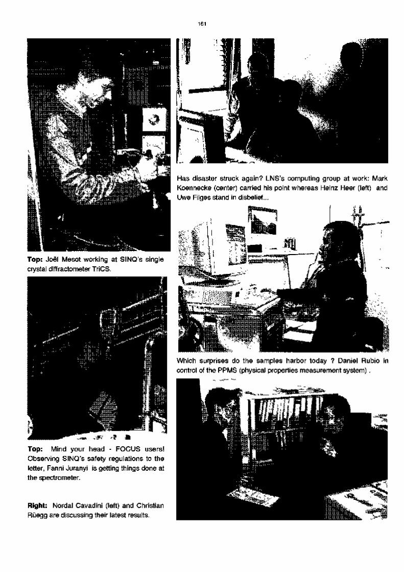
161
Has disaster struck again? LNS's computing group at work: Mark Koennecke (center) carried his point whereas Heinz Heer (left) and Uwe Filges stand in disbelief...
Top: Joel Mesot working at SINQ's single crystal diffractometer TriCS.
•***Vi"' ...
Top: Mind your head - FOCUS users! Observing SINQ's safety regulations to the letter, Fanni Juranyi is getting things done at the spectrometer.
Right: Nordal Cavadini (left) and Christian Ruegg are discussing their latest results.
D Which surprises do the samples harbor today ? Daniel Rubio in control of the PPMS (physical properties measurement system).

162


![3} tj- 7] - OSTI.GOV](https://static.fdokumen.com/doc/165x107/631c4c86b8a98572c10cd705/3-tj-7-ostigov.jpg)