Vaccines for preventing enterotoxigenic Escherichia coli (ETEC) diarrhoea

Journal of General Microbiology (1982), 128, 3037 3050. Printed in Great Brituin 303 7
Isolation, Properties and Nucleolytic Degradation of Chromatin from Escherichia coli
By K . SJASTAD, ' P. F A D N E S , ' P . G . K R U G E R , 2 I . LOSSIUS1 A N D K . KLEPPE1*
Departments of Biochemistry and Anatomy, University of Bergen, Bergen, Norway
(Received 4 February 1 Y82 ; .revised 16 Muy 1982)
A new procedure has been developed for the isolation of the chromosome complex, termed chromatin, from Escherichiu coli. The bacteria were subjected to low ionic strength and T4 lysozyme, followed by detergent treatment analogous to that employed for the isolation of eukaryotic chromosomes. The chromatin was an insoluble viscous material which contained approximately equal amounts of DNA and RNA. The protein content of the chromatin was almost three times greater than the nucleic acid content. Electron microscopy revealed that the chromatin was highly condensed, having multiple loops and beaded structures with various diameters. The chromatin could be completely solubilized by both micrococcal nuclease and DNAase I, whereas RNAase had no effect. The initial degradation by micrococcal nuclease resulted in the production of a DNA-protein particle, sedimentation coefficient lOS, and an RNA-protein complex of 24s. Further degradation led to a decrease in sedimentation coefficient of the DNA-protein complex, but not of the RNA-protein particle. The peak size of the DNA of the initial DNA-protein particle was approximately 2400 bp. The action of micro- coccal nuclease also resulted in the production of several discrete RNA species of various sizes. Several low molecular weight proteins (1 2000-27000) were found in the DNA-protein complex. The DNA-binding protein HU was present in the undigested chromatin; varying amounts of HU were, however, detected in the DNA-protein and RNA-protein particles.
I N T R O D U C T I O N
It is now well established that eukaryotic chromosomes possess well defined DNA-protein complexes termed nucleosomes (Kornberg, 1977). These may be packed further into more compact, higher order structures. The histone proteins are well characterized and the details of the interactions between DNA and the histones are being unravelled (Klug et al., 1980).
In the case of prokaryotic organisms several models for the organization of the chromosome have been presented (Kleppe et ul., 1979; Pettijohn, 1976; Worcel & Burgi, 1972). One major difficulty in interpreting the results obtained is the isolation procedure usually employed for the nucleoid. This involves opening the bacterial cells with large amounts of egg-white lysozyme, followed by treatments with ionic detergents in 1 M-NaCl and subsequent sucrose gradient centrifugations. Under such conditions most of the proteins bound to DNA in vivo will dissociate and consequently specific DNA-protein complexes are destroyed. Other artefacts may also arise (Kleppe etal . , 1979). Thus, in order to make accurate predictions about the in civo structure of the Escherichia coZi chromosome and the proteins and RNA species associated with it, the chromosome complex should be isolated using lower salt concentrations.
In the present work we have developed a new method for the isolation of the bacterial chromosome complex from E. coli at low ionic strength. T4 lysozyme is used to open the cells, followed by non-ionic detergent treatment and centrifugation. The latter conditions are similar to those employed for the isolation of eukaryotic chromatin (Hancock, 1974). The complex isolated is termed bacterial chromatin by analogy with eukaryotic systems. The complex does
Abbreviations: DTT, dithiothreitol; P M S F , phenylmethylsulphony1 fluoride.
0022-1287/82/0001-0395 $02.00 0 1982 SGM

3038 K . S J A S T A D A N D O T H E R S
not behave as individual chromosome particles, as obtained in the standard nucleoid procedure, but rather as a multi-chromosome aggregate. We feel that the term chromosome should only be used when individual chromosome particles can be isolated or described genetically or biochemically. The present study also deals with the properties and nucleolytic degradation of the bacterial chromatin from E. coli.
M E T H O D S
Reagents and enzymes. The isotopes [3H]thymidine and [5-3H]uridine were obtained from Amersham. Nonidet P-40 non-ionic detergent was obtained from Sigma. Micrococcal nuclease, DNAase I and RNAase were from Sigma. T4 lysozyme was a gift from Dr H. B. Jensen, Department of Biochemistry, University of Bergen. Norway; it had been purified as previously described by Jensen & Kleppe (1972). The protein gel markers were obtained from Bio-Rad Laboratories. The DNA markers, Hoe111 digested 4x174 RFI DNA, and Hind111 digested A DNA, were from New England Bio-Labs.
Organisms. Escherichia coli K12, DG75 (F-) thyleu was from the laboratory stock of the Department of Biochemistry, University of Bergen, Norway.
Proteins. HU protein and BHl protein were purified essentially as described by RouviZre-Yaniv & Gros (1975) and Varshavsky et al. (1977).
Growth oj'labelled bacteria. Escherichia coli K12 DG75 thy leu was grown in the following media. (a) L-broth, containing (per litre): 10 g tryptone, 5 g yeast extract, 10 g NaCl and 4 g glucose; pH 7.4. (b) M-9, containing (per litre): 1 g NH,Cl, 0.1 3 g MgClz. 6 H Z 0 , 3 g KH2P0,, 6 g Na,HPO,. 2H,O, 100 mg L-leucine, 20 mg thymine and 4 g glucose. The bacteria were grown to a turbidity ( A 6 s 0 ) of 0.8. When the bacteria were grown in M-9, 1 ml from an overnight culture in L-broth was added to 100ml M-9 medium. For radioactive labelling of DNA, ["]thymidine was added [250 pCi 1-' (9.25 MBq I- ' ) at A650 = 0.2, followed by 250 pCi 1- I a t A650 = 0.41. For radioactive labelling of RNA, [5-"H]uridine was employed.
Lysis of'bacterial cells. 3H-Labelled cells from one litre of medium were suspended in buffer A [20 mM-Tris/HCl pH 8.1, 50 mM-NaC1, 5 mM-EDTA, 0-5 mM-dithiothreitol and 10% (v/v) glycerol], final volume approximately 4 ml. Chloroform (25 pl).was added to the suspension to partially destroy the lipopolysaccharide membrane of the bacteria. The mixture was kept at 37 OC for 15 min. Excess chloroform was removed by gently bubbling air through the solution. T4 lysozyme was then added to a final concentration of 2 pg ml-' and the mixture incubated at 37 "C for 15 min. Lysis was completed by freezing and thawing the cells once.
Preparation qf' bacterial chromatin. Approximately 4 ml (1 vol.) buffer B [O-5 % (v/v) Nonidet P-40, 0.2 mM- EDTA and 0.1 mM-phenylmethylsulphonyl fluoride (PMSF), pH 7-51 was added to the lysate, carefully mixed, and left on ice for 1Omin. A sample (1 ml) of the viscous mixture was then layered on top of 10ml buffer C (100 mM-sucrose, 0.2 mM-KNa2P0, and 0.1 mM-PMSF, pH 7.5) in a 15 ml Corex centrifuge tube and centrifuged for 15 rnin at 5800 r.p.m., 4 "C, using an HB-4 rotor in a Sorvall RC5 centrifuge, After centrifugation the supernatant was carefully removed, the pellet resuspended in 1 vol. buffer B and left on ice for 10 min. It was then layered on top of 10ml buffer C and centrifuged again as above. This procedure was repeated four times (Hancock, 1974).
Determination oj'nucleic acids in chromatin. The isolated chromatin was diluted 10 times in TBE buffer (90 mM- Tris base, 90 ~ M - H , B O , and 2.5 mM-EDTA); NaCl and SDS were then added to final concentrations of 1 M and 1 "/, (w/v), respectively. One volume of chloroform/isoamyl alcohol (24 : 1, v/v) was then added, and the mixture shaken and centrifuged for 2 rnin in an Eppendorf centrifuge at 12000g. The nucleic acids were in the top layer and the ,4260 of the deproteinized chromatin was measured. The chromatin was diluted to a nucleic acid density corresponding to an AZbo of 20 before any further treatment.
Nuclease treatment. (a) Micrococcal nuclease. Various amounts of enzyme (see Results) were added to 400 1.11 chromatin (A260 = 20) in 10 mM-Tris/HCl pH 8.0,0.5 mM-CaCIZ, and the mixture incubated at 37 "C for different times. The reaction was stopped by addition of EDTA to a final concentration of 10 mM. Insoluble material was removed by centrifugation in an Eppendorf centrifuge (12000g for 2 min). One unit of micrococcal nuclease gives an increase in A260 of 1.0 in 30 min at pH 8-8, 37 " c in a 3.55 ml reaction volume, light path 1 cm. (b) DNAase I . Pancreatic DNAase I (200 units) was added to 400 pl chromatin in 10 mM-Tris/HCl pH 8.0,0.5 m~-CaCl , , 5 mM- MgCI: and the mixture incubated at 37 "C for 15 min. The reaction was stopped by adding EDTA to a final concentration of 10 mM. (c) RNAase. RNAase was added to 400 p1 chromatin in TBE buffer to give a final con- centration of 20pgml-', and the incubation carried out for 15min at 37°C.
Polyacrylamide gel electrophoresis of D N A and R N A . To determine the sizes of the nucleic acid fragments after micrococcal nuclease treatment, electrophoresis was carried out in a 4 or 5 % (w/v) polyacrylamide slab gel with 2.5% (w/v) N,N'-methylenediacrylamide (Maniatis ef al., 1975). The size of the slab gel was 18 x 13.5 x 0.1 cm. The gel was run in TBE buffer at a constant 180 V for 2 h, room temperature. The micrococcal nuclease digest of chromatin was deproteinized, and the nucleic acids were precipitated with ethanol and resuspended in 10-20 pl TBE buffer. Sample buffer (1-2 pl), consisting of 90 mM-Tris base, 90 ~ M - H ~ B O ~ , 2.5 mM-EDTA, 78% (v/v)
Chromatin jrom E. coli 3039
glycerol, 0.25% (wlv) xylene cyanol and 0.25% (w/v) bromophenol blue, was added to the nucleic acid sample and the mixture loaded on to the gel.
The gel was stained with ethidium bromide (2 pg ml-I) for 30 min and thereafter destained in TBE buffer for 30 min. Alternatively the gel was stained with methylene blue (0-02%, w/v) in 10 mM-Tris/acetate pH 8 . 3 for 30 rnin and destained in TBE buffer for 30 min essentially as described by Jeppesen (1980). The gel was then placed on a Whatman 3MM paper and dried under vacuum. The radioactivity in the gel was determined by cutting the dry gel into 5 mm pieces which were incubated with 750 pl H,Oz (30%, v/v) at 60 "C for 3 h. Thereafter, 10 ml aqueous scintillation liquid was added to each sample and the radioactivity determined in a Packard 460 scintillation counter.
Agarose gel electrophoresis of DNA. To determine the accurate size of the major DNA fragment, agarose gel electrophoresis was carried out using a 4 mm horizontal 10% (w/v) agarose gel (A-6877; Sigma) in TBE buffer. The electrophoresis was performed at a constant 6 V cm-' for 4.5 h. The samples were incubated for 15 min at 60 "C in sample buffer [ 1.25% (w/v) SDS, 6.25% (v/v) glycerol and 62.5 ng bromophenol blue ml-I, final concentrations] before application to the gel. Whatman 3MM chromatography papers (three layers) were used as wicks. The wet gel was cut into 5 mm pieces and the radioactivity determined as described above.
Sucrose gradient centrifugation. Nucleic acid-protein complexes were separated on a 5.0-28-8 % (w/v) isokinetic sucrose gradient containing 1 m ~ - E D T A , pH 7.5. The gradient volume was 11 ml and a 100 pl sample was layered on the top. Centrifugations were carried out in a Spinco L-70 ultracentrifuge using a SW 41 Ti rotor and run at 36000 r.p.m. for 18 h, 4 "C. The gradient was harvested from the bottom of the tube in 370 pl fractions, The radio- activity in the fractions was determined using aqueous scintillation liquid. The Atso was measured using a Pharmacia UV 1 single-path monitor, fitted with a 10 mm flow cell.
Determination of protein, DNA and RNA. Protein was determined according to the procedure of Sedmark & Grossberg (1977). The DNA content was estimated by the diphenylamine method (Burton, 1956) and RNA by the orcinol procedure. An alternative method for determination of nucleic acids in the nuclease digest was also used. In this case the sample was first deproteinized and the AZbo and Azso values recorded. The sample was then treated with RNAase (50pgml-') at 37°C for 15 min, the DNA precipitated with cold ethanol and the AZbo and values again measured. By this procedure both DNA and RNA could be determined from the absorbance values. Since the DNA was 3H-labelled, the loss of DNA in each step was corrected by monitoring the radioactivity in the sample.
Isolation of calf thymus nuclei and preparation oJ nucleosomes. Fresh or frozen ( - 80 "C) calf thymus was used. Connective tissue and blood veins were removed and the thymus was cut into small pieces. Three volumes of homogenization buffer (H buffer; 0.25 M-SUCTOSe, 10 mM-Tris/HCl pH 7.4, 6 ~ M - K C I , 5 mM-magnesium acetate and 0.1 mM-EGTA) supplemented with 1 mM-PMSF, were added (Wallace et al., 1977). The mixture was homogenized with two strokes in a Teflon homogenizer. H buffer (9 vol.) was added to the homogenate and the mixture sieved through two layers of cheesecloth. The nuclei were then pelleted by centrifugation at 280g, 4 "C, for 7min and the pellet was resuspended in 10vol. H buffer supplemented with 0.1 ~ M - P M S F and 0.5% (v/v) Nonidet P-40. The nuclei were then centrifuged and the pellet was washed with 1Ovol. H buffer and again pelleted. The purified nuclei were resuspended in H buffer and the concentration was adjusted to 3 x lo8 nuclei ml-l. One volume of 87% (v/v) glycerol, 5 mwmagnesium acetate was added to this suspension and the nuclei were kept at - 20 "C.
The micrococcal nuclease treatment was carried out directly on the nuclei or on the purified chromatin. The eukaryotic chromatin was prepared essentially as described for E. coli chromatin. Micrococcal nuclease (1 50 units) was added to 1.5 x lo8 nuclei in 10 mM-Tris/HCI pH 8.0,0.5 mM-CaClz and the mixture incubated for 10 min at 37 "C. The reaction was then stopped by adding EDTA to a final concentration of 10 mM. The digested nuclei were homogenized with 10 strokes in a Teflon homogenizer. Insoluble material was removed by centrifugation at 10000r.p.m., 4"C, for 30min using an SS34 rotor in a Sorvall RC5 centrifuge.
Preparation of chromatin for electron microscopy. Appropriate dilutions of indigested and digested chromatin were fixed in 1 % (v/v) glutaraldehyde. A 5 pl sample of chromatin was placed on a 200 mesh carbonated grid and the excess carefully removed. The grids were then stained with 1 % (w/v) uranyl acetate and rinsed in distilled water before examination in a JEOL 100 CX electron microscope.
R E S U L T S
Preparation of bacterial chromatin Chromatin from E. coli was prepared as described in Methods. The procedure, which is
simple and gives reproducible results, involves the use of T4 lysozyme, rather than egg-white lysozyme, to digest the peptidoglycan layer. The E. coli chromatin obtained was an insoluble viscous material which sedimented easily and could be washed repeatedly. The composition of the chromatin in terms of protein and nucleic acids is given in Table 1. The amount of protein present was almost three times greater than the nucleic acid content, with the RNA content

3040 K . S J A S T A D A N D O T H E R S
Table 1 . DNA, RNA and protein content of chromatin digested by micrococcal nuclease
The nucleic acid concentration in the chromatin was adjusted to an AZbo of 20 and 50 units micrococcal nuclease ml-' was added. The mixture was incubated for 15 min at 37 "C and the DNA, RNA and protein content was analysed in the supernatant after centrifugation at 12000g for 2 min.
Con tent Constituent (mg ml- )
DNA 0.30 RNA 0.4 1 Protein 2-05
Fig. 1 . (a, 6) Electron micrographs of undigested chromatin. The arrows indicate DNA loops.

Chromatin .from E. coli 304 1
approximately the same as that of the DNA. The RNA content was also found to be somewhat dependent on the extent of washing of the chromatin and on the centrifugation conditions. Several preparations were examined in the electron microscope using the same procedure as employed for eukaryotic chromosomes and nucleosomes. The conventional Kleinschmidt technique could not be used here as the high salt and cytochrome c concentration destroys the protein-nucleic acid complexes. All electron micrographs showed the essential absence of membranes, ribosomes, cell wall fragments and unlysed cells. Two different complexes were detected, a loose complex (Fig. 1 a) and a more compact structure (Fig. 1 b). The latter form were the dominant species. Beaded fibres with diameters varying from 20 to 150 nm were observed in both complexes. No repeating bead structures were detected. Loops up to 2 pm in length and extending from a main chain could also be seen. Most of the loops had lengths between 0-4 and 0.8 pm. Larger complexes than the ones shown were also found in preparations from undigested chromatin. Some of the side chains must obviously be RNA-protein complexes.
Digestion by micrococcal nuclease
The bacterial chromatin was readily digested by micrococcal nuclease under essentially the same conditions as for the degradation of eukaryotic chromatin (Noll, 1974). In our studies insoluble undigested material was removed by a brief centrifugation (12OOOg for 2 min) and the supernatant analysed for the presence of DNA, RNA and proteins. Virtually all the [3H]- thymidine-labelled DNA present in the chromatin was released into the supernatant following digestion with micrococcal nuclease at 25 units ml- ; at lower enzyme concentrations less material was liberated (Table 2). The time course of the degradation of DNA into acid-soluble products using three different concentrations of micrococcal nuclease is shown in Fig. 2. With an
Table 2. Solubilization of' chromatin by micrococcal nuclease
The nucleic acid concentration in the chromatin was adjusted to an AZbo of 20, in Tris/HCl pH 8.0, 0.5 mM-CaC12. Various amounts of micrococcal nuclease were added. The mixtures were incubated for 15 min at 37 "C and the radioactivity in the supernatants was determined after centrifugation at 12000g for 2 min.
Percentage of Enzyme concentration "-labelled DNA
(units rn1-I) in the supernatant
0 12.5 25
125
2 56 78 98
I I I I
5 10 15 20 Incubation time ( m i n )
Fig. 2. Kinetics of micrococcal nuclease digestion. Chromatin ( A ? ( , , , = 20) was treated with 12.5 (A), 25 (0) or 125 (0) units enzyme ml-I. The reaction conditions were as described in Methods. The conditions used for precipitation of nucleic acids and proteins with trichloroacetic acid were as previously described (Kleppe el d., 1971 ).

3042 K . S J A S T A D A N D O T H E R S
I I I I I I
A
0.4
0.3 OD N
-T
0 .2
0.1
5 10 15 20 25 30 Fraction no.
0.4
0.3 0
N
T
0-2
0. I
0.4
0.3 0 m N
T
0.2
0 .1
I I I I I I
B
5 10 15 20 25 30 Fraction no.
I I I I I I
C
/
I I I I
5 10 15 20 25 30
8: 2 4
x 6 .-
Y 0
0 .- 4 2 d X
rn
2 2
Fraction no. Fig. 3. Sucrose gradient centrifugations of chromatin from bacteria grown in M-9 medium and digested with micrococcal nuclease. 0 , 0, radioactivity in 3H-labelled DNA. Chromatin was treated with 12.5 units enzyme ml-' (gradient A), 25 units enzyme ml-I (gradient B) or 125 units enzyme ml-' (gradient C). The enzyme digestion and centrifugation conditions were as described in Methods; incubation time was 15 min (37°C).
Table 3 . AZ6O : AZso ratio in the RNA-protein and DNA-protein complexes from sucrose gradients
Fractions 4-8 (RNA-protein) and fractions 15-20 (DNA-protein) from gradient A (Fig. 3) and fractions 3-6 (RNA-protein) and 20-24 (DNA-protein) from gradient C (Fig. 3) were pooled. The A,, , and values were measured against appropriate blanks.
A 2 6 0 : A 2 8 0
Gradient Complexes ratio
RNA-protein 1.83 DNA-protein 1.7 1 RNA-protein 1.74 DNA-protein 1.68
enzyme concentration of 125 units ml-l approximately 90% of the DNA had become acid soluble after 20 min. Using a 10-fold lower concentration of enzyme (12.5 units ml-l), a plateau corresponding to approximately 25 % acid solubility was seen after the same period.
The products of the micrococcal nuclease action released into the supernatant were subjected to neutral sucrose gradient centrifugation. The conditions used were again similar to those

Chromatin ,from E. coli 3043
Fig. 4. Electron micrograph of the DNA-protein complex from sucrose gradient A (Fig. 3). fractions 15-20.
2L
v a* I I I 5 10
1 1 1 1 350 bp
I I
5 10 15 Migration (cm)
Fig. 5. (a) Agarose gel electrophoresis of chromatin (A260 = 20) digested with 25 units micrococcal nuclease m1-I. The chromatin was deproteinized and treated with RNAase before application to the gel. The arrows indicate the mobility of 3, DNA fragments produced by Hind111 digestion. (b) Polyacryl- amide gel (4%, w/v) electrophoresis of chromatin (A260 = 20) digested with 125 units micrococcal nuclease ml-I. The chromatin was deproteinized and treated with RNAase before application to the gel. The arrows indicate the mobility of 4x174 RFI DNA fragments produced by HaeIII digestion.
employed for separation of micrococcal nuclease products of eukaryotic chromatin (Noll, 1974). The results for three different enzyme concentrations employed are shown in Fig. 3. In all three cases the main absorbance peaks at 280nm were found near the bottom of the tubes, corresponding to a sedimentation coefficient of 24s. The main radioactive DNA peaks, on the other hand, were detected in the middle of the tubes and upwards and had sedimentation coefficients of approximately lOS, 9s and 7s in the three gradients. The 260 : 280 nm absorbance ratios of the bottom peaks and the radioactive DNA peaks were measured in gradients A and C (Table 3); the results suggest that both peaks are nucleic acid-protein complexes. The bottom peak, 24S, was an RNA-protein complex. This was confirmed by labelling the chromatin with [5-3H]uridine followed by digestion with micrococcal nuclease, and sucrose gradient centrifugation (results not shown).
3044 K . S J A S T A D A N D O T H E R S
When 35S-labelled chromatin was treated with micrococcal nuclease and then subjected to sucrose gradient centrifugation, peaks of radioactivity were seen at the position of both the RNA-protein and DNA-protein complexes in addition to the top of the gradient (results not shown). Little radioactivity was detected on the shelf, confirming the view that membranes were mostly absent. The 35S-labelled DNA-protein complex was also treated with DNAase and subjected to gel chromatography on a Sepharose column. The protein peak had a higher elution volume than the protein-DNA complex, clearly showing that the DNA and the protein exist as a complex and do not simply co-sediment (results not shown).
The DNA-protein complexes obtained were examined by electron microscopy ; and an
1373
872
603
234
118
7 2
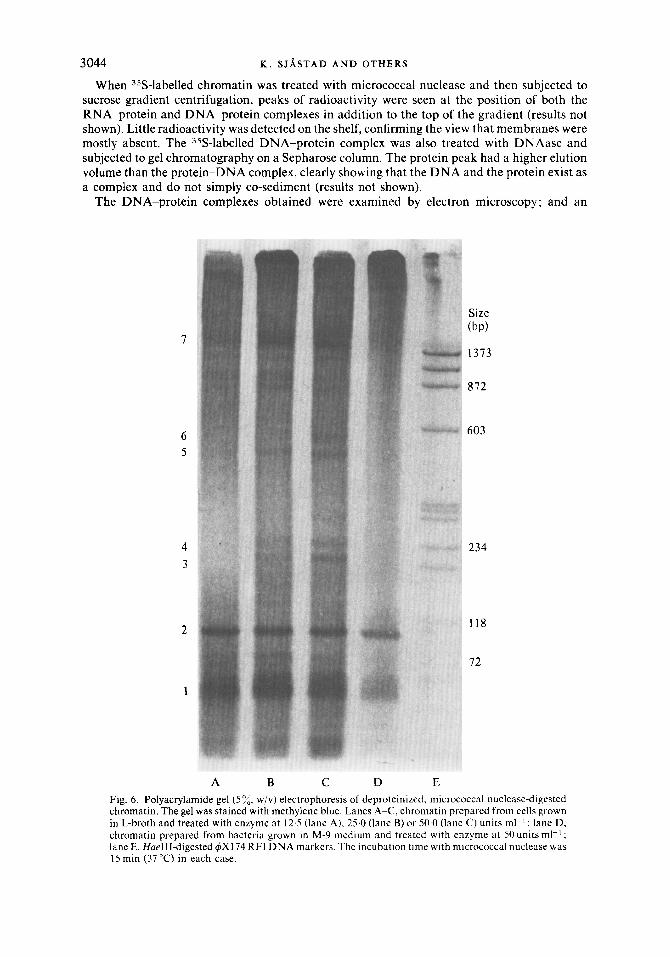
A B c D E Fig. 6. Polyacrylamide gel ( 5 y;, w/v) electrophoresis of deproteinized, micrococcal nuclease-digested chromatin. The gel was stained with methylene blue. Lanes A-C, chromatin prepared from cells grown in L-broth and treated with enzyme at 12.5 (lane A), 25.0 (lane B) or 50.0 (lane C) units ml-' ; lane D, chromatin prepared from bacteria grown in M-9 medium and treated with enzyme at 50 units ml-' ; lane E, HueIII-digested 4x1 74 RFI DNA markers. The incubation time with micrococcal nuclease was 15 min (37 "C) in each case.

Chromatin .from E. coli 3045 example of the 10s complex is shown in Fig. 4. Elongated particles approximately 0.8 pn in length with a fibre diameter of approximately 20-30 nm were detected.
The sizes of the DNA fragments in the digested chromatin were deduced from agarose and polyacrylamide gel electrophoresis using appropriate DNA markers (with the radioactivity in the gel determined as described). The digested chromatin was deproteinized and the nucleic acids precipitated with ethanol prior to electrophoresis. The peak size of the DNA was found to be 2400 bp when a low concentration (25 units ml-l) of micrococcal nuclease was used (Fig. 5 0 ) . At an enzyme concentration of 125 units m1-l the DNA was more spread out on the gel, having a peak size of 350 bp (Fig. 5b).
The nucleic acid components in the gel were also detected by staining with methylene blue or ethidium bromide. An example of a gel stained with methylene blue is shown in Fig. 6 (Jeppesen, 1980). In chromatin from bacteria grown in L-broth as well as minimal medium,
A B C Fig. 7. Polyacrylamide gel (5%, w/v) electrophoresis of chromatin digested with 25 units micrococcal nuclease ml-l, deproteinized and treated with RNAase. The gel was stained with methylene blue. Lane A, deproteinized chromatin treated with 20 pg R N Aase ml-' for 15 min at 37 "C ; lane B, deproteinked chromatin treated with 2Oyg RNAase ml-' for 1 min at 37 " C ; lane C , micrococcal nuclease-digested calf thymus chromatin in which the sample was deproteinized and precipitated with ethanol prior to electrophoresis; the lower band corresponds to D N A from mononucleosomes (145 bp) and the others correspond to DNA from dinucleosomes. trinucleosomes, etc.

3046 K . S J A S T A D A N D OTHERS
three major RNA bands were seen. Bands 1 and 2 appeared immediately after addition of micrococcal nuclease. The sizes of the RNA species were determined from the DNA markers using the relationship between mobilities of native denatured DNA and RNA obtained by Maniatis et al. (1975). For the three major RNA species (bands 1, 2 and 7) the lengths were estimated to be approximately 70, 100 and 1000 nucleotides, respectively. In addition to the major RNA bands four minor RNA species (bands 3, 4, 5 and 6) were also found in the micrococcal nuclease-digested chromatin from bacteria grown in L-broth, but not from those grown in minimal medium. The sizes of these were approximately 180, 200, 320 and 350 nucleotides, respectively. The fact that these bands were RNA was confirmed by digestion of the samples with RNAase prior to gel electrophoresis (Fig. 7). Treatment of the sample for 15 min at 37 "C (lane A) completely eliminated the RNA bands, whereas only the major DNA band was visible. When the sample was incubated with less RNAase for shorter periods (lane B), small molecular weight RNA oligonucleotides at the bottom were clearly seen in addition to the DNA band. Several control experiments with micrococcal nuclease were carried out using digestion of eukaryotic chromatin. Nuclei from calf thymus were employed and the hydrolysis conditions were identical to those for prokaryotic chromatin. Mononucleosomes, dinucleo- somes, etc. were produced as expected (lane C).
Size (kDal)
94
68
43
30
21
14.3
10
A B C D E Fig. 8. SDS-PAGE gel electrophoresis (15%, w/v polyacrylamide) of DNA-binding proteins. Lane A, Bio-Rad molecular weight markers; lane B, HU protein; lane C, BH1 protein; lane D, total protein from bacterial chromatin; lane E, proteins in the DNA-protein peak from sucrose gradient B (Fig. 3).

Chromatin .from E. coli 3047 A separate control experiment was also carried out using deproteinized E. coli chromatin. In
this case no distinct DNA or RNA species were detected, in contrast to the results for intact chromatin.
Digestion of E. coli chromatin with pancreatic DNAase I and RNAase
The DNA in the chromatin was readily digested with pancreatic DNAase I. However, the discrete bands of RNA were not as apparent as those produced by micrococcal nuclease. Several experiments were carried out studying the digestion of intact E. coli chromatin with pancreatic RNAase. Virtually no UV-absorbing material was detected in the supernatant after incubation for prolonged periods, showing that pancreatic RNAase does not act on intact chromatin to any extent.
Analysis of the proteins present in the chromatin
The protein species present in the intact chromatin and in the micrococcal nuclease digested products were analysed by means of SDS-PAGE essentially as described by Laemmli & Favre (1973). The results (Fig. 8) show the total chromatin proteins as well as the proteins in the DNA peak. Up to 30 different protein bands were detected in the total chromatin, their molecular weights ranging from 10000 to approximately 100000. the seven major species were, however, of low molecular weight (10000-27000). Several low molecular weight (1 2 000-27 000) protein bands were found in the DNA-protein complex isolated by sucrose gradient centrifugation. Only small amounts of the HU protein (Rouvikre-Yaniv & Gros, 1975) were detected in both this complex and the RNA-protein particle. The amount of HU protein present in the DNA- protein complex, however, varied somewhat from one preparation to another. A band corres- ponding to the BH1 protein (Varshavsky et al., 1977) was seen in the DNA-protein particle. Protein bands corresponding to the p’ and p subunits of RNA polymerase were also detected in the chromatin, but only as minor species. A histone-like protein with a molecular weight of 28 000 has been isolated from E. coli by Hubscher et al. (1980) and it might also be present in the DNA-protein complex.
DISCUSSION
The present work deals with the isolation, properties and nucleolytic degradation of chromatin from E. coli. An important aspect in the isolation procedure was the use of T4 lysozyme. In the standard procedure for the lysis of E. coli cells, egg-white lysozyme is normally employed (Korch et al., 1976), with high concentrations of enzyme needed. Egg-white lysozyme, being a basic protein, will, at normal physiological ionic strength, bind to nucleic acids and may thus even displace other DNA- and RNA-binding proteins. Insoluble nucleic acid-egg-white lysozyme aggregates can also easily form. At higher ionic strength (1 M-NaC1) such as that employed for the isolation of the nucleoid (Korch et al., 1976), lysozyme and most other DNA binding proteins are not bound to DNA. According to Jensen & Miron (1980), T4 lysozyme is approximately 400 times more efficient than egg-white lysozyme in hydrolysing the peptidoglycan layer. Only small amounts of this enzyme are therefore required (2 pg ml-l vs 800 pg ml-l) and this does not significantly affect the chromatin structure. The subsequent isolation procedure used for the bacterial chromatin was mostly identical to one of several methods commonly used for the isolation of chromatin from eukaryotic cells.
In many ways the properties of the chromatin isolated in this study are in agreement with the recently postulated model of the structure of the in vivo nucleoid (Kleppe et a/., 1979). In the earlier model of the E. coli chromosome (Pettijohn, 1976; Worcel & Burgi, 1972), the so-called clover leaf structure, multiple (lOCrl50) supercoiled DNA loops were presumed to radiate from a common centre and to be held together in the centre by RNA molecules. Several lines of evidence suggest, however, that the RNA stabilization may be an artefact of high salt concentration or action of detergents. A more thorough discussion of these aspects is given by Kleppe et al. (1979). DNA loops are also central to the new model but here they extend mostly into the cytoplasm and along the membranes and have nascent RNA chains with ribosomes

3048 K . S J A S T A D A N D O T H E R S
bound to them. During the present isolation procedure the ribosomes dissociate from the RNA. The DNA loops observed in the present work (1-2 pm) are smaller than those detected in the nucleoid in high salt concentration (10-20 pm); however, the former loops are more condensed. The highly condensed structures seen in the electron microscope resemble those observed by Materman & van Goo1 (1978) (which were isolated by different methods) with regard to both the shape and the diameters of beaded fibres. The structures observed in the present work differ markedly from those observed by Griffith (1976). Here the fibres emanating from partially disrupted E. coii cells had a diameter of 12 nm and a repeating bead structure of 13 nm. In the latter experiments, however, the detergent and ionic conditions were different from those used in the present study.
The RNA found in the E . coli chromatin in the the present work is presumably nascent RNA similar to that detected in the bacterial nucleoid isolated in high salt (Pettijohn, 1976; Pettijohn & Hecht, 1973). In contrast to that in the nucleoid, the KNA in the intact chromatin was not attacked by pancreatic RNAase, suggesting that it is protected by a layer of protein. Digestion with micrococcal nuclease on the other hand led to an almost immediate release of most of the RNA from the chromatin and, moreover, the enzyme produced a number of distinct RNA species which after deproteinization could be separated on polyacrylamide gels. Apart from this immediate hydrolysis, the RNA-protein complex seemed to be attacked at a slower rate than the DNA-protein complex. Micrococcal nuclease is an enzyme which works equally well on RNA and DNA and has both endonucleolytic and exonucleolytic activities (Laskowski, 1961). Two of the major RNA species observed were similar in size to tRNA and precursor tRNA. It is possible that these and the other RNA species produced are degradation products of precursor RNA molecules such as precursor rRNA. The cleavage of chromatin-associated RNA by micrococcal nuclease but not by pancreatic RNAase could be due to micrococcal nuclease catalysing hydrolysis of DNA at specific sites prior to RNA cleavage. This would allow the chromatin structure to open up, thus making the RNA more accessible to the enzyme. Little information is currently available about the nature of the association between DNA and RNA but presumably the same type of binding as observed in the nucleoid could exist, namely via RNA polymerase bound to DNA, or part of the RNA being in a DNA-RNA hybrid form (Pettijohn, 1976).
The DNA-protein products obtained by the action of micrococcal nuclease differed in size and nature from those produced during hydrolysis of eukaryotic chromatin under identical conditions. No nucleosome-type structures were produced. These results clearly show that the organization of the prokaryotic chromatin is different from that in the eukaryotic cell. In the initial action of micrococcal nuclease, DNA-protein particles were produced in which the DNA had a peak size of approximately 2400bp. From the sedimentation properties it can be estimated that the DNA-protein particle must be of a rod-type shape, and not spherical, since a DNA molecule of 2000 bp alone would have a sedimentation coefficient of approximately 10s. There is good correspondence between this model, the actual electron microscopic data obtained and the expected length of such a DNA molecule. In addition the DNA in such a particle could be partially supercoiled, hence the DNA particle may in fact constitute a loop in the chromatin. Further degradation led to a decrease in size of the DNA and an increase in the amount of low molecular weight DNA on the top of the sucrose gradient. This could be explained by assuming that the initial particles produced are the result of the endonucleolytic activity of the enzyme whereas further breakdown is due mostly to the exonucleolytic activity of the enzyme. A similar mechanism has been postulated for other DNA substrates and also for eukaryotic chromatin (Noll, 1974). Micrococcal nuclease has a greater activity in single-stranded than on double- stranded DNA and, moreover, it has a preference for AT sequences flanked by GC-rich sequences (Horz & Altenburger, 1981 ; Laskowski, 1961). Thus, the initial cut made could be governed by the above factors. It is also interesting to note that the DNA fragments initially produced are about the size of an average gene. In many cases AT-rich sequences are found at the operator and termination sites of genes. An alternative explanation of the unique size of the initial DNA fragments could of course be that certain sites are not protected by DNA-binding proteins. This view is supported by the finding that digestion of deproteinized chromatin with

Chromatin from E. coli 3049
micrococcal nuclease did not give rise to distinct DNA species. Varshavsky et al. (1977) reported that a minimum-sized DNA fragment of approximately 120 bp was produced by the action of micrococcal nuclease. We have not been able to detect a fragment of 120 bp or multimers thereof. It should be noted however that we used different procedures to prepare the chromatin.
The analysis of the proteins of the DNA-protein complex revealed the presence of several proteins and several minor protein components. Previously, three DN A-binding proteins have been characterized, namely HU (Rouviere-Yaniv & Gros, 1975), BH1 (Varshavsky et al., 1977) and a protein of molecular weight 28000 (Hiibscher et al., 1980). The HU protein is found associated with nucleoids isolated in low salt, and it alone can give rise to nucleosome-like structures in the presence of relaxed circular DNA. However, recent studies have suggested that it is a more general nucleic acid-binding protein since it is also found associated with ribosomes (Berthold & Geider, 1976). In the present work only small amounts of HU were found in the DNA-protein and RNA-protein particles but it was present in larger concentrations in the undigested chromatin. It is possible, therefore, that HU could be a type of linker protein analogous to histone H1 in eukaryotic cells, or may bind to low molecular weight RNA species (Noll, 1977). The two other proteins, BHl and the histone-like protein of molecular weight 28000, are most probably present in the DNA-protein particle. Studies of the properties and function of the DNA-binding proteins described in this work are currently in progress in this laboratory. We believe that these proteins play a central role in the organization of the prokaryotic chromosome, possibly in the condensation of the DNA into rod domains and in the organization of the DNA loops.
This study was supported by grants from the Norwegian Research Council for Science and Humanities.
R E F E R E N C E S
BERTHOLD, V. & GEIDER, K. (1976). Interaction of DNA with DNA-binding proteins. European Journal of Biochemistry 71, 443449.
BURTON, K . (1956). A study of conditions and mechanism of the diphenyl reaction of colorimetric estimation of deoxyribonucleic acid. Biochcmicul Journal 62, 3 15-323.
GRIFFITH, J . D. (1976). Visualization of prokaryotic DNA in a regulatory condensed chromatin-like fiber. Proceedings ofthe National Academy u j Sciences qf ' the United States of America 63, 563-567. ANCOCK, R. (1974). Interphase chromosomal deoxy- ribonucleoprotein isolated as a discrete structure from cultured cells. Journal of' Moleculur Biology 86,
ORZ, W . & ALTENBURGER, W . (1981). Sequence specific cleavage of DNA by micrococcal nuclease. Nucleic Acids Research 9, 2643-2658.
HUBSCHER, U., LUTZ, H . & KORNBERG, A . (1980). Novel histone H2A-like protein from E. cdi . Pro- ceedings qf the National Academ), uj ' Sciences of ' the C'nited States of America 77, 5097-5 101.
JENSEN, H . B. & KLEPPE, K. (1972). Studies on T, lysozyme. Affinity for chitin and the use of chitin in the purification of the enzyme. European Journul of Biochemistry 26, 305-3 12.
JENSEN, H. B. & MIRON, T. (1980). Preparation and properties of insoluble forms of bacteriophage T, lysozyme and chicken egg white lysoryme. JourFlai o f Solid-Phuse Biochemistry 5, 45-60.
JEPPESEN, P. G. (1980). Separation and isolation of DNA fragments using linear polyacrylamide gradi- ent gel electrophoresis. Methods in Enzymologj. 65,
649-663.
305-319.
KLEPPE, K., OHTSUKA, E. , KLEPPE, R.. MOLINEUX, I . & KHORANA, H. G. (1971). Studies on polynucleotides. XCVI. Repair replication of short synthetic DNAs as catalyzed by DNA polymerases. Journul uf ' Molecular Biology 56, 34 1-36 1.
KLEPPE, K., ~ V R E B B , s. & LOSSIUS. I . (1979). The bacterial nucleoid. Journal of General Microbiology
KLUG, A,, RHODES, J . , SMITH, J., FINCH, J. T. & THOMAS, J . 0. (1 980). A low resolution structure for the histone core of the nucleosome. Nature, London
KORCH, c., 0VREB0, s. & KLEPPE, K. (1976). Envel- ope-associated folded chromosomes from Escheri- chiu coli: variations under diffferent physiological conditions. Journal of Bacteriology 127, 904-9 16.
KORNBERG, R. D. (1977). Structure of chromatin. Annual Reriew of' Biochemistry 46, 93 1-954.
LAEMMLI, U . K. & FAVRE, M. (1973). Maturation of the head of bacteriophage T,. Journal of Molecular Biology 80, 575-599.
LASKOWSKI, M. (1961). Deoxyribonucleases. In The En;ymes, pp. 123-147. Edited by P. D. Boyer, H. Lardy & K. Myrback. New York: Academic Press.
MANIATIS. T.. JEFFREY, A. &VAN DE SANDE, H. (1975). Chain length determination of small double- and single-stranded DNA molecules by polyacrylamide gel electrophoresis. Biochemistry 14, 3787-3794.
MATERMAN. E. c. &VAN GOOL, A. P. (1978). Compact Eschuichia coli nucleoids in a highly supercoiled con- formation. Journal of Bacteriology 135, 703-706.
NOLL, M. (1974). Subunit structure of chromatin. Nature, London 251, 249-25 1.
112, 1-13.
287, 509-5 16.
3050 K . S J A S T A D A N D O T H E R S
NOLL, M. (1977). The linkage of chromatin subunits and the role of histone. 1. In Nucleic Acid-Protein Recognition, pp. 139-150. Edited by H. J . Vogel. New York: Academic Press.
PETTIJOHN, D. E. (1976). Prokaryotic DNA in nucleoid structure. CRC Critical Reciews in Biochemistry 4,
PETTIJOHN, D. E. & HECHT, R. (1973). RNA molecules bound to the folded bacterial genome stabilize DNA folds and segregate domains of supercoiling. Cold Spring Harbor Symposia on Quantitatizre Biology 38,
ROUVIERE-YANIV, J. & GROS, F. (1975). Characteriza- tion of a novel, low-molecular weight DN A-binding protein from Escherichia coli. Proceedings of the National Academy of Sciences of the United States of America 72, 3428-3432.
175-202.
31-42.
SEDMARK, J . J . & GROSSBERG, S. E. (1977). A rapid, sensitive and versatile assay for protein using Coomassie blue G250. Analytical Biochemistry 79,
VARSHAVSKY, A. J., NEWSPASOV, S. A., BAKAYEV, V. V., BAKAYEVA, T. G. & GEORGIEV, G. P. (1977). Histone-like proteins in the purified E. coli deoxy- ribonucleoprotein. Nucleic Acids Research 4, 2725- 2745.
WALLACE, R. B., SARGENT, T. D., MURPHY, R. F. & BONNER, J. (1977). Physical properties of chemically acetylated rat liver chromatin. Proceedings of the National Academy of Sciences of the United States of America 14, 3244-3248.
WORCEL, A. & BURGI, E. (1972). On the structure of the folded chromosome of Escherichia coli. Journal of Molecular Biology 71, 127-147.
544-552.
Copyright © 2022 FDOKUMEN