Iontophoresis - An Approach for Controlled Drug Delivery: A Review
10
Current Drug Delivery, 2007, 4, 1-10 1 1567-2018/07 $50.00+.00 © 2007 Bentham Science Publishers Ltd. Iontophoresis - An Approach for Controlled Drug Delivery: A Review Nitin Dixit, Vikas Bali, Sanjula Baboota*, Alka Ahuja and Javed Ali Department of Pharmaceutics, Faculty of Pharmacy, Jamia Hamdard, (Hamdard University), New Delhi-110062, India Abstract: The recent approval of lidocaine hydrochloride and epinephrine combined iontophoretic patch (Lidosite ® Vys- teris Inc.) for localized pain treatment by FDA has invigorated the gaining interest in iontophoretic drug delivery systems for the transdermal delivery of drugs. This technique of facilitated movement of ions across a membrane under the influ- ence of an externally applied electric potential difference, is one of the most promising physical skin penetration enhanc- ing method. The rationale behind using this technique is the capability of this method to increase the systemic delivery of high molecular weight compounds with controlled input kinetics and minimum inter-subject variability, which is other- wise achieved only when parentral route of administration is used. Recently, good permeation of larger peptides like insu- lin has been achieved through this technique in combination with chemical enhancers. This review briefly describes the factors which affect iontophoretic drug delivery and summarizes the studies conducted recently using this technique in or- der to achieve higher systemic absorption of the drugs having low passive diffusion otherwise. The effect of permeation enhancers (chemical enhancers) on iontophoretic flux of drugs has also been described. Present review also provides an insight into reverse iontophoresis. Various parameters which affect the transdermal absorption of drugs through iontopho- resis like drug concentration, polarity of drugs, pH of donor solution, presence of co-ions, ionic strength, electrode polar- ity etc. have also been reviewed in detail. Keywords: Iontophoresis, electric current, potential difference, transdermal, permeation enhancers. INTRODUCTION The skin has been investigated for several decades as a route of drug administration and so far many drug delivery techniques which utilize alternative forms of energy have been explored to facilitate permeation of drugs across the skin. Amongst these, iontophoresis, which is the facilitated movement of ions across a membrane under the influence of an externally applied small electrical potential difference (0.5 mA/cm 2 or less), is one of the most promising novel drug delivery system, which has proved to enhance the skin penetration and the release rate of a number of drugs having poor absorption/permeation profile through the skin [1-4]. It is a localized, non-invasive, convenient and rapid method of delivering water soluble, ionized medication into the skin. This technique has already been reviewed by Banga and Chien [5] and Banga et al. [6]. Recently it was reviewed by Kalia et al. [7]. Wang et al., described an overview of im- proved permeation of hydrophilic as well as the lipophilic drugs using iontophoreis in combination with several strate- gies like permeation enhancers, sonophoresis etc. [8]. Ionto- phoresis provides the usual advantages of a trandermal route like, therapeutic efficacy improvement by bypassing hepatic “first pass” metabolism, avoidance of inconvenience caused by parenteral drug delivery and prevention of variation in the absorption seen with oral administration. Besides this, it also reduces the chance of dosing variation by providing pro- grammed delivery of the drug. Iontophoresis also provides a therapeutic regimen leading to better patient compliance. It *Address correspondence to this author at the Department of Pharmaceutics , Faculty of Pharmacy, Jamia Hamdard, (Hamdard University), New Delhi- 110062, India; Tel:/Fax: 26059663; E-mails: [email protected] or [email protected] permits the use of a drug with a short biological half life since the drug is delivered to the target area without the need to recirculate in the blood. Moreover, the drug is delivered into the bloodstream directly without any delay. It also pro- vides a rapid termination of the effect by turning off the ion- tophoretic delivery system. Thus, because of many advan- tages associated with this system, it has been an area of growing interest in the local and the systemic delivery of drugs. Iontophoresis is gaining wide popularity in the area of pain relief as it provides a non invasive means of systemic administration of minute amount of drugs. The potential of this technique has been exploited for the transdermal deliv- ery of many drugs with poor penetration properties e.g., high molecular weight electrolytes such as proteins, peptides and oligonucleotides which are normally difficult to administer except through parenteral route. It also offers a great poten- tial for the delivery of charged peptides used as drugs. Al- though iontophoresis has been able to achieve significant increase in the transdermal absorption of many drugs, it has not been able to show significant permeation of larger pep- tides like insulin. This has lead to many studies involving the use of various chemical enhancers (permeation enhancers) along with iontophoresis [9]. Such combination approaches have been found to significantly improve the absorption of insulin and many other drugs, which could not be delivered using ionotphoresis alone. The permeation rate of thiocolchi- cocide, a muscle relaxant used in the treatment of orthopedic, traumatic and rheumometallogic disorders, which can not permeate the skin easily due to high molecular weight and low octanol /water partition coefficient, has been enhanced using iontophoresis in combination with chemical enhancer like lauric acid. This produced a significant increase in the
-
Upload
independent -
Category
Documents
-
view
2 -
download
0
Transcript of Iontophoresis - An Approach for Controlled Drug Delivery: A Review
Current Drug Delivery, 2007, 4, 1-10 1
1567-2018/07 $50.00+.00 © 2007 Bentham Science Publishers Ltd.
Iontophoresis - An Approach for Controlled Drug Delivery: A Review
Nitin Dixit, Vikas Bali, Sanjula Baboota*, Alka Ahuja and Javed Ali
Department of Pharmaceutics, Faculty of Pharmacy, Jamia Hamdard, (Hamdard University), New Delhi-110062, India
Abstract: The recent approval of lidocaine hydrochloride and epinephrine combined iontophoretic patch (Lidosite® Vys-teris Inc.) for localized pain treatment by FDA has invigorated the gaining interest in iontophoretic drug delivery systems for the transdermal delivery of drugs. This technique of facilitated movement of ions across a membrane under the influ-ence of an externally applied electric potential difference, is one of the most promising physical skin penetration enhanc-ing method. The rationale behind using this technique is the capability of this method to increase the systemic delivery of high molecular weight compounds with controlled input kinetics and minimum inter-subject variability, which is other-wise achieved only when parentral route of administration is used. Recently, good permeation of larger peptides like insu-lin has been achieved through this technique in combination with chemical enhancers. This review briefly describes the factors which affect iontophoretic drug delivery and summarizes the studies conducted recently using this technique in or-der to achieve higher systemic absorption of the drugs having low passive diffusion otherwise. The effect of permeation enhancers (chemical enhancers) on iontophoretic flux of drugs has also been described. Present review also provides an insight into reverse iontophoresis. Various parameters which affect the transdermal absorption of drugs through iontopho-resis like drug concentration, polarity of drugs, pH of donor solution, presence of co-ions, ionic strength, electrode polar-ity etc. have also been reviewed in detail.
Keywords: Iontophoresis, electric current, potential difference, transdermal, permeation enhancers.
INTRODUCTION
The skin has been investigated for several decades as a route of drug administration and so far many drug delivery techniques which utilize alternative forms of energy have been explored to facilitate permeation of drugs across the skin. Amongst these, iontophoresis, which is the facilitated movement of ions across a membrane under the influence of an externally applied small electrical potential difference (0.5 mA/cm2 or less), is one of the most promising novel drug delivery system, which has proved to enhance the skin penetration and the release rate of a number of drugs having poor absorption/permeation profile through the skin [1-4]. It is a localized, non-invasive, convenient and rapid method of delivering water soluble, ionized medication into the skin. This technique has already been reviewed by Banga and Chien [5] and Banga et al. [6]. Recently it was reviewed by Kalia et al. [7]. Wang et al., described an overview of im-proved permeation of hydrophilic as well as the lipophilic drugs using iontophoreis in combination with several strate-gies like permeation enhancers, sonophoresis etc. [8]. Ionto-phoresis provides the usual advantages of a trandermal route like, therapeutic efficacy improvement by bypassing hepatic “first pass” metabolism, avoidance of inconvenience caused by parenteral drug delivery and prevention of variation in the absorption seen with oral administration. Besides this, it also reduces the chance of dosing variation by providing pro-grammed delivery of the drug. Iontophoresis also provides a therapeutic regimen leading to better patient compliance. It
*Address correspondence to this author at the Department of Pharmaceutics , Faculty of Pharmacy, Jamia Hamdard, (Hamdard University), New Delhi-110062, India; Tel:/Fax: 26059663; E-mails: [email protected] or [email protected]
permits the use of a drug with a short biological half life since the drug is delivered to the target area without the need to recirculate in the blood. Moreover, the drug is delivered into the bloodstream directly without any delay. It also pro-vides a rapid termination of the effect by turning off the ion-tophoretic delivery system. Thus, because of many advan-tages associated with this system, it has been an area of growing interest in the local and the systemic delivery of drugs. Iontophoresis is gaining wide popularity in the area of pain relief as it provides a non invasive means of systemic administration of minute amount of drugs. The potential of this technique has been exploited for the transdermal deliv-ery of many drugs with poor penetration properties e.g., high molecular weight electrolytes such as proteins, peptides and oligonucleotides which are normally difficult to administer except through parenteral route. It also offers a great poten-tial for the delivery of charged peptides used as drugs. Al-though iontophoresis has been able to achieve significant increase in the transdermal absorption of many drugs, it has not been able to show significant permeation of larger pep-tides like insulin. This has lead to many studies involving the use of various chemical enhancers (permeation enhancers) along with iontophoresis [9]. Such combination approaches have been found to significantly improve the absorption of insulin and many other drugs, which could not be delivered using ionotphoresis alone. The permeation rate of thiocolchi-cocide, a muscle relaxant used in the treatment of orthopedic, traumatic and rheumometallogic disorders, which can not permeate the skin easily due to high molecular weight and low octanol /water partition coefficient, has been enhanced using iontophoresis in combination with chemical enhancer like lauric acid. This produced a significant increase in the
2 Current Drug Delivery, 2007, Vol. 4, No. 1 Dixit et al.
flux of thiocolchicocide through the skin [10]. Iontophoretic delivery of hydrophilic thyrotropic releasing hormone (TRH), having a molecular weight of 362 and pKa of 6.2, has been increased 2.5 to 3 times due to decrease in diffusional resistance of skin by ethanol pretreatment which raised the hydrophilic region [11]. This study conclusively proofed that chemical permeation enhancers can be applied to enhance and control the transdermal delivery of peptides. For this technique to be successful clinically, some basic require-ments must be met like small size of solute so that permea-tion can take place easily with iontophoresis alone, ionized state of the solute and suitable vehicle so as to facilitate de-livery in the skin [12]. Iontophoresis has been used in a wide variety of bio-medical fields viz. dermatology (palmar hyperhydrosis, male contraception, ulcers, allergy testing, cystic fibrosis, scleroderma), ophthalmology (delivery of atropine, sco-polamine, gentamycin, fluorescein), ENT (providing anaes-thesia of external ear canal in facial prosthetic surgeries), dentistry (local anaesthesia for multiple tooth extraction), neuropsychological (as a research tool for studying neuro-muscular junction, peripheral and central nervous system), muscle skeletal disorders (Mg for bursitis, Ca for myopathy, Ag for osteomylitis, local anaesthetics and steroids into el-bow, shoulder and knee joints) and drug delivery for coun-terirritants, antihypertensives, antidiabetic, antirheumatoids, hormones, vasodialators etc. This technique not only provides usual benefits of trans-dermal delivery but it can also be used for programmed and controlled delivery of drugs by adjusting the current, as the flux of drug into the system is in proportion to the current. This not only avoids dependence on biological variables but also improves patient compliance [13]. In iontophoresis, a small electric current forces molecules into the skin. An elec-trode patch containing the drug is placed on the skin and this acts as the working electrode. This can be either positive or negative, depending on the characteristics of the drug. An-other electrode is placed elsewhere to complete the electrical circuit and a small current of 0.5 mA/cm2 is applied to de-liver the drug through the skin. Iontophoresis thus uses an electrode of same polarity as the charge on the drug to drive ionic (charged) drugs into the body by electrostatic repulsion [2, 4, 14, 15]. The mechanism of iontophoresis is based on the physical phenomenon that “like charges repel and opposite charges attract”. The drugs are forced across the skin by simple elec-tronic repulsion of similar charges. Thus, anionic drugs can cross the skin by using a negatively charged working elec-trode. Similarly, cationic drugs enter the skin more success-fully when a positively charged electrode is used. While de-livering a negatively charged drug across biological mem-brane, it is placed between the negative electrode (cathode), and the skin. The drug ion is then attracted through the skin towards the positive electrode (anode) by the electromotive force provided by the cell. In case of positively charged drug, the electrode polarities are opposite. Once the drug has passed through the outer barrier layer of skin, it reaches to its site of action by rapidly going into the circulation. The elec-tric circuit is completed by the movement of endogenous counter ions from within the skin. In vitro iontophoretic
studies conducted on peptides have shown an increase in the passive permeability of skin post iontophoresis. This shows, that the alteration of the skin barrier function due to current passage in vitro is, one of the mechanisms for enhanced permeability following iontophoresis [1, 5, 15]. Mechanism of iontophoretic transport of drugs across the skin involves either diffusion, migration or electroosmosis. Electroosmosis is the bulk flow of fluid occurring in the same direction as the flow of counter ions when a voltage difference is applied across a charged, porous membrane. This flow involves motion of fluid without concentration gradient and is a significant factor affecting iontophoresis. At physiological pH, human skin has a slight negative charge and counter ions are usually cations. Therefore, flow occurs from anode to cathode electroosmotically thus, enhancing the flux of cationic drugs [16]. Although normal iontophoresis is done with the help of continuous DC current, pulsed waveform of DC has also been used, which has been able to produce significant and rapid delivery of drugs e.g., penetration of thyrotropic releas-ing hormone (TRH) was significantly increased when given by pulsed form than by continuous current [17]. Moreover, this has been found to be less damaging to the skin [18]. An-other type of iontophoretic technique which is gaining wide popularity is reverse iontophoresis. This technique of ionto-phoresis is used for the extraction of a molecule from the body rather than its delivery into the body. This reverse technique thus can have important applications in diagnosis and has shown tremendous potential in glucose monitoring [19]. Sekkat et al., have recently proved its potential in sam-pling of caffeine and theophylline, which is used to monitor the progress of premature infants [20]. Some of the commercially developed iontophoretic de-livery systems include Lidosite®,a system developed by Vys-teris to deliver lidocaine, an anesthetic agent. The system consist of a small wearable device containing a patch filled with the drug. The patch is connected to an electronic con-troller that can be programmed to deliver a desired level of current. Iomed Phoresor® II is another commercial system developed to deliver botulinum molecule which is used for the treatment of hyperhydrosis. This molecule was considered too large for delivery into the skin but the use of a small iontophoresis unit has allowed botox molecule to be deliv-ered successfully [21]. E-Trans® manufactured by Alza, is a patch-sized iontophoretic device used to deliver fentanyl [22]. It not only provides continuous drug delivery but can also be programmed for on-demand or patterned delivery of the drug. Phoresor® iontophoretic drug delivery system from Iomed is used to deliver iontocaine (lidocaine and epinephrine combination) for local dermal anesthesia. This allows pa-tients with focal hyperhydrosis to get rid of painful injec-tions and sometimes even surgery [23]. Ocuphor™ ocular drug delivery technology by Iomed is also a novel iontopho-retic drug delivery method to deliver the drugs safely and non invasively on the back of the eye for the treatment of retinal diseases like age-related macular degeneration and diabetic retinopathy where other conventional methods like topical (eye drops), oral or injections are undesirable because of the non-surety of these routes to achieve therapeutic levels in ocular tissues and risk of local injuries and retinal detach-

Iontophoresis - An Approach for Controlled Drug Delivery Current Drug Delivery, 2007, Vol. 4, No. 1 3
ment [23]. In a recently conducted study Chaturvedula et al., showed that iontophoretic patch of salmon calcitonin, a modified WEDD® iontophoretic system delivered therapeuti-cally relevant concentrations of the drug in the body which was comparable to conventional route like subcutaneous besides providing improved patient compliance [24]. Hence, iontophoresis is an area which has wide scope for expansion. There are several devices, reusable or disposable available to help suit the individual needs and to improve patient compliance. Factors affecting the iontophoretic proc-ess and the studies conducted recently are now described in detail.
FACTORS INFLUENCING IONTOPHORETIC PRO-CESS
The factors influencing iontophoretic delivery of a drug can be broadly classified into operational and biological fac-tors [6, 25]. These factors are enlisted in Table 1.
Operational Factors
Composition of Formulation
Concentration: Concentration of drug is one of the most important factors affecting iontophoretic process. The effect of the concentration has been studied on a number of drugs. An increase in concentration was shown to increase the ap-parent steady state flux of a number of drugs e.g., AVP [26], metoprolol [27], butyrate [28], diclofenac sodium [29], do-pamine agonist 5-OH DPAT [30], rotigotine [31], atenolol HCl [32] and ketorolac [33]. All these drugs showed a pro-portional increase in flux with an increase in concentration. With drugs like benzoate [34] and LHRH [35], a modest increase was observed. But this is not the general observa-tion since, an increase in concentration increases flux upto a point, after which the flux becomes independent of the donor concentration. This is probably due to the charge saturation of the aqueous conducting pathways of skin also called as
boundary layer saturation [36]. Methyl phenidate showed a little change in flux when concentration was increased be-yond 0.1M [37]. pH: Since iontophoresis is widely used for peptide deliv-ery, pH plays a vital role and it determines the ionization of peptides, which depends upon isoelectric point and respec-tive pKa of charged amino acid. Moreover, skin permeability is also dependent upon pH e.g., AVP (pI- 10.8) showed maximum flux when donor having a wide range of pH (4-8) were used [38, 39] but calcitonin (pI-6.5) showed optimum flux at pH 4.0 and not at higher pH [40]. 5-OH DPAT showed enhanced flux when pH was increased from 3 to 5 but not at higher pH [30]. In case of leuprolide (LHRH ago-nist) a two fold increase in flux at pH 7.2 was observed than at pH 4.5 [41]. There was a three fold increase in flux of buprenorphine at pH 4.0 than at pH 5.0 [42]. Glibenclamide, when given by pulsed iontophoresis, showed higher flux at pH 8.5 than at pH 7.4 or 8.0 [43]. Since pH influences the charge on protein, polarity of electrodes is an important fac-tor to be taken into consideration during drug delivery e.g. anodal delivery of insulin is preferred [44] but below its isoelectric point [45] whereas in case of pilocarpine a mod-erate pH of 5.98 is required to achieve maximum permeation [46]. Thus, the optimum pH for iontophoretic delivery of a compound is one where it exists predominantly in an ionized form. The effect of pH of aqueous vehicle on rate and extent of iontophoretic delivery of lidocaine was investigated. The rate was found to be maximum when the drug was in an ion-ized form [47]. Thus, pH is an important factor governing the iontophoretic delivery of drugs. Moreover, it also influ-ences the chemical stability of the drug involved. Ionic strength & presence of other ions: In iontophoresis the main aim is that the drug ion should carry maximum charge across the membrane. It follows that an increase in ionic strength will decrease drug delivery, as extraneous ions compete with the drug ions. The buffering agents used to maintain pH of the donor medium is a source of co-ions.
Table 1. Factors Affecting Iontophoretic Delivery of the Drug
Operational Factors Biological Factors
I. Composition of formulation: Concentration of drug solution pH of donor solution Ionic strength Presence of co-ions
II. Physicochemical properties of the permeant: Molecular size Charge Polarity Molecular weight
III. Experimental conditions: Current density Current profile Duration of treatment Electrode material Polarity of electrodes
I. Intra and inter subject variability II. Regional blood flow III. Skin pH IV. Condition of skin
4 Current Drug Delivery, 2007, Vol. 4, No. 1 Dixit et al.
These co-ions are generally more mobile and smaller in size than the drug ions itself and can dominate the penetration into the skin thereby causing a decrease in transdermal flux of the drug. Many peptides widely studied for ionic strength showed a higher flux occurring at low electrolyte concentra-tion [26, 38, 48-50]. Similarly, drugs like ketorolac showed increased flux with decrease in ionic strength [33]. A 50% reduction in benzoate flux occurred when an approximately equimolar amount of NaCl was added to donor compartment [35]. Salicylic acid flux was found to decrease with the in-crease in concentration of HEPES buffer [51] and 5-OH DPAT flux decreased with addition of NaCl [30]. But occa-sionally an increase in ionic strength leads to an increased flux e.g., iontophoresis facilitated an increased skin permea-tion of AVP as the ionic strength of donor solution increased [52].
Physicochemical Properties
Molecular size and molecular weight: The molecular size of the solute is a major factor governing its feasibility for iontophoretic delivery and hence the amount transported. When the iontophoretic delivery of carboxylate ions was studied, flux for acetate was found to be more than that of hexanoate and dodecanoate. This suggests that smaller and more hydrophilic ions are transported at a faster rate than larger ions [53, 54]. Many studies correlating flux as a func-tion of molecular weight have been conducted and it was concluded that for electro repulsive iontophoresis, when all other conditions were kept constant, transport of compounds decreased with increase in molecular weight (chlo-ride>amino acid>nucleotide>tripeptide>insulin) [55-59]. But due to the use of advanced techniques like iontophoresis, electroporation and phonophoresis, delivery of even large molecule like peptides is possible now. Charge: Charge on a molecule is an important physico-chemical property governing iontophoretic transport, since the sign of the charge determines the mechanism by which iontophoresis will proceed e.g., electrorepulsion or electrore-pulsion and electroosmosis [60]. Although the transport of cations has been shown to be better than anions for amino acids and peptides [55, 56, 61], this however is not so simple because an increase in charge will require pH to be de-creased, which inturn shall directly decrease the electroos-mosis and electrotransport process. An increased positive charge on peptide, cause it to bind tightly to the membrane creating a reservoir which in turn can decrease the rate at which the steady state flux will be achieved [60]. Polarity: Generally, the compounds which are hydro-philic are considered ideal candidates for optimum flux e.g., nalbuphine and its ester showed an increased flux as the lipophilicity of the compound decreased [62].
Experimental Conditions
Current strength: Since current can easily be controlled by the use of electronics, it is a convenient mean to control delivery of drugs to the body. However, a large increase be-yond the permissible limits causes irritation and can damage the skin. A linear relationship has been observed between the apparent flux of a number of compounds and the applied current. Methyl phenidate showed a linear relationship be-
tween the applied current and its iontophoretic flux [37]. A linear increase in the flux with current has also been found for TRH [63], verapamil [64], GRH [35], diclofenac [65] and ketorolac [33]. In general, 0.5 mA/cm2 is often stated to be the maximum iontophoretic current which should be used on human beings [13]. Current profile: Mostly, in the studies conducted on ani-mals in vitro, current is kept constant and very low voltage of about 10 V is applied. Pulsed current: The persistent use of direct current (DC), proportional to time, can reduce the iontophoretic flux because of its polarization effect on the skin [66]. This can be overcome by the use of pulsed DC which is a direct cur-rent delivered in a periodic manner [5]. During “off stage” the skin gets depolarized and returns to the initial polarized state. However, Bagniefski and Burnett showed that en-hanced skin depolarization can decrease the efficiency of drug transport, if the frequency of pulsed current is very high [67]. A two fold increase in the transdermal flux of vaso-pressin was observed when pulsed current was used in vivo in rabbits [68]. Enhanced transport of proteins and peptides has been reported using pulsed DC e.g., insulin [69]. But in many cases like sufentanil [70], fentanyl [71] and ketorolac [33,] a decreased flux was observed when pulsed current was used as compared to constant direct current. Electrode material: Iontophoretic studies have been con-ducted using both platinum wire and Ag/AgCl wires. How-ever, platinum electrodes or other inert electrodes like nickel or stainless steel have been found to cause pH drift and gas bubbling due to decomposition of water and thus causing production of H+ and OH- ions [26] in the following manner: Anode: H2O 2H+ + 1/2 O2 + 2e- Cathode: H2O + e- OH- + 1/2 H2 Thus, Ag/AgCl electrodes with redox potential lower than that of water which help to maintain electroneutrality at both anode and cathode have been used for this purpose. Phipps et al. [72] studied the electrode material selection in optimizing the delivery of lithium across polyvinyl alco-hol (PVA) hydrogel membrane. They showed use of plati-num anode in donor caused a pH decrease due to production of hydronium ion as shown above, which are more mobile and no efficient delivery of lithium was observed while the use of Ag/AgCl electrodes in place caused no pH drift and a significant increase in lithium flux almost double of the above case was observed. Regional blood flow: During iontophoresis, the dermal blood supply determines the systemic and underlying tissue solute absorption. Blood supply however, does not appear to affect the drug penetration fluxes through the epidermis dur-ing iontophoretic delivery. Cross and Roberts [73] showed that solute in the upper layer of the skin following iontopho-resis was comparable in anaesthetized rats and sacrificed rats. It can thus be presumed that the blood did not affect the penetration through the epidermis since the latter has no blood supply. Condition of skin: In iontophoresis, skin condition also affects the penetrating properties of permeant. Roberts et al., studied the in vivo passive diffusion of methyl salicylate us-

Iontophoresis - An Approach for Controlled Drug Delivery Current Drug Delivery, 2007, Vol. 4, No. 1 5
ing skin from different areas of human body and observed the following rank order: abdomen> forearm> instep> heel> planter, for all subjects[74]. Feldman et al., showed that the passive diffusion of hydrocortisone occured maximally from the area with numerous hair follicle while lesser in area with thickest stratum corneum [75]. Table 2 enlists the drugs which have been studied re-cently for iontophoretic drug delivery.
Use of Chemical Enhancers in Iontophoresis
For a transdermal delivery system to be successful, it should make the drug permeate through the skin to the sys-temic circulation in quantities sufficient to show the thera-peutic effect. In many cases, iontophoresis itself has been able to show an increased permeation of the drug molecule but, in many others, iontophoresis alone has not given de-sired results which have lead to the use of various chemical enhancers along with iontophoresis to enhance the delivery of drugs especially larger peptide molecules. By both chemi-cal and physical alteration of stratum corneum barrier, the extent of drug absorption can be increased dramatically. Ion-tophoresis, thus, has been used along with chemicals like permeation enhancers to produce a synergistic effect on the permeation of many drugs. Table 3 provides some of the studies conducted using iontophoresis along with chemical enhancers.
OTHER IONTOPHORETIC DELIVERY SYSTEMS
1. PULSATILE/SWITCHING IONTOPHORESIS
Many studies have been conducted where instead of us-ing constant DC iontophoresis; DC in the form of short pulses has been used. Table 4 summarizes the list of studies conducted recently.
2. IONTOPHORESIS (IP) AND ELECTROPORATION (EP) COMBINATION
Iontophoresis has also been used along with other skin penetration enhancing techniques like electroporation, which involves the application of high voltage (> 100 V) pulses for short duration (µs-ms) to increase the permeability through the skin [6]. Electroporation is usually applied before ionto-phoresis, which causes the creation of permeablized skin due to exposure to high voltage pulses. Iontophoresis thus, when applied after electroporation helps in extending the perme-ablized state of the skin resulting in the rapid onset (which is not possible with iontophoresis alone) and sometimes in-creased flux. Optimum time for electroporation is desired since if it is not applied for proper time, it may not reduce the lag time sufficiently to produce the desired permeablized skin state which would otherwise facilitate the flux of the drug [76]. The increased transport by electroporation has been found due to creation of electropores as well as local field induced electrophoretic drift [94, 95]. Fang et al., stud-ied the effect of electroporation on the delivery of buprenor-phine [76]. They showed that application of 300 V or 500 V pulses increased the buprenorphine flux by several folds over passive transport. Despite the pulsing time of 10 min, the cumulative amount of buprenorphine in the receptor com-partment increased constantly till the end of 8 h. This sug-
gested that a drug reservoir was created within the skin from where the drug was able to permeate to receptor site after 10 min of application, at a constant rate, thus authenticating the studies conducted previously [96-100]. Table 5 shows the drugs where iontophoresis and electroporation have been used in combination.
3. REVERSE IONTOPHORESIS
Reverse iontophoresis, a technique in which low electric current is applied to draw intestinal fluid through the skin, is widely applied now a days in devices meant for diagnostic application. This provides a convenient and non-invasive method for sampling of body fluids so as to permit simulta-neous measurement of the desired substance in the body fluid and thus to monitor them efficiently e.g., devices like Glucowatch® uses the reverse iontophoretic process to con-tinuously monitor the glucose level in the blood. This system provides a needleless means of monitoring blood glucose levels in diabetic patients and uses an electrical signal that is proportional to the amount of glucose in the extracellular fluid. The GlucoWatch® technology requiring calibration with traditional finger-stick glucose measurements is able to provide readings every 20 min for 12 h. This is a patient friendly mechanism as regular finger pricks are avoided. GlucoWatch® is approved for use in children and adults, and is currently indicated only as an adjunctive therapy to con-ventional blood glucose monitoring. The technique of re-verse iontophoresis provides a feasible method for rapid, linear extraction of phenylalanine and for easy detection (by instrument like biosensors) of monitoring diseases like phenylketonuria [103]. This technique not only provides non-invasive sampling but also provides filtered samples free from large molecules. Although this technique provides for less tedious sampling, for it to be successful, it needs a very sensitive analytical method since the amount extracted is very low. Table 6 lists recent applications of reverse iontophoresis in extraction of drug substances for diagnostic purposes.
CONCLUSION
Iontophoresis, the technique of facilitated movement of ions across a membrane under the influence of an externally applied electrical potential difference, is one of the most promising methods to enhance delivery of drugs with poor permeation profile through the skin. This is especially true for high molecular weight compounds e.g. proteins, peptides, and oligonucleotides, which can only be administered through parenteral route having many obvious disadvan-tages. Iontophoresis dramatically enhances both the rate of release and the extent of penetration of the salt form of the drugs. Without iontophoresis, such charged species are largely incapable of transdermal penetration due to the skin's lipophilic nature. Iontophoresis is gaining wide popularity as it provides a non invasive and convenient means of systemic administration of drugs with poor bioavailability profile, short half life and with multiple dosing schedules. Iontopho-resis, in comparison to oral route, definitely provides bene-fits of improved efficacy and/or reduction in adverse effects. For topical delivery of drugs like lidocaine (Lidosite®), ion-tophoresis provides an obvious advantage of getting quicker

6 Current Drug Delivery, 2007, Vol. 4, No. 1 Dixit et al.
Table 2. List of Drugs Investigated Recently for Iontophoretic Delivery
Drug Animal/ Membrane Model
Used
Experimental Conditions Results References
Thiocolchicoside Rabbit and human skin In vitro: Glass-Franz type cell. Enhanced flux of the drug over passive control.
[10]
Salbutamol Non rate limiting artificial membrane
In vitro: Release of drug from a liquid crystalline vehicle was
studied.
Enhanced flux from the vehicle. [77]
Timolol maleate (TM)
Excised rat, rabbit, guinea pig, mouse and human skin
In vitro: Valia-Chien side by side diffusion cell.
Studied effect of species.
Iontophoretic transport highest in human skin and lowest in
rabbits.
[78]
Dextran sulphate Full thickness pig skin or epi-dermis separated from human
cadaver skin
In vitro: Valia-Chien cell; 500 V;
Current- 0.5mA/cm2;
Time – 6 h.
Cumulative amount fluxed from cathode was approximately 300
times more over passive and from anode it
was 15 times more.
[79]
Diclofenac Guinea Pig skin In vitro: Current- 0.2 and 0.5 mA/cm2;
Time- 6 h. Studied effect of current on drug
delivery.
Full plasma concentration achieved in 1 h. Drug delivery
was proportional to current (371± 141 µgm / lt at 0.5
mA/cm2 and 132 ± 62 µ gm/ lt at 0.2 mA/ cm2 ).
[65]
Rotigotine Human stratum corneum In vitro: Side-by-side diffusion cell;
Studied effect of drug concentra-tion and effect of co-ions (
triethylamine (TEA) & tributy-lamine
(TBA)) on flux; 0.05mA/ cm2.
Flux increased with drug con-centration. With co-ions
viz.TEA, flux of rotigotine increased while TBA showed
no effect on flux.
[31]
Leuprolide (LHRH agonist)
Human epidermal skin In vitro: Conducted using buff-ers with pH- 4.5 and pH- 7.2;
Current- 0.5-2.3 mA/cm2.
Iontophoretic permeation was found to be double at pH-7.2
than at pH-4.5 (increased trans-ference number was observed).
[41]
5-Amino Levulinic acid (Ala) & its methyl ester
(m-Ala).
Porcine skin In vitro: Anodal iontophoresis for 15 h at 0.4 mA/cm2
Ala - steady state - 10-12 h. Flux- 65 nmole/cm2 .
m-Ala - steady state - 2.5-4.0 h flux- 145 nmole/cm2.
[80]
Nalbuphine (Nb) & prodrug Nalbuphine pivalate, de-canoate and enduthate.
Intact skin, stratum corneum stripped skin,
dilipidised skin, Wistar rat skin.
In vitro: Done to asses the effect of prodrug lipophilicity on pas-sive and iontophoretic permea-
tion.
Enhancement ratio highest for Nb & decreased as the lipophil-icity of the prodrug increased.
[62]
Arginine &Vasopressin (AVP) Rat skin In vitro: Franz diffusion cell; Ionic strength - 0.05 M and
0.5M; Current- 0.5 mA/cm2; Time- 4h.
Studied the effect of ionic strength
Enhancement ratio was found to be 6 folds at 0.5 M compared to
0.05 M ionic strength.
[52]
Atenolol hydrochloride Porcine buccal mucosa In vitro: Horizontal three cham-ber permeation cell;
Current densities - 0.1, 0.2, 0.3, 0.4 mA/cm2; Time-8h
Studied the effect of donor concentration.
Delivery of atenolol hydrochlo-ride increased with increase in
donor concentration.
[32]
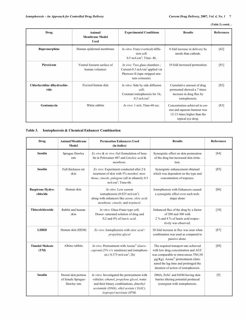
Iontophoresis - An Approach for Controlled Drug Delivery Current Drug Delivery, 2007, Vol. 4, No. 1 7 (Table 2) contd…
Drug Animal/ Membrane Model
Used
Experimental Conditions Results References
Buprenorphine Human epidermal membrane In vitro: Franz (vertical) diffu-sion cell.
0.5 mA/cm2; Time- 4h.
8 fold increase in delivery by anode than cathode.
[42]
Piroxicam Ventral forearm surface of human volunteer
In vivo: Two glass chambers ; Current-0.3 mA/cm2 applied via Phoresor II (tape stripped stra-
tum corneum).
10 fold increased permeation. [81]
Chlorhexidine dihydrochlo-ride
Excised human skin In vitro: Side by side diffusion cell;
Constant iontophoresis for 1h; 0.5 mA/cm2.
Cumulative amount of drug permeated showed a 7 times
increase in drug flux by iontophoresis.
[82]
Gentamycin White rabbits In vivo: 1 mA; Time-60 sec. Concentration achieved in cor-nea and aqueous humour was 12-15 times higher than the
topical eye drop.
[83]
Table 3. Iontophoresis & Chemical Enhancer Combination
Drug Animal/Membrane Model
Permeation Enhancers Used (in italics)
Results References
Insulin Sprague Dawley rats
Ex vivo & in vivo: Gel formulation of Insu-lin in Poloxamer 407 and Linoleic acid &
menthone.
Synergistic effect on skin permeation of the drug but increased skin irrita-
tion.
[84]
Insulin Full thickness rat skin
Ex vivo: Experiment conducted after 2 h trearment of skin with 5% menthol, men-
thone, cineole, pulegone (all in ethanol); 0.5 mA/cm2; Time-6h.
Synergistic enhancement obtained which was dependent on the type and
concentration of terpenes.
[85]
Buspirone Hydro-chloride
Human skin In vitro: Low current iontophoresis (0.025 mA/cm2)
along with enhancers like azone, oleic acid, menthone, cineole, and terpineol.
Iontophoresis with Enhancers caused a synergistic effect over each tech-
nique alone
[86]
Thiocolchicoside Rabbit and human skin
In vitro: Glass-Franz type cell; Donor- saturated solution of drug and
0,2 and 4% of lauric acid.
Enhanced flux of the drug by a factor of 200 and 300 with
2 % and 4 % of lauric acid respec-tively was observed.
[10]
LHRH Human skin (HEM) Ex vivo: Iontophoresis with oleic acid / propylene glycol
30 fold increase in flux was seen when combination was used as compared to
passive alone.
[87]
Timolol Maleate (TM)
Albino rabbits In vivo: Pretreatment with Azone® (lauro-capram) (3% v/v emulsion) and iontophore-
sis ( 0.375 mA/cm2; 2h)
The required transport rate achieved with low drug concentration and AUC was comparable to intravenous TM (30 µg/Kg). Azone® pretreatment elimi-nated the lag time and prolonged the duration of action of iontophoresis.
[88]
Insulin Dorsal skin portion of female Sprague-
Dawley rats
In vitro: Investigated the pretreatment with vehicles: ethanol, propylene glycol, water and their binary combinations, dimethyl acetamide (DMA), ethyl acetate ( EtAC),
isopropyl myristate (IPM).
DMA, EtAC and EtOH having skin barrier altering potential produced
synergism with iontophoresis.
[9]
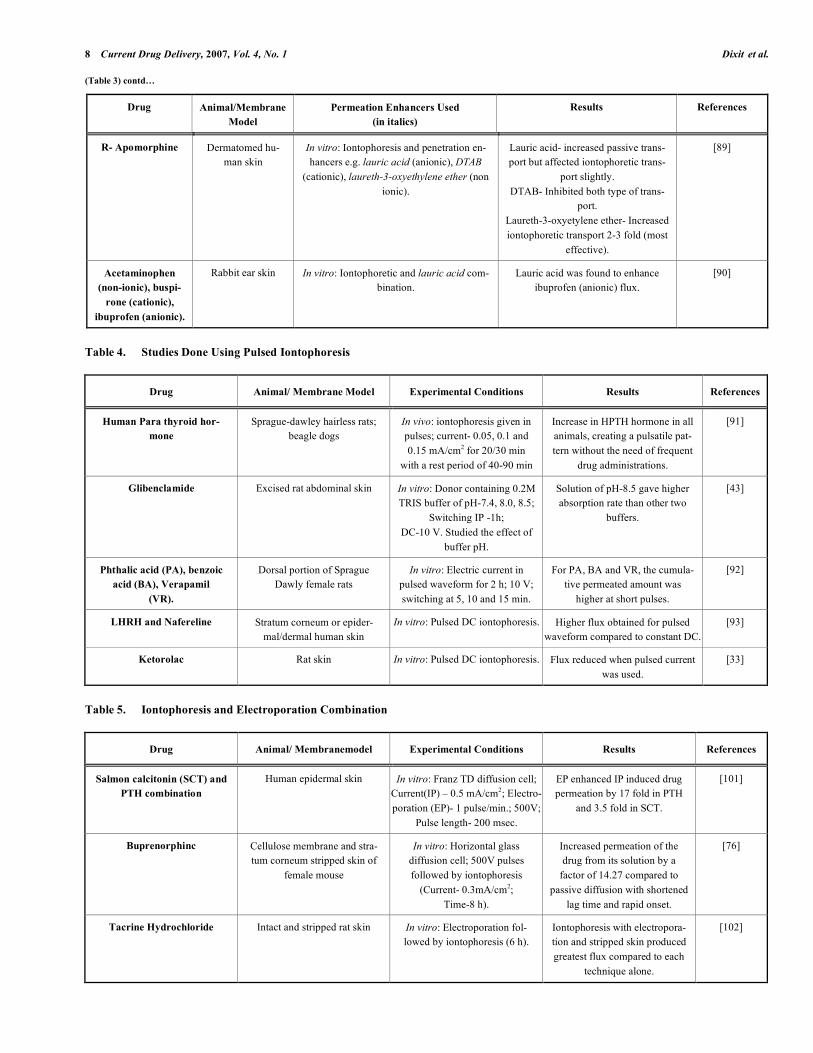
8 Current Drug Delivery, 2007, Vol. 4, No. 1 Dixit et al.
(Table 3) contd…
Drug Animal/Membrane Model
Permeation Enhancers Used (in italics)
Results References
R- Apomorphine Dermatomed hu-man skin
In vitro: Iontophoresis and penetration en-hancers e.g. lauric acid (anionic), DTAB
(cationic), laureth-3-oxyethylene ether (non ionic).
Lauric acid- increased passive trans-port but affected iontophoretic trans-
port slightly. DTAB- Inhibited both type of trans-
port. Laureth-3-oxyetylene ether- Increased iontophoretic transport 2-3 fold (most
effective).
[89]
Acetaminophen (non-ionic), buspi-
rone (cationic), ibuprofen (anionic).
Rabbit ear skin In vitro: Iontophoretic and lauric acid com-bination.
Lauric acid was found to enhance ibuprofen (anionic) flux.
[90]
Table 4. Studies Done Using Pulsed Iontophoresis
Drug Animal/ Membrane Model Experimental Conditions Results References
Human Para thyroid hor-mone
Sprague-dawley hairless rats; beagle dogs
In vivo: iontophoresis given in pulses; current- 0.05, 0.1 and 0.15 mA/cm2 for 20/30 min
with a rest period of 40-90 min
Increase in HPTH hormone in all animals, creating a pulsatile pat-tern without the need of frequent
drug administrations.
[91]
Glibenclamide Excised rat abdominal skin In vitro: Donor containing 0.2M TRIS buffer of pH-7.4, 8.0, 8.5;
Switching IP -1h; DC-10 V. Studied the effect of
buffer pH.
Solution of pH-8.5 gave higher absorption rate than other two
buffers.
[43]
Phthalic acid (PA), benzoic acid (BA), Verapamil
(VR).
Dorsal portion of Sprague Dawly female rats
In vitro: Electric current in pulsed waveform for 2 h; 10 V; switching at 5, 10 and 15 min.
For PA, BA and VR, the cumula-tive permeated amount was
higher at short pulses.
[92]
LHRH and Nafereline Stratum corneum or epider-mal/dermal human skin
In vitro: Pulsed DC iontophoresis. Higher flux obtained for pulsed waveform compared to constant DC.
[93]
Ketorolac Rat skin In vitro: Pulsed DC iontophoresis. Flux reduced when pulsed current was used.
[33]
Table 5. Iontophoresis and Electroporation Combination
Drug Animal/ Membranemodel Experimental Conditions Results References
Salmon calcitonin (SCT) and PTH combination
Human epidermal skin In vitro: Franz TD diffusion cell; Current(IP) – 0.5 mA/cm2; Electro-poration (EP)- 1 pulse/min.; 500V;
Pulse length- 200 msec.
EP enhanced IP induced drug permeation by 17 fold in PTH
and 3.5 fold in SCT.
[101]
Buprenorphinc Cellulose membrane and stra-tum corneum stripped skin of
female mouse
In vitro: Horizontal glass diffusion cell; 500V pulses followed by iontophoresis
(Current- 0.3mA/cm2; Time-8 h).
Increased permeation of the drug from its solution by a
factor of 14.27 compared to passive diffusion with shortened
lag time and rapid onset.
[76]
Tacrine Hydrochloride Intact and stripped rat skin In vitro: Electroporation fol-lowed by iontophoresis (6 h).
Iontophoresis with electropora-tion and stripped skin produced greatest flux compared to each
technique alone.
[102]

Iontophoresis - An Approach for Controlled Drug Delivery Current Drug Delivery, 2007, Vol. 4, No. 1 9
Table 6. Reverse Iontophoresis in Diagnostic Applications
Drug Animal Model Experimental Conditions
Results References
Caffeine, Theophyline
Porcine ear skin ( intact and tape stripped)
In vitro: Three compartment vertical diffusion cell; 0.3 mA/cm2; Time-5 h.
Better extraction through stra-tum corneum than the stratum
corneum stripped skin.
[20]
lithium Dermatomed pig ear skin In vitro: Extraction via electro migration to cathode.
Excellent correlation between subdermal lithium and ionto-phoretic extraction flux and
iontophoresis tracked sudden changes in lithium concentra-
tion.
[104 ]
Phenytoin Dermatomed pig ear skin In vitro: Extraction of ionized solution at anode by electromi-gration and neutral at cathode
by electroosmosis.
Ratio of extracted amount corre-lated well with subdermal con-
centration.
[105 ]
onset of action and minimization of side effects due to avoidance of large oral doses to get local action on the skin. Many characteristics of iontophoresis must be controlled to achieve successful drug delivery. These include mainly the factors which can affect the iontophoretic delivery of drugs like the operational factors and the biological factors. The charge, concentration, and drug combinations must be com-patible with the entire process of iontophoresis. Besides this, preservatives, buffers, osmotic agents, stability and conduc-tivity of the vehicle, are also important for the process of iontophoresis to be successful. Many studies involving the use of combination approaches like use of various chemical enhancers along with iontophoresis have lead to significant improvement in the absorption of insulin and many other drugs, which could not be delivered using ionotphoresis alone. Use of pulsed waveform of DC has also been used and has shown to produce significant and rapid delivery of drugs. Other types of iontophoretic techniques like reverse ionto-phoresis, have opened a new era in the diagnostic field, as it provides a non-invasive and less tedious sampling of body fluids. The major advantages of iontophoretic delivery sys-tem which makes its future use hopeful on large scale are the accurate control over drug input kinetics and optimization of drug input rates. In the future, this system might be used to deliver therapeutic proteins or vaccines transdermally. Even though iontophoresis has so many advantages, a considerable amount of research and judicious use of technology is needed to make further improvement in these microelectron-ics devices and to make the iontophoretic delivery products available to public on a large scale. Thus, iontophoresis may prove to be an important alternative method of drug delivery in the near future.
REFERENCES [1] Tyle, P. Pharm. Res. 1986, 3 (6), 318-26. [2] Green, P.G.; Flanagan, M.; Shroot, B.; Guy, R.H. In Physical Skin
Penetration Enhancement, Walters, K.A.; Hadgraft. J. Eds.; Marcel Dekker Inc.: New York, 1993, 311-33.
[3] Green, Philip G. J. Control. Release, 1996, 41(1-2), 33-48.
[4] Sage, B.H. In Encyclopedia of pharmaceutical Technology, Swar-brick, J.; Boylan, J.C., Eds.; Marcel Dekker Inc.: New York, 1993, Vol. 8, 217-47.
[5] Banga, A.K.; Chien, Y.W. J. Control. Release, 1988, 7(1), 1-24. [6] Banga, A.K.; Bose, S.; Ghosh, T.K. Int. J. Pharm. 1999, 179(1), 1-
19. [7] Kalia, Y. N.; Naik, A.; Garrison, J.; Guy, R.H. Adv. Drug Deliv.
Rev. 2004, 56(5), 619-58. [8] Wang, Y.; Thakur, R.; Fan, Q.; Michniak, B. Eur. J. Pharm. Bio-
pharm. 2005, 60(2), 179-91. [9] Pillai, O.; Nair, V.; Panchagnula, R. Int. J. Pharm. 2004, 269(1),
109-20. [10] Artusi, M.; Nicoli, S.; Colombo, P.; Bettini, R.; Sacchi, A.; Sanli,
P. J. Pharm. Sci. 2004, 93 (10), 2431-8. [11] Chou, W.-L.; Cheng, C.-H.; Yen, S.-C.; Jiang, T.-S. Drug Dev. Ind.
Pharm. 1996, 22(9&10), 943-50. [12] Lai, P. M.; Roberts, M. S. In Dermal absorption and toxicity as-
sessment, Roberts, M.S.; Walters, Kenneth A., Eds.; Marcel Dekker Inc.: New York, 1998, Vol. 91, pp. 371-414.
[13] Banga, A.K. Electrically assisted transdermal and topical drug delivery, Taylor and Francis, London, 1998.
[14] Singh, J.; Bhatia, K.S. Med. Res. Rev. 1996, 16, 285-96. [15] Singh, J.; Maibach, H.I. Crit. Rev. Ther. Drug Carr. Syst. 1994, 11,
161-213. [16] Pikal, M.J. Adv. Drug. Deliv. Rev. 1992, 9(2-3), 201-37. [17] Huang, Y.-Y.; Wu, S.-M.; Wang, C.-Y. Pharm. Res. 1996, 13(4),
547-52. [18] Clemessy, M.; Couarraze, G.; Bevan, B.; Puisieux, F. Int. J.
Pharm. 1994, 101(3), 219-26. [19] Rao, G.; Glikfeld, P.; Guy, R.H. Pharm. Res. 1993, 10(12), 1751-5. [20] Sekkat, N.; Naik, A.; Kalia, Y.N.; Glikfeld, P.; Guy, R.H. J. Con-
trol. Release 2002, 81(1-2), 83-9. [21] Kavanagh, G. M.; Oh, C.; Shams, K. Brit. J. Dermatol. 2004, 151,
1093–95. [22] Alza product literature, www.alza.com. [23] Iomed product literature, http:// www.iomed.com. [24] Chaturvedula, A.; Joshi, D. P.; Anderson, C.; Morris, R. L.; Sem-
browich, Walter L.; Banga, A. K. Int. J. Pharm. 2005, 297(1-2), 190-6.
[25] Turner, N. G.; Guy, R.H. J. Pharm. Sci. 1997, 86(12), 1385-9. [26] Lelawongs, P.; Liu, J-C; Siddiqui, O.; Chien, Y. W. Int. J. Pharm.
1989, 56(1), 13-22. [27] Thysman, S.; Préat, V.; Roland, M. J. Pharm. Sci. 1992, 81(7),
670-5. [28] Delterzo, S.; Behl, C.R.; Nash, R.A. Pharm. Res. 1989, 6(1), 89-90. [29] Koizumi, T.; Kakemi, M.; Katayama, K.; Inada, H.; Sudeji, K.;
Kawasaki, M. Chem. Pharm. Bull. 1990, 38(4), 1022-3. [30] Nugroho, A. K.; Li, L.; Dijkstra, D.; Wikström, H.; Danhof, M.;
Bouwstra, J.A. J. Control. Release 2005, 103(2), 393-403.

10 Current Drug Delivery, 2007, Vol. 4, No. 1 Dixit et al.
[31] Nugroho, A. K.; Li, G.; Arne, G.; Danhof, M.; Bouwstra, J. A. J. Control. Release 2004, 96(1), 159-67.
[32] Jacobsen, J. J. Control. Release 2001, 70(1-2), 83-95. [33] Tiwari, S. B.; Udupa, N. Int. J. Pharm. 2003, 260(1), 93-103. [34] Bellantone, N.H.; Rim, S.; Francoeur, M.L.; Rasadi, B. Int. J.
Pharm. 1986, 30(1), 63-72. [35] Miller, L.L.; Kolaskie, C.J.; Smith, G.A.; Rivier, J. J. Pharm. Sci.
1990, 79(6), 490-3 [36] Sanderson, J.E.; Riel, S.D.; Dixon, R. J. Pharm. Sci. 1989, 78, 361-
64. [37] Singh, P.; Boniello, S.; Liu, P.; Dinh, S. Pharm. Res. 1997,
14(suppl.), S309-10 [38] Morimoto, K.; Iwakura, Y.; Miyazaki, M.; Natakani, E. Int. J.
Pharm. 1992, 81(2-3), 119-25. [39] Iwakura, Y.; Morimoto, K. S.T.P. Pharm. Sci. 1991, 1, 387. [40] Morimoto, K.; Iwakura, Y.; Nakatani, E.; Miyazaki, M.; Tojima, H.
J. Pharm. Pharmacol. 1992, 44(3), 216-8. [41] Kochhar, C.; Imanidis, G. J. Control. Release 2004, 98(1), 25-35. [42] Bose, S.; Ravis, W.R.; Lin, Y.-J.; Zhang, L.; Hofmann, G.A.;
Banga, A.K. J. Control. Release 2001, 73(2-3), 197-203. [43] Takahashi, Y.; Iwata, M.; Mahila, Y. Yakugaku Zasshi, 2001,
121(2), 161-6. [44] Pillai, O.; Nair, V.B.; Ramarao, P.; Panchagnula, R. J. Pharm.
Pharmacol. 2000, 52(suppl.), 92. [45] Siddiqui, O.; Sun, Y.; Liu, J.-C.; Chien, Y.W. J. Pharm. Sci. 1987,
76(4), 341-5. [46] Huang, Y.-Y.; Wu, S.-M.; Wang, C.-Y.; Jiang, T.-S. Drug Develop.
Ind. Pharm. 1995, 21(14), 1631-48. [47] Siddiqui, O.; Roberts, M.S.; Polack, A.E. J. Pharm. Pharmacol.
1985, 37(10), 732-5. [48] Craane-Van, Heinsberg, W.H.M.; Bax, L.; Flinterman, N.H.M.;
Verhoef, J.; Junginger, H.E.; Boddé, H.E. Pharm. Res. 1994, 11(9), 1296-1308.
[49] Knoblauch, P.; Moll, F. J. Control. Release 1993, 26(3), 203-12. [50] Fu, L.M.; Lee, D.; Carlson, R.; Rao, G.S.; Hui, H.W.; Adjei, L.;
Herrin, M.; Sunberg, D.; Hsu, L. Drug Develop. Ind. Pharm. 1993, 19(13), 1557-71.
[51] Yoshida, N.H.; Roberts, M.S. J. Pharm. Pharmacol. 1995, 47(11), 883-90.
[52] Nair, V.B.; Pillai, O.; Ramarao, P.; Panchagnula, R. J. Pharm. Pharmacol. 2000, 52(Suppl.), 79.
[53] Miller, L.L.; Smith, G.A.; Chang, An-Chang.; Zhou, Q.-X. J. Con-trol. Release 1987, 6(1), 293-6.
[54] Miller, L.L.; Smith, G.A. Int. J. Pharm. 1989, 49(1), 15-22. [55] Green, P.G.; Hinz, R.S.; Cullander, C.; Yamane, G.; Guy, R.H.
Pharm. Res. 1991, 8(9), 1113-20. [56] Green, P.G.; Shroot, B.; Bernard, F.; Pilgrim, W.R.; Guy, R.H. J.
Control. Release 1992, 20(3), 209-17. [57] Langkjaer, L.; Brange, J.; Grodosky, G.M.; Guy, R.H. Proc. Int.
Symp. Control. Rel. Biact. Mater. 1994, 21, 172. [58] Vander-Geest, R.; Hueber, F.; Szoka, F.C. Jr.; Guy, R.H. Pharm.
Res. 1996, 13 (4), 553-8. [59] Burnett, R.R.; Ongpipattanakul, B. J. Pharm. Sci. 1987, 76(10),
765-73. [60] Berner, B.; Dinh, S.M. Electronically assisted controlled drug
delivery, CRC: Washington DC. 1998. [61] Green, P.G.; Hinz, R.S.; Kim, A.; Cullander, C.; Yamane, G.;
Szoka, F.C. Jr.; Guy, R.H. J. Control. Release 1992, 21 (1-3), 187-190.
[62] Sung, K.C.; Fang, J.-Y.; Hu, O.Y.-P. J. Control. Release 2000, 67 (1), 1-8.
[63] Burnette, R.R.; Marrero, D. J. Pharm. Sci. 1986, 75(8), 738-43. [64] Wearley, L.; Liu, J.-C.; Chien, Y.W. J. Control. Release 1989,
8(3), 237-50. [65] Hui, X.; Anigbogu, A.; Singh, P.; Xiong, G.; Poblet, N.; Liu, P.;
Maibach, H.I. J. Pharm. Sci. 2001, 90(9), 1269-76. [66] Lawler, J.C.; Davis, M.J.; Griffith, E. J. Invest. Dermatol. 1960, 34,
301-8. [67] Bagniefski, T.; Burnett, R.R. J. Control. Release 1990, 11(1-3),
113-22.
[68] Singh, P.; Puchun, L.; Dinh, S.M. In Percutaneous Absorption Drug-cosmetic-mechanism and methods, Bronaugh, R.L.; Maibach, H.I. Eds.; 3rdedition, Marcel Dekker Inc., New York, 1998.
[69] Chien, Y.W.; Lelawong, P.; Siddiqui, O.; Sun, Y.; Shi, W.M. J. Control. Release 1990, 13(2-3), 263- 78.
[70] Préat, V.; Thysman, S. Int. J. Pharm. 1993, 96(1-3), 189-96. [71] Thysman, S.; Tasset, C.; Préat, V. Int. J. Pharm. 1994, 101(1-2),
105-13. [72] Phipps, J.B.; Padmanabham, R.V.; Lattin, G.A. J. Pharm. Pharma-
col. 1989, 78, 365. [73] Cross, S.E.; Roberts, M.S. J. Pharm. Sci. 1995, 84, 584-92. [74] Roberts, M.S.; Favretto, W.A.; Meyer, A.; Rechmann, M.; Wong-
seelashote, T. Aust. N Z J. Med. 1982, 12, 305. [75] Feldman, R.H.; Maibach, H.I. Arch. Dermatol. 1967, 48, 181. [76] Fang, J.-Y.; Sung, K.C.; Wang, J.-J.; Chu, C.-C.; Chen, K.-T. J.
Pharm. Pharmacol. 2002, 54(10), 1329-37. [77] Nolan, L.M.A.; Corish, J.; Corrigan, O.I.; Fitzpatrick, D. Int. J.
Pharm. 2003, 257(1-2), 41-55. [78] Kanikannan, J.N.; Singh, J.; Ramarao, P. J. Control. Release 2001,
71(1), 99-105. [79] Badkar, A.V.; Banga, A.K.; J. Pharm. Pharmaco. 2002, 54(7),
907-12. [80] Merclin, N.; Bramer T.; Edsman, K. J. Control. Release 2004,
98(1), 57-65. [81] Curdy, C.; Kalia, Y.N.; Naik, A.; Guy, R.H. J. Control. Release
2001, 76(1-2), 73-9. [82] Amini, T.; Conway, B.R.; Irwin, W.J.; Lambert, P.A.; Elliot, T.S.J.
J. Pharm. Pharmacol. 2000, 52(Suppl.), 25. [83] Esther, E.-B.; Raiskup, F.; Stepensky, D.; Domb, A.J. J. Invest.
Opthal. Vis. Sci. 2004, 45, 2543-8. [84] Pillai, O.; Panchagnula, R. J. Control. Release 2003, 89(1), 127-40. [85] Pillai, O.; Panchagnula, R. J. Control. Release 2003, 88(2), 287-96. [86] Meidan, V. M.; Al-Khalili, M.; Michniak, B. B. Int. J. Pharm.
2003, 264(1-2), 73-83. [87] Smyth, H.D.C.; Becket, G.; Mehta, S. J. Pharm. Sci. 2002, 91(5),
1296-1307. [88] Kanikkannan, N.; Singh, J.; Ramarao, P. Int. J. Pharm. 2000,
197(1-2), 69-76. [89] Li, G.L.; Van der Geest, R.; Chanet, L.; Zanten, E. V.; Danhof, M.;
Bouwastra, J. A. J. Control. Release 2002, 84(1-2), 49-57. [90] Sebastiani, P.; Nicoli, S.; Santi, P. Int. J. Pharm. 2005, 292(1-2),
119-26. [91] Suzuki, Y.; Iga, K.; Yanai, S.; Matsumoto, Y.; Kawase, M.; Fu-
kuda, T.; Adachi, H.; Higo, N.; Ogawa, Y. J. Pharm. Pharmaco. 2001, 53(9), 1227-34.
[92] Ishikawa, O.; Kato, Y.; Onishi, H.; Nagai, T.; Machida, Y. Int. J. Pharm. 2002, 249(1-2), 81-8.
[93] Johanna, R.; Maija, k.; Katri, H.; Risto, K.; Jouni, H. Eur. J. Pharm. Sci. 2004, 21(2-3), 371-7.
[94] Prausnitz, M.R. J. Control. Release 1996, 40(3), 321-6. [95] Riviere, J. E.; Heit, M.C. Pharm. Res. 1997, 14(6), 687-97. [96] Vanbever, R.; Demorre, N.; Préat, V. Pharm. Res. 1996, 13(9),
1360-6. [97] Bose, S.; Ravis, W.R.; Lin, Y.J.; Zhang, L.; Hofmann, G.A.;
Banga, A.K. J. Control. Release 2001, 73(2-3), 197-203. [98] Jadoul, A.; Lecouturier, N.; Mesens, J.; Caers, W.; Préat, V. J.
Control. Release 1998, 54(3), 265-72. [99] Regnier, V.; De Moore, N.; Jadoul, A.; Préat, V. Int. J. Pharm.
1999, 184(2), 147-56. [100] Merino, V.; Kalia, Y.N.; Guy, R.H. Trends. Biotech. 1997, 15, 288-
90. [101] Chang, S.L.; Hofmann, G.A.; Zhang, Lei; Deftor, L. J.; Banga,
A.K. J. Control. Release 2000, 66(2-3), 127-33. [102] Hirsch, A.C.; Upasani, R.S.; Banga, A.K. J. Control. Release 2005,
103(1), 113-21. [103] Merino, V.; López, A.; Hochstrasser, D.; Guy, R.H. J. Control.
Release 1999, 61(1-2), 65-9. [104] Leboulanger, B.; Fathi, M.; Guy, R.H.; Delgado-Charro, M.B.
Pharm. Res. 2004, 21(7), 1214-22. [105] Leboulanger, B.; Guy, R. H.; Delgado-Charro, M.B. Eur. J. Pharm.
Sci. 2004, 22(5), 427-33. Received: October 19, 2005 Revised: April 25, 2006 Accepted: April 27, 2006