Controlled gene and drug release from a liposomal delivery ...
11
ARTICLE Controlled gene and drug release from a liposomal delivery platform triggered by X-ray radiation Wei Deng 1,7 , Wenjie Chen 1 , Sandhya Clement 1,7 , Anna Guller 1,2,3,7 , Zhenjun Zhao 2 , Alexander Engel 4,5,6 & Ewa M. Goldys 1,7 Liposomes have been well established as an effective drug delivery system, due to simplicity of their preparation and unique characteristics. However conventional liposomes are unsui- table for the on-demand content release, which limits their therapeutic utility. Here we report X-ray-triggerable liposomes incorporating gold nanoparticles and photosensitizer verteporfin. The 6 MeV X-ray radiation induces verteporfin to produce singlet oxygen, which destabilises the liposomal membrane and causes the release of cargos from the liposomal cavity. This triggering strategy is demonstrated by the efficiency of gene silencing in vitro and increased effectiveness of chemotherapy in vivo. Our work indicates the feasibility of a combinatorial treatment and possible synergistic effects in the course of standard radiotherapy combined with chemotherapy delivered via X-ray-triggered liposomes. Importantly, our X-ray-mediated liposome release strategy offers prospects for deep tissue photodynamic therapy, by removing its depth limitation. DOI: 10.1038/s41467-018-05118-3 OPEN 1 ARC Centre of Excellence for Nanoscale Biophotonics, Faculty of Science and Engineering, Macquarie University, North Ryde, 2109 New South Wales, Australia. 2 Faculty of Medicine and Health Sciences, Macquarie University, North Ryde, 2109 NSW, Australia. 3 Sechenov University, Moscow, 119992, Russia. 4 Sydney Medical School, University of Sydney, Sydney, 2050 NSW, Australia. 5 Department of Colorectal Surgery, Royal North Shore Hospital, St Leonards, 2065 NSW, Australia. 6 Sydney Vital Translational Cancer Research, Kolling Institute of Medical Research, Northern Sydney Local Health District, St Leonards, 2065 NSW, Australia. 7 Present address: The Graduate School of Biomedical Engineering, University of New South Wales, Sydney, Kensington, 2052 NSW, Australia. These authors contributed equally: Wenjie Chen, Sandhya Clement. Correspondence and requests for materials should be addressed to W.D. (email: [email protected] or (email: [email protected]) or to E.M.G. (email: [email protected]) NATURE COMMUNICATIONS | (2018)9:2713 | DOI: 10.1038/s41467-018-05118-3 | www.nature.com/naturecommunications 1 1234567890():,;
-
Upload
khangminh22 -
Category
Documents
-
view
0 -
download
0
Transcript of Controlled gene and drug release from a liposomal delivery ...
ARTICLE
Controlled gene and drug release from a liposomaldelivery platform triggered by X-ray radiationWei Deng1,7, Wenjie Chen1, Sandhya Clement1,7, Anna Guller 1,2,3,7, Zhenjun Zhao2,
Alexander Engel4,5,6 & Ewa M. Goldys1,7
Liposomes have been well established as an effective drug delivery system, due to simplicity
of their preparation and unique characteristics. However conventional liposomes are unsui-
table for the on-demand content release, which limits their therapeutic utility. Here we report
X-ray-triggerable liposomes incorporating gold nanoparticles and photosensitizer verteporfin.
The 6MeV X-ray radiation induces verteporfin to produce singlet oxygen, which destabilises
the liposomal membrane and causes the release of cargos from the liposomal cavity. This
triggering strategy is demonstrated by the efficiency of gene silencing in vitro and increased
effectiveness of chemotherapy in vivo. Our work indicates the feasibility of a combinatorial
treatment and possible synergistic effects in the course of standard radiotherapy combined
with chemotherapy delivered via X-ray-triggered liposomes. Importantly, our X-ray-mediated
liposome release strategy offers prospects for deep tissue photodynamic therapy, by
removing its depth limitation.
DOI: 10.1038/s41467-018-05118-3 OPEN
1 ARC Centre of Excellence for Nanoscale Biophotonics, Faculty of Science and Engineering, Macquarie University, North Ryde, 2109 New South Wales,Australia. 2 Faculty of Medicine and Health Sciences, Macquarie University, North Ryde, 2109 NSW, Australia. 3 Sechenov University, Moscow, 119992,Russia. 4 Sydney Medical School, University of Sydney, Sydney, 2050 NSW, Australia. 5 Department of Colorectal Surgery, Royal North Shore Hospital, StLeonards, 2065 NSW, Australia. 6 Sydney Vital Translational Cancer Research, Kolling Institute of Medical Research, Northern Sydney Local Health District,St Leonards, 2065 NSW, Australia. 7Present address: The Graduate School of Biomedical Engineering, University of New South Wales, Sydney, Kensington,2052 NSW, Australia. These authors contributed equally: Wenjie Chen, Sandhya Clement. Correspondence and requests for materials should be addressedto W.D. (email: [email protected] or (email: [email protected]) or to E.M.G. (email: [email protected])
NATURE COMMUNICATIONS | (2018) 9:2713 | DOI: 10.1038/s41467-018-05118-3 | www.nature.com/naturecommunications 1
1234
5678
90():,;
The development and application of various nanomaterialdesigns for gene and drug delivery is currently one of thekey focus areas in nanomedicine. Although viral carriers
have been traditionally used as a gene/drug delivery method1,2,their application is hindered by a range of limitations includingimmunogenicity, limited size of transgenic materials, packagingdifficulties and the risk of recombination3. Furthermore, viralcarriers do not offer any temporal control over transfectionwhich, once introduced, cannot be deliberately stopped4. Toovercome these limitations, synthetic nanomaterial-based systemshave been extensively studied and developed. Among thesenanomaterials, liposomes have been well established as an effec-tive drug delivery system, due to simplicity of their preparationand unique characteristics5,6. Liposomes consist of an aqueouscore surrounded by a lipid bilayer similar to cell membranes,which facilitates cellular uptake of liposomes. The lipids formingliposomes are amphipathic, thus allowing the encapsulation ofboth hydrophobic and hydrophilic molecules or (and) colloidalparticles7. Liposomes are usually biocompatible and biodegrad-able, which makes them suitable for clinical applications5,8.
However conventional liposomes, for example, commerciallipofectamine 2000, are unsuitable for the on-demand contentrelease, which limits their therapeutic utility, although they pos-sess high efficiency of delivery. By contrast, triggerable liposomesare able to release genes/drugs in a more controlled manner,usually much faster and, depending on triggering modality, alsoto a specific area, and these properties contribute to theirpotentially greater clinical success. Several strategies have beenpreviously employed to design responsive liposomes whosebilayer could be destabilised by using physiological and externalstimuli. The triggering approaches previously reported includechanges in pH (typical in cancer)9,10, externally delivered heat, forexample via alternating magnetic field or infrared light11,12,enzymes13,14 and non-thermal effects caused by lightirradiation15,16. These approaches have certain limitations, inparticular triggering of light-sensitive liposomes by visible light islimited by its relatively shallow (few mm) penetration of light intobiological tissues17. As a result of this modest penetration depth,visible light can not activate photosensitizers (PS) located deeplyin the body and generate sufficient amount of singlet oxygen(1O2) or other reactive oxygen species (ROS) to release theliposome cargo required for the therapeutic effects18. With itsexcellent tissue penetration depth, X-ray radiation explored inthis work for liposome triggering offers an alternative approach toyield both spatial targeting (such as to a tumour site) via standardradiotherapy approaches such as the Gamma-knife19 and trig-gered release of encapsulated contents from the liposomes oncethey are located at the target site. Importantly, the X-ray liposometriggering can be used concurrently with radiation therapy, acommon treatment modality in cancer.
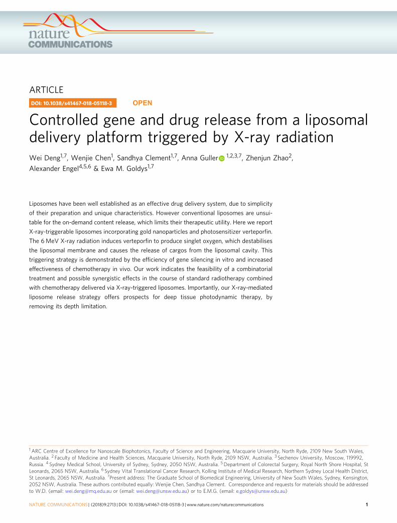
Herein we design X-ray triggered liposomes by co-embeddingphotosensitizers and gold nanoparticles (3–5 nm) inside a lipidbilayer. Gold is chosen in this work as, due to its high atomicnumber, it strongly interacts with X-ray radiation as shown, forexample, by gold nanoparticle-induced radiation enhancementinside biological tissue20–22. Although in our design the photo-sensitisers are the primary source of reactive oxygen species(ROS) to oxidise unsaturated lipids and destabilise liposomalmembranes, gold nanoparticles exposed to X-rays also generatesome level of ROS23. More complex effects are also possible; forexample, secondary electrons produced during the interaction ofX-rays with gold nanoparticles may transfer from gold to aphotosensitizer and lead to PS-induced generation of 1O2 or otherROS24–26. As a photosensitizer we choose verteporfin (VP),clinically approved for photodynamic therapy (PDT) of age-related macular degeneration27,28. 1,2-dioleoyl-sn-glycero-3-
phosphocholine (DOPC) and 1, 2-di-(9Z-octadecenoyl)-3-tri-methylammonium-propane (DOTAP) are chosen as lipid com-ponents in the liposome formulation because DOPC can loadhighly hydrophobic molecules and DOTAP can facilitate cellularuptake due to its positive charge29. The 1O2 generation fromdifferent liposome samples and destabilization of the lipid bilayerby 1O2 under 365 nm LED illumination with different time points(2, 4, 6, 8 and 10 min) and X-ray radiation with different dosage(1, 2 and 4 Gy) are assessed by using singlet oxygen green sensor(SOSG) and calcein release assays, respectively. SOSG is a com-monly used and highly specific fluorescence probe for thedetection of 1O2 generation30. It is identified to be fluoresceincovalently bound with an anthracene moiety31. Calcein is afluorescent dye that self-quenches at high concentration32,33
which makes it possible to detect its release from the liposomes tothe surrounding environment by monitoring the increase incalcein fluorescence intensity upon X-ray radiation34,35. Addi-tionally, 1O2 quantum yield under UV light illumination and thenumber of 1O2 generated as a result of X-ray radiation are alsocalculated based on experimental data36,37. Triggered release ofthe liposome cargo by X-rays is verified by (a) demonstrating theefficiency of X-ray triggered gene silencing in vitro and (b) theincreased effectiveness of chemotherapy in vivo (Fig.1). For genesilencing, an antisense oligonucleotide complementary to a spe-cific pituitary adenylate cyclase-activating polypeptide (PACAP)receptor, PAC1R, is encapsulated inside the liposomes. Followingthe liposome take-up by rat PC12 cells, the X-ray radiation at adose of 4 Gy is applied. As a result of exposure to ionisingradiation, the 1O2 generated in a lipid bilayer destabilises theliposomes, leading to the release of antisense oligonucleotides.This antisense nucleotide is then able to prevent the translation ofthe PAC1R mRNA by blocking the translation initiation complex.Gene knockdown is monitored by observing a decrease in thefluorescence intensity from indirect immunofluorescence staining
Cancer therapy
X-ray
Gene silencing
Targeted liposomes loaded with VP and gold in a lipidbilayer would release gene or drug under X-ray radiation
Fig. 1 The schematic illustration of gene silencing and cancer cell-killing byX-ray-triggered liposomes. This liposomal delivery platform incorporatesverteporfin (VP) and gold nanoparticles. Two types of cargos, antisenseoligonucleotide and Doxorubicin, are respectively entrapped inside aliposomal middle cavity for demonstration of in vitro gene release andin vivo drug delivery
ARTICLE NATURE COMMUNICATIONS | DOI: 10.1038/s41467-018-05118-3
2 NATURE COMMUNICATIONS | (2018) 9:2713 | DOI: 10.1038/s41467-018-05118-3 | www.nature.com/naturecommunications

of PAC1R in cells after X-ray irradiation. For X-ray-triggeredchemotherapy, an antitumour drug, doxorubicin (Dox), is loadedinto the liposomes. The liposomes are taken up by human col-orectal cancer HCT 116 cells and X-rays applied. In vivo anti-tumour effect is evaluated by monitoring tumour developmentand body weight of mice bearing colorectal cancer xenografts andby conducting histological analysis of tumour tissues after thetreatments.
Results1O2 generation tests by using light and X-rays respectively. Thegeneration of singlet oxygen is a key factor in the oxidation ofunsaturated lipids, resulting in the disruption of the liposomestructure26. 1O2 generation was confirmed by using SOSG andmonitoring the enhancement of fluorescence intensity at 488 nmexcitation. 1O2 reacts with SOSG to produce endoperoxideswhich have a strong fluorescence signal at 525 nm for 488 nmexcitation, while it has weak fluorescence in the absence of 1O2.The SOSG fluorescence intensity enhancement as a function oflight illumination time and X-ray dose, respectively, is plotted inFig. 2. Figure 2a shows that the liposomes loaded with goldnanoparticles and VP generate more singlet oxygen than theother samples, with an increase of about 102% after 10 minillumination. Singlet oxygen quantum yield (SOQY) from thissample (liposomes loaded with gold nanoparticles and VP) iscalculated to be 0.75 ± 0.18 (mean value ± standard deviation),indicating an enhancement factor of 1.42 compared with the
liposomes loaded with VP alone. The details of this calculationare explained in Supplementary Note 2. We attribute thisenhancement of 1O2 generation from VP to near-field enhance-ment of electromagnetic field induced by gold nanoparticles38,39.However such enhancement was dependent on one of experi-mental factors, the distance between gold and photosensitisers. Inthis study the distance between gold nanoparticles and VPmolecules was not controllable under the current conditionbecause both were randomly loaded in the liposomal bilayer, withsome molecules less than optimally placed in terms of the dis-tance for optimal enhancement of singlet oxygen generation. Thismay partially contribute to the singlet oxygen generationenhancement limited within a certain range as observed in thisstudy. In particular, the interaction between gold and photo-sensitisers would not contribute to the singlet oxygen generationwhen they are extremely close40,41.
Similarly, the enhancement of 1O2 generation was observed inliposomes loaded with gold nanoparticles and VP in our X-rayradiation experiments as well, but to a lesser extent. As shown inFig. 2b, liposomes doped with gold nanoparticles and VPmolecules generate the highest amount of 1O2, with a percentageincrease of approximately 79% under X-ray radiation with 4 Gy,while liposomes containing gold nanoparticles alone and thesample containing VP alone produced a limited amount of 1O2,with a percentage increase of approximately 48% and 40%,respectively, under the same experimental conditions. Wecalculated the number of singlet oxygen generated fromliposomes loaded with VP and gold nanoparticles under X-ray
160a b
c d70
60
50
40
Cal
cein
rel
ease
per
cent
age
(%)
30
20
10
00 2 4 6 8 10 43210
0
10
20
Cal
cein
rel
ease
per
cent
age
(%)
Inte
nsity
incr
ease
per
cent
age
(%)
30
Illumination time (min) Dosage (Gy)
Liposomes loaded with gold nanoparticlesand VP
Liposomes loaded with VP aloneLiposomes loaded with goldnanoparticles alone
Pure liposomes
Liposomes loaded with gold nanoparticlesand VP
Liposomes loaded with VP alone
Pure liposomes
Liposomes loaded with gold nanoparticlesand VP
Liposomes loaded with gold nanoparticlesand VP
Liposomes loaded with VP
Liposomes loaded with VP alone
Liposomes loaded with goldnanoparticles
Pure liposomes
Pure liposomes
120In
tens
ity in
crea
se p
erce
ntag
e (%
)
80
40
0
120
80
40
00 2 4 6
Illumination time (min) X-ray dosage (Gy)8 10 12 0 1 2 3 4
Fig. 2 Singlet oxygen generation and calcein release from liposomes under light and X-ray triggering. a, b Percentage increase of SOSG fluorescenceintensities from different liposome samples under (a) 360 nm irradiation at different time points and (b) X-ray radiation with different doses. c, d Calceinrelease profiles from liposomes under (c) 360 nm irradiation and (d) X-ray radiation. Error bars show standard deviation from four measurements
NATURE COMMUNICATIONS | DOI: 10.1038/s41467-018-05118-3 ARTICLE
NATURE COMMUNICATIONS | (2018) 9:2713 | DOI: 10.1038/s41467-018-05118-3 | www.nature.com/naturecommunications 3
radiation with 4 Gy, to be 7250 per a single liposome. Thecalculation is provided in Supplementary Note 3. The observedenhancement of X-ray induced 1O2 generation in the presence ofgold nanoparticles can be explained by the following mechanism.Gold is a heavy metal element strongly interacting with X-rays,which leads to a significant increase of energy deposition inbiological tissues when irradiated with such rays42–44. Thereforegold nanoparticles are well-known radiosensitizers able toamplify the radiation doses in tumour tissue45–47. In addition,gold nanoparticles can selectively scatter and (or) absorb the highenergy X-ray radiation20–22, leading to enhanced energy transferfrom X-ray to photosensitizers. With such contribution, the VPmolecules in the presence of gold nanoparticles are able tointeract more strongly with ionising radiation than the VP on itsown, causing enhanced 1O2 generation.
Calcein release assays under two external stimuli. Havingconfirmed the 1O2 generation from VP entrapped inside lipo-somes using two stimulating modalities, we attempted to evaluatethe liposome content release by using a calcein release assay,which is based on the principle of fluorescence self-quenching48,49. Figure 2c, d shows the proportion of calceinrelease from different liposome samples under UV illuminationand X-ray exposure, respectively. The amount of calcein releasedfrom liposomes doped with both gold nanoparticles and VPreaches a maximum of 44% after 10 min light illumination(Fig. 2c) and 19% after X-ray radiation with 4 Gy (Fig. 2d),respectively. However, lower leakage is observed in the controls(liposomes doped with VP alone), with only 31 and 13% of cal-cein being released at the same experimental conditions. Similarlyto our results of the 1O2 generation, our findings show thatintroduction of gold nanoparticles inside liposomes contributes toincreased release of entrapped calcein, compared with samples
containing VP molecules only, under both UV illumination andX-ray radiation.
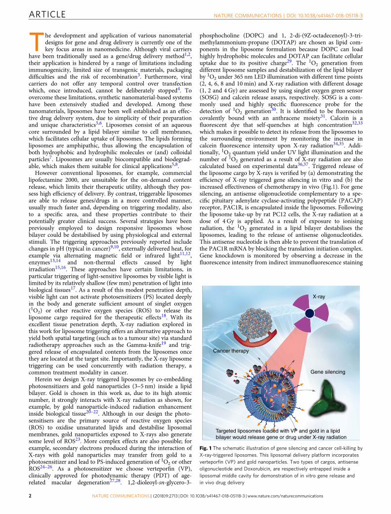
Cellular uptake of liposomes in PC12 cells. In order to inves-tigate the cellular uptake of liposomes, the PC12 cells were treatedwith liposomes for 1 h, 4 h and 10 h. As shown in Fig. 3, higherred fluorescence signal from VP was observed after 4-h incuba-tion compared with cells treated for 1 h. Detailed characterisationof the cellular uptake of liposomes after 4 h incubation with PC12cells is provided in the Supplementary Fig. 6. In addition, greenfluorescence from fluorescein amidite (FAM)-labelled oligonu-cleotide is also clearly observed after 4 h incubation (Supple-mentary Fig. 7). After 10 h incubation with liposomes, cells weresurrounded by large red clusters, indicating a large amount ofliposomes loaded with VP were internalised by cells. However,some clusters were also observed in other regions due to non-specific binding (Fig. 3). Therefore, we chose 4 h incubation timefor PC12 cells. Based on the concentration of fluorescentlylabelled lipid internalised by cells, we estimated that 2550 ± 89liposomes were internalised by each PC12 cell. The number ofgold nanoparticles per liposome is estimated to be 156 ± 24 on thebasis of the ICP-MS data. Therefore, the number of gold nano-particles internalised by each PC12 cell is estimated to be 3.98 ×10⁵ in this study. The detailed calculation of the number ofliposome per cell and the number of gold nanoparticles perliposome is provided in Supplementary Note 4.
Cellular uptake of folate-conjugated liposomes. The folatereceptor (FR) is significantly expressed in many types of cancercells while its expression in most normal tissues is generallylow50. Folic acid (FA) has a very high affinity for the FRs with aminimal effect on its binding ability even after conjugation withother nanomaterials. Therefore FA can significantly enhance the
Nuclei stained withHoechst 333421 h incubation
4 h incubation10 h incubation
VP fluorescence DIC image of cells Overlay offluorescence and DIC
a
b
c
Fig. 3 Cellular uptake activity of liposomes in rat PC12 cells. a–c Representative confocal laser scanning microscopy images of PC12 cells incubated withliposome nanoparticles (25 µM) for 1, 4 and 10 h, respectively. Scale bar is 20 µm
ARTICLE NATURE COMMUNICATIONS | DOI: 10.1038/s41467-018-05118-3
4 NATURE COMMUNICATIONS | (2018) 9:2713 | DOI: 10.1038/s41467-018-05118-3 | www.nature.com/naturecommunications

capability of nanoparticle-based delivery systems to target cancercells51,52. In this study, we modified the liposome surface withfolate and determined the average number of the folate moleculesper liposome (estimated to be approximately 480) based on thetotal amount of folate and liposomes in the sample. To evaluatethe targeting specificity of the folate-targeted liposomes totumour cells, the uptake activity of liposomes by colorectal cancerHCT 116 cells, was compared to the uptake by normal humancolonic cell line, CCD 841 CoN. As shown in Fig. 4a, cancer cellstreated with folate-conjugated liposome nanoparticles clearlyexhibited red signal from VP in the cytoplasm after 1 h incuba-tion. By contrast, the level of liposome uptake by CCD 841 CoNcells is shown to be fairly low under the same experimentalconditions (Fig. 4b). These results indicated that FA- inducedspecific binding to the folate receptor expressed on HCT 116 cellsurface resulted in a higher internalisation rate of targeted lipo-somes, compared to the normal CCD 841 CoN cells.
X-ray triggered in vitro gene silencing and chemotherapy. Wefurther applied the liposomes loaded with antisense oligonu-cleotide to carry out the PAC1R gene knockdown by deliveringthe liposomes to PC12 cells and applying 4 Gy of X-ray radiation.The fluorescently labelled PAC1R expressed by PC12 cells wasimaged by using confocal microscopy at various time points. Forcomparison, the cells treated with liposomes alone, but withouttriggering were also imaged using the same imaging conditions.As shown in Fig. 5a, decreased fluorescence in cell samples wasclearly observed 24 h after X-ray exposure, indicating that theantisense oligonucleotide released from liposomes effectivelyknocked down the PAC1R gene expression. For cells treated withliposomes alone, a decreased PAC1R fluorescence signal was alsoobserved at 24 h after treatment, but the decrease was less pro-nounced compared to cells treated with X-ray radiation (Fig. 5b).We quantitatively analysed the PAC1R inhibition at differenttime points based on cellular fluorescence images. After 24 h sinceX-ray exposure the density of PAC1R decreased by about 45%,while the level of PAC1R in cells which were not exposed to X-rays but received the liposomes with antisense oligonucleotidesdecreased by only 30% (Fig. 5c, d).
In addition to the demonstration of gene silencing by using X-ray-triggered liposomes, we also investigated the in vitro cell-
killing effect of the liposomes loaded with varying amounts ofantitumour drugs, Dox and etoposide (ETP), in HCT 116 cells. Aseries of drug-dilution assays presented in SupplementaryFigure 8a reveals that 50% cell-killing (IC50) was achieved at1.6 µM of Dox encapsulated in the liposomes (LipoDox) andtriggered by X-ray radiation. However, the LipoDox alone,without X-ray triggering but with same Dox concentration of 1.6µM killed only about 10% of cancer cells (Supplementary Fig. 8b).This illustrates, not unexpectedly, that the efficacy of LipoDox forcell killing was higher with X-ray radiation, compared withLipoDox only. The results of our X-ray-triggered LipoDoxtreatment described here indicates that a combination of X-ray-triggered chemo- and radiotherapy with the same X-rays appearsto produce an enhanced effect and it yields improved efficacy ofcancer cell-killing. It should be mentioned that simultaneouschemo- and radiotherapy may result in the development ofcardiotoxicity, whose incidence is associated with differentfactors, including the type of antitumour drugs53. Therefore, weevaluated the cell-killing effect of a second chemotherapy drug,ETP, in combination with X-ray radiation. ETP is associated withreduced incidence of cardiotoxicity, compared with Dox54. Asshown in Supplementary Fig. 8c, higher cytotoxicity of LipoETPin HCT 116 cells was observed at 24 h after X-ray radiation of 4Gy, compared with LipoETP alone.
Toxicity assays of liposomes and X-ray exposure. We firstassessed the toxicity of liposomes doped with gold nanoparticlesand VP. Compared with the control group, no significant changewas observed in the viability of PC12 cells treated with liposomeconcentrations up to 50 µM, higher than those used for gene anddrug delivery in our study (Supplementary Fig. 9a). Theliposome-formulated Dox designed in this study should also haveminimal toxicity effect on normal cells without X-ray-triggering.To verify this, we examined the toxicity of LipoDox on CCD 841CoN cells by varying Dox concentration. As shown in Supple-mentary Fig. 9b, we did not observe a noticeable reduction in cellsurvival (up to 14% cell death) at 24 h after incubation withliposome-formulated Dox samples (Dox concentration: 3 µg mL−1 and 2 µg mL−1), suggesting that under in vitro conditions, ourLipoDox samples with these two Dox concentrations are likelynot to affect the viability of CCD 841 CoN cells.
Nuclei stained withHoechst 33342
a
b
1 h incubation1 h incubation
Verteporfin fluorescence DIC image of cellsOverlay offluorescence and DIC Inset
Fig. 4 Cellular uptake of folate-conjugated liposomes in HCT 116 cells and CCD 841 CoN cells. a, b Representative confocal laser scanning microscopyimages of incubated (a) HCT 116 cells and (b) CCD 841 CoN cells with folate-conjugate liposomes (25 µM) for 1 h. Scale bar is 75 µm
NATURE COMMUNICATIONS | DOI: 10.1038/s41467-018-05118-3 ARTICLE
NATURE COMMUNICATIONS | (2018) 9:2713 | DOI: 10.1038/s41467-018-05118-3 | www.nature.com/naturecommunications 5

It is well known that radiolysis of water molecules as a result ofX-ray radiation damages DNA molecules by producing toxicradicals. Although cells attempt to repair the damage, completerepair may not be possible at higher doses55. The surviving cellsmay suffer residual DNA damage, potentially contributing toadverse long-term health effects. In this study we assessed the X-ray-induced damage in both cultured cells and genetic materials.In cell experiments, the MTS test did not reveal a clear decrease insurvival of PC12 cells, HCT 116 cells and CCD 841 CoN cells at24 h and 48 h after X-ray exposure (Supplementary Fig. 9c). Withregard to the X-ray effects on genes, the DNA gel electrophoresisdid not show obvious dispersion of DNA bands after X-rayradiation compared to the control, indicating that X-ray radiationwith our applied dosage did not cause obvious damage to theDNA molecules (Supplementary Fig. 9d).
In addition, we also checked the effect of the singlet oxygen ongenetic material by irradiating a mixture solution of oligonucleo-tides and VP with X-ray. As shown in Supplementary Fig. 9d,there was no clear oligonucleotide damage observed comparedwith the control. Singlet oxygen is the primary cytotoxic agentresponsible for photobiological activity involved in the PDTtechnique. It can damage cells by reacting with many biomole-cules, including amino acids, nucleic acids and unsaturated fattyacids that have double bonds as well as sulphur-containing aminoacids56,57. Short lifetime of singlet oxygen prevents it fromtravelling larger distances, therefore it mainly causes localised58,near the photosensitizer molecule where it was generated. In thisstudy, singlet oxygen generated from VP loaded in a lipid bilayer
mainly destabilises the unsaturated lipids and consequentlyinduces drug release. This reaction with lipids consumes singletoxygen radicals59. Therefore, adverse effect of singlet oxygen onoligonucleotides will be minimised.
Therapeutic effect of X-ray-triggered liposomes in vivo. Todetermine the efficacy of X-ray-triggered liposomes in vivo, wedetected their ability to control tumour growth in a xenograftmouse model bearing HCT 1116 cells. Based on the in vitro work,4 Gy was chosen for irradiation on mice. The sizes of tumours inmice treated with different conditions are presented in Fig. 6a.PBS-, liposome- and X-ray-treated tumours respectively increased3.0-fold, 2.9-fold, and 3.4-fold during the study period (two weekspost treatment), indicating that these treatments failed to delaytumour progression. By contrast, in the group treated with X-ray-triggered liposomes the tumour sizes gradually shrunk over thisperiod, with 74% reduction in tumour volume compared to thePBS control group. The size of tumours in mice exposed to dif-ferent treatments were also photographed and presented inSupplementary Fig. 10a. This figure shows that tumours in micetreated with X-ray-triggered liposomes grew more slowly incomparison with PBS control, X-ray radiation alone, and lipo-somes alone. In addition, no mortality was observed during14 days after treatment with X-ray-triggered liposomes, and noweight loss of treated mice was observed compared to the control,suggesting that this combined technique is well tolerated by miceunder the present conditions (Fig. 6b). Histological analysis wasalso performed to further verify the tumour response to the
Control group
+ X-ray
– X-ray
120110
100
90
80
70
60
Control 4 h
100
+ X-ray – X-ray***
80
Tota
l flu
ores
cenc
e (%
)
Tota
l flu
ores
cenc
e (%
)
60
40
20Control 4 h 24 h 48 h 24 h 48 h
c
4 h 24 h 48 ha
b
d
Fig. 5 In vitro gene silencing by X-ray triggered liposomes loaded with antisense oligonucleotide. a, b Representative confocal images of indirectimmunofluorescence staining of PAC1R at different time points after cells were treated with (a) X-ray-triggered liposomes and (b) liposomes alone. Theconcentration of liposomes incubated with cells was 25 µM. Scale bar was 75 µm. Boxplots in c, d show quantitative assessment of PAC1R gene silencinginduced by antisense oligonucleotide released from liposomes at different time points (c) with and (d) without X-ray radiation. Decreased PAC1Rfluorescence intensity was expressed as percentage of the control. The box is bounded by the first and third quartile with a horizontal line at the medianand whiskers extend to 1.5 times the interquartile range. The mean value was analysed using the t test (n= 5). *** P < 0.001, compared with the controlgroup
ARTICLE NATURE COMMUNICATIONS | DOI: 10.1038/s41467-018-05118-3
6 NATURE COMMUNICATIONS | (2018) 9:2713 | DOI: 10.1038/s41467-018-05118-3 | www.nature.com/naturecommunications
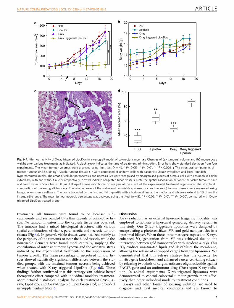
treatments. All tumours were found to be localised sub-cutaneously and surrounded by a thin capsule of connective tis-sue. No tumour invasion into the capsule tissue was observed.The tumours had a mixed histological structure, with variousspatial combinations of viable, paranecrotic and necrotic tumourtissues (Fig.6c). In general, viable tissues were localized mainly atthe periphery of the tumours or near the blood vessels, while thenon-viable elements were found more centrally, implying thecontribution of intrinsic tumour hypoxia and the oxidative stressinduced by the experimental treatments to the suppression oftumour growth. The mean percentage of necrotised tumour tis-sues showed statistically significant differences between the stu-died groups, with the maximal tumour necrosis being achievedwhen treated with X-ray triggered LipoDox (Fig. 6d). Thesefindings further confirmed that this strategy can achieve bettertherapeutic effect compared with individual modality treatment.More detailed histological analysis for each treatment (PBS-, X-ray-, LipoDox-, and X-ray-triggered LipoDox-treated) is providedin Supplementary Note 6.
DiscussionX-ray radiation, as an external liposome triggering modality, wasemployed to activate a liposomal gene/drug delivery system inthis study. Our X-ray- triggerable liposomes were designed byencapsulating a photosensitizer, VP, and gold nanoparticles in aliposomal bilayer. When these liposomes were exposed to X-rays,enhanced 1O2 generation from VP was achieved due to theinteraction between gold nanoparticles with incident X-rays. This1O2 oxidises unsaturated lipids and destabilises the membrane,allowing the release of entrapped cargos from the liposomes. Wedemonstrated that this release strategy has the capacity forin vitro gene knockdown and enhanced cancer cell-killing efficacyby releasing two kinds of cargos, antisense oligonucleotide againstPAC1R gene and an antitumour drug (Dox) upon X-ray radia-tion. In animal experiments, X-ray-triggered liposomes weredemonstrated to control colorectal tumour growth more effec-tively than other individual modality treatment conditions.
X-rays and other forms of ionising radiation are used todiagnose and treat medical conditions and are known to
50020
19
18******
17
Bod
y w
eigh
t (g)
16
15
PBS
LipoDox
X-ray
X-ray triggered LipoDox
PBS LipoDox X-ray X-ray triggeredLipoDox
PBSLipoDoxX-rayX-ray triggered LipoDox
400
300
Tum
our
volu
me
(mm
3 )
200
100
100
80
1
2
c
60M
ean
necr
otic
are
a (%
)
40
50 µm
******
**
00 4 8 12 16 20 0 4 8 12 16 20
Days Days
d
ba
Fig. 6 Antitumour activity of X-ray triggered LipoDox in a xenograft model of colorectal cancer. a,b Changes of (a) tumours’ volume and (b) mouse bodyweight after various treatments as indicated. A black arrow indicates the time of treatment administration. Error bars show standard deviation from fourexperiments. The mean tumour volumes were analysed using the t test (n= 4). * P < 0.05, ** P < 0.01, *** P < 0.001. c The structural components oftreated tumour (H&E staining). Viable tumour tissues (1) were composed of uniform cells with basophilic (blue) cytoplasm and large roundishhyperchromatic nuclei. The areas of cellular paranecrosis and necrosis (2) were recognised by disorganised groups of tumour cells with eosinophilic (pink)cytoplasm, with and without nuclei, respectively. Arrows indicate congested blood vessels. Note the spatial association between the viable tumour tissueand blood vessels. Scale bar is 50 µm. d Boxplot shows morphometric analysis of the effect of the experimental treatment regimens on the structuralcomposition of the xenograft tumours. The relative areas of the viable and non-viable (paranecrotic and necrotic) tumour tissues were measured usingImageJ open source software. The box is bounded by the first and third quartile with a horizontal line at the median and whiskers extend to 1.5 times theinterquartile range. The mean tumour necrosis percentage was analysed using the t test (n= 5). * P < 0.05, ** P < 0.01, *** P < 0.001, compared with X-ray-triggered LipoDox-treated group
NATURE COMMUNICATIONS | DOI: 10.1038/s41467-018-05118-3 ARTICLE
NATURE COMMUNICATIONS | (2018) 9:2713 | DOI: 10.1038/s41467-018-05118-3 | www.nature.com/naturecommunications 7

contribute to DNA mutations that may lead to dose-dependentand stochastic toxic effects. Compared with light, however, X-rayswith the suitable energy can easily penetrate the human body,activating gene/drug release in deep tissues once the X-ray-triggered liposomes reach their target. This feature will openmany opportunities for biomedical research and clinical medi-cine, from triggered gene therapies and chemotherapy, through toenhanced PDT which currently suffers from limited penetrationdepth of illumination light (usually in the UV and visible region).
Additionally, the strategy described here has been designed tobe compatible with future clinical translation. The materials andapproaches used in this study, such as VP, lipids, Dox, and X-rays, are clinically used in treatment of tumours. Although goldnanoparticles used in this study have not yet been approved bythe regulatory agencies, their size is compatible with therequirements of renal clearance60. In this way, long-term nano-particle toxicity is likely to be minimised if not eliminated.Moreover, the ease of conjugation of targeting ligands to lipo-some surface with appropriate linkers, for example, lipid-polyethylene glycol (PEG)61, would be an added advantagewhen applied to the targeted therapy, in particular for tumourtreatment. From a clinical point of view, it would be beneficial tohave access to this multimodality treatment, given our evidence ofbetter therapeutic effect (or, potentially, equal therapeutic effect)at diminished toxicity in the case when single modality treatmentoptions alone can only produce desired therapeutic effects at asignificant cost of short- and long-term toxicity.
MethodsPreparation of liposomes loaded with gold and VP. 350 µL of DOTAP (AvantiPolar Lipids, no. 890890 P) dissolved in chloroform (100 mgmL−1, Sigma-Aldrich,no. 288306-1 L) was mixed with 370 µL of DOPC (Avanti Polar Lipids, no. 850375P) dissolved in chloroform (100 mgmL−1), followed by addition of 40 µL of goldnanoparticle suspension (Nanocomposix, Inc) and 50 µL of VP (Sigma-Aldrich, no.SML0534-5MG) dissolved in dimethyl sulfoxide (DMSO, 2.3 mgmL−1, Sigma-Aldrich, no. 472301-500ML). For the synthesis of empty liposomes, VP and goldnanoparticles were omitted in the mixture solution. The mixture was diluted to 1.0mL in total volume using chloroform and vortexed gently for 10 min. Chloroformwas evaporated off with a stream of Argon and the remaining DMSO was eva-porated under freeze-drying, which was carried out in a freeze dryer (Alpha 1–4LDplus, John Morris Scientific Pty Ltd). The lipid film was hydrated by adding 1.0mL of DI water to a glass test tube, followed by vigorous stirring until the sus-pension was homogenised. The hydrated lipid suspension was left overnight toallow the maximal swelling of liposomes. The suspension was then extruded eleventimes in an extruder (Avanti Polar Lipids, Inc) with two 1.0 mL glass syringes. Thepore size of the polycarbonate membrane (Avanti Polar Lipids, Inc) was 200 nm.The resulting suspension was stored at 4 °C under argon. For encapsulation ofcalcein inside liposomes, 1.0 mL calcein solution (100 mM, Sigma-Aldrich, no.C0875-5G) was used as lipid hydration solution, instead of DI water. For encap-sulation of oligonucleotides, 1.0 mL PBS (pH 7.4) solution containing antisenseoligonucleotide (10 µM, 5′-TGGTGCTTCCCAGCCACTAT-3′) with 3′ FAMlabelling against PAC1R gene (Integrated DNA Technologies Pte. Ltd.) was used tohydrate lipid film, followed by the hydration procedure described above. In orderto remove calcein and oligonucleotides present in the supernatant after hydration,liposomes were then centrifuged at 14000×g for 10 min by using Pall Nanosepcentrifugal devices (Sigma-Aldrich) as per manufacturer’s instructions.
Synthesis of LipoDox. The encapsulation of doxorubicin inside of liposomes wasconducted as per a published protocol, using a gradient exchange method withminor modifications62. 1 mL ammonium sulphate (250 mM, Sigma-Aldrich, no.A4418-100G) was added to the glass test tube where the lipid film was producedafter evaporation of organic solvent, followed by the hydration procedure describedabove. Free ammonium sulphate was removed by dialysis in the PBS solution (pH7.4) with buffer exchange repeated four times. The Dox solution (10 mgmL−1,Sigma-Aldrich, no. D1515-10MG) was subsequently added to hydrated liposomesuspension with a drug to lipid mass ratio of 1:10, followed by incubation at 60 °Cfor 1 h. Unloaded Dox was removed by dialysis in PBS solution (pH 7.4) with fourtime buffer exchange.
Preparation of liposome incorporating ETP. Liposomes incorporating ETP, VPand gold nanoparticles were prepared by thin film hydration with some mod-ifications. Briefly, 100 µL of DOTAP (50 mgmL−1in chloroform) was mixed with54 µL of DOPC (100 mgmL−1 in chloroform), followed by addition of 6 µL of gold
nanoparticle suspension, 7 µL of VP (2.3 mgmL−1 in DMSO) and 83.5 µL of ETP(Sigma-Aldrich, no. E1383-25MG, 1 mgmL−1 in chloroform and ethanol (1:1 V/V)). After evaporation of organic solvent, the lipid film was hydrated with 1 mLPBS (pH 7.4). The hydration and extrusion procedure was the same as describedabove. The unloaded etoposide was removed by dialysis in the PBS solution (pH7.4) with buffer exchange repeated four times.
Preparation of folate-conjugated liposomes. Folate-conjugated liposomes wereprepared by postinsertion of DSPE-PEG2000-Folate micelles into preformedliposomes with slight modifications63,64. In brief, 1 mg DSPE-PEG2000-folate(Avanti Polar Lipids, no. 880124) was dissolved in 320 µL DMSO, followed byhydration with 3.1 mL of distilled water, producing 100 µM micelle suspension.The suspension was then dialysed three times in a 10000 MWCO dialysis tubingagainst 1 L water to remove DMSO. After this, 40 µL of micelles were added to 1mL of the preformed liposome suspension in ammonium sulphate (250 mM) andheated at 60 °C for 1 h to produce folate-tethered liposomes. Leaked ammoniumsulphate and unincorporated micelles were removed by dialysis. To determine thefolate content conjugated with liposomes, bare liposomes was used in conjugationprocedure instead of VP-loaded liposomes. After preparation, the folate amountwas determined by measuring the UV absorbance at 285 nm after lysing liposomeswith 0.1% Triton X-100 and comparing with a standard curve of folic acid with theknown concentration.
Characterisation of liposomes. The extinction spectra of liposomes loaded withgold nanoparticles and VP, VP alone and gold nanoparticles alone were measuredusing a spectrophotometer (Cary 5000 UV-Vis-NIR, Varian Inc.). Size distributionand zeta potentials of liposomes were measured with a Zetasizer Nano Series fromMalvern Instruments. The morphology of liposomes was documented usingTransmission Electron Microscopy (TEM). For TEM imaging, the liposomesamples were prepared by placing a drop of suspension onto a copper grid and air-dried, following negative staining with one drop of 2% aqueous Uranyl Acetate forcontrast enhancement. The air-dried samples were then imaged using a PHILIPSCM 10 system at an accelerating voltage of 100 KV. Images were captured with anOlympus Megaview G10 camera and iTEM software. To determine the encapsu-lation efficiency of oligonucleotides, Dox and etoposide loaded inside of liposomes,Triton X-100 (0.1%, Sigma-Aldrich, no. T8787-50ML) was added to as-preparedliposome solution, resulting in the gene/drug release. The FAM fluorescence fromoligonucleotides (Ex/Em: 494 nm/520 nm) and Dox fluorescence (Ex/Em 485/590nm) was recorded on a Fluorolog-Tau-3 system (Jobin Yvon-Horiba, US) andcompared with the corresponding oligonucleotide and Dox standard curves,respectively. The epotoside amount was determined by measuring the UV absor-bance at 285 nm under Cary UV-VIS-NIR absorption spectrophotometer (VarianIncl.) and comparing with the epotoside standard curve.
1O2 generation tests with light and X-ray triggering. For light illumination, a365 nm LED was used to illuminate the samples. 16 µL of SOSG (0.5 mM, ThermoFisher Scientific Inc, no. S36002) was mixed with 3 mL of liposome suspension andthe mixture was then placed in a cuvette, followed by illumination under a 365 nmLED (2.5 mW cm−2, irradiation for 10 min). After illumination, the SOSG fluor-escence at 525 nm upon 488 nm excitation was recorded using a fluorescencespectrophotometer. For X-ray radiation, a linear accelerator (6 MeV LINAC, ElektaAB, Sweden) was used to deliver different doses (1 Gy, 2 Gy and 4 Gy) to thesamples. 96-well plates with 200 µL of liposome suspension and 2 µL of SOSG (0.5mM) in each well were exposed to X-ray radiation. The irradiation of samples wascarried out using 6MeV X-ray photons from the anterior and posterior directedradiation fields. After irradiation, the SOSG fluorescence was recorded using amicroplate reader (PHERAstar FS system, BMG LABTECH, Germany).
Calcein release assay with light and X-ray irradiation. Liposomes loaded withcalcein were separated from free calcein molecules by using Pall Nanosep® cen-trifugal devices (Sigma-Aldrich) equilibrated with 10 mM Tris/HCl. They werethen activated by light illumination and ionizing radiation, respectively. Theexperiment process was the same as described in the 1O2 generation test, apartfrom the omission of SOSG. The induced release and subsequent dilution of thecalcein previously contained in the liposome s, leading to an increase of calceinfluorescence65,66. The calcein fluorescence signal was recorded at 510 nm uponexcitation at 485 nm. The percentage of calcein release (Rc(%)) at various illumi-nation time points or X-ray dosage was calculated as follows:
Rc %ð Þ ¼ FtðdÞ � F0Fmax � F0
´ 100% ð1Þ
where Ft and F0 respectively indicate the fluorescence intensity of calcein at variousillumination time points and without illumination. Fmax refers to the total fluor-escence intensity of calcein after the disruption of liposomes by adding 0.1% TritonX-100. For X-ray radiation, Fd is the fluorescence intensity of calcein at variousradiation doses, d.
ARTICLE NATURE COMMUNICATIONS | DOI: 10.1038/s41467-018-05118-3
8 NATURE COMMUNICATIONS | (2018) 9:2713 | DOI: 10.1038/s41467-018-05118-3 | www.nature.com/naturecommunications

Serum and pH stability studies of PEGylated liposomes. 200 µL LipoDoxsamples with and without PEG modification were respectively diluted in PBS (pH7.4) containing foetal bovine serum (FBS) with different concentrations (0%, 10%,25 and 50%). All samples were dialyzed in Slide-A-Lyzer MINI dialysis devices(Thermo Fisher Scientific). These devices were then kept in 50 mL centrifuge tubeswith 10 mL PBS at 37 °C for 48 h. At various time points (0 h, 2 h, 4 h, 18 h, 24 hand 48 h), an aliquot of PBS was taken for the fluorescence characterisation of thereleased Dox. The total Dox fluorescence was measured by disrupting liposomeswith 0.1% Triton X-100. The percentage of Dox release at various time points wascalculated by using the same formula as that applied to the calcein release assays. Inour pH-triggered drug release studies, 200 µL Dox-loaded PEGylated liposomesuspension was incubated with PBS (containing 10% FBS) with pH respectivelyadjusted to 7.4 (control), 6.0 and 5.0, followed by the same dialysis procedure andfluorescence measurement described above.
Cell preparation and ionizing radiation treatment of cells. Rat PC12 cells,human colon adenocarcinoma HCT 116 cells and normal human colon epithelialcells (CCD 841 CoN) were purchased from the American Type Culture Collection.PC12 cells were cultured in Dulbecco’s modified Eagle’s medium (DMEM); HCT116 cells were cultured in McCoy’s 5 A (modified) medium; CCD 841 CoN cellswere cultured in Eagle’s Minimum Essential Medium (EMEM). All culture mediawere supplemented with 10% foetal bovine serum and 1% antibiotic-antimycotic.The flasks were maintained in a 37 °C incubator with 5% CO2 humidified air. Thecells were detached with trypsin and transferred at appropriate dilutions into 96-well plates for cell viability assays or glass-bottom petri dishes for cell imaging. ForX-ray radiation experiments, the cells were radiated by using the same acceleratoras described in the 1O2 generation test.
Imaging and analysis of cellular uptake of liposomes. The PC12 cells(3 × 10⁴ mL−1) were attached to glass-bottom petri dishes and incubated at 37 °Cfor 24 h. After removing the culture medium, the cells were incubated with lipo-some suspension (25 µM) in culture medium supplemented with 10% FBS for 1 h,4 h and 10 h. The cells were then washed with PBS (1 × , PH 7.4) three times toremove free liposomes. To assess the uptake of liposome nanoparticles, the cellswere fixed with 2.5% paraformaldehyde for 10 min at room temperature, washedtwice with PBS (1 × , PH 7.4) and stained with Hoechst 33342 (5 µg ml−1) for 10min at room temperature before imaging. The cells were imaged using a Leica SP2confocal laser scanning microscopy system. A violet laser at 405 nm and an argonlaser at 496 nm were used for the excitation of VP and FAM-labelled oligonu-cleotide entrapped inside liposomes, respectively. The imaging of uptake activity ofFA-targeted liposomes into HCT 116 cells and CCD 841CoN cells were alsoconducted as mentioned above.
For quantitative analysis, fluorescently labelled DOTAP (Avanti Polar Lipids,no. 810890 P), was employed, instead of standard DOTAP in order to preparefluorescent liposomes. PC12 cells (1 × 10⁴ mL−1) were cultured in petri dishes at37 °C for 24 h. After removing the old culture medium, 1 mL of a fresh mediumcontaining 10 μL of fluorescently labelled liposomes (0.5 mg mL−1) was added tothe petri dishes and the cells were incubated at 37 °C for a further 4 h. Afterincubation, the cells were washed with fresh medium three times to remove freeliposomes, detached with trypsin from the petri dishes and counted using a cellcounter (Countess II FL automated cell counter from Thermo Scientific). 100 µLNaOH (1M) and 100 µL Triton X-100 (1% v/v) were subsequently added to 800 µLof cell suspension. The cells were lysed at R.T. for 2 h with constant shaking. Aftercell lysis, fluorescence (Ex/Em: 460/535 nm) was recorded on a Fluorolog-Tau-3system and compared with the standard curve of free fluorescent DOTAP solution.A detailed calculation of the number of liposomes per cell is described inSupplementary Note 4.
Indirect immunofluorescence staining of PAC1R. The PC12 cells were fixed with2.5% paraformaldehyde for 10 min and permeabilized with 0.1% Triton X-100 foranother 10 min at room temperature, followed by blocking with 5% bovine serumalbumin for 30 min. The cells were then incubated with goat anti-PAC1R primaryantibody (1:50 dilution, Santa Cruz Biotechnology, no. sc-15964) for 90 min anddonkey anti goat IgG secondary antibody (1:100 dilution, Santa Cruz Bio-technology, no. sc-2024) conjugated to FITC for 30 min at room temperature.
Cytotoxicity assays of X-ray-triggered LipoDox and LipoETP. The in vitroantitumour effect of X-ray-triggered LipoDox and LipoETP was evaluated usingthe MTS test. Before treatment, the HCT 116 cells (2 × 10⁴ mL−1) were grown on96-well plates in the culture medium with 10% FBS for 24 h. After removing the oldmedium, the cells were respectively incubated with a series of LipoDox andLipoETP samples diluted in the culture medium with 10% FBS for 4 h. Afterincubation, the old medium was removed and a fresh medium was added to cells,followed by X-ray radiation with 4 Gy. The cytotoxicity of X-ray-triggered LipoDoxand LipoETP on HCT 116 cells at various time points (0 h, 2 h, 4 h and 24 h) wasdetermined by the MTS test (Promega Co., WI, USA, no. G3582) according tomanufacturer’s instructions and compared with control cells without any treat-ment. Cell viability was then calculated as a percentage of the absorbance of theuntreated control sample. The latter was set to 100%. For comparison purposes, the
viability of cells treated with LipoDox alone was also evaluated in the sameexperimental conditions.
Toxicity assays of LipoDox and X-ray on cells and gene. The PC12, HCT 116and CCD 841 CoN cells (1–4 × 10⁴ mL−1) were, respectively, grown on 96-wellplates in a culture medium with 10% FBS for 24 h. For liposome and LipoDoxtreatment experiments, the PC12 cells and CCD 841 CoN cells were, respectively,incubated with different liposome and LipoDox samples for 4 h, followed byincubation in a fresh medium for further 24 h. For the X-ray exposure experimentson cells, all three types of cells were radiated with 4 Gy, followed by incubation in afresh medium for further 24 and 48 h. Cell viability was assessed by using the samemethod as described above. For X-ray treatment of pure DNA molecules andmixture of DNA and verteporfin, 50 µL of antisense oligonucleotide solution (10µg mL−1) and 50 µL of mixture solution (10 µg mL−1 DNA and 32 µg mL−1 ver-teporfin) was respectively exposed to X-ray radiation with different dosages (1, 2and 4 Gy). After treatment, the gel electrophoresis was carried out in 1.2 % agarosegel in Tris-acetate-EDTA (TAE) buffer at 95 V for 45 min. The gel was stained withSYBR Safe DNA Gel Stain (Thermo Fisher) and photographed under UV lightusing a Bio-Rad imaging system.
In vivo antitumour efficacy by X-ray-triggered drug release. All procedureswere carried out with the approval from Macquarie University Animal EthicsCommittee (animal ethics approval No. 2017/001). 6–7 weeks old BALB/c nu/nufemale mice (The Animal Resources Centre, Perth, Australia) were injected sub-cutaneously with 5 × 10⁶ HCT 116 cells, suspended in 100 µl McCoy’s 5 A (mod-ified) medium without FBS, to the flank. Tumours were measured every two dayswith a caliper and volume (V) was calculated by using the following formula:
V ¼ π=6 ´ L ´W2 ð2Þ
where L and W are the length (large diameter) and width (short diameter) of thetumour. When tumour volume reached approximately 100 mm3, mice were ran-domly divided into 4 groups (n= 4 per group) for different treatments: Group Atreated PBS via intratumour injection (20 µL); Group B treated with liposomesuspension via intratumour injection (20 µL, 10 mg kg−1, approximately 10 µMgold nanoparticles and 20 µM VP used for each mouse); Group C treated with X-ray radiation (4 Gy, single fraction) and Group D treated with liposome suspensionvia intratumour injection (20 µL, 10 mg kg−1) and X-ray radiation (4 Gy, singlefraction). Mice were then maintained for additional 2 weeks. Body weight andtumour volume were measured every other day. After two weeks, mice weresacrificed and tumours were removed, photographed and fixed with 10% neutral-buffered formalin for histological analysis. Tumour tissues were cryosectioned intoserial sections of 6 µm in thickness and stained with haematoxylin and eosin(H&E) following conventional protocol. The histological preparations wereexamined using an upright research microscope Axio Imager Z2 (Zeiss, Germany)equipped with dry-air EC Plan-Neofluar (5 × /NA0.16; 10 × /NA0.30; 20 × /NA0.50Ph) and oil-immersion α Plan Apochromat (100×/NA1.46 oil) objectives (Zeiss,Germany). Images were recorded using a digital video camera AxioCam(1388×1040, Zeiss, Germany) in a single-frame and stitching modes.
Data availability. The relevant data generated and (or) analysed in the currentstudy are available from the corresponding author upon reasonable request.
Received: 18 November 2016 Accepted: 24 May 2018
References1. Thomas, C. E., Ehrhardt, A. & Kay, M. A. Progress and problems with the use
of viral vectors for gene therapy. Nat. Rev. Genet. 4, 346 (2003).2. Zhang, Y., Satterlee, A. & Huang, L. In vivo gene delivery by nonviral vectors:
overcoming hurdles? Mol. Ther. 20, 1298–1304 (2012).3. Luo, D. & Saltzman, W. M. Synthetic DNA delivery systems. Nat. Biotechnol.
18, 33–37 (2000).4. Liu, D., Yang, F., Xiong, F. & Gu, N. The smart drug delivery system and its
clinical potential. Theranostics 6, 1306 (2016).5. Allen, T. M. & Cullis, P. R. Liposomal drug delivery systems: from concept to
clinical applications. Adv. Drug Deliv. Rev. 65, 36–48 (2013).6. Pattni, B. S., Chupin, V. V. & Torchilin, V. P. New developments in liposomal
drug delivery. Chem. Rev. 115, 10938–10966 (2015).7. Khan, D. R., Rezler, E. M., Lauer‐Fields, J. & Fields, G. B. Effects of drug
hydrophobicity on liposomal stability. Chem. Biol. Drug Des. 71, 3–7 (2008).8. Akbarzadeh, A. et al. Liposome: classification, preparation, and applications.
Nanoscale Res. Lett. 8, 102 (2013).9. Nahire, R. et al. pH-triggered echogenicity and contents release from
liposomes. Mol. Pharm. 11, 4059–4068 (2014).
NATURE COMMUNICATIONS | DOI: 10.1038/s41467-018-05118-3 ARTICLE
NATURE COMMUNICATIONS | (2018) 9:2713 | DOI: 10.1038/s41467-018-05118-3 | www.nature.com/naturecommunications 9
10. Ferreira, D. d. S., Lopes, S. C. d. A., Franco, M. S. & Oliveira, M. C. pH-sensitive liposomes for drug delivery in cancer treatment. Ther. Deliv. 4,1099–1123 (2013).
11. Dicheva, B. M. et al. Targeted and heat-triggered doxorubicin delivery totumors by dual targeted cationic thermosensitive liposomes. J. Control Release195, 37–48 (2014).
12. Kono, K. et al. Highly temperature-sensitive liposomes based on athermosensitive block copolymer for tumor-specific chemotherapy.Biomaterials 31, 7096–7105 (2010).
13. Sarkar, N. R. et al. “Uncorking” of liposomes by matrix metalloproteinase-9.Chem. Commun. 0, 999–1001 (2005).
14. Arouri, A. et al. Development of a cell-based bioassay for phospholipase A2-triggered liposomal drug release. PLoS ONE. 10, e0125508 (2015).
15. Leung, S. J. & Romanowski, M. Light-activated content release fromliposomes. Theranostics 2, 1020 (2012).
16. Puri, A. Phototriggerable liposomes: current research and future perspectives.Pharmaceutics 6, 1–25 (2013).
17. Wilson, B. C. & Patterson, M. S. The physics, biophysics and technology ofphotodynamic therapy. Phys. Med. Biol. 53, R61 (2008).
18. Shum, P., Kim, J.-M. & Thompson, D. H. Phototriggering of liposomal drugdelivery systems. Adv. Drug Deliv. Rev. 53, 273–284 (2001).
19. Begg, A. C., Stewart, F. A. & Vens, C. Strategies to improve radiotherapy withtargeted drugs. Nat. Rev. Cancer 11, 239–253 (2011).
20. Kwatra, D., Venugopal, A. & Anant, S. Nanoparticles in radiation therapy: asummary of various approaches to enhance radiosensitization in cancer.Transl. Cancer Res. 2, 330–342 (2013).
21. Jain, S., Hirst, D. & O’sullivan, J. Gold nanoparticles as novel agents for cancertherapy. Br. J. Radiol. 85, 101–113 (2012).
22. Apanasevich, V. et al. Enhance the absorption of gamma-ray energy inside thetumor using gold nanoparticles and iodine particles. Cancer Oncol. Res. 2,17–20 (2014).
23. Butterworth, K. T. et al. Evaluation of cytotoxicity and radiation enhancementusing 1.9 nm gold particles: potential application for cancer therapy. Nanotech21, 295101 (2010).
24. Kotova, E. A., Kuzevanov, A. V., Pashkovskaya, A. A. & Antonenko, Y. N.Selective permeabilization of lipid membranes by photodynamic action viaformation of hydrophobic defects or pre-pores. Biochim. Biophys. ActaBiomembr. 1808, 2252–2257 (2011).
25. Yavlovich, A., Smith, B., Gupta, K., Blumenthal, R. & Puri, A. Light-sensitivelipid-based nanoparticles for drug delivery: design principles and futureconsiderations for biological applications.Mol. Membr. Biol. 27, 364–381 (2010).
26. Tejero, I., González-Lafont, A., Lluch, J. M. & Eriksson, L. A. Photo-oxidationof lipids by singlet oxygen: a theoretical study. Chem. Phys. Lett. 398, 336–342(2004).
27. Brodowska, K. et al. The clinically used photosensitizer Verteporfin (VP)inhibits YAP-TEAD and human retinoblastoma cell growth in vitro withoutlight activation. Exp. Eye Res. 124, 67–73 (2014).
28. Liu-Chittenden, Y. et al. Genetic and pharmacological disruption of theTEAD–YAP complex suppresses the oncogenic activity of YAP. Genes Dev.26, 1300–1305 (2012).
29. Chang, H.-I. & Yeh, M.-K. Clinical development of liposome-based drugs:formulation, characterization, and therapeutic efficacy. Int. J. Nanomed. 7, 49(2012).
30. Shen, Y. et al. in Photonics Asia 2010 78451F-78451F-78456 (InternationalSociety for Optics and Photonics, 2010).
31. Kim, S., Fujitsuka, M. & Majima, T. Photochemistry of singlet oxygen sensorgreen. J. Phys. Chem. B 117, 13985–13992 (2013).
32. Allen, T. & Cleland, L. Serum-induced leakage of liposome contents. Biochim.Biophys. Acta Biomembr. 597, 418–426 (1980).
33. Kundrot, C. E., Spangler, E. A., Kendall, D. A., MacDonald, R. C. & MacDonald,R. I. Sendai virus-mediated lysis of liposomes requires cholesterol. Proc. Natl.Acad. Sci. 80, 1608–1612 (1983).
34. Yavlovich, A. et al. Design of liposomes containing photopolymerizablephospholipids for triggered release of contents. J. Therm. Anal. Calorim. 98,97–104 (2009).
35. Maherani, B., Arab-Tehrany, E., Kheirolomoom, A., Geny, D. & Linder, M.Calcein release behavior from liposomal bilayer; influence of physicochemical/mechanical/structural properties of lipids. Biochimie 95, 2018–2033 (2013).
36. Clement, S., Sobhan, M., Deng, W., Camilleri, E. & Goldys, E. M.Nanoparticle-mediated singlet oxygen generation from photosensitizers. J.Photochem. Photobiol. A 332, 66–71 (2017).
37. Clement, S., Deng, W., Camilleri, E., Wilson, B. C. & Goldys, E. M. X-rayinduced singlet oxygen generation by nanoparticle-photosensitizer conjugatesfor photodynamic therapy: determination of singlet oxygen quantum yield.Sci. Rep. 6, 1–9 (2016).
38. Hutter, T., Huang, F. M., Elliott, S. R. & Mahajan, S. Near-field plasmonics ofan individual dielectric nanoparticle above a metallic substrate. J. Phys.Chem. C. 117, 7784–7790 (2013).
39. Tanabe, K. Field enhancement around metal nanoparticles and nanoshells:a systematic investigation. J. Phys. Chem. C. 112, 15721–15728(2008).
40. Conklin, D. et al. Electronic transport in porphyrin supermolecule-goldnanoparticle assemblies. Nano. Lett. 12, 2414–2419 (2012).
41. Shaikh, A. J. et al. Binding strength of porphyrin−gold nanoparticlehybrids based on number and type of linker moieties and a simple methodto calculate inner filter effects of gold nanoparticles using fluorescencespectroscopy. J. Phys. Chem. A 119, 1108–1116 (2015).
42. Chithrani, D. B. et al. Gold nanoparticles as radiation sensitizers in cancertherapy. Radiat. Res. 173, 719–728 (2010).
43. Hainfeld, J. F. et al. Gold nanoparticles enhance the radiation therapy of amurine squamous cell carcinoma. Phys. Med. Biol. 55, 3045 (2010).
44. Rahman, W. N. et al. Enhancement of radiation effects by gold nanoparticlesfor superficial radiation therapy. Nanomed. Nanotechnol. Biol. Med. 5,136–142 (2009).
45. Cho, S. H. Estimation of tumour dose enhancement due to gold nanoparticlesduring typical radiation treatments: a preliminary Monte Carlo study. Phys.Med. Biol. 50, N163 (2005).
46. Cooper, D. R., Bekah, D. & Nadeau, J. L. Gold nanoparticles and theiralternatives for radiation therapy enhancement. Front. Chem. 2, 86(2014).
47. Hainfeld, J. F., Slatkin, D. N. & Smilowitz, H. M. The use of gold nanoparticlesto enhance radiotherapy in mice. Phys. Med. Biol. 49, N309 (2004).
48. Slepushkin, V. A. et al. Sterically stabilized pH-sensitive liposomesintracellular delivery of aqueous contents and prolonged circulation in vivo. J.Biol. Chem. 272, 2382–2388 (1997).
49. Nam, J. et al. pH-responsive gold nanoparticles-in-liposome hybridnanostructures for enhanced systemic tumor delivery. Nanoscale 5,10175–10178 (2013).
50. Parker, N. et al. Folate receptor expression in carcinomas and normal tissuesdetermined by a quantitative radioligand binding assay. Anal. Biochem. 338,284–293 (2005).
51. Hong, E. J., Choi, D. G. & Shim, M. S. Targeted and effective photodynamictherapy for cancer using functionalized nanomaterials. Acta Pharmacol. Sin. B6, 297–307 (2016).
52. Zhang, S., Lu, C., Zhang, X., Li, J. & Jiang, H. Targeted delivery of etoposide tocancer cells by folate-modified nanostructured lipid drug delivery system.Drug Deliv. 23, 1838–1845 (2016).
53. Bovelli, D., Plataniotis, G., Roila, F. & Group, E. G. W. Cardiotoxicity ofchemotherapeutic agents and radiotherapy-related heart disease: ESMOClinical Practice Guidelines. Ann. Oncol. 21, v277–v282 (2010).
54. Nemade, H. et al. Cell death mechanisms of the anti-cancer drug etoposide onhuman cardiomyocytes isolated from pluripotent stem cells. Arch. Toxicol. 92,1507–1524 (2018).
55. Baskar, R., Dai, J., Wenlong, N., Yeo, R. & Yeoh, K.-W. Biological response ofcancer cells to radiation treatment. Front. Mol. Biosci. 1, 24 (2014).
56. Niedre, M., Patterson, M. S. & Wilson, B. C. Direct near-infraredluminescence detection of singlet oxygen generated by photodynamictherapy in cells in vitro and tissues in vivo. Photochem. Photobiol. 75, 382–391(2002).
57. DeRosa, M. C. & Crutchley, R. J. Photosensitized singlet oxygen and itsapplications. Coord. Chem. Rev. 233, 351–371 (2002).
58. Krieger-Liszkay, A. Singlet oxygen production in photosynthesis. J. Exp. Bot.56, 337–346 (2005).
59. Redmond, R. W. & Kochevar, I. E. Spatially resolved cellular responses tosinglet gen. Photochem. Photobiol. 82, 1178–1186 (2006).
60. Rengan, A. K. et al. In vivo analysis of biodegradable liposome goldnanoparticles as efficient agents for photothermal therapy of cancer. Nano.Lett. 15, 842–848 (2015).
61. Milla, P., Dosio, F. & Cattel, L. PEGylation of proteins and liposomes: apowerful and flexible strategy to improve the drug delivery. Curr. Drug.Metab. 13, 105–119 (2012).
62. Li, X., Ding, L., Xu, Y., Wang, Y. & Ping, Q. Targeted delivery of doxorubicinusing stealth liposomes modified with transferrin. Int. J. Pharm. 373, 116–123(2009).
63. Ishida, T., Iden, D. L. & Allen, T. M. A combinatorial approach to producingsterically stabilized (Stealth) immunoliposomal drugs. FEBS Lett. 460,129–133 (1999).
64. Yoshino, K. et al. Comparative studies of irinotecan-loaded polyethyleneglycol-modified liposomes prepared using different PEG-modificationmethods. Biochim. Biophys. Acta Biomembr. 1818, 2901–2907 (2012).
65. Shimanouchi, T., Ishii, H., Yoshimoto, N., Umakoshi, H. & Kuboi, R. Calceinpermeation across phosphatidylcholine bilayer membrane: effects ofmembrane fluidity, liposome size, and immobilization. Colloids Surf. BBiointerfaces 73, 156–160 (2009).
66. Han, H. D., Kim, T. W., Shin, B. C. & Choi, H. S. Release of calcein fromtemperature-sensitive liposomes. Macromol. Res. 13, 54–61 (2005).
ARTICLE NATURE COMMUNICATIONS | DOI: 10.1038/s41467-018-05118-3
10 NATURE COMMUNICATIONS | (2018) 9:2713 | DOI: 10.1038/s41467-018-05118-3 | www.nature.com/naturecommunications

AcknowledgementsAll TEM images in this work were performed in the Microscopy Unit, Faculty of Scienceand Engineering at Macquarie University. We thank Dr. Michael Grace, Dr.VaughanMoutrie and Dr. Daniel Santos from Genesis Cancer Care NSW and Macquarie Uni-versity Hospital, for helping us with X-ray radiation experiments. We also thank MrPeter Wieland from the Department of Earth and Planetary Science at MacquarieUniversity for his assistance in ICP-MS measurement. This work is supported by Dis-covery Early Career Researcher Award scheme (DE130100894), Centre of Excellencescheme (CE140100003) from the Australian Research Council and Sydney VitalTranslational Cancer Research Centre. The experimental work was carried out at Mac-quarie University and Genesis Cancer Care NSW.
Author contributionsW. D. and E. G. designed this study, conducted experiments and draft the manuscript.W. C. conducted animal experiments, cell lysis experiments and gel electrophoresis. S. C.conducted calculation of SOQY and 1O2 generation enhancement factor under lightillumination and the number of 1O2 generated under X-ray radiation. A. G. contributedto histological analysis and interpretation. Z. Z. helped with measurement of SOSGfluorescence intensity and calcein release assays after X-ray radiation. He also con-tributed to animal ethics application and experiments. A. E. contributed to manuscriptpreparation.
Additional informationSupplementary Information accompanies this paper at https://doi.org/10.1038/s41467-018-05118-3.
Competing interests: The authors declare no competing interests.
Reprints and permission information is available online at http://npg.nature.com/reprintsandpermissions/
Publisher's note: Springer Nature remains neutral with regard to jurisdictional claims inpublished maps and institutional affiliations.
Open Access This article is licensed under a Creative CommonsAttribution 4.0 International License, which permits use, sharing,
adaptation, distribution and reproduction in any medium or format, as long as you giveappropriate credit to the original author(s) and the source, provide a link to the CreativeCommons license, and indicate if changes were made. The images or other third partymaterial in this article are included in the article’s Creative Commons license, unlessindicated otherwise in a credit line to the material. If material is not included in thearticle’s Creative Commons license and your intended use is not permitted by statutoryregulation or exceeds the permitted use, you will need to obtain permission directly fromthe copyright holder. To view a copy of this license, visit http://creativecommons.org/licenses/by/4.0/.
© The Author(s) 2018
NATURE COMMUNICATIONS | DOI: 10.1038/s41467-018-05118-3 ARTICLE
NATURE COMMUNICATIONS | (2018) 9:2713 | DOI: 10.1038/s41467-018-05118-3 | www.nature.com/naturecommunications 11