The Optimized Delivery of Triterpenes by Liposomal ... - MDPI
51
Citation: Milan, A.; Mioc, A.; Prodea, A.; Mioc, M.; Buzatu, R.; Ghiulai, R.; Racoviceanu, R.; Caruntu, F.; ¸ Soica, C. The Optimized Delivery of Triterpenes by Liposomal Nanoformulations: Overcoming the Challenges. Int. J. Mol. Sci. 2022, 23, 1140. https://doi.org/10.3390/ ijms23031140 Academic Editor: Christian Celia Received: 20 December 2021 Accepted: 18 January 2022 Published: 20 January 2022 Publisher’s Note: MDPI stays neutral with regard to jurisdictional claims in published maps and institutional affil- iations. Copyright: © 2022 by the authors. Licensee MDPI, Basel, Switzerland. This article is an open access article distributed under the terms and conditions of the Creative Commons Attribution (CC BY) license (https:// creativecommons.org/licenses/by/ 4.0/). International Journal of Molecular Sciences Review The Optimized Delivery of Triterpenes by Liposomal Nanoformulations: Overcoming the Challenges Andreea Milan 1,2,† , Alexandra Mioc 1,2,† , Alexandra Prodea 1,2 , Marius Mioc 1,2, *, Roxana Buzatu 3, * , Roxana Ghiulai 1,2 , Roxana Racoviceanu 1,2 , Florina Caruntu 4 and Codru¸ ta ¸ Soica 1,2 1 Faculty of Pharmacy, “Victor Babe¸ s” University of Medicine and Pharmacy, 2 E. Murgu Sq., 300041 Timi¸ soara, Romania; [email protected] (A.M.); [email protected] (A.M.); [email protected] (A.P.); [email protected] (R.G.); [email protected] (R.R.); [email protected] (C.¸ S.) 2 Research Centre for Pharmaco-Toxicological Evaluation, “Victor Babes” University of Medicine and Pharmacy, Eftimie Murgu Sq., No. 2, 300041 Timi¸ soara, Romania 3 Faculty of Dental Medicine, “Victor Babe¸ s” University of Medicine and Pharmacy Timi¸ soara, 2 Eftimie Murgu Street, 300041 Timi¸ soara, Romania 4 Faculty of Medicine, “Victor Babe¸ s” University of Medicine and Pharmacy Timi¸ soara, 2 Eftimie Murgu Street, 300041 Timi¸ soara, Romania; caruntu.fl[email protected] * Correspondence: [email protected] (M.M.); [email protected] (R.B.); Tel.: +40-256-494-604 (M.M. & R.B.) † These authors contributed equally to this work. Abstract: The last decade has witnessed a sustained increase in the research development of modern- day chemo-therapeutics, especially for those used for high mortality rate pathologies. However, the therapeutic landscape is continuously changing as a result of the currently existing toxic side effects induced by a substantial range of drug classes. One growing research direction driven to mitigate such inconveniences has converged towards the study of natural molecules for their promising therapeutic potential. Triterpenes are one such class of compounds, intensively investigated for their therapeutic versatility. Although the pharmacological effects reported for several representatives of this class has come as a well-deserved encouragement, the pharmacokinetic profile of these molecules has turned out to be an unwelcomed disappointment. Nevertheless, the light at the end of the tunnel arrived with the development of nanotechnology, more specifically, the use of liposomes as drug delivery systems. Liposomes are easily synthesizable phospholipid-based vesicles, with highly tunable surfaces, that have the ability to transport both hydrophilic and lipophilic structures ensuring superior drug bioavailability at the action site as well as an increased selectivity. This study aims to report the results related to the development of different types of liposomes, used as targeted vectors for the delivery of various triterpenes of high pharmacological interest. Keywords: liposomes; triterpenes; targeted delivery; nanocarriers; nano-therapy 1. Introduction The introduction of drugs derived from synthetic organic chemistry in the XXth cen- tury has marked the phenomenal advance of therapy in all medical fields due to their intense biologic effects and economic feasibility for large-scale preparation. However, a major disadvantage of synthetic drugs lies in their adverse effects such as the high, intolerable organ toxicity exhibited by anti-neoplastic drugs. Currently, this issue is mit- igated by the development of modern and sophisticated therapies such as gene therapy, immunotherapy, or targeted therapy which show promising clinical outcomes but at high costs. A more approachable path consists of the replacement of synthetic drugs with nat- ural compounds which exhibit similar pharmacological activities and can be chemically modified in order to obtain a suitable bioavailability [1]. Amongst all-natural bioactive compounds, triterpenes represent one of the most promising categories, not only for their Int. J. Mol. Sci. 2022, 23, 1140. https://doi.org/10.3390/ijms23031140 https://www.mdpi.com/journal/ijms
-
Upload
khangminh22 -
Category
Documents
-
view
3 -
download
0
Transcript of The Optimized Delivery of Triterpenes by Liposomal ... - MDPI
�����������������
Citation: Milan, A.; Mioc, A.; Prodea,
A.; Mioc, M.; Buzatu, R.; Ghiulai, R.;
Racoviceanu, R.; Caruntu, F.; Soica, C.
The Optimized Delivery of
Triterpenes by Liposomal
Nanoformulations: Overcoming the
Challenges. Int. J. Mol. Sci. 2022, 23,
1140. https://doi.org/10.3390/
ijms23031140
Academic Editor: Christian Celia
Received: 20 December 2021
Accepted: 18 January 2022
Published: 20 January 2022
Publisher’s Note: MDPI stays neutral
with regard to jurisdictional claims in
published maps and institutional affil-
iations.
Copyright: © 2022 by the authors.
Licensee MDPI, Basel, Switzerland.
This article is an open access article
distributed under the terms and
conditions of the Creative Commons
Attribution (CC BY) license (https://
creativecommons.org/licenses/by/
4.0/).
International Journal of
Molecular Sciences
Review
The Optimized Delivery of Triterpenes by LiposomalNanoformulations: Overcoming the Challenges
Andreea Milan 1,2,† , Alexandra Mioc 1,2,† , Alexandra Prodea 1,2 , Marius Mioc 1,2,*, Roxana Buzatu 3,* ,
Roxana Ghiulai 1,2, Roxana Racoviceanu 1,2 , Florina Caruntu 4 and Codruta Soica 1,2
1 Faculty of Pharmacy, “Victor Babes” University of Medicine and Pharmacy, 2 E. Murgu Sq.,300041 Timisoara, Romania; [email protected] (A.M.); [email protected] (A.M.);[email protected] (A.P.); [email protected] (R.G.); [email protected] (R.R.);[email protected] (C.S.)
2 Research Centre for Pharmaco-Toxicological Evaluation, “Victor Babes” University of Medicine and Pharmacy,Eftimie Murgu Sq., No. 2, 300041 Timisoara, Romania
3 Faculty of Dental Medicine, “Victor Babes” University of Medicine and Pharmacy Timisoara,2 Eftimie Murgu Street, 300041 Timisoara, Romania
4 Faculty of Medicine, “Victor Babes” University of Medicine and Pharmacy Timisoara, 2 Eftimie Murgu Street,300041 Timisoara, Romania; [email protected]
* Correspondence: [email protected] (M.M.); [email protected] (R.B.);Tel.: +40-256-494-604 (M.M. & R.B.)
† These authors contributed equally to this work.
Abstract: The last decade has witnessed a sustained increase in the research development of modern-day chemo-therapeutics, especially for those used for high mortality rate pathologies. However, thetherapeutic landscape is continuously changing as a result of the currently existing toxic side effectsinduced by a substantial range of drug classes. One growing research direction driven to mitigatesuch inconveniences has converged towards the study of natural molecules for their promisingtherapeutic potential. Triterpenes are one such class of compounds, intensively investigated for theirtherapeutic versatility. Although the pharmacological effects reported for several representatives ofthis class has come as a well-deserved encouragement, the pharmacokinetic profile of these moleculeshas turned out to be an unwelcomed disappointment. Nevertheless, the light at the end of thetunnel arrived with the development of nanotechnology, more specifically, the use of liposomes asdrug delivery systems. Liposomes are easily synthesizable phospholipid-based vesicles, with highlytunable surfaces, that have the ability to transport both hydrophilic and lipophilic structures ensuringsuperior drug bioavailability at the action site as well as an increased selectivity. This study aims toreport the results related to the development of different types of liposomes, used as targeted vectorsfor the delivery of various triterpenes of high pharmacological interest.
Keywords: liposomes; triterpenes; targeted delivery; nanocarriers; nano-therapy
1. Introduction
The introduction of drugs derived from synthetic organic chemistry in the XXth cen-tury has marked the phenomenal advance of therapy in all medical fields due to theirintense biologic effects and economic feasibility for large-scale preparation. However,a major disadvantage of synthetic drugs lies in their adverse effects such as the high,intolerable organ toxicity exhibited by anti-neoplastic drugs. Currently, this issue is mit-igated by the development of modern and sophisticated therapies such as gene therapy,immunotherapy, or targeted therapy which show promising clinical outcomes but at highcosts. A more approachable path consists of the replacement of synthetic drugs with nat-ural compounds which exhibit similar pharmacological activities and can be chemicallymodified in order to obtain a suitable bioavailability [1]. Amongst all-natural bioactivecompounds, triterpenes represent one of the most promising categories, not only for their
Int. J. Mol. Sci. 2022, 23, 1140. https://doi.org/10.3390/ijms23031140 https://www.mdpi.com/journal/ijms
Int. J. Mol. Sci. 2022, 23, 1140 2 of 51
large plethora of compounds but also for their wide range of pharmacological activities,such as anti-tumor, antioxidant, anti-viral, anti-microbial, cardioprotective, antidiabetic,neuroprotective, etc., [2]. Even though triterpenes exhibit such a significant pharmacologi-cal potential, their low solubility, hence bioavailability, leads to the need of finding properformulations to enhance their biomedical activities [3].
Microtechnology and nanotechnology have revolutionized the 21st century of thepharmaceutical and biotechnological industry that are being viewed as powerful tools forbasic research, imaging, and especially for enhanced drug delivery. The introduction ofmicro and nano-drug delivery systems provided the opportunity to obtain an improvedtherapeutic response with lower drug quantities while maintaining a high safety profile.The development of nanotechnology had a huge impact on creating new pharmaceuticalproducts with significantly improved bioavailability [4], safety, and patient compliance,thus offering the possibility of delivering highly lipophilic or chemically unstable drugs [5].The use of nanoparticles is not new to today’s technology; it originated in the medievalperiod when nano-sized gold particles were used in order to stain glass windows ofchurches, coloring them in orange, purple, red, and green according to their size [6].
Besides the clear distinction in particle size (1–100 nm for nanoparticles, 100–1000 nmfor sub-microparticles, and 1–1000 µm microparticles), when comparing nanoparticles withmicroparticles as delivery systems, both possess properties that can make either of themmore or less useful depending on the application type (chapter). Microparticles as wellas microspheres and microcapsules, were developed for multiparticulate drug deliverysystem formulations. Due to their structural and functional abilities, they are considered tobe suitable, tolerable, and convenient for drug administration via several routes. Despitetheir advantage to act locally, as microcarriers do not traverse into the interstitium and canconsecutively incorporate toxic substances, when aiming for a systemic targeted deliverythis is a shortcoming [7]. Consecutively, nanocarriers have evolved in order to overcomethe disadvantage of microcarriers for systemic targeted delivery, as nanocarriers are able tocross through biological barriers [8].
Among various types of nanoparticles, lipidic vesicular systems, including liposomes,are nano-vehicles with many advantages due to their unique physical and chemical prop-erties as well as high potency and ability to encapsulate a large number of differentmolecules [9]. Additionally, this type of nanoformulation exhibits high flexibility in prepa-ration allowing it to undergo several types of surface modifications for extensive use incutaneous applications, gene therapy, or as drug delivery systems in many pathologiessuch as cancer, HIV, tuberculosis, and brain pathologies [10].
This research aims to review the main methods used for triterpenoids inclusions indifferent types of liposomes while focusing on the positive biological results that couldcontribute to the future development of natural treatments. The literature data used to pre-pare the present review were identified by accessing internationally recognized databasessuch as PubMed, Web of Science, Science Direct, and Springer. Primary Medical SubjectHeadings [MeSH] terms such as “liposomes”, “triterpenes”, “pentacyclic triterpenes”,“drug delivery systems”, “nanotechnology” and regular keywords such as “vesicular sys-tems”, “ liposomal formulations”, ‘’betulinic acid liposomes”, oleanolic acid liposomes”,‘’ursolic acid liposomes”, lupeol liposones”, ‘’boswellic acid liposomes”, “glycyrrhetinicacid liposomes”, “target therapy” combined with Boolean terms, were used to obtain thearticles of interest. Only original articles and clinical trials published in English-languagewere included in this review. The reference list of the cited papers was examined in detailin order to extract other additional relevant literature data. No restrictions were appliedregarding the publication date.
2. Nanotechnology and Nano-Therapy
In comparison to traditional therapeutics, which focus mostly on obtaining stable andtolerable therapies, nano-therapy aims for safer, more efficient, and less toxic treatment thatcan also increase patient compliance [11].
Int. J. Mol. Sci. 2022, 23, 1140 3 of 51
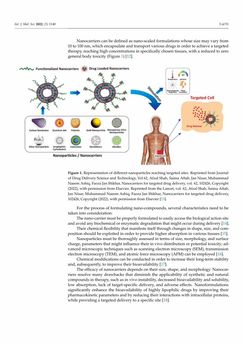
Nanocarriers can be defined as nano-scaled formulations whose size may vary from10 to 100 nm, which encapsulate and transport various drugs in order to achieve a targetedtherapy, reaching high concentrations in specifically chosen tissues, with a reduced to zerogeneral body toxicity (Figure 1) [12].
Figure 1. Representation of different nanoparticles reaching targeted sites. Reprinted from Journalof Drug Delivery Science and Technology, Vol 62, Afzal Shah, Saima Aftab, Jan Nisar, MuhammadNaeem Ashiq, Faoza Jan Iftikhar, Nanocarriers for targeted drug delivery, vol. 62, 102426, Copyright(2022), with permission from Elsevier. Reprinted from the Lancet, vol. 62, Afzal Shah, Saima Aftab,Jan Nisar, Muhammad Naeem Ashiq, Faoza Jan Iftikhar, Nanocarriers for targeted drug delivery,102426, Copyright (2022), with permission from Elsevier [13].
For the process of formulating nano-compounds, several characteristics need to betaken into consideration:
The nano-carrier must be properly formulated to easily access the biological action siteand avoid any biochemical or enzymatic degradation that might occur during delivery [14].
Their chemical flexibility that manifests itself through changes in shape, size, and com-position should be exploited in order to provide higher absorption in various tissues [15].
Nanoparticles must be thoroughly assessed in terms of size, morphology, and surfacecharge, parameters that might influence their in vivo distribution or potential toxicity; ad-vanced microscopic techniques such as scanning electron microscopy (SEM), transmissionelectron microscopy (TEM), and atomic force microscopy (AFM) can be employed [16].
Chemical modifications can be conducted in order to increase their long-term stabilityand, subsequently, to improve their bioavailability [17].
The efficacy of nanocarriers depends on their size, shape, and morphology. Nanocar-riers resolve many drawbacks that diminish the applicability of synthetic and naturalcompounds in therapy, such as in vivo instability, decreased bioavailability and solubility,low absorption, lack of target-specific delivery, and adverse effects. Nanoformulationssignificantly enhance the bioavailability of highly lipophilic drugs by improving theirpharmacokinetic parameters and by reducing their interactions with intracellular proteins,while providing a targeted delivery to a specific site [18].

Int. J. Mol. Sci. 2022, 23, 1140 4 of 51
Besides the various advantages of nanoformulated drugs, several drawbacks need tobe addressed. These include: (i) the difficulty in the manufacturing processes and potentialstability issues which may appear during formulation, storing, and shipping; (ii) highpressure or temperature could severely cause damage to the formulated drug by causingchanges in particles’ crystallinity; (iii) sedimentation, crystal growth, or agglomeration mayoccur during storing and shipping [19].
2.1. Classification of Nanocarriers
The properties of nanoparticles highly depend on their size and morphology, factorsthat have a significant influence on particles’ target efficiency [20]. According to theirmorphology, nanocarriers can be divided into four categories: (i) spheres and sphericalcore/shape-like nanoparticles; (ii) ellipsoids and ellipsoidal core/shell-like nanoparticles;(iii) cylindrical, rod-like and tubular nanoparticles; and (iv) planar and disk-like nanoparti-cles, which are all presented in Figure 2.
Figure 2. Classification of nanoparticles according to their morphology.
Depending on the transformation it undergoes in the organism, nanocarriers can alsobe classified into another two main categories: (i) disintegrative and (ii) non-disintegrative.The disintegrative nanoparticles, represented by organic nanoparticles such as polymericnanoparticles and lipidic vesicular systems, undergo hydrolysis and degradation andrelease the active compound after reaching the targeted sites [21,22]. Contrarily, non-disintegrative nanoparticles, represented by inorganic nanoparticles such as metallicnanoparticles and quantum dots, are non-biodegradable; after delivering the active com-pound to the targeted site they are rapidly eliminated, due to their high toxicity, renallyor hepatobiliary as such. Non-disintegrative nanoparticles possess a better therapeuticpotential and are usually used in the field of imaging and photothermal therapy [23].
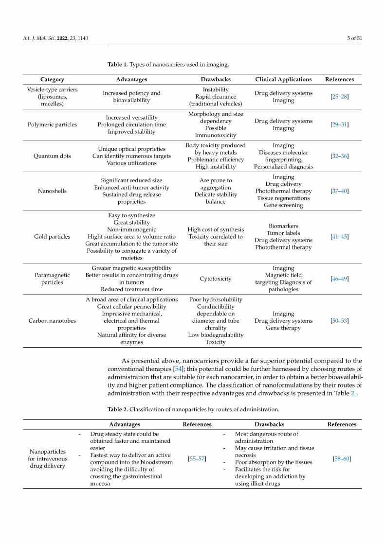
Nanotechnology is rapidly advancing in the field of personalized therapy; currently,various nanotools are under development and investigation due to their medical applicationin in vivo imaging and therapeutics [24]. Some main types of nanocarriers that have beenfurther explored in the last decades for their medical applications in personalized therapyand imaging are presented in Table 1.
Int. J. Mol. Sci. 2022, 23, 1140 5 of 51
Table 1. Types of nanocarriers used in imaging.
Category Advantages Drawbacks Clinical Applications References
Vesicle-type carriers(liposomes,
micelles)
Increased potency andbioavailability
InstabilityRapid clearance
(traditional vehicles)
Drug delivery systemsImaging
[25–28]
Polymeric particlesIncreased versatility
Prolonged circulation timeImproved stability
Morphology and sizedependency
Possibleimmunotoxicity
Drug delivery systemsImaging
[29–31]
Quantum dotsUnique optical proprieties
Can identify numerous targetsVarious utilizations
Body toxicity producedby heavy metals
Problematic efficiencyHigh instability
ImagingDiseases molecular
fingerprinting,Personalized diagnosis
[32–36]
Nanoshells
Significant reduced sizeEnhanced anti-tumor activity
Sustained drug releaseproprieties
Are prone toaggregation
Delicate stabilitybalance
ImagingDrug delivery
Photothermal therapyTissue regenerations
Gene screening
[37–40]
Gold particles
Easy to synthesizeGreat stability
Non-immunogenicHight surface area to volume ratio
Great accumulation to the tumor sitePossibility to conjugate a variety of
moieties
High cost of synthesisToxicity correlated to
their size
BiomarkersTumor labels
Drug delivery systemsPhotothermal therapy
[41–45]
Paramagneticparticles
Greater magnetic susceptibilityBetter results in concentrating drugs
in tumorsReduced treatment time
Cytotoxicity
ImagingMagnetic field
targeting Diagnosis ofpathologies
[46–49]
Carbon nanotubes
A broad area of clinical applicationsGreat cellular permeability
Impressive mechanical,electrical and thermal
proprietiesNatural affinity for diverse
enzymes
Poor hydrosolubilityConductibilitydependable on
diameter and tubechirality
Low biodegradabilityToxicity
ImagingDrug delivery systems
Gene therapy[50–53]
As presented above, nanocarriers provide a far superior potential compared to theconventional therapies [54]; this potential could be further harnessed by choosing routes ofadministration that are suitable for each nanocarrier, in order to obtain a better bioavailabil-ity and higher patient compliance. The classification of nanoformulations by their routes ofadministration with their respective advantages and drawbacks is presented in Table 2.
Table 2. Classification of nanoparticles by routes of administration.
Advantages References Drawbacks References
Nanoparticlesfor intravenousdrug delivery
- Drug steady state could beobtained faster and maintainedeasier
- Fastest way to deliver an activecompound into the bloodstreamavoiding the difficulty ofcrossing the gastrointestinalmucosa
[55–57]
- Most dangerous route ofadministration
- May cause irritation and tissuenecrosis
- Poor absorption by the tissues- Facilitates the risk for
developing an addiction byusing illicit drugs
[58–60]

Int. J. Mol. Sci. 2022, 23, 1140 6 of 51
Table 2. Cont.
Advantages References Drawbacks References
Nanoparticlesfor oral drug
delivery
- Easy administration- High compliance- Possibility of an easy
self-administration- Flexibility in dosage adjustments
and painless administration
[61–63]
- Low absorption rate due to theenzymes and bacterial florapresent in the gut mucosa
- Poor stability- Low bioavailability
[64–66]
Nanoparticlesfor transdermaldrug delivery
- Reduced interfacial tensions- Prolonged delivery- Penetration through the skin of
Both lipophilic and hydrophilicdrugs
- Avoiding the first-passmetabolism of thegastrointestinal tract
[67–70]
- Allergenic potential- Excessive local drug clearance- Only small lipophilic
compounds can penetrate theskin
[71–73]
Nanoparticlesfor pulmonarydrug delivery
- Higher safety- Little to no side effects- Delivery of big sized molecules
and obtaining a steadydistribution of the drug in thealveolar space
[74–76]
- The efficacy depends on manyfactors amongst which: thephysicochemical proprieties ofthe drug and the condition of thepatient
[77–80]
Nanoparticlesin ocular drug
delivery
- Intensifies the permeation anddrug solubility thus providing asolution to the problematicmeans of classic administrationof ocular treatment (poorbioavailability/low absorptionin the ocular mucosa)
- The toxicity and side-effectsproduced by the drug could beminimized
[81–85]- Challenges in finding a suitable
carrier for the drug that canenhance its proprieties
[86–89]
2.2. Biomedical Applications of Nanocarriers
One of the greatest challenges when developing nanocarriers loaded with variousdrugs for biomedical applications is ensuring a direct and efficient delivery. Consecutively,to preferentially deliver nanocarriers to diseased cells and tissues whilst maintaining mini-mal accumulation in healthy cells and tissues, various targeting strategies were developed:
Active targeting relies on a pair of nanocarriers loaded with a specific drug and ligandthat specifically targets the selected cells or tissues [90].
Physical targeting is based on physical properties of the system nanocarrier–targettissue, by using pH-sensitive, temperature-sensitive, ultrasound-sensitive, and magnetic-sensitive systems [91].
Passive targeting relies on the accumulation of the drug-loaded nanoparticles in adiseased area through its leaky vasculature; usually in tumors and inflamed regions [92].
The ‘’stealth” effect refers directly to nanocarriers used for passive targeting whoseshell is modified with polymers (polyethylene glycol-PEG, polyacrylamide, polyvinylpyrroli-done, polysaccharides, or dextrans), used to protect the nanocarriers from being degradedby the reticuloendothelial system (RES) [93].
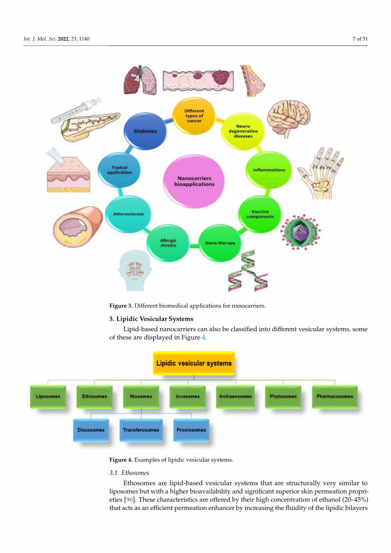
Nanocarriers’ wide range of biomedical applications makes them prone to constantevaluation and improvement at an accelerated rate over the years. Despite their primaryuse as carriers of anti-neoplastic drugs, there are many other applications in diseasessuch as diabetes, neurodegenerative diseases (Alzheimer’s disease, Parkinson’s disease,Huntington’s disease), chronic inflammations, inflammatory diseases (inflammatory boweldisease), venous thromboembolism [94,95], and others that are summarized in Figure 3.
Int. J. Mol. Sci. 2022, 23, 1140 7 of 51
Figure 3. Different biomedical applications for nanocarriers.
3. Lipidic Vesicular Systems
Lipid-based nanocarriers can also be classified into different vesicular systems, someof these are displayed in Figure 4.
Figure 4. Examples of lipidic vesicular systems.
3.1. Ethosomes
Ethosomes are lipid-based vesicular systems that are structurally very similar toliposomes but with a higher bioavailability and significant superior skin permeation propri-eties [96]. These characteristics are offered by their high concentration of ethanol (20–45%)that acts as an efficient permeation enhancer by increasing the fluidity of the lipidic bilayers

Int. J. Mol. Sci. 2022, 23, 1140 8 of 51
of the cellular membranes and by lowering the ethosome density [96]. Furthermore, itseems that ethosomes are deformable in many ways and can pass between skin corneo-cytes [97]. As a result, ethosomes can easily penetrate into the stratum corneum and skinbarrier, otherwise a limiting step in the transdermal route [96,98].
The pursuit to improve the properties of this type of carrier led researchers to thedevelopment of a new generation of ethosomes: binary ethosomes and transethosomes,depicted in Figure 5 [99].
Figure 5. Structural representation of ethosomes: classical ethosomes (A), binary ethosomes (B), andtransethosomes (C). Created with BioRender.com (accessed on 10 January 2022).
Binary ethosomes are formulated not only with ethanol but also with propyleneglycol (PEG) and isopropyl alcohol. The last two offer improved stability and permeationproprieties due to their higher hygroscopicity and viscosity. Moreover, this new type ofbinary ethosome is able to carry an increased amount of drugs into the deeper layers of theskin [100].
Transethosomes are lipid vesicles that were based on transfersomes and ethosomes.They contain phospholipids, high ethanol amounts (∼=30%), and edge activators (Span 60,65, 80; Tween 20, 60, 80; sodium deoxycholate, etc.) that offer, as compared to ethosomes,enhanced vesicle elasticity and increased skin permeation/penetration capacity [101,102].
3.2. Niosomes (Non-Ionic Surfactant- Based Vesicles)
Niosomes are vesicular systems that are formed from a non-ionic surfactant andan aqueous core, that form bilayer structures due to their amphiphilic nature (Figure 6).Niosomes can be prepared by the same methods as liposomes and can be used in thecosmetic, pharmaceutical, and food science fields [103]. However, compared to liposomes,they possess several advantages including increased physical stability, lower productioncost, and ease of storage [104]. Niosomes can be utilized as transdermal delivery systemson account of their superior skin penetration proprieties, for topical vaccine delivery, andocular delivery due to their low toxicity [105,106].
Chen S et al. classified niosomes into different groups based on their composition andbiomedical applications [107]:
(a) Elastic niosomes are composed of cholesterol, surfactants, water, and ethanol. They areflexible and can infiltrate into pores that are significantly smaller than their size, with-out altering their structure, and hence, are prone to be used in topical or transdermaldrug delivery [108].
(b) Discomes are large vesicular thermosensitive systems (their structure changes withtemperatures above 37 ◦C) that are generally used as ocular delivery systems [109].
Int. J. Mol. Sci. 2022, 23, 1140 9 of 51
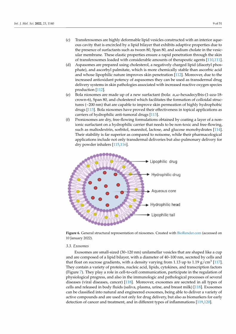
(c) Transferosomes are highly deformable lipid vesicles constructed with an interior aque-ous cavity that is encircled by a lipid bilayer that exhibits adaptive properties due tothe presence of surfactants such as tween 80, Span 80, and sodium cholate in the vesic-ular membrane. These elastic properties ensure a rapid penetration through the skinof transferosomes loaded with considerable amounts of therapeutic agents [110,111].
(d) Aspasomes are prepared using cholesterol, a negatively charged lipid (diacetyl phos-phate), and ascorbyl palmitate, which is more chemically stable than ascorbic acidand whose lipophilic nature improves skin penetration [112]. Moreover, due to theincreased antioxidant potency of aspasomes they can be used as transdermal drugdelivery systems in skin pathologies associated with increased reactive oxygen speciesproduction [112].
(e) Bola niosomes are made up of a new surfactant (bola: α,ω-hexadecylbis-(1-aza-18-crown-6), Span 80, and cholesterol which facilitates the formation of colloidal struc-tures (~200 nm) that are capable to improve skin permeation of highly hydrophobicdrugs [113]. Bola niosomes have proved their effectiveness in topical applications ascarriers of hydrophilic anti-tumoral drugs [113].
(f) Proniosomes are dry, free-flowing formulations obtained by coating a layer of a non-ionic surfactant on a hydrophilic carrier that needs to be non-toxic and free-flowing,such as maltodextrin, sorbitol, mannitol, lactose, and glucose monohydrates [114].Their stability is far superior as compared to noisome, while their pharmacologicalapplications include not only transdermal deliveries but also pulmonary delivery fordry powder inhalers [115,116].
Figure 6. General structural representation of niosomes. Created with BioRender.com (accessed on10 January 2022).
3.3. Exosomes
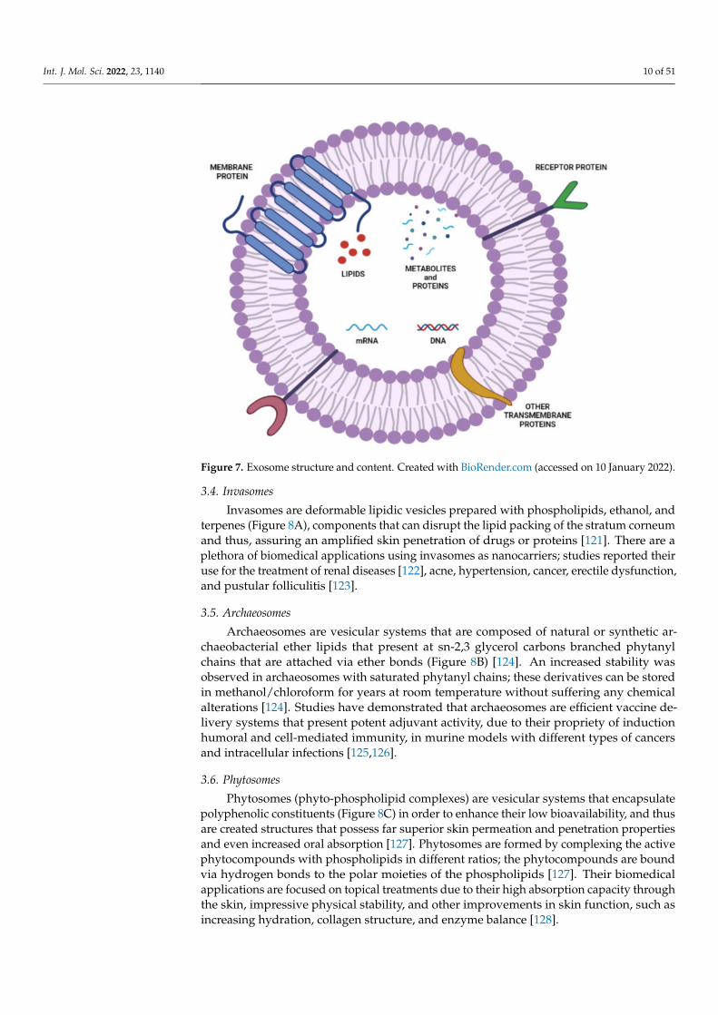
Exosomes are small-sized (30–120 nm) unilamellar vesicles that are shaped like a cupand are composed of a lipid bilayer, with a diameter of 40–100 nm, secreted by cells andthat float on sucrose gradients, with a density varying from 1.13 up to 1.19 g/cm3 [117].They contain a variety of proteins, nucleic acid, lipids, cytokines, and transcription factors(Figure 7). They play a role in cell-to-cell communication, participate in the regulation ofphysiological progress, and also in the immunologic and pathological processes of severaldiseases (viral diseases, cancer) [118]. Moreover, exosomes are secreted in all types ofcells and released in body fluids (saliva, plasma, urine, and breast milk) [118]. Exosomescan be classified into natural and engineered exosomes, being able to deliver a variety ofactive compounds and are used not only for drug delivery, but also as biomarkers for earlydetection of cancer and treatment, and in different types of inflammations [119,120].
Int. J. Mol. Sci. 2022, 23, 1140 10 of 51
Figure 7. Exosome structure and content. Created with BioRender.com (accessed on 10 January 2022).
3.4. Invasomes
Invasomes are deformable lipidic vesicles prepared with phospholipids, ethanol, andterpenes (Figure 8A), components that can disrupt the lipid packing of the stratum corneumand thus, assuring an amplified skin penetration of drugs or proteins [121]. There are aplethora of biomedical applications using invasomes as nanocarriers; studies reported theiruse for the treatment of renal diseases [122], acne, hypertension, cancer, erectile dysfunction,and pustular folliculitis [123].
3.5. Archaeosomes
Archaeosomes are vesicular systems that are composed of natural or synthetic ar-chaeobacterial ether lipids that present at sn-2,3 glycerol carbons branched phytanylchains that are attached via ether bonds (Figure 8B) [124]. An increased stability wasobserved in archaeosomes with saturated phytanyl chains; these derivatives can be storedin methanol/chloroform for years at room temperature without suffering any chemicalalterations [124]. Studies have demonstrated that archaeosomes are efficient vaccine de-livery systems that present potent adjuvant activity, due to their propriety of inductionhumoral and cell-mediated immunity, in murine models with different types of cancersand intracellular infections [125,126].
3.6. Phytosomes
Phytosomes (phyto-phospholipid complexes) are vesicular systems that encapsulatepolyphenolic constituents (Figure 8C) in order to enhance their low bioavailability, and thusare created structures that possess far superior skin permeation and penetration propertiesand even increased oral absorption [127]. Phytosomes are formed by complexing the activephytocompounds with phospholipids in different ratios; the phytocompounds are boundvia hydrogen bonds to the polar moieties of the phospholipids [127]. Their biomedicalapplications are focused on topical treatments due to their high absorption capacity throughthe skin, impressive physical stability, and other improvements in skin function, such asincreasing hydration, collagen structure, and enzyme balance [128].

Int. J. Mol. Sci. 2022, 23, 1140 11 of 51
Figure 8. The structure and content of some types of lipidic vesicular systems: invasomes (A),archaeosomes (B), phytosomes (C), and pharmacosomes (D). Created with BioRender.com (accessedon 10 January 2022).
3.7. Pharmacosomes
Pharmacosomes are amphiphilic vesicular systems of drugs that possess at least oneactive hydrogen atom, covalently bound to phospholipids in equimolar concentrations,thus providing a greater drug load (Figure 8D) [129]. Their amphiphilic nature offersthe advantage of increased absorption through tissues and dissolution in gastrointestinalfluid [129]. Compared to conventional liposomes, they provide a superior targeted deliveryby significantly increasing the bioavailability of several lipophilic drugs, providing greaterentrapment results by ensuring no drug leakage due to their covalent bonds [130].
4. Liposomes as Targeted Delivery Systems
Liposomes were discovered by Alec D. Bangham in 1960 at the Braham Institute atCambridge University and are mainly made up of one or more lipid layers that encapsu-late a hydrophilic layer [131]. They possess a cylindrical shape and a diameter rangingfrom nanometres to several hundred micrometres; liposomes with a diameter between50–450 nm are used in therapy [131]. It is considered that particles with a size of up to100 nm can be classified as nanoparticles, whereas particles with a size ranging from 100 to1000 nm can be classified as sub-microparticles [132].
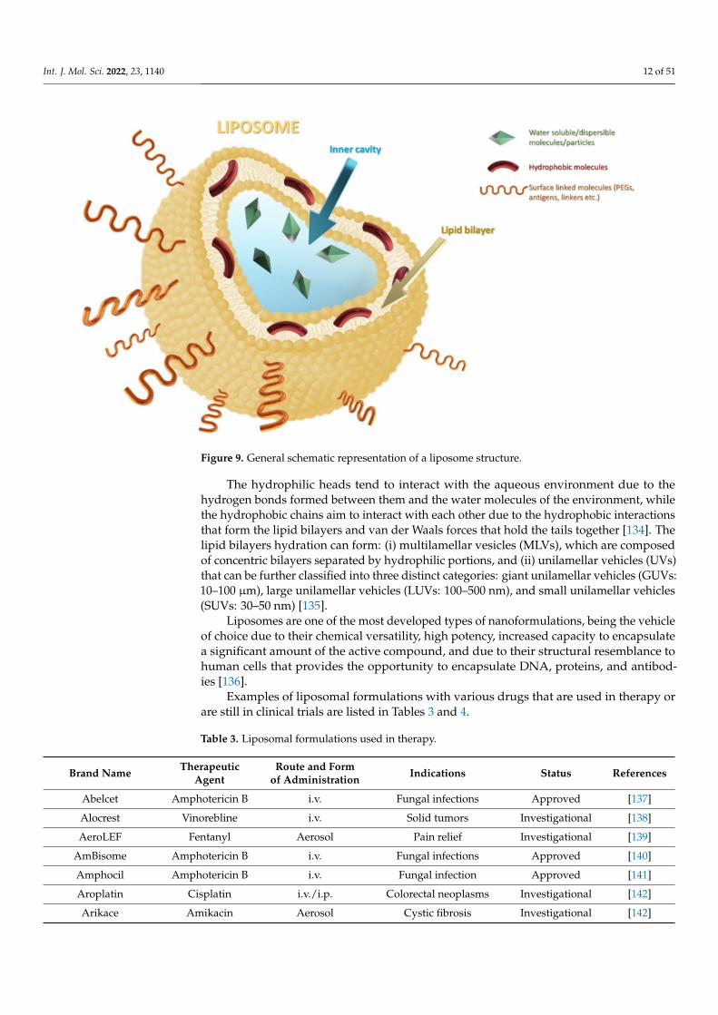
This complex structure of liposomes is stable due to the interactions between thephospholipids which are composed of a hydrophilic head and two hydrophobic tails. Onaccount of their amphiphilic characteristics, liposomes can entrap both hydrophilic andlipophilic drugs (Figure 9) [133].
Int. J. Mol. Sci. 2022, 23, 1140 12 of 51
Figure 9. General schematic representation of a liposome structure.
The hydrophilic heads tend to interact with the aqueous environment due to thehydrogen bonds formed between them and the water molecules of the environment, whilethe hydrophobic chains aim to interact with each other due to the hydrophobic interactionsthat form the lipid bilayers and van der Waals forces that hold the tails together [134]. Thelipid bilayers hydration can form: (i) multilamellar vesicles (MLVs), which are composedof concentric bilayers separated by hydrophilic portions, and (ii) unilamellar vehicles (UVs)that can be further classified into three distinct categories: giant unilamellar vehicles (GUVs:10–100 µm), large unilamellar vehicles (LUVs: 100–500 nm), and small unilamellar vehicles(SUVs: 30–50 nm) [135].
Liposomes are one of the most developed types of nanoformulations, being the vehicleof choice due to their chemical versatility, high potency, increased capacity to encapsulatea significant amount of the active compound, and due to their structural resemblance tohuman cells that provides the opportunity to encapsulate DNA, proteins, and antibod-ies [136].
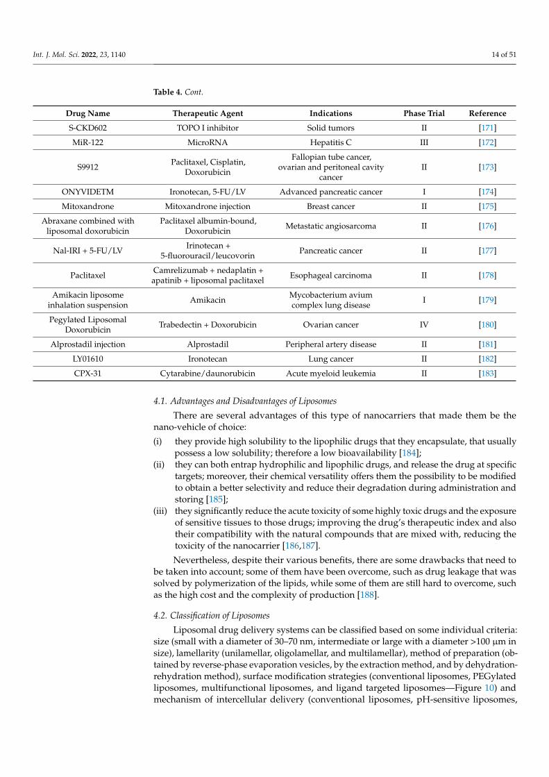
Examples of liposomal formulations with various drugs that are used in therapy orare still in clinical trials are listed in Tables 3 and 4.
Table 3. Liposomal formulations used in therapy.
Brand NameTherapeutic
AgentRoute and Form
of AdministrationIndications Status References
Abelcet Amphotericin B i.v. Fungal infections Approved [137]
Alocrest Vinorebline i.v. Solid tumors Investigational [138]
AeroLEF Fentanyl Aerosol Pain relief Investigational [139]
AmBisome Amphotericin B i.v. Fungal infections Approved [140]
Amphocil Amphotericin B i.v. Fungal infection Approved [141]
Aroplatin Cisplatin i.v./i.p. Colorectal neoplasms Investigational [142]
Arikace Amikacin Aerosol Cystic fibrosis Investigational [142]

Int. J. Mol. Sci. 2022, 23, 1140 13 of 51
Table 3. Cont.
Brand NameTherapeutic
AgentRoute and Form
of AdministrationIndications Status References
Atragen Tretinoin i.v. Solid tumors Investigational [143]
Atu027 siRNA i.v. Solid tumors Investigational [144]
Brakiva Topotecan i.v. Solid tumors Investigational [145]
DepoDur Morphine sulfate Epidural Pain management Approved [146]
DepoCyt Cytarabine i.v.Lymphomatous
meningitisApproved [147]
Dimericine T4N4 Oral Precancerous condition Investigational [148]
Doxisome Doxorubicin i.v. Solid tumors Investigational [149]
EpaxalInactivated
hepatitis A virus(strain RG-SB)
i.m. Hepatitis A Approved [150]
Lipo-Dox Doxorubicin i.v. Solid tumors Approved [151]
Lipoplatin Cisplatin i.v. Solid tumors Investigational [152]
Liposomalalendronate
Alendronate i.v. Coronary artery stenosis Investigational [153]
Liprostin Prostaglandin i.v.Peripheral vascular
diseaseInvestigational [154]
L-annamycin Annamycin i.v. Acute lymphocytic Investigational [155]
Marqibo Vincristine i.v. Solid tumors Investigational [156]
Mifamurtide Mepacti.v./injection/
powder
High-grade, resectable,non-metastatic
osteosarcoma in childrenand young adults
Approved [157]
Nanocort Prednisolone i.v. Rheumatoid arthritis Investigational [158]
NanoVNB Vinorelbine i.v. Colon cancer Investigational [159]
OctinoxateEucerin, Meijer,Sumadan Wash
Topical/emulsionProtection against UV
lightApproved,
Investigational[160]
RVCLUV Ropivacaine i.v. Anesthetic Investigational [161]
Stimuvax BLP25 vaccine i.v. Lung cancer Investigational [162]
VaxiSome Influenza i.m. Influenza Investigational [163]
7-ethyl-10-hydroxycamptothecin
- - Colorectal cancer Investigational [164]
Table 4. Liposomal formulations of various anti-cancer agents under clinical trials.
Drug Name Therapeutic Agent Indications Phase Trial References
BP1001 Grb2 antisense oligonucleotideLeukemia, myelodysplastic
syndrome, Ph1-positive CMLI [165]
LiPlaCis Cisplatin Solid tumors I/II [166]
LDF01Rhodamine-labeled cationic
liposomesHead and neck squamous
cell carcinomasI [167]
Atu027 siRNA Solid tumors I [168]
LEP-ETU Paclitaxel Ovarian cancer II [169]
OSI-211 Lurtotecan Head and neck carcinomas II [170]
Int. J. Mol. Sci. 2022, 23, 1140 14 of 51
Table 4. Cont.
Drug Name Therapeutic Agent Indications Phase Trial Reference
S-CKD602 TOPO I inhibitor Solid tumors II [171]
MiR-122 MicroRNA Hepatitis C III [172]
S9912Paclitaxel, Cisplatin,
Doxorubicin
Fallopian tube cancer,ovarian and peritoneal cavity
cancerII [173]
ONYVIDETM Ironotecan, 5-FU/LV Advanced pancreatic cancer I [174]
Mitoxandrone Mitoxandrone injection Breast cancer II [175]
Abraxane combined withliposomal doxorubicin
Paclitaxel albumin-bound,Doxorubicin
Metastatic angiosarcoma II [176]
Nal-IRI + 5-FU/LVIrinotecan +
5-fluorouracil/leucovorinPancreatic cancer II [177]
PaclitaxelCamrelizumab + nedaplatin +apatinib + liposomal paclitaxel
Esophageal carcinoma II [178]
Amikacin liposomeinhalation suspension
AmikacinMycobacterium aviumcomplex lung disease
I [179]
Pegylated LiposomalDoxorubicin
Trabedectin + Doxorubicin Ovarian cancer IV [180]
Alprostadil injection Alprostadil Peripheral artery disease II [181]
LY01610 Ironotecan Lung cancer II [182]
CPX-31 Cytarabine/daunorubicin Acute myeloid leukemia II [183]
4.1. Advantages and Disadvantages of Liposomes
There are several advantages of this type of nanocarriers that made them be thenano-vehicle of choice:
(i) they provide high solubility to the lipophilic drugs that they encapsulate, that usuallypossess a low solubility; therefore a low bioavailability [184];
(ii) they can both entrap hydrophilic and lipophilic drugs, and release the drug at specifictargets; moreover, their chemical versatility offers them the possibility to be modifiedto obtain a better selectivity and reduce their degradation during administration andstoring [185];
(iii) they significantly reduce the acute toxicity of some highly toxic drugs and the exposureof sensitive tissues to those drugs; improving the drug’s therapeutic index and alsotheir compatibility with the natural compounds that are mixed with, reducing thetoxicity of the nanocarrier [186,187].
Nevertheless, despite their various benefits, there are some drawbacks that need tobe taken into account; some of them have been overcome, such as drug leakage that wassolved by polymerization of the lipids, while some of them are still hard to overcome, suchas the high cost and the complexity of production [188].
4.2. Classification of Liposomes
Liposomal drug delivery systems can be classified based on some individual criteria:size (small with a diameter of 30–70 nm, intermediate or large with a diameter >100 µm insize), lamellarity (unilamellar, oligolamellar, and multilamellar), method of preparation (ob-tained by reverse-phase evaporation vesicles, by the extraction method, and by dehydration-rehydration method), surface modification strategies (conventional liposomes, PEGylatedliposomes, multifunctional liposomes, and ligand targeted liposomes—Figure 10) andmechanism of intercellular delivery (conventional liposomes, pH-sensitive liposomes,

Int. J. Mol. Sci. 2022, 23, 1140 15 of 51
cationic liposomes, immuno-liposomes, long-circulating liposomes, and thermo-sensitiveliposomes) [189–191].
Figure 10. Classification of liposomes by surface modification strategies: conventional liposomes(A), PEGylated liposomes (B), multifunctional liposomes (C), and ligand-targeted liposomes (D).Reprinted with permission from [192]; copyright (2022). International Journal of Applied Pharmaceutics.
4.2.1. Unilamellar and Multilamellar Liposomes
Unilamellar vesicles (ULVs) or multilamellar vesicles (MLVs) can be obtained bychoosing a certain method of preparation and synthesis such as the Bangham methodfor MLVs and ISCRPE and the SCRPE method for ULVs [191]. When comparing theproperties of the two types of liposomes, it seems that UVLs (50–250 nm), which consistof only one lipid bilayer, tend to incorporate hydrophilic drugs since they possess a largeaqueous interior, while MLVs (1–5 µm), that contain two or more lipidic bilayers orientedconcentrically, are more suitable for lipophilic drugs (Figure 11) [191].
4.2.2. Conventional Liposomes
Liposomal formulations have been explored widely for at least two decades due totheir plethora of advantages and mostly for their possibility of incorporating various typesof drugs in very small or very high quantities and delivering them on targeted sites thatare otherwise hard to reach [189]. The most inconvenient drawback that it is encounteredwhile using conventional liposomes is the fact that they are easily recognized and captured

Int. J. Mol. Sci. 2022, 23, 1140 16 of 51
by the mononuclear phagocyte system (MPS) and cleared from blood circulation; moreover,as a result of their chemical composition, they are very unstable in the plasma because theyinteract with lipoproteins, thus they release the drug load into the plasma [193].
Figure 11. Structural representation of unilamellar and multilamellar vesicles. Created withBioRender.com (accessed on 10 January 2022).
4.2.3. Temperature-Sensitive Liposomes (TSLs)
TSLs are made up of temperature-sensitive lipids that change their microarchitecturefrom a solid and compact structure of gel to a loose structure of a liquid-crystal state;providing the property of releasing the drug at a temperature in which the lipids suffer aphase change—temperatures usually combine with local hyperthermia (40–45 ◦C), whichis clinically established as a suitable treatment for solid tumors (Figure 12) [194]. TSLs arecomposed of dipalmitoyl phosphatidylcholine (which has a transition temperature = 41 ◦C)and might suffer modification for an increased drug release, better chemical stability (theymight incorporate polyethylene glycol on TSLs), or for the adjustment of the temperaturein which the TSLs release the payload (cholesterol, hydrogenated soy phosphatidylcholine-HSPC and 1,2-distearoyl-sn-glycero-3-phosphocholine might be incorporated) [195].
Figure 12. Schematic representation of temperature-sensitive liposomes. Created with BioRender.com(accessed on 10 January 2022).
4.2.4. pH-Sensitive Liposomes (PSLs)
Both TSLs and PSLs were designed as targeted delivery systems for their property toencapsulate the drugs and only release them when the body conditions are suitable fordelivery, in a certain temperature range and pH range, accordingly [196].
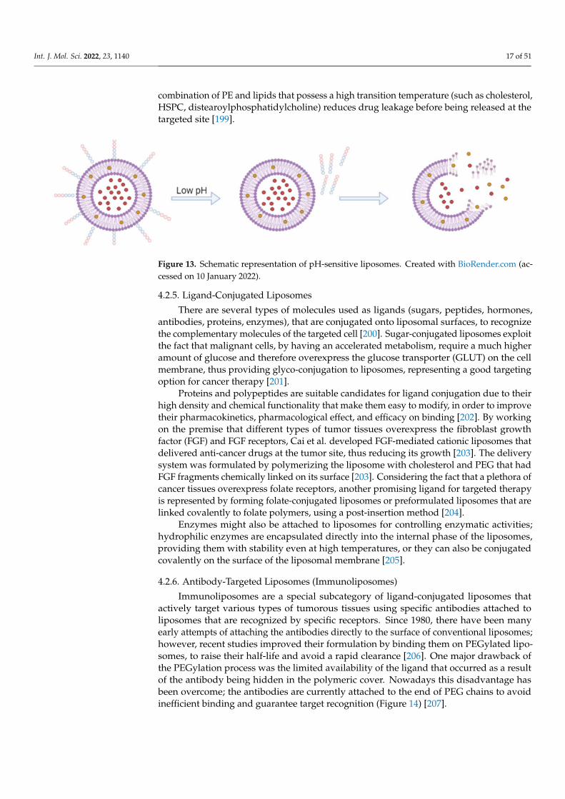
Commonly, PSLs are stable at a physiological pH, but at slightly acidic values of pH,usually in case of inflammations and infections, they are released when the lipidic bilayersare destabilized (Figure 13); being the most promising types of carriers for deliveringgenes, antisense oligonucleotides, and proteins [197]. The most used compound for theformulation of PSLs is phosphatidylethanolamine (PE) or its derivatives that containan acidic moiety which is a stabilizer at a neutral pH; however, similar to conventionalliposomes, PSLs are degraded by the MPS, lowering their pharmacological activity [198].To address this issue many types of formulations have been tested; it was noticed that the
Int. J. Mol. Sci. 2022, 23, 1140 17 of 51
combination of PE and lipids that possess a high transition temperature (such as cholesterol,HSPC, distearoylphosphatidylcholine) reduces drug leakage before being released at thetargeted site [199].
Figure 13. Schematic representation of pH-sensitive liposomes. Created with BioRender.com (ac-cessed on 10 January 2022).
4.2.5. Ligand-Conjugated Liposomes
There are several types of molecules used as ligands (sugars, peptides, hormones,antibodies, proteins, enzymes), that are conjugated onto liposomal surfaces, to recognizethe complementary molecules of the targeted cell [200]. Sugar-conjugated liposomes exploitthe fact that malignant cells, by having an accelerated metabolism, require a much higheramount of glucose and therefore overexpress the glucose transporter (GLUT) on the cellmembrane, thus providing glyco-conjugation to liposomes, representing a good targetingoption for cancer therapy [201].
Proteins and polypeptides are suitable candidates for ligand conjugation due to theirhigh density and chemical functionality that make them easy to modify, in order to improvetheir pharmacokinetics, pharmacological effect, and efficacy on binding [202]. By workingon the premise that different types of tumor tissues overexpress the fibroblast growthfactor (FGF) and FGF receptors, Cai et al. developed FGF-mediated cationic liposomes thatdelivered anti-cancer drugs at the tumor site, thus reducing its growth [203]. The deliverysystem was formulated by polymerizing the liposome with cholesterol and PEG that hadFGF fragments chemically linked on its surface [203]. Considering the fact that a plethora ofcancer tissues overexpress folate receptors, another promising ligand for targeted therapyis represented by forming folate-conjugated liposomes or preformulated liposomes that arelinked covalently to folate polymers, using a post-insertion method [204].
Enzymes might also be attached to liposomes for controlling enzymatic activities;hydrophilic enzymes are encapsulated directly into the internal phase of the liposomes,providing them with stability even at high temperatures, or they can also be conjugatedcovalently on the surface of the liposomal membrane [205].
4.2.6. Antibody-Targeted Liposomes (Immunoliposomes)
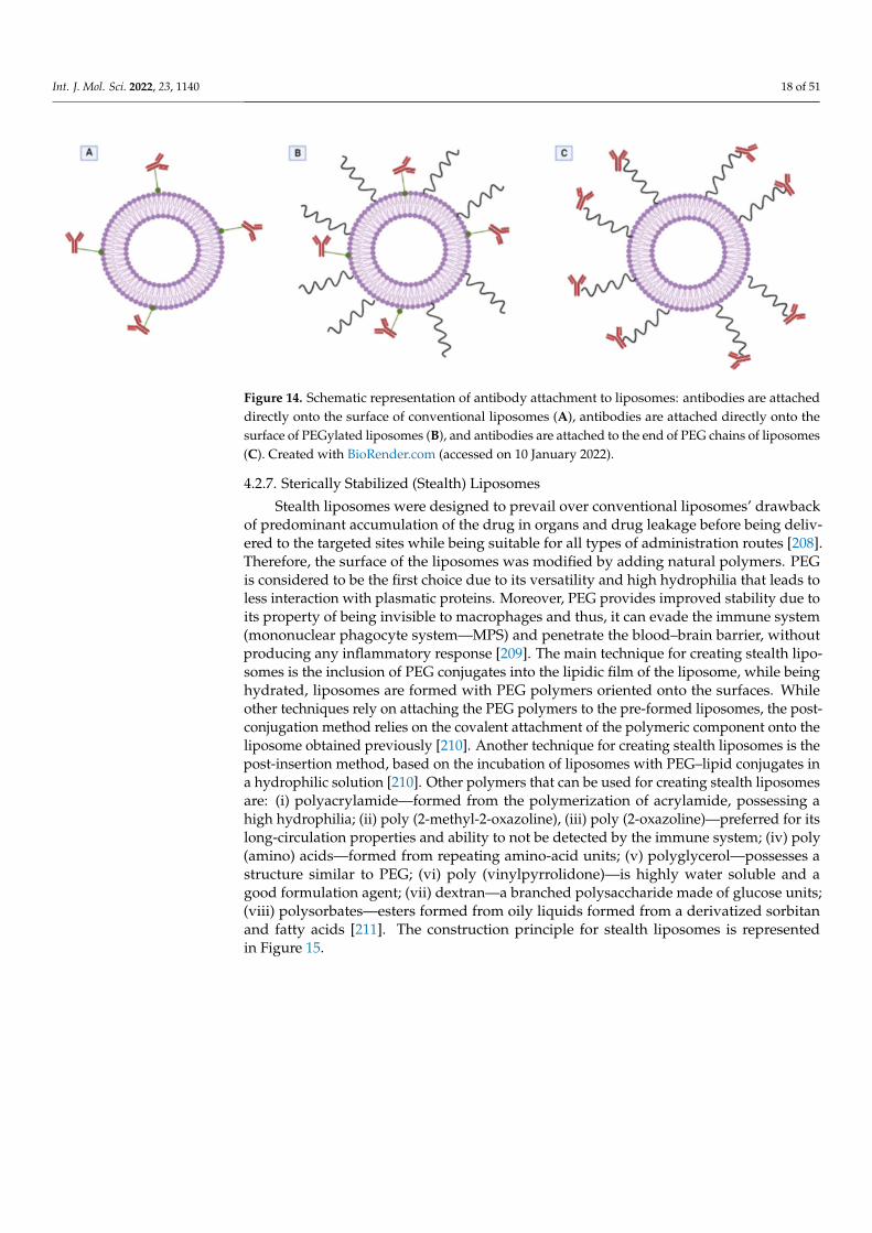
Immunoliposomes are a special subcategory of ligand-conjugated liposomes thatactively target various types of tumorous tissues using specific antibodies attached toliposomes that are recognized by specific receptors. Since 1980, there have been manyearly attempts of attaching the antibodies directly to the surface of conventional liposomes;however, recent studies improved their formulation by binding them on PEGylated lipo-somes, to raise their half-life and avoid a rapid clearance [206]. One major drawback ofthe PEGylation process was the limited availability of the ligand that occurred as a resultof the antibody being hidden in the polymeric cover. Nowadays this disadvantage hasbeen overcome; the antibodies are currently attached to the end of PEG chains to avoidinefficient binding and guarantee target recognition (Figure 14) [207].
Int. J. Mol. Sci. 2022, 23, 1140 18 of 51
Figure 14. Schematic representation of antibody attachment to liposomes: antibodies are attacheddirectly onto the surface of conventional liposomes (A), antibodies are attached directly onto thesurface of PEGylated liposomes (B), and antibodies are attached to the end of PEG chains of liposomes(C). Created with BioRender.com (accessed on 10 January 2022).
4.2.7. Sterically Stabilized (Stealth) Liposomes
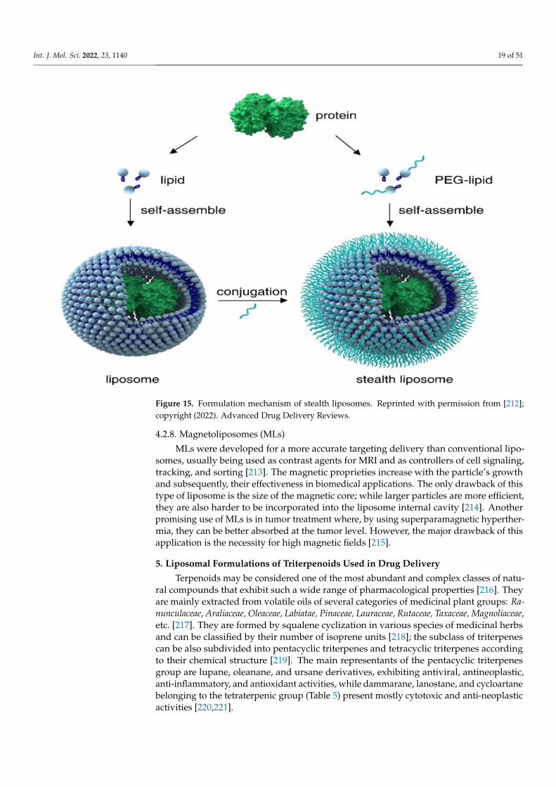
Stealth liposomes were designed to prevail over conventional liposomes’ drawbackof predominant accumulation of the drug in organs and drug leakage before being deliv-ered to the targeted sites while being suitable for all types of administration routes [208].Therefore, the surface of the liposomes was modified by adding natural polymers. PEGis considered to be the first choice due to its versatility and high hydrophilia that leads toless interaction with plasmatic proteins. Moreover, PEG provides improved stability due toits property of being invisible to macrophages and thus, it can evade the immune system(mononuclear phagocyte system—MPS) and penetrate the blood–brain barrier, withoutproducing any inflammatory response [209]. The main technique for creating stealth lipo-somes is the inclusion of PEG conjugates into the lipidic film of the liposome, while beinghydrated, liposomes are formed with PEG polymers oriented onto the surfaces. Whileother techniques rely on attaching the PEG polymers to the pre-formed liposomes, the post-conjugation method relies on the covalent attachment of the polymeric component onto theliposome obtained previously [210]. Another technique for creating stealth liposomes is thepost-insertion method, based on the incubation of liposomes with PEG–lipid conjugates ina hydrophilic solution [210]. Other polymers that can be used for creating stealth liposomesare: (i) polyacrylamide—formed from the polymerization of acrylamide, possessing ahigh hydrophilia; (ii) poly (2-methyl-2-oxazoline), (iii) poly (2-oxazoline)—preferred for itslong-circulation properties and ability to not be detected by the immune system; (iv) poly(amino) acids—formed from repeating amino-acid units; (v) polyglycerol—possesses astructure similar to PEG; (vi) poly (vinylpyrrolidone)—is highly water soluble and agood formulation agent; (vii) dextran—a branched polysaccharide made of glucose units;(viii) polysorbates—esters formed from oily liquids formed from a derivatized sorbitanand fatty acids [211]. The construction principle for stealth liposomes is representedin Figure 15.
Int. J. Mol. Sci. 2022, 23, 1140 19 of 51
Figure 15. Formulation mechanism of stealth liposomes. Reprinted with permission from [212];copyright (2022). Advanced Drug Delivery Reviews.
4.2.8. Magnetoliposomes (MLs)
MLs were developed for a more accurate targeting delivery than conventional lipo-somes, usually being used as contrast agents for MRI and as controllers of cell signaling,tracking, and sorting [213]. The magnetic proprieties increase with the particle’s growthand subsequently, their effectiveness in biomedical applications. The only drawback of thistype of liposome is the size of the magnetic core; while larger particles are more efficient,they are also harder to be incorporated into the liposome internal cavity [214]. Anotherpromising use of MLs is in tumor treatment where, by using superparamagnetic hyperther-mia, they can be better absorbed at the tumor level. However, the major drawback of thisapplication is the necessity for high magnetic fields [215].
5. Liposomal Formulations of Triterpenoids Used in Drug Delivery
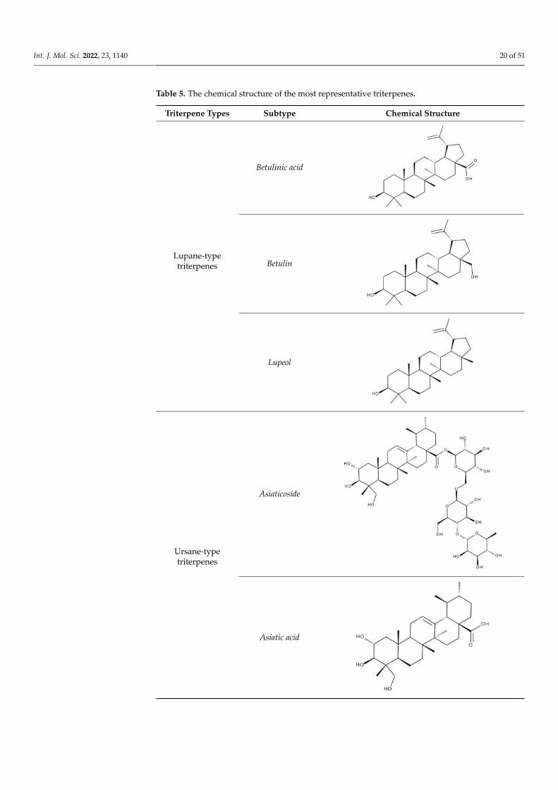
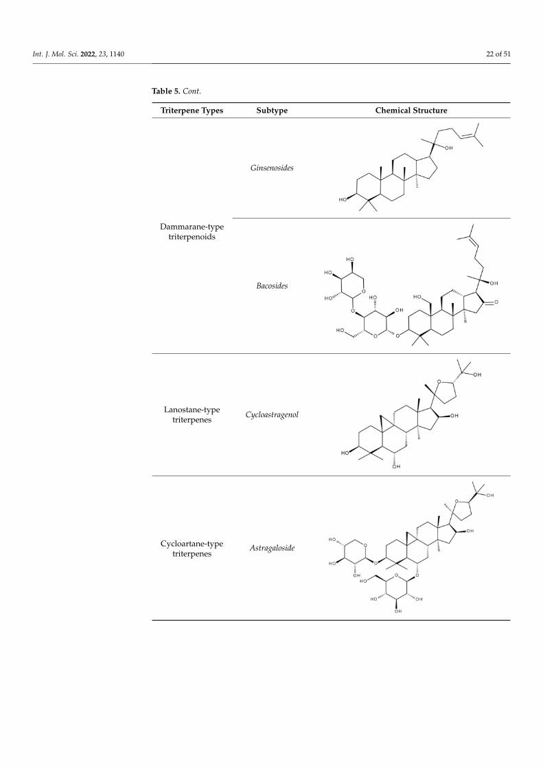
Terpenoids may be considered one of the most abundant and complex classes of natu-ral compounds that exhibit such a wide range of pharmacological properties [216]. Theyare mainly extracted from volatile oils of several categories of medicinal plant groups: Ra-nunculaceae, Araliaceae, Oleaceae, Labiatae, Pinaceae, Lauraceae, Rutaceae, Taxaceae, Magnoliaceae,etc. [217]. They are formed by squalene cyclization in various species of medicinal herbsand can be classified by their number of isoprene units [218]; the subclass of triterpenescan be also subdivided into pentacyclic triterpenes and tetracyclic triterpenes accordingto their chemical structure [219]. The main representants of the pentacyclic triterpenesgroup are lupane, oleanane, and ursane derivatives, exhibiting antiviral, antineoplastic,anti-inflammatory, and antioxidant activities, while dammarane, lanostane, and cycloartanebelonging to the tetraterpenic group (Table 5) present mostly cytotoxic and anti-neoplasticactivities [220,221].
Int. J. Mol. Sci. 2022, 23, 1140 20 of 51
Table 5. The chemical structure of the most representative triterpenes.
Triterpene Types Subtype Chemical Structure
Lupane-typetriterpenes
Betulinic acid
Betulin
Lupeol
Ursane-typetriterpenes
Asiaticoside
Asiatic acid

Int. J. Mol. Sci. 2022, 23, 1140 21 of 51
Table 5. Cont.
Triterpene Types Subtype Chemical Structure
Madecassoside
Madecassic acid
Oleane-typetriterpenes
Oleanolic acid
Glycyrrhizin
Int. J. Mol. Sci. 2022, 23, 1140 22 of 51
Table 5. Cont.
Triterpene Types Subtype Chemical Structure
Dammarane-typetriterpenoids
Ginsenosides
Bacosides
Lanostane-typetriterpenes
Cycloastragenol
Cycloartane-typetriterpenes
Astragaloside
Int. J. Mol. Sci. 2022, 23, 1140 23 of 51
Table 5. Cont.
Triterpene Types Subtype Chemical Structure
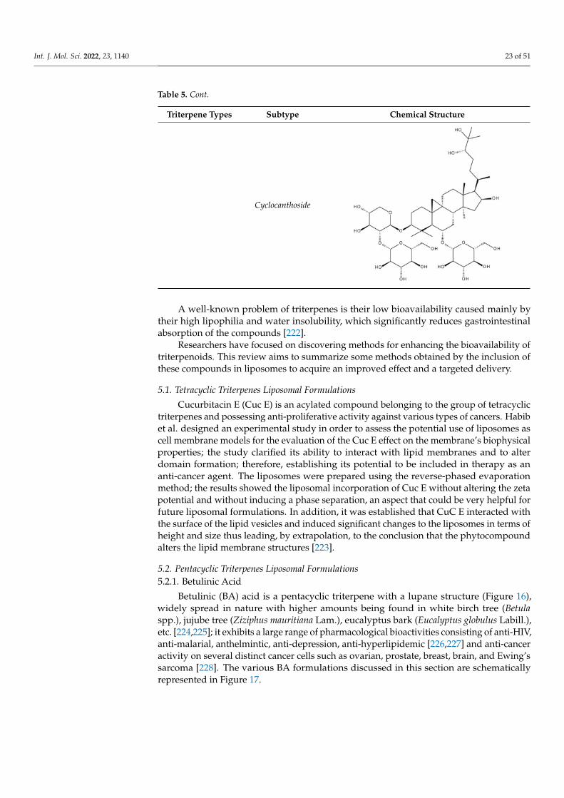
Cyclocanthoside
A well-known problem of triterpenes is their low bioavailability caused mainly bytheir high lipophilia and water insolubility, which significantly reduces gastrointestinalabsorption of the compounds [222].
Researchers have focused on discovering methods for enhancing the bioavailability oftriterpenoids. This review aims to summarize some methods obtained by the inclusion ofthese compounds in liposomes to acquire an improved effect and a targeted delivery.
5.1. Tetracyclic Triterpenes Liposomal Formulations
Cucurbitacin E (Cuc E) is an acylated compound belonging to the group of tetracyclictriterpenes and possessing anti-proliferative activity against various types of cancers. Habibet al. designed an experimental study in order to assess the potential use of liposomes ascell membrane models for the evaluation of the Cuc E effect on the membrane’s biophysicalproperties; the study clarified its ability to interact with lipid membranes and to alterdomain formation; therefore, establishing its potential to be included in therapy as ananti-cancer agent. The liposomes were prepared using the reverse-phased evaporationmethod; the results showed the liposomal incorporation of Cuc E without altering the zetapotential and without inducing a phase separation, an aspect that could be very helpful forfuture liposomal formulations. In addition, it was established that CuC E interacted withthe surface of the lipid vesicles and induced significant changes to the liposomes in terms ofheight and size thus leading, by extrapolation, to the conclusion that the phytocompoundalters the lipid membrane structures [223].
5.2. Pentacyclic Triterpenes Liposomal Formulations
5.2.1. Betulinic Acid
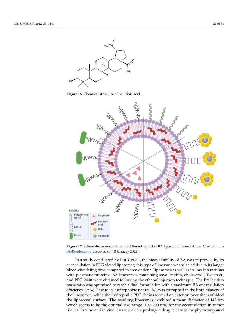
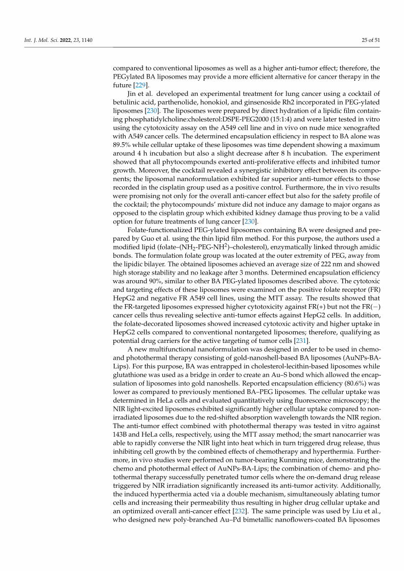
Betulinic (BA) acid is a pentacyclic triterpene with a lupane structure (Figure 16),widely spread in nature with higher amounts being found in white birch tree (Betulaspp.), jujube tree (Ziziphus mauritiana Lam.), eucalyptus bark (Eucalyptus globulus Labill.),etc. [224,225]; it exhibits a large range of pharmacological bioactivities consisting of anti-HIV,anti-malarial, anthelmintic, anti-depression, anti-hyperlipidemic [226,227] and anti-canceractivity on several distinct cancer cells such as ovarian, prostate, breast, brain, and Ewing’ssarcoma [228]. The various BA formulations discussed in this section are schematicallyrepresented in Figure 17.
Int. J. Mol. Sci. 2022, 23, 1140 24 of 51
Figure 16. Chemical structure of betulinic acid.
Figure 17. Schematic representation of different reported BA liposomal formulations. Created withBioRender.com (accessed on 10 January 2022).
In a study conducted by Liu Y et al., the bioavailability of BA was improved by itsencapsulation in PEG-ylated liposomes; this type of liposome was selected due to its longerblood-circulating time compared to conventional liposomes as well as its low interactionswith plasmatic proteins. BA liposomes containing soya lecithin, cholesterol, Tween-80,and PEG-2000 were obtained following the ethanol injection technique. The BA:lecithinmass ratio was optimized to reach a final formulation with a maximum BA encapsulationefficiency (95%). Due to its hydrophobic nature, BA was entrapped in the lipid bilayers ofthe liposomes, while the hydrophilic PEG chains formed an exterior layer that enfoldedthe liposomal surface. The resulting liposomes exhibited a mean diameter of 142 nmwhich seems to be the optimal size range (100–200 nm) for the accumulation in tumortissues. In vitro and in vivo tests revealed a prolonged drug release of the phytocompound
Int. J. Mol. Sci. 2022, 23, 1140 25 of 51
compared to conventional liposomes as well as a higher anti-tumor effect; therefore, thePEGylated BA liposomes may provide a more efficient alternative for cancer therapy in thefuture [229].
Jin et al. developed an experimental treatment for lung cancer using a cocktail ofbetulinic acid, parthenolide, honokiol, and ginsenoside Rh2 incorporated in PEG-ylatedliposomes [230]. The liposomes were prepared by direct hydration of a lipidic film contain-ing phosphatidylcholine:cholesterol:DSPE-PEG2000 (15:1:4) and were later tested in vitrousing the cytotoxicity assay on the A549 cell line and in vivo on nude mice xenograftedwith A549 cancer cells. The determined encapsulation efficiency in respect to BA alone was89.5% while cellular uptake of these liposomes was time dependent showing a maximumaround 4 h incubation but also a slight decrease after 8 h incubation. The experimentshowed that all phytocompounds exerted anti-proliferative effects and inhibited tumorgrowth. Moreover, the cocktail revealed a synergistic inhibitory effect between its compo-nents; the liposomal nanoformulation exhibited far superior anti-tumor effects to thoserecorded in the cisplatin group used as a positive control. Furthermore, the in vivo resultswere promising not only for the overall anti-cancer effect but also for the safety profile ofthe cocktail; the phytocompounds’ mixture did not induce any damage to major organs asopposed to the cisplatin group which exhibited kidney damage thus proving to be a validoption for future treatments of lung cancer [230].
Folate-functionalized PEG-ylated liposomes containing BA were designed and pre-pared by Guo et al. using the thin lipid film method. For this purpose, the authors used amodified lipid (folate–(NH2-PEG-NH2)–cholesterol), enzymatically linked through amidicbonds. The formulation folate group was located at the outer extremity of PEG, away fromthe lipidic bilayer. The obtained liposomes achieved an average size of 222 nm and showedhigh storage stability and no leakage after 3 months. Determined encapsulation efficiencywas around 90%, similar to other BA PEG-ylated liposomes described above. The cytotoxicand targeting effects of these liposomes were examined on the positive folate receptor (FR)HepG2 and negative FR A549 cell lines, using the MTT assay. The results showed thatthe FR-targeted liposomes expressed higher cytotoxicity against FR(+) but not the FR(−)cancer cells thus revealing selective anti-tumor effects against HepG2 cells. In addition,the folate-decorated liposomes showed increased cytotoxic activity and higher uptake inHepG2 cells compared to conventional nontargeted liposomes; therefore, qualifying aspotential drug carriers for the active targeting of tumor cells [231].
A new multifunctional nanoformulation was designed in order to be used in chemo-and photothermal therapy consisting of gold-nanoshell-based BA liposomes (AuNPs-BA-Lips). For this purpose, BA was entrapped in cholesterol-lecithin-based liposomes whileglutathione was used as a bridge in order to create an Au–S bond which allowed the encap-sulation of liposomes into gold nanoshells. Reported encapsulation efficiency (80.6%) waslower as compared to previously mentioned BA–PEG liposomes. The cellular uptake wasdetermined in HeLa cells and evaluated quantitatively using fluorescence microscopy; theNIR light-excited liposomes exhibited significantly higher cellular uptake compared to non-irradiated liposomes due to the red-shifted absorption wavelength towards the NIR region.The anti-tumor effect combined with photothermal therapy was tested in vitro against143B and HeLa cells, respectively, using the MTT assay method; the smart nanocarrier wasable to rapidly converse the NIR light into heat which in turn triggered drug release, thusinhibiting cell growth by the combined effects of chemotherapy and hyperthermia. Further-more, in vivo studies were performed on tumor-bearing Kunming mice, demonstrating thechemo and photothermal effect of AuNPs-BA-Lips; the combination of chemo- and pho-tothermal therapy successfully penetrated tumor cells where the on-demand drug releasetriggered by NIR irradiation significantly increased its anti-tumor activity. Additionally,the induced hyperthermia acted via a double mechanism, simultaneously ablating tumorcells and increasing their permeability thus resulting in higher drug cellular uptake andan optimized overall anti-cancer effect [232]. The same principle was used by Liu et al.,who designed new poly-branched Au–Pd bimetallic nanoflowers-coated BA liposomes

Int. J. Mol. Sci. 2022, 23, 1140 26 of 51
(BA–Lips–Pd–Au NFs) in order to exploit the synergistic anti-cancer effects induced by thecombination of chemo- and photothermal therapy; the bimetallic nanostructure showedstrong photothermal conversion properties while BA was identified as an effective anti-tumor agent. Their cytotoxic effect was tested in vitro against HeLa cells by using the MTTassay and in vivo on U14 tumor-bearing mice; both in vitro and in vivo studies showedan increased inhibitory activity in cancer cells exerted by the combination therapy whichproved significantly more effective than either therapy alone. The anti-tumor effect wasalso revealed as synergistic due to the hyperthermia which enhanced the cellular uptakeof BA and also triggered its release thus subsequently increasing its cytotoxic activity; inaddition, hyperthermia induced the ablation of cancer cells. Equally important, the novelmultifunctional platform revealed high biocompatibility which promotes it as a futurecandidate for bimodal chemo-photothermal therapy [233].
Glycolipid biosurfactants such as mannosylerythritol lipid A (MEL-A) have the re-markable potential to stabilize liposomes and to increase their drug-carrying ability; anovel BA soy phosphatidylcholine-cholesterol liposome modified with biosurfactant wasdeveloped and tested on HepG2 cells. The liposomes were obtained through the lipid filmhydration method using various mass ratios of cholesterol, soy phosphatidylcholine, andMEL-A, to reach a final optimal formulation regarding zeta potential, PDI, and encapsula-tion efficiency. The MEL-A modification affected the particle size and zeta potential, theresulting liposomes showing improved stability, although the encapsulation efficiency wasnot significantly changed showing a slight 1.77% decrease; moreover, MEL-A increased thetransfection efficacy and accelerated the liposome’s entry into the tumor cells. Therefore,the authors concluded that the biosurfactant promoted the delivery of BA-loaded liposomesto HepG2 cells which, together with its intrinsic anti-tumor activity led to a synergisticanti-cancer effect [234].
Mullauer et al. conducted an in vivo study on A549 (lung cancer) and SW480 (coloncancer) xenograft models on athymic nude Foxn1 mice using BA formulated as small andlarge liposomes. The liposomes were synthesized by the film hydration method using amixture of egg-phosphatidylcholine and egg-phosphatidylglycerol in a 10:2 molar ratioand various ratios of BA that later enabled to afford small (0.1–0.2 µm) and large liposomes(1–1.5 µm). Although the small liposomes exhibited the maximal drug incorporation(up to 1 mg/mL BA) while also preferentially targeting the tumor in a passive manner,when tested in vivo they failed to inhibit tumor growth presumably due to the overallsmall drug amount. Therefore, large liposomes were designed and prepared withoutthe use of cholesterol, resulting in the incorporation of a drug amount six times highercompared to small liposomes (6 mg/mL BA); the presence of the incorporated BA produceda significant increase in the liposomes’ in vitro stability. In vivo tests showed a reductionin lung and colon tumor growth by more than 50% compared to the control group as aresult of the parenteral/oral administration of BA-loaded large liposomes; the oral routeproved to be less efficient compared to the i.v. one presumably due to the digestive processwhich hampers BA absorption. The authors concluded that, instead of functioning astargeted drug carriers in a similar manner with small liposomes, the large vesicles actedas solubilizing vehicles for the active phytocompound; the BA formulation significantlyreduced colon and lung tumors and also succeeded in extending mice survival whilecompletely lacking systemic toxicity [235].
Farcas et al. developed a new targeted delivery method for BA using magnetolipo-somes as nanocarriers against breast cancer. A liposomal platform capable of incorporatingboth BA and magnetic iron oxide nanoparticles (MIONPs) was created in order to releasethe BA payload under hyperthermic conditions. These PEG-ylated liposomes were alsoobtained by the film hydration method where the lipid film containing the active BA washydrated with a magnetite nanosuspension. The BA-MIONPs showed suitable diameters(under 200 nm, with a mean of 198 nm) for their biological purposes. The BA MIONPswere tested in vitro against MCF7 and MDA-MB-231 cell lines and were later comparedto BA alone and BA liposomes under normothermic conditions. The BA-loaded magne-

Int. J. Mol. Sci. 2022, 23, 1140 27 of 51
toliposomes exhibited a biocompatible phase transition temperature, superparamagneticproperties, and heating ability; the induced hyperthermia enhanced the anti-tumor effectof BA-loaded liposomes while selectively targeting breast cancer cells [236].
5.2.2. Oleanolic Acid
Oleanolic acid (OA) is a pentacyclic triterpene (Figure 18) found in nature as a free acidor as the aglycone of triterpenoid saponins, regularly associated with its position isomer,ursolic acid [237,238]. OA can be extracted from edible and medicinal plants, reachinghigh concentrations in olive leaves (Olea europaea L.) [239]; it exhibits a broad spectrum ofpharmacological activities such as antioxidant, anti-cancer, anti-inflammatory, cardioprotec-tive, hepatoprotective, and anti-diabetic [240,241] and also shows a surprisingly significantinhibitory activity against HIV type 1 [242]. The various OA formulations discussed in thissection are schematically represented in Figure 19.
Figure 18. Chemical structure of oleanolic acid.
Figure 19. Schematic representation of different reported OA liposomal formulations. Created withBioRender.com (accessed on 10 January 2022).
Tang et al. carried out a study focused on increasing OA bioavailability by its inclusionin liposomes with modified surfaces; a modified ethanol injection method combined withsonication was used to encapsulate OA in PEG-ylated liposomes which provided goodstability, solubility, and diffusion permeability for the active phytocompound, combined

Int. J. Mol. Sci. 2022, 23, 1140 28 of 51
with a slow in vitro drug release which might lead to lower drug toxicity. The anti-canceractivity of these OA liposomes was tested in vitro against HeLa cells by MTT assay wherecell viability was decreased in a dose-dependent manner in higher percentages comparedto pure OA. The cytotoxic activity induced by the PEG-ylated liposomes displayed asimilar pattern with pure OA; the presence of the hydrophilic PEG outer layer provided asignificant reduction in the effective drug dose as well as a remarkable drug-loading ability(>98%) while also inducing a longer drug life by avoiding opsonization and macrophageuptake [243]. The researchers continued with the in vivo testing of the OA-loaded PEG-ylated liposomes on Kunming mice bearing U14 (cervical carcinoma) xenografted tumors;pure OA as well as OA-loaded liposomes were administered orally and significantlysuppressed tumor growth with higher efficiency for the entrapped phytocompound byinducing tumor cell apoptosis. In addition, no signs of systemic toxicity occurred asreflected by the lack of pathological changes in renal or hepatic tissues [244].
Gold nanoshells-coated liposomes mediated by chitosan were developed by Luo et al.in order to entrap OA (GNOLs) for a combined approach as anti-cancer agents by usingchemo- and photothermal therapy. The GNOLs were synthesized by the seed growthmethod using the chitosan’s amine groups to afford the liposome gold linkage. Theformulations reached an average diameter of 172.03 nm and a suitable zeta potential foran easier accumulation in tumor cells. The highest reported encapsulation efficiency andstability related to OA was 77.52 ± 1.23%, when the liposomes were stored at 4 ◦C. Thenanoformulation enabled the slow and controlled release of the active phytocompounddepending on the pH value of the environment; the pH-responsive effect is induced bychitosan and provides GNOLs with a pH-mediated drug release in tumor tissues. Their anti-cancer activity was tested in vitro on 143B cell lines under NIR irradiation; the generatedhyperthermia triggered drug release in a selective manner due to the possibility to controllight properties in space and in real time. The GNOLs were assessed in vivo on U14 tumor-bearing mice by combining the effects of photothermal ablation and chemotherapy; theresults showed a remarkable inhibition and apoptosis of tumor tissues [245].
OA has the ability to ameliorate organ toxicity of associated chemotherapeutics suchas doxorubicin; Sarfraz et al. assessed organ toxicity for a liposomal combination ofdoxorubicin with OA used as treatment against hepatocellular carcinoma. Nine liposomalformulations were prepared at fixed ratios with their entrapment efficiency consideredas the main criterion for selection as the most suitable experimental design; their particlesize was situated between 85–200 nm for all formulations. The apoptosis assay wasperformed on HepG2 cells where the co-delivery of liposomal OA and doxorubicin showeda synergistic anti-cancer effect. The in vivo tests on HepG2 tumor-bearing BALB/c nudemice treated with the new liposomal formulation resulted in higher tumor growth inhibitioncompared to either OA or doxorubicin alone; moreover, the combination of doxorubicinand OA exhibited limited cardiotoxicity and lacked any histopathological changes in themain organs hence representing a promising future therapeutic strategy for hepatocellularcarcinoma [246]. The same research group perfected the previously used ethanolic injectionmethod by eliminating the extrusion process in order to improve the entrapment andrelease of both active compounds, single and combined, while preserving particle size. Theanti-cancer activity of the resulting PEG-ylated liposomes was tested in vitro on HepG2and KB cancer cell lines, respectively, by means of the MTT assay, and in vivo on tumor-bearing Kunming mice. The MTT assay showed a synergistic apoptotic effect of the twocompounds against HepG2 cells when a fixed OA concentration was combined withvarious doxorubicin amounts or vice versa, either free or entrapped in liposomes; thesynergism phenomenon produced the 50% reduction in the dose of one drug able to inducea 50% cell viability when used in association with the other. The in vivo tests revealed alonger half-life of the active drugs entrapped in the liposomal nanoformulation due tothe presence of the PEG layer; the histopathological evaluation revealed the lack of toxicactivity in liver, kidney, or heart tissues which was attributed to the antioxidant effect ofOA that protects organs against oxidative stress. The authors concluded that the liposomal

Int. J. Mol. Sci. 2022, 23, 1140 29 of 51
formulation of OA combined with doxorubicin not only decreased the effective dose ofboth compounds but also eliminated doxorubicin’s organ toxicity without diminishing itsanti-cancer effect [247].
Multivesicular liposomes containing OA (OA-MVLs) were designed and developedas a treatment against hepatocellular carcinoma. OA-MVLs were prepared by the doubleemulsion method where the lipids (cholesterol, triolein, stearic acid) including OA andsoybean lecithin are dissolved in a solvent after which are subsequently emulsified withtwo water solutions to obtain a water-in-oil-in-water double emulsion from which thesolvent is removed by vacuum evaporation; the reported encapsulation efficiency for theOA-MVLs was rather high at 82.3 ± 0.61% while the in vitro release study revealed a drugrelease rate of 80.56 ± 1.27%/12 h; their anti-proliferative effect was tested on HepG2cells using the MTT assay technique for different concentrations while in vivo tests wereperformed on murine H22 hepatoma-bearing mice. OA-MVLs inhibited the growth ofhuman HepG2 cells and murine H22 hepatoma more efficiently compared to the purephytocompound; in both in vitro and in vivo experiments, the liposomal nanoformulationreleased the active drug in a sustained manner thus leading to a longer circulation timefor the entrapped phytocompound which prolonged the survival of the tumor-bearingmice. The histopathological evaluation showed no toxicity signs on the host; therefore,due to their simple preparation method combined with the promising biological effects,OA-MVLs were revealed as potential future candidates for the treatment of various types ofcancer [248]. The same group continued their research by effectively trying to improve drugrelease by analyzing the variable factors such as lipid composition and process parameters;they employed response surface methodology, a collection of statistical and mathematicalmethods able to quantify the relationship between various controllable parameters and thetriggered results, for the development of improved multivesicular nanoformulations interms of particle size and encapsulation efficiency by modulating their components’ ratio.The optimized resulting formulation induced selective cell toxicity in vitro against twohuman hepatocellular carcinoma cell lines, SMMC-7721 and HepG2, thus showing potentialto fight different cell types of hepatocellular carcinoma; cell viability percentages weremuch lower for the liposomal formulation compared to the pure phytocompound and thecytotoxic effect was dose dependent. Furthermore, even when used in low doses, OA-MVLsinhibited the adhesion, migration, and invasion of liver cancer cells without damagingnormal liver cells [249]. Another research group involved polyvinylpyrrolidone (PVP) as aprotective coating for OA-loaded liposomes by using the thin film dispersion-sonicationmethod and reaching >90% drug-encapsulation efficiency; the resulting nanoformulationswere orally administered to healthy adult male Sprague-Dawley rats in order to assesstheir in vivo pharmacokinetic parameters. Compared to commercially available OA, theliposomal OA exhibited approximately seven times the maximum plasma concentration ofOA thus providing improved oral bioavailability [250].
Bian et al. developed lecithin-cholesterol-based liposomes encapsulating OA andcoated with chitosan by using the ethanol injection method; the rationale behind usingchitosan resides in its positive charge which is preferentially attracted to the negativecharge of the tumor cells’ surface thus achieving targeted delivery to cancer cells. Thechitosan-coated liposomes showed higher rigidity and stability compared to conventionalOA liposomes thus preventing the premature release of the encapsulated drug. The authorsreported high determined encapsulation efficiency of 94.7%. In addition, chitosan exertedan enhanced release of the active drug under acidic conditions (pH = 5.5), reaching avalue of 97.74 ± 4.45% after 72 h (30% higher compared to the release at normal pH), thusproving suitable for tumor-targeted drug release. The cytotoxic activity of the chitosan-coated OA-loaded liposomes was tested in vitro using the MTT assay and revealed strongereffects compared to the control groups represented by pure OA as well as OA-loadedconventional liposomes. Therefore, the authors concluded that the chitosan-modifiedliposomal formulation loaded with OA might not only solve the low water solubility of theencapsulated drug but also provide an improved anti-cancer efficacy [251].

Int. J. Mol. Sci. 2022, 23, 1140 30 of 51
For the same purpose of achieving targeted delivery to cancer cells, Wang et al. devel-oped octreotide-modified liposomes loaded with OA aimed to enhance the activity of theactive drug against tumor cells overexpressing somatostatin; octreotide is an analog of theendogenous somatostatin which possesses a high affinity for the somatostatin receptorsthus being able to direct the delivery of liposomes to the targeted cells. The octreotideOA liposomes (O-OA-L) were prepared by the ethanol injection method which producedan adequate particle size (100–200 nm). The O-OA-L exhibited a slow release of the en-capsulated phytocompound (45.39%, after 72 h dialysis) and revealed that the presenceof octreotide did not alter its release profile compared to conventional liposomes (46.27%,after 72 h dialysis). An increased inhibitory effect on the A549 cell line bearing overex-pressed somatotropin receptors was recorded compared to free OA and OA-liposomes; nosuch increase occurred when the liposomes were tested against HeLa cells which show alow expression of the somatotropin receptors. Hence, O-OA-L exhibited high specificityfor somatostatin receptors-overexpressing tumor cells thus providing the opportunity toachieve an optimized receptor-ligand affinity and, subsequently, an optimized targeting ofthe tumor cells [252].
5.2.3. Glycyrrhetinic Acid
Glycyrrhetinic acid (GA) is a pentacyclic triterpenoid (Figure 20) serving as the ac-tive aglycone of glycyrrhizin, the main active component of licorice (Glycyrrhiza glabra L.)root extract [253]. GA is hydrolyzed during plant metabolism into two pentacyclic triter-penoids, 18α- and 18β-glycyrrhetinic acids, which are stereoisomers whose configurationsexhibit different pharmacological profiles [254]. Furthermore, GA exhibits a wide range ofbiomedical activities, including antioxidant, antiviral, anti-diabetic, anti-ulcer, hepatopro-tective, anti-microbial, neuroprotective, and anti-cancer effects [255,256]. The various GAformulations discussed in this section are schematically represented in Figure 21.
Figure 20. Chemical structure of glycyrrhetinic acid.
The cellular membrane of hepatocytes and hepatocellular carcinoma (HCC) cellspossesses an abundance of GA receptors whose binding to GA or GA derivatives maydepend on the ligand’s chemical structure. The two stereoisomers bearing C3-hydroxyl andC11-carbonyl active groups exhibit different stabilities, solubilities, and pharmacologicaleffects including anti-proliferative and protective effects against drug-induced organ toxic-ity. The targeting effect of various GA configurations and groups was investigated by Sunet al. who designed PEG-ylated liposomes decorated with GA derivatives through ami-dation and assessed their activity in vitro, on HepG2 cells, and in vivo, on tumor-bearingBALB/c mice xenografted with mouse ascites hepatoma (H22) cells. The liposomes wereobtained through the thin lipid film hydration method. The lipid film consisted of a 1:10GA derivative: phospholipid molar ratio, cholesterol (20 mg), and coumarin 6 (2 mg). TheGA-modified liposomes exhibited higher loading ability and stability compared to commonPEG-ylated liposomes up to 82.40% encapsulation efficiency for 3-Ace-GA-Cou6-Lip ascompared to 70.33% for conventional PEG liposomes. In addition, the presence of GA moi-eties induced a faster cellular uptake in vitro and a longer remanence in vivo in liver cancer
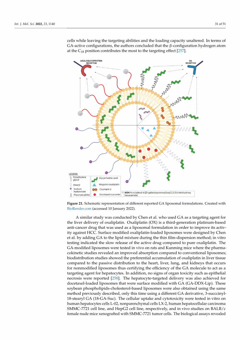
Int. J. Mol. Sci. 2022, 23, 1140 31 of 51
cells while leaving the targeting abilities and the loading capacity unaltered. In terms ofGA-active configurations, the authors concluded that the β-configuration hydrogen atomat the C18 position contributes the most to the targeting effect [257].
Figure 21. Schematic representation of different reported GA liposomal formulations. Created withBioRender.com (accessed 10 January 2022).
A similar study was conducted by Chen et al. who used GA as a targeting agent forthe liver delivery of oxaliplatin. Oxaliplatin (OX) is a third-generation platinum-basedanti-cancer drug that was used as a liposomal formulation in order to improve its activ-ity against HCC. Surface modified oxaliplatin-loaded liposomes were designed by Chenet al. by adding GA to the lipid mixture during the thin film-dispersion method; in vitrotesting indicated the slow release of the active drug compared to pure oxaliplatin. TheGA-modified liposomes were tested in vivo on rats and Kunming mice where the pharma-cokinetic studies revealed an improved absorption compared to conventional liposomes;biodistribution studies showed the preferential accumulation of oxaliplatin in liver tissuecompared to the passive distribution to the heart, liver, lung, and kidneys that occursfor nonmodified liposomes thus certifying the efficiency of the GA molecule to act as atargeting agent for hepatocytes. In addition, no signs of organ toxicity such as epithelialnecrosis were reported [258]. The hepatocyte-targeted delivery was also achieved fordocetaxel-loaded liposomes that were surface modified with GA (GA-DDX-Lip). Thesesoybean phospholipids-cholesterol-based liposomes were also obtained using the samemethod previously described, only this time using a different GA derivative, 3-succcinyl-18-stearyl GA (18-GA-Suc). The cellular uptake and cytotoxicity were tested in vitro onhuman hepatocytes cells L-02, nonparenchymal cells LX-2, human hepatocellular carcinomaSMMC-7721 cell line, and HepG2 cell line, respectively, and in vivo studies on BALB/cfemale nude mice xenografted with SMMC-7721 tumor cells. The biological assays revealed

Int. J. Mol. Sci. 2022, 23, 1140 32 of 51
that the addition of GA acid to docetaxel-loaded liposomes enhanced their selective tar-geting to hepatocytes due to the receptor-mediated endocytosis. As a result, the modifiedGA-DDX-Lip displayed an improved anti-cancer activity both in vivo and in vitro com-pared to the unmodified liposomes, while retaining the same pharmacokinetic profile [259].The research on docetaxel-loaded liposomes decorated with GA went further by co-loadingcopper sulfide nanoparticles which rendered the nanoformulation thermosensitive; thus,upon injection in liver tumor-bearing mice, the modified liposomes increased drug ac-cumulation at the tumor site and actively targeted liver cells while the copper sulfideenhanced the release of the active compound after being NIR irradiated. The modifiedliposomal formulation can be used for photothermal therapy combined with chemotherapythus exhibiting a significantly improved inhibitory activity of tumor growth as well aslower toxicity [260]. A similar study used GA as liver targeting ligand for wogonin-loadedliposomes (GA-WG-Lip); the liposomes were prepared by the reverse evaporation methodusing the same 18-GA-Suc derivative, only this time the active drug, being water solu-ble, was emulsified in the lipid solution, after which the solvent was removed throughvacuum evaporation. The anti-cancer efficiency of GA-WG-Lip, pure wogonin (WG), andWG-loaded passively targeted liposomes was tested in vitro on HepG2 cells and in vivoon liver tumor-bearing mice. The results showed that the entrapment efficiency was notaltered by the addition of GA which in turn induced higher cellular uptake (1.6 timeshigher) and similar IC50 value compared to conventional liposomes. A much-improvedanti-neoplastic effect was recorded for GA-modified liposomes loaded with wogonin whichwere able to specifically target the liver with a long retention time and reduce tumor growth.Collectively, the experimental results indicated an optimized biodistribution, tumor ac-cumulation, and anti-cancer efficacy for the GA-modified liposomes presumably due tothe receptor-mediated cellular uptake of the nanoformulation [261]. GA also induced theliver targeted delivery of curcumin loaded in cationic liposomes; a complex of GA andoctadecylamine was prepared through a simple static binding reaction and later usedfor the preparation of GA-modified curcumin-loaded cationic liposomes by means of theethanol injection method. This formulation was highly suitable for curcumin entrapmentgiven by the high entrapment efficiency value of 98.26± 1.33%. In vitro, the GA-modifiedliposomes produced a stronger inhibition of the HepG2 cells than free curcumin; however,the most remarkable results were revealed in vivo on H22 tumor-bearing mice where theanti-tumor effect following intravenous administration was similar to the one recorded forthe intratumor administration. The in vivo experiment emphasized the inhibition of tumorgrowth, an anti-angiogenic effect, and improved hematologic parameters for the treatedmice thus indicating a future potential treatment for liver cancer [262]. A further improve-ment of the hepatocyte-targeted delivery of an active drug was achieved by using surfacegalactose-modified liposomes which benefit from the recognition by the asialoglycoprotein(ASGP) receptors displayed at the hepatocytes’ surfaces. GA was used as a model drugto be loaded in liposomes whose surfaces were subjected to an enzymatic modification inorder to include β-galactose residues. The in vitro assessment of the resulting liposomesrevealed stable nanoformulations with high encapsulation efficiency (>90%) and sustaineddrug release [263].
A novel formulation of GA-modified cationic stealth liposomes (GA-PEG-CLs) wasdeveloped by He et al. in order to facilitate gene delivery to healthy and cancerous liver cells.The liposomes were obtained through the film dispersion method by mixing cholesterol, 1,2-dioleoyl-3- trimethylammonium-propane (DOTAP) a GA-PEG-cholesterol conjugate. Blankliposomes were incubated afterwards with a 5% glucose protein plasmid DNA solution.These formulations were subsequently tested in two different concentrations (1% and 5%)by comparison to cationic liposomes (CLs) and PEGylated cationic liposomes (PEG-CLs),respectively. The in vitro cell transfection efficiency tests on HCC HepG2 cells showed thatthe best transfection efficacy occurred for GA-PEG-CLs 5% which also displayed the lowesttoxicity on normal L02 liver cells; for the human embryonic kidney cell line HEK 293 whichdoes not overexpress the GA receptor, GA-PEG-CLs revealed the lowest transfection rate
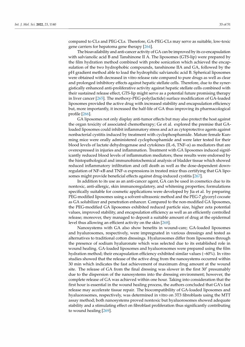
Int. J. Mol. Sci. 2022, 23, 1140 33 of 51
compared to CLs and PEG-CLs. Therefore, GA-PEG-CLs may serve as suitable, low-toxicgene carriers for hepatoma gene therapy [264].
The bioavailability and anti-cancer activity of GA can be improved by its co-encapsulationwith salvianolic acid B and Tanshinone II A. The liposomes (GTS-lip) were prepared bythe film hydration method combined with probe sonication which achieved the encap-sulation of the two hydrophobic compounds, tanshinone IIA and GA, followed by thepH gradient method able to load the hydrophilic salvianolic acid B. Spherical liposomeswere obtained with decreased in vitro release rate compared to pure drugs as well as clearand prolonged inhibitory effects against hepatic stellate cells. Therefore, due to the syner-gistically enhanced anti-proliferative activity against hepatic stellate cells combined withtheir sustained release effect, GTS-lip might serve as a potential future promising therapyin liver cancer [265]. The methoxy-PEG-poly(lactide) surface modification of GA-loadedliposomes provided the active drug with increased stability and encapsulation efficiencybut, more importantly, it increased the half-life of GA thus improving its pharmacologicalprofile [266].
GA liposomes not only display anti-tumor effects but may also protect the host againstthe organ toxicity of associated chemotherapy; Ge et al. explored the premise that GA-loaded liposomes could inhibit inflammatory stress and act as cytoprotective agents againstnonbacterial cystitis induced by treatment with cyclophosphamide. Mature female Kun-ming mice were orally administered cyclophosphamide and were later tested for theirblood levels of lactate dehydrogenase and cytokines (IL-6, TNF-α) as mediators that areoverexpressed in injuries and inflammation. Treatment with GA liposomes induced signif-icantly reduced blood levels of inflammation mediators; these results were endorsed bythe histopathological and immunohistochemical analysis of bladder tissue which showedreduced inflammatory infiltration and cell death as well as the dose-dependent down-regulation of NF-κB and TNF-α expressions in treated mice thus certifying that GA lipo-somes might provide beneficial effects against drug-induced cystitis [267].
In addition to its use as an anti-cancer agent, GA can be used in cosmetics due to itsnontoxic, anti-allergic, skin immunoregulatory, and whitening properties; formulationsspecifically suitable for cosmetic applications were developed by Jia et al. by preparingPEG-modified liposomes using a solvent-ultrasonic method and the PEG-7 glyceryl cocoateas GA solubilizer and penetration enhancer. Compared to the non-modified GA liposomes,the PEG-modified GA liposomes exhibited reduced particle size, higher zeta potentialvalues, improved stability, and encapsulation efficiency as well as an efficiently controlledrelease; moreover, they managed to deposit a suitable amount of drug at the epidermallevel thus allowing an efficient activity on the skin [268].
Nanosystems with GA also show benefits in wound-care; GA-loaded liposomesand hyalurosomes, respectively, were impregnated in various dressings and tested asalternatives to traditional cotton dressings. Hyalurosomes differ from liposomes throughthe presence of sodium hyaluronate which was selected due to its established role inwound healing. GA-loaded liposomes and hyaluorosomes were prepared using the filmhydration method; their encapsulation efficiency exhibited similar values (~60%). In vitrostudies showed that the release of the active drug from the nanosystems occurred within30 min which indicates the fast achievement of maximum drug amount at the woundsite. The release of GA from the final dressing was slower in the first 30′ presumablydue to the dispersion of the nanosystems into the dressing environment; however, thecomplete release of GA was achieved within one hour. Taking into consideration that thefirst hour is essential in the wound healing process, the authors concluded that GA’s fastrelease may accelerate tissue repair. The biocompatibility of GA-loaded liposomes andhyaluorosomes, respectively, was determined in vitro on 3T3 fibroblasts using the MTTassay method; both nanosystems proved nontoxic but hyaluorosomes showed adequatestability and a stimulating effect on fibroblast proliferation thus significantly contributingto wound healing [269].

Int. J. Mol. Sci. 2022, 23, 1140 34 of 51
Conventional liposome preparation methods lead to aqueous suspensions which dis-play poor long-term stability; an effective way to overcome this drawback is the preparationof proliposomes in the form of powders obtained through the dehydration of liposomecomponents which may be dispersed in water before application thus regenerating theliposomes. Liu et al. developed such GA-loaded proliposomes using the lyophilizationmonophase solution method; the authors optimized the formulation and processing vari-ables of GA liposomes in order to achieve adequate stability, encapsulation efficacy, andsustained drug release. In addition, the uptake process of regenerated liposomes by HepG2 liver cancer cells was time dependent [270].
5.2.4. Ursolic Acid
Ursolic acid (UA) is a pentacyclic triterpene (Figure 22) abundantly found in naturein various fruits and vegetables but also dietary fibers and brown mustard as well as theleaves and herbs of the Lamiaceae family [271]; it was revealed as non-toxic and accumu-lates predominantly in spleen and hepatic cells [272]. Even though UA possesses a broadspectrum of impressive biological activities such as anti-allergic, anti-tumor, cardiopro-tective, analgesic, neuroprotective, anti-obesity, anti-anxiety, and anti-depression [273],its low bioavailability induced by its high lipophilic structure requires various chemicalmodifications in order to improve its potential pharmacological use [274]. The various UAformulations discussed in this section are schematically represented in Figure 23.
Figure 22. Chemical structure of ursolic acid.
Due to its low water solubility and bioavailability, UA was incorporated in PEG-liposomes in order to modulate the tumor microenvironment and regulate T cell activityin cancer immunotherapy. The UA liposomes were prepared by the thin-film hydrationmethod using a hydrophilic cyclodextrin (hydroxypropyl-β-cyclodextrin—HPβCD) as asolubility enhancer for UA, following a multiple-step process. UA was first transformed toa metformin salt after which this salt was used to form an HPβCD. This pre-formulation af-forded an increased UA water solubility and therefore rendered the possibility of includingUA in the internal cavity of the liposome as opposed to normal UA liposome formulationswhere the triterpene would be immersed in the lipid bilayer of the particle. This processenabled the formation of thermodynamically stable nanosized crystalline UA inside theliposomes which facilitate the sustained in vivo UA release (>40% drugs quantity remainsinside liposomes after 72 h). The in vitro cytotoxicity studies were conducted on severalcancer cell lines including the 4T1 murine triple-negative breast cancer and failed to revealcytotoxic effects for the UA liposomes. However, when administered in vivo to tumor-bearing C57BL/6 mice, they were able to modulate CD4 + CD25 + Foxp3+ T cells thuscorrecting the tumor-mediated immune-suppressive microenvironment. Consequently, thetumor tissue may become more sensitive towards anti-tumor immune cells; therefore, theauthors concluded that the UA liposome treatment was able to inhibit tumor growth andact as an immunotherapy agent in future anti-cancer therapies [275].
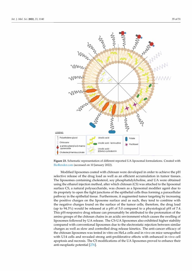
Int. J. Mol. Sci. 2022, 23, 1140 35 of 51
Figure 23. Schematic representation of different reported UA liposomal formulations. Created withBioRender.com (accessed on 10 January 2022).
Modified liposomes coated with chitosan were developed in order to achieve the pHselective release of the drug load as well as an efficient accumulation in tumor tissues.The liposomes containing cholesterol, soy phosphatidylcholine, and UA were obtainedusing the ethanol injection method, after which chitosan (CS) was attached to the liposomalsurface CS, a natural polysaccharide, was chosen as a liposomal modifier agent due toits propriety to open the tight junctions of the epithelial cells thus forming a paracellularpathway in the epithelial tissue. Furthermore, it augmented tumor targeting by increasingthe positive charges on the liposome surface and as such, they tend to combine withthe negative charges found on the surface of the tumor cells; therefore, the drug load(up to 94.3%) would be released at a pH of 5.0 compared to a physiological pH of 7.4.This pH-responsive drug release can presumably be attributed to the protonation of theamino groups of the chitosan chains in an acidic environment which causes the swelling ofliposomes followed by UA release. The CS-UA liposomes also exhibited higher stabilitycompared with conventional liposomes due to the electrostatic rejection between similarcharges as well as slow and controlled drug release kinetics. The anti-cancer efficacy ofthe chitosan liposomes was tested in vitro on HeLa cells and in vivo on mice xenograftedwith U14 cells and revealed strong anti-proliferative effects with enhanced in vivo cellapoptosis and necrosis. The CS modifications of the UA liposomes proved to enhance theiranti-neoplastic potential [276].
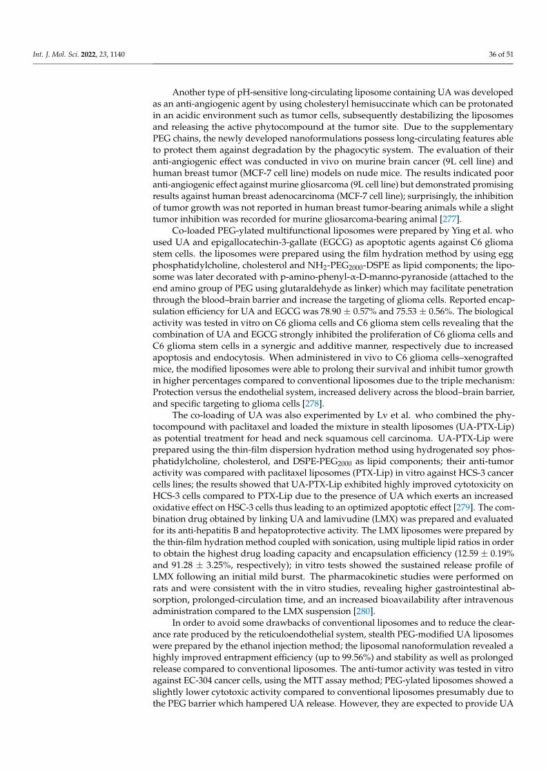
Int. J. Mol. Sci. 2022, 23, 1140 36 of 51
Another type of pH-sensitive long-circulating liposome containing UA was developedas an anti-angiogenic agent by using cholesteryl hemisuccinate which can be protonatedin an acidic environment such as tumor cells, subsequently destabilizing the liposomesand releasing the active phytocompound at the tumor site. Due to the supplementaryPEG chains, the newly developed nanoformulations possess long-circulating features ableto protect them against degradation by the phagocytic system. The evaluation of theiranti-angiogenic effect was conducted in vivo on murine brain cancer (9L cell line) andhuman breast tumor (MCF-7 cell line) models on nude mice. The results indicated pooranti-angiogenic effect against murine gliosarcoma (9L cell line) but demonstrated promisingresults against human breast adenocarcinoma (MCF-7 cell line); surprisingly, the inhibitionof tumor growth was not reported in human breast tumor-bearing animals while a slighttumor inhibition was recorded for murine gliosarcoma-bearing animal [277].
Co-loaded PEG-ylated multifunctional liposomes were prepared by Ying et al. whoused UA and epigallocatechin-3-gallate (EGCG) as apoptotic agents against C6 gliomastem cells. the liposomes were prepared using the film hydration method by using eggphosphatidylcholine, cholesterol and NH2-PEG2000-DSPE as lipid components; the lipo-some was later decorated with p-amino-phenyl-α-D-manno-pyranoside (attached to theend amino group of PEG using glutaraldehyde as linker) which may facilitate penetrationthrough the blood–brain barrier and increase the targeting of glioma cells. Reported encap-sulation efficiency for UA and EGCG was 78.90 ± 0.57% and 75.53 ± 0.56%. The biologicalactivity was tested in vitro on C6 glioma cells and C6 glioma stem cells revealing that thecombination of UA and EGCG strongly inhibited the proliferation of C6 glioma cells andC6 glioma stem cells in a synergic and additive manner, respectively due to increasedapoptosis and endocytosis. When administered in vivo to C6 glioma cells–xenograftedmice, the modified liposomes were able to prolong their survival and inhibit tumor growthin higher percentages compared to conventional liposomes due to the triple mechanism:Protection versus the endothelial system, increased delivery across the blood–brain barrier,and specific targeting to glioma cells [278].
The co-loading of UA was also experimented by Lv et al. who combined the phy-tocompound with paclitaxel and loaded the mixture in stealth liposomes (UA-PTX-Lip)as potential treatment for head and neck squamous cell carcinoma. UA-PTX-Lip wereprepared using the thin-film dispersion hydration method using hydrogenated soy phos-phatidylcholine, cholesterol, and DSPE-PEG2000 as lipid components; their anti-tumoractivity was compared with paclitaxel liposomes (PTX-Lip) in vitro against HCS-3 cancercells lines; the results showed that UA-PTX-Lip exhibited highly improved cytotoxicity onHCS-3 cells compared to PTX-Lip due to the presence of UA which exerts an increasedoxidative effect on HSC-3 cells thus leading to an optimized apoptotic effect [279]. The com-bination drug obtained by linking UA and lamivudine (LMX) was prepared and evaluatedfor its anti-hepatitis B and hepatoprotective activity. The LMX liposomes were prepared bythe thin-film hydration method coupled with sonication, using multiple lipid ratios in orderto obtain the highest drug loading capacity and encapsulation efficiency (12.59 ± 0.19%and 91.28 ± 3.25%, respectively); in vitro tests showed the sustained release profile ofLMX following an initial mild burst. The pharmacokinetic studies were performed onrats and were consistent with the in vitro studies, revealing higher gastrointestinal ab-sorption, prolonged-circulation time, and an increased bioavailability after intravenousadministration compared to the LMX suspension [280].
In order to avoid some drawbacks of conventional liposomes and to reduce the clear-ance rate produced by the reticuloendothelial system, stealth PEG-modified UA liposomeswere prepared by the ethanol injection method; the liposomal nanoformulation revealed ahighly improved entrapment efficiency (up to 99.56%) and stability as well as prolongedrelease compared to conventional liposomes. The anti-tumor activity was tested in vitroagainst EC-304 cancer cells, using the MTT assay method; PEG-ylated liposomes showed aslightly lower cytotoxic activity compared to conventional liposomes presumably due tothe PEG barrier which hampered UA release. However, they are expected to provide UA

Int. J. Mol. Sci. 2022, 23, 1140 37 of 51
with a longer-circulating time in the bloodstream thus reaching a more efficient anti-tumoractivity [281]. An in vivo study of PEGylated UA-loaded liposomes was conducted on CD-1female mice inoculated with U14 cervical carcinoma mouse cells; the results demonstratedthat the presence of the outer PEG shell extended the half-life and reduced the release rateof the incorporated drug while the rate of tumor apoptosis was heightened compared toconventional UA liposomes. The histopathological analysis showed no signs of toxicity onliver and kidney tissues thus recommending the nanoformulation for further developmentin anti-cancer treatment [282]. Improved stealth liposomes were designed and developedby coating their surfaces with folate-PEG-cholesteryl hemisuccinate; their pharmacokineticsand anti-tumor activity on folate receptor-positive human oral cancer KB cells were laterassessed. The folate-PEG-ylated liposomes significantly enhanced UA bioavailability andsolubility while preferentially targeting the folate receptor-positive cells thus providing apossible basis for future oral cancer treatment [283].
5.2.5. Lupeol
Lupeol is a lupan-type pentacyclic triterpene (Figure 24) also found under the namesclerodol, fagarasterol, and monogynol B; it can be extracted from a variety of vegetablesand fruits such as soybean (Glycine max L.) Merr., aloe (Aloe vera L. Burm.f.), black tea(Camellia sinensis var. assamica J.W.Mast. Kitam.), tomato (Solanum lycopersicum L.), mango(Mangifera indica L.), etc. [284]. Lupeol exhibits a wide range of pharmacological proprietiessuch as cardioprotective, hepatoprotective, anti-diabetic, anti-cancer, anti-oxidant, anti-microbial, and anti-inflammatory [285]. However, its lipophilic structure induces low watersolubility and bioavailability; therefore, requiring engineered formulations in order to exertan enhanced pharmacological activity [286,287]. The various lupeol formulations discussedin this section are schematically represented in Figure 25.
Figure 24. Chemical structure of lupeol.
Lupeol-loaded liposomes bearing PEG chains were developed by Zhang et al. andtested in vitro on the HepG2 cancer cell line, revealing high inhibition and apoptosis rate,blocking the cells in the G2M phase; the PEGylation process improved the encapsulationproprieties (86.2 ± 0.9%) and zeta potential (1.6 ± 0.15 with PDI = 0.25 ± 0.029) thus con-tributing to an increased stability of the nanoformulation. In vivo studies were performedon rats by intravenous administration in order to investigate the pharmacokinetics of theliposomal formulation which increased the half-life and AUC of lupeol several times [288].
In a similar manner with GA, lupeol was incorporated in galactose-decorated lipo-somes in order to benefit from the asialoglycoprotein receptors found at the surface ofhepatocytes and achieve a targeted delivery in liver cancer. Lupeol-loaded galactose-PEG-DSPE liposomes (GAL-L) were prepared by the thin-film dispersion method using HSPC,cholesterol, and galactose-PEG-DSPE as lipidic components; their encapsulated efficiencywas above 85% and showed long-term stability. The apoptotic effect and cellular uptake
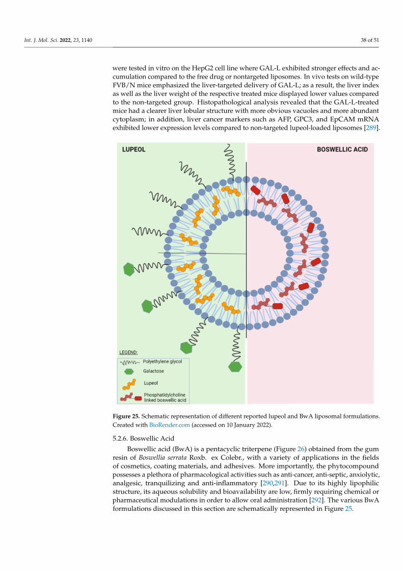
Int. J. Mol. Sci. 2022, 23, 1140 38 of 51
were tested in vitro on the HepG2 cell line where GAL-L exhibited stronger effects and ac-cumulation compared to the free drug or nontargeted liposomes. In vivo tests on wild-typeFVB/N mice emphasized the liver-targeted delivery of GAL-L; as a result, the liver indexas well as the liver weight of the respective treated mice displayed lower values comparedto the non-targeted group. Histopathological analysis revealed that the GAL-L-treatedmice had a clearer liver lobular structure with more obvious vacuoles and more abundantcytoplasm; in addition, liver cancer markers such as AFP, GPC3, and EpCAM mRNAexhibited lower expression levels compared to non-targeted lupeol-loaded liposomes [289].
Figure 25. Schematic representation of different reported lupeol and BwA liposomal formulations.Created with BioRender.com (accessed on 10 January 2022).
5.2.6. Boswellic Acid
Boswellic acid (BwA) is a pentacyclic triterpene (Figure 26) obtained from the gumresin of Boswellia serrata Roxb. ex Colebr., with a variety of applications in the fieldsof cosmetics, coating materials, and adhesives. More importantly, the phytocompoundpossesses a plethora of pharmacological activities such as anti-cancer, anti-septic, anxiolytic,analgesic, tranquilizing and anti-inflammatory [290,291]. Due to its highly lipophilicstructure, its aqueous solubility and bioavailability are low, firmly requiring chemical orpharmaceutical modulations in order to allow oral administration [292]. The various BwAformulations discussed in this section are schematically represented in Figure 25.
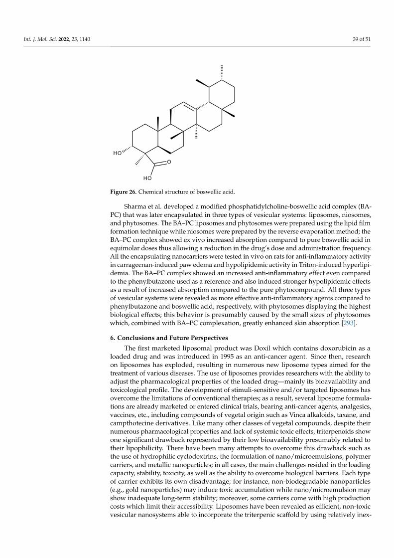
Int. J. Mol. Sci. 2022, 23, 1140 39 of 51
Figure 26. Chemical structure of boswellic acid.
Sharma et al. developed a modified phosphatidylcholine-boswellic acid complex (BA-PC) that was later encapsulated in three types of vesicular systems: liposomes, niosomes,and phytosomes. The BA–PC liposomes and phytosomes were prepared using the lipid filmformation technique while niosomes were prepared by the reverse evaporation method; theBA–PC complex showed ex vivo increased absorption compared to pure boswellic acid inequimolar doses thus allowing a reduction in the drug’s dose and administration frequency.All the encapsulating nanocarriers were tested in vivo on rats for anti-inflammatory activityin carrageenan-induced paw edema and hypolipidemic activity in Triton-induced hyperlipi-demia. The BA–PC complex showed an increased anti-inflammatory effect even comparedto the phenylbutazone used as a reference and also induced stronger hypolipidemic effectsas a result of increased absorption compared to the pure phytocompound. All three typesof vesicular systems were revealed as more effective anti-inflammatory agents compared tophenylbutazone and boswellic acid, respectively, with phytosomes displaying the highestbiological effects; this behavior is presumably caused by the small sizes of phytosomeswhich, combined with BA–PC complexation, greatly enhanced skin absorption [293].
6. Conclusions and Future Perspectives
The first marketed liposomal product was Doxil which contains doxorubicin as aloaded drug and was introduced in 1995 as an anti-cancer agent. Since then, researchon liposomes has exploded, resulting in numerous new liposome types aimed for thetreatment of various diseases. The use of liposomes provides researchers with the ability toadjust the pharmacological properties of the loaded drug—mainly its bioavailability andtoxicological profile. The development of stimuli-sensitive and/or targeted liposomes hasovercome the limitations of conventional therapies; as a result, several liposome formula-tions are already marketed or entered clinical trials, bearing anti-cancer agents, analgesics,vaccines, etc., including compounds of vegetal origin such as Vinca alkaloids, taxane, andcampthotecine derivatives. Like many other classes of vegetal compounds, despite theirnumerous pharmacological properties and lack of systemic toxic effects, triterpenoids showone significant drawback represented by their low bioavailability presumably related totheir lipophilicity. There have been many attempts to overcome this drawback such asthe use of hydrophilic cyclodextrins, the formulation of nano/microemulsions, polymercarriers, and metallic nanoparticles; in all cases, the main challenges resided in the loadingcapacity, stability, toxicity, as well as the ability to overcome biological barriers. Each typeof carrier exhibits its own disadvantage; for instance, non-biodegradable nanoparticles(e.g., gold nanoparticles) may induce toxic accumulation while nano/microemulsion mayshow inadequate long-term stability; moreover, some carriers come with high productioncosts which limit their accessibility. Liposomes have been revealed as efficient, non-toxicvesicular nanosystems able to incorporate the triterpenic scaffold by using relatively inex-

Int. J. Mol. Sci. 2022, 23, 1140 40 of 51
pensive materials and manufacturing procedures. In addition, liposomes showed a hugeability to support various modulations both at the surface and in the inner compositionwhich promoted significant drug loading, sustained controlled drug release, improvedpharmacokinetics, and targeted delivery thus inducing optimal pharmacological outcomesby avoiding acute and chronic organ toxicities as well as preventing premature drug degra-dation. Liposomes loaded with triterpenic compounds revealed higher pharmacodynamiceffects compared to the drug alone, in particular as anti-cancer and anti-inflammatoryagents, with limited toxicity on normal cells; moreover, triterpenes can be co-loaded withconventional anti-cancer drugs, managing to counteract their severe side effects. Besidesserving as loaded drugs, triterpenes may also act as organ-targeting ligands as is the caseof glycirrhetinic acid which is recognized by specific receptors located at the surface ofhepatocytes and hepatocellular carcinoma cells. Liposomes are highly malleable carriersthat are able to withstand various surface modulations; as such, chitosan-coated liposomesmanaged to achieve the pH-selective release of the drug load as well as an efficient accu-mulation in tumor tissues. In addition, chitosan was able to mediate the combination ofliposomes with another type of nanocarrier, i.e., gold nanoshells, in order to provide thecombination of chemo- and photothermal therapy. The long-term stability was improvedby the preparation of proliposomes which may regenerate the liposomes as a result ofwater dispersion. One of the most important advantages liposomes can provide is theirability to bind various ligands used in targeted therapies; the targeted use of triterpeneshas been proven highly useful in preclinical trials; however, with the exception of betulinand betulinic acid which have entered the pharmaceutical market or several clinical trials,respectively, triterpenes have not yet reached clinical expectations regardless of their phar-maceutical formulations. Through the advantages offered, their loading into liposomesmay lead to sufficient improvement of their pharmacological profile so as to justify theirinclusion in clinical trials; however, further research is mandatory in order to achievethis goal.
The fact that different types of liposome-based formulations are currently used intherapy or assessed in clinical trials stands as a driving milestone for the importance ofdeveloping such nanoformulations containing triterpenoid structures. There are, however,two important interconnected issues that need to be addressed. Firstly, the main triterpenerepresentatives, especially the ones with anti-cancer activity, do not have a completelyelucidated mechanism of action. Secondly, when available, the answers to this first issueshould be coupled with a more accurate quantification of the liposome-released amountof substance at the tumor site, available to exert its pharmacological effect. After reachingthese important checkpoints in the future, triterpenoid-containing liposomal formulationswill be able to reach clinical trials and thus impact the current therapeutic landscape.
Author Contributions: Conceptualization, A.M. (Andreea Milan) and A.M. (Alexandra Mioc);writing—original draft preparation: Section 1, A.M. (Andreea Milan) and A.M. (Alexandra Mioc);Section 2, A.M. (Andreea Milan) and A.P.; Section 3, A.M. (Alexandra Mioc) and F.C.; Section 4, A.M.(Andreea Milan) and A.M. (Alexandra Mioc), R.G.; Section 5, A.M. (Andreea Milan), A.M. (AlexandraMioc), R.R. and C.S.; visualization, C.S., M.M. and R.B.; writing—review and editing: M.M. andR.B.; supervision, C.S. and M.M.; project administration, C.S.; funding acquisition, C.S. and M.M. Allauthors have read and agreed to the published version of the manuscript.
Funding: This work was supported by the Romanian UEFISCDI national grant PN-III-P1-1.1-PD-2019-1078 and Internal grant “Victor Babes” University of Medicine and Pharmacy, Grant1EXP/1233/30.01.2020 LUPSKINPATH.
Conflicts of Interest: The authors declare no conflict of interest.
Int. J. Mol. Sci. 2022, 23, 1140 41 of 51
References
1. Surowiak, A.; Balcerzak, L.; Lochynski, S.; Strub, D. Biological Activity of Selected Natural and Synthetic Terpenoid Lactones. Int.
J. Mol. Sci. 2021, 22, 5036. [CrossRef]2. Mahizan, N.A.; Yang, S.-K.; Moo, C.L.; Song, A.A.-L.; Chong, C.-M.; Chong, C.-W.; Abushelaibi, A.; Lim, S.-H.E.; Lai, K.-S.
Terpene Derivatives as a Potential Agent against Antimicrobial Resistance (AMR) Pathogens. Molecules 2019, 24, 2631. [CrossRef]3. Valdés, K.; Morales, J.; Rodríguez, L.; Gunther, G. Potential use of nanocarriers with pentacyclic triterpenes in cancer treatments.
Nanomedicine 2016, 11, 3139–3156. [CrossRef]4. Soica, C.; Coricovac, D.; Dehelean, C.; Pinzaru, I.; Mioc, M.; Danciu, C.; Fulias, A.; Puiu, M.; Sitaru, C. Nanocarriers as Tools in
Delivering Active Compounds for Immune System Related Pathologies. Recent Pat. Nanotechnol. 2016, 10, 128–145. [CrossRef]5. Sahoo, S.K.; Labhasetwar, V. Nanotech approaches to drug delivery and imaging. Drug Discov. Today 2003, 8, 1112–1120.
[CrossRef]6. Petrak, K. Nanotechnology and site-targeted drug delivery. J. Biomater. Sci. Polym. Ed. 2006, 17, 1209–1219. [CrossRef] [PubMed]7. Lengyel, M.; Kállai-Szabó, N.; Antal, V.; Laki, A.J.; Antal, I. Microparticles, Microspheres, and Microcapsules for Advanced Drug
Delivery. Sci. Pharm. 2019, 87, 20. [CrossRef]8. Kohane, D.S. Microparticles and nanoparticles for drug delivery. Biotechnol. Bioeng. 2006, 96, 203–209. [CrossRef] [PubMed]9. Gregory, G. Engineering liposomes for drug delivery: Progress and problems. Trends Biotechnol. 1995, 13, 527–537.10. Lamichhane, N.; Udayakumar, T.S.; D’Souza, W.D.; Ii, C.B.S.; Raghavan, S.R.; Polf, J.; Mahmood, J.; Lamichhane, N.;
Udayakumar, T.S.; D’Souza, W.D.; et al. Liposomes: Clinical Applications and Potential for Image-Guided Drug Delivery.Molecules 2018, 23, 288. [CrossRef]
11. Chaturvedi, V.K.; Singh, A.; Singh, V.K.; Singh, M.P. Cancer Nanotechnology: A New Revolution for Cancer Diagnosis andTherapy. Curr. Drug Metab. 2019, 20, 416–429. [CrossRef]
12. Pérez-Herrero, E.; Fernández-Medarde, A. Advanced targeted therapies in cancer: Drug nanocarriers, the future of chemotherapy.Eur. J. Pharm. Biopharm. 2015, 93, 52–79. [CrossRef]
13. Shah, A.; Aftab, S.; Nisar, J.; Ashiq, M.N.; Iftikhar, F.J. Nanocarriers for targeted drug delivery. J. Drug Deliv. Sci. Technol. 2021,62, 102426. [CrossRef]
14. Jeevanandam, J.; Chan, Y.S.; Danquah, M. Nano-formulations of drugs: Recent developments, impact and challenges. Biochimie
2016, 128–129, 99–112. [CrossRef] [PubMed]15. Jiang, W.; Kim, B.Y.; Rutka, J.T.; Chan, W.C. Advances and challenges of nanotechnology-based drug delivery systems. Expert
Opin. Drug Deliv. 2007, 4, 621–633. [CrossRef]16. Pal, S.L.; Jana, U.; Manna, P.K.; Mohanta, G.P.; Manavalan, R. Nanoparticle: An overview of preparation and characterization. J.
Appl. Pharm. Sci. 2011, 1, 228–234.17. Torchilin, V.P. Nanocarriers. Pharm. Res. 2007, 24, 2333–2334. [CrossRef] [PubMed]18. Zhao, Z.; Ukidve, A.; Kim, J.; Mitragotri, S. Targeting Strategies for Tissue-Specific Drug Delivery. Cell 2020, 181, 151–167.
[CrossRef]19. Wu, L.; Zhang, J.; Watanabe, W. Physical and chemical stability of drug nanoparticles. Adv. Drug Deliv. Rev. 2011, 63, 456–469.
[CrossRef] [PubMed]20. Qi, W.H.; Wang, M.P.; Liu, Q.H. Shape factor of nonspherical nanoparticles. J. Mater. Sci. 2005, 40, 2737–2739. [CrossRef]21. Williams, K.J.; Phillips, M.C.; Rodrígueza, W.V. Structural and metabolic consequences of liposome-lipoprotein interactions. Adv.
Drug Deliv. Rev. 1998, 32, 31–43. [PubMed]22. Moreno Raja, M.; Lim, P.Q.; Wong, Y.S.; Xiong, G.M.; Zhang, Y.; Venkatraman, S.; Huang, Y. Polymeric Nanomaterials. In
Nanocarriers for Drug Delivery; Elsevier: Amsterdam, The Netherlands, 2019; pp. 557–653.23. Poon, W.; Zhang, Y.-N.; Ouyang, B.; Kingston, B.R.; Wu, J.L.Y.; Wilhelm, S.; Chan, W.C.W. Elimination Pathways of Nanoparticles.
ACS Nano 2019, 13, 5785–5798. [CrossRef] [PubMed]24. Cuenca, A.G.; Jiang, H.; Hochwald, S.N.; Delano, M.; Cance, W.G.; Grobmyer, S.R. Emerging implications of nanotechnology on
cancer diagnostics and therapeutics. Cancer 2006, 107, 459–466. [CrossRef] [PubMed]25. Kapoor, B.; Gupta, R.; Gulati, M.; Singh, S.; Khursheed, R.; Gupta, M. The Why, Where, Who, How, and What of the vesicular
delivery systems. Adv. Colloid Interface Sci. 2019, 271, 101985. [CrossRef]26. Danaei, M.; Dehghankhold, M.; Ataei, S.; Hasanzadeh Davarani, F.; Javanmard, R.; Dokhani, A.; Khorasani, S.; Mozafari, M.R.
Impact of Particle Size and Polydispersity Index on the Clinical Applications of Lipidic Nanocarrier Systems. Pharmaceutics 2018,10, 57. [CrossRef]
27. Bütün, V.; Sönmez, S.; Yarligan, S.; Taktak, F.F.; Atay, A.; Bütün, S. Micelles and ‘reverse micelles’ with a novel water-solublediblock copolymer. Polymer 2008, 49, 4057–4065. [CrossRef]
28. Record, M.; Carayon, K.; Poirot, M.; Silvente-Poirot, S. Exosomes as new vesicular lipid transporters involved in cell–cellcommunication and various pathophysiologies. Biochim. Biophys. Acta (BBA) Mol. Cell Biol. Lipids 2014, 1841, 108–120. [CrossRef][PubMed]
29. Elsabahy, M.; Heo, G.S.; Lim, S.-M.; Sun, G.; Wooley, K.L. Polymeric Nanostructures for Imaging and Therapy. Chem. Rev. 2015,115, 10967–11011. [CrossRef]
30. Pisani, E.; Tsapis, N.; Paris, J.; Nicolas, V.; Cattel, L.; Fattal, E.; Sud, U.V.P. Polymeric Nano/Microcapsules of Liquid Perfluorocar-bons for Ultrasonic Imaging: Physical Characterization. Langmuir 2006, 73, 4397–4402. [CrossRef] [PubMed]
Int. J. Mol. Sci. 2022, 23, 1140 42 of 51
31. Key, J.; Aryal, S.; Gentile, F.; Ananta, J.S.; Zhong, M.; Landis, M.D.; Decuzzi, P. Engineering discoidal polymeric nanoconstructswith enhanced magneto-optical properties for tumor imaging. Biomaterials 2013, 34, 5402–5410. [CrossRef]
32. Lodahl, P. Quantum-dot based photonic quantum networks Quantum-dot based photonic quantum networks. Quantum Sci.
Technol. 2018, 3, 013001. [CrossRef]33. Moon, H.; Lee, C.; Lee, W.; Kim, J.; Chae, H. Stability of Quantum Dots, Quantum Dot Films, and Quantum Dot Light-Emitting
Diodes for Display Applications. Adv. Mater. 2019, 31, e1804294. [CrossRef] [PubMed]34. Emin, S.; Singh, S.P.; Han, L.; Satoh, N.; Islam, A. Colloidal quantum dot solar cells. Sol. Energy 2011, 85, 1264–1282. [CrossRef]35. Fernando, K.A.S.; Sahu, S.; Liu, Y.; Lewis, W.K.; Guliants, E.A.; Jafariyan, A.; Wang, P.; Bunker, C.E.; Sun, Y.-P. Carbon Quantum
Dots and Applications in Photocatalytic Energy Conversion. ACS Appl. Mater. Interfaces 2015, 7, 8363–8376. [CrossRef] [PubMed]36. Jun, H.; Careem, M.; Arof, A. Quantum dot-sensitized solar cells—Perspective and recent developments: A review of Cd
chalcogenide quantum dots as sensitizers. Renew. Sustain. Energy Rev. 2013, 22, 148–167. [CrossRef]37. Bardhan, R.; Lal, S.; Joshi, A.; Halas, N.J. Theranostic Nanoshells: From Probe Design to Imaging and Treatment of Cancer. Acc.
Chem. Res. 2011, 44, 936–946. [CrossRef] [PubMed]38. Delcea, M.; Möhwald, H.; Skirtach, A.G. Stimuli-responsive LbL capsules and nanoshells for drug delivery. Adv. Drug Deliv. Rev.
2011, 63, 730–747. [CrossRef]39. Huschka, R.; Zuloaga, J.; Knight, M.W.; Brown, L.V.; Nordlander, P.; Halas, N.J. Light-Induced Release of DNA from Gold
Nanoparticles: Nanoshells and Nanorods. J. Am. Chem. Soc. 2011, 133, 12247–12255. [CrossRef]40. Liu, H.; Chen, D.; Li, L.; Liu, T.; Tan, L.; Wu, X.; Tang, F. Multifunctional Gold Nanoshells on Silica Nanorattles: A Platform for the
Combination of Photothermal Therapy and Chemotherapy with Low Systemic Toxicity. Angew. Chem. Int. Ed. 2010, 50, 891–895.[CrossRef]
41. Mioc, M.; Pavel, I.Z.; Ghiulai, R.; Coricovac, D.E.; Farcas, C.; Mihali, C.-V.; Oprean, C.; Serafim, V.; Popovici, R.A.; Dehelean, C.A.; et al.The Cytotoxic Effects of Betulin-Conjugated Gold Nanoparticles as Stable Formulations in Normal and Melanoma Cells. Front.
Pharmacol. 2018, 9, 429. [CrossRef]42. Vines, J.B.; Yoon, J.-H.; Ryu, N.-E.; Lim, D.-J.; Park, H. Gold Nanoparticles for Photothermal Cancer Therapy. Front. Chem. 2019,
7, 167. [CrossRef]43. Balfourier, A.; Luciani, N.; Wang, G.; Lelong, G.; Ersen, O.; Khelfa, A.; Alloyeau, D.; Gazeau, F.; Carn, F. Unexpected intracellular
biodegradation and recrystallization of gold nanoparticles. Proc. Natl. Acad. Sci. USA 2019, 117, 103–113. [CrossRef] [PubMed]44. Elahi, N.; Kamali, M.; Baghersad, M.H. Recent biomedical applications of gold nanoparticles: A review. Talanta 2018, 184, 537–556.
[CrossRef]45. Fratoddi, I.; Venditti, I.; Cametti, C.; Russo, M.V. How toxic are gold nanoparticles? The state-of-the-art. Nano Res. 2015,
8, 1771–1799. [CrossRef]46. Xu, M.; Li, J.; Yan, Y.; Zhao, X.; Yan, J.; Zhang, Y.; Lai, B.; Chen, X.; Song, L. Catalytic degradation of sulfamethoxazole through
peroxymonosulfate activated with expanded graphite loaded CoFe2O4 particles. Chem. Eng. J. 2019, 369, 403–413. [CrossRef]47. Lops, C.; Ancona, A.; Di Cesare, K.; Dumontel, B.; Garino, N.; Canavese, G.; Hérnandez, S.; Cauda, V. Sonophotocatalytic
degradation mechanisms of Rhodamine B dye via radicals generation by micro- and nano-particles of ZnO. Appl. Catal. B Environ.
2018, 243, 629–640. [CrossRef] [PubMed]48. Li, J.; Yan, J.; Yao, G.; Zhang, Y.; Li, X.; Lai, B. Improving the degradation of atrazine in the three-dimensional (3D) electrochemical
process using CuFe2O4 as both particle electrode and catalyst for persulfate activation. Chem. Eng. J. 2018, 361, 1317–1332.[CrossRef]
49. Zhang, X.; Feng, M.; Qu, R.; Liu, H.; Wang, L.; Wang, Z. Catalytic degradation of diethyl phthalate in aqueous solution bypersulfate activated with nano-scaled magnetic CuFe2O4/MWCNTs. Chem. Eng. J. 2016, 301, 1–11. [CrossRef]
50. Feng, W.; Ji, P. Enzymes immobilized on carbon nanotubes. Biotechnol. Adv. 2011, 29, 889–895. [CrossRef]51. Mittal, G.; Dhand, V.; Rhee, K.Y.; Park, S.-J.; Lee, W.R. A review on carbon nanotubes and graphene as fillers in reinforced polymer
nanocomposites. J. Ind. Eng. Chem. 2014, 21, 11–25. [CrossRef]52. Blackburn, J.L.; Ferguson, A.J.; Cho, C.; Grunlan, J.C. Carbon-Nanotube-Based Thermoelectric Materials and Devices. Adv. Mater.
2018, 30, 1704386. [CrossRef] [PubMed]53. Negri, V.; Pacheco, J.; Daniel, T. Carbon Nanotubes in Biomedicine. In Topics in Current Chemistry; Springer International
Publishing: Cham, Switzerland, 2020; pp. 1–41.54. Kumar, A.; Chen, F.; Mozhi, Y.; Zhang, X.; Xue, X. Inovative pharmaceutical development based on unique properties of nanoscale
delivery formulation. Nanoscale 2013, 5, 8307–8325. [CrossRef] [PubMed]55. Ferrari, R.; Sponchioni, M.; Morbidelli, M.; Moscatelli, D. Polymer nanoparticles for the intravenous delivery of anticancer drugs:
The checkpoints on the road from the synthesis to clinical translation. Nanoscale 2018, 10, 22701–22719. [CrossRef]56. Des Rieux, A.; Pourcelle, V.; Cani, P.; Marchand-Brynaert, J.; Préat, V. Targeted nanoparticles with novel non-peptidic ligands for
oral delivery. Adv. Drug Deliv. Rev. 2013, 65, 833–844. [CrossRef]57. Singh, S.R.; Grossniklaus, H.E.; Kang, S.J.; Edelhauser, H.F.; Ambati, B.K.; Kompella, U.B. Intravenous transferrin, RGD peptide
and dual-targeted nanoparticles enhance anti-VEGF intraceptor gene delivery to laser-induced CNV. Gene Ther. 2009, 16, 645–659.[CrossRef]
58. Ak, G.; Bozkaya, F.; Yılmaz, H.; Turgut, S.; Bilgin, I.; Tomruk, C.; Uyanıkgil, Y.; Sanlıer, H. An intravenous application of magneticnanoparticles for osteomyelitis treatment: An efficient alternative. Int. J. Pharm. 2020, 592, 119999. [CrossRef]

Int. J. Mol. Sci. 2022, 23, 1140 43 of 51
59. Duan, Y.; Xu, J.; Lin, Y.; Yu, H.; Gong, T.; Li, Y.; Zhang, Z. A preliminary study on MeO-PEG-PLGA-PEG-OMe nanoparticles asintravenous carriers. J. Biomed. Mater. Res. Part A 2008, 87, 515–523. [CrossRef]
60. Tadros, M.I.; Al-Mahallawi, A.M. Long-circulating lipoprotein-mimic nanoparticles for smart intravenous delivery of a practically-insoluble antineoplastic drug: Development, preliminary safety evaluations and preclinical pharmacokinetic studies. Int. J.
Pharm. 2015, 493, 439–450. [CrossRef]61. Date, A.; Hanes, J.; Ensign, L.M. Nanoparticles for oral delivery: Design, evaluation and state-of-the-art. J. Control. Release 2016,
240, 504–526. [CrossRef] [PubMed]62. Yun, Y.; Cho, Y.W.; Park, K. Nanoparticles for oral delivery: Targeted nanoparticles with peptidic ligands for oral protein delivery.
Adv. Drug Deliv. Rev. 2013, 65, 822–832. [CrossRef]63. Bakhru, S.H.; Furtado, S.; Morello, A.P.; Mathiowitz, E. Oral delivery of proteins by biodegradable nanoparticles. Adv. Drug Deliv.
Rev. 2013, 65, 811–821. [CrossRef] [PubMed]64. Cao, S.-J.; Xu, S.; Wang, H.-M.; Ling, Y.; Dong, J.; Xia, R.-D.; Sun, X.-H. Nanoparticles: Oral Delivery for Protein and Peptide
Drugs. AAPS PharmSciTech 2019, 20, 190. [CrossRef]65. Dilnawaz, F. Polymeric Biomaterial and Lipid Based Nanoparticles for Oral Drug Delivery. Curr. Med. Chem. 2017, 24, 2423–2438.
[CrossRef]66. Huang, F.-Y.J.; Chen, W.-J.; Lee, W.-Y.; Lo, S.-T.; Lee, T.-W.; Lo, J.-M. In Vitro and in Vivo Evaluation of Lactoferrin-Conjugated
Liposomes as a Novel Carrier to Improve the Brain Delivery. Int. J. Mol. Sci. 2013, 14, 2862–2874. [CrossRef]67. Gupta, R. Beena Rai Computer-Aided Design of Nanoparticles for Transdermal Drug Delivery. Methods Mol. Biol. 2020,
2059, 225–237.68. Palmer, B.C.; De Louise, L.A. Nanoparticle-Enabled Transdermal Drug Delivery Systems for Enhanced Dose Control and Tissue
Targeting. Molecules 2016, 21, 1719. [CrossRef] [PubMed]69. Ruan, R.; Chen, M.; Zou, L.; Wei, P.; Liu, J.; Ding, W.; Wen, L. Recent advances in peptides for enhancing transdermal
macromolecular drug delivery. Ther. Deliv. 2016, 7, 89–100. [CrossRef]70. Peña-Juárez, M.C.; Guadarrama-Escobar, O.R.; Escobar-Chávez, J.J. Transdermal Delivery Systems for Biomolecules. J. Pharm.
Innov. 2021, 1–14. [CrossRef]71. Carter, P.; Narasimhan, B.; Wang, Q. Biocompatible nanoparticles and vesicular systems in transdermal drug delivery for various
skin diseases. Int. J. Pharm. 2018, 555, 49–62. [CrossRef] [PubMed]72. Das Kurmi, B.; Tekchandani, P.; Paliwal, R.; Paliwal, S.R. Transdermal Drug Delivery: Opportunities and Challenges for Controlled
Delivery of Therapeutic Agents Using Nanocarriers. Curr. Drug Metab. 2017, 18, 481–495. [CrossRef] [PubMed]73. Diblíková, D.; Kopecná, M.; Školová, B.; Krecmerova, M.; Roh, J.; Hrabálek, A.; Vávrová, K. Transdermal Delivery and Cutaneous
Targeting of Antivirals using a Penetration Enhancer and Lysolipid Prodrugs. Pharm. Res. 2013, 31, 1071–1081. [CrossRef][PubMed]
74. Mangal, S.; Gao, W.; Li, T.; Zhou, Q. Pulmonary delivery of nanoparticle chemotherapy for the treatment of lung cancers:Challenges and opportunities. Acta Pharmacol. Sin. 2017, 38, 782–797. [CrossRef]
75. Schreier, H.; Gonzalez-rothib, R.J.; Stecenkoc, A.A. Pulmonary delivery of liposomes. J. Control. Release 1993, 24, 209–223.[CrossRef]
76. Kuruba, R.; Wilson, A.; Gao, X.; Li, S. Targeted Delivery of Nucleic Acid-Based Therapeutics to the Pulmonary Circulation. AAPS
J. 2009, 11, 23–30. [CrossRef] [PubMed]77. Chakraborty, A.; Royce, S.G.; Plebanski, M.; Selomulya, C. Glycine microparticles loaded with functionalized nanoparticles for
pulmonary delivery. Int. J. Pharm. 2019, 570, 118654. [CrossRef] [PubMed]78. Kunda, N.; Somavarapu, S.; Gordon, S.; Hutcheon, G.; Saleem, I.Y. Nanocarriers Targeting Dendritic Cells for Pulmonary Vaccine
Delivery. Pharm. Res. 2012, 30, 325–341. [CrossRef] [PubMed]79. Osman, N.; Kaneko, K.; Carini, V.; Saleem, I. Carriers for the targeted delivery of aerosolized macromolecules for pulmonary
pathologies. Expert Opin. Drug Deliv. 2018, 15, 821–834. [CrossRef]80. Mcbride, A.A.; Price, D.N.; Muttil, P.; Laboratories, N. Pulmonary Delivery of Magnetically Targeted Nano-in Microparticles.
Methods Enzymol. 2017, 1530, 369–378.81. Shen, J.; Lu, G.W.; Hughes, P. Targeted Ocular Drug Delivery with Pharmacokinetic/Pharmacodynamic Considerations. Pharm.
Res. 2018, 35, 217. [CrossRef]82. Janagam, D.R.; Wu, L.; Lowe, T.L. Nanoparticles for drug delivery to the anterior segment of the eye. Adv. Drug Deliv. Rev. 2017,
122, 31–64. [CrossRef]83. Ameeduzzafar; Ali, J.; Fazil, M.; Qumbar, M.; Khan, N.; Ali, A. Colloidal drug delivery system: Amplify the ocular delivery. Drug
Deliv. 2014, 23, 700–716. [CrossRef]84. Gaudana, R.; Jwala, J.; Boddu, S.H.S.; Mitra, A.K. Recent Perspectives in Ocular Drug Delivery. Pharm. Res. 2008, 26, 1197–1216.
[CrossRef]85. De la Fuente, M.; Raviña, M.; Paolicelli, P.; Sanchez, A.; Seijo, B.; Alonso, M.J. Chitosan-based nanostructures: A delivery platform
for ocular therapeutics. Adv. Drug Deliv. Rev. 2010, 62, 100–117. [CrossRef]86. Suri, R.; Beg, S.; Kohli, K. Target strategies for drug delivery bypassing ocular barriers. J. Drug Deliv. Sci. Technol. 2020, 55, 101389.
[CrossRef]
Int. J. Mol. Sci. 2022, 23, 1140 44 of 51
87. Jung, J.H.; Chiang, B.; Grossniklaus, H.E.; Prausnitz, M.R. Ocular drug delivery targeted by iontophoresis in the suprachoroidalspace using a microneedle. J. Control. Release 2018, 277, 14–22. [CrossRef]
88. Kim, Y.C.; Chiang, B.; Wu, X.; Prausnitz, M.R. Ocular delivery of macromolecules. J. Control. Release 2014, 190, 172–181. [CrossRef][PubMed]
89. Rozaria, A.; Pontillo, N.; Detsi, A. Nanoparticles for ocular drug delivery: Modified and non-modified chitosan as a promisingbiocompatible carrier. Nanomedicine 2019, 14, 1889–1909.
90. Béduneau, A.; Saulnier, P.; Benoit, J.-P. Active targeting of brain tumors using nanocarriers. Biomaterials 2007, 28, 4947–4967.[CrossRef]
91. Ganta, S.; Devalapally, H.; Shahiwala, A.; Amiji, M. A review of stimuli-responsive nanocarriers for drug and gene delivery. J.
Control. Release 2008, 126, 187–204. [CrossRef]92. Alavi, M.; Hamidi, M. Passive and active targeting in cancer therapy by liposomes and lipid nanoparticles. Drug Metab. Pers.
Ther. 2019, 34, 20180032. [CrossRef]93. Wang, M.; Thanou, M. Targeting nanoparticles to cancer. Pharmacol. Res. 2010, 62, 90–99. [CrossRef]94. Liu, J.; Li, Y.; Chen, S.; Lin, Y.; Lai, H.; Chen, B.; Chen, T. Biomedical Application of Reactive Oxygen Species—Responsive
Nanocarriers in Cancer, Inflammation, and Neurodegenerative Diseases. Front. Chem. 2020, 8, 1–24. [CrossRef]95. Zou, A.; Li, Y.; Chen, Y.; Angelova, A.; Garamus, V.M.; Li, N.; Drechsler, M.; Angelov, B.; Gong, Y. Self-assembled stable
sponge-type nanocarries for Brucea javanica oil delivery. Colloids Surf. B Biointerfaces 2017, 153, 310–319. [CrossRef]96. Park, S.N.; Lee, H.J.; Kim, H.S.; Park, M.A.; Gu, H.A. Enhanced transdermal deposition and characterization of quercetin-loaded
ethosomes. Korean J. Chem. Eng. 2012, 30, 688–692. [CrossRef]97. Nainwal, N.; Jawla, S.; Singh, R.; Saharan, V.A.S. Transdermal applications of ethosomes—A detailed review. J. Liposome Res.
2018, 29, 103–113. [CrossRef]98. Cândido, T.M.; De Oliveira, C.A.; Ariede, M.B.; Velasco, M.V.R.; Rosado, C.; Baby, A.R. Safety and Antioxidant Efficacy Profiles of
Rutin-Loaded Ethosomes for Topical Application. AAPS PharmSciTech 2018, 19, 1773–1780. [CrossRef] [PubMed]99. Zhang, J.-P.; Wei, Y.-H.; Zhou, Y.; Li, Y.-Q.; Wu, X.-A. Ethosomes, binary ethosomes and transfersomes of terbinafine hydrochloride:
A comparative study. Arch. Pharmacal Res. 2012, 35, 109–117. [CrossRef]100. Paiva-Santos, A.C.; Silva, A.L.; Guerra, C.; Peixoto, D.; Pereira-Silva, M.; Zeinali, M.; Mascarenhas-Melo, F.; Castro, R.; Veiga,
F. Ethosomes as Nanocarriers for the Development of Skin Delivery Formulations. Pharm. Res. 2021, 38, 947–970. [CrossRef][PubMed]
101. Ascenso, A.; Raposo, S.; Batista, C.; Cardoso, P.; Mendes, T.; Praça, F.G.; Bentley, M.V.; Simões, S. Development, characterization,and skin delivery studies of related ultradeformable vesicles: Transfersomes, ethosomes, and transethosomes. Int. J. Nanomed.
2015, 10, 5837–5851. [CrossRef] [PubMed]102. Niu, X.-Q.; Zhang, D.-P.; Bian, Q.; Feng, X.-F.; Li, H.; Rao, Y.-F.; Shen, Y.-M.; Geng, F.-N.; Yuan, A.-R.; Ying, X.-Y.; et al. Mechanism
investigation of ethosomes transdermal permeation. Int. J. Pharm. X 2019, 1, 100027. [CrossRef] [PubMed]103. Pando, D.; Matos, M.; Gutiérrez, G.; Pazos, C. Formulation of resveratrol entrapped niosomes for topical use. Colloids Surf. B
Biointerfaces 2015, 128, 398–404. [CrossRef]104. Uchegbu, I.F.; Vyas, S.P. Non-ionic surfactant based vesicles (niosomes) in drug delivery. Int. J. Pharm. 1998, 172, 33–70. [CrossRef]105. Pando, D.; Gutiérrez, G.; Coca, J.; Pazos, C. Preparation and characterization of niosomes containing resveratrol. J. Food Eng. 2013,
117, 227–234. [CrossRef]106. De, S.; Kundu, R.; Biswas, A. Synthesis of gold nanoparticles in niosomes. J. Colloid Interface Sci. 2012, 386, 9–15. [CrossRef]
[PubMed]107. Chen, S.; Hanning, S.; Falconer, J.; Locke, M.; Wen, J. Recent advances in non-ionic surfactant vesicles (niosomes): Fabrication,
characterization, pharmaceutical and cosmetic applications. Eur. J. Pharm. Biopharm. 2019, 144, 18–39. [CrossRef]108. Manosroi, A.; Jantrawut, P.; Akazawa, H.; Akihisa, T.; Manosroi, W.; Manosroi, J. Transdermal absorption enhancement of gel
containing elastic niosomes loaded with gallic acid from Terminalia chebula galls. Pharm. Biol. 2011, 49, 553–562. [CrossRef][PubMed]
109. Abdelkader, H.; Ismail, S.; Kamal, A.; Alany, R.G. Design and Evaluation of Controlled-Release Niosomes and Discomes forNaltrexone Hydrochloride Ocular Delivery. J. Pharm. Sci. 2011, 100, 1833–1846. [CrossRef]
110. Opatha, S.A.T.; Titapiwatanakun, V.; Chutoprapat, R. Transfersomes: A Promising Nanoencapsulation Technique for TransdermalDrug Delivery. Pharmaceutics 2020, 12, 855. [CrossRef]
111. Rai, S.; Pandey, V.; Rai, G. Transfersomes as versatile and flexible nano-vesicular carriers in skin cancer therapy: The state of theart. Nano Rev. Exp. 2017, 8, 1325708. [CrossRef]
112. Gopinath, D.; Ravi, D.; Rao, B.; Apte, S.; Renuka, D.; Rambhau, D. Ascorbyl palmitate vesicles (Aspasomes): Formation,characterization and applications. Int. J. Pharm. 2004, 271, 95–113. [CrossRef] [PubMed]
113. Cosco, D.; Paolino, D.; Muzzalupo, R.; Celia, C.; Citraro, R.; Caponio, D.; Picci, N.; Fresta, M. Novel PEG-coated niosomes basedon bola-surfactant as drug carriers for 5-fluorouracil. Biomed. Microdevices 2009, 11, 1115–1125. [CrossRef] [PubMed]
114. Yasam, V.R.; Jakki, S.L.; Natarajan, J.; Kuppusamy, G. A review on novel vesicular drug delivery: Proniosomes. Drug Deliv. 2013,21, 243–249. [CrossRef] [PubMed]

Int. J. Mol. Sci. 2022, 23, 1140 45 of 51
115. Gamal, A.; Saeed, H.; Sayed, O.M.; Kharshoum, R.M.; Salem, H.F. Proniosomal Microcarriers: Impact of Constituents on thePhysicochemical Properties of Proniosomes as a New Approach to Enhance Inhalation Efficiency of Dry Powder Inhalers. AAPS
PharmSciTech 2020, 21, 156. [CrossRef]116. Song, S.; Tian, B.; Chen, F.; Zhang, W.; Pan, Y.; Zhang, Q.; Yang, X.; Pan, W. Potentials of proniosomes for improving the oral
bioavailability of poorly water-soluble drugs. Drug Dev. Ind. Pharm. 2013, 41, 51–62. [CrossRef]117. Viaud, S.; Ullrich, E.; Zitvogel, L.; Chaput, N. Exosomes for the Treatment of Human Malignancies. Horm. Metab. Res. 2008,
40, 82–88. [CrossRef]118. Soung, Y.H.; Ford, S.; Zhang, V.; Chung, J. Exosomes in Cancer Diagnostics. Cancers 2017, 9, 8. [CrossRef]119. Zhang, Y.; Bi, J.; Huang, J.; Tang, Y.; Du, S.; Li, P. Exosome: A Review of Its Classification, Isolation Techniques, Storage, Diagnostic
and Targeted Therapy Applications. Int. J. Nanomed. 2020, 15, 6917–6934. [CrossRef] [PubMed]120. Hough, K.; Deshane, J.S. Exosomes in Allergic Airway Diseases. Curr. Allergy Asthma Rep. 2019, 19, 26. [CrossRef]121. Jain, S.; Tripathi, S.; Tripathi, P.K. Invasomes: Potential vesicular systems for transdermal delivery of drug molecules. J. Drug
Deliv. Sci. Technol. 2020, 61, 102166. [CrossRef]122. Matusz, P.; Miclăus, G.D.; Banciu, C.D.; Sas, I.; Joseph, S.C.; Pirtea, L.C.; Tubbs, R.S.; Loukas, M. Congenital solitary kidney with
multiple renal arteries: Case report using MDCT angiography. Rom. J. Morphol. Embryol. 2015, 56, 823–826.123. Shah, S.; Ashtikar, M.; Jain, A.S.; Makhija, D.T.; Nikam, Y.; Gude, R.P.; Steiniger, F.; Jagtap, A.A.; Nagarsenker, M.S.; Fahr, A.
LeciPlex, invasomes, and liposomes: A skin penetration study. Int. J. Pharm. 2015, 490, 391–403. [CrossRef] [PubMed]124. Patel, G.B.; Sprott, G.D. Archaeobacterial Ether Lipid Liposomes (Archaeosomes) as Novel Vaccine and Drug Delivery Systems.
Crit. Rev. Biotechnol. 1999, 19, 317–357. [CrossRef] [PubMed]125. Haq, K.; Jia, Y.; Krishnan, L. Archaeal lipid vaccine adjuvants for induction of cell-mediated immunity. Expert Rev. Vaccines 2016,
15, 1557–1566. [CrossRef]126. Patel, G.B.; Chen, G.B.P.A.W. Archaeosome Immunostimulatory Vaccine Delivery System. Curr. Drug Deliv. 2005, 2, 407–421.
[CrossRef]127. Lu, M.; Qiu, Q.; Luo, X.; Liu, X.; Sun, J.; Wang, C.; Lin, X.; Deng, Y.; Song, Y. Phyto-phospholipid complexes (phytosomes): A
novel strategy to improve the bioavailability of active constituents. Asian J. Pharm. Sci. 2018, 14, 265–274. [CrossRef]128. Alharbi, W.S.; Almughem, F.A.; Almehmady, A.M.; Jarallah, S.J.; Alsharif, W.K.; Alzahrani, N.M.; Alshehri, A.A. Phytosomes
as an Emerging Nanotechnology Platform for the Topical Delivery of Bioactive Phytochemicals. Pharmaceutics 2021, 13, 1475.[CrossRef] [PubMed]
129. Semalty, A.; Semalty, M.; Rawat, B.S.; Singh, D.; Rawat, M. Pharmacosomes: The lipid-based new drug delivery system. Expert
Opin. Drug Deliv. 2009, 6, 599–612. [CrossRef]130. Singh, D.; Pradhan, M.; Nag, M.; Singh, M.R. Vesicular system: Versatile carrier for transdermal delivery of bioactives. Artif. Cells
Nanomed. Biotechnol. 2013, 43, 282–290. [CrossRef]131. Bangham, A. Negative staining of phospholipids and their structural modification by surface-active agents as observed in the
electron microscope. J. Mol. Biol. 1964, 8, 660–668. [CrossRef]132. Shalu, S.; Gang, R.; Jessica, W.; Schmidt, C.E. Microparticles and Nanoparticles. In Classes of Materials Used in Medicine; Elsevier
Inc.: Amsterdam, The Netherlands, 2013; pp. 360–388.133. Kim, J.-S. Liposomal drug delivery system. J. Pharm. Investig. 2016, 46, 387–392. [CrossRef]134. Bozzuto, G.; Molinari, A. Liposomes as nanomedical devices. Int. J. Nanomed. 2015, 10, 975–999. [CrossRef]135. Caritá, A.C.; Eloy, J.; Chorilli, M.; Lee, R.J.; Leonardi, G. Recent Advances and Perspectives in Liposomes for Cutaneous Drug
Delivery. Curr. Med. Chem. 2018, 25, 606–635. [CrossRef] [PubMed]136. Edwards, K.; Baeumner, A. Analysis of liposomes. Talanta 2006, 68, 1432–1441. [CrossRef] [PubMed]137. Ledoux, M.; Guffroy, B.; Herbrecht, R. Invasive Pulmonary Aspergillosis. Semin. Respir. Crit. Care Med. 2020, 1, 80–98. [CrossRef]
[PubMed]138. Spectrum Pharmaceuticals, Inc. Available online: https://investor.sppirx.com/node/19271/html (accessed on 10 November 2021).139. Study of Single and Multiple Doses of Inhaled AeroLEF (Liposome-Encapsulated Fentanyl) in Healthy Subjects (Clinical Trial).
NCT00709254. Available online: https://clinicaltrials.gov/ct2/show/NCT00709254 (accessed on 8 November 2021).140. Wagner, V.; Dullaart, A.; Bock, A.-K.; Zweck, A. The emerging nanomedicine landscape. Nat. Biotechnol. 2006, 24, 1211–1217.
[CrossRef] [PubMed]141. Van Griensven, J.; Diro, E. Visceral Leishmaniasis: Recent Advances in Diagnostics and Treatment Regimens. Infect. Dis. Clin. N.
Am. 2019, 33, 79–99. [CrossRef] [PubMed]142. Wang, A.; Lin, W.; Liu, D.; He, C. Application of liposomal technologies for delivery of platinum analogs in oncology. Int. J.
Nanomed. 2013, 8, 3309–3319. [CrossRef] [PubMed]143. Manconi, M.; Sinico, C.; Valenti, D.; Loy, G.; Fadda, A.M. Niosomes as carriers for tretinoin. I. Preparation and properties. Int. J.
Pharm. 2001, 234, 237–248. [CrossRef]144. Titze-De-Almeida, R.; David, C.; Titze-De-Almeida, S.S. The Race of 10 Synthetic RNAi-Based Drugs to the Pharmaceutical
Market. Pharm. Res. 2017, 34, 1339–1363. [CrossRef]145. Topotecan Liposomes Injection for Small Cell Lung Cancer (SCLC), Ovarian Cancer and Other Advanced Solid Tumors (Clinical
Trial). NCT00765973. Available online: https://clinicaltrials.gov/ct2/show/NCT04047251 (accessed on 8 November 2021).

Int. J. Mol. Sci. 2022, 23, 1140 46 of 51
146. Bulbake, U.; Doppalapudi, S.; Kommineni, N.; Khan, W. Liposomal Formulations in Clinical Use: An Updated Review. Pharma-
ceutics 2017, 9, 12. [CrossRef]147. Cytarabine, E.S.; Murry, D.J.; Blaney, S.M. Clinical Pharmacology of Encapsulated Sustained-Release Cytarabine. Oncology 2000,
34, 1173–1178.148. Cafardi, A.J.; Elmets, A.C. T4 endonuclease V: Review and application to dermatology. Expert Opin. Biol. Ther. 2008, 8, 829–838.
[CrossRef]149. Taiwan Liposome Company, Ltd. Available online: https://www.tlcbio.com/en-global (accessed on 3 November 2021).150. Bovier, A.P. Epaxal®: A virosomal vaccine to prevent hepatitis A infection. Expert Rev. Vaccines 2008, 7, 1141–1150. [CrossRef]
[PubMed]151. Pignata, S.; Cecere, S.C.; Du Bois, A.; Harter, P.; Heitz, F. Treatment of recurrent ovarian cancer. Ann. Oncol. 2017, 28, viii51–viii56.
[CrossRef] [PubMed]152. Evaluation, D. Clinical overview on Lipoplatin TM: A successful liposomal formulation of cisplatin. Expert Opin. Investig. Drugs
2009, 18, 1197–1218.153. Gutman, D.; Golomb, G. Liposomal alendronate for the treatment of restenosis. J. Control. Release 2012, 161, 619–627. [CrossRef]154. Prostaglandin E1 (Liprostin) Treatment with Lower Limb Angioplasty for Peripheral Arterial Occlusive Disease (Clinical Trial)
Title. NoNCT00053716. Available online: https://clinicaltrials.gov/ct2/show/NCT00053716 (accessed on 3 November 2021).155. Apostolidou, E.; Swords, R.; Alvarado, Y.; Giles, F.J. Treatment of Acute A New Era. Drugs 2007, 67, 2153–2171. [CrossRef]156. Silverman, J.A.; Deitcher, S.R. Marqibo® (vincristine sulfate liposome injection) improves the pharmacokinetics and pharmacody-
namics of vincristine. Cancer Chemother. Pharmacol. 2012, 71, 555–564. [CrossRef] [PubMed]157. Frampton, J.E. Mifamurtide A Review of its Use in the Treatment of Osteosarcoma. Paediatr. Drugs 2010, 12, 141–153. [CrossRef]158. Nanocort in Acute Exacerbation of Relapsing-Remitting Multiple Sclerosis (MS). (Clinical Trial). NCT01039103. Available online:
https://clinicaltrials.gov/ct2/show/NCT01039103 (accessed on 10 November 2021).159. Chow, T.-H.; Lin, Y.-Y.; Hwang, J.-J.; Tseng, Y.-L.; Pang, V.F.; Liu, R.-S.; Lin, W.-J.; Yang, C.-S.; Ting, G. Therapeutic Efficacy
Evaluation of 111In-Labeled PEGylated Liposomal Vinorelbine in Murine Colon Carcinoma with Multimodalities of MolecularImaging. J. Nucl. Med. 2009, 50, 2073–2081. [CrossRef]
160. Ricci-Junior, E.; Ortiz, G.M.D.; Dos Santos, E.P.; Mota, A.D.C.V.; Ozzetti, R.A.; Vergnanini, A.L.; Santos-Oliveira, R.; Silva, R.S.;Ribeiro, V.L.; De Freitas, Z.M.F. In vivo and in vitro evaluation of octyl methoxycinnamate liposomes. Int. J. Nanomed. 2013,8, 4689–4701. [CrossRef]
161. Topical Formulations of Liposomal Local Anesthetics (Clinical Trial). NCT01054547. Available online: https://clinicaltrials.gov/ct2/show/NCT01054547 (accessed on 13 November 2021).
162. Bradbury, P.A.; Shepherd, F.A. Immunotherapy for Lung Cancer. J. Thorac. Oncol. 2008, 3, S164–S170. [CrossRef] [PubMed]163. Schwendener, R.A. Liposomes as vaccine delivery systems: A review of the recent advances. Ther. Adv. Vaccines 2014, 2, 159–182.
[CrossRef]164. Xuan, T.; Zhang, J.A.; Ahmad, I. HPLC method for determination of SN-38 content and SN-38 entrapment efficiency in a novel
liposome-based formulation, LE-SN38. J. Pharm. Biomed. Anal. 2006, 41, 582–588. [CrossRef] [PubMed]165. Clinical Trial of BP1001 (L-Grb-2 Antisense Oligonucleotide) in CML, AML, ALL & MDS. Available online: https://clinicaltrials.
gov/ct2/show/NCT01159028 (accessed on 10 November 2021).166. Phase I/II Study to Evaluate the Safety and Tolerability of LiPlaCis in Patients with Advanced or Refractory Tumours (LiPlaCis).
Available online: https://clinicaltrials.gov/ct2/show/NCT01861496 (accessed on 8 November 2021).167. Strieth, S.; Dunau, C.; Kolbow, K.; Knuechel, R.; Michaelis, U.; Ledderose, H.; Eichhorn, M.E.; Strelczyk, D.; Tschiesner, U.;
Wollenberg, B.; et al. Phase I clinical study of vascular targeting fluorescent cationic liposomes in head and neck cancer. Eur. Arch.
Oto-Rhino-Laryngol. 2012, 270, 1481–1487. [CrossRef] [PubMed]168. Aleku, M.; Schulz, P.; Keil, O.; Santel, A.; Schaeper, U.; Dieckhoff, B.; Janke, O.; Endruschat, J.; Durieux, B.; Röder, N.; et al. Atu027,
a Liposomal Small Interfering RNA Formulation Targeting Protein Kinase N3, Inhibits Cancer Progression. Cancer Res. 2008,68, 9788–9798. [CrossRef] [PubMed]
169. Liposome Entrapped Paclitaxel Easy to Use (LEP-ETU) in Patients with Advanced Cancer. Available online: https://clinicaltrials.gov/ct2/show/NCT00080418 (accessed on 10 November 2021).
170. Duffaud, F.; Borner, M.; Chollet, P.; Vermorken, J.; Bloch, J.; Degardin, M.; Rolland, F.; Dittrich, C.; Baron, B.; Lacombe, D.; et al.Phase II study of OSI-211 (liposomal lurtotecan) in patients with metastatic or loco-regional recurrent squamous cell carcinoma ofthe head and neck: An EORTC New Drug Development Group Study. Eur. J. Cancer 2004, 40, 2748–2752. [CrossRef]
171. Zamboni, W.C.; Ramalingam, S.; Friedland, D.M.; Edwards, R.P.; Stoller, R.G.; Strychor, S.; Maruca, L.; Zamboni, B.A.; Chandra, P.CKD-602 (S-CKD602) in Patients with Advanced Malignancies. Clin. Cancer Res. 2009, 15, 1466–1472. [CrossRef]
172. Jopling, C. Liver-specific microRNA-122: Biogenesis and function. RNA Biol. 2012, 9, 137–142. [CrossRef]173. S9912 Combination Chemo in Stage III Ovarian Cancer. Available online: https://clinicaltrials.gov/ct2/show/NCT00003896
(accessed on 10 November 2021).174. Bioequivalence Study of Irinotecan Liposome Injection in Chinese Advanced Pancreatic Cancer. Available online: https:
//clinicaltrials.gov/ct2/show/NCT04482257 (accessed on 10 November 2021).175. A Study of Mitoxantrone Hydrochloride Liposome Injection in Patients with Advanced HER2 Negative Breast Cancer. Available
online: https://clinicaltrials.gov/ct2/show/NCT04927481 (accessed on 10 November 2021).
Int. J. Mol. Sci. 2022, 23, 1140 47 of 51
176. Albumin-Bound Paclitaxel Combined with Liposomal Doxorubicin in the Treatment of Advanced or Unresectable Angiosarcoma.Available online: https://clinicaltrials.gov/ct2/show/NCT04859465 (accessed on 8 November 2021).
177. Ueno, M.; Nakamori, S.; Sugimori, K.; Kanai, M.; Ikeda, M.; Ozaka, M.; Furukawa, M.; Okusaka, T.; Kawabe, K.; Furuse, J.; et al.nal-IRI+5-FU/LV versus 5-FU/LV in post-gemcitabine metastatic pancreatic cancer: Randomized phase 2 trial in Japanesepatients. Cancer Med. 2020, 9, 9396–9408. [CrossRef]
178. Zhang, B.; Qi, L.; Wang, X.; Xu, J.; Liu, Y.; Mu, L.; Wang, X.; Bai, L.; Huang, J. Phase II clinical trial using camrelizumab combinedwith apatinib and chemotherapy as the first-line treatment of advanced esophageal squamous cell carcinoma. Cancer Commun.
2020, 40, 711–720. [CrossRef]179. Winthrop, K.L.; Flume, P.A.; Thomson, R.; Mange, K.C.; Yuen, D.W.; Ciesielska, M.; Morimoto, K.; Ruoss, S.J.; Codecasa, L.R.;
Yim, J.-J.; et al. Amikacin Liposome Inhalation Suspension for Mycobacterium avium Complex Lung Disease: A 12-MonthOpen-Label Extension Clinical Trial. Ann. Am. Thorac. Soc. 2021, 18, 1147–1157. [CrossRef]
180. Pignata, S.; Scambia, G.; Villanucci, A.; Naglieri, E.; Ibarbia, M.A.; Brusa, F.; Bourgeois, H.; Sorio, R.; Casado, A.; Reichert, D.; et al.A European, Observational, Prospective Trial of Trabectedin Plus Pegylated Liposomal Doxorubicin in Patients with Platinum-Sensitive Ovarian Cancer. Oncologist 2020, 26, e658–e668. [CrossRef] [PubMed]
181. Alprostadil Liposomes for Injection for Lower Extremity Arteriosclerosis Obliteran. Available online: https://clinicaltrials.gov/ct2/show/NCT04197323 (accessed on 10 November 2021).
182. Irinotecan Hydrochloride Liposome Injection (LY01610) for Small Cell Lung Cancer. Available online: https://www.clinicaltrials.gov/ct2/show/NCT04381910 (accessed on 10 November 2021).
183. Issa, G.C.; Kantarjian, H.M.; Xiao, L.; Ning, J.; Alvarado, Y.; Borthakur, G.; Daver, N.; Dinardo, C.D.; Jabbour, E.; Bose, P.; et al.Phase II trial of CPX-351 in patients with acute myeloid leukemia at high risk for induction mortality. Leukemia 2020, 34, 2914–2924.[CrossRef] [PubMed]
184. Mazur, F.; Bally, M.; Städler, B.; Chandrawati, R. Liposomes and lipid bilayers in biosensors. Adv. Colloid Interface Sci. 2017,249, 88–99. [CrossRef]
185. Pattni, B.S.; Chupin, V.V.; Torchilin, V.P. New Developments in Liposomal Drug Delivery. Chem. Rev. 2015, 115, 10938–10966.[CrossRef]
186. Shah, S.; Dhawan, V.; Holm, R.; Nagarsenker, M.S.; Perrie, Y. Liposomes: Advancements and innovation in the manufacturingprocess. Adv. Drug Deliv. Rev. 2020, 154–155, 102–122. [CrossRef] [PubMed]
187. Beltrán-Gracia, E.; López-Camacho, A.; Higuera-Ciapara, I.; Velázquez-Fernández, J.B.; Vallejo-Cardona, A.A. Nanomedicine
Review: Clinical Developments in Liposomal Applications; Springer: Vienna, Austria, 2019; Volume 10, ISBN 1264501900.188. Aryasomayajula, B.; Salzano, G.; Torchilin, P.T. Multifunctional liposomes. Cancer Nanotechnol. 2016, 1530, 41–195.189. Akbarzadeh, A.; Rezaei-sadabady, R.; Davaran, S.; Joo, S.W.; Zarghami, N.; Hanifehpour, Y.; Samiei, M.; Kouhi, M.; Nejati-Koshki,
K. Liposome: Classification, preparation, and applications. Nanoscale Res. Lett. 2013, 8, 102. [CrossRef]190. Le, N.T.T.; Du Cao, V.; Nguyen, T.N.Q.; Le, T.T.H.; Tran, T.T.; Thi, T.T.H. Soy Lecithin-Derived Liposomal Delivery Systems:
Surface Modification and Current Applications. Int. J. Mol. Sci. 2019, 20, 4706. [CrossRef]191. Has, C.; Sunthar, P. A comprehensive review on recent preparation techniques of liposomes. J. Liposome Res. 2019, 30, 336–365.
[CrossRef] [PubMed]192. Kusuma, P.M.D.; Vinod, K.; Damini, V.K.; Eswar, K.; Kadiri, R.R.; Britto, R.; Suchartitha, P. Somes: A Review On Composition,
Formulation Methods And Evaluations Of Different Types Of “Somes” Drug Delivery System. Int. J. Appl. Pharm. 2020, 12, 7–18.[CrossRef]
193. Sofou, S. Surface-active liposomes for targeted cancer therapy. Nanomedicine 2007, 2, 711–724. [CrossRef] [PubMed]194. Lindner, L.H.; Eichhorn, M.E.; Eibl, H.; Teichert, N.; Schmitt-Sody, M.; Issels, R.; Dellian, M. Novel temperature-sensitive
liposomes with prolonged circulation time. Clin. Cancer Res. 2004, 10, 2168–2178. [CrossRef] [PubMed]195. Saxena, V.; Johnson, C.G.; Negussie, A.H.; Sharma, K.V.; Dreher, M.R.; Wood, B.J. Temperature-sensitive liposome-mediated
delivery of thrombolytic agents. Int. J. Hyperth. 2015, 31, 67–73. [CrossRef] [PubMed]196. Aoki, A.; Akaboshi, H.; Ogura, T.; Aikawa, T.; Kondo, T.; Tobori, N.; Yuasa, M. Preparation of pH-sensitive Anionic Liposomes
Designed for Drug Delivery System (DDS) Application. J. Oleo Sci. 2015, 64, 233–242. [CrossRef]197. Momekova, D.; Rangelov, S.; Lambov, N. Long-Circulating, pH-Sensitive Liposomes; Humana Press: Totowa, NJ, USA, 2017;
Volume 1522, ISBN 9781493965915.198. Hazemoto, N.; Harada, M.; Suzuki, S.; Kaiho, F.; Haga, M.; Kato, Y. Effect of phosphatidylcholine and cholesterol on pH-sensitive
liposomes. Chem. Pharm. Bull. 1993, 41, 1003–1006. [CrossRef]199. Simões, S. On the formulation of pH-sensitive liposomes with long circulation times. Adv. Drug Deliv. Rev. 2004, 56, 947–965.
[CrossRef]200. Zhang, C.; Chen, Z.; Li, W.; Liu, X.; Tang, S.; Jiang, L.; Li, M.; Peng, H.; Lian, M. Influences of different sugar ligands on targeted
delivery of liposomes. J. Drug Target. 2020, 28, 789–801. [CrossRef]201. Wang, C.; Chen, Z.; Tang, X.; Liu, X.; Na, W.; Li, W.; Liu, T. Influences of galactose ligand on the uptake of TADF liposomes by
HepG2 cells. Photodiagnosis Photodyn. Ther. 2020, 32, 102014. [CrossRef]202. Davis, B.G.; Robinson, A.M. Drug delivery systems based on sugar-macromolecule conjugates. Curr. Opin. Drug Discov. Dev.
2002, 5, 279–288.
Int. J. Mol. Sci. 2022, 23, 1140 48 of 51
203. Chen, L.; Cai, L.; Wang, X.; Qiu, N.; Wen, J.; Duan, X.; Li, X.; Yang, L.; Qian, Z.; Wei, Y.; et al. Peptide ligand and PEG-mediatedlong-circulating liposome targeted to FGFR overexpressing tumor in vivo. Int. J. Nanomed. 2012, 7, 4499–4510. [CrossRef]
204. Press, D. Functional coating of liposomes using a folate—Polymer conjugate to target folate receptors. Int. J. Nanomed. 2012,7, 3679–3688.
205. Yoshimoto, M.; Takaki, N.; Yamasaki, M. Catalase-conjugated liposomes encapsulating glucose oxidase for controlled oxidation ofglucose with decomposition of hydrogen peroxide produced. Colloids Surf. B Biointerfaces 2010, 79, 403–408. [CrossRef] [PubMed]
206. Torchilin, V. Antibody-modifi ed liposomes for cancer chemotherapy expected. Expert Opin. Drug Deliv. 2008, 5, 175–204.[CrossRef]
207. Merino, M.; Zalba, S.; Garrido, M.J. Immunoliposomes in clinical oncology: State of the art and future perspectives. J. Control.
Release 2018, 275, 162–176. [CrossRef] [PubMed]208. Wang, Q.; He, L.; Fan, D.; Liang, W.; Fang, J. Improving the anti-inflammatory efficacy of dexamethasone in the treatment of
rheumatoid arthritis with polymerized stealth liposomes as a delivery vehicle. J. Mater. Chem. B 2020, 8, 1841–1851. [CrossRef]209. Mineart, K.P.; Venkataraman, S.; Yang, Y.Y.; Hedrick, J.L.; Prabhu, V.M. Fabrication and Characterization of Hybrid Stealth
Liposomes. Macromolecules 2018, 51, 3184–3192. [CrossRef] [PubMed]210. Hirota, K.; Czogala, E.; Pedrycz, W. Stealth liposomes: Review of the basic science, rationale, and clinical applications, existing
and potential. Int. J. Nanomed. 2006, 1, 297–315.211. Assanhou, A.G.; Alolga, R.N.; Onoja, V.; Agbokponto, J.E. Polymers used for surface modifications in stealth liposomes
preparations: A review. World J. Pharm. Res. 2015, 4, 2064–2086.212. Liu, Y.; Li, J.; Lu, Y. Enzyme therapeutics for systemic detoxification. Adv. Drug Deliv. Rev. 2015, 90, 24–39. [CrossRef]213. Barreto, G.R.; Kawai, C.; Tofanello, A.; Neves, A.; Araujo-Chaves, J.C.; Belleti, E.; Lanfredi, A.J.C.; Crespilho, F.N.; Nantes-Cardoso,
I.L. Magnetoliposomes as model for signal transmission. R. Soc. Open Sci. 2019, 6, 181108. [CrossRef]214. Choi, W.I.; Sahu, A.; Wurm, F.R.; Jo, S.-M. Magnetoliposomes with size controllable insertion of magnetic nanoparticles for
efficient targeting of cancer cells. RSC Adv. 2019, 9, 15053–15060. [CrossRef]215. Caizer, C.; Dehelean, C.; Coricovac, D.E.; Caizer, I.S.; Soica, C.M. Nanoformulations in Human Health; Springer: Cham, Switzerland, 2020.216. Laszczyk, M.N. Pentacyclic Triterpenes of the Lupane, Oleanane and Ursane Group as Tools in Cancer Therapy. Planta Med. 2009,
75, 1549–1560. [CrossRef]217. Yang, W.; Chen, X.; Xiuling, Y.; Guo, S.; Wang, Z.; Yu, X. Advances in Pharmacological Activities of Terpenoids. Nat. Prod.
Commun. 2020, 15. [CrossRef]218. Paduch, R.; Kandefer-Szerszen, M. Antitumor and Antiviral Activity of Pentacyclic Triterpenes. Mini-Rev. Org. Chem. 2014,
11, 262–268. [CrossRef]219. Sharma, H.; Kumar, P.; Deshmukh, R.R.; Bishayee, A.; Kumar, S. Pentacyclic triterpenes: New tools to fight metabolic syndrome.
Phytomedicine 2018, 50, 166–177. [CrossRef]220. Ghiulai, R.; Rosca, O.; Antal, D.; Mioc, M.; Mioc, A.; Racoviceanu, R.; Macasoi, I.; Olariu, T.; Dehelean, C.; Cretu, O.; et al.
Tetracyclic and Pentacyclic Triterpenes with High Therapeutic Efficiency in Wound Healing Approaches. Molecules 2020, 25, 5557.[CrossRef]
221. Soica, C.; Voicu, M.; Ghiulai, R.; Dehelean, C.; Racoviceanu, R.; Trandafirescu, C.; Ros, ca, O.-J.; Nistor, G.; Mioc, M.; Mioc, A.Natural Compounds in Sex Hormone-Dependent Cancers: The Role of Triterpenes as Therapeutic Agents. Front. Endocrinol. 2021,11, 1042. [CrossRef] [PubMed]
222. Furtado, N.A.J.C.; Pirson, L.; Edelberg, H.; Miranda, L.M.; Loira-Pastoriza, C.; Preat, V.; Larondelle, Y.; André, C.M. PentacyclicTriterpene Bioavailability: An Overview of In Vitro and In Vivo Studies. Molecules 2017, 22, 400. [CrossRef]
223. Habib, L.; Khreich, N.; Jraij, A.; Abbas, S.; Magdalou, J.; Charcosset, C.; Greige-Gerges, H. Preparation and characterization ofliposomes incorporating cucurbitacin E, a natural cytotoxic triterpene. Int. J. Pharm. 2013, 448, 313–319. [CrossRef]
224. Wang, X.; Lu, X.; Zhu, R.; Zhang, K.; Li, S.; Chen, Z.; Li, L. Betulinic Acid Induces Apoptosis in Differentiated PC12 Cells ViaROS-Mediated Mitochondrial Pathway. Neurochem. Res. 2017, 42, 1130–1140. [CrossRef] [PubMed]
225. Saneja, A.; Arora, D.; Kumar, R.; Dubey, R.D.; Panda, A.K.; Gupta, P.N. Therapeutic applications of betulinic acid nanoformulations.Ann. N. Y. Acad. Sci. 2018, 1421, 5–18. [CrossRef]
226. Ríos, J.L.; Máñez, S. New Pharmacological Opportunities for Betulinic Acid Authors Antidiabetic Properties. Planta Med. 2017,84, 8–19. [PubMed]
227. Ajala-Lawal, R.A.; Aliyu, N.; Ajiboye, T.O. Betulinic acid improves insulin sensitivity, hyperglycemia, inflammation and oxidativestress in metabolic syndrome rats via PI3K/Akt pathways. Arch. Physiol. Biochem. 2018, 126, 107–115. [CrossRef]
228. An, T.; Zha, W.; Zi, J. Biotechnological production of betulinic acid and derivatives and their applications. Appl. Microbiol.
Biotechnol. 2020, 104, 3339–3348. [CrossRef]229. Liu, Y.; Gao, D.; Zhang, X.; Liu, Z.; Dai, K.; Ji, B.; Wang, Q.; Luo, L. Antitumor drug effect of betulinic acid mediated by
polyethylene glycol modified liposomes. Mater. Sci. Eng. C 2016, 64, 124–132. [CrossRef] [PubMed]230. Jin, X.; Yang, Q.; Cai, N.; Zhang, Z. A cocktail of betulinic acid, parthenolide, honokiol and ginsenoside Rh2 in liposome systems
for lung cancer treatment. Nanomedicine 2020, 15, 41–54. [CrossRef]231. Guo, B.; Xu, D.; Liu, X.; Yi, J. Enzymatic synthesis and in vitro evaluation of folate-functionalized liposomes. Drug Des. Dev. Ther.
2017, 11, 1839–1847. [CrossRef]

Int. J. Mol. Sci. 2022, 23, 1140 49 of 51
232. Liu, Y.; Zhang, X.; Liu, Z.; Wang, L.; Luo, L.; Wang, M.; Wang, Q.; Gao, D. Gold nanoshell-based betulinic acid liposomes forsynergistic chemo-photothermal therapy. Nanomed. Nanotechnol. Biol. Med. 2017, 13, 1891–1900. [CrossRef]
233. Liu, Y.; Zhang, X.; Luo, L.; Li, L.; He, Y.; An, J.; Gao, D. Self-Assembly of Stimuli-Responsive Au-Pd Bimetallic Nanoflowers Basedon Betulinic Acid Liposomes for Synergistic Chemo-Photothermal Cancer Therapy. ACS Biomater. Sci. Eng. 2018, 4, 2911–2921.[CrossRef]
234. Shu, Q.; Wu, J.; Chen, Q. Synthesis, Characterization of Liposomes Modified with Biosurfactant MEL-A Loading Betulinic Acidand Its Anticancer Effect in HepG2 Cell. Molecules 2019, 24, 3939. [CrossRef] [PubMed]
235. Mullauer, F.B.; Van Bloois, L.; Daalhuisen, J.B.; Brink, M.S.T.; Storm, G.; Medema, J.P.; Schiffelers, R.M.; Kessler, J.H. Betulinic aciddelivered in liposomes reduces growth of human lung and colon cancers in mice without causing systemic toxicity. Anti-Cancer
Drugs 2011, 22, 223–233. [CrossRef]236. Farcas, C.G.; Dehelean, C.; Pinzaru, I.A.; Mioc, M.; Socoliuc, V.; Moaca, E.-A.; Avram, S.; Ghiulai, R.; Coricovac, D.; Pavel, I.; et al.
Thermosensitive Betulinic Acid-Loaded Magnetoliposomes: A Promising Antitumor Potential for Highly Aggressive HumanBreast Adenocarcinoma Cells Under Hyperthermic Conditions. Int. J. Nanomed. 2020, 15, 8175–8200. [CrossRef]
237. Ayeleso, T.B.; Matumba, M.G.; Mukwevho, E. Oleanolic Acid and Its Derivatives: Biological Activities and Therapeutic Potentialin Chronic Diseases. Molecules 2017, 22, 1915. [CrossRef] [PubMed]
238. Žiberna, L.; Šamec, D.; Mocan, A.; Nabavi, S.; Bishayee, A.; Farooqi, A.A.; Sureda, A. Oleanolic Acid Alters Multiple Cell SignalingPathways: Implication in Cancer Prevention and Therapy. Int. J. Mol. Sci. 2017, 18, 643. [CrossRef] [PubMed]
239. Baer-Dubowska, W.; Narozna, M.; Krajka-Kuzniak, V. Anti-Cancer Potential of Synthetic Oleanolic Acid Derivatives and TheirConjugates with NSAIDs. Molecules 2021, 26, 4957. [CrossRef]
240. Feng, A.; Yang, S.; Sun, Y.; Zhang, L.; Bo, F.; Li, L. Development and Evaluation of Oleanolic Acid Dosage Forms and ItsDerivatives. BioMed Res. Int. 2020, 2020, 1308749. [CrossRef]
241. Sultana, N.; Ata, A. Oleanolic acid and related derivatives as medicinally important compounds. J. Enzym. Inhib. Med. Chem.
2008, 23, 739–756. [CrossRef]242. Medina-O’Donnell, M.; Rivas, F.; Reyes-Zurita, F.J.; Cano-Muñoz, M.; Martinez, A.; Lupiañez, J.A.; Parra, A. Oleanolic Acid
Derivatives as Potential Inhibitors of HIV-1 Protease. J. Nat. Prod. 2019, 82, 2886–2896. [CrossRef]243. Gao, D.; Tang, S.; Tong, Q. Oleanolic acid liposomes with polyethylene glycol modification: Promising antitumor drug delivery.
Int. J. Nanomed. 2012, 7, 3517–3526. [CrossRef] [PubMed]244. Tang, S.; Gao, D.; Zhao, T.; Zhou, J.; Zhao, X. An evaluation of the anti-tumor efficacy of oleanolic acid-loaded PEGylated
liposomes. Nanotechnology 2013, 24, 235102. [CrossRef] [PubMed]245. Luo, L.; Bian, Y.; Liu, Y.; Zhang, X.; Wang, M.; Xing, S.; Li, L.; Gao, D. Combined Near Infrared Photothermal Therapy and
Chemotherapy Using Gold Nanoshells Coated Liposomes to Enhance Antitumor Effect. Small 2016, 12, 4103–4112. [CrossRef]246. Sarfraz, M.; Afzal, A.; Raza, S.M.; Bashir, S.; Madni, A.; Khan, M.W.; Ma, X.; Xiang, G. Liposomal co-delivered oleanolic acid
attenuates doxorubicin-induced multi-organ toxicity in hepatocellular carcinoma. Oncotarget 2017, 8, 47136–47153. [CrossRef][PubMed]
247. Sarfraz, M.; Afzal, A.; Yang, T.; Gai, Y.; Raza, S.M.; Khan, M.W.; Cheng, Y.; Ma, X.; Xiang, G. Development of Dual Drug LoadedNanosized Liposomal Formulation by A Reengineered Ethanolic Injection Method and Its Pre-Clinical Pharmacokinetic Studies.Pharmaceutics 2018, 10, 151. [CrossRef]
248. Zhong, Z.; Liu, Z.; Zhang, X.; Huang, J.; Yu, X.; Li, J.; Xiong, D.; Sun, X.; Luo, Y. Effect of a controlled-release drug deliverysystem made of oleanolic acid formulated into multivesicular liposomes on hepatocellular carcinoma in vitro and in vivo. Int. J.
Nanomed. 2016, 11, 3111–3129. [CrossRef]249. Wang, Y.; Luo, Y.; Li, C.; Zhang, X.; Pi, C.; Yu, L.; Wang, S.; Zhong, Z. Optimized formulation of multivesicular liposomes loaded
with oleanolic acid enhanced anticancer effect in vitro. Drug Des. Dev. Ther. 2017, 11, 955–968. [CrossRef]250. Liu, Y.; Luo, X.; Xu, X.; Gao, N.; Liu, X. Preparation, characterization and in vivo pharmacokinetic study of PVP-modified
oleanolic acid liposomes. Int. J. Pharm. 2017, 517, 1–7. [CrossRef]251. Bian, Y.; Gao, D.; Liu, Y.; Li, N.; Zhang, X.; Zheng, R.Y.; Wang, Q.; Luo, L.; Dai, K. Preparation and study on anti-tumor effect of
chitosan-coated oleanolic acid liposomes. RSC Adv. 2015, 5, 18725–18732. [CrossRef]252. Wang, Q.; Zhu, R.; Wang, M.; Xing, S.; Li, L.; He, Y.; Cao, W.; Gao, D. Targeted therapy of octreotide-modified oleanolic acid
liposomes to somatostatin receptor overexpressing tumor cells. Nanomedicine 2017, 12, 927–940. [CrossRef] [PubMed]253. Kowalska, A.; Kalinowska-Lis, U. 18 β-Glycyrrhetinic acid: Its core biological properties and dermatological applications. Int. J.
Cosmet. Sci. 2019, 41, 325–331. [CrossRef] [PubMed]254. Xu, B.; Wu, G.-R.; Zhang, Q.-W.; Yan, M.-M.; Zhao, R.; Xue, N.-N.; Fang, K.; Wang, H.; Chen, M.; Guo, W.-B.; et al. An Overview
of Structurally Modified Glycyrrhetinic Acid Derivatives as Antitumor Agents. Molecules 2017, 22, 924. [CrossRef]255. Cai, Y.; Xu, Y.; Chan, H.F.; Fang, X.; He, C.; Chen, M. Glycyrrhetinic Acid Mediated Drug Delivery Carriers for Hepatocellular
Carcinoma Therapy. Mol. Pharm. 2016, 13, 699–709. [CrossRef] [PubMed]256. Bian, M.; Zhen, D.; Shen, Q.-K.; Du, H.-H.; Ma, Q.-Q.; Quan, Z.-S. Structurally modified glycyrrhetinic acid derivatives as
anti-inflammatory agents. Bioorg. Chem. 2020, 107, 104598. [CrossRef]257. Sun, Y.; Dai, C.; Yin, M.; Lu, J.; Hu, H.; Chen, D. Hepatocellular carcinoma-targeted effect of configurations and groups of
glycyrrhetinic acid by evaluation of its derivative-modified liposomes. Int. J. Nanomed. 2018, 13, 1621–1632. [CrossRef]

Int. J. Mol. Sci. 2022, 23, 1140 50 of 51
258. Gao, Y.; Chen, J.; Jiang, H.; Wu, Y.; Li, Y. A novel glycyrrhetinic acid-modified oxaliplatin liposome for liver-targeting andin vitro/vivo evaluation. Drug Des. Dev. Ther. 2015, 9, 2265–2275. [CrossRef]
259. Li, J.; Xu, H.; Ke, X.; Tian, J. The anti-tumor performance of docetaxel liposomes surface-modified with glycyrrhetinic acid. J.
Drug Target. 2012, 20, 467–473. [CrossRef] [PubMed]260. Zhu, X.; Huang, S.; Li, L.; Wang, S.; Chen, J.; Guan, Y.; Wang, B.; Jia, Y. Glycyrrhetinic acid-decorated and docetaxel-loaded
thermosensitive liposomes for combination therapy against hepatocellular carcinoma. J. Nanopart. Res. 2021, 23, 1–16. [CrossRef]261. Tian, J.; Wang, L.; Wang, L.; Ke, X. A wogonin-loaded glycyrrhetinic acid-modified liposome for hepatic targeting with anti-tumor
effects. Drug Deliv. 2013, 21, 553–559. [CrossRef]262. Chang, M.; Wu, M.; Li, H. Antitumor activities of novel glycyrrhetinic acid-modified curcumin-loaded cationic liposomes in vitro
and in H22 tumor-bearing mice. Drug Deliv. 2018, 25, 1984–1995. [CrossRef]263. Guo, B.-H.; Cheng, Y.; Lin, L.-P.; Lin, D.-H.; Wu, W. Preparation and characterization of galactose-modified liposomes by a
nonaqueous enzymatic reaction. J. Liposome Res. 2010, 21, 255–260. [CrossRef]264. He, Z.Y.; Zheng, X.; Wu, X.H.; Song, X.R.; He, G.; Wu, W.F.; Yu, S.; Mao, S.J.; Wei, Y.Q. Development of glycyrrhetinic acid-modified
stealth cationic liposomes for gene delivery. Int. J. Pharm. 2010, 397, 147–154. [CrossRef] [PubMed]265. Lin, J.; Wang, X.; Wu, Q.; Dai, J.; Guan, H.; Cao, W.; He, L.; Wang, Y. Development of Salvianolic acid B–Tanshinone II A–
Glycyrrhetinic acid compound liposomes: Formulation optimization and its effects on proliferation of hepatic stellate cells. Int. J.
Pharm. 2013, 462, 11–18. [CrossRef]266. Lu, Y.; Li, J.; Wang, G. In vitro and in vivo evaluation of mPEG-PLA modified liposomes loaded glycyrrhetinic acid. Int. J. Pharm.
2008, 356, 274–281. [CrossRef]267. Ge, B.; Yang, D.; Wu, X.; Zhu, J.; Wei, W.; Yang, B. Cytoprotective effects of glycyrrhetinic acid liposome against cyclophosphamide-
induced cystitis through inhibiting inflammatory stress. Int. Immunopharmacol. 2018, 54, 139–144. [CrossRef] [PubMed]268. Jia, H.; Jia, F.; Zhu, B.; Zhang, W. Preparation and characterization of glycyrrhetinic-acid loaded PEG-modified liposome based on
PEG-7 glyceryl cocoate. Eur. J. Lipid Sci. Technol. 2017, 119, 1600010. [CrossRef]269. Abruzzo, A.; Cappadone, C.; Farruggia, G.; Luppi, B.; Bigucci, F.; Cerchiara, T. Glycyrrhetinic Acid Liposomes and Hyalurosomes
on Spanish Broom, Flax, and Hemp Dressings to Heal Skin Wounds. Molecules 2020, 25, 2558. [CrossRef]270. Liu, T.; Zhu, W.; Han, C.; Sui, X.; Liu, C.; Ma, X.; Dong, Y. Preparation of Glycyrrhetinic Acid Liposomes Using Lyophilization
Monophase Solution Method: Preformulation, Optimization, and In Vitro Evaluation. Nanoscale Res. Lett. 2018, 13, 324. [CrossRef][PubMed]
271. Chan, E.W.C.; Soon, C.Y.; Tan, J.B.L.; Wong, S.K.; Hui, Y.W. Ursolic acid: An overview on its cytotoxic activities against breast andcolorectal cancer cells. J. Integr. Med. 2019, 17, 155–160. [CrossRef]
272. Iqbal, J.; Abbasi, B.A.; Ahmad, R.; Mahmood, T.; Kanwal, S.; Ali, B.; Khalil, A.T.; Shah, A.; Alam, M.M.; Badshah, H. Ursolic acid apromising candidate in the therapeutics of breast cancer: Current status and future implications. Biomed. Pharmacother. 2018,108, 752–756. [CrossRef]
273. Jinhua, W. Ursolic acid: Pharmacokinetics processin vitroandin vivo, a mini review. Arch. Pharm. 2019, 352, e1800222. [CrossRef][PubMed]
274. Mlala, S.; Oyedeji, A.O.; Gondwe, M.; Oyedeji, O.O. Ursolic Acid and Its Derivatives as Bioactive Agents. Molecules 2019, 24, 2751.[CrossRef]
275. Zhang, N.; Liu, S.; Shi, S.; Chen, Y.; Xu, F.; Wei, X.; Xu, Y. Solubilization and delivery of Ursolic-acid for modulating tumormicroenvironment and regulatory T cell activities in cancer immunotherapy. J. Control. Release 2020, 320, 168–178. [CrossRef][PubMed]
276. Wang, M.; Zhao, T.; Liu, Y.; Wang, Q.; Xing, S.; Li, L.; Wang, L.; Liu, L.; Gao, D. Ursolic acid liposomes with chitosan modification:Promising antitumor drug delivery and efficacy. Mater. Sci. Eng. C 2017, 71, 1231–1240. [CrossRef]
277. Rocha, T.G.R.; Lopes, S.C.D.A.; Cassali, G.D.; Ferreira, E.; Veloso, E.S.; Leite, E.A.; Braga, F.; Ferreira, L.; Balvay, D.; Garofalakis,A.; et al. Evaluation of Antitumor Activity of Long-Circulating and pH-Sensitive Liposomes Containing Ursolic Acid in AnimalModels of Breast Tumor and Gliosarcoma. Integr. Cancer Ther. 2016, 15, 512–524. [CrossRef]
278. Ying, X.; Wang, Y.; Xu, H.; Li, X.; Yan, H.; Tang, H.; Wen, C.; Li, Y. The construction of the multifunctional targeting ursolic acidsliposomes and its apoptosis effects to C6 glioma stem cells. Oncotarget 2017, 8, 64129–64142. [CrossRef]
279. Lv, B.-H.; Tan, W.; Shang, X.; Zhang, L. Evaluation of clinical effectiveness of paclitaxel and ursolic acid co-loaded liposomes asenhanced treatment for head and neck squamous cell carcinoma. Trop. J. Pharm. Res. 2019, 17, 2115. [CrossRef]
280. Zhong, Y.; Wang, J.; Wang, Y.; Wu, B. Preparation and evaluation of liposome-encapsulated codrug LMX. Int. J. Pharm. 2012,438, 240–248. [CrossRef]
281. Zhao, T.; Liu, Y.; Gao, Z.; Gao, D.; Li, N.; Bian, Y.; Dai, K.; Liu, Z. Self-assembly and cytotoxicity study of PEG-modified ursolicacid liposomes. Mater. Sci. Eng. C 2015, 53, 196–203. [CrossRef]
282. Wang, Q.; Zhao, T.; Liu, Y.; Xing, S.; Li, L.; Gao, D. An evaluation of anti-tumor effect and toxicity of PEGylated ursolic acidliposomes. J. Nanopart. Res. 2016, 18, 34. [CrossRef]
283. Yang, G.; Yang, T.; Zhang, W.; Lu, M.; Ma, X.; Xiang, G. In Vitro and in Vivo Antitumor Effects of Folate-Targeted Ursolic AcidStealth Liposome. J. Agric. Food Chem. 2014, 62, 2207–2215. [CrossRef]
284. Tsai, F.; Lin, L.; Wu, C. Lupeol and Its Role in Chronic Diseases. Adv. Exp. Med. Biol. 2016, 929, 145–175.
Int. J. Mol. Sci. 2022, 23, 1140 51 of 51
285. Ahmad, R.; Khan, A.; Lee, H.J.; Rehman, I.U.; Khan, I.; Alam, S.I.; Kim, M.O. Lupeol, a Plant-Derived Triterpenoid, Protects MiceBrains against Aβ-Induced Oxidative Stress and Neurodegeneration. Biomedicines 2020, 8, 380. [CrossRef]
286. Correa, R.; Coelho, C.P.; Dos Santos, M.; Ellena, J.; Doriguetto, A.C. Lupeol. Acta Crystallogr. Sect. C Cryst. Struct. Commun. 2009,65, 97–99. [CrossRef]
287. Liu, K.; Zhang, X.; Xie, L.; Deng, M.; Chen, H.; Song, J.; Long, J.; Li, X.; Luo, J. Lupeol and its derivatives as anticancer andanti-inflammatory agents: Molecular mechanisms and therapeutic efficacy. Pharmacol. Res. 2020, 164, 105373. [CrossRef][PubMed]
288. Zhang, J.; Liang, H.; Yao, H.; Qiu, Z.; Chen, X.; Hu, X.; Hu, J.; Zheng, G. The preparation, characterization of Lupeol PEGylatedliposome and its functional evaluation in vitro as well as pharmacokinetics in rats. Drug Dev. Ind. Pharm. 2019, 45, 1052–1060.[CrossRef] [PubMed]
289. Zhang, J.; Hu, X.; Zheng, G.; Yao, H.; Liang, H. In vitro and in vivo antitumor effects of lupeol-loaded galactosylated liposomes.Drug Deliv. 2021, 28, 709–718. [CrossRef]
290. Roy, N.; Parama, D.; Banik, K.; Bordoloi, D.; Devi, A.K.; Thakur, K.K.; Padmavathi, G.; Shakibaei, M.; Fan, L.; Sethi, G.; et al. AnUpdate on Pharmacological Potential of Boswellic Acids against Chronic Diseases. Int. J. Mol. Sci. 2019, 20, 4101. [CrossRef][PubMed]
291. Al-Bahlani, S.; Burney, I.A.; Al-Dhahli, B.; Al-Kharusi, S.; Al-Kharousi, F.; Al-Kalbani, A.; Ahmed, I. Boswellic acid sensitizesgastric cancer cells to Cisplatin-induced apoptosis via p53-mediated pathway. BMC Pharmacol. Toxicol. 2020, 21, 64. [CrossRef]
292. Vijayarani, K.R.; Govindarajulu, M.; Ramesh, S.; Alturki, M.; Majrashi, M.; Fujihashi, A.; Almaghrabi, M.; Kirubakaran, N.; Ren, J.;Babu, R.J.; et al. Enhanced Bioavailability of Boswellic Acid by Piper longum: A Computational and Pharmacokinetic Study.Front. Pharmacol. 2020, 11, 551911. [CrossRef] [PubMed]
293. Sharma, A.; Gupta, N.K.; Dixit, V.K. Complexation with phosphatidyl choline as a strategy for absorption enhancement ofboswellic acid. Drug Deliv. 2010, 17, 587–595. [CrossRef] [PubMed]