Insulin at normal physiological levels does not prolong QT c interval in thorough QT studies...
12
Insulin at normal physiological levels does not prolong QT c interval in thorough QT studies performed in healthy volunteers Jorg Taubel, 1 Ulrike Lorch, 1 Georg Ferber, 1 Jatinder Singh, 1 Velislav N. Batchvarov, 2 Irina Savelieva 2 & A. John Camm 2 1 Richmond Pharmacology Ltd, St George’s University of London, London, UK, 2 Cardiovascular Sciences Research Centre, Division of Clinical Sciences, St George’s University of London, London, UK Correspondence Dr Jorg Taubel, Richmond Pharmacology Ltd, St George’s University of London, Cranmer Terrace, Tooting, London, SW17 0RE, UK. Tel.: +44 (0)20 8664 5200 Fax: +44 (0)20 8664 5201 E-mail: [email protected] ----------------------------------------------------------------------- Keywords C-peptide, euglycaemic insulin clamp, clinical trial, meal effects, QTc prolongation, QTc shortening ----------------------------------------------------------------------- Received 9 February 2012 Accepted 18 June 2012 Accepted Article Published Online 10 July 2012 AIMS Food is known to shorten the QTc (QTcI and QTcF) interval and has been proposed as a non- pharmacological method of confirming assay sensitivity in thorough QT (TQT) studies and early phase studies in medicines research. Intake of food leads to a rise in insulin levels together with the release of C-peptide in equimolar amounts. However, it has been reported that euglycaemic hyperinsulinemia can prolong the QTc interval, whilst C-peptide has been reported to shorten the QTc interval. Currently there is limited information on the effects of insulin and C-peptide on the electrocardiogram (ECG). This study was performed to assess the effect of insulin, glucose and C-peptide on the QTc interval under the rigorous conditions of a TQT study. METHODS Thirty-two healthy male and female, Caucasian and Japanese subjects were randomized to receive six treatments: (1) placebo, (2) insulin euglycaemic clamp, (3) carbohydrate rich ‘continental’ breakfast, (4) calorie reduced ‘American’ FDA breakfast, (5) moxifloxacin without food, and (6) moxifloxacin with food. Measurements of ECG intervals were performed automatically with subsequent adjudication in accordance with the ICH E14 guideline and relevant amendments. RESULTS No effect was observed on QTcF during the insulin euglycaemic clamp period (maximal shortening of QTcF by 2.6 ms, not significant). Following ingestion of a carbohydrate rich ‘continental’ breakfast or a calorie reduced ‘American’ FDA standard breakfast, a rapid increase in insulin and C-peptide concentrations were observed. Insulin concentrations showed a peak response after the ‘continental’ breakfast observed at the first measurement time point (0.25 h) followed by a rapid decline. Insulin concentrations observed with the ‘American’ breakfast were approximately half of those seen with the ‘continental’ breakfast and showed a similar pattern. C-peptide concentrations showed a peak response at the first measurement time point (0.25 h) with a steady return to baseline at the 6 h time point. The response to the ‘continental’ breakfast was approximately double that of the ‘American’ FDA breakfast. A rapid onset of the effect on QTcF was observed with the ‘continental’ breakfast with shortening by >5 ms in the time interval from 1 to 4 h. After the ‘American’ FDA breakfast, a similar but smaller effect was seen. CONCLUSIONS The findings of this study demonstrate that there was no change in QTc during the euglycaemic clamp. Given that insulin was raised to physiological concentrations comparable with those seen after a meal, whilst the release of C-peptide was suppressed, insulin appears to have no effect on the QTc interval in either direction. The results suggest a relationship exists between the shortening of QTc and C-peptide concentrations and indicate that glucose may have a QTc prolonging effect, which will require further research. WHAT IS ALREADY KNOWN ABOUT THIS SUBJECT • A meal, in particular if rich in carbohydrates alters the QT/RR relationship. This is of interest since this shows that normal physiological changes can alter QTc significantly. • The underlying effects of QTc shortening after a meal are currently unknown. A meal leads to the release of C-peptide and insulin in equimolar amounts. • QTc shortening after administration of C-peptide has been described by other authors and one study has reported that insulin prolongs the QTc. WHAT THIS STUDY ADDS • This study investigates the effect of insulin on the QT interval under the rigorous conditions of a thorough QT (TQT) study using a single dose of 400 mg moxifloxacin as a positive control. • Insulin concentrations during the euglycaemic clamp were raised to constant concentrations of about 45 mlU ml -1 during which endogenous C-peptide release was successfully suppressed. • During the euglycaemic clamp, no significant change was observed in QTcF. The greatest effect observed was a QTcF shortening of 2.6 ms (95% CI -5.3, 0.2), 1.5 h into the clamp. The observation that insulin does not influence QTcF was confirmed by a concentration-response analysis which revealed that QTc shortening was due to C-peptide and that glucose counteracted this thereby partially offsetting the effect of C-peptide leading to a net QTcF decrease. British Journal of Clinical Pharmacology DOI:10.1111/j.1365-2125.2012.04376.x 392 / Br J Clin Pharmacol / 75:2 / 392–403 © 2012 The Authors British Journal of Clinical Pharmacology © 2012 The British Pharmacological Society
Transcript of Insulin at normal physiological levels does not prolong QT c interval in thorough QT studies...

Insulin at normalphysiological levels does notprolong QTc interval inthorough QT studiesperformed in healthyvolunteersJorg Taubel,1 Ulrike Lorch,1 Georg Ferber,1 Jatinder Singh,1
Velislav N. Batchvarov,2 Irina Savelieva2 & A. John Camm2
1Richmond Pharmacology Ltd, St George’s University of London, London, UK, 2Cardiovascular Sciences
Research Centre, Division of Clinical Sciences, St George’s University of London, London, UK
CorrespondenceDr Jorg Taubel, Richmond PharmacologyLtd, St George’s University of London,Cranmer Terrace, Tooting, London,SW17 0RE, UK.Tel.: +44 (0)20 8664 5200Fax: +44 (0)20 8664 5201E-mail:j.taubel@richmondpharmacology.com-----------------------------------------------------------------------
KeywordsC-peptide, euglycaemic insulin clamp,clinical trial, meal effects, QTc
prolongation, QTc shortening-----------------------------------------------------------------------
Received9 February 2012
Accepted18 June 2012
Accepted ArticlePublished Online10 July 2012
AIMSFood is known to shorten the QTc (QTcI and QTcF) interval and has been proposed as a non-pharmacological method of confirming assay sensitivity in thorough QT (TQT) studies and earlyphase studies in medicines research. Intake of food leads to a rise in insulin levels together withthe release of C-peptide in equimolar amounts. However, it has been reported that euglycaemichyperinsulinemia can prolong the QTc interval, whilst C-peptide has been reported to shortenthe QTc interval. Currently there is limited information on the effects of insulin and C-peptide onthe electrocardiogram (ECG).This study was performed to assess the effect of insulin, glucoseand C-peptide on the QTc interval under the rigorous conditions of a TQT study.
METHODSThirty-two healthy male and female, Caucasian and Japanese subjects were randomized toreceive six treatments: (1) placebo, (2) insulin euglycaemic clamp, (3) carbohydrate rich‘continental’ breakfast, (4) calorie reduced ‘American’ FDA breakfast, (5) moxifloxacin withoutfood, and (6) moxifloxacin with food. Measurements of ECG intervals were performedautomatically with subsequent adjudication in accordance with the ICH E14 guideline andrelevant amendments.
RESULTSNo effect was observed on QTcF during the insulin euglycaemic clamp period (maximalshortening of QTcF by 2.6 ms, not significant). Following ingestion of a carbohydrate rich‘continental’ breakfast or a calorie reduced ‘American’ FDA standard breakfast, a rapid increasein insulin and C-peptide concentrations were observed. Insulin concentrations showed a peakresponse after the ‘continental’ breakfast observed at the first measurement time point (0.25 h)followed by a rapid decline. Insulin concentrations observed with the ‘American’ breakfastwere approximately half of those seen with the ‘continental’ breakfast and showed a similarpattern. C-peptide concentrations showed a peak response at the first measurement timepoint (0.25 h) with a steady return to baseline at the 6 h time point. The response to the‘continental’ breakfast was approximately double that of the ‘American’ FDA breakfast. A rapidonset of the effect on QTcF was observed with the ‘continental’ breakfast with shortening by>5 ms in the time interval from 1 to 4 h. After the ‘American’ FDA breakfast, a similar butsmaller effect was seen.
CONCLUSIONSThe findings of this study demonstrate that there was no change in QTc during theeuglycaemic clamp. Given that insulin was raised to physiological concentrations comparablewith those seen after a meal, whilst the release of C-peptide was suppressed, insulin appearsto have no effect on the QTc interval in either direction. The results suggest a relationshipexists between the shortening of QTc and C-peptide concentrations and indicate that glucosemay have a QTc prolonging effect, which will require further research.
WHAT IS ALREADY KNOWN ABOUTTHIS SUBJECT• A meal, in particular if rich in carbohydrates alters the
QT/RR relationship. This is of interest since this showsthat normal physiological changes can alter QTc
significantly.• The underlying effects of QTc shortening after a meal
are currently unknown. A meal leads to the release ofC-peptide and insulin in equimolar amounts.
• QTc shortening after administration of C-peptide hasbeen described by other authors and one study hasreported that insulin prolongs the QTc.
WHAT THIS STUDY ADDS• This study investigates the effect of insulin on the QT
interval under the rigorous conditions of a thorough QT(TQT) study using a single dose of 400 mg moxifloxacinas a positive control.
• Insulin concentrations during the euglycaemic clampwere raised to constant concentrations of about45 mlU ml-1 during which endogenous C-peptiderelease was successfully suppressed.
• During the euglycaemic clamp, no significant changewas observed in QTcF. The greatest effect observed wasa QTcF shortening of 2.6 ms (95% CI -5.3, 0.2), 1.5 h intothe clamp. The observation that insulin does notinfluence QTcF was confirmed by aconcentration-response analysis which revealed thatQTc shortening was due to C-peptide and that glucosecounteracted this thereby partially offsetting the effectof C-peptide leading to a net QTcF decrease.
British Journal of ClinicalPharmacology
DOI:10.1111/j.1365-2125.2012.04376.x
392 / Br J Clin Pharmacol / 75:2 / 392–403 © 2012 The AuthorsBritish Journal of Clinical Pharmacology © 2012 The British Pharmacological Society

Introduction
The effect of food on QT interval has been investigated innumerous studies over the past two decades. These haveincluded reports of prolonged QT in response to lowcalorie meals and starvation [1–3], reports of post-prandialQT shortening [4, 5] and post-prandial increases in heartrate [6, 7].The effect of food has recently been re-examinedin a study that measured the effect of food on QTc inhealthy subjects in the resting state. Heart rate was foundto rapidly increase by approximately 9.4 beats min–1 fol-lowing food and lasted for approximately 4 h. A maximumQTc interval (Fridericia’s formula, QTcF) shortening ofapproximately 8.2 ms (two-sided 95% confidence interval(CI) 6, 10) was observed 2 h post dose [8]. A similar increasein heart rate of 12 beats min–1 has also been reported afterfood intake which lasted for approximately 3 h [9]. There isgood consistency between these more substantive studiesand various other reported studies.
Transient endogenous physiological insulinaemia hasbeen associated with meals of high carbohydrate content[6]. A 10 beats min–1 increase in heart rate combined withpeak increases in blood glucose concentrations at 40 minand peak insulin concentrations at 1 h 20 min wereobserved following carbohydrate ingestion [6]. Anotherstudy using an insulin euglycaemic clamp demonstratedan effect of insulin on the heart rate and QTcB [10].However, it has been shown that Bazett’s formula artifi-cially prolongs the QTc interval with an increase in heartrate [11]. Therefore, studies reporting prolongation of theQTc (Bazett, QTcB) interval post-prandially or after insulinadministration (both of which increase the heart rate)should be interpreted with caution. None of these studiesmeasured the concentrations of C-peptide which isexcreted in similar amounts to insulin and has been asso-ciated with reductions in QTc [12]. In one study [12], admin-istration of C-peptide to type 1 diabetes patients withautonomic neuropathy shortened the QT interval. It can
therefore be surmised that C-peptide may be responsiblefor the QTc shortening effects observed beyond the imme-diate post-prandial period.
Based on the available evidence reported in the litera-ture, there is limited information on the effects of insulinand C-peptide on the ECG following food ingestion. Theaim of this study was to characterize the effect of insulin/glucose and C-peptide on QT compared with placebounder the rigorous conditions of a TQT study. A single oraldose of 400 mg moxifloxacin was used as a positive controlto confirm assay sensitivity.
Methods
SubjectsThe study population consisted of 32 healthy non-smoking,Caucasian and Japanese male and female subjects, agedbetween 20–45 years (inclusive) with a body mass index ofbetween 18 to 25 kg m-2, using an effective contraceptivemethod (or were abstinent). Subjects were excluded if theyhad (1) any risk factor for the occurrence of torsades depointes (marked baseline prolongation of QT/QTc interval,e.g. QTcB > 450 ms, congenital long QT syndrome), (2) anypathology or abnormality with possible clinically signifi-cant influence on the ECG (e.g. electrolyte imbalance),(3) impaired drug metabolism/clearance, (4) the useof concomitant medications or foods that impair drugmetabolizing capacity, (5) concomitant use of drugs withQT-prolonging effects and (6) impaired glucose toleranceon screening.
Study designThe study was designed as a single centre, randomized,placebo- and positive-controlled, crossover study. Sub-jects participating in the study attended for screening,two treatment periods (periods 1 and 2) of 4 assess-ment days each, and a follow-up visit (Table 1). General
Table 1Summary of study design. This was a randomized, placebo- and positive-controlled, crossover study. Subjects participating in the study attended forscreening and two treatment periods (periods 1 and 2) separated by a 3 day wash-out interval
Period 1 Washout* Period 2
Day -1 Day 1 Day 2 Day 3 Minimum of3 days
Day -1 Day 1 Day 2 Day 3
P P I M P B F M + B
I B M F P M + B
B F M P I M + B
F P M I B M + B
P I M + B B F M
I B M + B F P M
B F M + B P I M
F P M + B I B M
P = Placebo, I = Insulin + Glucose (clamp), B = high carbohydrate breakfast (>70% carbohydrates), F = calorie reduced FDA standard breakfast, M = Moxifloxacin. *At least a 3 daywash-out interval between day 3 of period 1 and day -1 of period 2.
Insulin does not prolong QT/QTc interval
Br J Clin Pharmacol / 75:2 / 393

eligibility of subjects for participation in this studywas assessed at screening which took place within21 days of the first study treatment. Subjects’ eligibilityregarding specific ECG criteria was evaluated duringscreening and confirmed on day -1, prior to the firststudy treatment.
Eligible subjects were randomized to one of eightsequences of the treatments, insulin euglycaemic clamp, acarbohydrate rich ‘continental’ breakfast, a calorie reduced‘American’ FDA standard breakfast, a single dose of moxi-floxacin 400 mg, a single dose of moxifloxacin 400 mg witha carbohydrate rich ‘continental’ breakfast and placebo.These sequences provided balance for period and preced-ing treatment (Williams squares), but always had the treat-ments including moxifloxacin on day 3 of the two periodsto avoid any carry over effects from the treatment withmoxifloxacin. Randomization was performed stratified bygender and race. Full nutritional breakdown and calorificcontent of the carbohydrate rich ‘continental’ breakfastand calorie reduced ‘American’ FDA breakfast is describedin Table 2.
Each period consisted of a baseline ECG day (day -1)and treatment days (day 1, day 2 and day 3). The ECGand samples for PK/PD analysis on the treatment dayswere taken at the corresponding clock time points ason the baseline day. Each subject received three treat-ments per period and all the comparisons betweentreatment effects were intra-individual reducing theanticipated variability and thereby reducing the samplesize.
The study was approved by the local NHS Ethics Com-mittee (London Surrey-Borders, UK) and the Medicines andHealthcare products Regulatory Authority (MHRA) andwas conducted in accordance with Good Clinical Practiceand the Declaration of Helsinki.
ECG assessments and QTc evaluationTwelve-lead ECGs were recorded using a MAC1200®(500 samples s-1, 4.88 mV amplitude resolution, GEHealthcare, Milwaukee, WI, USA) recorder connected via afixed network connection to the MUSE® Cardiology Infor-mation System (MUSE). All ECGs recorded during thestudy were stored electronically on the MUSE informationsystem. Only ECGs recorded electronically at a stable heartrate were valid ECGs for any purpose other than safetyassessment.
12-lead ECG recordings were made at the followingtime points, pre-dose (to establish a baseline), 0.25, 0.5,0.75, 1.0, 1.5, 2.0, 2.5, 3.0, 3.5, 4.0 and 6.0 h post-dose/infusion on days -1, 1, 2, and 3 after the subjects had beenresting in a supine position for at least 10 min.The subjectsavoided postural changes during the ECG recordings andclinical staff ensured that subjects were awake during theECG recording. The use of a semi-permanent skin markerwas to ensure consistent placement of the leads for con-secutive study days.
At each time point, the ECGs were recorded in triplicate,to reduce variance and improve the precision of measure-ment. The triplicates were performed at approximately1 min intervals. Each ECG recording lasted 10 s. RepeatECGs were performed until at least three 10 s ECG record-ings per scheduled time point met the pre-defined qualitycriteria to enable reading and analyzing at least five com-plexes per derivation. Simultaneous 12-lead Holter ECGrecordings were taken from pre-dose to 6 h post-dose forlater analysis; data not presented.
During treatment days subjects were served lunch (7 hpost-dose), dinner (11 h post-dose) and a snack (13.5 hpost-dose). When required, the different breakfasts wereprovided 30 min prior to anticipated dosing and were tobe consumed 10 min before anticipated dosing.
Table 2Nutritional breakdown and calorific content of meals
Meal type Food Iitem Servings Calories (kcal) Protein (g) Carbohydrates (g) Fat (g) Fibre (g)
Calorie reduced ‘American’FDA breakfast
One fried egg in butter (7.9 g) 1 egg 90.0 6.3 0.4 6.8 0.0
One slice of bacon (grilled) 1 slice 44.0 2.9 0.1 3.5 0.0
Hash browns 54 g 95.0 1.2 13.0 3.9 1.4
White toast medium 1 slice 64.0 2.0 12.0 0.9 0.6
Whole milk 85 ml 54.6 2.8 4.3 3.1 0.0
Butter (spreadable) 7.9 g 58.0 0.0 0.0 6.5 0.0Total nutrition of the breakfast 405.6 15.2 29.8 24.7 2.0
Carbohydrate rich ‘continental’breakfast
Cornflakes 30 g 112.0 2.1 25.0 0.2 0.9
Semi-skimmed milk 150 ml 73.2 5.1 25.0 2.6 0.0
Sugar 10 g 40.0 0.0 10.0 0.0 0.0
Wholemeal hoagie 98 g 235.5 11.0 39.4 2.3 6.4
Jam 20 ml 55.6 0.1 13.8 0.0 0.2
Butter 7.9 g 58.0 0.0 0.0 6.5 0.0
Apple juice 200 ml 88.0 0.2 20.7 0.2 0.0Total nutrition of the breakfast 661.8 18.5 133.9 11.8 7.5
J. Taubel et al.
394 / 75:2 / Br J Clin Pharmacol
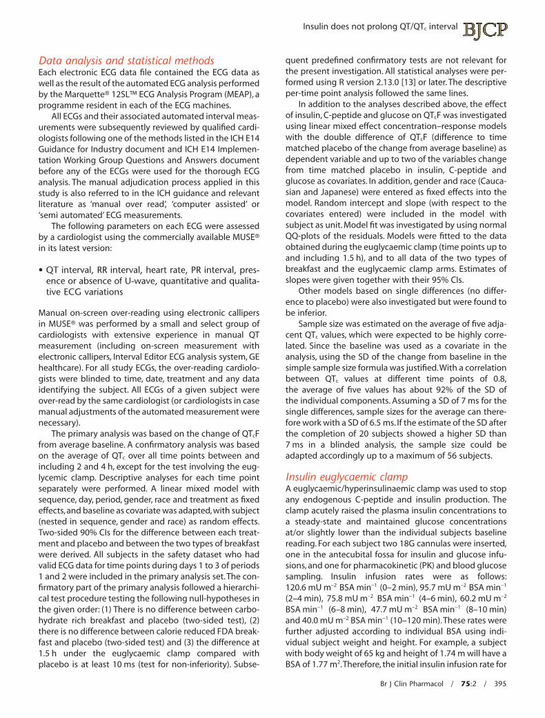
Data analysis and statistical methodsEach electronic ECG data file contained the ECG data aswell as the result of the automated ECG analysis performedby the Marquette® 12SL™ ECG Analysis Program (MEAP), aprogramme resident in each of the ECG machines.
All ECGs and their associated automated interval meas-urements were subsequently reviewed by qualified cardi-ologists following one of the methods listed in the ICH E14Guidance for Industry document and ICH E14 Implemen-tation Working Group Questions and Answers documentbefore any of the ECGs were used for the thorough ECGanalysis. The manual adjudication process applied in thisstudy is also referred to in the ICH guidance and relevantliterature as ‘manual over read’, ‘computer assisted’ or‘semi automated’ ECG measurements.
The following parameters on each ECG were assessedby a cardiologist using the commercially available MUSE®in its latest version:
• QT interval, RR interval, heart rate, PR interval, pres-ence or absence of U-wave, quantitative and qualita-tive ECG variations
Manual on-screen over-reading using electronic callipersin MUSE® was performed by a small and select group ofcardiologists with extensive experience in manual QTmeasurement (including on-screen measurement withelectronic callipers, Interval Editor ECG analysis system, GEhealthcare). For all study ECGs, the over-reading cardiolo-gists were blinded to time, date, treatment and any dataidentifying the subject. All ECGs of a given subject wereover-read by the same cardiologist (or cardiologists in casemanual adjustments of the automated measurement werenecessary).
The primary analysis was based on the change of QTcFfrom average baseline. A confirmatory analysis was basedon the average of QTc over all time points between andincluding 2 and 4 h, except for the test involving the eug-lycemic clamp. Descriptive analyses for each time pointseparately were performed. A linear mixed model withsequence, day, period, gender, race and treatment as fixedeffects,and baseline as covariate was adapted,with subject(nested in sequence, gender and race) as random effects.Two-sided 90% CIs for the difference between each treat-ment and placebo and between the two types of breakfastwere derived. All subjects in the safety dataset who hadvalid ECG data for time points during days 1 to 3 of periods1 and 2 were included in the primary analysis set.The con-firmatory part of the primary analysis followed a hierarchi-cal test procedure testing the following null-hypotheses inthe given order: (1) There is no difference between carbo-hydrate rich breakfast and placebo (two-sided test), (2)there is no difference between calorie reduced FDA break-fast and placebo (two-sided test) and (3) the difference at1.5 h under the euglycaemic clamp compared withplacebo is at least 10 ms (test for non-inferiority). Subse-
quent predefined confirmatory tests are not relevant forthe present investigation. All statistical analyses were per-formed using R version 2.13.0 [13] or later. The descriptiveper-time point analysis followed the same lines.
In addition to the analyses described above, the effectof insulin, C-peptide and glucose on QTcF was investigatedusing linear mixed effect concentration–response modelswith the double difference of QTcF (difference to timematched placebo of the change from average baseline) asdependent variable and up to two of the variables changefrom time matched placebo in insulin, C-peptide andglucose as covariates. In addition, gender and race (Cauca-sian and Japanese) were entered as fixed effects into themodel. Random intercept and slope (with respect to thecovariates entered) were included in the model withsubject as unit. Model fit was investigated by using normalQQ-plots of the residuals. Models were fitted to the dataobtained during the euglycaemic clamp (time points up toand including 1.5 h), and to all data of the two types ofbreakfast and the euglycaemic clamp arms. Estimates ofslopes were given together with their 95% CIs.
Other models based on single differences (no differ-ence to placebo) were also investigated but were found tobe inferior.
Sample size was estimated on the average of five adja-cent QTc values, which were expected to be highly corre-lated. Since the baseline was used as a covariate in theanalysis, using the SD of the change from baseline in thesimple sample size formula was justified.With a correlationbetween QTc values at different time points of 0.8,the average of five values has about 92% of the SD ofthe individual components. Assuming a SD of 7 ms for thesingle differences, sample sizes for the average can there-fore work with a SD of 6.5 ms. If the estimate of the SD afterthe completion of 20 subjects showed a higher SD than7 ms in a blinded analysis, the sample size could beadapted accordingly up to a maximum of 56 subjects.
Insulin euglycaemic clampA euglycaemic/hyperinsulinaemic clamp was used to stopany endogenous C-peptide and insulin production. Theclamp acutely raised the plasma insulin concentrations toa steady-state and maintained glucose concentrationsat/or slightly lower than the individual subjects baselinereading. For each subject two 18G cannulas were inserted,one in the antecubital fossa for insulin and glucose infu-sions, and one for pharmacokinetic (PK) and blood glucosesampling. Insulin infusion rates were as follows:120.6 mU m–2 BSA min-1 (0–2 min), 95.7 mU m–2 BSA min-1
(2–4 min), 75.8 mU m–2 BSA min-1 (4–6 min), 60.2 mU m–2
BSA min-1 (6–8 min), 47.7 mU m–2 BSA min-1 (8–10 min)and 40.0 mU m–2 BSA min-1 (10–120 min).These rates werefurther adjusted according to individual BSA using indi-vidual subject weight and height. For example, a subjectwith body weight of 65 kg and height of 1.74 m will have aBSA of 1.77 m2.Therefore, the initial insulin infusion rate for
Insulin does not prolong QT/QTc interval
Br J Clin Pharmacol / 75:2 / 395

0–2 min would be: 120.6 mU m–2 BSA min-1 ¥ 1.77 m2 =213.5 mU min-1. Blood glucose concentrations were meas-ured to determine basal concentrations, 4 and 10 min afterinsulin infusion had started and then every 5 min thereaf-ter until the end of the clamp.
Pharmacodynamic assessmentsVenous blood samples for the determination of concentra-tions of insulin, glucose and C-peptide in plasma weretaken during the study up to 6 h post-dose in each studyperiod. The blood samples (4.5 ml) were taken at pre-dose(to establish a baseline), 0.25, 0.5, 0.75, 1.0, 1.5, 2.0, 2.5, 3.0,3.5, 4.0 and 6.0 h post-dose/infusion. All samples were cen-trifuged and transferred to polypropylene tubes (NuncCryotube) and stored at -80°C.
The concentration of C-peptide, insulin and glucose inhuman plasma was determined by Analytical ServicesInternational (ASI, London, UK) using commercially avail-able solid phase, enzyme labelled chemiluminescentimmunoassay and enzymatic (hexokinase) assay methods.For C-peptide and insulin, a solid phase (bead), enzyme-labelled chemiluminescent immunometric assay was per-formed on the Immulite 1000 auto-analyzer using acommercially available kit supplied by Siemens HealthcareDiagnostic Products. C-peptide results falling below0.1 ng ml-1 and insulin results falling below 2.0 mIU ml-1
were reported as below the limit of quantification (BLQ).For glucose, the enzymatic (hexokinase) assay was per-formed on the Olympus AU400 auto-analyzer using a com-mercially available kit supplied by Randox.
Results
Subject demographics and dispositionAfter 24 subjects had completed the study, the SD for thesingle differences was estimated to be 8.5 ms assuming atreatment effect of 5 ms, and 7.5 ms for the time 0 values(assuming no treatment effect). Therefore, it was decidedto recruit another eight subjects.Thus a total of 32 subjectswere included in the study to be randomized to one of theeight treatment sequences. Subject demographics are pre-sented in Table 3.
Plasma concentrations of C-peptide, glucoseand insulin at baselineAverage plasma concentrations of C-peptide, glucose andinsulin at baseline for the different treatments (carbohy-drate rich‘continental’breakfast,calorie reduced‘American’FDA breakfast and euglycaemic insulin clamp) are pre-sented in Table 4.
Plasma concentrations of insulin, glucose andC-peptide following a carbohydrate rich‘continental’ breakfastFollowing consumption of a carbohydrate rich ‘continen-tal’ breakfast, plasma concentrations for insulin, glucoseand C-peptide rose rapidly reaching maximum valuesat approximately 0.25 h post completion of breakfast(62.6 mIU ml-1 [insulin], 3.2 mmol l-1 [glucose], 7.3 ng ml-1
[C-peptide] change from baseline) (Figure 1A). Insulin andglucose plasma concentrations then showed a steadydecline up to 1 h post-completion of breakfast, C-peptideplasma concentration was found to be steady up to 2.5 hpost-completion of breakfast.
Plasma concentrations of insulin, glucose andC-peptide following a calorie reduced‘American’ FDA breakfastA similar pattern was also observed following consump-tion of a calorie reduced ‘American’ FDA breakfast withplasma concentrations approximately half those seen fol-lowing a carbohydrate rich ‘continental’ breakfast. Plasmaconcentrations for insulin, glucose and C-peptide roserapidly reaching maximum values at approximately 0.25 hpost-completion of breakfast (27.2 mIU ml-1 [insulin],1.0 mmol l-1 [glucose], 3.8 ng ml-1 [C-peptide] change frombaseline) (Figure 1B). Insulin, glucose and C-peptideplasma concentrations then showed a steady decline up to1 h post-completion of breakfast after which plasma con-centrations were maintained at a steady level up 2.5 h.
Plasma concentrations of insulin, glucoseand C-peptide following an insulineuglycaemic clampThe glucose target concentration during the clamp wasthe basal blood glucose minus 0.5 mmol l-1 if the bloodglucose was 5.0 mmol l-1 or above or the basal bloodglucose minus 0.3 mmol l-1 if the basal blood glucose wasbelow 5.0 mmol l-1. Following administration of the insulineuglycaemic clamp, the plasma concentration of insulinrose rapidly, reaching a value of 43.7 mIU ml-1 withinapproximately 0.25 h (Figure 1C). This concentration wasmaintained for the remainder of the clamp time (up to 2 hpost-infusion start) due to the continuous infusion ofglucose. Plasma glucose concentrations were noted tohave decreased during the first 0.5 h (-0.3 mml l-1) whichwas then maintained until the end of the clamp. PlasmaC-peptide concentrtions did not change and endogenous
Table 3Subject demographics for the study
Male (n = 18) Female (n = 14)
Age (years) 26 � 3.77 (20–34) 27.8 � 4.20 (21–34)Height (cm) 174 � 6.52 (164–190) 163.6 � 7.44 (154–175)
Weight (kg) 63.0 � 7.29 (51.3–76.8) 58.0 � 6.35 (48.6–69)BMI (kg m-2) 20.8 � 1.96 (18.4–24.6) 21.6 � 1.35 (18.3–23.1)
Values are given as mean � SD with ranges in parentheses.
J. Taubel et al.
396 / 75:2 / Br J Clin Pharmacol

release was suppressed allowing the separation of insulin,glucose and C-peptide effects.
Plasma concentrations of insulin, glucose andC-peptide following placeboUnder placebo conditions there was no change observedfrom baseline for insulin, glucose and C-peptide concen-trations (Figure 1D).
Effects on QTcF: confirmatory analysisThe three relevant confirmatory null hypotheses could allbe rejected on the 5% level (one sided), i.e. a difference in
QTcF between ‘continental’ breakfast and placebo andbetween ‘American’ FDA breakfast and placebo could beascertained. Furthermore, it could be ascertained that theeffect of insulin under the euglycaemic clamp at 1.5 h wastoo small to be of any relevance.
Effect of the euglycaemic clamp on QTcFThe point estimates of the difference between the eugly-caemic clamp compared with placebo on QTcF were smalland ranged from -0.3 to -1.4 ms showing a small shorten-
Table 4Average baseline plasma concentration of C-peptide, glucose and insulin for different treatments
TreatmentAverage pre-dose concentrationC-peptide (ng ml-1) Glucose (mmol l-1) Insulin (mIU ml-1)
Carbohydrate rich ‘continental’ breakfast 1.12 5.34 3.96Calorie reduced ‘American’ FDA breakfast 1.24 5.3 4.77
Euglycaemic insulin clamp 1.13 5.36 4.21Placebo 1.21 5.33 4.84
A
C-p
epti
te (
ng m
l−1),
gluc
ose
(mm
ol l
−1)
conc
entr
atio
n
Insu
lin c
onc
entr
atio
n(μ
IU m
l−1)
−10
−55152535455565
0 1 2 3Time (h)
4 6
12345678
Carbohydrate rich (‘continental’) breakfastAverage of difference from baseline
C-p
epti
te (
ng m
l−1),
gluc
ose
(mm
ol l
−1)
conc
entr
atio
n
Insu
lin c
onc
entr
atio
n(μ
IU m
l−1)
−10
−551525354555
65
0 1 2 3Time (h)
4 6
12345678
Calorie reduced (‘American’)FDA standard breakfast
Average of difference from baseline
B
C D
C-p
epti
te (
ng m
l−1),
gluc
ose
(mm
ol l
−1)
conc
entr
atio
n
Insu
lin c
onc
entr
atio
n(μ
IU m
l−1)
−2−1
10
−5
5
15
25
35
45
55
65
0 1 2 3Time (h)
4 6
234567
Glucose infusion
Insulin clamp
Euglycaemic insulin clamp for 2 hAverage of difference from baseline
C-p
epti
te (
ng m
l−1),
gluc
ose
(mm
ol l
−1)
conc
entr
atio
n
Insu
lin c
onc
entr
atio
n(μ
IU m
l−1)
−10
−55
15
25
35
45
55
65
0 1 2 3Time (h)
4 6
12345678
Fasting for 6 hAverage of difference from baseline
Figure 1Average change from baseline of insulin, glucose and C-peptide following carbohydrate rich ‘continental’ breakfast (A), calorie reduced ‘American’ FDAbreakfast (B), euglycaemic insulin clamp (C) and placebo (D). , C-peptide; , Glucose; , Insulin
Insulin does not prolong QT/QTc interval
Br J Clin Pharmacol / 75:2 / 397

ing of QTcF. The maximum QTcF shortening was observedat 1.5 h post-dose with a value of 2.6 ms (two-sided 90% CI-5.3, -0.2) (Figure 2A). The values observed were not sig-nificantly different from zero. It should be noted that aslight prolongation was observed at 0.25 h post infusion.The time course of the changes in QTcF did not followthose of insulin, but rather resembled those of glucose.Themean baseline QTcF interval value for euglycaemic clampwas 415.1 ms compared with 416.2 ms for placebo.
Effect of carbohydrate rich ‘continental’breakfast on QTcFFor the continental breakfast the maximum QTcF shorten-ing was observed at 3.5 h post-dose with a value of 7.9 ms(two-sided 90% CI -10.4, -5.5) (Figure 2B). The mean base-line QTcF interval value for the ‘continental’ breakfast was415.7 ms compared to placebo (416.2 ms).
Effect of calorie reduced ‘American’ FDAbreakfast on QTcFWith the carbohydrate reduced‘American’style FDA break-fast, the maximum QTcF shortening was observed at 2.5 hpost-dose with a value of 6.8 ms (two-sided 90% CI -9.3,
-4.3) (Figure 2C).The mean baseline QTcF interval value forthe ‘American’ FDA breakfast was 417.1 ms compared withplacebo (416.2 ms).
Effect of moxifloxacin 400 mg on QTcFThe assay sensitivity was shown to be adequate. From0.25 h to 6 h post-dose, the DDQTcF change from baselinefollowing moxifloxacin 400 mg given in fasting conditioncompared with placebo ranged from 1.7 to 14.4 ms withthe lower bound of the 95% CI clearly above 5 ms. Thelargest QTcF change from baseline was observed at 2.5 hpost-dose in the fasted state with a value of 14.4 ms (two-sided 90% CI 11.9, -16.8) (Figure 2D). The mean baselineQTcF interval value for moxifloxacin (fasted state) was416.7 ms compared with placebo (416.2 ms).
Effects on heart rateSince QTc and heart rate are closely linked, the same analy-sis as performed for QTcF was also repeated for heart rate.Results are given in Figure 3. For insulin a gradual increasein heart rate was observed up to 3 h (6.5 beats min–1),returning to near baseline values at the 6 h time point
A
−2
2
0 h
Insulin andglucose clamp
0.5 h 1 h 1.5 h 2 h 3 h 4 h 6 h2.5 h 3.5 h
6
10
−6
Effect of insulin on QTCF
ΔΔQ
TCF
(m
s)
0
0 h 0.5 h 1 h 1.5 h 2 h 3 h 4 h 6 h2.5 h 3.5 h
5
10
15
20
−5
ΔΔQ
TCF
(m
s)
B
C D
−8
−4
0 h 0.5 h 1 h 1.5 h 2 h 3 h 4 h 6 h2.5 h 3.5 h
4
0
8
−12
−8
−4
0 h 0.5 h 1 h 1.5 h 2 h 3 h 4 h 6 h2.5 h 3.5 h
4
0
8
−12
ΔΔQ
TCF
(m
s)
ΔΔQ
TCF
(m
s)
Effect of carbohydrate richbreakfast on QTCF
Effect of calorie reduced FDAbreakfast on QTCF
Effect of moxifloxacinon QTCF in the fasted state
Figure 2Average change from baseline on QTcF following euglycaemic clamp (A), carbohydrate rich ‘continental’ breakfast (B), calorie reduced ‘American’ FDAbreakfast (C) compared with placebo. Effect of 400 mg moxifloxacin in fasted state on QTcF (D)
J. Taubel et al.
398 / 75:2 / Br J Clin Pharmacol

(Figure 3A). A steep increase in heart rate was observed forboth the carbohydrate rich ‘continental’ and caloriereduced ‘American’ FDA breakfast. The maximum increasein change of heart rate was observed at 1 h following acarbohydrate rich breakfast (9.1 beats min–1) and 0:25 h fol-lowing a calorie reduced ‘American’ FDA breakfast (8.5beats min–1). It should be noted that the observed heartrate effect following a carbohydrate rich breakfast(Figure 3B) was greater in comparison with a caloriereduced‘American’FDA breakfast (Figure 3C).With a carbo-hydrate rich breakfast the change in heart rate stabilizedafter a steep increase and remained so up to 2 h beforegradually returning to near baseline values. A similar trendwas observed with a calorie reduced‘American’FDA break-fast but the magnitude of the effect was smaller. The mag-nitude of the observed change in heart rate for 400 mgmoxifloxacin (fasted state) is shown in Figure 3D.
Concentration–response analysis for insulin,glucose and C-peptideThe effect on QTc was investigated using linear mixedeffect models with placebo corrected QTcF (change fromaverage baseline) as a dependent variable and insulin,glucose and C-peptide (placebo corrected) as covariates
for the data obtained under the euglycaemic clamp as wellas for all data obtained under the clamp and the two typesof breakfast. The resulting regression coefficients (‘slopes’)are shown in Table 5. Insulin was shown to have no effecton QTc during the euglycaemic clamp, if the model control-led for the effect of glucose. The same holds in a modelbased on all data, if the model controlled for the influencesof both glucose and C-peptide. However, C-peptide andglucose were found to have significant negative and posi-tive effects on QTc, respectively, based on the data from allthree treatment arms and all time points. The analysis pre-dicts that an increase of C-peptide apparently causes adecrease in QTcF [-1.2 ms/(ng ml-1)] while an increase ofglucose increases QTcF [1.6 ms/(mmol ml-1)) Since bothconcentrations are positively correlated, their effectsantagonize one another. This can be demonstrated withthe considerably smaller apparent effect of C-peptide ofonly 0.7 ms/(ng ml-1) obtained in a model ignoringglucose, i.e. when ascribing the joint effect of C-peptideand glucose to C-peptide only. Likewise, in a model ignor-ing C-peptide, the apparent effect of glucose appears to bereversed, i.e. the QTc-shortening effect of C-peptide domi-nates the QTc-prolonging effect of glucose.
Figure 4 B and C illustrates this relationship. The influ-ence of insulin on QTcF during the euglycemic clamp is
A Time matched baseline difference (insulin) Time matched baseline difference(carbohydrate rich breakfast)
Time matched baseline difference(calorie reduced FDA breakfast)
Time matched baseline difference(moxifloxacin-fasted state)
B
C D
0
2
4
6
8
0 h 0.5 h 1 h 1.5 h 2 h 3 h 4 h 6 h2.5 h 3.5 h
14
12
10
−2
−4
ΔHR
(be
ats
min
−1)
0
2
4
6
8
0 h 0.5 h 1 h 1.5 h 2 h 3 h 4 h 6 h2.5 h 3.5 h
14
12
10
−2
−4
ΔHR
(be
ats
min
−1)
0
2
4
6
8
0 h 0.5 h 1 h 1.5 h 2 h 3 h 4 h 6 h2.5 h 3.5 h
14
12
10
−2
−4
ΔHR
(be
ats
min
−1)
0
2
4
6
8
0 h 0.5 h 1 h 1.5 h 2 h 3 h 4 h 6 h2.5 h 3.5 h
14
12
10
−2
ΔHR
(be
ats
min
−1)
Figure 3Effect on heart rate with 95% CI on insulin euglycaemic clamp (A), carbohydrate rich ‘continental’ breakfast (B), calorie reduced ‘American’ FDA breakfast (C).Effect of 400 mg moxifloxacin (fasted state) on heart rate (D)
Insulin does not prolong QT/QTc interval
Br J Clin Pharmacol / 75:2 / 399

shown in Figure 4A. In Figure 4B the effect of C-peptide isshown for various concentrations of glucose (aimed at tobe kept constant). In addition, the broken line gives theapparent effect of C-peptide if the influence of glucose isdisregarded by the model. Figure 4C shows the same forglucose. In addition to the influence of glucose for severalfixed concentrations of C-peptide and the apparent effectof glucose estimated in a model that does not control forthe influence of C-peptide, the relationship found underthe euglycaemic clamp is also added as a dotted line. Itshould be noted that this relationship, established in a situ-ation where the concentrations of C-peptide were keptconstant, in reality, agrees well with those that wereestablished by a model that simultaneously models the
influence of both substances. Figure 5 shows a three-dimensional plot of the change in DDQTcF (ms) as a func-tion of glucose (mmol ml-1) and C-peptide (ng ml-1).
Discussion
As per the ICH E14 guideline [14], the majority of TQTstudies use moxifloxacin 400 mg as a positive control todemonstrate assay sensitivity by utilizing its well charac-terized QTc prolonging effect compared with placebo [8,15–18]. A positive control is included in these studies toensure that a TQT study can demonstrate a change in QTc
of approximately 5 ms. Moxifloxacin has been shown to
Table 5Influence of insulin, C-peptide and glucose on QTcF
Parameter Units of slope Data used Controlling for EstitmateLower Upperbound of 95% CI
Insulin ms/(mIU ml-1) Clamp Glucose -0.01 -0.06 0.05
All Glucose and C-peptide 0.01 -0.02 0.04C-peptide ms/(ng ml-1) All Glucose -1.24 -1.59 -0.90
All None -0.71 -0.98 -0.45
Glucose ms/(mmol ml-1) Clamp Insulin 1.90 0.07 3.73
All C-peptide 1.57 0.92 2.22
All None -0.86 -1.45 -0.27
All models use difference from placebo values, QTcF is change from average baseline. Based on linear mixed effects models.
A B CInfluence of insulin on QTCF
QT
CF
(m
s) (
chan
ce fr
om
tim
e m
atch
ed b
asel
ine)
QT
CF
(m
s) (
chan
ce fr
om
tim
e m
atch
ed b
asel
ine)
QT
CF
(m
s) (
chan
ce fr
om
tim
e m
atch
ed b
asel
ine)
Insulin (μIU ml−1)
Influence of C peptide on QTCF Influence of glucose on QTCF
−30
−20
−10
0
0 20 40 60 80 0 5 10 15 4 6 8 10
10
20
−30
−20
−10
0
10
20
−30
−20
−10
0
10
20
C-peptide (ng ml−1) Glucose (mmol l−1)
Figure 4Plot of DDQTcF (ms) against change from time matched placebo of insulin (A), C-peptide (B) and glucose (C) concentration. The solid black lines give theregression lines for insulin after the correction for glucose (A), C-peptide after correction for glucose (B) and for glucose after the correction for C-peptide(C), which are taken as the mean values across the data. The yellow area is the corresponding 95% CI. (A) The grey lines give the predictions for maximumand minimum mean glucose seen over time (change from placebo). The dashed line gives the regression without correction for glucose. (B) The grey linesgive the predictions for the glucose concentration seen under placebo and for the maximum mean change of glucose across all time points in thecarbohydrate-rich breakfast condition, which occurred at 15 min. The dashed line gives the regression line obtained for C-peptide without correction forglucose. (C) The grey lines give the predictions for the C-peptide level seen under placebo and for the maximum mean change of C-peptide across all timepoints in the carbohydrate-rich breakfast condition, which occurred at 30 min. The dashed line gives the regression line obtained for glucose withoutcorrection for C-peptide.The dotted black line gives the relationship between change of glucose from time matched placebo and DDQTcF established underthe euglycaemic clamp. (A) Is based on data obtained under the euglycaemic clamp (time points up to 1.5 h), (B) and (C) are based on all time points of allthree regimens (euglycaemic clamp, carbohydrate rich ‘continental’ breakfast and calorie reduced ‘American’ FDA breakfast)
J. Taubel et al.
400 / 75:2 / Br J Clin Pharmacol

produce changes in QTc which are greater and this has ledto suggestions of having positive controls which producesmaller effects i.e. closer to that suggested in the guide-lines [16]. With the actual effect of moxifloxacin it is con-sidered necessary to ensure that the lower bound of thelower 90% CI excludes 5 ms at least at one time point [16,19], thereby addressing the concern that a study showing asmall moxifloxacin effect of only 5 ms following a 400 mgoral dose would obviously not be indicative of a suffi-ciently sensitive study able to detect small changes in QTc.
The reported effect of food on QTc provides potentialfor its use as a positive control in thorough ECG studies andpossibly as an alternative to moxifloxacin. One of the firststudies to report the relationship between consumption ofa standardized meal under the rigorous conditions of aTQT study demonstrated a maximum QTcF shortening ofapproximately 8.2 ms [8]. The carbohydrate content of themeal appears to be important since a carbohydrate richmeal has been associated with endogenous physiologicalinsulinaemia [6] and two studies have demonstratedincreased heart rate and QTcB in response to increasedinsulin concentrations [6, 20]. In the study by Scott et al. [6],heart rate increased by 17% and was correlated withinsulin concentration. However, only heart rate (not QTc)and insulin measurements were provided in that study.
If the expectation that post-prandial insulinaemia has arole in the observed effects of food on ECG is correct, thenmeals with high levels of carbohydrates would be expectedto show a greater effect.Three studies reporting QTc effectshave used meals with higher carbohydrate content (68%[8], 57% [9], 53% [3]) compared with, for example, an FDAstandard breakfast which has a greater fat (58%) contentand a much lower carbohydrate content. This difference in
carbohydrate content may lead to a difference in QTc effectobserved between the meals but there is currently onlylimited data to suggest that this is the case.
The exact effects of insulin on the QTc interval havebeen investigated in this study. C-peptide and insulin areexcreted in equimolar amounts [20] following a carbohy-drate challenge. It has been demonstrated that administra-tion of C-peptide to type I diabetic patients withneuropathy shortens the QTc [12]. To our knowledge, thedata presented in the current study are the first to reportthe relationship of insulin, glucose and C-peptide follow-ing a meal conducted under the conditions of a TQT study.The results show that following ingestion of a carbohy-drate rich ‘continental’ breakfast or a calorie reduced‘American’ FDA standard breakfast, a rapid increase ininsulin and C-peptide concentrations occurs. What isapparent is the transient nature of the insulin increase,reaching maximum physiological concentrations almostimmediately and then decreasing to half the concentra-tions by 1 h post ingestion. Of interest is the prolongedsteady C-peptide concentration lasting up to 2 h post-ingestion, particularly with the carbohydrate rich ‘conti-nental’ breakfast. Insulin at physiological levels was shownto have no effect on QTcF during the euglycaemic clampperiod.This finding contrasts with the finding of Gastaldelliet al. [10] who have reported a QTc prolongation withincreased concentrations of insulin. However, in the studyby Gastadelli et al. [10], an increase in heart rate of up to 71beats min–1 was demonstrated. It is known that the use ofthe Bazett’s correction formula (QTcB) leads to false QTc
prolongations [11], i.e. these are merely due to changes inheart rate rather than a true change in the QT/RR relation-ship. As far as the food effect was concerned [8], similarresults to those observed by Gastaldelli et al. [10] wereobserved up to 2 h i.e. increase in the QTcB. However, in thefood effect study there was a shortening of QTcB at 4 hpost-dose of about 4 ms [8]. This is important becauseGastaldelli et al. [10] did not report QTcB after 2 h and alsofailed to demonstrate a shortening effect on the uncor-rected QT. Although, it has been reported that QTcB isunsuitable when dealing with heart rate changes [11],QTcBis still widely used in the clinic and false positives have alsobeen demonstrated for other studies [3, 21]. Our findingsshowed that shortening of the QTc was correlated to anincrease in heart rate.The greatest change in heart rate wasobserved during a carbohydrate rich‘continental’breakfastwhich correspondingly had the greatest QTcF shorteningeffect. Notably, for insulin the increase in heart rate fol-lowed by a decrease after 3 h to near baseline values cor-responded to the changes in QTcF that were observed. Asreported previously [8], the effect on QT appears to begreater than proportional and becomes noticeable whenapplying heart rate correction factors such as QTcF.
In concentration–effect modelling, C-peptide emergesas the most likely candidate to be responsible for the QTc
shortening observed following ingestion of a carbohy-
ΔΔQ
TCF
ΔΔQ
TCF
−5−40
−30
−20
−10
0
10
20
30
−40
−30
−20
−10
0
10
20
30
−4−2
02
4
0 5 10 15
C-peptide
Glu
cose
Figure 5Change in DDQTc (ms) as a function of glucose (mmol ml-1) and C-peptide(ng ml-1).The dashed blue plane shows a concentration dependent short-ening with C-peptide and prolongation with glucose
Insulin does not prolong QT/QTc interval
Br J Clin Pharmacol / 75:2 / 401

drate rich ‘continental’ breakfast in this study and wherereported previously [8]. The concentration–responseanalysis has confirmed a QTcF shortening effect withincreasing C-peptide concentration. It further appears thatthe C-peptide (QT shortening) effect couples with theantagonizing (prolongation) effects of glucose. Furtherinvestigations are required to assess the direct impact ofC-peptide on QTc change under TQT conditions and under-standing its role would allow the use of concentration–response models. This finding is of importance because ithas been shown in another study with type 1 diabeticpatients that increases in glucose can shorten the QTc [22].However this study did not control for C-peptide. Under-standing the role of glucose and C-peptide might also leadto improved prophylaxis in the cardiac complications asso-ciated with patients with diabetes. It is well documentedthat patients with insulin dependent diabetes are prone tosudden arrhythmias that can lead to ‘dead in bed’ syn-drome due to QTc prolongation. It could be surmised thatbecause glucose by itself may prolong, with C-peptideantagonizing, the effects on the QTc interval, diabeticpatients and particularly those prone to ‘dead in bed’ syn-drome may further benefit from tight glucose regulationand may benefit from C-peptide treatment.
There is sufficient evidence in the literature to suggestthat the food effect observed in this study is well repro-ducible both in terms of magnitude of effect and timecourse. We were able to replicate the same ECG effects intwo TQT studies so far. Moreover, it compares favourablywith other non-pharmacological methods of experimen-tally prolonging QTc such as temporary changes in postureas it is not compromised by QT/RR hysteresis and is easierto control than an exercise protocol in an environment thatshould be free of autonomic effects on the heart [3, 6, 9].Our findings (Figure 2A) also suggest that late changesafter an insulin clamp may lead to significant increases inthe QTc interval. Significant increases in the QTc intervalwere observed at the 4 and 6 h time points, i.e. 2–4 h afterthe end of the insulin clamp.These changes might be asso-ciated with fluctuating serum potassium concentrations.Indeed, the effect of insulin on the QTc as reported by Gast-aldelli et al. [10] was postulated to have been mediated bythe decrease of serum potassium concentrations with con-comitant increase in norepinephrine concentrations. Thisleads to the notion that insulin augments cellular potas-sium uptake and hyperpolarization of the cell membrane.However, such changes attributed to serum concentra-tions of potassium should be treated with caution. Forexample, in another study [23], it was shown that adjust-ment for fasting insulin or potassium concentration didnot explain the change in QTc. It might be surmised thatglucose could itself be implicated in the mechanisms ofsuboptimal regulation of cardiac ion channels involved inventricular repolarization and thereby modulate the QTinterval. Further work would be necessary to unravel theprecise mechanism(s).
In summary, this study demonstrates that there was nochange in QTc during the euglycaemic clamp. Given thatinsulin was raised to physiological concentrations compa-rable with those seen after a meal, whilst the release ofC-peptide was suppressed, insulin appears to have noeffect on the QTc interval in either direction. From thesedata it could be postulated that some type of relationshipmight exist between shortening of QTc and C-peptide con-centrations. There is no doubt that further work would benecessary to tease out the precise mechanism by whichthis interplay could occur.
Competing Interests
All authors have completed the Unified Competing Inter-est form at http://www.icmje.org/coi_disclosure.pdf (avail-able on request from the corresponding author) anddeclare no support from any organization for the submit-ted work, no financial relationships with any organizationsthat might have an interest in the submitted work in theprevious 3 years and no other relationships or activitiesthat could appear to have influenced the submitted work.
We would like to thank Dilshat Djumanov for data man-agement, Juleen Gayed for medical work and Carike Coetzeefor organizing the trial.
REFERENCES
1 Thwaites BC, Bose M. Very low calorie diets and pre-fastingprolonged QT interval. A hidden potential danger. WestIndian Med J 1992; 41: 169–71.
2 Swenne I, Larsson PT. Heart risk associated with weight lossin anorexia nervosa and eating disorders: risk factors for QTcinterval prolongation and dispersion. Acta Paediatr 1999; 88:304–9.
3 Nagy D, DeMeersman R, Gallagher D, Pietrobelli A, Zion AS,Daly D, Heymsfield SB. QTc interval (cardiac repolarization):lengthening after meals. Obes Res 1997; 5: 531–7.
4 Hulhoven R, Rosillon D, Bridson WE, Meeus MA, Salas E,Stockis A. Effect of levetiracetam on cardiac repolarization inhealthy subjects: a single-dose, randomized, placebo- andactive-controlled, four-way crossover study. Clin Ther 2008;30: 260–70.
5 Bloomfield D, Kost J, Ghosh K, Hreniuk D, Hickey L, GuitierrezM, Gottesdiener K, Wagner J. The effect of moxifloxacin onQTc and implications for the design of thorough QT studies.Clin Pharmacol Ther 2008; 84: 475–80.
6 Scott EM, Greenwood JP, Vacca G, Stoker JB, Gilbey SG, MaryD. Carbohydrate ingestion, with transient endogenousinsulinaemia, produces both sympathetic activation andvasodilatation in normal humans. Clin Sci 2002; 102: 523–9.
7 Lu C, Zou X, Orr WC, Chen JDZ. Postprandial changes ofsympathovagal balance measured by heart rate variability.Dig Dis Sci 1999; 44: 857–61.
J. Taubel et al.
402 / 75:2 / Br J Clin Pharmacol

8 Taubel J, Wong AH, Naseem A, Ferber G, Camm AJ.Shortening of the QT interval after food can be used todemonstrate assay sensitivity in thorough QT studies. J ClinPharmacol 2011 [Epub ahead of print].
9 Widerlov E, Jostell K, Claesson L, Odlind E, Keisu M,Freyschuss U. Influence of food intake onelectrocardiograms of healthy male volunteers. Eur J ClinPharmacol 1999; 55: 619–24.
10 Gastaldelli FA, Emdin M, Conforti F, Camastra S, Ferrannini E.Insulin prolongs the QTc interval in humans. Am J PhysiolRegul Integr Comp Physiol 2000; 279: R2022–5.
11 Malik M. Problems of heart rate correction in assessment ofdrug-induced QT interval prolongation. J CardiovascElectrophysiol 2001; 12: 411–20.
12 Wahren J, Ekbery K, Jornvall H. C-peptide and neuropathy intype 1 diabetes. Immunol Endocr Metab Agents Med Chem2007; 7: 69–77.
13 R Development Core Team. 2011). R: a language andenvironment for statistical computing [online]. R Foundationfor Statistical Computing, Vienna, Austria. ISBN 3-900051-07-0, Available at http://www.R-project.org/ (last accessedMay 2012).
14 ICH Harmonized Tripartite Guideline E14. The clinicalevaluation of QT/QTc interval prolongation andproarrhythmic potential for nonantiarrhythmic drugs.International Conference on Harmonization, Step 4,Guideline, EMEA, CHMP/ICH/2/04, 2005.
15 Dixon R, Job S, Oliver R, Tompson D, Wright JG, Maltby K,Lorch U, Taubel J. Lamotrigine does not prolong QTc in athorough QT/QTc study in healthy subjects. Br J ClinPharmacol 2008; 66: 396–404. Epub 2008 Jul 23.
16 Taubel J, Naseem A, Harada T, Wang D, Arezina R, Lorch U,Camm AJ. Levofloxacin can be used effectively as a positive
control in thorough QT/QTc studies in healthy volunteers. BrJ Clin Pharmacol 2009; 69: 391–400.
17 Demolis JL, Kubitza D, Tenneze L, Funck-Brentano C. Effect ofa single oral dose of moxifloxacin (400mg and 800mg) onventricular repolarization in healthy subjects. Clin PharmacolTher 2000; 68: 658–66.
18 Florian J, Tornøe C, Brundage R, Parekh A, Garnett C.Population pharmacokinetic and concentration-QTc modelsfor moxifloxacin: pooled analysis of 20 thorough QT studies.J Clin Pharmacol 2011; 51: 1152–62. Epub 2011 Jan 12.
19 ICH Harmonized Tripartite Guideline E14. The clinicalevaluation of QT/QTc interval prolongation andproarrhythmic potential for non-antiarrhythmic drugs.International conference on harmonisation, E14Implementation and working group, Question andAnswers, 2008.
20 Johansson BL, Borg K, Fernqvist-Forbes E, Odergren T,Remahl S, Wahren J. C-peptide improves autonomic nervefunction in IDDM patients. Diabetologia 1996; 39: 687–95.
21 Browne KF, Zipes DP, Heger JJ, Prystowsky EN. Influence ofthe autonomic nervous system on the Q-T interval in man.Am J Cardiol 1982; 50: 1099–103.
22 Suys B, Heuten S, De Wolf D, Verherstraeten M, de BeeckLO, Matthys D, Vrints C, Rooman R. Glycemia andcorrected QT interval prolongation in young type 1diabetic patients: what is the relation? Diabetes Care2006; 2: 427–9.
23 Dekker JM, Crow RS, Hannan PJ, Schouten EG, AR F, StudyARIC. Heart rate-corrected QT interval prolongation predictsrisk of coronary heart disease in Black and Whitemiddle-aged men and women: the ARIC study. J Am CollCardiol 2004; 18: 565–71.
Insulin does not prolong QT/QTc interval
Br J Clin Pharmacol / 75:2 / 403