Characterization of Donor Dendritic Cells and Enhancement of Dendritic Cell Efflux With cc-Chemokine...
20
Characterization of donor dendritic cells and enhancement of dc efflux with cc- chemokine ligand 21: a novel strategy to prolong islet allograft survival Received on 13 October 2006 and accetped in revised form 15 January 2007. Paolo Fiorina 1,3* , Mollie Jurewicz 1* , Katsunori Tanaka 1 , Negin Behazin 1 , Andrea Augello 1 , Andrea Vergani 1,3* , Uli Von Adrian 4 , Neal R. Smith 2 , Mohamed H. Sayegh 1 , and Reza Abdi 1 1 Transplantation Research Center (TRC), Children’s Hospital and Brigham and Women's Hospital, Harvard Medical School, Boston, USA; 2 Pathology, Massachusetts General Hospital, Harvard Medical School, Boston; 3 Medicine, San Raffaele Scientific Institute, Milan, Italy; 4 CBR Institute for Biomedical Research and Department of Pathology, Harvard Medical School, Boston, USA. *PF and MJ contributed equally to this work. Running title: Chemokine-based therapy to reduce direct allorecognition Address for correspondence: Reza Abdi, MD Transplantation Research Center (TRC) Children’s Hospital and Brigham and Women's Hospital Harvard Medical School 221 Longwood Avenue Boston, MA 02115 E-mail: [email protected] This article includes an Online Appendix. 1 Diabetes In Press, published online February 7, 2007 Copyright American Diabetes Association, Inc., 2007
-
Upload
hms-harvard -
Category
Documents
-
view
1 -
download
0
Transcript of Characterization of Donor Dendritic Cells and Enhancement of Dendritic Cell Efflux With cc-Chemokine...

Characterization of donor dendritic cells and enhancement of dc efflux with cc-chemokine ligand 21:
a novel strategy to prolong islet allograft survival
Received on 13 October 2006 and accetped in revised form 15 January 2007.
Paolo Fiorina1,3*, Mollie Jurewicz1*, Katsunori Tanaka1, Negin Behazin1, Andrea Augello1, Andrea Vergani1,3*, Uli Von Adrian4, Neal R. Smith2, Mohamed H. Sayegh1,
and Reza Abdi1
1Transplantation Research Center (TRC), Children’s Hospital and Brigham and Women's Hospital, Harvard Medical School, Boston, USA; 2Pathology, Massachusetts General
Hospital, Harvard Medical School, Boston; 3Medicine, San Raffaele Scientific Institute, Milan, Italy; 4CBR Institute for Biomedical Research and Department of Pathology,
Harvard Medical School, Boston, USA. *PF and MJ contributed equally to this work.
Running title: Chemokine-based therapy to reduce direct allorecognition
Address for correspondence: Reza Abdi, MD
Transplantation Research Center (TRC) Children’s Hospital and Brigham and Women's Hospital
Harvard Medical School 221 Longwood Avenue
Boston, MA 02115 E-mail: [email protected]
This article includes an Online Appendix.
1
Diabetes In Press, published online February 7, 2007
Copyright American Diabetes Association, Inc., 2007
Abstract Dendritic cells (DC) are the most potent antigen-presenting cells, yet little data are available on the differential characteristics of donor and recipient DC (dDC and rDC) during the process of islet allograft rejection. DTR-GFP-DC mice provide a novel tool to monitor DC trafficking and characteristics during allograft rejection. We show rapid migration of dDC to recipient lymphoid tissues as early as 3 hours post islet allo-transplantation. Compared with rDC, dDC express different patterns of chemokine receptors, display differential proliferative capacity, and exhibit a higher level of maturity; these findings could be attributed to the effects of injury that dDC undergo during islet cell preparation and engraftment. Intriguingly, we detected dDC in the spleen of recipients long after rejection of islet allografts. Given that dDC express high levels of CCR7, islets were cultured before transplant with the ligand for CCR7 (CCL21). This novel method, which enabled us to enhance the efflux of dDC from islet preparations, resulted in a prolongation of islet allograft survival in immunocompetent recipients. This study introduces dDC and rDC as two distinct types of DC and provides novel data with clinical implications to use chemokine-based DC-depleting strategies to prolong islet allograft survival. Key-word: Dendritic cells, islet transplantation, chemokines. Word-count: 3999
2
INTRODUCTION Generation of alloreactive T cells, a pivotal event in allograft rejection, requires presentation of alloantigens to recipient T cells. Dendritic cells (DC) are regarded as the most potent antigen-presenting cells1-3. During the process of direct allorecognition, recipient T cells recognize intact allogeneic major histocompatibility complex (MHC) molecules on donor tissue resident dendritic cells (dDC)4-6. During indirect allorecognition, donor alloantigen shed from the allograft is released into the recipient’s circulation, is processed by recipient DC (rDC), and finally is presented to recipient T cells7. DC also express high levels of costimulatory molecules, rendering them potent initiators of alloimmune responses, and regulate immune responses by instructing T cells and by regulating the generation of suppressive (regulatory) T cell responses. However, many questions remain concerning DC in transplantation, the answers to which are of fundamental importance in better comprehending the roles of DC during the processes of engraftment or rejection. For instance, no data are yet available on distinct donor and recipient DC (dDC and rDC) trafficking or characteristics (maturity, longevity, proliferation, mobility and chemokine receptor expression) during the process of islet allograft destruction. Because of the lack of animal models to easily monitor rDC and dDC, characterization of such cells has been a difficult task and therefore remains poorly described. B6.FVB-Tg (Itgax-DTR/GFP)57Lan (DTR-GFP-DC) mice, which have a green fluorescent protein (GFP) linked to the CD11c promoter, provide a model that can be employed to circumvent this problem. Using these
mice as donors of islet allografts enabled us to better study DC characteristics and trafficking8,9. Because the capacity of DC to proficiently present antigen to T cells and to generate an alloimmune response relies on the ability of DC trafficking to lymphoid tissue, and given that chemokines tightly control DC migration trafficking, we also examined the expression of chemokine receptors of DC at different time points after islet transplantation5,10-12. In recognition of the importance of dDC in the generation of alloimmune responses, particularly in the case of islet cell transplantation, investigators have attempted to deplete dDC using a number of techniques with the goal of achieving prolongation of islet graft survival13-17. Here we aim to characterize dDC and to establish a chemokine-based strategy to deplete islet DC, by culturing islets in the presence of the chemokine CCL21 prior to transplantation to enhance the efflux of DC from islets.
3
MATERIALS AND METHODS Mice B6.FVB-Tg (ITgax-DTR/GF)57Lan (DTR-GFP-DC), CD45.1 (B6.SJL-Ptprca Pepcb/BoyJ), C57BL/6, and BALB/c mice were purchased from The Jackson Laboratory, Bar Harbor, Maine. Islet transplantation Pancreatic islets were isolated by collagenase digestion followed by density
gradient separation and then hand-picking, as described previously18. For more information, please refer to the online supplement. Trafficking studies Allogeneic islets isolated from DTR-GFP-DC mice (on a BALB/c background, H-2d) were transplanted under the kidney capsule of streptozotocin (STZ)-induced diabetic C57BL/6 mice (H-2b). For details of flow cytometric and immunohistological analysis, please see online supplement. Proliferation studies To address the proliferation of dDC and rDC, mice were injected i.p. with 1 mg of bromodeoxyuridine (BrdU, Sigma-Aldrich) dissolved in PBS as described previously19-22. DC were isolated from splenocytes using anti-CD11c magnetic beads (Miltenyi Biotec, Auburn, CA) with more than 90% purity and were assessed for BrdU uptake19. Depletion of DC using diphtheria toxin (DT) or CCL21 We established 2 groups of DC-depleting experiments by either administering DT to donor in BALB/c GFP-DTR-DT mice or by isolating islets and culturing them with CCL21. For details of both experiments, refer to the online supplement. DC isolation, culture, and adoptive transfer BALB/c DC were generated as previously described from murine bone marrow23. For details of these culture conditions as
well as specifics of adoptive transfer, please refer to the online supplement. Mixed lymphocyte reaction (MLR) Splenocytes were isolated at day 7 post-islet transplantation from STZ-induced diabetic C57BL/6 mice (responders) that received BALB/c CCL21-cultured or control islets and were challenged with donor irradiated naïve BALB/c splenocytes stimulators in a fully mismatched MLR. Cells were cultured for 3 days, and proliferation was measured following pulsing for 16 hours with [3H]TdR using a liquid scintillation counter. T cells were also stimulated with ConA as a positive control. Statistical analyses Survival of islet grafts has been evaluated with a survival Kaplan-Meier analysis. A Mann-Whitney test (for non-parametric data) or two-sided unpaired Student t-test (for parametric data) was used when the differences were compared among the groups cross-sectionally A P value of less than 0.05 (by two-tailed testing) was considered an indicator of statistical significance. Data are expressed as mean ± standard error. Prism Graph Pad statistical package for Windows (GraphPad Software, Inc. San Diego, CA) was used for data analysis.
4

RESULTS Evaluation of dDC trafficking using DTR-GFP-DC mice In DTR-GFP-DC mice, a green fluorescent protein (GFP) is linked to the CD11c promoter. As shown in Figure 1A, in contrast to BALB/c WT naïve mice, BALB/c DTR-GFP-DC naïve mice show GFP+ expression in the spleen (Figure 1B). Examination of the naïve pancreas of BALB/c DTR-GFP-DC (used as donors in islet transplantation studies) showed a cluster of GFP+DC surrounding the pancreatic islets (as shown by insulin staining) (Figure 1C). Hence, DTR-GFP-DC mice provide a unique tool to monitor dDC trafficking by transplanting islets from DTR-GFP-DC mice (BALB/c background) into STZ-induced diabetic C57BL/6 mice. Following transplantation of islets underneath the kidney capsule, islet grafts and secondary lymphoid tissues were also examined for the presence of GFP+ cells to evaluate the trafficking of dDC. dDC were shown to migrate from the islet graft (located under the kidney capsule) into the recipient renal parenchyma and spleen as early as 3 hours after allogeneic islet transplantation (Figures 1D and 1E, respectively). Compared with 3 hours post transplant, the number of dDC seen in the recipient spleen increased significantly at 24 hours (Figure 1F). Immunostaining showed that these GFP+ cells coexpress CD11c (Figure 1G) and are found within the vicinity of CD4+ cells in the spleen of islet allograft recipients (Figure 1H). The earliest time point at which dDC could be detected was at 3 hours in the spleen. While at 3 hours and day 1 post transplantation no dDC were detected in the recipient NDLN (data not shown), we observed very few dDC in the DLN at 3 hours (Figure 2A) with an increase at 24 hours after transplantation (Figure 2B). No dDC were detected in the
thymus at 3 hours and day 1. These data demonstrate a selective, robust and rapid migration of islet dDC into recipient spleen. Interestingly, we did detect dDC in the bone marrow of recipients after allogeneic islet transplantation 3 hours after transplant, highlighting the possible importance of bone marrow as a site of allorecognition or immunoregulation (Figure 2C). The percentage of CD11c+GFP+ remained constant at 24 hours as well (data not shown). Given that streptozotocin induces islet inflammation and could serve as a signal for dDC to migrate. we performed islets transplantation using donor islets from DTR-GFP-DC mice into STZ-induced diabetic mice. Naïve pancreata were examined following transplantation and showed no active presence of dDC (data not shown). Since spleen contained more dDC than other lymphoid tissues, we compared the percentage of CD11c+GFP+ cells in the spleen of islet allograft and isograft recipients by FACS. While there were very few CD11c+GFP+ cells in the spleen of syngeneic recipients (islets from C57BL/6 donor DTR-GFP-DC mice into C57BL/6 recipient) (Figure 2D), a large number of double-stained cells were noted in the spleen of allogeneic recipient mice (Figure 2E). These data are suggestive of much more prominent migration of dDC in the allogeneic setting, and suggest that most of the GFP+ cells also co-express CD11c. To study the kinetics of dDC and rDC trafficking, we recovered infiltrating cells from islet grafts and subjected them to our FACS analysis. We observed only a small increase in rDC at days 1 and 7, while a 20-fold increase in dDC recovered from islet grafts was noted at day 1 after islet transplantation (Figures 2F and 2G). This could be in part due to the
5

proliferative capacity of dDC (please refer to the data below). dDC exhibit a higher proliferative ability at an earlier time point compared to rDC No data exist on the proliferative capacity of DC in islet cell transplantation. To assess the proliferative capability of dDC and rDC, we injected BrdU, a proliferation marker that binds to newly synthesized DNA19-22, into recipients of islet allografts. By analyzing recipient splenocytes by FACS and gating on GFP+ and BrdU+ cells, we show that in the allogeneic setting dDC begin to proliferate at an early time point (3 hours) and that dDC exhibit a higher proliferative capacity than rDC (both recovered from recipient spleens) during the time course of the study (Figures 2H-2J). To further confirm the proliferative capability of dDC, we also used Ki67, a sensitive cycle marker for in vivo studies. Sections of the spleens recovered from islet allograft recipients of GFP+ donors, were stained with an anti-Ki67 antibody (red). dDC (GFP+ cells, green) undergoing proliferation were co-stained for Ki67 and appeared yellow (Figure 2K). Chemokine receptor and costimulatory molecule expression of dDC and rDC We then examined dDC and rDC expression of chemokine receptors and of CD86/CD80 (DC maturity markers) at 3 hours, day 1, and day 7 post-transplantation in recipient spleens (Figure 3). Compared with rDC, dDC displayed much higher levels of expression of the chemokine receptors CCR5, CXCR4, and CCR7 at 3 hours and day 1 (Figures 3A-3C and 3F-3H, respectively). At day 7, although dDC still exhibit higher levels of CXCR4 and CCR7 than rDC, this difference subsides for CCR5 (Figures 3K-3M).
At 3 hours and day 1, dDC also expressed much higher levels of CD86 and Class II compared to rDC (Figures 3D-3E and 3I-3J). At day 7, dDC and rDC display comparable levels of CD86 and Class II (Figures 3N-3O). These data are in accordance with the current thought that dDC (direct pathway) are the first initiators of early acute rejection of islet allografts24-27. Finally, these data could also suggest that similar to the in vitro DC maturation data previously reported23, CXCR4 and CCR7 appear to be important chemokine receptors involved in the migration and maturation of DC. Oxidative/ischemic stress and DC activation We examined the hypothesis that tissue injury during islet cell preparation and transplantation ischemia can contribute to the activation of dDC, an important aspect in the generation of alloimmune response that remains to be explored. A number of studies have highlighted the active recruitment and presence of DC in ischemic tissue28,29. Kostulas et al.28 showed an increase in the number and in the activation of DC in ischemic brain compared with sham hemispheres as early as 1 hour after arterial occlusion. Ischemia associated with the surgical procedure itself was reported to recruit DC to the site of injury in a model of renal ischemia28,29. We evaluated the maturation of islet dDC following 24-hour culture and compared them with islet DC analyzed immediately after isolation. The expression of CD80, MHC Class II molecules and CCR7 was upregulated on dDC recovered from cultured islets as compared with pre-culture islets (Figures 4A-4C). Class I expression studies showed no significant upregulation (data not shown). These data support the idea that due to prolonged ischemia time and tissue preparation, islet DC become more activated and migrate
6

faster because of chemokine receptor upregulation. Furthermore, to address the direct effect of oxidative stress on DC activation, we cultured BM-derived DC from BALB/c mice (our islet donors) with 200 µmol H2O2 for 30 minutes as described previously (in which B lymphocytes were treated with a similar strategy)30. FACS analysis using 7-AAD and AnnexinV showed no significant decrease in cell viability (less than 2%) following H2O2 treatment (data not shown). As shown in Figures 4D-4E, treating dDC with H2O2 significantly upregulated the expression of CD86/CD80 (Figures 4D-4E). These data suggest the potential role of ischemia/injury in the induction of chemokine receptors and in the maturation of dDC and could in part explain the higher activity of islet DC and their immunogenicity. dDC were found in recipient spleen long after islet allograft rejection One important unexpected observation was the detection of dDC in the spleen of the recipients long after rejection of islet allografts. This was observed when islets were transplanted from BALB/c GFP-DTR-DC donors into C57BL/6 recipients. Our FACS data showed the presence of GFP+ cells in the spleen of islet allograft recipients 30 days after islet transplantation (2 weeks after rejection of islet allografts) (Figure 4F). Similarly, when we transplanted islets from the CD45.1+ congenic strain into a CD45.2+ recipient and spleens of recipients were recovered 30 days after transplantation, persistent dDC (CD11c+CD45.1+ cells) were demonstrated by FACS in the islet allograft recipient spleens (Figure 4G). This transplant combination has been one of the classical models to investigate allografted donor cells in recipients31,32.
Depletion of islet DC using DTR-GFP-DC mice DTR-GFP-DC mice have a fusion protein that expresses both the simian DT receptor and GFP and is linked to the CD11c promoter. Murine cells, unlike primate cells, are insensitive to death by DT; transfer of primate DTR into mice via a transgene therefore confers DT sensitivity to murine cells8,33. DT was administered to BALB/c DTR-GFP-DT mice, and islets isolated from DT-treated mice were transplanted into STZ-induced diabetic C57BL/6 recipients. At baseline, CD11c+GFP+ cells are evident in the spleen and in the pancreas of DTR-GFP-DC mice (Figure 4H), but GFP+CD11c+ cells can be depleted with 250 ng of DT. Depletion is evident in the spleen and pancreas at 6 hours and more efficiently occurs at 48 hours post-injection (Figure 4H). Surprisingly, transplanting islets from DT-depleted mice into an allogeneic host resulted in a slight delay of islet allograft rejection (MST: 11 vs. 14, control and DT-depleted respectively, p=0.01), (Figure 4I). Examination of recipient spleens transplanted with DT-treated donors revealed that GFP+ cells still could be detected at day 7 after islet transplantation (data not shown). The presence of dDC in recipient spleens following depletion could explain modest prolongation of islet allograft survival. We speculate that a more robust depletion could be achieved by administering a higher dose of DT; however, a high mortality rate associated with increasing doses of DT has been a limiting factor in achieving this end. Establishing a chemokine-based DC depletion strategy for islet transplantation Previous studies to deplete dDC of islets have led to prolongation of islet allograft survival. Data from these studies indicate
7
that endocrine tissues such as islets are highly dependent on the function of dDC to develop an acute rejection response13-17. Here, we have established a novel strategy to enhance migration of dDC out of islets during preparation for transplantation. Given the overexpression of CCR7 in dDC during the alloresponse, we hypothesized that culturing islets with CCL21 (CCR7 ligand) would cause efflux of dDC from the islets. We cultured islets for 24 hours with CCL21 and performed FACS analysis of cells recovered from the aspirated medium above the islets. Medium obtained from islets cultured for 24 hours with 200 ng/ml of CCL21 showed that the percentage of CD11c+ cells (after gating on CD45+ cells) was almost 3-fold higher than in control cultures, and when 800 ng/ml of CCL21 was used, the percentage of CD45+CD11c+ cells was 7-fold higher than in controls (Figure 5A). In parallel, a depletion of CD11c+ cells was evident in the islets cultured with CCL21 compared to islets cultured with medium alone; islets were treated with collagenase, homogenized, and subjected to FACS analysis (Figure 5B). Transplanting BALB/c islets cultured with medium containing 800 ng/ml CCL21 into STZ-induced diabetic C57BL/6 recipients resulted in significant prolongation of islet allograft survival (MST: 11 and 19 days for control and CCL21-cultured islets, respectively, p<0.001), (Figure 5C) with 20% of mice free from rejection for more than 30 days. In order to elucidate the mechanism behind the observed prolongation of islet allograft survival, we studied and compared the status of host immune responses in two groups of recipients that received either CCL21-cultured or control islets (control islets are cultured for 24 hours without CCL21). At day 7, spleens
and DLN were harvested from recipients, and percentages of CD4+CD25+ cells (activated T lymphocytes), CD4+CD44hi cells (T effectors) and CD4+CD25+Foxp3+ cells (T regulatory cells) were determined. Compared to controls, mice receiving CCL21-cultured islets showed almost 10-fold fewer activated T cells (as shown by the absence of CD4+CD25+ cells) in the DLN (0.88±0.1 vs. 8.10±2.0%, p<0.01), (Figure 5D). A reduced number of effector cells defined as CD4+CD44hi were also recovered in the spleen of recipients of CCL21-cultured islets (p=0.02), (data not shown). No differences were noted in the percentage of CD4+CD25+Foxp3+ cells (T regulatory cells) in the DLN or spleen of both groups (data not shown). To compare the capacity to generate an alloresponse between mice that received CCL21-cultured or control islets, we measured their allostimulatory capacity in an MLR assay. BALB/c irradiated splenocytes were used to stimulate splenocytes extracted from primed C57BL/6 mice that received either CCL21-cultured islets or control islets. As shown in Figure 5E, the control group exhibited a higher T cell proliferative response (p<0.01). To confirm that the direct allorecognition pathway was affected by the CCL21 treatment, as an add-back experiment we adoptively transferred BALB/c DC extracted with CD11c+ microbeads into C57BL/6 recipients immediately following islet transplantation. Indeed, dDC adoptive transfer completely abrogated the protective effect of CCL21-culture treatment on islet allograft survival; all mice rejected within 10 days following islet transplantation (MST=9 days, Figure 5C). Finally, CCL21-treated and untreated islet allografts were harvested at day 7 post transplantation. Compared with the control group, CCL21-treated islets show
8

significantly less infiltrates and well-maintained insulin staining (Figure 5F and 5G, respectively).
9
DISCUSSION DC are migratory antigen-presenting cells that migrate to the lymphoid tissue DLN and spleen and present antigens to recipient T cells, a central event in generating an alloresponse1-3. Allorecognition occurs via both “direct” and “indirect” pathways. In direct allorecognition, recipient T cells recognize intact allogeneic MHC molecules on donor antigen-presenting cells (i.e. tissue resident dendritic cells/passenger leukocytes)4,5. Larsen et al6 demonstrated that donor-derived DC migrate from transplanted heart allografts into recipient lymphoid tissue. Similar results were demonstrated in a mouse model of heart transplantation, in which the efflux of dDC into recipient spleens as well as the presence of rDC in the grafts were noted19. Due to a dearth of suitable models, examining the specific contribution of dDC and rDC in transplantation has been a difficult task. No data are yet available on the distinct characteristics of dDC and rDC or on the role of specific chemokine receptors important in the migration of DC in islet transplantation. Using DTR-GFP-DC mice (which have GFP linked to CD11c) allowed us to explore the differential features of dDC and rDC, both of which are central in the generation and regulation of alloimmune responses. These features include DC trafficking, maturation, and proliferation, and their linkage with the temporal expression of chemokine expression. Our data demonstrate that dDC first quickly migrate to lymphoid tissue such as spleen instead of DLN. Compared with rDC, not only did dDC exhibit a higher level of maturity at earlier time points post transplantation, but they also showed differential levels of chemokine receptor expression as compared to rDC.
Current theory proposes that while dDC (direct pathway) are potent initiators of early acute rejection, the indirect pathway (rDC) is responsible for late acute/chronic rejection24-27. We have shown that dDC display much greater proliferative activity compared to rDC as well, and we show that ischemia and tissue preparation could in part contribute to this higher dDC activity. Our data suggest that oxidative stress and tissue injury could increase the level of expression of DC costimulatory molecules and CCR7, through which dDC may acquire superior mobility and allostimulatory capacity. Results of our studies may explain in part the mechanisms behind the excessive immunogenicity of islet allografts, in which panels of tissue injuries (due to ischemia and tissue preparation) are more likely to occur than in a transplant setting such as the heart. We also detected long-lasting dDC in spleens of islet allograft recipients more than 1 month following transplant. It is surprising to find these cells weeks after allograft rejection, as it has been commonly thought that the dDC half-life is much shorter34. The question of how dDC can escape recipient immune surveillance is unclear to us. We can hypothesize that they do so through acquiring a tolerogenic phenotype by failing to provide a second signal, but this is a subject of future studies. One of the important aspects of our study was to abrogate the effect of direct allorecognition. The importance of direct allorecognition is well-established, as stimulation with DC has been shown to be very potent in stimulating primary allogeneic MLRs, and depletion of donor DC can prolong islet and thyroid allograft survival35,36. We used a novel DT-based system that allows the inducible in vivo ablation of DC8,9. We show that DC can be depleted in islet allografts with DT
10

treatment of donors. Using these mice as donors, however, only minimally prolonged islet allograft survival. This could be due to the subsequent repopulation or expansion of dDC within a few days of depletion and after transplantation. We also report that islets can be cultured with CCL21 to increase the efflux of DC from islets. Our data show that this strategy leads to prolongation of islet allograft survival. A reduction in T cell activation and in the potency of the alloresponse was noted in recipients that received CCL21-cultured islets compared to the control group. Although all grafts eventually were rejected, we should note that this prolongation was achieved with no immunosuppression in the recipients and that this outcome is comparable with the prolongation investigators have reported when using considerable immunosuppression37. We view this as a potential adjunct therapeutic approach to combine with immunosuppressive or tolerogenic strategies to promote islet cell engraftment in the future. During maturation, DC upregulate surface expression of the chemokine receptor CCR7. CCL21, the ligand for CCR7, which was used in our in vitro assay, is constitutively expressed at high levels in lymph nodes38. Given that CCL21 exhibits chemotactic activity mostly for mature DC, it is plausible that immature DC or DC progenitors remained in the graft even after the CCL21-based depletion38,39. These remaining DC could mature following transplantation and mount a delayed alloimmune response, leading to eventual graft rejection. The differential survival outcomes observed between DT depletion and CCL21 strategies could be the fact that with CCL21 treatment we were able to selectively deplete CCR7high DC, which
are most likely responsible for mounting an allogeneic response. Further studies are necessary to optimize culture conditions to achieve more robust prolongation; possible adjustments include using higher concentrations of CCL21 and/or repetitive treatment as well as employing multiple key chemokines/ligands in culture. The latter requires precise study of chemokine receptors expressed on dDC in addition to CCR7. With improvement, this method could easily be translated to clinical practice. Common immunosuppressive agents used in islet cell transplantation are specifically associated with serious islet toxicity, and a method such as ours may allow clinicians to use significantly lower doses of immunosuppression to promote tolerance. In mice a prolongation of islet allograft survival was reported not only by immunosuppression strategies, but also by anti inflammatory treatments (e.g., the use of oxygen free radical scavengers and heme oxygenase-1 induction)40-41. Tissue pretreatment such as culturing at reduced temperatures or treating with anti-class II antibodies, with the goal of eliminating APC within endocrine tissues, has also resulted in prolongation of allograft survival in immunocompetent recipients13-
17. Of note, these strategies nonspecifically targeted all antigen-presenting cells. Our work is unique because our approach specifically deals with dendritic cells and can potentially lead to greater manipulation and selective transformation of the islet DC population by using specific chemokines to induce selectively the efflux of allogeneic DC so that tolerogenic DC remain. The efficacy of such strategy will be tested in NOD recipients in our future studies. We believe these results could easily be translated into clinical transplantation, and could have a tremendous impact on the outcome of human islet cell
11

Supported by the Juvenile Diabetes Research Foundation Center Grant on Immunological Tolerance in Type 1 Diabetes (to M.H. Sayegh).
transplantation, which requires further improvement42-45. Acknowledgments Reza Abdi is the recipient of a JDRF Career Development Award.
12

References 1. Fayette J, Durand I, Bridon JM, et al. Dendritic cells enhance the differentiation of naive B cells into plasma cells in vitro. Scand J Immunol. 1998;48:563-570. 2. Banchereau J, Briere F, Caux C, et al. Immunobiology of dendritic cells. Annu Rev Immunol. 2000;18:767-811. 3. Morelli AE, Thomson AW. Dendritic cells: regulators of alloimmunity and opportunities for tolerance induction. Immunol Rev. 2003;196:125-146. 4. Coates PT, Colvin BL, Kaneko K, Taner T, Thomson AW. Pharmacologic, biologic, and genetic engineering approaches to potentiation of donor-derived dendritic cell tolerogenicity. Transplantation. 2003;75:32S-36S. 5. Coates PT, Duncan FJ, Colvin BL, et al. In vivo-mobilized kidney dendritic cells are functionally immature, subvert alloreactive T-cell responses, and prolong organ allograft survival. Transplantation. 2004;77:1080-1089. 6. Larsen CP, Morris PJ, Austyn JM. Migration of dendritic leukocytes from cardiac allografts into host spleens. A novel pathway for initiation of rejection. J Exp Med. 1990;171:307-314. 7. Lechler RI, Batchelor JR. Restoration of immunogenicity to passenger cell-depleted kidney allografts by the addition of donor strain dendritic cells. J Exp Med. 1982;155:31-41. 8. Jung S, Unutmaz D, Wong P, et al. In vivo depletion of CD11c(+) dendritic cells abrogates priming of CD8(+) T cells by exogenous cell-associated antigens. Immunity. 2002;17:211-220. 9. Probst HC, Tschannen K, Odermatt B, Schwendener R, Zinkernagel RM, Van Den Broek M. Histological analysis of CD11c-DTR/GFP mice after in vivo depletion of dendritic cells. Clin Exp Immunol. 2005;141:398-404. 10. Gunn MD. Chemokine mediated control of dendritic cell migration and function. Semin Immunol. 2003;15:271-276. 11. Bedard EL, Kim P, Jiang J, et al. Chemokine-binding viral protein M-T7 prevents chronic rejection in rat renal allografts. Transplantation. 2003;76:249-252. 12. Cyster JG. Chemokines and the homing of dendritic cells to the T cell areas of lymphoid organs. J Exp Med. 1999;189:447-450. 13. Lacy PE, Davie JM, Finke EH. Prolongation of islet allograft survival following in vitro culture (24 degrees C) and a single injection of ALS. Science. 1979;204:312-313. 14. Ricordi C, Lacy PE, Sterbenz K, Davie JM. Low-temperature culture of human islets or in vivo treatment with L3T4 antibody produces a marked prolongation of islet human-to-mouse xenograft survival. Proc Natl Acad Sci U S A. 1987;84:8080-8084. 15. Faustman DL, Steinman RM, Gebel HM, Hauptfeld V, Davie JM, Lacy PE. Prevention of rejection of murine islet allografts by pretreatment with anti-dendritic cell antibody. Proc Natl Acad Sci U S A. 1984;81:3864-3868. 16. Coulombe M, Yang H, Guerder S, Flavell RA, Lafferty KJ, Gill RG. Tissue immunogenicity: the role of MHC antigen and the lymphocyte costimulator B7-1. J Immunol. 1996;157:4790-4795. 17. Nicolls MR, Coulombe M, Gill RG. The basis of immunogenicity of endocrine allografts. Crit Rev Immunol. 2001;21:87-101. 18. Abdi R, Smith RN, Makhlouf L, et al. The role of CC chemokine receptor 5 (CCR5) in islet allograft rejection. Diabetes. 2002;51:2489-2495.
13

19. Saiki T, Ezaki T, Ogawa M, Matsuno K. Trafficking of host- and donor-derived dendritic cells in rat cardiac transplantation: allosensitization in the spleen and hepatic nodes. Transplantation. 2001;71:1806-1815. 20. Kamath AT, Henri S, Battye F, Tough DF, Shortman K. Developmental kinetics and lifespan of dendritic cells in mouse lymphoid organs. Blood. 2002;100:1734-1741. 21. Kamath AT, Pooley J, O'Keeffe MA, et al. The development, maturation, and turnover rate of mouse spleen dendritic cell populations. J Immunol. 2000;165:6762-6770. 22. Lanzavecchia A, Sallusto F. The instructive role of dendritic cells on T cell responses: lineages, plasticity and kinetics. Curr Opin Immunol. 2001;13:291-298. 23. Sallusto F, Schaerli P, Loetscher P, et al. Rapid and coordinated switch in chemokine receptor expression during dendritic cell maturation. Eur J Immunol. 1998;28:2760-2769. 24. Makhlouf L, Kishimoto K, Smith RN, et al. The role of autoimmunity in islet allograft destruction: major histocompatibility complex class II matching is necessary for autoimmune destruction of allogeneic islet transplants after T-cell costimulatory blockade. Diabetes. 2002;51:3202-3210. 25. Makhlouf L, Yamada A, Ito T, et al. Allorecognition and effector pathways of islet allograft rejection in normal versus nonobese diabetic mice. J Am Soc Nephrol. 2003;14:2168-2175. 26. Kupfer TM, Crawford ML, Pham K, Gill RG. MHC-mismatched islet allografts are vulnerable to autoimmune recognition in vivo. J Immunol. 2005;175:2309-2316. 27. Beilke J, Johnson Z, Kuhl N, Gill RG. A major role for host MHC class I antigen presentation for promoting islet allograft survival. Transplant Proc. 2004;36:1173-1174. 28. Kostulas N, Li HL, Xiao BG, Huang YM, Kostulas V, Link H. Dendritic cells are present in ischemic brain after permanent middle cerebral artery occlusion in the rat. Stroke. 2002;33:1129-1134. 29. Schlichting CL, Schareck WD, Weis M. Renal ischemia-reperfusion injury: new implications of dendritic cell-endothelial cell interactions. Transplant Proc. 2006;38:670-673. 30. Farber CM, Liebes LF, Kanganis DN, Silber R. Human B lymphocytes show greater susceptibility to H2O2 toxicity than T lymphocytes. J Immunol. 1984;132:2543-2546. 31. Cinamon G, Matloubian M, Lesneski MJ, et al. Sphingosine 1-phosphate receptor 1 promotes B cell localization in the splenic marginal zone. Nat Immunol. 2004;5:713-720. 32. Hayashi S, Peranteau WH, Shaaban AF, Flake AW. Complete allogeneic hematopoietic chimerism achieved by a combined strategy of in utero hematopoietic stem cell transplantation and postnatal donor lymphocyte infusion. Blood. 2002;100:804-812. 33. Zaft T, Sapoznikov A, Krauthgamer R, Littman DR, Jung S. CD11chigh dendritic cell ablation impairs lymphopenia-driven proliferation of naive and memory CD8+ T cells. J Immunol. 2005;175:6428-6435. 34. Ochando JC, Krieger NR, Bromberg JS. Direct versus Indirect Allorecognition: Visualization of Dendritic Cell Distribution and Interactions During Rejection and Tolerization. Am J Transplant. 2006;6:2488-2496.
14
35. Talmage DW, Dart G, Radovich J, Lafferty KJ. Activation of transplant immunity: effect of donor leukocytes on thyroid allograft rejection. Science. 1976;191:385-388. 36. Bowen KM, Andrus L, Lafferty KJ. Successful allotransplantation of mouse pancreatic islets to nonimmunosuppressed recipients. Diabetes. 1980;29 Suppl 1:98-104. 37. Battaglia M, Stabilini A, Draghici E, et al. Rapamycin and interleukin-10 treatment induces T regulatory type 1 cells that mediate antigen-specific transplantation tolerance. Diabetes. 2006;55:40-49. 38. Sallusto F, Lanzavecchia A. Understanding dendritic cell and T-lymphocyte traffic through the analysis of chemokine receptor expression. Immunol Rev. 2000;177:134-140. 39. Bromley SK, Thomas SY, Luster AD. Chemokine receptor CCR7 guides T cell exit from peripheral tissues and entry into afferent lymphatics. Nat Immunol. 2005;6:895-901. 40 Ichii H, Wang X, Messinger S et al. Improved human islet isolation using nicotinamide. Am J Transplant. 2006; 6: 2060-8. 41 Pileggi A, Molano RD, Berney T et al. Heme oxygenase-1 induction in islet cells results in protection from apoptosis and improved in vivo function after transplantation. Diabetes. 2001; 50: 1983-91. 42. Fiorina P, Folli F, Zerbini G, et al. Islet transplantation is associated with improvement of renal function among uremic patients with type I diabetes mellitus and kidney transplants. J Am Soc Nephrol. 2003;14:2150-2158. 43. Fiorina P, Gremizzi C, Maffi P, et al. Islet transplantation is associated with an improvement of cardiovascular function in type 1 diabetic kidney transplant patients. Diabetes Care. 2005;28:1358-1365. 44. Ryan EA, Paty BW, Senior PA, et al. Five-year follow-up after clinical islet transplantation. Diabetes. 2005;54:2060-2069. 45. Shapiro AM, Lakey JR, Ryan EA, et al. Islet transplantation in seven patients with type 1 diabetes mellitus using a glucocorticoid-free immunosuppressive regimen. N Engl J Med. 2000;343:230-238.
15

Figure legends Figure 1. Spleens were removed from naïve BALB/c (control) and BALB/c DTR-GFP-DC mice. Compared to background (autofluorescence) (Panel A), DTR-GFP-DC mice showed GFP+ cells in the spleen (Panel B). A cluster of GFP+ cells is evident in the vicinity of the islet complex, identified by insulin staining, in the pancreas of DTR-GFP-DC mice (Panel C). Three hours after allogeneic islet transplantation, dDC-GFP+ migrate through the recipient renal parenchyma (Panel D) and into the recipient spleen (Panel E) with an increase in number observed at day 1 (Panel F). GFP+ cells detected in the spleen of islet allograft recipients are costained with CD11c (Panel G). Finally, dDC-GFP+ cells (green) were noted in the CD4+ cell zone (red) of recipients’ spleens (Panel H). All fields originally at 400 x magnification. n=4 mice per group.
Islet GFP+ DC
A B
FE
GFP+ DC
GFP+ DCGFP+ DC
C Cluster of GFP+ DC
Insulin
D
GFP+ DC
Renal capsule
Islet allograft
Islet GFP+ DC
CD4+ cells
Islet GFP+ DC
HGFP+CD11c+ cells G
16
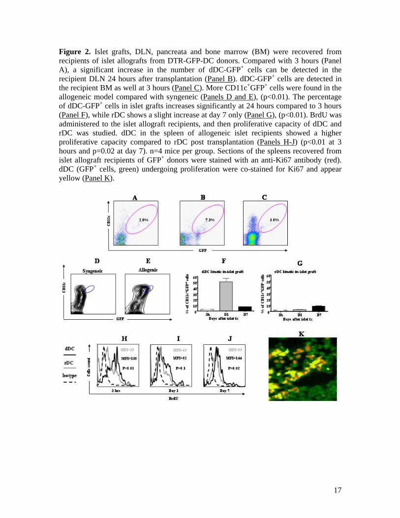
Figure 2. Islet grafts, DLN, pancreata and bone marrow (BM) were recovered from recipients of islet allografts from DTR-GFP-DC donors. Compared with 3 hours (Panel A), a significant increase in the number of dDC-GFP+ cells can be detected in the recipient DLN 24 hours after transplantation (Panel B). dDC-GFP+ cells are detected in the recipient BM as well at 3 hours (Panel C). More CD11c+GFP+ cells were found in the allogeneic model compared with syngeneic (Panels D and E), (p<0.01). The percentage of dDC-GFP+ cells in islet grafts increases significantly at 24 hours compared to 3 hours (Panel F), while rDC shows a slight increase at day 7 only (Panel G), (p<0.01). BrdU was administered to the islet allograft recipients, and then proliferative capacity of dDC and rDC was studied. dDC in the spleen of allogeneic islet recipients showed a higher proliferative capacity compared to rDC post transplantation (Panels H-J) (p<0.01 at 3 hours and p=0.02 at day 7). n=4 mice per group. Sections of the spleens recovered from islet allograft recipients of GFP+ donors were stained with an anti-Ki67 antibody (red). dDC (GFP+ cells, green) undergoing proliferation were co-stained for Ki67 and appear yellow (Panel K).
17

Figure 3. Spleens of islet allograft recipients were removed and splenocytes were subjected to FACS analysis. dDC (CD11c+GFP+) and rDC (CD11c+GFP-) were studied for chemokine receptor and CD86/Class II expression. A higher level of CXCR4, CCR5, CCR7, CD86 and Class II is noted in dDC extracted from recipient spleens at 3 hours after allogeneic islet transplantation (Tx), (Panels A-E). This higher expression of chemokine receptor and maturity markers (CD86/Class II) in dDC vs. rDC is maintained at day 1 (Panels F-J), while at day 7, dDC and rDC showed comparable levels of expression of CCR5 and maturity markers (Panels K-O). All experiments n=4 mice per group. * p<0.001; # p<0.05; ns not statistically significant.
3 hrs after
islet tx
Cel
ls c
ount
Recipient DC
Donor DC
A B C D
F G H I
K L M N
MFI=34
MFI=186
MFI=18
MFI=67
MFI=26
MFI=331
MFI=41
MFI=225
MFI=36
MFI=232
MFI=22
MFI=151
MFI=27
MFI=413
MFI=63
MFI=295
MFI=29
MFI=74
MFI=50
MFI=78
MFI=52
MFI=110MFI=178
MFI=260
CXCR4 CCR5 CCR7 CD86
Day 1 after
islet tx
Day 7 after
islet tx
E
J
O
Class II
MFI=60
MFI=400
MFI=490
MFI=550
MFI=120
MFI=680
ns ns ns#
*
*
#
* * *
* * *
#
*
18
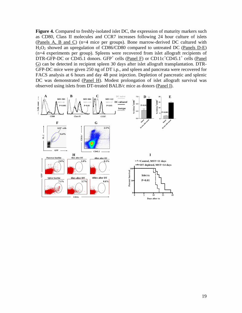
Figure 4. Compared to freshly-isolated islet DC, the expression of maturity markers such as CD80, Class II molecules and CCR7 increases following 24 hour culture of islets (Panels A, B and C) (n=4 mice per groups). Bone marrow-derived DC cultured with H2O2 showed an upregulation of CD86/CD80 compared to untreated DC (Panels D-E) (n=4 experiments per group). Spleens were recovered from islet allograft recipients of DTR-GFP-DC or CD45.1 donors. GFP+ cells (Panel F) or CD11c+CD45.1+ cells (Panel G) can be detected in recipient spleen 30 days after islet allograft transplantation. DTR-GFP-DC mice were given 250 ng of DT i.p., and spleen and pancreata were recovered for FACS analysis at 6 hours and day 48 post injection. Depletion of pancreatic and splenic DC was demonstrated (Panel H). Modest prolongation of islet allograft survival was observed using islets from DT-treated BALB/c mice as donors (Panel I).
0 5 10 15 200
50
100
Control, MST=11 daysDT depleted, MST=14 days
Days after tx
Perc
ent S
urvi
val
Islet tx
P=0.01
10 2CD80 102Class II 10 2CCR7
Naive
30 m
in 200 m
mol0.0
2.5
5.0
7.5
% C
D11
c+ CD
86+
P=0.01
2.5%
CD45.1
Cel
ls co
unt
A B C D
G
H I
DC cultured
DC naive
F
CD
11c+
21.64%
Pancreas baseline 6hrs after DT 48hrs after DT
6hrs after DT 48hrs after DTSpleen baseline
CD11c
GFP
GFP
FL-2
GFP+-cells
6.4%
2.0% 1.0% 0.3%
7.1% 3.7% 0.6%
MFI=905MFI=40
MFI=804MFI=225
MFI=122MFI=57
Naive
30 m
in 200 m
mol0
5
10
15
20
% C
D11
c+ CD
80+
E
P<0.001
P<0.001 P<0.01 P<0.05Isotype
19

Figure 5. An increase in the percentage of CD11c+CD45+ cells was noted in the medium of CCL21-cultured islets after 24 hours of culture (Panel A, p<0.01), with a parallel decrease of CD11c+CD45+ cells recovered from islets (Panel B, p<0.01). Prolongation of islet allograft survival was observed when using CCL21-cultured islets in allogeneic islet transplantation (Panel C, p<0.001). An abrogation of prolonged islet allograft survival occurred when WT-dDC were adoptively transferred (Panel C). A reduction in activated T cells (CD4+CD25+ cells) in the draining lymph nodes (Panels D, p<0.01) and a lower proliferative response (measured by MLR) were noted in the recipients of CCL21-cultured islets donor compared to mice that received control islets (Panel E, both experiments at day 7 after islet transplantation p<0.01). Islet grafts harvested at day 7 after transplantation of CCL21-cultured islets appeared less infiltrated by mononuclear cells and showed a well-maintained insulin staining (Panel F) compared to control islets (Panel G).
0 10 20 30 400
50
100 control, MST=11 days200 ng, MST=13 days800 ng, MST=19 days800 ng+AT-dDC=9 days
Days after tx
Perc
ent S
urvi
val
B
C
800 ng vs. control/AT dDC, p<0.001
AControl 200 ng 800 ng
7.0% 17.5% 48.8% 20.7%800 ngControl
6.3%
CD45+CD11c+ cells
Cel
ls co
unt
P<0.01
CCL21 Untreated0
5000
10000
15000
20000
3 H-th
ymid
ine
inco
rpor
atio
n
17.5%48.8%
7.0%
CD25
Cel
ls c
ount
P<0.01
CCL21
Control
MFI=6MFI=41
Cel
ls co
unt
CD45+CD11c+ cells
D E
F G
20