21 Gene expression profiling in acute allograft
22
REVIEW Open Access Gene expression profiling in acute allograft rejection: challenging the immunologic constant of rejection hypothesis Tara L Spivey 1,2,3 , Lorenzo Uccellini 1,4 , Maria Libera Ascierto 1,5,6 , Gabriele Zoppoli 5,7 , Valeria De Giorgi 1 , Lucia Gemma Delogu 8 , Alyson M Engle 1 , Jaime M Thomas 1 , Ena Wang 1 , Francesco M Marincola 1* and Davide Bedognetti 1,5,9* Abstract In humans, the role and relationship between molecular pathways that lead to tissue destruction during acute allograft rejection are not fully understood. Based on studies conducted in humans, we recently hypothesized that different immune-mediated tissue destruction processes (i.e. cancer, infection, autoimmunity) share common convergent final mechanisms. We called this phenomenon the “Immunologic Constant of Rejection (ICR).” The elements of the ICR include molecular pathways that are consistently described through different immune- mediated tissue destruction processes and demonstrate the activation of interferon-stimulated genes (ISGs), the recruitment of cytotoxic immune cells (primarily through CXCR3/CCR5 ligand pathways), and the activation of immune effector function genes (IEF genes; granzymes A/B, perforin, etc.). Here, we challenge the ICR hypothesis by using a meta-analytical approach and systematically reviewing microarray studies evaluating gene expression on tissue biopsies during acute allograft rejection. We found the pillars of the ICR consistently present among the studies reviewed, despite implicit heterogeneity. Additionally, we provide a descriptive mechanistic overview of acute allograft rejection by describing those molecular pathways most frequently encountered and thereby thought to be most significant. The biological role of the following molecular pathways is described: IFN-g, CXCR3/CCR5 ligand, IEF genes, TNF-a, IL-10, IRF-1/STAT-1, and complement pathways. The role of NK cell, B cell and T-regulatory cell signatures are also addressed. Introduction Defining the interplay between molecular pathways within highly complex biological systems, such as those between immune cell networks and target tissues, is cer- tainly a daunting task. The advent of high-throughput gene expression technology has served as an extremely useful tool to enable investigators to characterize biolo- gical events taking place within humans, reducing the inherent bias often generated by testing specific but restricted hypotheses derived from animal models. Pre- viously, we applied this approach to profiling tumor lesions in humans, before and after immunotherapy, to identify molecular pathways activated during immune- mediated tumor rejection. These pathways illustrate a process characterized by the coordinated activation of interferon stimulated genes (ISGs), the recruitment of cytotoxic cells through the massive production of speci- fic chemokine ligands, and the activation of immune effector function (IEF) genes (genes expressed by NK cells and CD8 T cells upon activation) [1-4]. Similar pathways have been described among other immune- mediated tissue destruction processes such as those occurring during autoimmunity, graft versus host dis- ease (GVHD), infection clearance, acute cardiovascular events, chronic obstructive pulmonary disease, and pla- cental villitis [5-10]. These observations suggest that these distinct tissue destruction processes share com- mon final immune-mediated molecular mechanisms. We termed this phenomenon as the “ Immunologic * Correspondence: [email protected]; [email protected] 1 Infectious Disease and Immunogenetics Section (IDIS), Department of Transfusion Medicine, Clinical Center and trans-NIH Center for Human Immunology (CHI), National Institutes of Health, Bethesda, Maryland, 20892, USA Full list of author information is available at the end of the article Spivey et al. Journal of Translational Medicine 2011, 9:174 http://www.translational-medicine.com/content/9/1/174 © 2011 Spivey et al; licensee BioMed Central Ltd. This is an Open Access article distributed under the terms of the Creative Commons Attribution License (http://creativecommons.org/licenses/by/2.0), which permits unrestricted use, distribution, and reproduction in any medium, provided the original work is properly cited.
Transcript of 21 Gene expression profiling in acute allograft

REVIEW Open Access
Gene expression profiling in acute allograftrejection: challenging the immunologic constantof rejection hypothesisTara L Spivey1,2,3, Lorenzo Uccellini1,4, Maria Libera Ascierto1,5,6, Gabriele Zoppoli5,7, Valeria De Giorgi1,Lucia Gemma Delogu8, Alyson M Engle1, Jaime M Thomas1, Ena Wang1, Francesco M Marincola1* andDavide Bedognetti1,5,9*
Abstract
In humans, the role and relationship between molecular pathways that lead to tissue destruction during acuteallograft rejection are not fully understood. Based on studies conducted in humans, we recently hypothesized thatdifferent immune-mediated tissue destruction processes (i.e. cancer, infection, autoimmunity) share commonconvergent final mechanisms. We called this phenomenon the “Immunologic Constant of Rejection (ICR).” Theelements of the ICR include molecular pathways that are consistently described through different immune-mediated tissue destruction processes and demonstrate the activation of interferon-stimulated genes (ISGs), therecruitment of cytotoxic immune cells (primarily through CXCR3/CCR5 ligand pathways), and the activation ofimmune effector function genes (IEF genes; granzymes A/B, perforin, etc.).Here, we challenge the ICR hypothesis by using a meta-analytical approach and systematically reviewing microarraystudies evaluating gene expression on tissue biopsies during acute allograft rejection. We found the pillars of theICR consistently present among the studies reviewed, despite implicit heterogeneity.Additionally, we provide a descriptive mechanistic overview of acute allograft rejection by describing thosemolecular pathways most frequently encountered and thereby thought to be most significant. The biological roleof the following molecular pathways is described: IFN-g, CXCR3/CCR5 ligand, IEF genes, TNF-a, IL-10, IRF-1/STAT-1,and complement pathways. The role of NK cell, B cell and T-regulatory cell signatures are also addressed.
IntroductionDefining the interplay between molecular pathwayswithin highly complex biological systems, such as thosebetween immune cell networks and target tissues, is cer-tainly a daunting task. The advent of high-throughputgene expression technology has served as an extremelyuseful tool to enable investigators to characterize biolo-gical events taking place within humans, reducing theinherent bias often generated by testing specific butrestricted hypotheses derived from animal models. Pre-viously, we applied this approach to profiling tumorlesions in humans, before and after immunotherapy, to
identify molecular pathways activated during immune-mediated tumor rejection. These pathways illustrate aprocess characterized by the coordinated activation ofinterferon stimulated genes (ISGs), the recruitment ofcytotoxic cells through the massive production of speci-fic chemokine ligands, and the activation of immuneeffector function (IEF) genes (genes expressed by NKcells and CD8 T cells upon activation) [1-4]. Similarpathways have been described among other immune-mediated tissue destruction processes such as thoseoccurring during autoimmunity, graft versus host dis-ease (GVHD), infection clearance, acute cardiovascularevents, chronic obstructive pulmonary disease, and pla-cental villitis [5-10]. These observations suggest thatthese distinct tissue destruction processes share com-mon final immune-mediated molecular mechanisms.We termed this phenomenon as the “Immunologic
* Correspondence: [email protected]; [email protected] Disease and Immunogenetics Section (IDIS), Department ofTransfusion Medicine, Clinical Center and trans-NIH Center for HumanImmunology (CHI), National Institutes of Health, Bethesda, Maryland, 20892,USAFull list of author information is available at the end of the article
Spivey et al. Journal of Translational Medicine 2011, 9:174http://www.translational-medicine.com/content/9/1/174
© 2011 Spivey et al; licensee BioMed Central Ltd. This is an Open Access article distributed under the terms of the Creative CommonsAttribution License (http://creativecommons.org/licenses/by/2.0), which permits unrestricted use, distribution, and reproduction inany medium, provided the original work is properly cited.

Constant of Rejection (ICR) [3].” The molecular con-stants shared among these different tissue destructionprocesses include the coordinated activation of the fol-lowing pathways: I) STAT-1/IRF-1/T-bet/IFN-g/IL-15pathway; II) CXCR3 ligand chemokine pathway(CXCL9, -10, -11) III) CCR5 ligand chemokine pathway(CCL3, -4, -5) and IV) TIA-1 pathway/granzyme A/B/granulysin/perforin pathway [3,4].Over the past decade gene expression microarrays
have been employed to study allograft rejection inhumans. The intrinsic heterogeneity among differentinvestigators in terms of patient selection, microarrayplatforms, gene coverage, statistical analysis, sample col-lection and study design makes cross-comparisonbetween different studies very challenging. Furthermore,since microarray profiling is a relatively new technology,it has continued to evolve in sophistication and has onlyrecently become standardized [11,12]. For this reasonwe believe that despite the non-uniformity among stu-dies, genes that are consistently reported across differentstudies and in different organs command attention. Inthis review we challenge the concept of the ICR byexamining multiple studies to evaluate the presence ofthe “constants of rejection.” We tested the ICR hypoth-esis by describing the most frequently reported immunepathways activated during acute allograft rejection inhumans as reported by publications using microarraytechnologies. Biological explanations for relevant path-ways are provided based on pertinent literature.
Data Extraction CriteriaIn this review we focused on high-throughput geneexpression profiling studies which sought to characterizethe molecular features of acute allograft rejection.Accordingly, we searched various combinations of thefollowing MeSH terms/keywords in PubMed: “geneexpression, “ “acute, “ “allograft, “ “rejection, “ and“microarray.” Searches were performed independently bytwo investigators. Gene Expression Omnibus (GEO) andreference lists of original articles and review articles alsoserved as additional search methods. Microarray studiesproviding original data and performed on human tissuebiopsies during established acute allograft rejection wereselected and evaluated [13-46]). Studies analyzing geneexpression profiles of peripheral blood mononuclearcells and urine sediments during acute rejection will notbe considered here, despite their potential utility as non-invasive diagnostic/predictive tools [47-51].The compiled list of key genes in this review came
from those reported as upregulated in the original publi-cations, most of which were predominantly immune-related and are reported in Table 1. In total, 15 uniquedatasets met the search criteria, and comprise Tables 1and 2. Of these datasets, four comparative analyses were
among those selected for inclusion. Of note, all of thestudies contained original data from de novoinvestigation.The Ingenuity Systems Pathway Analysis (IPA) http://
www.ingenuity.com and MetaCore http://www.genego.com were used to illustrate the relationships among thecompiled list of key genes. Additional detail regardingthe data extraction is provided in Additional File 1.
Overview of microarray studiesConsidering the heterogeneity among the selected stu-dies in terms of platform used, purpose, design, andinterpretation (see also Table 2), a quantitative approachwas not feasible, making this review qualitative in nat-ure. The diversity of the clinical setting (pediatric oradult patients; heart, lung, liver or kidney transplants)also added complexity to this analysis. The purposes ofthe original studies included here ranged between classdiscovery, class comparison, and/or class prediction. Dif-ferent methods (summarized in Table 2) were used bydifferent investigators to provide a list of genes modu-lated during acute allograft rejection. Not surprisingly,little overlap exists among studies with respect to speci-fic ‘genes’ described as upregulated during acute rejec-tion, yet, we found a striking consistency in terms ofpathway overrepresentation suggesting that each indivi-dual study identified different pieces of the same puzzle.It should be noted that these studies lacked the use of
micro/macro-dissection which prohibited identificationof the cellular source of the transcripts differentiallyexpressed during acute rejection. It is logical to thinkthat analysis of RNA from the whole tissue samplescould influence gene expression patterns. With this inmind, Sarwal et al. [22], investigated if the molecularchanges observed during allograft rejection could havebeen related to the differential sampling of cortical andmedullary kidney sections. For this analysis the authorsexcluded the genes whose expression was shown to becorrelated with the depth of biopsy in a previous investi-gation. The introduction of this filter did not signifi-cantly change the results. In another study involvingkidney recipients, Rodder et al. [29] performed qRT-PCR on isolated glomeruli, and on proximal and distaltubules. Although qRT-PCR of targets genes (metzincinsand related genes) revealed some differences betweenglomeruli and tubules, it confirmed, overall, the differ-ences between acute rejection and normal samplesdetected by microarray analysis.
The Immunologic Constant of Rejection pathways inacute allograft rejectionAfter reviewing the literature, we found that pathwaysinvolved in the ICR hypothesis are frequently activatedduring acute allograft rejection across studies conducted
Spivey et al. Journal of Translational Medicine 2011, 9:174http://www.translational-medicine.com/content/9/1/174
Page 2 of 22

Table 1 Key genes associated with acute allograft rejection according to human microarray studies
Author: Tannapfel et al. 2001 [14]Organ: Liver
IL3 MMP9 TGF-B1-3 TIMP1 TNF CLUSTERIN(CLU)
IL10
Author: Sreekumar et al. 2002 [15]Organ: Liver
C1QB C3 LIPC GZMB HSPA1A IGF1 IL2 (IRF9*) STAT1 ACADM
HLA-I HLA-II PCK1 SELPLG TGFB1 TNF TNFAIP3 TNFSF10 UBB3# UBA1
UBE2N ADAM17 GYS2 CTPS
Inkinen et al. 2005 [16]Organ: Liver
HLA-D IL2RB IL2RG CASP1 CASP3 GZMA GZMB SELL ICAM3 ITGA4
SELE VCAM1 IFNG IL1B
Asaoka et al. 2009 [28]Organ: Liver
AKAP11 ALOX15 CASP8 CFLAR FFAR3 IFNAR1 IGFBP3 IL12RB1 LTA POU4F1
PPP1R8 PPP1R3A PVRL1 TNK2
Author: Gimino et al. 2003 [17]Lande et al. 2006 [30](MINNEAPOLIS Dataset)Organ: Lung
C4B CCR7 CD28 CD3E CD84 CTLA4 CXCR3 GZMK GZMA IFNG
IGKC ITK KIR PRF1 STAT4 IL2RA IL2RB Zap70 LCK
Patil et al. 2008 (MINNEAPOLIS2 Dataset) [18]Organ: Lung
IFITM1 CD8A MARCKS CCL3 GZMB ITM2A IL32 IL8 CCL4
Author: Karason et al. 2006 [19]Organ: Heart
C3 C4A CXCL9 CXCL10 GBP1 HLA-C HLA-F HLA-J IGFBP4 NPPA
PSME2 RARRES3 STAT1
Author: Akalin et al. 2001 [21]Organ: Kidney
Humig(CXCL9)
C3 CD18(ITGB2)
ISGF-3(STAT1)
MCL1 MIP-3b(CCL19)
NNMT RING4(TAP1)
TCRB# IL2-SP2#
(LCP1)
Author: Sarwal et al. 2003 (STANFORD Dataset) [22]Organ: Kidney
HLA-A HLA-B HLA-C HLA-E HLA-DR HLA-DQ HLA-DMA HLA-DRB4 TGFBR2 TGFR1
TCR DARC C4B CXCL9 SCYA3(CCL3)
SCYA5(CCL5)
CCR5 SCYA2(CCL2)
CD20 PERFORIN
CD53 NFKB1 NK4(IL32)
CX3CR1 GZMA STK17B IL6R IL2RB IL15RA IL16
STAT1 CASP10 IFNGR1 IGHG3 IGKC IGL IGHM LENG4 CD59 VCAM1
CXCL9 CXCL10 CCR5
Author: Flechner et al. 2004 (CLEVELAND Dataset) [23]Organ: Kidney
C1QB CCL5 CD14 CD163 TRB@ CD16 CD2 CD27 CD3D CD48
CD53 CD64(FCGR1A)
CD8 CDW52 CXCR4 GZMA HLA-F IFI30 IL10RA IL10RB
IL4R ISG20 PKR(PRKRA)
RAGE4(RAGE)
TNFRSF1b
Reeve et al. 2009 (EDMONTON Dataset) [24]Organ: Kidney
APOBEC3G CCL4 CCL5 CD8A CRTAM CXCL9 CXCL10 CXCL11 FAM26F GBP1
GBP2 GBP4 GBP5 GZMA GZMB INDO LCP2 LILRB1 NLRC5 PSMB9
Spivey et al. Journal of Translational Medicine 2011, 9:174http://www.translational-medicine.com/content/9/1/174
Page 3 of 22
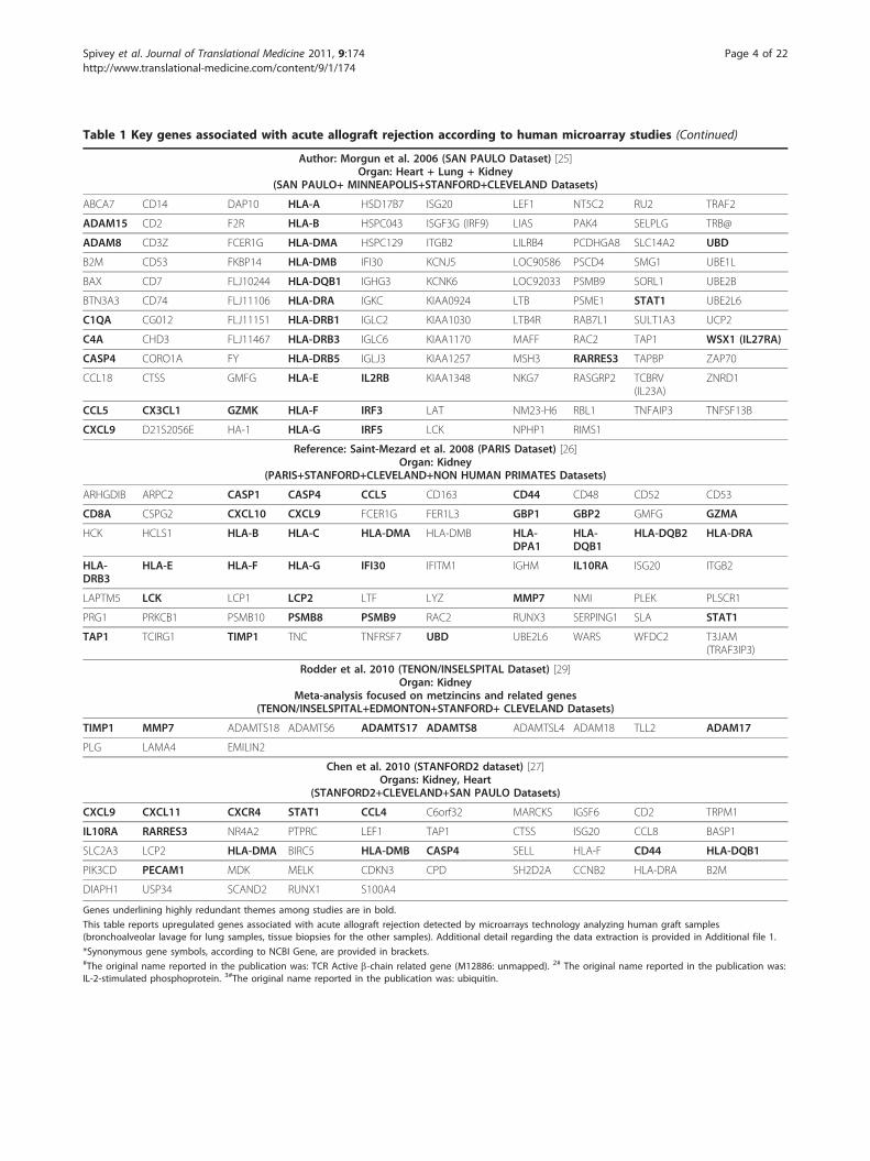
Table 1 Key genes associated with acute allograft rejection according to human microarray studies (Continued)
Author: Morgun et al. 2006 (SAN PAULO Dataset) [25]Organ: Heart + Lung + Kidney
(SAN PAULO+ MINNEAPOLIS+STANFORD+CLEVELAND Datasets)
ABCA7 CD14 DAP10 HLA-A HSD17B7 ISG20 LEF1 NT5C2 RU2 TRAF2
ADAM15 CD2 F2R HLA-B HSPC043 ISGF3G (IRF9) LIAS PAK4 SELPLG TRB@
ADAM8 CD3Z FCER1G HLA-DMA HSPC129 ITGB2 LILRB4 PCDHGA8 SLC14A2 UBD
B2M CD53 FKBP14 HLA-DMB IFI30 KCNJ5 LOC90586 PSCD4 SMG1 UBE1L
BAX CD7 FLJ10244 HLA-DQB1 IGHG3 KCNK6 LOC92033 PSMB9 SORL1 UBE2B
BTN3A3 CD74 FLJ11106 HLA-DRA IGKC KIAA0924 LTB PSME1 STAT1 UBE2L6
C1QA CG012 FLJ11151 HLA-DRB1 IGLC2 KIAA1030 LTB4R RAB7L1 SULT1A3 UCP2
C4A CHD3 FLJ11467 HLA-DRB3 IGLC6 KIAA1170 MAFF RAC2 TAP1 WSX1 (IL27RA)
CASP4 CORO1A FY HLA-DRB5 IGLJ3 KIAA1257 MSH3 RARRES3 TAPBP ZAP70
CCL18 CTSS GMFG HLA-E IL2RB KIAA1348 NKG7 RASGRP2 TCBRV(IL23A)
ZNRD1
CCL5 CX3CL1 GZMK HLA-F IRF3 LAT NM23-H6 RBL1 TNFAIP3 TNFSF13B
CXCL9 D21S2056E HA-1 HLA-G IRF5 LCK NPHP1 RIMS1
Reference: Saint-Mezard et al. 2008 (PARIS Dataset) [26]Organ: Kidney
(PARIS+STANFORD+CLEVELAND+NON HUMAN PRIMATES Datasets)
ARHGDIB ARPC2 CASP1 CASP4 CCL5 CD163 CD44 CD48 CD52 CD53
CD8A CSPG2 CXCL10 CXCL9 FCER1G FER1L3 GBP1 GBP2 GMFG GZMA
HCK HCLS1 HLA-B HLA-C HLA-DMA HLA-DMB HLA-DPA1
HLA-DQB1
HLA-DQB2 HLA-DRA
HLA-DRB3
HLA-E HLA-F HLA-G IFI30 IFITM1 IGHM IL10RA ISG20 ITGB2
LAPTM5 LCK LCP1 LCP2 LTF LYZ MMP7 NMI PLEK PLSCR1
PRG1 PRKCB1 PSMB10 PSMB8 PSMB9 RAC2 RUNX3 SERPING1 SLA STAT1
TAP1 TCIRG1 TIMP1 TNC TNFRSF7 UBD UBE2L6 WARS WFDC2 T3JAM(TRAF3IP3)
Rodder et al. 2010 (TENON/INSELSPITAL Dataset) [29]Organ: Kidney
Meta-analysis focused on metzincins and related genes(TENON/INSELSPITAL+EDMONTON+STANFORD+ CLEVELAND Datasets)
TIMP1 MMP7 ADAMTS18 ADAMTS6 ADAMTS17 ADAMTS8 ADAMTSL4 ADAM18 TLL2 ADAM17
PLG LAMA4 EMILIN2
Chen et al. 2010 (STANFORD2 dataset) [27]Organs: Kidney, Heart
(STANFORD2+CLEVELAND+SAN PAULO Datasets)
CXCL9 CXCL11 CXCR4 STAT1 CCL4 C6orf32 MARCKS IGSF6 CD2 TRPM1
IL10RA RARRES3 NR4A2 PTPRC LEF1 TAP1 CTSS ISG20 CCL8 BASP1
SLC2A3 LCP2 HLA-DMA BIRC5 HLA-DMB CASP4 SELL HLA-F CD44 HLA-DQB1
PIK3CD PECAM1 MDK MELK CDKN3 CPD SH2D2A CCNB2 HLA-DRA B2M
DIAPH1 USP34 SCAND2 RUNX1 S100A4
Genes underlining highly redundant themes among studies are in bold.
This table reports upregulated genes associated with acute allograft rejection detected by microarrays technology analyzing human graft samples(bronchoalveolar lavage for lung samples, tissue biopsies for the other samples). Additional detail regarding the data extraction is provided in Additional file 1.
*Synonymous gene symbols, according to NCBI Gene, are provided in brackets.#The original name reported in the publication was: TCR Active b-chain related gene (M12886: unmapped). 2# The original name reported in the publication was:IL-2-stimulated phosphoprotein. 3#The original name reported in the publication was: ubiquitin.
Spivey et al. Journal of Translational Medicine 2011, 9:174http://www.translational-medicine.com/content/9/1/174
Page 4 of 22
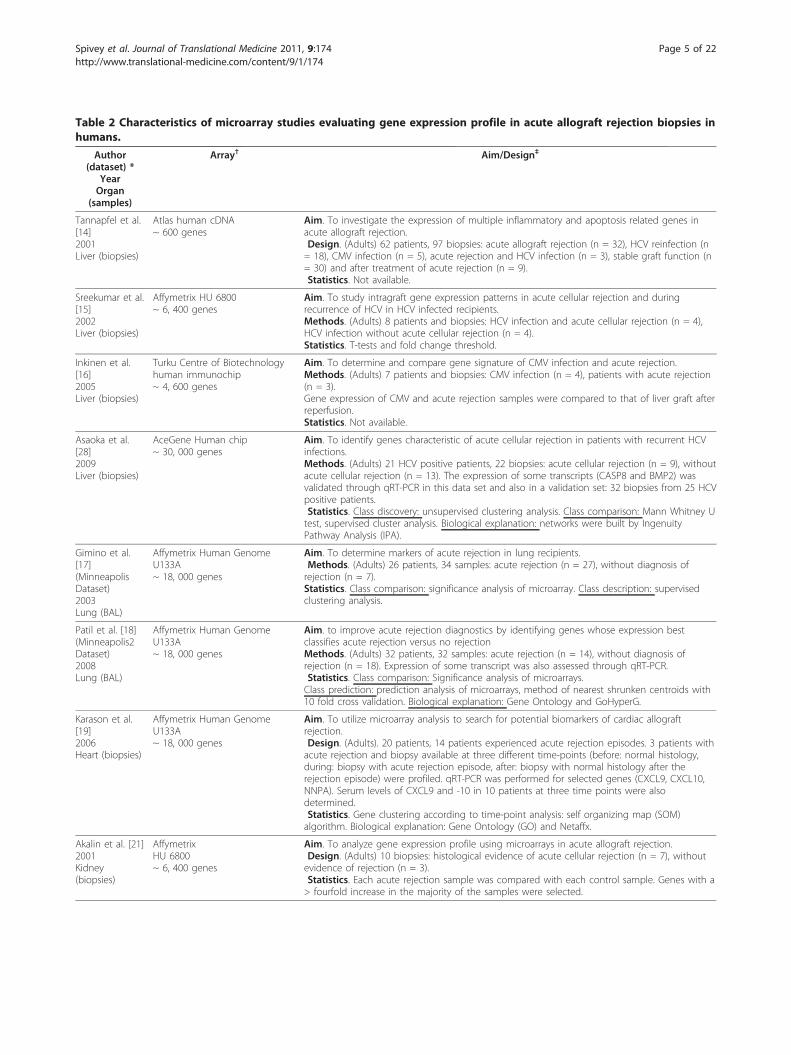
Table 2 Characteristics of microarray studies evaluating gene expression profile in acute allograft rejection biopsies inhumans.
Author(dataset) *
YearOrgan
(samples)
Array† Aim/Design‡
Tannapfel et al.[14]2001Liver (biopsies)
Atlas human cDNA~ 600 genes
Aim. To investigate the expression of multiple inflammatory and apoptosis related genes inacute allograft rejection.Design. (Adults) 62 patients, 97 biopsies: acute allograft rejection (n = 32), HCV reinfection (n= 18), CMV infection (n = 5), acute rejection and HCV infection (n = 3), stable graft function (n= 30) and after treatment of acute rejection (n = 9).Statistics. Not available.
Sreekumar et al.[15]2002Liver (biopsies)
Affymetrix HU 6800~ 6, 400 genes
Aim. To study intragraft gene expression patterns in acute cellular rejection and duringrecurrence of HCV in HCV infected recipients.Methods. (Adults) 8 patients and biopsies: HCV infection and acute cellular rejection (n = 4),HCV infection without acute cellular rejection (n = 4).Statistics. T-tests and fold change threshold.
Inkinen et al.[16]2005Liver (biopsies)
Turku Centre of Biotechnologyhuman immunochip~ 4, 600 genes
Aim. To determine and compare gene signature of CMV infection and acute rejection.Methods. (Adults) 7 patients and biopsies: CMV infection (n = 4), patients with acute rejection(n = 3).Gene expression of CMV and acute rejection samples were compared to that of liver graft afterreperfusion.Statistics. Not available.
Asaoka et al.[28]2009Liver (biopsies)
AceGene Human chip~ 30, 000 genes
Aim. To identify genes characteristic of acute cellular rejection in patients with recurrent HCVinfections.Methods. (Adults) 21 HCV positive patients, 22 biopsies: acute cellular rejection (n = 9), withoutacute cellular rejection (n = 13). The expression of some transcripts (CASP8 and BMP2) wasvalidated through qRT-PCR in this data set and also in a validation set: 32 biopsies from 25 HCVpositive patients.Statistics. Class discovery: unsupervised clustering analysis. Class comparison: Mann Whitney Utest, supervised cluster analysis. Biological explanation: networks were built by IngenuityPathway Analysis (IPA).
Gimino et al.[17](MinneapolisDataset)2003Lung (BAL)
Affymetrix Human GenomeU133A~ 18, 000 genes
Aim. To determine markers of acute rejection in lung recipients.Methods. (Adults) 26 patients, 34 samples: acute rejection (n = 27), without diagnosis ofrejection (n = 7).Statistics. Class comparison: significance analysis of microarray. Class description: supervisedclustering analysis.
Patil et al. [18](Minneapolis2Dataset)2008Lung (BAL)
Affymetrix Human GenomeU133A~ 18, 000 genes
Aim. to improve acute rejection diagnostics by identifying genes whose expression bestclassifies acute rejection versus no rejectionMethods. (Adults) 32 patients, 32 samples: acute rejection (n = 14), without diagnosis ofrejection (n = 18). Expression of some transcript was also assessed through qRT-PCR.Statistics. Class comparison: Significance analysis of microarrays.Class prediction: prediction analysis of microarrays, method of nearest shrunken centroids with10 fold cross validation. Biological explanation: Gene Ontology and GoHyperG.
Karason et al.[19]2006Heart (biopsies)
Affymetrix Human GenomeU133A~ 18, 000 genes
Aim. To utilize microarray analysis to search for potential biomarkers of cardiac allograftrejection.Design. (Adults). 20 patients, 14 patients experienced acute rejection episodes. 3 patients withacute rejection and biopsy available at three different time-points (before: normal histology,during: biopsy with acute rejection episode, after: biopsy with normal histology after therejection episode) were profiled. qRT-PCR was performed for selected genes (CXCL9, CXCL10,NNPA). Serum levels of CXCL9 and -10 in 10 patients at three time points were alsodetermined.Statistics. Gene clustering according to time-point analysis: self organizing map (SOM)algorithm. Biological explanation: Gene Ontology (GO) and Netaffx.
Akalin et al. [21]2001Kidney(biopsies)
AffymetrixHU 6800~ 6, 400 genes
Aim. To analyze gene expression profile using microarrays in acute allograft rejection.Design. (Adults) 10 biopsies: histological evidence of acute cellular rejection (n = 7), withoutevidence of rejection (n = 3).Statistics. Each acute rejection sample was compared with each control sample. Genes with a> fourfold increase in the majority of the samples were selected.
Spivey et al. Journal of Translational Medicine 2011, 9:174http://www.translational-medicine.com/content/9/1/174
Page 5 of 22

Table 2 Characteristics of microarray studies evaluating gene expression profile in acute allograft rejection biopsies inhumans. (Continued)
Sarwal et al.[22](StanfordDataset)2003Kidney(biopsies)
Lymphochip> 12, 000 genes
Aim. To investigate the possibility that variations in gene-expression patterns in allograft-biopsysamples from patients with acute rejection and related disorders could identify molecularlydistinct subtypes of acute rejection to possibly explain differences in clinical behavior.Design. (Pediatric patients) 50 patients. 67 biopsies: biopsies during acute or chronic allograftdysfunctions (n = 52) and at the time of the engraftment or when graft function was stable (n= 15). The possibility of different sampling of the medullary and the cortical regions was alsoaddressed.Statistics. Class discovery: unsupervised clustering analysis. Class comparison: significanceanalysis of microarray. Survival analysis: Kaplan-Meyer/Cox log-rank method. Biologicalexplanation: enrichment of specific functional groups through evaluation of hypergeometricdistribution. The exclusion of data from genes whose expression was correlated with the depthof biopsy did not change the cluster analysis.
Flechner et al.[23](ClevelandDataset)2004Kidney(biopsies andPBLs)
AffymetrixHG-U95Av2~ 10, 000 genes
Aim. To determine gene expression profiling in transplant patients including: normal donorkidneys, well functioning transplants without rejection, kidneys undergoing acute rejection, andtransplants with renal dysfunction without rejection.Design. (Adults) 23 graft recipients and 9 donors. Acute rejection biopsies (n = 7), renaldysfunction without rejection on biopsies (n = 6), biopsies carried out more than one year posttransplant in patient with good transplant function and normal histology (n = 10), biopsiesfrom living donor controls (n = 9). PBLs were also collected and profiled. Expression of sometranscript was also assessed through qRT-PCR.Statistics. Class discovery: unsupervised clustering analysis. Class comparison: significanceanalysis of microarray filtered with limit fold model and MAS 5.0 present/absent calls. Class-prediction: leave-one-out method. Biological explanation: analysis of functional classes of thedifferentially expressed genes.
Reeve et al. [24](EdmontonDataset)2009Kidney(biopsies)
Affymetrix Human Genome U133Plus 2.0> 38, 000 genes
Aim. To define a classifier to distinguish rejection vs non rejection using predictive analysis formicroarrays.Design. (Adults) 143 patients, 186 biopsies: acute rejection samples (acute cellular rejection,antibody mediated rejection or mixed) (n = 51), non-rejection samples (n = 135).Statistics. Class comparisons: Bayesian t-test and false discovery rate. Class prediction:prediction analysis of microarrays. Biological explanation: analysis of functional classes of thedifferentially expressed genes according to KEGG pathways and to authors’ definedpathogenesis-based transcripts.
Morgun et al.[25](San PauloDataset)2006Heart (biopsies)
Qiagen/Operon Array~ 14, 000 genes
Aim. To analyze gene expression differences between rejection, non rejection andTrypanosoma cruzi infection.Design. (Adults) 40 patients, 76 biopsies (rejection, no rejection and Trypanosoma cruziinfection recurrence). Expression of some transcripts was also assessed through qRT-PCR.Statistics. Class comparison: random variance t-test filtered with univariate/multivariate tests forfalse discovery rates; supervised clustering analysis. Class prediction: 6 different multivariatemodels models (compound covariate predictor, diagonal linear discriminant analysis, 1- and 3-nearest neighbor predictor, nearest centroid predictor, support vector machine) and leave-one-out cross validation. The authors validated the predictor-set in independent datasets of biopsies(collected on different continents and analyzed with different chip batches). The authors alsotested the predictor set by analyzing the data from data from Cleveland (Kidney) Stanford(Kidney) and Minneapolis (Lung) datasets.Biological explanation: Database Annotation, Visualization and Integrated Discovery (DAVID)/Gene Ontology and KEGG Pathways.
Saint-Mezard etal. [26](Paris Dataset)2008Kidney(biopsies)
Affymetrix Human Genome U133Plus 2.0> 38, 000 genes
Aim. To identify a robust and reliable molecular signature for acute rejection in humans.Design. (Adults) 45 patients, 47 biopsies: acute rejection (n = 8), acute rejection and chronicallograft nephropathy (n = 8), borderline (n = 3), non rejection (n = 7), and chronic allograftnephropathy (n = 22). Normal kidney tissue was obtained from histopathologically unaffectedareas of the cortex of native nephrectomies performed for renal carcinoma was used ascontrol.Statistics. Genes differentially expressed (Paris Dataset) were intersected with those from with 2public human datasets: 1) Stanford Dataset and 2) Cleveland Dataset and with one Non HumanPrimate (NHP) model of acute renal allograft. However, the authors used biopsy microarray datafrom Edmonton Dataset as in independent confirmation set. Score from the identified classifierwas correlated with the histopathological Banff score. Expression of some transcripts was alsoassessed through qRT-PCR.Class comparison: ANOVA with or without false discovery rate and additional cutoff based ontwofold change. Class discovery: Principal component analysis, supervised clustering analysis(using the genes differentially expressed in all four datasets); Class prediction: leave-one-outcross-validation and 10-fold cross-validation. Biological explanation: Gene regulatory networkswere generated using MetaCore.
Spivey et al. Journal of Translational Medicine 2011, 9:174http://www.translational-medicine.com/content/9/1/174
Page 6 of 22

by different investigators in different organs (see Table1). Figure 1 provides a visualization of the relationshipsamong the key genes described. Here, we attempt toillustrate the hypothetical role of these pathways duringacute allograft rejection.IFN-g pathwayIFN-g is a pleiotropic cytokine that plays a role in themodulation of many aspects of the immune response.Studies conducted involving IFN-g -/- mice suggest thatthis cytokine, in addition to its pro-inflammatory func-tions, might be important for graft acceptance, prevent-ing early graft necrosis, and maintaining microvascularviability [52,53]. However, the molecular mechanismsthrough which IFN-g exerts its anti-inflammatory actionduring the early phases of the engraftment are unclear.On the other hand, this interferon is considered a cen-tral cytokine in sustaining inflammation during allograftrejection both in humans and in murine models.IFN-g is predominantly produced by NK cells as part
of the innate immune response, and by CD4 T helper 1cells (Th1) and CD8 cytotoxic T cells (CTLs) as a partof the adaptive immune response once antigen-specificimmunity develops [54,55]. Its overexpression has alsobeen observed in acute allograft rejection in severalhuman studies where RT-PCR was applied [17,56-58].Microarray studies have not only enabled the detectionof the expression of the IFN-g gene itself, but also the
detection of its downstream effects (IFN-g stimulatedgenes) [15,17,21-24,59]. Figure 1A represents the firstnetwork generated by IPA after analysis of the compiledlist of key immune-related genes (Table 1), with IFN-gserving as the hub of this important network. Detectionof IFN-g stimulated genes alone is not sufficient to dis-criminate its effect from the effects of other cytokines,such as IFN-a, which can also activate many of theIFN-g stimulated genes [60,61]. However, the prevalenceof IFN-g versus IFN-a transcripts in addition to the acti-vation of pathways that specifically enhance the INF-gloop (e.g. TNF-a, CCR5, and CXCR3) implicate IFN-gas a driver gene involved in sustaining acute allograftrejection [22,24,26,31,59,62]. Although some functionshave been described for individual IFN-g stimulatedgenes, the overall orchestration is not completely under-stood. A partial description of the relationship amongIFN-g stimulated genes detected in microarray studies isillustrated in Figure 1A.Primarily through interferon regulatory factor 1 (IRF-
1), IFN-g upregulates both MHC class I and II, byincreasing the expression of antigen peptide transportersTAP1-2, or class II transactivator CIITA, for example[54,63,64]. Indeed, IFN-g promotes the differentiation ofnaïve CD4 T cells into Th1 cells which are, among thelineage of CD4 T cells (Th1, Th2, Th17 and T Reg), theonly ones that produce a consistent amount of IFN-g
Table 2 Characteristics of microarray studies evaluating gene expression profile in acute allograft rejection biopsies inhumans. (Continued)
Rodder et al.[29](Tenon/InelspitalDataset)2011Kidney(biopsies)
Affymetrix Human Genome U133Plus 2.0> 38, 000 genes
Aim. To identify the expression of metzincins and related genes in allograft rejection biopsies.Design. (Adults) 41 biopsies: normal histology (n = 20), borderline changes (n = 4), acuterejection (n = 10) and acute rejection and interstitial fibrosis/tubular atrophy (n = 7). Expressionof some transcripts was also assessed through qRT-PCR.Statistics. Class prediction: ANOVA and shrinking centroids methods were used for variableselection and a variety of classification methods were tested. Leave-one-out method wasperformed as internal cross-validation. Classifier performance was estimated as correct rate after1-level cross validation. The model was validated in Edmonton, Cleveland and Stanforddatasets. Gene set scores from biopsies were also determined and correlated with Banff scores.
Chen et al. [27](Stanford2Dataset)2011Kidney(biopsies)
Affymetrix Human Genome U133Plus 2.0> 38, 000 genes
Aim. To identify biomarkers across similar conditions through integration of related datasets.Methods. (Pediatric patients) 36 patients and biopsies: acute rejection biopsies (n = 18), stablefunction biopsies (n = 18).Statistics. Class comparison: significance analysis of microarrays and fold change filter. Theupregulated genes during acute rejection were intersected with genes upregulated duringacute rejection in two other datasets (Cleveland and San Paulo).
Notes
*For the Minneapolis Dataset only the publication by Gimino et al. is described;†Microarray chips details: Atlas human cDNA microarrays ~ 588 gene analyzed; Affymetrix GeneChip HU6800 Array containing > 7, 000 oligonucleotide probesets representing ~ 6, 400 human genes (Affymetrix, Santa Clara, CA);
Affymetrix Human Genome U133A Array containing > 22, 000 oligonucleotide probe sets representing > 18, 000 transcripts (~ 14, 500 human genes) (Affymetrix,Santa Clara, CA); Lymphochip: in-house microarrays containing > 28, 000 cDNA probes representing > 12, 000 genes (Stanford University); Affymetrix GeneChipHG-U95Av2 Array containing ~ 12, 000 oligonucleotide probes representing ~ 10, 000 human genes; Affymetrix Human Genome U133A Array containing > 22,000 oligonucleotide probe sets representing > 18, 000 transcripts (~ 14, 500 human genes) (Affymetrix, Santa Clara, CA); Affymetrix Human Genome U133 Plus2.0 Array containing > 54, 000 oligonucleotide probe sets representing > 47, 000 transcripts (~ 38, 500 human genes) (Affymetrix, Santa Clara, CA); Qiagen/Operon array: in-house oligonucleotide array platform designed by Qiagen/Operon (Alameda, CA) and printed at NIAID Microarray facility, representing ~ 14, 000human genes;‡Study aim/design is referred to gene expression experiments;
Abbreviations: BOS: Bronchiolitis obliterans syndrome; PBLs: Peripheral blood lymphocyte; CMV: cytomegalovirus; HCV: hepatitis C virus; qRT-PCR: quantitative realtime polymerase chain reaction;
Spivey et al. Journal of Translational Medicine 2011, 9:174http://www.translational-medicine.com/content/9/1/174
Page 7 of 22

[55,65]. IFN-g in turn, and usually in synergy withtumor necrosis factor-a (TNF-a), induces the expres-sion of CXCR3 ligands [54,66,67] and CCR5 ligands[68].CXCR3 and CCR5 ligand pathwaysCXCR3 ligands (CXCL9, -10, -11) and CCR5 ligands(CCL3, -4, -5) are the chemokines most frequently upre-gulated during acute allograft rejection as described byhuman microarray studies [18,21-27] and RT-PCR (SeeTable 1). The upregulation of the related receptors,CXCR3 and CCR5, has also been frequently described,though not as much specifically within microarray stu-dies [17,22,24,69-72]. Interestingly, high urinary CXCR3ligand protein levels were detected in clinical trials in
patients experiencing acute kidney rejection [48-51].More recently, Chen et al. [27] described CXCL9 as abiomarker of acute rejection in a cross-organ microarraystudy evaluating pediatric renal, adult renal, and adultcardiac transplant patients. Additionally, CXCL9 pro-teins were also found to be elevated in serum. Indeed,the CCR5Δ32 polymorphism that encodes for a non-functional CCR5 receptor, conferred a greatly reducedrisk for the development of acute rejection in kidney[73] and in liver transplantation [74].The driving role of these two pathways in allograft
rejection was suggested by in vivo murine models onedecade ago [75-78]. The lack of host CCR5 was asso-ciated with a three-fold increase in allograft survival
A
B
C
E
D
STAT 1/IFN Network
Immune
EffectorFunction
Genes
Netw
ork
TNF Network
Chem
okine/Ch
emok
ine
Receptor
Network
NF kB Network
Network 1
Network 2
Network 3Network 4
Network 7
Blue genes = IFN Overlay
Figure 1 First 5 Immune networks according to Ingenuity Pathways Analysis (IPA), representing schematic relationships among keygenes upregulated in acute allograft rejection (Network number 1 (A), 2 (B), 3 (C), 4 (D) and 7(E), generated by IPA). The gene listuploaded represents the key gene list (Table 1). Red: The genes and gene complexes from the key gene list are represented in red background(no color fill is used for the genes that are part of the network but not part of the key gene list). Blue: IFN-g stimulated genes (designated IFN-gstimulated genes identified as those upregulated in peripheral monocytes after IFN-g stimulation). A. the first network is centered around IFN-g;B. the second network is centered around TNF- a; C. the third network focuses on Interferon Regulatory Factors (IRFs) and chemokine/chemokine receptor interaction (i.e., CCR5/CCR5 ligands and CXCR3/CXCR3 ligands); D. the fourth network focuses on Immune Effector Function(IEF) genes (i.e., around granzyme B, perforin, caspases); E. the fifth network is centered around the NF-kB complex. Bold lines indicate directinteraction. Dotted lines indicate indirect interaction.
Spivey et al. Journal of Translational Medicine 2011, 9:174http://www.translational-medicine.com/content/9/1/174
Page 8 of 22

rate, but the targeting of any of the three main ligandsusing knockout mice or monoclonal antibodies had noeffect on allograft survival [79]. Similarly, lack of CXCR3led to graft acceptance indefinitely [75] and in addition,a significant survival benefit has been observed inCXCL10 -/- recipients [76]. This supposed non-redun-dant effect of CXCR3 that has been assumed correct foralmost ten years, is currently object of debate [80].Recently, in fact, two independent studies reported thatdisruption or blockade of recipient CXCR3 had rela-tively little effect on rejection [81-83]. These observa-tions, as well as the abundance of CXCR3 ligandspresent during acute allograft rejection, raise questionsabout the functional importance of CXCR3 during rejec-tion and the possibility of alternative targets of CXCR3ligands [80].Upon antigen stimulation, the co-expression of
CXCR3 and CCR5 is a marker of Th1 cell polarization,whereas CCR3, CCR4, CCR8, and CRTh2 are expressedby Th2 cells [65]. The genes and gene pathways fre-quently overexpressed during acute allograft rejectionare consistent with the predominance of Th1 cell polari-zation. Among CXCR3 and CCR5 ligands, CXCL9, -10and CCL4, -5 were the most frequently reported chemo-kines associated with acute rejection in microarray stu-dies [19,21-24,31] (see Table 1).CCR5 and CXCR3 ligands can be secreted differen-
tially by dendritic cells, activated macrophages and Tcells, endothelial cells, and NK cells [69,70,84-87]. How-ever, studies that define the cell-specific production ofchemokines in allograft rejection in humans are scant.Consequently, evidence-based descriptions of cell-speci-fic chemokine-mediated recruitment have not beenwell-defined in humans either.Hoffmann et al. [88] described that a significant pro-
portion of both CD4+ and CD8+ T-cells detected inhuman renal biopsies during rejection express CXCR3.It has also been speculated that CXCR3 may act as adecoy receptor by binding CCL11, preventing therecruitment of granulocytes via the CCL11/CCR3 bind-ing interaction [80,89]. This might explain why granulo-cytes are not classically found in acute cellular rejection.Furthermore, peripheral blood monocytes that are lowerin CCR5 expression could be recruited through CCR1that also binds CCL-3, -4, -5. In addition to Th1 andCTL cells, NK cells could also possibly be recruitedthrough this pathway since they are all known toexpress CXCR3 and CCR5 receptors [85]. However, NKcells are rarely present in allograft infiltrates and areespecially rare in T-cell mediated rejection [39,90].Recruitment and activation of CCR5 and CXCR3
ligands can lead to increased production of IFN-g, witha resultant amplification of the inflammatory stimuliand further release of chemoattractant molecules. Thus,
in a concerted fashion, these molecules orchestrate theswitch from innate to adaptive immunity, meanwhilesustaining and strengthening the innate cytotoxicmechanisms with a persistent “NK-like” response.Finally, even though up to 25% of circulating B cells
express CXCR3 [90-92], and can also produce CXCR3ligands [87,93], the recruitment of B cells through thismechanism during acute allograft rejection has not yetbeen defined in humans.This complex cascade of cytokines and the coordinate
activation of specific pathways so far described, leads tothe activation of IEF genes (perforin, granzymes A/B,Fas/Fas ligand, and caspases) during the process of tis-sue destruction.IEF genesThe release of granzymes, perforins, and granulysin andthe interaction between the Fas/Fas ligand and caspaseactivation represent the major effector mechanisms ofcell-mediated immunity [94]. These IEF transcripts havebeen consistently described as being associated withacute allograft rejection using transcriptome analyses[17,22,23,26].By profiling PBMCs, Hidalgo et al. [40] found that
cytotoxic molecular transcripts (i.e. granzyme B, Fasligand, perforin) are commonly overexpressed in CTLCD4+ cells, CTL CD8+ cells, and NK cells. These obser-vations highlight the existence of a common molecularcytotoxic “NK-like” effector mechanism that is sharedamong the different arms of the immune system, theclassically distinct innate and adaptive immune arms.Taking this one step further, Mueller et al. [31] foundthat gene expression patterns of T-cell mediated rejec-tion are surprisingly similar to the expression patternsfound in antibody-mediated rejection. In particular,interferon-g affected transcripts and IEF genes such asperforin, granzyme B, and Fas ligand were overexpressedin both of them. This observation suggests that effectorT cells and antibodies lead to the activation of a com-mon final pathway in tissue destruction and supportsthe proposed theory of the immunologic constant ofrejection [3].
NK cell, B cell, and T-reg signaturesNK signatureNK cells in murine skin and rat liver allografts are theimmune cells responsible for early chemokine produc-tion of CCR5 and CXCR3 ligands (i.e., CCL3, CCL4,CXCL10) which are important in initiating and sustain-ing acute allograft rejection [95,96]. Nevertheless, NKcells do not seem to be sufficient to reject solid organsdirectly since mice that have intact NK cell function butabsent adaptive immunity (RAG-/- or SCID) are able toaccept skin and cardiac graft transplants indefinitely[97-99]. However, the inability to reject the graft does
Spivey et al. Journal of Translational Medicine 2011, 9:174http://www.translational-medicine.com/content/9/1/174
Page 9 of 22
not prove that innate cells (in this case NK cells) areunable to mediate tissue destruction. A possible expla-nation could be that, in these models, the lack of stimuliderived from a reciprocal feedback between innate andadaptive cells, does not allow triggering or sustaining astrong enough “NK-like” cytotoxic effector function.Recently it has been observed that nude mice treated
with oncolytic viruses can reject tumor xenografts [100].This rejection was associated with the activation of ISGs(both IFN-g and IFN-a stimulated genes), upregulationof CXCR3 and CCR5 ligands, and activation of IEFgenes (granzyme B, caspase 8). Since these mice lack Tcells and secondarily lack B cell responses, this immune-mediated tissue destruction is thought to be induced byinnate immune effectors such as NK cells and activatedmacrophages. This study suggests that, at least in thismodel, innate immunity can be an independent effectorof tissue-specific destruction not requiring adaptiveimmunity. It is possible that the oncolytic virus used inthis model primes the innate immune system in a man-ner that bypasses the need for the adaptive immune sys-tem interaction.In humans, however, studies analyzing the individual
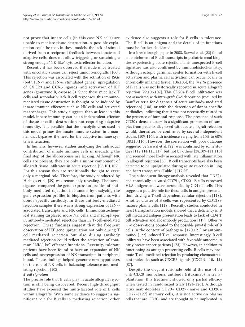
contribution of innate immune cells in mediating thefinal step of the alloresponse are lacking. Although NKcells are present, they are only a minor component ofallograft tissue infiltrates in acute rejection [98,101,102].For this reason they are traditionally thought to exertonly a marginal role. Therefore, the study conducted byHidalgo et al. [39] was remarkably revealing. The inves-tigators compared the gene expression profiles of anti-body-mediated rejection in humans by analyzing thegene expression profiles in biopsies from patients withdonor specific antibody. In these antibody-mediatedrejection samples there was a strong expression of IFN-gassociated transcripts and NK cells. Immunohistochem-ical staining displayed more NK cells and macrophagesin antibody-mediated rejection than in T-cell-mediatedrejection. These findings suggest that the frequentobservation of IEF gene upregulation not only during Tcell mediated rejection but also during antibodymediated rejection could reflect the activation of com-mon “NK-like” effector functions. Recently, tolerantpatients have been found to have an expansion of NKcells and overexpression of NK transcripts in peripheralblood. These findings helped generate new hypotheseson the role of NK cells in balancing tolerance and med-iating rejection [103].B cell signatureThe precise role that B cells play in acute allograft rejec-tion is still being discovered. Recent high-throughputstudies have exposed the multi-faceted role of B cellswithin allografts. With some evidence to suggest a sig-nificant role for B cells in mediating rejection, other
evidence also suggests a role for B cells in tolerance.The B cell is an enigma and the details of its functionsmust be further elucidated.In a breakthrough paper in 2003, Sarwal et al. [22] found
an enrichment of B cell transcripts in pediatric renal biop-sies experiencing acute rejection. This unexpected B-cellsignature was also confirmed by immunohistochemistry.Although ectopic germinal center formation with B-cellactivation and plasma cell activation can occur locally inchronically inflamed tissue [104,105], the in situ presenceof B cells was not historically reported in acute allograftrejection [22,106,107]. This CD20+ B cell infiltration wasnot associated with intra-graft C4d deposition (required byBanff criteria for diagnosis of acute antibody-mediatedrejection) [108] or with the detection of donor-specificantibodies, indicating that it was not necessarily related tothe presence of humoral response. The presence of suchCD20+ dense clusters in a significant proportion of sam-ples from patients diagnosed with acute allograft rejectionwould, thereafter, be confirmed by several independentstudies [109-116], with incidence varying from 15% to 60%[38,113,116]. However, the correlation with poor outcomesuggested by Sarwal et al. [22] was confirmed by some stu-dies [112,114,115,117] but not by others [38,109-111,113]and seemed more likely associated with late inflammationin allograft rejection [38]. B cell transcripts have also beendetected to be upregulated during acute rejection in lungand heart transplants (Table 1) [17,25].The subsequent lineage analysis revealed that CD27+
and chronically activated CD79+, CD20+ B cells expressedHLA antigens and were surrounded by CD4+ T cells. Thissuggests a putative role for these cells in antigen presenta-tion, driving a T-cell dependent cellular rejection [118].Another cluster of B cells was represented by CD138+mature plasma cells [118]. Recently, studies conducted inheart transplantation models showed that a deficiency in Bcell mediated antigen presentation leads to lack of CD4 Tcell activation and alloantibody production [119]. Other invivo observations pointed to the possible pivotal role of Bcells in the context of pathogen- [120,121] or autoim-mune- [122] induced T cell response. Interestingly, B cellinfiltrates have been associated with favorable outcome inearly breast cancer patients [123]. However, in addition tofunctioning as antigen presenting cells, B cells may pro-mote T cell mediated rejection by producing chemoattrac-tant molecules such as CXCR3 ligands (CXCL9, -10, -11)[87].Despite the elegant rationale behind the use of an
anti-CD20 monoclonal antibody (rituximab) in trans-plantation, this treatment showed only partial efficacywhen tested in randomized trials [124-126]. Althoughrituximab depletes CD20+ CD27- naïve and CD20+CD27+[127] memory cells, it is not active on plasmacells that are CD20- and are thought to be implicated in
Spivey et al. Journal of Translational Medicine 2011, 9:174http://www.translational-medicine.com/content/9/1/174
Page 10 of 22

the pathophysiology of acute antibody-mediated rejec-tion. Additionally, two high-throughput studies evaluat-ing several parameters in peripheral blood [103,128] andurine [128] of patients with drug-free spontaneous renalallograft tolerance found an expansion of B-cells in per-ipheral blood, confirming a previous report [129]. Theparticular phenotype of these B cells seems to be repre-sented by an expression of memory activated B-cellsand increased expression of inhibitory molecules [130].These observations could explain the increased rate ofrejection reported in rituximab-treated patients in arecent randomized controlled trial that was forced tostop prematurely [126]. With B-cells implicated in bothrejection and in tolerance, their precise functions remainpuzzling.T-reg signatureThe recent detection of the association between the tran-scription factor forkhead box 3 (FOXP3) transcripts andacute rejection deserves comment. The recruitment ofCD25+, FOXP3+ T regulatory cells (T-regs) is a well-defined mechanism for controlling autoimmunity inhumans and animal models. It is known that humans car-rying X-linked FOXP3 mutations manifest an autoimmunesyndrome consisting of immune dysregulation, polyendo-crinopathy and enteropathy, termed IPEX syndrome.Additionally, FOXP3 knockout mice manifest severe auto-immune diseases as well [131,132]. However, the presenceof FOXP3+ cells and/or the expression of FOXP3 are notalways associated with a decreased immune response andtheir biological significance remains unclear. Interestingly,the pre-treatment presence of FOXP3+ T cells was asso-ciated with favorable outcome in colon cancer patientsundergoing chemotherapy or immunochemotherapy[133,134]. In kidney transplantation, however, higherFOXP3 transcripts in cells obtained from urine sampleswas associated with acute rejection [135]. Additionally, inanother study, FOXP3 expression was found to be higherin antibody-mediated and T-cell mediated acute rejectionsamples than it was in the non-rejection samples [136].Since FOXP3 mRNA directly correlated with post-trans-plantation time the authors speculated that FOXP3 posi-tive cells possessed the key to control the potential forautoimmunity in these sites rather than representing acognate immune-response. Nevertheless, it is presentlyunclear if FOXP3 (acting as a transcription factor) canmodulate the immune-response per se through unknownindependent pathways.
TNF-a, Complement and IL-10: the link between theinnate and adaptive immunityTNF-aThe upregulation of the TNF-a pathway is another sig-nature often associated with acute allograft rejection(Table 1, Figure 1B). Many of the genes expressed
during allograft rejection are associated with innateimmunity: TNF-a, ubiquitin, C3, Heat shock protein 70(HSPA1A, which is the endogenous ligand of Toll-likereceptor (TLR)-4) [137,138] and IRF-9 (a protein thatinteracts with STAT-1 and STAT-2 to form ISGF3, atranscription factor for IFN-a) [139,140].The presence of TNF-a is not indicative of acute
inflammation, and it is typically also present in chronicinflammation [3,141,142]. Although the transformationfrom an indolent process to an acute one is unknown, itseems plausible that an innate stimulus that leads toincreased TNF-a, could help elicit a cascade of eventsassociated with acute response [3]. Rather than theincrease of TNF-a per se, these stimuli could produce aseries of interconnected events, of which TNF-a upre-gulation might be one of the consequences. For exam-ple, the engagement of toll-like-receptors (TLRs) by theendogenous danger-associated molecules (the rise ofwhich can be caused by the intervention itself or by theischemic-reperfusion injury) [97,143], may lead to NF-kB (nuclear factor kappa B) activation and transcriptionof NF-kB induced genes, including TNF-a [144]. TNF-ais a potent activator of NF-kB, thereby amplifying apositive feedback mechanism. Moreover, NF-kB, byinducing transcription of CXCR3 and CCR5 ligands[144], could trigger and sustain the IFN-g cascade bypromoting the migration of IFN-g-producing Th1 cells,cytotoxic T cells, and NK cells. Concurrently, the activa-tion of TLRs on antigen presenting cells (APCs) couldalso enhance antigen presentation and induce upregula-tion of co-stimulatory molecules, promoting adaptiveresponses and recruiting CTLs [145,146], with furtheramplification of the immune response.In allograft rejection, the continuous and abundant
availability of antigens from the surface of donor cells,and the interaction with T and possibly with B cells(directly through interaction of B cell receptor andMHCs) cause a labile condition particularly vulnerableto being switched to a destructive acute response. Thus,whether this condition is sufficient per se to determinean acute response (according to the self non-self model)or needs to be prompted (in accordance with the dangermodel), is object of ongoing debate [137,147].In conclusion, we could hypothesize that both innate
and adaptive mechanisms synergize in generating/sus-taining the immune response. Indeed, the dual presenceof such strong stimuli leads almost inevitably to a pro-gressive destructive response, thereby requiring lifelongimmunosuppression, with the exception of the rarecases of spontaneous tolerance [103,128,148].ComplementComplement is the archetypal innate defense mechan-ism and provides a vital link between innate and adap-tive functions [149-151]. Briefly, the central event in
Spivey et al. Journal of Translational Medicine 2011, 9:174http://www.translational-medicine.com/content/9/1/174
Page 11 of 22
complement activation is the proteolysis of C3 (acti-vated by antibodies or microbial cell surfaces) to gen-erate biologically active products that lead to theformation of membrane attack complexes that result inthe activation of granulocytes and cell lysis [149-151].The majority of complement is synthesized in the liver;however, local sources of complement includeendothelial cells, macrophages, neutrophils, and epithe-lial cells (particularly renal tubular epithelial cells). Themolecular pathways that lead to the activation of com-plement transcription during the alloresponse are notcompletely clear, but activation of the NF-kB pathwayhas been suggested to be a potential stimulus for localC3 production [152,153]. It has been proposed that C3could also be responsible for the Th1 responseobserved during allorejection, directly by sustainingTh1 development [154], or indirectly by inhibiting Th2polarization [155]. Priming C3 deficient mice with den-dritic cells led to delayed skin allograft rejection. Addi-tionally, complement can activate B cells and initiatehumoral responses [156]. In kidney transplantation inanimal models, it has been shown that local renal C3production leads to faster allograft rejection [157,158].However, opposing results were reached in three inde-pendent studies analyzing liver transplantation in ani-mal models. In these cases, an association betweenoverexpression of C3 and tolerance was found[159-161]. Thus, at least in animal models, it is possi-ble to hypothesize the existence of diverse regulatorymechanisms in different organs.The presence of C4d (a C4 split product) by immuno-
histochemical staining is a feature associated with anti-body-mediated rejection since it can activate theclassical pathway of complement. Since the majority ofcirculating complement is produced by the liver, com-plement is not typically detected by microarray analysis.Thus, detection of complement transcripts during acuteallograft rejection by gene expression suggests local pro-duction within the graft.C3 and/or other complement components (C1 and
C4) have been associated with acute allograft rejectionin several microarray studies conducted in renal [23,21],liver [15], heart [19,25], and lung [17] transplants. Cur-rently, interest in the role of complement in the regula-tion of the alloresponse is rising [149,150,162]. In arecent study conducted by Naesens et al. (Stanfordgroup [162]), the authors observed upregulation of com-plement genes before transplantation in deceased donorkidney biopsies compared to living donors. In the samepublication, the authors reported a significant overex-pression of genes involved in the complement cascade(including C1 and C3) when comparing 32 acute rejec-tion samples to 20 non-rejection samples obtained frompediatric kidney recipients [162].
IL-10Contrary to the popular belief that IL-10 is principallyan anti-inflammatory cytokine, the IL-10 pathway is fre-quently described as upregulated during acute allograftrejection in kidney and liver transplants in humans[14,23,26] (Table 1).Although the canonical effects of IL-10 are regulatory
and function in the termination of inflammatory pro-cesses [163], this cytokine cannot merely be classified asanti-inflammatory, due to its pleiotropic ability to bothpositively and negatively influence the function of innateand adaptive immunity in pre-clinical models [164-166].In humans, intravenous administration of recombinantIL-10 produces pro-inflammatory effects by enhancingthe release of IFN-g, TNF-a, and IL-1, and appears toinduce the activation of CTLs and NK cells, as reflectedby increased plasma levels of granzyme-B [167,168].Interestingly, high levels of serum IL-10 were associatedwith anti-tumor response in a clinical trial involvingmetastatic melanoma patients treated with immuno-chemotherapy (i.e., bevacizumab and fotemustine) [169].In human monocyte lineage cells, IL-10 increases theexpression of TLRs, which might sensitize these cells to‘danger signal’ mediators. This suggests that IL-10 playsa key role in the early phases of the acute immuneresponse. Systemic administration of IL-10 exacerbatesalloreactions in murine models [170,171], and, accord-ingly, the administration of anti-IL-10 monoclonal anti-body prolongs graft survival [172]. In addition, byinhibiting APC maturation and postponing their migra-tion to lymph nodes, this cytokine may lead to moreefficient antigen loading, and might activate locallyadaptive effectors [164-166]. In humans, post-transplantlevels of IL-10 [173] and a specific IL-10 polymorphism[174] were associated with risk of acute rejection in kid-ney transplants.The evidence provided supports IL-10 involvement in
tumor rejection and allograft rejection in humans, andsuggests that this cytokine defies its reputation of havingsolely anti-inflammatory properties.IRF-1 and STAT-1By using MetaCore algorithms, IRF-1 and STAT-1 werepredicted to be regulators of several of the key tran-scripts after analysis of our key genes list extracted frommicroarray studies (Figure 2). IRF-1 is an inducible IFN-g transcription factor and it is transcribed in response toIFN-g via STAT-1 [54,175]. This transcription factorcould mediate the upregulation of several gene/genepathways during acute allograft rejection, as shown inFigure 2. Genes upregulated by IRF-1 include pro-inflammatory cytokines (e.g. TNF-a [176]), chemokines(e.g. CXCL10 [66,67], CCL5 [68]), and MHC class I andclass II molecules [54,177]. It could also drive the synth-esis of IL-10 RA [66]. Another important pro-
Spivey et al. Journal of Translational Medicine 2011, 9:174http://www.translational-medicine.com/content/9/1/174
Page 12 of 22

inflammatory function of this gene is the induction ofIL-12 [178] and IL-15 [179] with consequent enhance-ment of the IFN-g cascade.IRF-1 has been better described with relation to tumor
rejection. In a study conducted in melanoma patients byWang et al. [2], IRF-1 was the most significantly andconsistently upregulated transcript in metastatic mela-noma lesions undergoing clinical regression after thesystemic administration of high-dose interleukin-2. IRF-1 appeared to play a central role in orchestrating theimmune response, generating the switch from chronicto acute inflammation in this as well as several subse-quent studies [3,4].Regarding the allograft, although statistical algorithms
recognize IRF-1 as one of the main transcription factorsthat regulate genes involved in acute allograft rejection,
it should be noted that its overexpression per se has notyet been identified as relevant according to humanmicroarray studies. Thus, these data must be interpretedcautiously. Nevertheless, STAT-1 has been massivelydescribed as upregulated during acute allograft rejection(see Table 1), suggesting the regulation of IRF-1 throughthe IFN-g/STAT-1 pathway as a plausible mechanism.In a recent mouse liver transplant model microarraystudy, IRF-1 was one of the two genes overexpressedboth in leukocytes and intragraft during acute cellularrejection (GBP2 was the other gene, also an IFN-g indu-cible gene) [180]. Accordingly, studies have reported anassociation between IRF-1 and acute cellular rejection inheart transplant models [181,182]. On the other hand,other groups have reported STAT-1/IRF-1 pathway tobe upregulated in tolerant models [159,160]. In order to
Immune Effector Function Genes CCR5/CCR5 Ligands
IFNCXCR3/CXCR3 LigandsFigure 2 Transcription Regulatory Network Analysis according to MetaCore algorithms. This figure shows possible genes regulated bySTAT-1 and IRF-1. The gene list uploaded represents the key gene list (Table 1).
Spivey et al. Journal of Translational Medicine 2011, 9:174http://www.translational-medicine.com/content/9/1/174
Page 13 of 22

explain these findings, investigators proposed the induc-tion of T cell apoptosis by IFN-g signaling [159]: thetranscripts STAT-1 and IRF-1 were also found to beinvolved in the induction of apoptosis via a caspase-mediated mechanism [183] (Figure 2). Thus, it is likelythat IRF-1 plays a different role according to the inde-pendent co-activation of different pathways, which cangreatly differ from cell to cell but can also vary withchanges in the surrounding environment [183].Although IRF-1 seems to be regulated primarily by
IFN-g signaling [175], in vivo and in vitro observationssuggested that IRF-1 regulation does not necessarilyrequire the interaction of this cytokine. Indeed, IRF-1has been observed in response to IL-2 stimulation invitro [184] and in the absence of interferon upregulationin animal models [100]. In addition, IRF-1-/-mice havedefects not observed in IFN-g or IFN-g- receptor -/- ani-mals, (such as alterations in CD8+ T cells and thymo-cyte development), supporting the existence of an IFN-g-independent activation pathway of IRF-1, [54,185].Vice versa, even supposing a central role for this proteinin the induction of pro-inflammatory mediators, arecent microarray study in heart transplanted mice sug-gested that IRF-1 functions could be bypassed by othermediators [186]. That same study showed that theexpression profile of the allograft from IRF-1-/- miceand wild type mice were nearly identical to each otherand very different from the profile of isograft control.
Comparative analysesDespite discrepancies among different studies, cross-comparison of datasets has been remarkably revealing[25,26], probably because of the highly conserved mole-cular patterns associated with immune-mediated tissuedestruction. The first comparative analysis was per-formed by Morgun et al. [25] who, after identifying agene set predictive of acute-rejection in a series of heartallograft recipients, analyzed the data from two pub-lished studies on kidney (Stanford dataset [187] and Cle-veland dataset [23]) and lung (Minneapolis dataset [17])transplants. The authors observed a striking agreementwith the histological diagnosis of the three studies. Thepredictive accuracy of the gene set obtained from study-ing hearts was close to 95% in kidney and lung acuterejection illustrating the similarity of activated pathwaysfrom different rejected organs. Similar to observations inrenal transplants [22], B cell transcripts (immunoglobu-lins) were among the most upregulated, suggesting thatB cells may also have a local effect in heart rejection.Another interesting finding was the similar pattern ofimmune-response-related gene expression (antigen pre-sentation, innate immunity, chemotaxis, immunoglobu-lins and cytokines) among samples with diagnosis ofacute rejection versus infection. Here, the gene
expression pattern of transplant recipients who under-went rejection was similar to that of patients with Try-panosoma cruzi infection (which represents a frequentcause of chronic heart failure and consequent need forheart transplant in Latin America) [25]. The similaritiesin inflammatory/immune expression patterns betweenacute rejection and infection have also been describedby Sarwal et al. [22].By utilizing an established protein prediction model
for discovering serum biomarkers of disease, (IntegratedRNA Data Driven Proteomics (IRDDP)), Chen et al. [27]applied this model to cross-organ acute allograft rejec-tion datasets. In this analysis, three existing gene expres-sion datasets were analyzed to identify candidate serumprotein biomarkers. Evaluation of the three datasetsrevealed 45 genes commonly differentially expressed inacute allograft rejection (see Table 1). The datasets wereextracted from GEO and were derived from microarraystudies conducted on pediatric renal, adult renal, andadult cardiac human tissue biopsies during acute allo-graft rejection. Interestingly, by applying this proteinbiomarker prediction model, this data guided the inves-tigators to discover three serum protein biomarkers,PECAM1, CXCL9, and CD44, that could distinguishacute rejection from stable allograft function. Notably,since gene expression data was compared in heart andkidney samples, it reinforces the principal that commonmolecular mechanisms exist in acute allograft rejectionacross different organs.Another comparative analysis was conducted by Saint-
Mezard et al. [26] analyzing three datasets from humanrenal acute allograft rejection microarray studies. Theseauthors compared their own data, which consisted ofhuman and non-human primate kidney acute rejectionbiopsy specimens, to the Stanford [22] and Cleveland[23] datasets. By doing so, the authors analyzed 36 acuterejection samples, identifying 70 genes that were upre-gulated during acute allograft rejection. Importantly,they successfully validated their findings by using 143microarrays from the Edmonton dataset [31].Using GeneGo MetaCore algorithms (a web-based
suite for functional analysis of experimental data http://www.genego.com) STAT-1, Interferon Regulatory Factor(IRF-1), Nuclear Factor Kappa B (NF-kB), and PU.1 (atranscription factor involved in the in the developmentof myeloid and lymphoid cells [188]) were identified asthe main transcription factors that regulate the 70 genesconsistently represented during kidney acute rejection inaccording to the Saint-Mezard comparative analysis[26]. The relationship among different protein-proteininteractions, activation of transcription factors, andfunctional response is often difficult to establish becauseof its complexity and due to the incompletely under-stood association among signaling pathways. In simple
Spivey et al. Journal of Translational Medicine 2011, 9:174http://www.translational-medicine.com/content/9/1/174
Page 14 of 22

terms, during the alloresponse, NF-kB represents a con-stitutive activation of innate immunity (e.g. TNF-a path-way), and IRF-1 superimposes a switch toward adaptiveimmunity (through the IFN-g pathway). As describedpreviously, these two pathways can amplify each other,and can also collaborate in inducing the transcription ofcommon genes. For example, IFN-g (through IRF-1) andTNF-a (through NF-kB) can synergize in promoting theoverexpression of common genes such as CXCR3ligands [67] and CCR5 ligands [144]. Figure 3 sum-marizes a likely reciprocal enhancement of functionbetween the NF-kB and the STAT-1/IRF-1 pathwaysduring allograft rejection. Beyond the function of masterregulator of innate immunity, NF-kB is also importantin driving the adaptive response. In fact, it plays a key
role in IL-2 and TCR signaling, and in the regulation ofimmunoglobulin production [152]. It should be notedthat most of the drugs effective in the treatment and/orprevention of acute allograft rejection (e.g. glucocorti-coids, cyclosporine, and tacrolimus), interact with NF-kB pathway, and result in reduced production of severalcytokines such as IL-2 and TNF-a [152]. Accordingly,NF-kB activity impairment leads to an attenuation ofacute rejection in heart [189-191], lung [192] and skin[193] in animal models.
Metzincins and Related GenesRecently, attention has been brought to the role of themetzincins (a superfamily of endopeptide cleaving extra-cellular matrix proteins implicated in remodeling and
TLRsTNFreceptor
Antigen
Antigen receptor
CCL5
IFN
TNF
TNF Danger signals
NF kB
TNF IRF 1
STAT 1
CCL5
CXCL9
CCL5 CXCL9CXCL10
CXCL10
CXCL9
CXCR3+CCR5+
Th1 CTLNK
CXCL10
Figure 3 Possible mechanism of reciprocal enhancement between innate and adaptive immunity, through NF-kB and STAT-1/IRF-1pathway. This sketch is built according to genes often described as upregulated during acute allograft rejection in human studies. NF-kB can beactivated by a variety of inflammatory stimuli. For example, the engagement of toll-like receptors (TLRs) by the endogenous danger-associatedmolecules may lead to NF-kB activation and transcription of NF-kB induced genes, including TNF-a. TNF-a is a potent activator of NF-kB, thusforming an amplifying feed-forward loop. Indeed, NF-kB, through inducing transcription of CXCR3 and CCR5 ligands (e.g. CXCL9, -10 and CCL5respectively), engages Th1 cells, CTLs and NK cells since all express CXCR3 and CCR5. These cells in turn produce IFN-g with consequentactivation of the STAT-1/IRF-1 pathway leading to further production of chemoattractants (CCR5 and CXCR3 ligands) with amplification of theIFN-g response. IRF-1 can also induce TNF-a production, with further amplification of the loop.
Spivey et al. Journal of Translational Medicine 2011, 9:174http://www.translational-medicine.com/content/9/1/174
Page 15 of 22
modulation of cell signaling) in acute allograft rejection.In the Rodder meta-analysis, expression of Metzincinsand Related Genes (MARGs) were analyzed from fourseparate microarray study databases to characterize mar-kers of acute rejection in renal transplantation[22,23,29,31], revealing MMP7 and TIMP1 as the mosthighly upregulated genes [29]. Interestingly, MMP9,TIMP1, and ADAM genes have also been noted to beassociated with liver and heart acute allograft rejection[14,15,25] as listed in Table 1 of this review. Further,MMP9 and MMP2 were also described to be upregu-lated in a case report of small bowel acute rejection pro-filed by microarray. Of note, IFN-g was also upregulatedin this case report emphasizing the similarity cross-organ in acute allograft rejection [20].
ConclusionsHigh-throughput gene expression profiling has emergedas a powerful and reliable tool in investigating immuneresponse in vivo in humans [11,12]. Bypassing the tradi-tional hypothesis-driven approach, microarray studieshave revealed unsuspected mechanisms that mediate thebalance between rejection and tolerance.The pathways thought to be central during acute allo-
graft rejection have been described in this review. Mostof the pathways analyzed (IFN-g/STAT-1/IRF-1 path,CXCR3/CXCR3 ligands path, CCR5/CCR5 ligands path,and IEFs path) have also been associated with otherimmune-mediated processes, strengthening the conceptthat there are common convergent molecular mechan-isms in tissue specific destruction, as described by theImmunologic Constant of Rejection [3,194-198]. Even ifthe pathways analyzed are consistently observed inhumans, previous experiments in animal models failedto demonstrate them as necessary or sufficient for thedevelopment of rejection, in concordance with the highredundancy of mammalian immune system [54,82,83].Moreover, some of the genes associated with acuterejection also seem to play a role in tolerance models (e.g.STAT-1/IRF-1 [159,160]), stressing the pleiotropism ofsuch molecules, as well as illustrating the complexity ofthese networks and the necessity of investigatingimmune-response mechanisms in vivo in humans.Despite the wide-ranging observations at molecular levelwhich could be significantly influenced by multiple fac-tors including sample collection time, sample type, sam-ple handling and storage conditions, patientphysiological condition, coexisting pathological condi-tions, environmental factors and genetic predisposition,distinct molecular patterns associated with tissuedestruction have been revealed and summarized in thisreview.In conclusion, the purpose of this review was to con-
tribute to the understanding of how tissue specific
destruction occurs. Understanding why this occurs isone of the most challenging and intriguing questionsfacing modern human immunology.
Additional material
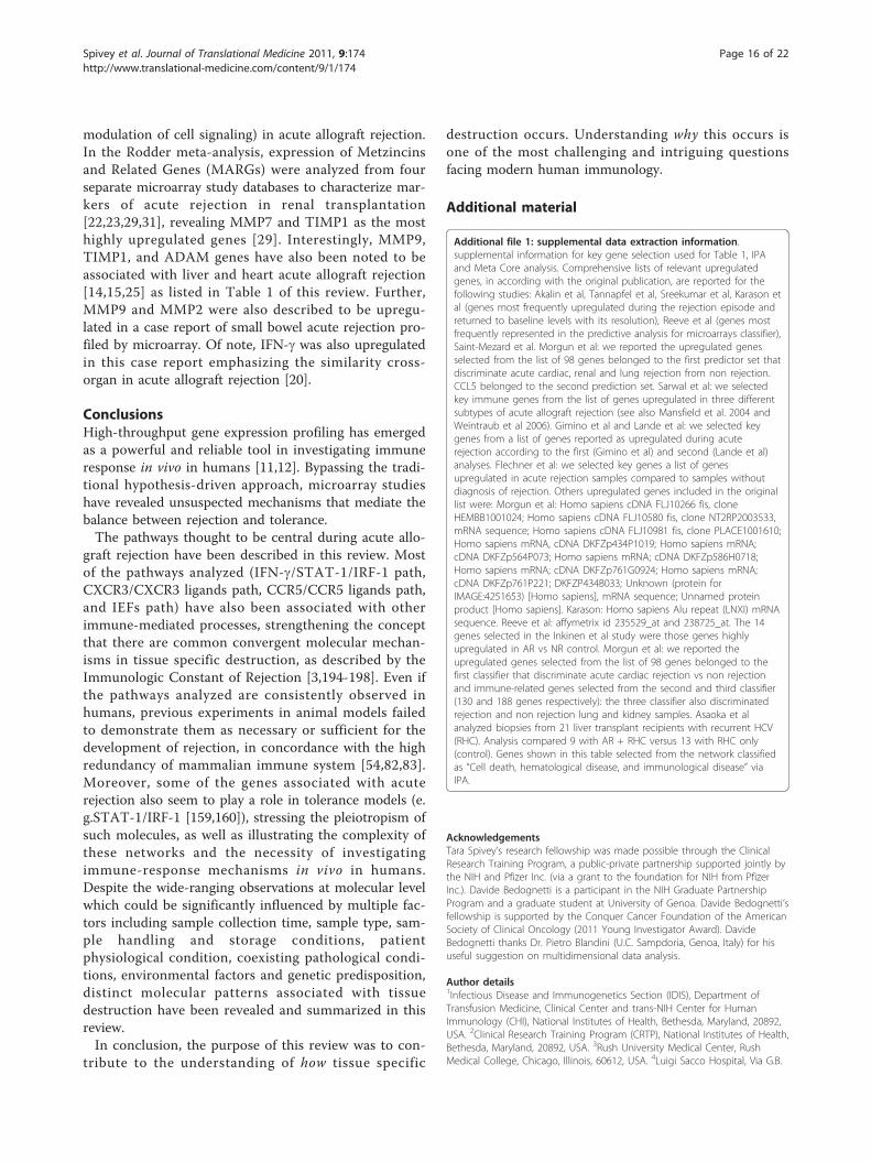
Additional file 1: supplemental data extraction information.supplemental information for key gene selection used for Table 1, IPAand Meta Core analysis. Comprehensive lists of relevant upregulatedgenes, in according with the original publication, are reported for thefollowing studies: Akalin et al, Tannapfel et al, Sreekumar et al, Karason etal (genes most frequently upregulated during the rejection episode andreturned to baseline levels with its resolution), Reeve et al (genes mostfrequently represented in the predictive analysis for microarrays classifier),Saint-Mezard et al. Morgun et al: we reported the upregulated genesselected from the list of 98 genes belonged to the first predictor set thatdiscriminate acute cardiac, renal and lung rejection from non rejection.CCL5 belonged to the second prediction set. Sarwal et al: we selectedkey immune genes from the list of genes upregulated in three differentsubtypes of acute allograft rejection (see also Mansfield et al. 2004 andWeintraub et al 2006). Gimino et al and Lande et al: we selected keygenes from a list of genes reported as upregulated during acuterejection according to the first (Gimino et al) and second (Lande et al)analyses. Flechner et al: we selected key genes a list of genesupregulated in acute rejection samples compared to samples withoutdiagnosis of rejection. Others upregulated genes included in the originallist were: Morgun et al: Homo sapiens cDNA FLJ10266 fis, cloneHEMBB1001024; Homo sapiens cDNA FLJ10580 fis, clone NT2RP2003533,mRNA sequence; Homo sapiens cDNA FLJ10981 fis, clone PLACE1001610;Homo sapiens mRNA, cDNA DKFZp434P1019; Homo sapiens mRNA;cDNA DKFZp564P073; Homo sapiens mRNA; cDNA DKFZp586H0718;Homo sapiens mRNA; cDNA DKFZp761G0924; Homo sapiens mRNA;cDNA DKFZp761P221; DKFZP434B033; Unknown (protein forIMAGE:4251653) [Homo sapiens], mRNA sequence; Unnamed proteinproduct [Homo sapiens]. Karason: Homo sapiens Alu repeat (LNXI) mRNAsequence. Reeve et al: affymetrix id 235529_at and 238725_at. The 14genes selected in the Inkinen et al study were those genes highlyupregulated in AR vs NR control. Morgun et al: we reported theupregulated genes selected from the list of 98 genes belonged to thefirst classifier that discriminate acute cardiac rejection vs non rejectionand immune-related genes selected from the second and third classifier(130 and 188 genes respectively): the three classifier also discriminatedrejection and non rejection lung and kidney samples. Asaoka et alanalyzed biopsies from 21 liver transplant recipients with recurrent HCV(RHC). Analysis compared 9 with AR + RHC versus 13 with RHC only(control). Genes shown in this table selected from the network classifiedas “Cell death, hematological disease, and immunological disease” viaIPA.
AcknowledgementsTara Spivey’s research fellowship was made possible through the ClinicalResearch Training Program, a public-private partnership supported jointly bythe NIH and Pfizer Inc. (via a grant to the foundation for NIH from PfizerInc.). Davide Bedognetti is a participant in the NIH Graduate PartnershipProgram and a graduate student at University of Genoa. Davide Bedognetti’sfellowship is supported by the Conquer Cancer Foundation of the AmericanSociety of Clinical Oncology (2011 Young Investigator Award). DavideBedognetti thanks Dr. Pietro Blandini (U.C. Sampdoria, Genoa, Italy) for hisuseful suggestion on multidimensional data analysis.
Author details1Infectious Disease and Immunogenetics Section (IDIS), Department ofTransfusion Medicine, Clinical Center and trans-NIH Center for HumanImmunology (CHI), National Institutes of Health, Bethesda, Maryland, 20892,USA. 2Clinical Research Training Program (CRTP), National Institutes of Health,Bethesda, Maryland, 20892, USA. 3Rush University Medical Center, RushMedical College, Chicago, Illinois, 60612, USA. 4Luigi Sacco Hospital, Via G.B.
Spivey et al. Journal of Translational Medicine 2011, 9:174http://www.translational-medicine.com/content/9/1/174
Page 16 of 22

Grassi, 20157 Milano, Italy. 5Department of Internal Medicine (DiMI),University of Genoa, Viale Benedetto XV, 6, 16132 Genoa, Italy. 6Center ofExcellence for Biomedical Research (CEBR), University of Genoa, VialeBenedetto XV, 6, 16132 Genoa, Italy. 7Laboratory of Molecular Pharmacology,Center for Cancer Research, National Cancer Institute, National Institutes ofHealth, Bethesda Maryland, 20892, USA. 8University of Sassari, Department ofDrug Sciences, Via Muroni 23 A, 07100, Sassari, Italy. 9Department ofOncology, Biology, and Genetics, University of Genoa, and National CancerResearch Institute, Largo Rosanna Benzi 10, 16132 Genoa, Italy.
Authors’ contributionsDB and TLS performed data extraction from the available literature andprepared the manuscript collaboratively with input and review by all co-authors. FMM and EW reviewed data extraction and literature research andrevised the manuscript for important intellectual contents. All authors readand approved the final manuscript.
Competing interestsThe authors declare that they have no competing interests.
Received: 4 May 2011 Accepted: 12 October 2011Published: 12 October 2011
References1. Panelli MC, Stashower ME, Slade HB, Smith K, Norwood C, Abati A, Fetsch P,
Filie A, Walters SA, Astry C, et al: Sequential gene profiling of basal cellcarcinomas treated with imiquimod in a placebo-controlled studydefines the requirements for tissue rejection. Genome Biology 2007, 8.
2. Wang E, Miller LD, Ohnmacht GA, Mocellin S, Perez-Diaz A, Petersen D,Zhao YD, Simon R, Powell JI, Asaki E, et al: Prospective molecular profilingof melanoma metastases suggests classifiers of immune responsiveness.Cancer Research 2002, 62:3581-3586.
3. Wang E, Worschech A, Marincola FM: The immunologic constant ofrejection. Trends in Immunology 2008, 29:256-262.
4. Bedognetti D, Wang E, Sertoli MR, Marincola FM: Gene-expression profilingin vaccine therapy and immunotherapy for cancer. Expert Review ofVaccines 2010, 9:555-565.
5. Imanguli MM, Swaim WD, League SC, Gress RE, Pavletic SZ, Hakim FT:Increased T-bet+ cytotoxic effectors and type I interferon-mediatedprocesses in chronic graft-versus-host disease of the oral mucosa. Blood2009, 113:3620-3630.
6. He XS, Ji X, Hale MB, Cheung R, Ahmed A, Guo Y, Nolan GP, Pfeffer LM,Wright TL, Risch N, et al: Global transcriptional response to interferon is adeterminant of HCV treatment outcome and is modified by race.Hepatology 2006, 44:352-359.
7. Feld JJ, Nanda S, Huang Y, Chen W, Cam M, Pusek SN, Schweigler LM,Theodore D, Zacks SL, Liang TJ, Fried MW: Hepatic gene expressionduring treatment with peginterferon and ribavirin: Identifying molecularpathways for treatment response. Hepatology 2007, 46:1548-1563.
8. Okamoto Y, Folco EJ, Minami M, Wara AK, Feinberg MW, Sukhova GK,Colvin RA, Kihara S, Funahashi T, Luster AD, Libby P: Adiponectin inhibitsthe production of CXC receptor 3 chemokine ligands in macrophagesand reduces T-lymphocyte recruitment in atherogenesis. Circ Res 2008,102:218-225.
9. Costa C, Rufino R, Traves SL, Lapa ESJR, Barnes PJ, Donnelly LE: CXCR3 andCCR5 chemokines in induced sputum from patients with COPD. Chest2008, 133:26-33.
10. Kim MJ, Romero R, Kim CJ, Tarca AL, Chhauy S, LaJeunesse C, Lee DC,Draghici S, Gotsch F, Kusanovic JP, et al: Villitis of Unknown Etiology IsAssociated with a Distinct Pattern of Chemokine Up-Regulation in theFeto-Maternal and Placental Compartments: Implications for ConjointMaternal Allograft Rejection and Maternal Anti-Fetal Graft-versus-HostDisease. Journal of Immunology 2009, 182:3919-3927.
11. Patterson TA, Lobenhofer EK, Fulmer-Smentek SB, Collins PJ, Chu TM,Bao W, Fang H, Kawasaki ES, Hager J, Tikhonova IR, et al: Performancecomparison of one-color and two-color platforms within the MicroArrayQuality Control (MAQC) project. Nat Biotechnol 2006, 24:1140-1150.
12. Shi L, Reid LH, Jones WD, Shippy R, Warrington JA, Baker SC, Collins PJ, deLongueville F, Kawasaki ES, Lee KY, et al: The MicroArray Quality Control(MAQC) project shows inter- and intraplatform reproducibility of geneexpression measurements. Nat Biotechnol 2006, 24:1151-1161.
13. Eikmans M, Roos-van Groningen MC, Sijpkens YWJ, Ehrchen J, Roth J,Baelde HJ, Bajema IM, de Fijter JW, de Heer E, Bruijn JA: Expression ofsurfactant protein-C, S100A8, S100A9, and B cell markers in renalallografts: Investigation of the prognostic value. Journal of the AmericanSociety of Nephrology 2005, 16:3771-3786.
14. Tannapfel A, Geissler F, Witzigmann H, Hauss J, Wittekind C: Analysis ofliver allograft rejection related genes using cDNA-microarrays in liverallograft specimen. Transplantation Proceedings 2001, 33:3283-3284.
15. Sreekumar R, Rasmussen DL, Wiesner RH, Charlton MR: Differential allograftgene expression in acute cellular rejection and recurrence of hepatitis Cafter liver transplantation. Liver Transplantation 2002, 8:814-821.
16. Inkinen K, Lahesmaa R, Brandt A, Katajamaa M, Halme L, Hockerstedt K,Lautenschlager I: DNA microarray-based gene expression profiles ofcytomegalovirus infection and acute rejection in liver transplants.Transplantation Proceedings 2005, 37:1227-1229.
17. Gimino VJ, Lande JD, Berryman TR, King RA, Hertz MI: Gene expressionprofiling of bronchoalveolar lavage cells in acute lung rejection.American Journal of Respiratory and Critical Care Medicine 2003,168:1237-1242.
18. Patil J, Lande JD, Li N, Berryman TR, King RA, Hertz MI: Bronchoalveolarlavage cell gene expression in acute lung rejection: Development of adiagnostic classifier. Transplantation 2008, 85:224-231.
19. Karason K, Jernas M, Hagg DA, Svensson PA: Evaluation of CXCL9 andCXCL10 as circulating biomarkers of human cardiac allograft rejection.BMC CardiovascDisord 2006, 6:29.
20. Bradley BA: Prognostic assays for rejection and tolerance in organtransplantation. Transplant Immunology 2005, 14:193-201.
21. Akalin E, Hendrix RC, Polavarapu RG, Pearson TC, Neylan JF, Larsen CP,Lakkis FG: Gene expression analysis in human renal allograft biopsysamples using high-density oligoarray technology. Transplantation 2001,72:948-953.
22. Sarwal M, Chua MS, Kambham N, Hsieh SC, Satterwhite T, Masek M,Salvatierra O: Molecular heterogeneity in acute renal allograft rejectionidentified by DNA microarray profiling. New England Journal of Medicine2003, 349:125-138.
23. Flechner SM, Kurian SM, Head SR, Sharp SM, Whisenant TC, Zhang J,Chismar JD, Horvath S, Mondala T, Gilmartin T, et al: Kidney transplantrejection and tissue injury by gene profiling of biopsies and peripheralblood lymphocytes. American Journal of Transplantation 2004, 4:1475-1489.
24. Reeve J, Einecke G, Mengel M, Sis B, Kayser N, Kaplan B, Halloran PF:Diagnosing Rejection in Renal Transplants: A Comparison of Molecular-and Histopathology-Based Approaches. American Journal ofTransplantation 2009, 9:1802-1810.
25. Morgun A, Shulzhenko N, Perez-Diez A, Diniz VZ, Sanson GF, Almeida DR,Matzinger P, Gerbase-DeLima M: Molecular profiling improves diagnosesof rejection and infection in transplanted organs. Circulation Research2006, 98:E74-E83.
26. Saint-Mezard P, Berthier CC, Zhang H, Hertig A, Kaiser S, Schumacher M,Wieczorek G, Bigaud M, Kehren J, Rondeau E, et al: Analysis ofindependent microarray datasets of renal biopsies identifies a robusttranscript signature of acute allograft rejection. Transplant International2009, 22:293-302.
27. Chen R, Sigdel TK, Li L, Kambham N, Dudley JT, Hsieh SC, Klassen RB,Chen A, Caohuu T, Morgan AA, et al: Differentially expressed RNA frompublic microarray data identifies serum protein biomarkers for cross-organ transplant rejection and other conditions. PLoS Comput Biol 2010,6.
28. Asaoka T, Kato T, Marubashi S, Dono K, Hama N, Takahashi H, Kobayashi S,Takeda Y, Takemasa I, Nagano H, et al: Differential transcriptome patternsfor acute cellular rejection in recipients with recurrent hepatitis C afterliver transplantation. Liver Transpl 2009, 15:1738-1749.
29. Rodder S, Scherer A, Korner M, Eisenberger U, Hertig A, Raulf F, Rondeau E,Marti HP: Meta-analyses qualify metzincins and related genes as acuterejection markers in renal transplant patients. Am J Transplant 2010,10:286-297.
30. Lande JD, Patil J, Li N, Berryman TR, King RA, Hertz MI: Novel insights intolung transplant rejection by microarray analysis. Proc Am Thorac Soc2007, 4:44-51.
31. Mueller TF, Einecke G, Reeve J, Sis B, Mengel M, Jhangri GS, Bunnag S,Cruz J, Wishart D, Meng C, et al: Microarray analysis of rejection in human
Spivey et al. Journal of Translational Medicine 2011, 9:174http://www.translational-medicine.com/content/9/1/174
Page 17 of 22

kidney transplants using pathogenesis-based transcript sets. AmericanJournal of Transplantation 2007, 7:2712-2722.
32. Bunnag S, Einecket G, Reeve J, Jhangri GS, Mueller TF, Sis B, Hidalgo LG,Mengel M, Kayser D, Kaplan B, Halloran PF: Molecular Correlates of RenalFunction in Kidney Transplant Biopsies. Journal of the American Society ofNephrology 2009, 20:1149-1160.
33. Perco P, Kainz A, Wilflingseder J, Soleiman A, Mayer B, Oberbauer R:Histogenomics: Association of Gene Expression Patterns WithHistological Parameters in Kidney Biopsies. Transplantation 2009,87:290-295.
34. Sis B, Jhangri GS, Bunnag S, Allanach K, Kaplan B, Halloran PF: EndothelialGene Expression in Kidney Transplants with Alloantibody IndicatesAntibody-Mediated Damage Despite Lack of C4d Staining. AmericanJournal of Transplantation 2009, 9:2312-2323.
35. Sis B, Halloran PF: Endothelial transcripts uncover a previously unknownphenotype: C4d-negative antibody-mediated rejection. Current Opinion inOrgan Transplantation 2010, 15:42-48.
36. Mengel M, Reeve J, Bunnag S, Einecke G, Jhangri GS, Sis B, Famulski K,Guembes-Hidalgo L, Halloran PF: Scoring Total Inflammation Is Superiorto the Current Banff Inflammation Score in Predicting Outcome and theDegree of Molecular Disturbance in Renal Allografts. American Journal ofTransplantation 2009, 9:1859-1867.
37. Einecke G, Mengel M, Hidalgo L, Allanach K, Famulski KS, Halloran PF: TheEarly Course of Kidney Allograft Rejection: Defining the Time WhenRejection Begins. American Journal of Transplantation 2009, 9:483-493.
38. Einecke G, Reeve J, Mengel M, Sis B, Bunnag S, Mueller TF, Halloran PF:Expression of B cell and immunoglobulin transcripts is a feature ofinflammation in late allografts. American Journal of Transplantation 2008,8:1434-1443.
39. Hidalgo LG, Sis B, Sellares J, Campbell PM, Mengel M, Einecke G, Chang J,Halloran PF: NK cell transcripts and NK cells in kidney biopsies frompatients with donor-specific antibodies: evidence for NK cellinvolvement in antibody-mediated rejection. Am J Transplant 2010,10:1812-1822.
40. Hidalgo LG, Einecke G, Allanach K, Mengel M, Sis B, Mueller TF, Halloran PF:The transcriptome of human cytotoxic T cells: Measuring the burden ofCTL-Associated transcripts in human kidney transplants. American Journalof Transplantation 2008, 8:637-646.
41. Mengel M, Chang J, Kayser D, Gwinner W, Schwarz A, Einecke G,Broecker V, Famulski K, De Freitas DG, Guembes-Hidalgo L, et al: TheMolecular Phenotype of 6-Week Protocol Biopsies from Human RenalAllografts: Reflections of Prior Injury but Not Future Course. Am JTransplant 2010.
42. Mengel M, Sis B, Kim D, Chang J, Famulski KS, Hidalgo LG, Einecke G, deFreitas DG, Tymchak W, Burton J, Halloran PF: The molecular phenotype ofheart transplant biopsies: relationship to histopathological and clinicalvariables. Am J Transplant 2010, 10:2105-2115.
43. Famulski KS, Einecke G, Sis B, Mengel M, Hidalgo LG, Kaplan B, Halloran PF:Defining the canonical form of T-cell-mediated rejection in humankidney transplants. Am J Transplant 2010, 10:810-820.
44. Einecke G, Kayser D, Vanslambrouck JM, Sis B, Reeve J, Mengel M,Famulski KS, Bailey CG, Rasko JE, Halloran PF: Loss of solute carriers in Tcell-mediated rejection in mouse and human kidneys: an activeepithelial injury-repair response. Am J Transplant 2010, 10:2241-2251.
45. Einecke G, Reeve J, Sis B, Mengel M, Hidalgo L, Famulski KS, Matas A,Kasiske B, Kaplan B, Halloran PF: A molecular classifier for predictingfuture graft loss in late kidney transplant biopsies. J Clin Invest 2010,120:1862-1872.
46. Mengel M, Allanach K, Einecke G, Sis B, Hidalgo L, Mueller T, Halloran PF:Comparing microarray versus RT-PCR assessment of renal allograftbiopsies: Similar performance despite different dynamic ranges.American Journal of Transplantation 2008, 8:349-349.
47. Pham MX, Teuteberg JJ, Kfoury AG, Starling RC, Deng MC, Cappola TP,Kao A, Anderson AS, Cotts WG, Ewald GA, et al: Gene-expression profilingfor rejection surveillance after cardiac transplantation. N Engl J Med 2010,362:1890-1900.
48. Schaub S, Nickerson P, Rush D, Mayr M, Hess C, Golian M, Stefura W,HayGlass K: Urinary CXCL9 and CXCL10 Levels Correlate with the Extentof Subclinical Tubulitis. American Journal of Transplantation 2009,9:1347-1353.
49. Hauser IA, Spiegler S, Kiss E, Gauer S, Sichler O, Scheuermann EH,Ackermann H, Pfeilschifter JM, Geiger H, Grone HJ, Radeke HH: Predictionof acute renal allograft rejection by urinary monokine induced by IFN-gamma (MIG). Journal of the American Society of Nephrology 2005,16:1849-1858.
50. Hu HZ, Aizenstein BD, Puchalski A, Burmania JA, Hamawy MM, Knechtle SJ:Elevation of CXCR3-binding chemokines in urine indicates acute renal-allograft dysfunction. American Journal of Transplantation 2004, 4:432-437.
51. Tatapudi RR, Muthukumar T, Dadhania D, Ding R, Li BG, Sharma VK, Lozada-Pastorio E, Seetharamu N, Hartono C, Serur D, et al: Noninvasive detectionof renal allograft inflammation by measurements of mRNA for IP-10 andCXCR3 in urine. Kidney International 2004, 65:2390-2397.
52. Hidalgo LG, Urmson J, Ramassar V, Halloran PF: IFN-gamma directlymodulates lymphocyte cytotoxicity. Faseb Journal 2002, 16:A1086-A1086.
53. Hidalgo LG, Urmson J, Halloran PF: IFN-gamma decreases CTL generationby limiting IL-2 production: A feedback loop controlling effector cellproduction. American Journal of Transplantation 2005, 5:651-661.
54. Hidalgo LG, Halloran PF: Role of IFN-gamma in allograft rejection. CriticalReviews in Immunology 2002, 22:317-349.
55. Schoenborn JR, Wilson CB: Regulation of interferon-gamma during innateand adaptive immune responses. Advances in Immunology 2007,96:41-101.
56. Kirk AD, Bollinger RR, Finn OJ: Rapid, Comprehensive Analysis of HumanCytokine Messenger-Rna and Its Application to the Study of AcuteRenal-Allograft Rejection. Human Immunology 1995, 43:113-128.
57. Moudgil A, Bagga A, Toyoda M, Nicolaidou E, Jordan SC, Ross D:Expression of gamma-IFN mRNA in bronchoalveolar lavage fluidcorrelates with early acute allograft rejection in lung transplantrecipients. Clinical Transplantation 1999, 13:201-207.
58. Nast CC, Zuo XJ, Prehn J, Danovitch GM, Wilkinson A, Jordan SC: Gamma-Interferon Gene-Expression in Human Renal-Allograft Fine-NeedleAspirates. Transplantation 1994, 57:498-502.
59. Lande JD, Gimino VJ, Hertz MI, King RA: Characterization of lung allograftrejection using microarray gene expression profiling. American Journal ofHuman Genetics 2002, 71:392-392.
60. Pos Z, Selleri S, Spivey TL, Wang JK, Liu H, Worschech A, Sabatino M,Monaco A, Leitman SF, Falus A, et al: Genomic scale analysis of racialimpact on response to IFN-alpha. Proceedings of the National Academy ofSciences of the United States of America 2010, 107:803-808.
61. de Veer MJ, Holko M, Frevel M, Walker E, Der S, Paranjape JM, Silverman RH,Williams BRG: Functional classification of interferon-stimulated genesidentified using microarrays. Journal of Leukocyte Biology 2001, 69:912-920.
62. Reeve J, Einecke G, Kaplan B, Halloran PF: A microarray-based classifier fordiagnosing rejection in kidney transplants: The top diagnostic genes forrejection are interferon-gamma induced. American Journal ofTransplantation 2008, 8:246-246.
63. Ramassar V, Urmson J, Hobart M, Goes N, Halloran PF: Class IItransactivator (CIITA) mediates the in vivo induction of class II in mousetissues. Faseb Journal 1996, 10:2262-2262.
64. Goes N, Hobart M, Ramassar V, Halloran PF: Role of IFN-gamma and IRF-1in injury-induced and basal MHC class I and II expression. Faseb Journal1996, 10:2184-2184.
65. Zhu JF, Paul WE: CD4 T cells: fates, functions, and faults. Blood 2008,112:1557-1569.
66. Ramsey SA, Klemm SL, Zak DE, Kennedy KA, Thorsson V, Li B, Gilchrist M,Gold ES, Johnson CD, Litvak V, et al: Uncovering a macrophagetranscriptional program by integrating evidence from motif scanningand expression dynamics. Plos Computational Biology 2008, 4.
67. Yeruva S, Ramadori G, Raddatz D: NF-kappa B-dependent synergisticregulation of CXCL10 gene expression by IL-1 beta and IFN-gamma inhuman intestinal epithelial cell lines. International Journal of ColorectalDisease 2008, 23:305-317.
68. Liu JG, Guan XQ, Ma XJ: Interferon regulatory factor 1 is an essential anddirect transcriptional activator for interferon gamma-induced RANTES/CC15 expression in macrophages. Journal of Biological Chemistry 2005,280:24347-24355.
69. Zhao DXM, Hu YY, Miller GG, Luster AD, Mitchell RN, Libby P: Differentialexpression of the IFN-gamma-inducible CXCR3-binding chemokines, IFN-inducible protein 10, monokine induced by IFN, and IFN-inducible T cellalpha chemoattractant in human cardiac allografts: Association with
Spivey et al. Journal of Translational Medicine 2011, 9:174http://www.translational-medicine.com/content/9/1/174
Page 18 of 22
cardiac allograft vasculopathy and acute rejection. Journal of Immunology2002, 169:1556-1560.
70. Fahmy NM, Yamani MH, Starling RC, Ratliff NB, Young JB, McCarthy PM,Feng JY, Novick AC, Fairchild RL: Chemokine and chemokine receptorgene expression indicates acute rejection of human cardiac transplants.Transplantation 2003, 75:72-78.
71. Belperio JA, Keane MP, Burdick MD, Lynch JP, Zisman DA, Xue YY, Li KW,Ardehali A, Ross DJ, Strieter RM: Role of CXCL9/CXCR3 chemokine biologyduring pathogenesis of acute lung allograft rejection. Journal ofImmunology 2003, 171:4844-4852.
72. Melter M, Exeni A, Reinders MEJ, Fang JC, McMahon G, Ganz P,Hancock WW, Briscoe DM: Expression of the chemokine receptor CXCR3and its ligand IP-10 during human cardiac allograft rejection. Circulation2001, 104:2558-2564.
73. Fischereder M, Luckow B, Hocher B, Wuthrich RP, Rothenpieler U,Schneeberger H, Panzer U, Stahl RAK, Hauser IA, Budde K, et al: CCchemokine receptor 5 and renal-transplant survival. Lancet 2001,357:1758-1761.
74. Heidenhain C, Pulhl G, Moench C, Lautem A, Neuhaus P: Chemokinereceptor 5 Delta 32 mutation reduces the risk of acute rejection in livertransplantation. Annals of Transplantation 2009, 14:36-44.
75. Hancock WW, Lu B, Gao W, Csizmadia V, Faia K, King JA, Smiley ST, Ling M,Gerard NP, Gerard C: Requirement of the chemokine receptor CXCR3 foracute allograft rejection. Journal of Experimental Medicine 2000,192:1515-1519.
76. Hancock WW, Gao W, Csizmadia V, Faia KL, Shemmeri N, Luster AD: Donor-derived IP-10 initiates development of acute allograft rejection. Journalof Experimental Medicine 2001, 193:975-980.
77. Hancock WW, Wang LQ, Ye QR, Han RX, Lee I: Chemokines and theirreceptors as markers of allograft rejection and targets forimmunosuppression. Current Opinion in Immunology 2003, 15:479-486.
78. Gao W, Faia KL, Csizmadia V, Smiley ST, Soler D, King JA, Danoff TM,Hancock WW: Beneficial effects of targeting CCR5 in allograft recipients.Transplantation 2001, 72:1199-1205.
79. Gao P, Zhou XY, Yashiro-Ohtani Y, Yang YF, Sugimoto N, Ono S,Nakanishi T, Obika S, Imanishi T, Egawa T, et al: The unique targetspecificity of a nonpeptide chemokine receptor antagonist: selectiveblockade of two Th1 chemokine receptors CCR5 and CXCR3. Journal ofLeukocyte Biology 2003, 73:273-280.
80. Halloran PF, Fairchild RL: The puzzling role of CXCR3 and its ligands inorgan allograft rejection. American Journal of Transplantation 2008,8:1578-1579.
81. Haskova Z, Izawa A, Contreras AG, Flynn E, Boulday G, Briscoe DM: Organ-specific differences in the function of MCP-1 and CXCR3 during cardiacand skin allograft rejection. Transplantation 2007, 83:1595-1601.
82. Kwun J, Hazinedaroglu SM, Schadde E, Kayaoglu HA, Fechner J, Hu HZ,Roenneburg D, Torrealba J, Shiao L, Hong X, et al: Unaltered graft survivaland intragraft lymphocytes infiltration in the cardiac allograft of Cxcr3(-/-) mouse recipients. American Journal of Transplantation 2008,8:1593-1603.
83. Zerwes HG, Li J, Kovarik J, Streiff M, Hofmann M, Roth L, Luyten M, Pally C,Loewe RP, Wieczorek G, et al: The chemokine receptor Cxcr3 is notessential for acute cardiac allograft rejection in mice and rats. AmericanJournal of Transplantation 2008, 8:1604-1613.
84. Morris MA, Ley K: Trafficking of natural killer cells. Curr Mol Med 2004,4:431-438.
85. Murphy PM, Baggiolini M, Charo IF, Hebert CA, Horuk R, Matsushima K,Miller LH, Oppenheim JJ, Power CA: International union of pharmacology.XXII. Nomenclature for chemokine receptors. Pharmacological Reviews2000, 52:145-176.
86. Moser B, Wolf M, Walz A, Loetscher P: Chemokines: multiple levels ofleukocyte migration control. Trends in Immunology 2004, 25:75-84.
87. Deola S, Panelli MC, Maric D, Selleri S, Dmitrieva NI, Voss CY, Klein H,Stroncek D, Wang E, Marincola FM: Helper B cells promote cytotoxic T cellsurvival and proliferation independently of antigen presentationthrough CD27/CD70 interactions. Journal of Immunology 2008,180:1362-1372.
88. Hoffmann U, Sergerer S, Rummele P, Kruger B, Pietrzyk M, Hofstadter F,Banas B, Kramer BK: Expression of the chemokine receptor CXCR3 inhuman renal allografts-a prospective study. Nephrology DialysisTransplantation 2006, 21:1373-1381.
89. Xanthou G, Duchesnes CE, Williams TJ, Pease JE: CCR3 functionalresponses are regulated by both CXCR3 and its ligands CXCL9, CXCL10and CXCL11. Eur J Immunol 2003, 33:2241-2250.
90. Halloran PF: T cell-mediated rejection of kidney transplants: a personalviewpoint. Am J Transplant 2010, 10:1126-1134.
91. Qin SX, Rottman JB, Myers P, Kassam N, Weinblatt M, Loetscher M, Koch AE,Moser B, Mackay CR: The chemokine receptors CXCR3 and CCR5 marksubsets of T cells associated with certain inflammatory reactions. Journalof Clinical Investigation 1998, 101:746-754.
92. Jones D, Benjamin RJ, Shahsafaei A, Dorfman DM: The chemokine receptorCXCR3 is expressed in a subset of B-cell lymphomas and is a marker ofB-cell chronic lymphocytic leukemia. Blood 2000, 95:627-632.
93. Drayton DL, Liao S, Mounzer RH, Ruddle NH: Lymphoid organdevelopment: from ontogeny to neogenesis. Nat Immunol 2006,7:344-353.
94. Krensky AM, Clayberger C: Biology and clinical relevance of granulysin.Tissue Antigens 2009, 73:193-198.
95. Obara H, Nagasaki K, Hsieh CL, Ogura Y, Esquivel CO, Martinez OM,Krams SM: IFN-gamma, produced by NK cells that infiltrate liverallografts early after transplantation, links the innate and adaptiveimmune responses. Am J Transplant 2005, 5:2094-2103.
96. Kondo T, Morita K, Watarai Y, Auerbach MB, Taub DD, Novick AC, Toma H,Fairchild RL: Early increased chemokine expression and production inmurine allogeneic skin grafts is mediated by natural killer cells.Transplantation 2000, 69:969-977.
97. Larosa DF, Rahman AH, Turka LA: The innate immune system in allograftrejection and tolerance. Journal of Immunology 2007, 178:7503-7509.
98. Kitchens WH, Uehara S, Chase CM, Colvin RB, Russell PS, Madsen JC: Thechanging role of natural killer cells in solid organ rejection andtolerance. Transplantation 2006, 81:811-817.
99. Bingaman AW, Ha J, Waitze SY, Durham MM, Cho HR, Tucker-Burden C,Hendrix R, Cowan SR, Pearson TC, Larsen CP: Vigorous allograft rejectionin the absence of danger. Journal of Immunology 2000, 164:3065-3071.
100. Worschech A, Chen NH, Yu YA, Zhang Q, Pos Z, Weibel S, Raab V,Sabatino M, Monaco A, Liu H, et al: Systemic treatment of xenografts withvaccinia virus GLV-1h68 reveals the immunologic facet of oncolytictherapy. Bmc Genomics 2009, 10.
101. Andersen CB, Ladefoged SD, Larsen S: Acute Kidney Graft-Rejection - AMorphological and Immunohistological Study on Zero-Hour and Follow-Up Biopsies with Special Emphasis on Cellular Infiltrates and AdhesionMolecules. Apmis 1994, 102:23-37.
102. Meehan SM, McCluskey RT, Pascual M, Preffer FI, Anderson P,Schlossman SF, Colvin RB: Cytotoxicity and apoptosis in human renalallografts: Identification, distribution, and quantitation of cells with acytotoxic granule protein GMP-17 (TIA-1) and cells with fragmentednuclear DNA. Laboratory Investigation 1997, 76:639-649.
103. Sagoo P, Perucha E, Sawitzki B, Tomiuk S, Stephens DA, Miqueu P,Chapman S, Craciun L, Sergeant R, Brouard S, et al: Development of across-platform biomarker signature to detect renal transplant tolerancein humans. J Clin Invest 2010, 120:1848-1861.
104. Cassese G, Lindenau S, de Boer B, Arce S, Hauser A, Riemekasten G, Berek C,Hiepe F, Krenn V, Radbruch A, Manz RA: Inflamed kidneys of NZB/W miceare a major site for the homeostasis of plasma cells. European Journal ofImmunology 2001, 31:2726-2732.
105. Magalhaes R, Stiehl P, Morawietz L, Berek C, Krenn V: Morphological andmolecular pathology of the B cell response in synovitis of rheumatoidarthritis. Virchows Archiv 2002, 441:415-427.
106. Platt JL, Lebien TW, Michael AF: Interstitial Mononuclear Cell-Populationsin Renal Graft-Rejection - Identification by Monoclonal-Antibodies inTissue-Sections. Journal of Experimental Medicine 1982, 155:17-30.
107. Zarkhin V, Sarwal MM: Microarrays: Monitoring for Transplant Toleranceand Mechanistic Insights. Clinics in Laboratory Medicine 2008, 28:385-+.
108. Racusen LC, Colvin RB, Solez K, Mihatsch MJ, Halloran PF, Campbell PM,Cecka MJ, Cosyns JP, Demetris AJ, Fishbein MC, et al: Antibody-mediatedrejection criteria - an addition to the Banff ‘97 Classification of RenalAllograft Rejection. American Journal of Transplantation 2003, 3:708-714.
109. Alausa M, Almagro U, Siddiqi N, Zuiderweg R, Medipalli R, Hariharan S:Refractory acute kidney transplant rejection with CD20 graft infiltratesand successful therapy with rituximab. Clinical Transplantation 2005,19:137-140.
Spivey et al. Journal of Translational Medicine 2011, 9:174http://www.translational-medicine.com/content/9/1/174
Page 19 of 22

110. Bagnasco SM, Tsai W, Rahman MH, Kraus ES, Barisoni L, Vega R, Racusen LC,Haas M, Mohammed BS, Zachary AA, Montgomery RA: CD20-positiveinfiltrates in renal allograft biopsies with acute cellular rejection are notassociated with worse graft survival. American Journal of Transplantation2007, 7:1968-1973.
111. Doria C, di Francesco F, Ramirez CB, Frank A, Iaria M, Francos G, Marino IR,Farber JL: The presence of B-cell nodules does not necessarily portend aless favorable outcome to therapy in patients with acute cellularrejection of a renal allograft. Transplantation Proceedings 2006,38:3441-3444.
112. Hippen BE, DeMattos A, Cook WJ, Kew CE, Gaston RS: Association of CD20+infiltrates with poorer clinical outcomes in acute cellular rejection ofrenal allografts. American Journal of Transplantation 2005, 5:2248-2252.
113. Kayler LK, Lakkis FG, Morgan C, Basu A, Blisard D, Tan HP, Shapiro R,Randhawa PS: Acute cellular rejection with CD20-Positive lymphoidclusters in kidney transplant patients following lymphocyte depletion.American Journal of Transplantation 2007, 7:357-357.
114. Krukemeyer MG, Moeller J, Morawietz L, Rudolph B, Neumann U,Theruvath T, Neuhaus P, Krenn V: Description of B lymphocytes andplasma cells, complement, and chemokines/receptors in acute liverallograft rejection. Transplantation 2004, 78:65-70.
115. Tsai EW, Rianthavorn P, Gjertson DW, Wallace WD, Reed EF, Ettenger RB:CD20+lymphocytes in renal allografts are associated with poor graftsurvival in pediatric patients. Transplantation 2006, 82:1769-1773.
116. Mengel M, Bogers J, Bosmans JL, Seron D, Moreso F, Carrera M, Gwinner W,Schwarz A, De Broe M, Kreipe H, Haller H: Incidence of C4d stain inprotocol biopsies from renal allografts: Results from a multicenter trial.American Journal of Transplantation 2005, 5:1050-1056.
117. Martins HL, Silva C, Martini D, Noronha IL: Detection of B lymphocytes(CD20(+)) in renal allograft biopsy specimens. Transplantation Proceedings2007, 39:432-434.
118. Zarkhin V, Kambham N, Li L, Kwok S, Hsieh SC, Salvatierra O, Sarwal MM:Characterization of intra-graft B cells during renal allograft rejection.Kidney International 2008, 74:664-673.
119. Noorchashm H, Reed AJ, Rostami SY, Mozaffari R, Zekavat G, Koeberlein B,Caton AJ, Naji A: B cell-mediated antigen presentation is required for thepathogenesis of acute cardiac allograft rejection. Journal of Immunology2006, 177:7715-7722.
120. Schultz KR, Klarnet JP, Gieni RS, Hayglass KT, Greenberg PD: The Role of B-Cells for Invivo T-Cell Responses to A Friend Virus-Induced Leukemia.Science 1990, 249:921-923.
121. Yang X, Brunham RC: Gene knockout B cell-deficient mice demonstratethat B cells play an important role in the initiation of T cell responses toChlamydia trachomatis (mouse pneumonitis) lung infection. Journal ofImmunology 1998, 161:1439-1446.
122. Cross AH, Stark JL, Lauber J, Ramsbottom MJ, Lyons JA: Rituximab reducesB cells and T cells in cerebrospinal fluid of multiple sclerosis patients.Journal of Neuroimmunology 2006, 180:63-70.
123. Ascierto M, Kmieciak M, Idowu M, Manjili R, Zhao Y, Nikizad H, Grimes M,Dumur C, Wang E, Wang X, et al: A signature of immune function genesassociated with recurrence-free survival in breast cancer patients. BreastCancer Resarch and Treatment 2011.
124. Zarkhin V, Chalasani G, Sarwal MM: The yin and yang of B cells in graftrejection and tolerance. Transplant Rev (Orlando) 2010, 24:67-78.
125. Tyden G, Genberg H, Tollemar J, Ekberg H, Persson NH, Tufveson G,Wadstrom J, Gabel M, Mjornstedt L: A randomized, doubleblind, placebo-controlled, study of single-dose rituximab as induction in renaltransplantation. Transplantation 2009, 87:1325-1329.
126. Clatworthy MR, Watson CJ, Plotnek G, Bardsley V, Chaudhry AN, Bradley JA,Smith KG: B-cell-depleting induction therapy and acute cellular rejection.N Engl J Med 2009, 360:2683-2685.
127. Bedognetti D, Zoppoli G, Massucco C, Zanardi E, Zupo S, Bruzzone A,Sertoli MR, Balleari E, Racchi O, Messina M, et al: Impaired response toinfluenza vaccine associated with persistent memory B cell depletion innon-Hodgkin’s lymphoma patients treated with rituximab-containingregimens. J Immunol 2011, 186:6044-55.
128. Newell KA, Asare A, Kirk AD, Gisler TD, Bourcier K, Suthanthiran M,Burlingham WJ, Marks WH, Sanz I, Lechler RI, et al: Identification of a B cellsignature associated with renal transplant tolerance in humans. J ClinInvest 2010, 120:1836-1847.
129. Louis S, Braudeau C, Giral M, Dupont A, Moizant F, Robillard N, Moreau A,Soulillou JP, Brouard S: Contrasting CD25hiCD4+T cells/FOXP3 patterns inchronic rejection and operational drug-free tolerance. Transplantation2006, 81:398-407.
130. Pallier A, Hillion S, Danger R, Giral M, Racape M, Degauque N, Dugast E,Ashton-Chess J, Pettre S, Lozano JJ, et al: Patients with drug-free long-term graft function display increased numbers of peripheral B cells witha memory and inhibitory phenotype. Kidney Int 2010, 78:503-513.
131. Sharma R, Jarjour WN, Zheng L, Gaskin F, Fu SM, Ju ST: Large functionalrepertoire of regulatory T-cell suppressible autoimmune T cells in scurfymice. J Autoimmun 2007, 29:10-19.
132. Wildin RS, Freitas A: IPEX and FOXP3: clinical and research perspectives. JAutoimmun 2005, , 25 Suppl: 56-62.
133. Lee WS, Park S, Lee WY, Yun SH, Chun HK: Clinical impact of tumor-infiltrating lymphocytes for survival in stage II colon cancer. Cancer 2010,116:5188-5199.
134. Correale P, Rotundo MS, Del Vecchio MT, Remondo C, Migali C,Ginanneschi C, Tsang KY, Licchetta A, Mannucci S, Loiacono L, et al:Regulatory (FoxP3+) T-cell tumor infiltration is a favorable prognosticfactor in advanced colon cancer patients undergoing chemo orchemoimmunotherapy. J Immunother 2010, 33:435-441.
135. Muthukumar T, Dadhania D, Ding R, Snopkowski C, Naqvi R, Lee JB,Hartono C, Li B, Sharma VK, Seshan SV, et al: Messenger RNA for FOXP3 inthe urine of renal-allograft recipients. N Engl J Med 2005, 353:2342-2351.
136. Bunnag S, Allanach K, Jhangri GS, Sis B, Einecke G, Mengel M, Mueller TF,Halloran PF: FOXP3 expression in human kidney transplant biopsies isassociated with rejection and time post transplant but not withfavorable outcomes. American Journal of Transplantation 2008, 8:1423-1433.
137. Matzinger P: The danger model: A renewed sense of self. Science 2002,296:301-305.
138. Vabulas RM, Ahmad-Nejad P, Ghose S, Kirschning CJ, Issels RD, Wagner H:HSP70 as endogenous stimulus of the toll/interleukin-1 receptor signalpathway. Journal of Biological Chemistry 2002, 277:15107-15112.
139. Platanias LC: Mechanisms of type-I- and type-II-interferon-mediatedsignalling. Nature Reviews Immunology 2005, 5:375-386.
140. Reich NC, Liu L: Tracking STAT nuclear traffic. Nature Reviews Immunology2006, 6:602-612.
141. Muller-Ladner U, Alten R, Heiligenhaus A, Kekow J, Koletzko S, Mrowietz U,Ochsenkuhn T, Radke M, Reich K, Rudwaleit M, Schreiber S: “TRECID”, TNFalpha related chronic inflammatory diseases - A new multiple diseasesbridging concept. Deutsche Medizinische Wochenschrift 2009,134:2132-2136.
142. Connolly MK, Bedrosian AS, Clair JMS, Mitchell AP, Ibrahim J, Stroud A,Pachter HL, Bar-Sagi D, Frey AB, Miller G: In liver fibrosis, dendritic cellsgovern hepatic inflammation in mice via TNF-alpha. Journal of ClinicalInvestigation 2009, 119:3213-3225.
143. Le Moine A, Goldman M, Abramowicz D: Multiple pathways to allograftrejection. Transplantation 2002, 73:1373-1381.
144. Bonizzi G, Karin M: The two NF-kappa B activation pathways and theirrole in innate and adaptive immunity. Trends in Immunology 2004,25:280-288.
145. Akira S, Uematsu S, Takeuchi O: Pathogen recognition and innateimmunity. Cell 2006, 124:783-801.
146. Iwasaki A, Medzhitov R: Toll-like receptor control of the adaptive immuneresponses. Nature Immunology 2004, 5:987-995.
147. Brent L: The 50th anniversary of the discovery of immunologic tolerance.New England Journal of Medicine 2003, 349:1381-1383.
148. Brouard S, Mansfield E, Braud C, Li L, Giral M, Hsieh SC, Baeten D, Zhang M,Ashton-Chess J, Braudeau C, et al: Identification of a peripheral bloodtranscriptional biomarker panel associated with operational renalallograft tolerance. Proceedings of the National Academy of Sciences of theUnited States of America 2007, 104:15448-15453.
149. Lin T, Zhou WD, Sacks SH: The role of complement and Toll-like receptorsin organ transplantation. Transplant International 2007, 20:481-489.
150. Sacks S, Lee Q, Wong W, Zhou W: The role of complement in regulatingthe alloresponse. Current Opinion in Organ Transplantation 2009, 14:10-15.
151. Sacks SH, Chowdhury P, Zhou WD: Role of the complement system inrejection. Current Opinion in Immunology 2003, 15:487-492.
152. Lee JI, Burckart GJ: Nuclear factor kappa B: Important transcription factorand therapeutic target. Journal of Clinical Pharmacology 1998, 38:981-993.
Spivey et al. Journal of Translational Medicine 2011, 9:174http://www.translational-medicine.com/content/9/1/174
Page 20 of 22

153. Schreiber J, Jenner RG, Murray HL, Gerber GK, Gifford DK, Young RA:Coordinated binding of NF-kappa B family members in the response ofhuman cells to lipopolysaccharide. Proceedings of the National Academy ofSciences of the United States of America 2006, 103:5899-5904.
154. Peng Q, Li K, Patel H, Sacks SH, Zhou WD: Dendritic cell synthesis of C3 isrequired for full T cell activation and development of a Th1 phenotype.Journal of Immunology 2006, 176:3330-3341.
155. Kawamoto S, Yalcindag A, Laouini D, Brodeur S, Bryce P, Lu B, Humbles AA,Oettgen H, Gerard C, Geha RS: The anaphylatoxin C3a downregulates theTh2 response to epicutaneously introduced antigen. Journal of ClinicalInvestigation 2004, 114:399-407.
156. Carroll MC: The complement system in B cell regulation. MolecularImmunology 2004, 41:141-146.
157. Pratt JR, Basheer SA, Sacks SH: Local synthesis of complement componentC3 regulates acute renal transplant rejection. Nature Medicine 2002,8:582-587.
158. Li K, Patel H, Farrar CA, Hargreaves REG, Sacks SH, Zhou WD: Complementactivation regulates the capacity of proximal tubular epithelial cell tostimulate alloreactive T cell response. Journal of the American Society ofNephrology 2004, 15:2414-2422.
159. Cordoba SP, Wang CM, Williams R, Li J, Smit L, Sharland A, Allen R,McCaughan G, Bishop A: Gene array analysis of a rat model of livertransplant tolerance identifies increased complement C3 and the STAT-1/IRF-1 pathway during tolerance induction. Liver Transplantation 2006,12:636-643.
160. Fujino M, Kitazawa Y, Kawasaki M, Funeshima N, Kimura H, Nakajima T,Saito H, Li XK: Differences in lymphocyte gene expression betweentolerant and syngeneic liver grafted rats. Liver Transplantation 2004,10:379-391.
161. Pan TL, Wang PW, Huang CC, Goto S, Chen CL: Expression, by functionalproteomics, of spontaneous tolerance in rat orthotopic livertransplantation. Immunology 2004, 113:57-64.
162. Naesens M, Li L, Ying LH, Sansanwal P, Sigdel TK, Hsieh SC, Kambham N,Lerut E, Salvatierra O, Butte AJ, Sarwal MM: Expression of ComplementComponents Differs Between Kidney Allografts from Living andDeceased Donors. Journal of the American Society of Nephrology 2009,20:1839-1851.
163. Moore KW, Malefyt RD, Coffman RL, O’Garra A: Interleukin-10 and theinterleukin-10 receptor. Annual Review of Immunology 2001, 19:683-765.
164. Mocellin S, Panelli MC, Wang E, Nagorsen D, Marincola FM: The dual roleof IL-10. Trends in Immunology 2003, 24:36-43.
165. Mocellin S, Panelli M, Wang E, Rossi CR, Pilati P, Nitti D, Lise M,Marincola FM: IL-10 stimulatory effects on human NK cells explored bygene profile analysis. Genes and Immunity 2004, 5:621-630.
166. Mocellin S, Marincola FM, Young HA: Interleukin-10 and the immuneresponse against cancer: a counterpoint. Journal of Leukocyte Biology2005, 78:1043-1051.
167. Tao M, Li BR, Nayini J, Sivaraman S, Song SW, Larson A, Toofanfard M,Chen H, Venugopal P, Preisler HD: In vivo effects of IL-4, IL-10, andamifostine on cytokine production in patients with acute myelogenousleukemia. Leukemia & Lymphoma 2001, 41:161-168.
168. Lauw FN, Pajkrt D, Hack CE, Kurimoto M, van Deventer SJH, van der Poll T:Proinflammatory effects of IL-10 during human endotoxemia. Journal ofImmunology 2000, 165:2783-2789.
169. Del Vecchio M, Mortarini R, Canova S, Di Guardo L, Pimpinelli N, Sertoli MR,Bedognetti D, Queirolo P, Morosini P, Perrone T, et al: Bevacizumab plusfotemustine as first-line treatment in metastatic melanoma patients:clinical activity and modulation of angiogenesis and lymphangiogenesisfactors. Clin Cancer Res 2010, 16:5862-5872.
170. Qian SG, Li W, Li YP, Fu FM, Lu LN, Fung JJ, Thomson AW: Systemicadministration of cellular interleukin-10 can exacerbate cardiac allograftrejection in mice. Transplantation 1996, 62:1709-1714.
171. Li W, Lu L, Li Y, Fu F, Fung JJ, Thomson AW, Qian S: High-dose cellular IL-10 exacerbates rejection and reverses effects of cyclosporine andtacrolimus in mouse cardiac transplantation. Transplantation Proceedings1997, 29:1081-1082.
172. Li W, Fu F, Lu L, Narula SK, Fung JJ, Thomson AW, Qian S: Systemicadministration of anti-interleukin-10 antibody prolongs organ allograftsurvival in normal and presensitized recipients. Transplantation 1998,66:1587-1596.
173. Karczewski J, Karczewski M, Wiktorowicz K, Glyda M: Cytometric Analysis ofTh1/Th2 Cytokines in the Urine of Patients Prior to the KidenyTransplantation. Transplant International 2009, 22:145-145.
174. Grinyo J, Vanrenterghem Y, Nashan B, Vincenti F, Ekberg H, Lindpaintner K,Rashford M, Nasmyth-Miller C, Voulgari A, Spleiss O, et al: Association offour DNA polymorphisms with acute rejection after kidneytransplantation. Transplant International 2008, 21:879-891.
175. Honda K, Taniguchi T: IRFs: master regulators of signalling by Toll-likereceptors and cytosolic pattern-recognition receptors. Nature ReviewsImmunology 2006, 6:644-658.
176. Vila-del Sol V, Punzon C, Fresno M: IFN-gamma-induced TNF-alphaexpression is regulated by interferon regulatory factors 1 and 8 inmouse macrophages. Journal of Immunology 2008, 181:4461-4470.
177. Penninger JM, Sirard C, Mittrucker HW, Chidgey A, Kozieradzki I, Nghiem M,Hakem A, Kimura T, Timms E, Boyd R, et al: The interferon regulatorytranscription factor IRF-1 controls positive and negative selection of CD8(+) thymocytes. Immunity 1997, 7:243-254.
178. Taki S, Sato T, Ogasawara K, Fukuda T, Sato M, Hida S, Suzuki G,Mitsuyama M, Shin EH, Kojima S, et al: Multistage regulation of Th1-typeimmune responses by the transcription factor IRF-1. Immunity 1997,6:673-679.
179. Ogasawara K, Hida S, Azimi N, Tagaya Y, Sato T, Yokochi-Fukuda T,Waldmann TA, Taniguchi T, Taki S: Requirement for IRF-1 in themicroenvironment supporting development of natural killer cells (vol391, pg 700, 1998). Nature 1998, 392:843-843.
180. Hama N, Yanagisawa Y, Dono K, Kobayashi S, Marubashi S, Nagano H,Umeshita K, Watanabe S, Uchiyama Y, Monden M: Gene ExpressionProfiling of Acute Cellular Rejection in Rat Liver Transplantation UsingDNA Microarrays. Liver Transplantation 2009, 15:509-521.
181. Stegall M, Park W, Kim D, Kremers W: Gene expression during acuteallograft rejection: Novel statistical analysis of microarray data. AmericanJournal of Transplantation 2002, 2:913-925.
182. Erickson LM, Pan F, Ebbs A, Kobayashi M, Jiang HS: Microarray-based geneexpression profiles of allograft rejection and immunosuppression in therat heart transplantation model. Transplantation 2003, 76:582-588.
183. Kroger A, Koster M, Schroeder K, Hauser H, Mueller PP: Activities of IRF-1.Journal of Interferon and Cytokine Research 2002, 22:5-14.
184. Jin P, Wang E, Provenzano M, Stroncekl D, Marincoial FM: Gene expressionsignatures of interleukin-2 in vivo and in vitro and their relation toanticancer therapy. Critical Reviews in Immunology 2007, 27:437-448.
185. Matsuyama T, Kimura T, Kitagawa M, Pfeffer K, Kawakami T, Watanabe N,Kundig TM, Amakawa R, Kishihara K, Wakeham A, et al: TargetedDisruption of Irf-1 Or Irf-2 Results in Abnormal Type-I Ifn Gene Inductionand Aberrant Lymphocyte Development. Cell 1993, 75:83-97.
186. Erickson L, Crews G, Pan F, Fisniku O, Jang MS, Wynn C, Kobayashi M,Jiang HS: Unique gene expression profiles of heart allograft rejection inthe interferon regulatory factor-1-deficient mouse. TransplantImmunology 2004, 13:169-175.
187. Mansfield ES, Sarwal MM: Arraying the orchestration of allograftpathology. American Journal of Transplantation 2004, 4:853-862.
188. Bonadies N, Neururer C, Steege A, Vallabhapurapu S, Pabst T, Mueller BU:PU.1 is regulated by NF-kappaB through a novel binding site in a 17 kbupstream enhancer element. Oncogene 2010, 29:1062-1072.
189. Finn PW, Stone JR, Boothby MR, Perkins DL: Inhibition of NF-kappa B-dependent T cell activation abrogates acute allograft rejection. Journal ofImmunology 2001, 167:5994-6001.
190. Basil CF, Zhao YD, Zavaglia K, Jin P, Panelli MC, Voiculescu S,Mandruzzato S, Lee HM, Seliger B, Freedman RS, et al: Common cancerbiomarkers. Cancer Research 2006, 66:2953-2961.
191. Tiao MM, Lu L, Tao R, Wang L, Fung JJ, Qian SG: Prolongation of cardiacallograft survival by systemic administration of immature recipientdendritic cells deficient in NF-kappa B activity. Annals of Surgery 2005,241:497-505.
192. Ohmori K, Takeda S, Miyoshi S, Minami M, Nakane S, Ohta M, Sawa Y,Matsuda H: Attenuation of lung injury in allograft rejection using NF-kappa B decoy transfection - novel strategy for use in lungtransplantation. European Journal of Cardio-Thoracic Surgery 2005, 27:23-27.
193. Zhou P, Hwang KW, Palucki DA, Guo Z, Boothby M, Newell KA, Alegre ML:Impaired NF-kappa B activation in T cells permits tolerance to primaryheart allografts and to secondary donor skin grafts. American Journal ofTransplantation 2003, 3:139-147.
Spivey et al. Journal of Translational Medicine 2011, 9:174http://www.translational-medicine.com/content/9/1/174
Page 21 of 22
194. Marincola FM, Wang E: Immunologic signatures of rejection. New York,NY: Springer; 2010.
195. Pos Z, Galon J: Predictors of favorable outcome in cancer. In Immunologicsignatures of rejection. Edited by: Marincola FM, Wang E. New York: Springer;2010:199-210.
196. Bedognetti D: Signatures Associated with Acute Rejection: AllograftRejection. In: Immunologic signatures of rejection.Edited by: MarincolaFM, Wang E. New York: Springer; 2010:305-345.
197. Ascierto ML, De Giorgi V, Liu Q, Bedognetti D, Spivey T, Murtas D,Uccellini L, Ayotte BD, Stroncek DF, Chouchane L, et al: An immunologicportrait of cancer. J Transl Med 2011.
198. Castiello L, Sabatino M, Jin P, Clayberger C, Marincola FM, Krensky AM,Stroncek DF: Monocyte-derived DC maturation strategies and relatedpathways: a transcriptional view. Cancer Immunol Immunother 2011,60:457-466.
doi:10.1186/1479-5876-9-174Cite this article as: Spivey et al.: Gene expression profiling in acuteallograft rejection: challenging the immunologic constant of rejectionhypothesis. Journal of Translational Medicine 2011 9:174.
Submit your next manuscript to BioMed Centraland take full advantage of:
• Convenient online submission
• Thorough peer review
• No space constraints or color figure charges
• Immediate publication on acceptance
• Inclusion in PubMed, CAS, Scopus and Google Scholar
• Research which is freely available for redistribution
Submit your manuscript at www.biomedcentral.com/submit
Spivey et al. Journal of Translational Medicine 2011, 9:174http://www.translational-medicine.com/content/9/1/174
Page 22 of 22