Soil organism influence on ecosystem-level processes – bypassing the ecological hierarchy?
Upload
khangminh22Category
view
5download
0
Hussainzada et al. BMC Pharmacology and Toxicology 2014, 15:15http://www.biomedcentral.com/2050-6511/15/15
RESEARCH ARTICLE Open Access
Whole adult organism transcriptional profiling ofacute metal exposures in male ZebrafishNaissan Hussainzada1, John A Lewis2, Christine E Baer4, Danielle L Ippolito5, David A Jackson3
and Jonathan D Stallings2,6*
Abstract
Background: A convergence of technological breakthroughs in the past decade has facilitated the development ofrapid screening tools for biomarkers of toxicant exposure and effect. Platforms using the whole adult organism toevaluate the genome-wide response to toxicants are especially attractive. Recent work demonstrates the feasibilityof this approach in vertebrates using the experimentally robust zebrafish model. In the present study, we evaluatedgene expression changes in whole adult male zebrafish following an acute 24 hr high dose exposure to threemetals with known human health risks. Male adult zebrafish were exposed to nickel chloride, cobalt chloride orsodium dichromate concentrations corresponding to their respective 96 hr LC20, LC40 and LC60. Histopathologywas performed on a subset of metal-exposed zebrafish to phenotypically anchor transcriptional changes associatedwith each metal.
Results: Comparative analysis identified subsets of differentially expressed transcripts both overlapping and uniqueto each metal. Application of gene ontology (GO) and transcription factor (TF) enrichment algorithms revealed anumber of key biological processes perturbed by metal poisonings and the master transcriptional regulatorsmediating gene expression changes. Metal poisoning differentially activated biological processes associated withribosome biogenesis, proteosomal degradation, and p53 signaling cascades, while repressing oxygen-generatingpathways associated with amino acid and lipid metabolism. Despite appreciable effects on gene regulation, nickelpoisoning did not induce any morphological alterations in male zebrafish organs and tissues. Histopathologicaleffects of cobalt remained confined to the olfactory system, while chromium targeted the gills, pharynx, andintestinal mucosa. A number of enriched transcription factors mediated the observed gene response to metalpoisoning, including known targets such as p53, HIF1α, and the myc oncogene, and novel regulatory factors such asXBP1, GATA6 and HNF3β.Conclusions: This work uses an experimentally innovative approach to capture global responses to metal poisoningand provides mechanistic insights into metal toxicity.
Keywords: Metals, Toxicity mechanisms, Zebrafish, Whole organism, Nickel, Chromium, Cobalt, Toxicogenomics
BackgroundToxicogenomics is a powerful tool for evaluating toxicityprofiles of known and potentially hazardous compounds.The zebrafish, a classic model for developmental toxicity,has recently proven to be an effective model organism forchemical screening [1,2] and environmental sentinel
* Correspondence: [email protected] Program, US Army Center for Environmental Health Research,Fort Detrick, Frederick, Maryland 21702-5010, USA6Environmental Health Program, US Army Center for Environmental HealthResearch, Fort Detrick, Frederick, Maryland, USAFull list of author information is available at the end of the article
© 2014 Hussainzada et al.; licensee BioMed CeCreative Commons Attribution License (http:/distribution, and reproduction in any mediumDomain Dedication waiver (http://creativecomarticle, unless otherwise stated.
applications, including sewage testing and chemicalhazard detection [3-6]. The low husbandry costs, smallsize, ease of genetic manipulation, and wealth of genomedatabase resources distinguish the zebrafish as a highlypromising model organism for toxicological studies.Responses to toxic insults usually affect multiple organsand tissues, supporting a role for gene profiling in thewhole animal to evaluate toxic responses. Although wholeorganism toxicogenomics has routinely been conducted ininvertebrate models such as the worm Caenorhabditiselegans and the fly Drosophila melanogaster [7-9] andin ecoindicator species such as Daphnia magna and
ntral Ltd. This is an Open Access article distributed under the terms of the/creativecommons.org/licenses/by/2.0), which permits unrestricted use,, provided the original work is properly credited. The Creative Commons Publicmons.org/publicdomain/zero/1.0/) applies to the data made available in this

Hussainzada et al. BMC Pharmacology and Toxicology 2014, 15:15 Page 2 of 15http://www.biomedcentral.com/2050-6511/15/15
Pimpephales promelas [10,11], only one study to datehas evaluated whole organism gene profiling in adultzebrafish [12]. In this study, robust expression signaturesdifferentiated between potent aryl hydrocarbon andestrogen receptor agonists, and accurately identified targettissues. We hypothesized that gene profiling in wholeadult zebrafish could also be used to infer toxic responsesto hazardous chemicals.Nickel, cobalt and chromium are environmentally
ubiquitous metals with recognized human health hazards[13]. Recently, applications in mining, smelting, industry,medicine, and agriculture have increased the environmentaldistribution of these metals, elevating elevated exposurerisk and incidences of occupational exposure. Nickel,cobalt, or chromium exposure can cause incapacitatingacute toxicity and/or long-term damage (e.g., carcinogenesis)[14-16]. Primary mechanisms of metal toxicity includethe production of free radicals which can trigger oxidativestress, induce mutagenesis by DNA-metal interactions, andimpair protein function by covalently modifying proteinsor competing with metal binding sites. Metal-derivedreactive oxygen species (ROS) may perturb a number oftightly regulated cellular processes (e.g., cell growth andproliferation), activate transcription factors and genes, andtrigger cellular adaptive programs including metal stressresponse, DNA repair mechanisms, and inflammation [17].Numerous studies have examined gene responses to
acute poisoning by nickel, cobalt, or chromium [18,19], butbesides a few studies in invertebrates, most of these studiesmeasure gene responses in isolated tissues or tissue-derivedcell lines [8,19-23]. Although analyzing isolated tissues isthe ideal approach to unambiguously identify gene changesin an organ of interest, it is experimentally impractical tomicrodissect and to analyze all potentially affected organsfrom zebrafish individually. Alternatively, a whole-organismapproach with post hoc gene ontology enrichment analysishas the advantage of predicting biomolecular pathwayslinked to observed histopathologic endpoints for informinglater organ-specific experiments. In this study, we used awhole-organism approach exposing adult male zebrafish toincreasing concentrations of nickel chloride, cobaltchloride, and sodium dichromate, and evaluating wholegenome transcriptional responses using DNA microarrays.We identified differentially regulated biological processesusing gene ontology enrichment analysis in order to infertoxicity mechanisms [21]. We also identified transcriptionfactors upstream of the differentially enriched geneswhich are predicted to directly activate or repressgene expression in order to characterize regulatoryprocesses involved in metal toxicity. Histopathologicalchanges in the whole organism were compared withgene changes. Overall, our study provides (i) insightinto transcriptomic changes corresponding to toxicindicators of metal poisoning and (ii) an experimental
evaluation of whole organism toxicogenomics in thezebrafish model.
MethodsResearch was conducted in compliance with the UnitedStates Animal Welfare Act, and other Federal statutesand regulations relating to animals and experimentsinvolving animals and adheres to principles stated in theGuide for the Care and Use of Laboratory Animals(NRC 2011) in facilities that are fully accredited by theAssociation for the Assessment and Accreditation ofLaboratory Animal Care, International. Approvals weregranted for this study by USACEHR’s Institutional AnimalCare and Use Committee.
Water qualityAll husbandry and aquatic exposures are performedusing USACEHR’s well water which is processed to en-sure proper conditioning of the water supply. The wateris supplied from a mix of onsite ground water wells andfrom municipal tap (domestic) water. Domestic water isused to produce “RO permeate” which is later mixedwith raw well water to produce water of appropriatehardness and alkalinity (150 – 210 mg/L as CaCO3;110 – 180 mg/L as CaCO3 respectively). Domesticwater is carbon filtered to remove chlorine levels(maintained below 0.1 mg/L), treated by a water softener,processed through reverse osmosis (RO) membranes thenstored for distribution. Prior to use, the RO processeddomestic water is blended with well water, filteredthrough a 10 μm particle filter, carbon filtered, thenheated and aerated to near 100% saturation at 25 ± 1°C.This processed water is then passed through another 10μm particle filter and UV sterilized prior to distributionthroughout the facility. This water is continuously moni-tored to maintain the following ranges: pH = 6.5 – 8.5;alkalinity = 110 – 180 mg/L CaCO3; hardness = 150 –210 mg/L as CaCO3; conductivity = 400 – 1000 mS/cm;total ammonia < 0.1 mg/L as NH3; dissolved oxygen(DO) = 80 – 100% saturation (6.8 – 8.5 mg/L at 25°C).Contaminant analysis is performed quarterly by ourin-house analytical chemistry department as well asannually by an external, certified testing facility.
Fish exposuresExposures were conducted using USACEHR well waterin 5-gallon glass aquaria adapted for flow-through use(60 mL/min; 5.4 turnovers/day) and maintained at 25°Cwith a 12 hr:12 hr (light:dark) photoperiod. During bothacclimation and exposure periods, water quality for eachtank is monitored daily (temperature, pH, alkalinity,hardness, DO, and conductivity (data not shown). Weestimated the concentration of each metal necessary for20% (LC20; low), 40% (LC40; mid) and 60% lethality
Hussainzada et al. BMC Pharmacology and Toxicology 2014, 15:15 Page 3 of 15http://www.biomedcentral.com/2050-6511/15/15
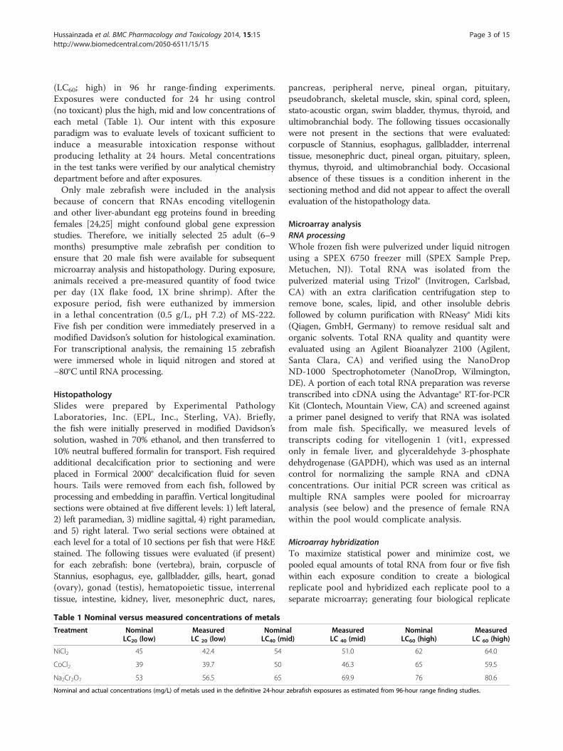
(LC60; high) in 96 hr range-finding experiments.Exposures were conducted for 24 hr using control(no toxicant) plus the high, mid and low concentrations ofeach metal (Table 1). Our intent with this exposureparadigm was to evaluate levels of toxicant sufficient toinduce a measurable intoxication response withoutproducing lethality at 24 hours. Metal concentrationsin the test tanks were verified by our analytical chemistrydepartment before and after exposures.Only male zebrafish were included in the analysis
because of concern that RNAs encoding vitellogeninand other liver-abundant egg proteins found in breedingfemales [24,25] might confound global gene expressionstudies. Therefore, we initially selected 25 adult (6–9months) presumptive male zebrafish per condition toensure that 20 male fish were available for subsequentmicroarray analysis and histopathology. During exposure,animals received a pre-measured quantity of food twiceper day (1X flake food, 1X brine shrimp). After theexposure period, fish were euthanized by immersionin a lethal concentration (0.5 g/L, pH 7.2) of MS-222.Five fish per condition were immediately preserved in amodified Davidson’s solution for histological examination.For transcriptional analysis, the remaining 15 zebrafishwere immersed whole in liquid nitrogen and stored at−80°C until RNA processing.
HistopathologySlides were prepared by Experimental PathologyLaboratories, Inc. (EPL, Inc., Sterling, VA). Briefly,the fish were initially preserved in modified Davidson’ssolution, washed in 70% ethanol, and then transferred to10% neutral buffered formalin for transport. Fish requiredadditional decalcification prior to sectioning and wereplaced in Formical 2000® decalcification fluid for sevenhours. Tails were removed from each fish, followed byprocessing and embedding in paraffin. Vertical longitudinalsections were obtained at five different levels: 1) left lateral,2) left paramedian, 3) midline sagittal, 4) right paramedian,and 5) right lateral. Two serial sections were obtained ateach level for a total of 10 sections per fish that were H&Estained. The following tissues were evaluated (if present)for each zebrafish: bone (vertebra), brain, corpuscle ofStannius, esophagus, eye, gallbladder, gills, heart, gonad(ovary), gonad (testis), hematopoietic tissue, interrenaltissue, intestine, kidney, liver, mesonephric duct, nares,
Table 1 Nominal versus measured concentrations of metals
Treatment NominalLC20 (low)
MeasuredLC 20 (low)
NominLC40 (m
NiCl2 45 42.4 54
CoCl2 39 39.7 50
Na2Cr2O7 53 56.5 65
Nominal and actual concentrations (mg/L) of metals used in the definitive 24-hour
pancreas, peripheral nerve, pineal organ, pituitary,pseudobranch, skeletal muscle, skin, spinal cord, spleen,stato-acoustic organ, swim bladder, thymus, thyroid, andultimobranchial body. The following tissues occasionallywere not present in the sections that were evaluated:corpuscle of Stannius, esophagus, gallbladder, interrenaltissue, mesonephric duct, pineal organ, pituitary, spleen,thymus, thyroid, and ultimobranchial body. Occasionalabsence of these tissues is a condition inherent in thesectioning method and did not appear to affect the overallevaluation of the histopathology data.
Microarray analysisRNA processingWhole frozen fish were pulverized under liquid nitrogenusing a SPEX 6750 freezer mill (SPEX Sample Prep,Metuchen, NJ). Total RNA was isolated from thepulverized material using Trizol® (Invitrogen, Carlsbad,CA) with an extra clarification centrifugation step toremove bone, scales, lipid, and other insoluble debrisfollowed by column purification with RNeasy® Midi kits(Qiagen, GmbH, Germany) to remove residual salt andorganic solvents. Total RNA quality and quantity wereevaluated using an Agilent Bioanalyzer 2100 (Agilent,Santa Clara, CA) and verified using the NanoDropND-1000 Spectrophotometer (NanoDrop, Wilmington,DE). A portion of each total RNA preparation was reversetranscribed into cDNA using the Advantage® RT-for-PCRKit (Clontech, Mountain View, CA) and screened againsta primer panel designed to verify that RNA was isolatedfrom male fish. Specifically, we measured levels oftranscripts coding for vitellogenin 1 (vit1, expressedonly in female liver, and glyceraldehyde 3-phosphatedehydrogenase (GAPDH), which was used as an internalcontrol for normalizing the sample RNA and cDNAconcentrations. Our initial PCR screen was critical asmultiple RNA samples were pooled for microarrayanalysis (see below) and the presence of female RNAwithin the pool would complicate analysis.
Microarray hybridizationTo maximize statistical power and minimize cost, wepooled equal amounts of total RNA from four or five fishwithin each exposure condition to create a biologicalreplicate pool and hybridized each replicate pool to aseparate microarray; generating four biological replicate
alid)
MeasuredLC 40 (mid)
NominalLC60 (high)
MeasuredLC 60 (high)
51.0 62 64.0
46.3 65 59.5
69.9 76 80.6
zebrafish exposures as estimated from 96-hour range finding studies.

Hussainzada et al. BMC Pharmacology and Toxicology 2014, 15:15 Page 4 of 15http://www.biomedcentral.com/2050-6511/15/15
pools per experimental condition for a total of 16 microar-rays per toxicant screened (i.e. four control replicates, fourlow dose replicates, four mid dose replicates and four highdose replicates). Statistical modeling demonstrates thatperforming microarray analysis on four biologicalreplicates comprised of RNA pooled from five samplesapproaches the statistical power attained by analyzing 20individual samples [26]. Numerous theoretical discussionsof the pooling procedure can be found in the literature[27-29]. Although pooling eliminates the ability to assessfish-to-fish variability in gene expression, it does provide astatistically powerful approach to identify clear toxicantresponses, which is the main focus of the current work.The microarrays used in this study were custom
designed in-house using the eArray microarray designtool (https://earray.chem.agilent.com/earray/; AgilentTechnologies, Inc.) and manufactured by Agilent. Eacharray contains 44,000 60-mer oligonucleotides representing21,904 zebrafish gene targets derived from Ensembl build46 (Zv7 genome build) and Vega build 26. Two probeswere designed per transcript wherever possible; only 94target transcripts have only one probe. Probes weredesigned using genes that are annotated, i.e., matched tonamed genes in the published databases, and representgood coverage of the whole zebrafish genome.Microarrays were processed following Agilent’s
One-Color Microarray-Based Gene Expression AnalysisProtocol (Version 5.5, February, 2007) for processing4 x 44 K microarray slides using an initial 1 μg pooledRNA input and an 18 hr overnight hybridization at 65°C.A final step for preventing ozone related degradationof signal using the Stabilization and Drying solution(Agilent Technologies, Inc.) was included after the requiredspecificity washes prior to scanning the arrays. Microarrayslides were scanned with a GenePix Autoloader 4200 ALscanner (Molecular Devices, Union City, CA) and rawimages processed using GenePix Pro 6.0 (MolecularDevices). All microarray data from this study have beendeposited in NCBI’s Gene Expression Omnibus under theaccession number GSE50648.
Statistical analysisRaw microarray data was analyzed with Partek GenomicsSuite software with probe intensities based on the mediansignal intensity of each feature and signal-to-noise ratio(SNR) data imported from GenePix Pro 6.0. GenePix Procalculates SNR as the difference between median spotsignal and median background divided by the standarddeviation of the background signal. Data preprocessingcomprised manual inspection of each extracted genefeature and quality control. We selected only unsaturatedprobes with an SNR greater than or equal to three(SNR ≥ 3) for analysis and performed quantilenormalization across arrays to control for inter-array
variability. Normalized probe intensities were then logtransformed. We also removed probes without Ensemblannotation producing a subset of 15,818 probes whichmapped to 7,909 genes (Additional file 1: Table S1). Weperformed three sets of ANOVAs using Partek GenomicsSuite to identify probes that were differentially expressedbetween each treatment group and its respective control.Each set consisted of data from all the replicate pools offish exposed to a specific metal and the replicate pools ofunexposed fish housed in adjacent tanks during the metalexposure. The ANOVA model included terms fortreatment (unexposed or exposed), concentration(control, low, mid, or high) and in interaction termfor treatment*concentration. Contrasts were performed todetermine significance between each concentration andcontrol. We used a step-up Benjamini and Hochberg falsediscovery rate (FDR) of 0.01 to select differentiallyexpressed probes. An FDR alpha value equal to 0.01 waschosen as the cut-off for the combined datasets of allreplicate pools. Probes not meeting this threshold werefiltered out and the resultant list was submitted to asecond filter specifying a 1.8-fold-difference betweentreated vs. control samples. Only transcripts for whichprobes passed these filters were included in the final list(Additional file 1: Table S1). Fold changes for each probepair (single probe transcripts excluded) were thenaveraged to generate a single value for each transcript.Gene Ontology (GO) enrichment analysis was performed
using the web-based tool GOTree Machine (GOTM;http://genereg.ornl.gov/gotm/), which generates a tree-likestructure to navigate the GO Directed Acyclic Graph forinput gene sets. GOTM supports analysis of the zebrafishgenome; however, this analysis had to be performed at thegene rather than the transcript level. GO term andKEGG pathway (http://bioinfo.vanderbilt.edu/webgestalt/)enrichment analyses were then performed on the secondarylists to determine biological processes that are significantly(FDR = 0.1) enhanced or depressed by each metal. Theenriched GO terms and KEGG pathways were alsomanually annotated into top level biological categoriesto clarify the overarching biological “themes” related tometal-induced gene perturbations. Finally, transcriptionfactor enrichment was performed using MetaCore’salgorithm (GeneGo, Inc.) with settings enabled foridentification of node relationships encompassing onlydirect downstream transcriptional regulation. Zebrafishgenes were mapped to their human homologs using theBiomart feature in Ensembl with genes mapping one-to-many discarded from subsequent analysis. Backgroundreference sets comprised the set of all transcripts withSNR ≥ 3 in each platform that could be subsequentlymapped in a one-to-one fashion to their human homologs.Thresholds were set at an FDR = 0.1 with at least threeDEG target identified for each enriched transcription factor.

Hussainzada et al. BMC Pharmacology and Toxicology 2014, 15:15 Page 5 of 15http://www.biomedcentral.com/2050-6511/15/15
Metacore does not explicitly provide zebrafish transcrip-tion factor regulatory networks; however, we anticipatedthat there would be high degree of conservation betweenthe zebrafish and human networks [30], and mapped thezebrafish DEGs to their human orthologs before performingenrichment. Each differentially expressed transcript wasmapped to its corresponding gene using the Ensembldatabase and resultant gene lists queried against theirappropriate reference gene lists. Background referencesets comprised the set of all transcripts with SNR ≥ 3 ineach exposure condition that could be subsequentlymapped to Ensembl genes. Significantly enriched GO terms,or those GO terms that are statistically over-represented ineach treatment compared to the reference set, weredetermined using the hypergeometric test with p-valuesadjusted using the Benjamini & Hochberg FDR correction(α = 0.1) and setting a threshold for the minimum numberof genes per category (n = 3). While many statistical testshave been used for GO enrichment evaluation, thehypergeometric distribution provides an appropriate methodfor modeling data in which genes can be selected only once,i.e. sampling without replacement, as occurs in GOenrichment analysis [31].
Results and discussionGross changes, behavior, and histopathologyFigure 1 provides a schematic of the experimentalparadigm. During range-finding metal exposure studies,fish were qualitatively assessed for gross changes in behavior
Figure 1 Schematic of experimental design. Diagram of the experiment
and general appearance. At study termination, tissueswere stained with hematoxylin and eosin (H&E) todetermine the morphological changes associated with eachmetal poisoning at the concentrations listed in Table 1.Nickel-exposed fish appeared fuzzy, an appearance which
is generally attributable to the excretion of mucus fromgoblet cells following irritation (personal communication,Dr. Donald K. Nichols). There were no deaths observed atany of the nickel concentrations nor did any of the nickelconcentrations lead to any discernible histopathologicalterations (Table 2) although behavioral differences andskin abnormalities were qualitatively different betweencontrol and treated fish (i.e. sluggish swimming and fuzzyskin appearance). Previous studies indicate that gills, liver,and kidney are histopathological targets of nickel poisoning[22,23,32-34], but differences in fish species, exposure timeand concentration could account for the discrepancy in thehistopathological endpoints.Chromium poisoning caused visible changes in fish
behavior and general appearance, including sluggishswimming, gasping and ulcerations near the tail in somefish. Four zebrafish died at the highest dose. Exposure toall three concentrations of chromium histopathologicallyaffected the gills, intestine, and pharynx (see Table 2 andFigure 2A). Both gill and pharynx epithelium exhibitedmononuclear cell infiltration, which is indicative of acuteinflammation. The most prominent change in intestinewas moderate to moderately severe atrophy of the mucosalfolds and a mild infiltration of the intestinal lamina propria
al design used for rangefinding and exposures.
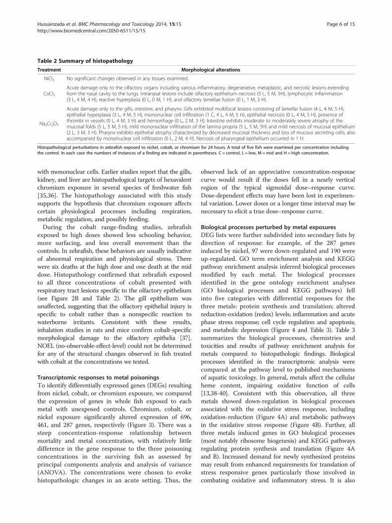
Table 2 Summary of histopathology
Treatment Morphological alterations
NiCl2 No significant changes observed in any tissues examined.
CoCl2Acute damage only to the olfactory organs including various inflammatory, degenerative, metaplastic, and necrotic lesions extendingfrom the nasal cavity to the lungs. Intranasal lesions include olfactory epithelium necrosis (5 L, 5 M, 5H), lymphocytic inflammation(3 L, 4 M, 4 H), reactive hyperplasia (0 L, 0 M, 1 H), and olfactory lamellae fusion (0 L, 1 M, 3 H).
Na2Cr2O7
Acute damage only to the gills, intestine, and pharynx. Gills exhibited multifocal lesions consisting of lamellar fusion (4 L, 4 M, 5 H),epithelial hyperplasia (3 L, 4 M, 5 H), mononuclear cell infiltration (1 C, 4 L, 4 M, 5 H), epithelial necrosis (0 L, 4 M, 5 H), presence ofthrombi in vessels (0 L, 4 M, 3 H) and hemorrhage (0 L, 2 M, 3 H). Intestine exhibits moderate to moderately severe atrophy of themucosal folds (5 L, 5 M, 5 H), mild mononuclear infiltration of the lamina propria (5 L, 5 M, 5H) and mild necrosis of mucosal epithelium(2 L, 3 M, 3 H). Pharynx exhibits epithelial atrophy characterized by decreased mucosal thickness and loss of mucous secreting cells; alsoaccompanied by mononuclear cell infiltration (0 L, 2 M, 4 H). Necrosis of pharyngeal epithelium occurred in 1 H.
Histopathological perturbations in zebrafish exposed to nickel, cobalt, or chromium for 24 hours. A total of five fish were examined per concentration includingthe control. In each case the numbers of instances of a finding are indicated in parentheses. C = control, L = low, M =mid and H = high concentration.
Hussainzada et al. BMC Pharmacology and Toxicology 2014, 15:15 Page 6 of 15http://www.biomedcentral.com/2050-6511/15/15
with mononuclear cells. Earlier studies report that the gills,kidney, and liver are histopathological targets of hexavalentchromium exposure in several species of freshwater fish[35,36]. The histopathology associated with this studysupports the hypothesis that chromium exposure affectscertain physiological processes including respiration,metabolic regulation, and possibly feeding.During the cobalt range-finding studies, zebrafish
exposed to high doses showed less schooling behavior,more surfacing, and less overall movement than thecontrols. In zebrafish, these behaviors are usually indicativeof abnormal respiration and physiological stress. Therewere six deaths at the high dose and one death at the middose. Histopathology confirmed that zebrafish exposedto all three concentrations of cobalt presented withrespiratory tract lesions specific to the olfactory epithelium(see Figure 2B and Table 2). The gill epithelium wasunaffected, suggesting that the olfactory epithelial injury isspecific to cobalt rather than a nonspecific reaction towaterborne irritants. Consistent with these results,inhalation studies in rats and mice confirm cobalt-specificmorphological damage to the olfactory epithelia [37].NOEL (no-observable-effect-level) could not be determinedfor any of the structural changes observed in fish treatedwith cobalt at the concentrations we tested.
Transcriptomic responses to metal poisoningsTo identify differentially expressed genes (DEGs) resultingfrom nickel, cobalt, or chromium exposure, we comparedthe expression of genes in whole fish exposed to eachmetal with unexposed controls. Chromium, cobalt, ornickel exposure significantly altered expression of 696,461, and 287 genes, respectively (Figure 3). There was asteep concentration-response relationship betweenmortality and metal concentration, with relatively littledifference in the gene response to the three poisoningconcentrations in the surviving fish as assessed byprincipal components analysis and analysis of variance(ANOVA). The concentrations were chosen to evokehistopathologic changes in an acute setting. Thus, the
observed lack of an appreciative concentration-responsecurve would result if the doses fell in a nearly verticalregion of the typical sigmoidal dose–response curve.Dose-dependent effects may have been lost in experimen-tal variation. Lower doses or a longer time interval may benecessary to elicit a true dose–response curve.
Biological processes perturbed by metal exposuresDEG lists were further subdivided into secondary lists bydirection of response: for example, of the 287 genesinduced by nickel, 97 were down-regulated and 190 wereup-regulated. GO term enrichment analysis and KEGGpathway enrichment analysis inferred biological processesmodified by each metal. The biological processesidentified in the gene ontology enrichment analyses(GO biological processes and KEGG pathways) fellinto five categories with differential responses for thethree metals: protein synthesis and translation; alteredreduction-oxidation (redox) levels; inflammation and acutephase stress response; cell cycle regulation and apoptosis;and metabolic depression (Figure 4 and Table 3). Table 3summarizes the biological processes, chemistries andtoxicities and results of pathway enrichment analysis formetals compared to histopathologic findings. Biologicalprocesses identified in the transcriptomic analysis werecompared at the pathway level to published mechanismsof aquatic toxicology. In general, metals affect the cellularheme content, impairing oxidative function of cells[13,38-40]. Consistent with this observation, all threemetals showed down-regulation in biological processesassociated with the oxidative stress response, includingoxidation-reduction (Figure 4A) and metabolic pathwaysin the oxidative stress response (Figure 4B). Further, allthree metals induced genes in GO biological processes(most notably ribosome biogenesis) and KEGG pathwaysregulating protein synthesis and translation (Figure 4Aand B). Increased demand for newly synthesized proteinsmay result from enhanced requirements for translation ofstress responsive genes particularly those involved incombating oxidative and inflammatory stress. It is also

Epithelial Cell Necrosis
Inflammation
Cellular Infiltrates
Necrosis
Necrosis
Cellular Debris
Cellular Debris
Nares/olfactory organ-control (4X) bar=100 m
Nares/olfactory organ-high dose CoCl3
(4X) bar=100 m
Olfactory lamellae-control (20X) bar=25 m
Olfactory lamellae-high dose CoCl3
(20X) bar=25 m
Pharynx-control (4X) bar=100 m
Intestine-control (4X) bar=100 m
Gill-control (20X) bar=25 m
Pharynx-high dose Na Cr O (4X) bar=100 m2 2 7
Intestine-high dose Na Cr O (4X) bar=100 m2 2 7
Gill-high dose Na Cr O (20X) bar=25 m2 2 7
A
B
Figure 2 Histopathology of affected tissues. Histopathology of affected tissues after (A) chromium – pharynx, intestine, and gill or (B) cobalt – naresand olfactory lamellae – exposure compared to unexposed controls (left panels). Hematoxylin and eosin staining. Arrows indicate regions withmetal-induced pathology.
Hussainzada et al. BMC Pharmacology and Toxicology 2014, 15:15 Page 7 of 15http://www.biomedcentral.com/2050-6511/15/15

Figure 3 Observed genes altered in expression. Total number ofobserved genes significantly (FDR = 0.01) altered in expression by atleast 1.8 fold in response to chromium (Cr), cobalt (Co), or nickel (Ni)versus untreated controls.
Hussainzada et al. BMC Pharmacology and Toxicology 2014, 15:15 Page 8 of 15http://www.biomedcentral.com/2050-6511/15/15
possible that the enhanced protein synthesis is a compen-satory mechanism to replenish cells lost through apoptosisin tissues specifically targeted by metal exposures. Theliterature is conflicting regarding ribosome biogenesis inresponse to toxicant poisoning [41-46]. The discrepanciesin our study between metals may reflect differences in theseverity of toxic insult among the metals.Nickel poisoning causes oxidative damage to DNA and
inhibits antioxidant defenses [47-49]. Nickel-poisonedfish had less observable histopathology than the othermetals and no histopathology directly attributable tonickel poisoning, but gene expression was more enrichedin processes involving cell cycle regulation and apoptosis(Figure 4A) and protein synthesis and translation than theother metals (Figure 4A and B). The lack of histopatho-logical signs of nickel poisoning at 24 hr may suggest thatour nickel exposure scheme captures mechanisms whichdrive the initial response to metal poisoning (i.e. at 24 hr)prior to histologic alterations.Cobalt poisoning mimics hypoxia, stimulating the
production of reactive oxygen species and increasinglipid peroxides [50]. Consistent with this observation,cobalt showed more up-regulation of genes regulating thebiological processes of the oxidative stress response thanany other metal (Figures 4A and B).Chromium redox cycling and ROS generation induce
DNA damage and activate subsequent repair mechanisms[51,52]. Corroborating these mechanisms, chromium up-regulated p53 and MAPK signaling pathways (Figure 4B)[53]. Genes associated with inflammation and acute phasestress responses were up-regulated more with chromiumthan the other metals (Figure 4A and B). These resultsare consistent with the histopathology observations thatchromium exposure induced the most inflammatory cell
infiltration (Figure 2 and Table 2). The genes in pathwaysassociated with metabolic depression support a molecularmechanism underlying the reduced feeding behavior andintestinal abnormalities observed in chromium exposedfish (Figure 2). Chromium poisoning was associated witha marked down-regulation in genes involved in cellularmetabolism, including lipid and steroid metabolicpathways. These processes included the citrate (TCA)cycle and fatty acid metabolism, which are regulatedby degradation of amino acids into acetyl-CoA andproprionyl-CoA intermediates respectively [54]. Genescontrolling the biological and molecular processescontrolling the processing of pyruvate, a degradationproduct of glucogenic amino acids, also significantlydecreased in fish exposed to chromium (Figure 4A).These results suggest that chromium poisoning, butnot cobalt or nickel, caused significant reductions inmetabolic capacity, particularly for amino acids andprocesses associated with their metabolic by-products.These results are consistent with the observed intestinalmucosa atrophy and mild necrosis (Figure 2A). Chromiumis readily absorbed by the intestinal tissue. Since gutmucosa represents the primary site for whole-body aminoacid metabolism [55,56], mucosal atrophy can significantlydiminish the gut’s amino acid metabolic capacity anddecrease amino acid requirements and use.
Enriched transcription factors in response to metalpoisoningWe performed transcription factor (TF) enrichmentanalysis using Metacore on each secondary enhanced/repressed gene list in an attempt to identify upstreamregulatory networks that mediate metal induced geneexpression linked to GO biological processes (Figure 5,Table 3, and Additional file 1: Table S1). The metals showeda significant discrepancy in the type and direction of theregulation of the transcription factors expressed (Figure 5A).Common to all metals was the up-regulation of the highlyconserved mini-chromosome maintenance 4 (mcm4) genewith DNA helicase activity essential for the inhibition ofeukaryotic genome replication and the origin recognitioncomplex (orc61) which facilitates replication [57-60](Figure 5B, Table 3, and Additional file 1: Table S1).In nickel poisoning, transcripts for the DNA replication
processes genes mcm3, mcm5, and rbb4 were up-regulatedin addition to mcm4 and orc61 (Table 3, Figure 5B, andAdditional file 1: Table S1). Nickel induced transcriptionfactor changes consistent with redox signaling, includingup-regulation of hif1α and xbp1 (Figure 5A and Table 3)and their associated gene targets (Figure 5B and Table 3).These gene products are cellular regulators that senseoxygen status and trigger adaptive cascades to maintainnormoxia [61-63]. It is known that nickel stabilizes Hif-1αby preventing the degradation of the protein either

BNi Co Cr Ni Co CrAribonucleoprotein complex biogenesisribosome biogenesisRNA processingncRNA processingcellular amino acid and derivative metabolic processncRNA metabolic processrRNA processingrRNA metabolic processcellular nitrogen compound metabolic processcellular component biogenesisDNA replication initiationDNA-dependent DNA replicationDNA replicationcellular homeostasiscell redox homeostasisoxidation reductionglutathione metabolic processglutamine family amino acid metabolic processpeptide metabolic processmacromolecule catabolic processmulti-organism processresponse to biotic stimulusresponse to bacteriumresponse to other organismmetabolic processsteroid metabolic processsterol metabolic processlipid metabolic processcellular lipid metabolic processlipid biosynthetic processsteroid biosynthetic processterpenoid metabolic processapocarotenoid metabolic processvitamin A metabolic processfat-soluble vitamin metabolic processretinal metabolic processregulation of hormone levelshormone metabolic processcellular hormone metabolic processhormone biosynthetic processcellular aldehyde metabolic processcellular nitrogen compound catabolic processamine metabolic processamine catabolic processcellular amino acid derivative catabolic processcellular biogenic amine catabolic processcellular biogenic amine metabolic processorganic acid metabolic processorganic acid catabolic processcellular ketone metabolic processoxoacid metabolic processcarboxylic acid metabolic processmonocarboxylic acid metabolic processcarboxylic acid catabolic processcellular amino acid catabolic processcellular aromatic compound metabolic processaromatic amino acid family catabolic processaromatic compound catabolic processaromatic amino acid family metabolic processL-phenylalanine metabolic processL-phenylalanine catabolic processlipid transportlipid localizationresponse to chemical stimulus
O-glycan biosynthesisfructose and mannose metabolismamino sugar & nucleotide sugar metabolismaminoacyl-tRNA biosynthesiscell cyclep53 signaling pathwayMAPK signaling pathwayDNA replicationgap junctioninsulin signaling pathwayECM-receptor interactionarachidonic acid metabolismcytosolic DNA-sensing pathwaymetabolic pathwaysproteasomegalactose metabolismglutathione metabolismmetabolism of xenobiotics by CYP450drug metabolism - CYP450drug metabolism - other enzymessteroid biosynthesisandrogen & estrogen metabolismarginine & proline metabolismtyrosine metabolismglycine, serine & threonine metabolismtryptophan metabolismhistidine metabolismnicotinate & nicotinamide metabolismvaline, leucine & isoleucine degradation-alanine metabolism
phenylalanine metabolismretinol metabolismPPAR signaling pathwayprimary bile acid biosynthesislinoleic acid metabolismfatty acid metabolismbiosynthesis of unsaturated fatty acidsbutanoate metabolismpyruvate metabolismcitrate cycle (TCA cycle)
KEGG pathwayGO Biological Process
>4
>4
log10
p-value
0
up
down
Gene Level
Protein synthesis & translation
Cell cycle regulation & apoptosis
ER stress (UPR response)
Oxidative stress response
Inflammation & acute phase stress response
Lipids
Hormones
Amines
Organic acids
Ketones
Aromatics
Lipid movement
Metabolic depression
Protein synthesis & translation
Cell cycle regulation & apoptosis
Inflammation & acute phase stress response
Oxidative stress response
Metabolic detoxification
Hormones
Amino acids
Lipids
Fatty acids
Metabolic pathways
Figure 4 Enrichment analyses. A – Gene ontology term enrichment analysis for biological processes that are significantly (FDR = 0.1) enrichedfor up-regulated or down-regulated gene sets with at least 3 genes/transcripts present in a category to be considered significant by each metal.NOTE: The yellow highlight indicates that the enriched GO term “metabolic process” contained both up-regulated and down-regulated gene sets.B – KEGG pathway enrichment analysis for biological processes that are significantly (FDR = 0.1) enriched for up-regulated or down-regulatedgene sets with at least 3 genes/transcripts present in a category to be considered significant by each metal. Metals listed on each heat map fromleft to right: nickel (Ni), cobalt (Co) and chromium (Cr) respectively. Color scale is log p-value of enrichment. White is for up-regulated gene setsand blue for down-regulated gene sets.
Hussainzada et al. BMC Pharmacology and Toxicology 2014, 15:15 Page 9 of 15http://www.biomedcentral.com/2050-6511/15/15
through the depletion of ascorbate or by replacingiron in the hydroxylases responsible for Hif-1α deg-radation [64-66], leading to transcriptional activationof downstream targets. In our study, nickel poisoningup-regulated other genes critical for redox sensingand homeostasis, including periredoxin (zgc:110343),thioredoxin (zgc:92903), thioredoxin-like 1 (txnl1), andprotein disulfide isomerase 4 and 5 (pdia4, pdip5)(Additional file 1: Table S1 and Figure 5B). Levels of hif1αdownstream targets (abcf2, pfkfb3, il1b, egln3, hk2) weresignificantly up-regulated. xbp1 expression was enhanced1.52 fold in parallel with robust up-regulation of its genetargets (hspa5, pdia4, dnajb11) (Figure 5A and B). Thexpb1 gene target hspa5 (which encodes Grp78/BiP) alsorepresents a specific and key marker for induction of theunfolded protein response (UPR) [67-69], an adaptive
response that prevents protein aggregation by enhancingexpression of molecular chaperones and diminishingnascent polypeptide flux into the ER [70,71]. Nickel dose-dependently increased expression of hspa5 (Figure 5B).Surprisingly, nickel poisoning did not induce genesinvolved in clearing terminally misfolded proteins(i.e. members of the ER-associated protein degradationmachinery [ERAD] family) (Additional file 1: Table S1 andFigure 5B) [71]. However, nickel poisonings up-regulatedan additional ensemble of ER chaperones (dnaja4,jnajb11, ahsa11, hsp701, hspe1, hsp90b1, hspa41) whichplay a crucial role in ensuring proper protein folding.Taken together, our data suggest that nickel exposuresinduced the UPR via xbp1 transcriptional activation.Cobalt poisoning resulted in up-regulation of only
hnf3β in the Metacore analysis (Figure 5A). Genes

Table 3 Comparison of histopathology, chemistry, toxicity and observed gene response among metals
Metal Predicted Histopathologytarget
GO biological process KEGG pathway Genes References
All (Ni, Co, Cr)Effects on hemecarriers (respiration)
n/aCell cycle regulationand apoptosis
Cell cycle regulation and apoptosis mcm4, gadd45bl, orc6l[57]
NiOxidative damageto DNA
None
Oxidative stress response Oxidative stress response
Periredoxin (zgc:110343), thioredoxin(zgc:92903); txnl1; pdia4; pdia4; pdip5;hif1a; xbp1; abcf2; pfkfb3; il1b; egln3;hk2; hspa5; pdia4, dnajb11, dnaja4,jnajb11, ahsa11, hsp70l, hspe1, hsp90b1,hspa4l
[61–66. 70–91]
Endoplasmic reticulum (ER)stress and UPR (unfoldedprotein response)
n/adnaja4, jnajb11, ahsa11, hsp70l, hspe1,hsp90b1, hspa4l; hspa5; pdia4; dnajb11
[61-69]
Protein synthesis andtranslation (DNAreplication processes)
Protein synthesis and translation mcm4, orc6l; mcm3, mcm5; rbb4[57-60]
CoInducer of hypoxicresponse
Olfactory organs:inflammatory,degenerative,metaplastic,necrotic lesions
Oxidative stress response Oxidative stress responsegss (zgc:101574); gclc; zgc:110010; psmd3,psmd7, psma6b, psma5, psmc3, psmc4,psmd11b, psmd1, psmc6, psme3, psmc1b
[40,72-74]
Inflammation and acutephase stress response
Increased metabolic detoxificationProtein serum amyloid A (zgc:103580 );il1b; c/ebp; atf3
[75-78]
CrOxidizing agent; inducesoxidative stress andreactive oxygen species
Gills: hyperplasia,necrosis, hemorrhage,inflammation
Inflammation and acutephase stress response
Inflammation and acute phasestress response
pcna, nfkbiaa, hamp1, ptgs2a, ptgs2b,ptges; dusp2; dusp5; dusp1; gadd45g;hmox1; socs1; egr2; hpx; il1b; c/ebp; atf3
[52,53,75-78]
Intestine: atrophyof mucosal folds,necrosis, inflammation
Ribosome biogenesis Cell cycle regulation and apoptosis ccne2, rrm2, zgc:77806; pcna; stat3, p53[89-91]
Pharynx: atrophy,mononuclear cellinfiltration
Metabolic depression Metabolic pathwaysg6pca, gys2, fabp1a, abcc2; hnf3 α,hnf3 β, hnf1 β, tcf8, bmal, ppargc1,lxr α, coup-tfi, gata-6
[80-82,84-88]
Summary of gene responses consistent with histopathology and predicted chemistry and toxicity of metals after 24 hr exposure.
Hussainzada
etal.BM
CPharm
acologyand
Toxicology2014,15:15
Page10
of15
http://www.biom
edcentral.com/2050-6511/15/15

Figure 5 Enriched transcription factors and predicted gene expression direction. A- Statistically enriched transcription factors thatmodulate gene expression for both enhanced and repressed gene responses to metal exposures. B - Predicted direction of gene expressionregulation (i.e. gene activation or repression) derived from the MetaCore knowledge base for human orthologs.
Hussainzada et al. BMC Pharmacology and Toxicology 2014, 15:15 Page 11 of 15http://www.biomedcentral.com/2050-6511/15/15
were up-regulated in two predominant biological processes:(1) altered redox levels (e.g., the glutathione synthesis genes[gss (zgc:101574), gclc, and zgc:110010] (Additional file 1:Table S1), and genes which encode multiple structuralcomponents of both the catalytic (20S) and regulatory(19S) ribosomal subunits [psmd3, psmd7, psma6b, psma5,psmc3, psmc4, psmd11b, psmd1, psmc6, psme3, psmc1b])[40,72-74] (Additional file 1: Table S1) and (2) inflamma-tion and acute phase stress response (genes encoding pro-tein serum amyloid A [zgc:103580], the pro-inflammatorycytokine il1b, atf3, c/epb) [75,76] (Table 3, Additional file 1:Table S1, and Figure 5B). The negative regulator atf3dampens the inflammation response by antagonizing thepro-inflammatory factor NF-κB, while c/ebp is robustlyup-regulated by pro-inflammatory signals, including il1b,
and functions as an enhancer of the inflammatoryresponse [57,75-79].The transcription factor analysis in chromium poisoning
is consistent with the chromium-induced metabolicdepression reported in the gene ontology analysis(Figures 4 and 5A). Chromium poisoning caused down-regulation of an entire set of genes encoding importantregulators of energy metabolism, glucose, cholesterol,amino acid, and fatty acid metabolism and transport inmany tissues, including the intestine and liver, includinghnf3α, hnf3β, and hnf1β [80] (Figure 5A). hnf4α is a keytranscriptional target of both hnf3α and gata-6 [81,82]which represents a master transcriptional activator ofenergy metabolism genes in multiple tissue types [83].Orphan nuclear receptors with putative roles in gut

Hussainzada et al. BMC Pharmacology and Toxicology 2014, 15:15 Page 12 of 15http://www.biomedcentral.com/2050-6511/15/15
and liver metabolism of nucleic acids (e.g., tcf8; Figure 5A),carbohydrates and lipids (bmal1, ppargc1; Figure 5A), andsterol and steroid hormones (lxr-α, coup-tfi; Figure 5A)were also down-regulated [84-87] (Figure 5A). Genesincluding g6pca, gys2, fabp1a, and abcc2 (involved inglucose metabolism, lipid metabolism, and canalicularbile acid transport, respectively) (Figure 5B andAdditional file 1: Table S1) [88]. Inflammation and acutephase response genes were, for the most part, up-regulatedby chromium, including pro-inflammatory genes(pcna, nfkbiaa, hamp2, ptgs2a, ptgs2b, ptges) (Figure 5Band Additional file 1: Table S1) and genes encodingmultiple dual-specificity phosphatases which modulateinflammatory MAPK cascades (dusp2, dusp5, dusp1)(Figure 5B and Additional file 1: Table S1). Similar tonickel, chromium also up-regulated il1b, c/ebp, and atf3(Figure 5B and Additional file 1: Table S1). Chromiumalso up-regulated cell cycle regulation and apoptosis genesnot enhanced in the other metals, including genesinvolved in the G1/S and G2/M cell-cycle checkpoints(ccne2, rrm2; Additional file 1: Table S1 and Figure 5B;[89]) and in the RAD6-dependent DNA repair pathway(pcna) [90,91] (Figure 5B). The transcription factorp53 was also up-regulated (Figure 5A), which mediatesexpression of protective genes that repair damagedDNA, power the immune system, arrest the proliferation ofdamaged cells, and induce apoptosis [92], as well as guardsthe cell-cycle checkpoint by inducing apoptosis under con-ditions of excessive oxidative stress and DNA damage [93].The transcriptomic results for chromium in particular are
consistent with the observed intestinal mucosa atrophy andmild necrosis observed in the histopathology (Figure 2A).Chromium is readily absorbed by the intestinal tissue. Sincegut mucosa represents the primary site for whole-bodyamino acid metabolism [55,56], mucosal atrophy cansignificantly diminish the gut’s amino acid metaboliccapacity and decrease amino acid requirements anduse. This hypothesis is supported by the observeddown-regulation of liver-specific genes after chromiumexposure. Robust, differential down-regulation of keymediators of glucose metabolism (g6pca, gys2), lipidmetabolism (fabp1), and canalicular bile acid transport(abcc2) (Figure 5B) suggest metabolic perturbations in theliver and/or gut-liver axis [88]. Taken together, the bio-logical processes, transcription factors and histopathologydata may suggest modulation of intestinal metabolicpathways due to chromium exposure. It is unclearwhether this is a direct consequence of chromiumpoisoning or a secondary consequence of chromium-mediated decreases in feeding over the 24 hr exposureperiod. A previous study in freshwater trout exposed tononlethal hexavalent chromium (1/10 of the 24 hr LC50)reported intestinal atrophy and decreased brush borderenzymatic activities consistent with decreased feeding
behavior [94]. However, since animal care personneldid not observe changes in feeding behavior, it ismore likely that chromium mediates specific perturbationsto the gut microenvironment that ultimately triggermetabolic depression.
LimitationsThe technical approaches used in this work have bothstrengths and weaknesses. In using whole adult organismRNA preparations, we introduce the ability to detecttoxicity responses at the whole organism level. We didobtain gene expression level data that corroborates thehistological observations in some cases (e.g., chromium-induced gut histopathology), but we acknowledge that thedetection of clear organ-specific effects was not possiblewith this approach because of the dilution of expressionsignals for genes that have highly tissue-specific distri-butions. Highly similar gene expression profiles areprobably shared by multiple tissues, making it difficultto differentiate among toxicity signatures for individualtarget organs from the system-wide response.Although we measured tank levels of each respective
metal, we acknowledge that this does not necessarilyindicate bioavailable dose, and that the comparisonacross studies would require internal measurement ofmetal concentration within the organism. The lack of adose–response relationship in histopathology is a significantlimitation to this study. The high doses required to observehistopathologic changes in the acute time interval of thestudy precluded identification of subtle, dose-dependentand tissue-specific responses. It is striking that despiteestablishing an exposure regimen from a 96 hr mortalitycurve, there were few differences in gene responses acrossthe exposure levels, even though we tested gene responses48 hr earlier than the mortality curve end points. This ef-fect could be the result of selecting doses in a nearly verticalregion of the typically sigmoidal dose–response curve withlittle difference between the LC20 and LC60 doses. Lowerdoses or longer time intervals could better delineate the dif-ference in histopathologic response in future experiments.This observation also suggests that range-finding for
mechanistic toxicity studies such as this one shouldnot solely be based on mortality data. Some method ofestablishing dose responsiveness based on gene expressionor other molecular endpoints are necessary as well.Nonetheless, the present study supports the use of
transcriptomics in the intact organism to predict candidategenes associated with toxicity endpoints in response to anexternal chemical insult. Using this approach, we haveidentified novel gene and transcription factor targets thatmediate the response to metal toxicity. Finally, thesenew players provide hypothesis-generating targets forfuture evaluation in classically designed studies of themechanisms of heavy metal toxicity.

Hussainzada et al. BMC Pharmacology and Toxicology 2014, 15:15 Page 13 of 15http://www.biomedcentral.com/2050-6511/15/15
ConclusionWe developed an exposure paradigm for comparing theeffects of various metals with varying toxicity mechanismsof action using whole animal transcriptomics in the zebra-fish vertebrate model. Using this technique, we identifiedchanges in expression of groups of genes consistent withadaptive responses to toxicity induced by nickel, cobalt, orchromium, including acute phase response, cell cycleregulation, apoptosis, and metabolic depression, amongothers. Histopathological evaluations corroborate thetoxicity endpoints derived from gene-level based pathwayanalysis for chromium and cobalt.Many of the genes enriched for biological processes’
gene responses reported in this study are consistent withknown physiological endpoints in there metals. Nickelinduces oxidative damage to DNA and proteins; geneswere up-regulated for biological processes includingprotein synthesis and translation, and cell-cycle regulationand apoptosis. Cobalt induces hypoxia; genes regulatingbiological processes of redox response, protein synthesisand translation, and inflammation and acute phase stressresponse were up-regulated. Chromate is a strongoxidizing agent and damages DNA integrity; biomolecularpathways and genes associated with inflammation andacute phase stress response were up-regulated and genesignatures suggested metabolic depression occurred.Further, a number of novel transcription factors thatmediate gene induction at the transcriptional level inresponse to metal exposures were identified.Enrichment of several functional categories of genes
plausibly involved in a variety of biological responseswas identified using unsupervised gene ontology analysisof metal-specific gene responses. Unique histopathologicalalterations were identified for each metal exposure,consistent with metal-specific toxicity in target organs andtissues. For nickel, we find that our toxicogenomicapproach using whole organism RNA preparations maybe more sensitive for identifying targets of nickel toxicitythan the classic toxicology approach of histopathology.These results suggest that toxicogenomics in the wholeadult zebrafish may provide a robust model for identifyingleading indicators of toxicity and intervention approachesfor exposure to toxic chemicals.Using a transcriptomics approach, we identify a number
of upstream modulators of metal-induced gene expression.A number of these transcription factors have been previ-ously implicated in triggering metal-specific gene responsesto toxicity (p53, Hif1a). Multiple novel mediators of thetoxic response to nickel, cobalt, or chromium inwhole adult zebrafish, including Xbp1, various Hnfmembers, and Gata6 were also identified. These findingsprovide additional mechanistic information on metaltoxicity mechanisms and highlight novel potential pointsof intervention for treatment of metal poisoning.
Additional file
Additional file 1: Final gene set. Final gene set used for analysis – 7,909genes with 3 additional tabs each one containing a list of differentiallyexpressed genes (DEG) per chemical with fold change data. A gene wasconsidered DE if it was significantly (FDR = 0.01) altered in expression by atleast 1.8 fold in response to chromium (Cr), cobalt (Co), or nickel (Ni) versusuntreated controls. Microsoft Excel workbook.
Competing interestsThe authors declare that they have no competing interests.
Authors’ contributionsConceived and designed the experiments: DAJ, JAL. Performed zebrafishexposures, sample processing and microarray analysis: CEB. Analyzed thedata: NH, JAL, DAJ. Wrote the paper: NH, JAL, DAJ, CEB, DLI, JDS. All authorsread and approved the final manuscript.
AcknowledgementsThis research was supported in part by an appointment to the PostgraduateResearch Participation Program at the U.S. Army Center for EnvironmentalHealth Research (USACEHR) administered by the Oak Ridge Institute forScience and Education through an interagency agreement between the U.S.Department of Energy and USACEHR.
DisclaimerOpinions, interpretations, conclusions, and recommendations are those ofthe authors and are not necessarily endorsed by the U.S. Army. Research wasconducted in compliance with the Animal Welfare Act, and other Federalstatutes and regulations relating to animals and experiments involvinganimals and adheres to principles stated in the Guide for the Care and Useof Laboratory Animals (NRC 2011) in facilities that are fully accredited by theAssociation for the Assessment and Accreditation of Laboratory Animal Care,International. The research described herein was sponsored by the U.S. ArmyMedical Research and Materiel Command, Military Operational MedicineResearch Program. Citations of commercial organizations or trade names inthis report do not constitute an official Department of the Armyendorsement or approval of the products or services of these organizations.
Author details1ORISE Postdoctoral Fellow, Ft. Detrick, Frederick, Maryland 21702, USA.2Biomarkers Program, US Army Center for Environmental Health Research,Fort Detrick, Frederick, Maryland 21702-5010, USA. 3Pulmonary HealthProgram, US Army Center for Environmental Health Research, Fort Detrick,Frederick, Maryland 21702-5010, USA. 4Excet Inc., Fort Detrick, Frederick,Maryland 21702-5010, USA. 5ORISE, Fort Detrick, Frederick, Maryland21702-5010, USA. 6Environmental Health Program, US Army Center forEnvironmental Health Research, Fort Detrick, Frederick, Maryland, USA.
Received: 26 September 2013 Accepted: 27 February 2014Published: 10 March 2014
References1. Hill AJ, Teraoka H, Heideman W, Peterson RE: Zebrafish as a model
vertebrate for investigating chemical toxicity. Toxicol Sci 2005, 86(1):6–19.2. Spitsbergen JM, Kent ML: The state of the art of the zebrafish model for
toxicology and toxicologic pathology research–advantages and currentlimitations. Toxicol Pathol 2003, 31(Suppl):62–87.
3. Carvan MJ III, Dalton TP, Stuart GW, Nebert DW: Transgenic zebrafish assentinels for aquatic pollution. Ann N Y Acad Sci 2000, 919:133–147. 133–147.
4. Carvan MJ III, Sonntag DM, Cmar CB, Cook RS, Curran MA, Miller GL:Oxidative stress in zebrafish cells: potential utility of transgenic zebrafishas a deployable sentinel for site hazard ranking. Sci Total Environ 2001,274(1–3):183–196.
5. Amanuma K, Takeda H, Amanuma H, Aoki Y: Transgenic zebrafish fordetecting mutations caused by compounds in aquatic environments.Nat Biotechnol 2000, 18(1):62–65.
6. Ng HBG, Lam SH, Sukardi H, Gong Z: Potential applications of transgenicfish to environmental monitoring and toxicology. In Aquaculture

Hussainzada et al. BMC Pharmacology and Toxicology 2014, 15:15 Page 14 of 15http://www.biomedcentral.com/2050-6511/15/15
Biotechnology. 1st edition. Edited by Fletcher GL, Rise ML. West Sussex:John Wiley & Sons, Ltd; 2012:267–280.
7. Reichert K, Menzel R: Expression profiling of five different xenobioticsusing a Caenorhabditis elegans whole genome microarray.Chemosphere 2005, 61(2):229–237.
8. Ruden DM, Chen L, Possidente D, Possidente B, Rasouli P, Wang L, Lu X,Garfinkel MD, Hirsch HVB, Page GP: Genetical toxicogenomics inDrosophila identifies master-modulatory loci that are regulated bydevelopmental exposure to lead. Neurotoxicology 2009, 30(6):898–914.
9. Neumann NF, Galvez F: DNA microarrays and toxicogenomics:applications for ecotoxicology? Biotechnol Adv 2002, 20(5–6):391–419.
10. Poynton HC, Varshavsky JR, Chang B, Cavigiolio G, Chan S, Holman PS,Loguinov AV, Bauer DJ, Komachi K, Theil EC: Daphnia magnaecotoxicogenomics provides mechanistic insights into metal toxicity.Environ Sci Technol 2007, 41(3):1044–1050.
11. Martyniuk CJ, Houlahan J: Assessing gene network stability and individualvariability in the fathead minnow (Pimephales promelas) transcriptome.Comp Biochem Physiol Part D Genomics Proteomics 2013, 8(4):283–291.
12. Lam SH, Mathavan S, Tong Y, Li H, Karuturi RKM, Wu Y, Vega VB, Liu ET,Gong Z: Zebrafish whole-adult-organism chemogenomics for large-scalepredictive and discovery chemical biology. PLoS Genet 2008,4(7):e1000121.
13. Valko M, Morris H, Cronin MTD: Metals, toxicity and oxidative stress.Curr Med Chem 2005, 12(10):1161–1208.
14. Fu H, Boffetta P: Cancer and occupational exposure to inorganic leadcompounds - a metaanalysis of published data. Occup Environ Med 1995,52(2):73–81.
15. Steenland K, Loomis D, Shy C, Simonsen N: Review of occupational lungcarcinogens. Am J Ind Med 1996, 29(5):474–490.
16. Integrated Risk Information System (IRIS). [http://www.epa.gov/iris/].17. Simmons SO, Fan C-Y, Ramabhadran R: Cellular stress response pathway
system as a sentinel ensemble in toxicological screening. Toxicol Sci 2009,111(2):202–225.
18. Beyersmann D, Hechtenberg S: Cadmium, gene regulation, and cellularsignalling in mammalian cells. Toxicol Appl Pharmacol 1997,144(2):247–261.
19. Salnikow K, Zhitkovich A: Genetic and epigenetic mechanisms in metalcarcinogenesis and cocarcinogenesis: nickel, arsenic, and chromium.Chem Res Toxicol 2007, 21(1):28–44.
20. Janssens TKS, Roelofs D, van Straalen NM: Molecular mechanisms of heavymetal tolerance and evolution in invertebrates. Insect Science 2009, 16(1):3–18.
21. Subramanian A, Tamayo P, Mootha VK, Mukherjee S, Ebert BL, Gillette MA,Paulovich A, Pomeroy SL, Golub TR, Lander ES, Mesirov JP: Gene setenrichment analysis: a knowledge-based approach for interpretinggenome-wide expression profiles. Proc Natl Acad Sci U S A 2005,102(43):15545–15550.
22. Athikesavan S, Vincent S, Ambrose T, Velmurugan B: Nickel inducedhistopathological changes in the different tissues of freshwater fish,Hypophthalmichthys molitrix (Valenciennes). J Environ Biol 2006,27(2):391–395.
23. Nath K, Kumar N: Nickel-induced histopathological alterations in the gillarchitecture of a tropical fresh-water perch, colisa-fasciatus (bloch and schn).Sci Total Environ 1989, 80(2–3):293–296.
24. Wang H, Tan JT, Emelyanov A, Korzh V, Gong Z: Hepatic and extrahepaticexpression of vitellogenin genes in the zebrafish, Danio rerio. Gene 2005,356:91–100.
25. Gong Z, Yan T, Liao J, Lee SE, He J, Hew CL: Rapid identification andisolation of zebrafish cDNA clones. Gene 1997, 201(1–2):87–98.
26. Peng X, Wood CL, Blalock EM, Chen KC, Landfield PW, Stromberg AJ:Statistical implications of pooling RNA samples for microarrayexperiments. BMC Bioinforma 2003, 4:26.
27. Kendziorski C, Irizarry RA, Chen KS, Haag JD, Gould MN: On the utility ofpooling biological samples in microarray experiments. Proc Natl Acad SciU S A 2005, 102(12):4252–4257.
28. Kendziorski CM, Zhang Y, Lan H, Attie AD: The efficiency of pooling mRNAin microarray experiments. Biostatistics 2003, 4(3):465–477.
29. Zhang SD, Gant TW: A statistical framework for the design of microarrayexperiments and effective detection of differential gene expression.Bioinformatics 2004, 20(16):2821–2828.
30. Chen K, Rajewsky N: The evolution of gene regulation by transcriptionfactors and microRNAs. Nat Rev Genet 2007, 8(2):93–103.
31. Drǎghici S, Khatri P, Martins RP, Ostermeier GC, Krawetz SA: Globalfunctional profiling of gene expression. Genomics 2003, 81(2):98–104.
32. Casillas E, Myers M, Ames WE: Relationship of serum chemistry values toliver and kidney histopathology in english sole (Parophrys-vetulus) afteracute exposure to carbon-tetrachloride. Aquat Toxicol 1983, 3(1):61–78.
33. Ptashynski MD, Klaverkamp JF: Accumulation and distribution of dietarynickel in lake whitefish (Coregonus clupeaformis). Aquat Toxicol 2002,58(3–4):249–264.
34. Ptashynski MD, Pedlar RM, Evans RE, Baron CL, Klaverkamp JF: Toxicology ofdietary nickel in lake whitefish (Coregonus clupeaformis). Aquat Toxicol2002, 58(3–4):229–247.
35. Mishra AK, Mohanty B: Chronic exposure to sublethal hexavalentchromium affects organ histopathology and serum cortisol profile of ateleost, Channa punctatus (Bloch). Sci Total Environ 2009, 407(18):5031–5038.
36. Parvathi K, Sivakumar P, Sarasu C: Effects of chromium on histologicalalterations of gill, liver and kidney of fresh water teleost, Cyprinus carpio(L.). J Fisheries Int 2011, 6(1):1–5.
37. Bucher JR, Elwell MR, Thompson MB, Chou BJ, Renne R, Ragan HA:Inhalation toxicity studies of cobalt sulfate in F344N rats and B6C3F1mice. Fundam Appl Toxicol 1990, 15(2):357–372.
38. Maines MD, Kappas A: Metals as regulators of heme metabolism.Science 1977, 198(4323):1215–1221.
39. Salnikow K, Su W, Blagosklonny MV, Costa M: Carcinogenic metals inducehypoxia-inducible factor-stimulated transcription by reactive oxygenspecies-independent mechanism. Cancer Res 2000, 60(13):3375–3378.
40. Njalsson R, Norgren S: Physiological and pathological aspects of GSHmetabolism. Acta Paediatr 2005, 94(2):132–137.
41. Koskinen H, Pehkonen P, Vehniainen E, Krasnov A, Rexroad C, Afanasyev S,Molsa H, Oikari A: Response of rainbow trout transcriptome to modelchemical contaminants. Biochem Biophys Res Commun 2004,320(3):745–753.
42. Maier MSV, Legare ME, Hanneman WH: The aryl hydrocarbon receptoragonist 3,3′,4,4′,5-pentachlorobiphenyl induces distinct patterns of geneexpression between hepatoma and glioma cells: chromatin remodelingas a mechanism for selective effects. Neurotoxicology 2007, 28(3):594–612.
43. Williams TD, Diab A, Ortega F, Sabine VS, Godfrey RE, Falciani F, Chipman JK,George SG: Transcriptomic responses of European flounder (Platichthys flesus)to model toxicants. Aquat Toxicol 2008, 90(2):83–91.
44. Sanchez BC, Carter B, Hammers HR, Sepúlveda MS: Transcriptionalresponse of hepatic largemouth bass (Micropterus salmoides) mRNAupon exposure to environmental contaminants. J Appl Toxicol 2011,31(2):108–116.
45. Romero G, Lasheras B, Sainz Suberviola L, Cenarruzabeitia E: Protectiveeffects of calcium channel blockers in carbon tetrachloride-induced livertoxicity. Life Sci 1994, 55(13):981–990.
46. Chung H, Kim H-J, Jang K-S, Kim M, Yang J, Kim JH, Lee Y-S, Kong G:Comprehensive analysis of differential gene expression profiles ondiclofenac-induced acute mouse liver injury and recovery. Toxicol Lett2006, 166(1):77–87.
47. Eisler R: Nickel Hazards to Fish, Wildlife, and Invertebrates: A Synoptic Review.Laurel: U.S. Department of the Interior, U.S. Geological Survey, PatuxentWildlife Research Center; 1998.
48. Brix KV, Keithly J, DeForest DK, Laughlin J: Acute and chronic toxicity ofnickel to rainbow trout (Oncorhynchus mykiss). Environ Toxicol Chem2004, 23(9):2221–2228.
49. Pane E, Richards J, Wood C: Acute waterborne nickel toxicity in therainbow trout (Oncorhynchus mykiss) occurs by a respiratory rather thanionoregulatory mechanism. Aquat Toxicol 2003, 63(1):65–82.
50. Kubrak OI, Husak VV, Rovenko BM, Storey JM, Storey KB, Lushchak VI:Cobalt-induced oxidative stress in brain, liver and kidney of goldfishCarassius auratus. Chemosphere 2011, 85(6):983–989.
51. Zhang Z, Huang CS, Li JX, Leonard SS, Lanciotti R, Butterworth L, Shi XL:Vanadate-induced cell growth regulation and the role of reactiveoxygen species. Arch Biochem Biophys 2001, 392(2):311–320.
52. Lushchak VI: Environmentally induced oxidative stress in aquatic animals.Aquat Toxicol 2011, 101(1):13–30.
53. Koj A: Initiation of acute phase response and synthesis of cytokines.Biochim Biophys Acta 1996, 1317(2):84–94.
54. Hütter R, Niederberger P: Biochemical pathways and mechanismsnitrogen, amino acid, and carbon metabolism. Biotechnol Adv 1983,1(2):179–191.

Hussainzada et al. BMC Pharmacology and Toxicology 2014, 15:15 Page 15 of 15http://www.biomedcentral.com/2050-6511/15/15
55. Johnson LR, Lichtenberger LM, Copeland EM, Dudrick SJ, Castro GA: Actionof gastrin on gastrointestinal structure and function.Gastroenterology 1975, 68(5 Pt 1):1184–1192.
56. Bertolo RFP, Chen CZL, Pencharz PB, Ball RO: Intestinal atrophy has agreater impact on nitrogen metabolism than liver by-pass in piglets fedidentical diets via gastric, central venous or portal venous routes. J Nutr1999, 129(5):1045–1052.
57. You ZY, Komamura Y, Ishimi Y: Biochemical analysis of the intrinsic Mcm4-Mcm6-Mcm7 DNA helicase activity. Mol Cell Biol 1999, 19(12):8003–8015.
58. Madine MA, Swietlik M, Pelizon C, Romanowski P, Mills AD, Laskey RA: Theroles of the MCM, ORC, and Cdc6 proteins in determining the replicationcompetence of chromatin in quiescent cells. J Struct Biol 2000,129(2–3):198–210.
59. Musahl C, Holthoff HP, Lesch R, Knippers R: Stability of the replicativeMcm3 protein in proliferating and differentiating human cells. Exp CellRes 1998, 241(1):260–264.
60. Stoeber K, Tisty TD, Happerfield L, Thomas GA, Romanov S, Bobrow L,Williams ED, Williams GH: DNA replication licensing and human cellproliferation. J Cell Sci 2001, 114(11):2027–2041.
61. Tu BP, Weissman JS: Oxidative protein folding in eukaryotes: mechanismsand consequences. J Cell Biol 2004, 164(3):341–346.
62. Tu BP, Ho-Schleyer SC, Travers KJ, Weissman JS: Biochemical basis ofoxidative protein folding in the endoplasmic reticulum. Science 2000,290(5496):1571–1574.
63. Iwai K, Naganuma A, Kuge S: Peroxiredoxin Ahp1 acts as a receptor foralkylhydroperoxides to induce disulfide bond formation in the Cad1transcription factor. J Biol Chem 2010, 285(14):10597–10604.
64. Salnikow K, Donald SP, Bruick RK, Zhitkovich A, Phang JM, Kasprzak KS:Depletion of intracellular ascorbate by the carcinogenic metals nickeland cobalt results in the induction of hypoxic stress. J Biol Chem 2004,279(39):40337–40344.
65. Permenter MG, Lewis JA, Jackson DA: Exposure to nickel, chromium, orcadmium causes distinct changes in the gene expression patterns of arat liver derived cell line. PLoS One 2011, 6(11):e27730.
66. Maxwell P, Salnikow K: HIF-1, an oxygen and metal responsivetranscription factor. Cancer Biol Ther 2004, 3(1):29–35.
67. Gazit G, Hung G, Chen XK, Anderson WF, Lee AS: Use of the glucosestarvation-inducible glucose-regulated protein 78 promoter in suicidegene therapy of murine fibrosarcoma. Cancer Res 1999, 59(13):3100–3106.
68. Koong AC, Auger EA, Chen EY, Giaccia AJ: The regulation of GRP78 andmessenger RNA levels by hypoxia is modulated by protein kinase Cactivators and inhibitors. Radiat Res 1994, 138(1):S60.
69. Lee AS: The ER chaperone and signaling regulator GRP78/BiP as amonitor of endoplasmic reticulum stress. Methods 2005, 35(4):373–381.
70. Mori K: Frame switch splicing and regulated intramembrane proteolysis: keywords to understand the unfolded protein response. Traffic 2003, 4(8):519–528.
71. Schroder M, Kaufman RJ: ER stress and the unfolded protein response.Mutat Res 2005, 569(1–2):29–63.
72. Ciechanover A: Proteolysis: from the lysosome to ubiquitin and theproteasome. Nat Rev Mol Cell Biol 2005, 6(1):79–86.
73. Finley D: Recognition and Processing of Ubiquitin-Protein Conjugates bythe Proteasome. In Annu Rev Biochem, Volume 78. Palo Alto: AnnualReviews; 2009:477–513.
74. Pickart CM, Cohen RE: Proteasomes and their kin: Proteases in themachine age. Nat Rev Mol Cell Biol 2004, 5(3):177–187.
75. Zheng H, Fletcher D, Kozak W, Jiang M, Hofmann KJ, Conn CA, Soszynski D,Grabiec C, Trumbauer ME, Shaw A, Kostura MJ, Stevens K, Rosen H, North RJ,Chen HY, Tocci MJ, Kluger MJ, Van der Ploeg LHT: Resistance to fever inductionand impaired acute-phase response in interleukin-1 beta-deficient mice.Immunity 1995, 3(1):9–19.
76. Fantuzzi G, Ku G, Harding MW, Livingston DJ, Sipe JD, Kuida K, Flavell RA,Dinarello CA: Response to local inflammation of IL-1 beta-convertingenzyme-deficient mice. J Immunol 1997, 158(4):1818–1824.
77. Burgess-Beusse BL, Darlington GJ: C/EBP alpha is critical for the neonatalacute-phase response to inflammation. Mol Cell Biol 1998, 18(12):7269–7277.
78. Gilchrist M, Thorsson V, Li B, Rust AG, Korb M, Kennedy K, Hai T, Bolouri H,Aderem A: Systems biology approaches identify ATF3 as a negativeregulator of Toll-like receptor 4. Nature 2006, 441(7090):173–178.
79. Lekstrom-Himes J, Xanthopoulos KG: Biological role of the CCAATenhancer-binding protein family of transcription factors. J Biol Chem1998, 273(44):28545–28548.
80. Costa RH, Kalinichenko VV, Holterman A-XL, Wang X: Transcription factorsin liver development, differentiation, and regeneration. Hepatology 2003,38(6):1331–1347.
81. Morrisey EE, Tang ZH, Sigrist K, Lu MM, Jiang F, Ip HS, Parmacek MS: GATA6regulates HNF4 and is required for differentiation of visceral endodermin the mouse embryo. Genes Dev 1998, 12(22):3579–3590.
82. Duncan SA, Navas MA, Dufort D, Rossant J, Stoffel M: Regulation of atranscription factor network required for differentiation and metabolism.Science 1998, 281(5377):692–695.
83. Sladek FM, Zhong WM, Lai E, Darnell JE: Liver-enriched transcription factorHnf-4 is a novel member of the steroid-hormone receptor superfamily.Genes Dev 1990, 4(12B):2353–2365.
84. Esterbauer H, Oberkofler H, Linnemayr V, Iglseder B, Hedegger M,Wolfsgruber P, Paulweber B, Fastner G, Krempler F, Patsch W: Peroxisomeproliferator-activated receptor-gamma coactivator-1 gene locus - Associationswith obesity indices in middle-aged women. Diabetes 2002, 51(4):1281–1286.
85. Inoue I, Shinoda Y, Ikeda M, Hayashi K, Kanazawa K, Nomura M, Matsunaga T,Xu HY, Kawai S, Awata T, Komoda T, Katayama S: CLOCK/BMAL1 is involved inlipid metabolism via transactivation of the peroxisome proliferator-activatedreceptor (PPAR) response element. J Atheroscler Thromb 2005, 12(3):169–174.
86. Janowski BA, Willy PJ, Devi TR, Falck JR, Mangelsdorf DJ: An oxysterolsignalling pathway mediated by the nuclear receptor LXR alpha.Nature 1996, 383(6602):728–731.
87. Hoshizaki DK, Blackburn T, Miles K, Sweis R: Fat-cell determination anddifferentiation - identification of genes necessary for fat-cell gene-expression.J Cell Biochem 1994, 18a:161–161.
88. Zeuzem S: Gut-liver axis. Int J Colorectal Dis 2000, 15(2):59–82.89. Giglia-Mari G, Zotter A, Vermeulen W: DNA damage response. Cold Spring
Harb Perspect Biol 2011, 3(1):a000745.90. Hoege C, Pfander B, Moldovan GL, Pyrowolakis G, Jentsch S: RAD6-dependent
DNA repair is linked to modification of PCNA by ubiquitin and SUMO.Nature 2002, 419(6903):135–141.
91. Chong JP, Blow JJ: DNA replication licensing factor. Prog Cell Cycle Res1996, 2:83–90.
92. Klaunig JE, Kamendulis LM: The role of oxidative stress in carcinogenesis.Annu Rev Pharmacol Toxicol 2004, 44:239–267.
93. Vogelstein B, Lane D, Levine AJ: Surfing the p53 network. Nature 2000,408(6810):307–310.
94. Boge G, Ndiaye P, Roche H, Peres G: Effects of Non lethal concentrationsof hexavalent chromium on intestinal enzymology of salmo-gairdneriand dicentrarchus-labrax (pisces). J Physiol Paris 1988, 83(2):57–63.
doi:10.1186/2050-6511-15-15Cite this article as: Hussainzada et al.: Whole adult organismtranscriptional profiling of acute metal exposures in male Zebrafish.BMC Pharmacology and Toxicology 2014 15:15.
Submit your next manuscript to BioMed Centraland take full advantage of:
• Convenient online submission
• Thorough peer review
• No space constraints or color figure charges
• Immediate publication on acceptance
• Inclusion in PubMed, CAS, Scopus and Google Scholar
• Research which is freely available for redistribution
Submit your manuscript at www.biomedcentral.com/submit
Copyright © 2022 FDOKUMEN