MyD88 signaling and donor-specific liver allograft tolerance
16
© 2015 Hu et al. This work is published by Dove Medical Press Limited, and licensed under Creative Commons Attribution – Non Commercial (unported, v3.0) License. The full terms of the License are available at http://creativecommons.org/licenses/by-nc/3.0/. Non-commercial uses of the work are permitted without any further permission from Dove Medical Press Limited, provided the work is properly attributed. Permissions beyond the scope of the License are administered by Dove Medical Press Limited. Information on how to request permission may be found at: http://www.dovepress.com/permissions.php International Journal of Nanomedicine 2015:10 4367–4382 International Journal of Nanomedicine Dovepress submit your manuscript | www.dovepress.com Dovepress 4367 ORIGINAL RESEARCH open access to scientific and medical research Open Access Full Text Article http://dx.doi.org/10.2147/IJN.S81413 Inhibition of myeloid differentiation factor 88 signaling mediated by histidine-grafted poly( β-amino ester) ester nanovector induces donor-specific liver allograft tolerance Fanguo Hu 1, * Hanjie Wang 2, * Shuangnan Zhang 2 Yao Peng 2 Lin Su 2 Jin Chang 2 Gang Liu 1 1 Department of General Surgery, Tianjin Medical University General Hospital, Tianjin, People’s Republic of China; 2 School of Life Sciences, Tianjin University, Collaborative Innovation Center of Chemical Science and Engineering, Tianjin Engineering Center of Micro-Nano Biomaterials and Detection-Treatment Technology, Tianjin, People’s Republic of China *These authors contributed equally to this work Abstract: Toll-like receptors (TLRs) activate biochemical pathways that evoke activation of innate immunity, which leads to dendritic cell maturation and initiation of adaptive immune responses that provoke allograft rejection. We aimed to prolong allograft survival by selectively inhibiting expression of myeloid differentiation factor 88 (MyD88), which is an essential adaptor in TLR signaling. We designed and synthesized a novel histidine-grafted poly(β-amino ester) (HGPAE) nanovector, which was shown to be safe and efficient both in vitro and in vivo for the delivery of a plasmid containing shRNA targeting MyD88 (pMyD88). We also demonstrated that the pMyD88/HGPAE complex mediated remarkable inhibition of MyD88 expression in rat liver in vivo. We transplanted Dark Agouti rat livers lacking MyD88 as result of transfec- tion with the pMyD88/HGPAE complex into Lewis rats. The recipients survived longer and graft rejection of the donor liver as well as serum levels of IL-2 and IFN-γ in the recipient were significantly reduced. Keywords: immune recognition, allograft rejection, MyD88, short hairpin RNA (shRNA), gene delivery, PAE Introduction Liver transplantation has achieved great success in patients with terminal liver diseases. Nevertheless, the success is limited by the requirement for lifelong use of immunosup- pressants to prevent allograft rejection. Current immunosuppressants are not completely effective and result in complications, which limit graft and patient survival. Therefore, a novel therapeutic strategy for suppressing graft rejection with limited side effects is required. Immunosuppression targets the adaptive alloimmune response primarily; however, innate immunity is also important in allograft rejection because it both medi- ates inflammation and promotes adaptive alloimmune responses. 1–3 Toll-like receptors (TLRs), which are innate immune receptors expressed by a variety of immune cells, recognize pathogen-associated molecular patterns present on microorganisms and also recognize endogenous ligands released from damaged tissue. 4 All TLRs, except TLR3, signal through an adaptor molecule, myeloid differentiation factor 88 (MyD88), which leads to nuclear translocation of NF-κB and IRF7, with consequent upregulation of proinflammatory cytokines (Figure 1); this upregulation subsequently promotes the development of effective adaptive immunity through activation of antigen-presenting cells (APCs), via upregulation of major histocom- patibility complex (MHC) class II antigens, costimulatory molecules, chemokines, Correspondence: Gang Liu Department of General Surgery, Tianjin Medical University General Hospital, 154 Anshan Road, Heping District, Tianjin 300052, People’s Republic of China Email [email protected] Jin Chang School of Life Sciences, Tianjin University, Collaborative Innovation Center of Chemical Science and Engineering, Tianjin Engineering Center of Micro-Nano Biomaterials and Detection-Treatment Technology, Tianjin 300072, People’s Republic of China Email [email protected]
Transcript of MyD88 signaling and donor-specific liver allograft tolerance

© 2015 Hu et al. This work is published by Dove Medical Press Limited, and licensed under Creative Commons Attribution – Non Commercial (unported, v3.0) License. The full terms of the License are available at http://creativecommons.org/licenses/by-nc/3.0/. Non-commercial uses of the work are permitted without any further
permission from Dove Medical Press Limited, provided the work is properly attributed. Permissions beyond the scope of the License are administered by Dove Medical Press Limited. Information on how to request permission may be found at: http://www.dovepress.com/permissions.php
International Journal of Nanomedicine 2015:10 4367–4382
International Journal of Nanomedicine Dovepress
submit your manuscript | www.dovepress.com
Dovepress 4367
O r I g I N a l r e s e a r c h
open access to scientific and medical research
Open access Full Text article
http://dx.doi.org/10.2147/IJN.S81413
Inhibition of myeloid differentiation factor 88 signaling mediated by histidine-grafted poly(β-amino ester) ester nanovector induces donor-specific liver allograft tolerance
Fanguo hu1,*hanjie Wang2,*shuangnan Zhang2
Yao Peng2
lin su2
Jin chang2
gang liu1
1Department of general surgery, Tianjin Medical University general hospital, Tianjin, People’s republic of china; 2school of life sciences, Tianjin University, collaborative Innovation center of chemical science and engineering, Tianjin engineering center of Micro-Nano Biomaterials and Detection-Treatment Technology, Tianjin, People’s republic of china
*These authors contributed equally to this work
Abstract: Toll-like receptors (TLRs) activate biochemical pathways that evoke activation of
innate immunity, which leads to dendritic cell maturation and initiation of adaptive immune
responses that provoke allograft rejection. We aimed to prolong allograft survival by selectively
inhibiting expression of myeloid differentiation factor 88 (MyD88), which is an essential adaptor
in TLR signaling. We designed and synthesized a novel histidine-grafted poly(β-amino ester)
(HGPAE) nanovector, which was shown to be safe and efficient both in vitro and in vivo for the
delivery of a plasmid containing shRNA targeting MyD88 (pMyD88). We also demonstrated
that the pMyD88/HGPAE complex mediated remarkable inhibition of MyD88 expression in
rat liver in vivo. We transplanted Dark Agouti rat livers lacking MyD88 as result of transfec-
tion with the pMyD88/HGPAE complex into Lewis rats. The recipients survived longer and
graft rejection of the donor liver as well as serum levels of IL-2 and IFN-γ in the recipient were
significantly reduced.
Keywords: immune recognition, allograft rejection, MyD88, short hairpin RNA (shRNA),
gene delivery, PAE
IntroductionLiver transplantation has achieved great success in patients with terminal liver diseases.
Nevertheless, the success is limited by the requirement for lifelong use of immunosup-
pressants to prevent allograft rejection. Current immunosuppressants are not completely
effective and result in complications, which limit graft and patient survival. Therefore,
a novel therapeutic strategy for suppressing graft rejection with limited side effects
is required. Immunosuppression targets the adaptive alloimmune response primarily;
however, innate immunity is also important in allograft rejection because it both medi-
ates inflammation and promotes adaptive alloimmune responses.1–3
Toll-like receptors (TLRs), which are innate immune receptors expressed by a
variety of immune cells, recognize pathogen-associated molecular patterns present on
microorganisms and also recognize endogenous ligands released from damaged tissue.4
All TLRs, except TLR3, signal through an adaptor molecule, myeloid differentiation
factor 88 (MyD88), which leads to nuclear translocation of NF-κB and IRF7, with
consequent upregulation of proinflammatory cytokines (Figure 1); this upregulation
subsequently promotes the development of effective adaptive immunity through
activation of antigen-presenting cells (APCs), via upregulation of major histocom-
patibility complex (MHC) class II antigens, costimulatory molecules, chemo kines,
correspondence: gang liuDepartment of general surgery, Tianjin Medical University general hospital, 154 anshan road, heping District, Tianjin 300052, People’s republic of chinaemail [email protected]
Jin changschool of life sciences, Tianjin University, collaborative Innovation center of chemical science and engineering, Tianjin engineering center of Micro-Nano Biomaterials and Detection-Treatment Technology, Tianjin 300072, People’s republic of chinaemail [email protected]
Journal name: International Journal of NanomedicineArticle Designation: Original ResearchYear: 2015Volume: 10Running head verso: Hu et alRunning head recto: MyD88 signaling and donor-specific liver allograft toleranceDOI: http://dx.doi.org/10.2147/IJN.S81413

International Journal of Nanomedicine 2015:10submit your manuscript | www.dovepress.com
Dovepress
Dovepress
4368
hu et al
TLR5TLR11
TLR2/1TLR2/6 TLR4
TLR7
TLR9
MyD88
NF-κB
NucleusProinflammatory
cytokines Type I IFNs
1 2 3 45
IRF7pMyD88/HGPAE complex
Endosome
Cell surface
Figure 1 The mechanism of MyD88 acting as an adaptor during Tlr signaling transduction in conventional dendritic cells.Notes: all TLRs, except Tlr3, recruit MyD88. MyD88 activates NF-κB and IrFs via complicated interactions, respectively. NF-κB initiates the transcription of proinflammatory cytokines, whereas IrFs initiate the transcription of type I IFNs. The pMyD88/hgPae complex acts on MyD88 to block the Tlr signaling.Abbreviation: hgPae, histidine-grafted poly(β-amino ester).
and cytokines.5–7 It has been reported that skin allografts in
mice with targeted deletion of the MyD88 adaptor protein
are received without rejection.8 Therefore, MyD88 is impli-
cated as an ideal target to inhibit innate immune responses
by preventing TLR signal transduction.9,10
The attenuation of allograft rejection by inhibiting MyD88
expression in liver transplantation has not yet been reported.
Therefore, in this study, a plasmid expressing a short hairpin
RNA (shRNA) targeting MyD88 (pMyD88) was designed,
synthesized, and combined with a new histidine-grafted poly(β-
amino ester) (HGPAE) nanovector to form the pMyD88/
HGPAE complex. The complex was then used to attenuate
graft rejection in a rat liver transplantation model by inhibiting
the expression of MyD88 in vivo. To protect the recipient, we
chose to inhibit MyD88 expression in the donor liver.
Materials and methodsMaterials and animals1,4-Butanediol diacrylate (90%), 4-amino-1-butanol
(98%), 4-dimethylaminopyridine (99%), N,N′-dicyclohe-
xylcarbodiimide (99%), N-cbz-L-histidine, 10% Pd-C, meth-
ylene chloride, and ethyl ether were purchased from Alfa Aesar
(Ward Hill, MA, USA). pMyD88 and the negative control
plasmid containing nonspecific shRNA sequence (pHK) were
both designed and synthesized by Genesil Biotechnology
(Wuhan, People’s Republic of China). Sprague Dawley rats
and Lewis rats weighing approximately 250 g were obtained
from the Experimental Animal Center of China’s Military
Academy of Medical Sciences (Beijing, People’s Republic of
China). DA rats were obtained from the Experimenta Animal
Center of the Second Affiliated Hospital of Harbin Medical
University (Harbin, People’s Republic of China).
synthesis of poly(β-amino esters) and hgPaes by Michael addition reactionPoly(β-amino esters) (PAEs) containing degradable ester
bonds were synthesized through the conjugation Michael
addition reaction between 1,4-butanediol diacrylate and
4-amino-1-butanol. The details are as follows: 2.22 g
1,4-butanediol diacrylate powder and 2.50 g 4-amino-1-bu-
tanol were dissolved into 10 mL methylene chloride and both
solutions were added into a flask with stirring. The mixed
solution was heated to 60°C and the reaction was continued
for 48 hours under argon. Ethyl ether was then added into the
mixed solution to precipitate the polymers. The precipitates
were centrifuged and washed with ethyl ether three times.

International Journal of Nanomedicine 2015:10 submit your manuscript | www.dovepress.com
Dovepress
Dovepress
4369
MyD88 signaling and donor-specific liver allograft tolerance
Finally, the products were stored in a vacuum drying oven
for subsequent experiments.
HGPAEs were synthesized by modification of the PAEs
with histidine, which improves the protonation of PAEs.
The details are as follows: 144.6 mg N-cbz-L-histidine,
6.1 mg 4-dimethylaminopyridine, and 158.2 mg PAEs were
dissolved into 4 mL N,N-dimethyl formamide (Alfa Aesar).
Then 113.4 mg N,N′-dicyclohexylcarbodiimide dissolved
into 4 mL N,N-dimethyl formamide was added into the
mixture and stirred at room temperature for 2 days under
argon. Subsequently, the insoluble products were filtered
out using oily membrane with aperture of 220 nm, and
the remaining solution was precipitated with ethyl ether.
The purified product was then dispersed into cyclohexene/
ethanol (5/95% v/v) (Alfa Aesar) solution in the presence
of 0.5 g 10% Pd-C. The solution was heated to 65°C and
the reaction was continued for 8 hours under argon for the
deprotection of carboxybenzyl groups of the conjugated
N-cbz-L-histidine. Ethyl ether was added into the mixed
solution to precipitate the polymers. The precipitates
were centrifuged and washed with ethyl ether three times.
Finally, the products were stored in a vacuum drying oven
for subsequent experiments.
structure and property characterization of Paes and hgPaeThe chemical structure was characterized based on proton
nuclear magnetic resonance spectra, which were recorded
on a Varian UNITY Plus-400 nuclear magnetic resonance
instrument (Palo Alto, CA, USA) using dimethyl sulfoxide
as a solvent.
The buffering ability of PAEs and HGPAE was deter-
mined by acid/base titration. The details are as follows: the
polymer solution was first adjusted to above pH 10 with
0.1 M NaOH and was then titrated with 0.1 M HCl. Titration
profiles were plotted as changes in pH against the volume
of HCl solution.
In addition, the pH sensitivity was tested by detecting
the absorbance of HGPAE solutions at different pH values
at 500 nm with UV spectrophotometry using a UV-2450
(Shimadzu, Kyoto, Japan).
Preparation and property characterization of the plasmid phK/hgPae complexesThe plasmid pHK was diluted in sodium acetate buffer and
mixed with HGPAE to form the pHK/HGPAE complexes
at a concentration of 1 mg/mL. After incubation at room
temperature for 30 minutes, the complexes were used for
further characterization.
Agarose gel retardation assays were used to test the gene
combining ability and gene protection ability of HGPAE
at different weight ratios of HGPAE to pHK. The size and
potential changes of pHK/HGPAE complexes with different
weight ratios were tested using a laser granulo meter and a
zeta potentiometer (BI-90Plus; Brookhaven, Brookhaven,
NY, USA), respectively. The morphology of the complexes
were observed with a JEOL-100 CXII transmission electron
microscope (JEOL, Tokyo, Japan) at an acceleration voltage
of 100 kV.
To verify the ability of the pHK/HGPAE complexes
to expand and disassemble under acid conditions, the size
changes were monitored kinetically by dynamic light scat-
tering under different pH conditions using a Brookhaven
BI-90Plus particle size analyzer.
Safety and transfection efficiency of hgPae in vitro3-(4,5-dimethylthiazol-2-yl)-2,5-diphenyltetrazolium bromide
(MTT)-based assay was performed to test the cytotoxicity of
HGPAE polymers in vitro. MIA PaCa-2 pancreatic cancer cells
were seeded into a 96-well plate at a density of 4,000 cells per
well and incubated at 37°C for 24 hours in Dulbecco’s Modi-
fied Eagle’s Medium (DMEM) (Thermo Fisher Scientific,
Waltham, MA, USA) supplemented with 10% fetal bovine
serum and 5% CO2. Then, 100 μL of medium containing differ-
ent concentrations of HGPAE was added. The cells were incu-
bated for an additional 24 hours at 37°C and 20 μL of 0.5 mg/mL
MTT solution was added to each well; cells were incubated
for another 4 hours. The culture medium was subsequently
removed and 200 μL of dimethyl sulfoxide was then added.
Absorbance was measured at 570 nm in a microplate reader
(model 680; Bio-Rad Laboratories Inc., Hercules, CA,
USA). Cell survival was then calculated as a percentage of
the untreated cell number, which was designated as 100%
survival.
The transfection efficiency of HGPAE nanovectors
in vitro was evaluated by observing the EGFP expression
in cells. Briefly, MIA PaCa-2s were seeded into a 24-well
plate at a density of 3×10⁴ cells per well and incubated at
37°C for 24 hours in 500 μL DMEM supplemented with 10%
fetal bovine serum. The cells were treated with two different
samples: pHK/Lipofectamine2000 (Thermo Fisher Scientific,
Waltham, MA, USA) and pHK/HGPAE complexes. After
48 hours in culture, the expression of EGFP was observed
by fluorescence microscopy.

International Journal of Nanomedicine 2015:10submit your manuscript | www.dovepress.com
Dovepress
Dovepress
4370
hu et al
Transfection safety and efficiency of hgPae in vivoTo evaluate the transfection safety and efficiency of nanovec-
tor HGPAE in vivo, the following three groups were estab-
lished: saline group, HGPAE vector group, and pHK/HGPAE
group. Liver function and renal function were used to test
the transfection safety of HGPAE in vivo. Serum aspartate
transaminase (AST), alanine transaminase (ALT), and
serum total bilirubin were used as liver function indexes and
creatinine and blood urea nitrogen were used as renal function
indexes. To evaluate the transfection efficiency of HGPAE in
vivo, liver specimens were homogenized and EGFP expres-
sion was measured by fluorimetry (excitation wavelength of
485 nm and emission wavelength of 530 nm).
The details of transfections in vivo are as follows:
Sprague Dawley rats (n=8 per group) were anesthetized by
intraperitoneal injection of ketamine (100 mg/kg) and xyla-
zine (5 mg/kg), and maintained with isoflurane inhalation.
A midline abdominal incision was made and the portal vein
was gently exposed and clamped at a distal point. Then 2 mL
of different samples of pHK/HGPAE containing 200 μg
shRNA were injected into the proximal portal vein over
approximately 20 seconds, and the portal vein was opened
2 minutes later. Tests were carried out on the first day and
the third day after transfection.
Transfection of rat liver with pMyD88/hgPae in vivoFour groups of rats (saline control group, HGPAE vector
control group, pHK/HGPAE control group, and pMyD88/
HGPAE group) were used to evaluate the inhibitory effect of
pMyD88/HGPAE on MyD88 gene expression in vivo. The
details are as described in the section “Transfection safety
and efficiency of HGPAE in vivo”. Liver specimens were
harvested 72 hours after transfection and then assessed for
MyD88 expression by real-time polymerase chain reaction
(PCR) and Western blot analyses.
real-time PcrTotal RNA was extracted from liver tissue using TRIzol
reagent (Thermo Fisher Scientific). RNA was reverse-
transcribed using oligo-(dT) primer and reverse transcriptase
(Thermo Fisher Scientific). Primers used for the amplification
of rat MyD88 and β-actin genes were as follows: MyD88,
5′-AGGACAAACGCCGGAACTTTT-3′ (forward) and
5′-GCC GATAGTCTGTCGTTCTAGT-3′ (reverse); and
β-actin, 5′-GTCGTACCACTGGCATTGTG-3′ (forward)
and 5′-CTCTCAGCTGTGGTGGTGAA-3′ (reverse).
Real-time PCR reactions were performed in a PTC-200
PCR machine (Bio-Rad Laboratories Inc.) using SYBR
green PCR Master Mix (Bio-Rad Laboratories Inc.) and
100 nM of forward and reverse primers. The PCR reaction
conditions were 94°C for 2 minutes, 94°C for 15 seconds,
58°C for 45 seconds, and 72°C for 30 seconds (35 cycles),
followed by 72°C for 10 minutes.
Western blotLiver tissue was homogenized in RIPA lysis buffer and used
for Western blot analysis. Briefly, 40 μg of protein extracts
were boiled with sodium dodecyl sulfate sample buffer for
5 minutes before being electrophoretically resolved on
sodium dodecyl sulfate polyacrylamide gels and transferred
to polyvinylidene fluoride membranes (Pierce Chemical
Company, Rockford, Illinois, USA). The membranes were
then blocked for 1 hour at room temperature in 5% skimmed
milk containing 1× Tris-buffered saline and 0.1% Tween 20.
After blocking, the membranes were incubated overnight at
4°C with rabbit-anti-rat monoclonal antibodies (1:500 dilu-
tion; Cell Signaling, Boston, MA, USA). The membranes
were incubated for 2 hours with goat-anti-rabbit secondary
antibodies (1:2,000 dilution; Cell Signaling). Finally, the
membranes were washed and an electrochemiluminescence
(ECL) signal detection kit (Amersham/GE Healthcare,
Piscataway, NJ, USA) was used for visualization of the
protein bands.
rat orthotopic liver transplantationDA and Lewis rats were used as donors and recipients,
respectively. Rats were allocated to the following four
groups: saline control group, HGPAE vector control group,
pHK/HGPAE control group, and pMyD88/HGPAE group
(n=16 per group). Transfection of the donor liver was per-
formed as described in the section “Transfection of rat liver
with pMyD88/HGPAE in vivo”. The liver transplantation
was performed 3 days after transfection using the modi-
fied two-cuffed technique as reported previously.11 Five days
after transplantation, eight recipients (Lewis rats) from each
group were sacrificed humanely with cervical dislocation and
both the liver and blood were harvested to evaluate the graft
rejection and detect expression of MyD88, IL-2, and IFN-γ.
The remaining rats in each group were used to observe the
survival time.
graft histologyLiver graft samples were fixed in 10% formaldehyde, embed-
ded in paraffin, and sectioned for hematoxylin and eosin

International Journal of Nanomedicine 2015:10 submit your manuscript | www.dovepress.com
Dovepress
Dovepress
4371
MyD88 signaling and donor-specific liver allograft tolerance
staining. The Banff pathological schema was used to evaluate
pathological features such as portal area inflammation, bile
duct inflammation injury, and venous endothelial cell inflam-
mation.12 The severity of pathological changes were scored as
none (0–3), mild (4–5), moderate (6–7), or severe (8–9).
enzyme-linked immunosorbent assay analysis of serum Il-2 and IFN-γ concentrationsThe blood samples of recipient rats were collected 5 days
after liver transplantation, and the concentrations of IL-2
and IFN-γ analyzed using IL-2 and IFN-γ enzyme-linked
immunosorbent assay kits (Boster Biological Engineering,
Wuhan, People’s Republic of China), respectively, accord-
ing to the manufacturer’s instructions. The absorbance was
measured at a test wavelength of 570 nm with a microplate
reader. The concentrations of IL-2 and IFN-γ were calculated
according to a standard curve prepared using samples of
known concentration.
statistical analysisThe survival time of recipients is reported as median survival
time and comparisons were made using the Kaplan–Meier
cumulative survival method. Histological findings were
analyzed using analysis of variance on rank tests. Statistical
comparisons of gene expression (real-time PCR) were per-
formed using one-way analyses of variance. Differences
with P-values less than 0.05 were considered to indicate
statistical significance.
ethics statementAll animal experimental procedures were carried out accord-
ing to the regulations and internal biosafety and bioethics
guidelines of Tianjin Medical University (Tianjin, People’s
Republic of China) and the Tianjin Municipal Science and
Technology Commission (Tianjin, People’s Republic of
China).
Resultsstructure and properties of Paes and hgPaePAEs as gene vectors can protect DNA under physiological
pH and promote gene release by responding to the intracel-
lular acid environment. In this study, a positively charged
histidine residue was grafted onto HGPAE to further improve
the gene loading efficiency. The synthesis process and proton
nuclear magnetic resonance spectra are shown in Figures 2
and 3. As shown in Figure 3, the peaks corresponding to
HN
65°C, 8 h
Pd-C
25°C, 24 h
DCCO
O
O
OO
OO
O
R1 R2
O
N-cbz-his cbz-HGPAEs HGPAE
OO
ororHHR1: or O
OO
OOC
C
C C
CHNH
O
n nHO+PAEs(2)
N
N
N N N NH
NH2
NHNH
NH
NH
NH2C
OH
1,4-butanediol diacrylate 4-amino-I-propanol
OO O
O
OH Michael addition
65°C, 24 hO
O O
PAEs
O n(1)
NH2N+
O
OO
R2:
Figure 2 The synthesis process of Paes and hgPae.Note: a positively charged histidine residue was grafted onto Paes to synthesize hgPae.Abbreviations: Dcc, N,N′-dicyclohexylcarbodiimide; hgPae, histidine-grafted Pae; Pae, poly(β-amino ester).

International Journal of Nanomedicine 2015:10submit your manuscript | www.dovepress.com
Dovepress
Dovepress
4372
hu et al
ab
O c
fg
e
dn
h
ij
k
k
NH2
Solvent
HGPAEs
PAEs
h
h
b
b
i
j
d
d
c + e
c + e
f + g
f + g
a
a
N NHI
I
O
N
O
O
OO
a
bO c
fg
e
dn
h
N
O
OH
012345678910
OO
ppmFigure 3 The proton nuclear magnetic resonance spectra of Paes and hgPaes.Notes: The peaks corresponding to signals δ (ppm) 5.9, 6.5, and 2.9 that are characteristic of acrylate-terminated Paes indicated successful copolymerization of Paes. some additional peaks at 7.80 –7.83 ppm that are characteristic of histidine were detected on the hgPae spectrum, which indicated the success of the conjugation of histidine and the chain of Paes. The lowercase letter labels mean different position of hydrogen on synthetic polymer.Abbreviations: hgPae, histidine-grafted Pae; Pae, poly(β-amino ester).
signals δ (ppm) 5.9, 6.5, and 2.9 that are characteristic of
acrylate-terminated PAEs indicated successful copolymeriza-
tion of PAEs. Some additional peaks at 7.80 –7.83 ppm that
are characteristic of histidine were detected on the HGPAE
spectrum, which indicated the success of the conjugation of
histidine and the chain of PAEs.
The ion buffering capacity, or the potential for resistance
to pH changes in different ionic environments, is one of the
most important properties of the gene vector that is required
for binding and release of the gene from endosome by the
“proton sponge effect”. The buffering capacity of PAEs
and HGPAE was investigated using the acid/base titration
method. The titration curves are shown in Figure 4A. The
NaCl solution profile showed a dramatic decrease in the pH
range of 7.4 to 5.2, whereas that of the PAEs underwent
a gradual decrease in the same pH range. Compared with
the PAE profile, the decrease in the HGPAE profile was
much more gradual. The ion buffering capacity according
to the ratio between d[H] and d[pH] is shown in Figure 4B.
Compared with PAEs, HGPAE slowed the change in pH
much more significantly, indicating the larger buffering
capacity of HGPAE. The reason for this may be that the
imidazole and amine groups on the histidine moiety become
protonated under acidic conditions.
As shown in Figure 4C, HGPAE showed a sensitive pH-
dependent transmittance property. When exposed to basic
conditions (pH .6.75), HGPAEs are prone to associate into
stable nanoparticles and thus easily condense with the gene,
while in acidic media (pH ,6.5), they disintegrate, all of
which indicates that HGPAEs are acid-sensitive and release
the gene under acidic endosomal conditions.
Properties of phK/hgPaeAs the weight ratio of HGPAE to pHK increases, the surface
charge of the pHK/HGPAE complex decreases. As seen in
Figure 5A, when the weight ratio of HGPAE to pHK reached
80:1, no DNA separation was detected, indicating complete
association of the DNA with HGPAE. Therefore, the ratio

International Journal of Nanomedicine 2015:10 submit your manuscript | www.dovepress.com
Dovepress
Dovepress
4373
MyD88 signaling and donor-specific liver allograft tolerance
A
C
B11
10
9
8
7
6
pH
d(H
)/d[p
H]
Tran
smitt
ance
(%)
5
4
3
2
0.0020
0.0018
0.0016
0.0014
0.0012
0.0010
0.0008
0.0006
0.0002
0.0004
0.0000
100
80
60
40
20
0
0 50 100 150 200 250
V(HCI) (μL)
pH
300
5.0 5.5 6.0 6.5 7.0 7.5 8.0
350 400 2 4 6
pH8 10 12
0.1 M NaCIPAEsHGPAEs
PAEsHGPAEs
HGPAEs
Figure 4 Properties of Paes and hgPaes.Notes: (A) Titration curves obtained by titrating aqueous solutions of Paes and hgPaes (0.1 mg/ml) in 0.01 M aqueous Nacl. (B) Derivative curve of the titration profile of hgPaes and Paes. (C) Transmittance of hgPae solution as ph changes.Abbreviations: hgPae, histidine-grafted Pae; Pae, poly(β-amino ester).
80:1 was selected as the ideal weight ratio for preparation
of the pDNA/HGPAE complexes.
Polyionic heparin sodium, which carries a strong nega-
tive charge, was used to imitate the in vivo environment for
evaluation of the stability of the pHK/HGPAE complexes. As
shown in Figure 5B, at heparin sodium concentrations rang-
ing from 0.4 IU to 2.4 IU, no DNA was detected in the DNA
electrophoresis samples, which showed the pHK/HGPAE
complexes were sufficiently stable to avoid destruction of
the polyanionic material.
As shown in Figure 5C, with increased incorporation
of HGPAE, the particle size and the zeta potential of the
pHK/HGPAE complexes became larger and more positive,
respectively. As shown in Figure 5D, when HGPAE com-
bined with pHK at a weight ratio of 80:1, the complexes
displayed a spherical shape with a relatively homogeneous
size distribution, which revealed the efficient condensation
capability of HGPAE.
As shown in Figure 5E, after incubation in pH 7 buf-
fer for 12 hours, the size of the pHK/HGPAE complexes
remained in the range of 200–300 nm, indicating that
HGPAE maintain the complex stability and protect pDNA
sufficiently. In contrast, a rapid size increase was observed
after incubation in pH 5.2 buffer and pH 6.3 buffer for
12 hours, which suggested the dissociation of the pHK/
HGPAE complexes as a result of proton buffering capabil-
ity and degradability. The pH response of pDNA/HGPAE
complexes may be important in protecting pDNA from
degradation by the endosome and allowing escape from the
acidic endosome into cytoplasm.
Safety and transfection efficiency of hgPae in vitroThe cytotoxicity of HGPAE in vitro was preliminarily esti-
mated by MTT assays. As shown in Figure 6A, the cytotoxic
activity was evaluated ranging from 60 μg/mL to 300 μg/mL.

International Journal of Nanomedicine 2015:10submit your manuscript | www.dovepress.com
Dovepress
Dovepress
4374
hu et al
Incubation time (h)
Hyd
rody
nam
ic d
iam
eter
(nm
) 1,200
1,000
800
600
400
200
00 2 4 8 10 126
pH =5.2pH =6.3pH =7.4
100 nm
100/1 90/1 80/1 70/1 60/1 50/1 DNA
V:P
(w/w
)
Con
tent
of s
odiu
mhe
parin
(IU
)
2.4 2.0 1.6 1.2 0.8 0.4 DNAA B
D
E
Particle size (nm) Zeta potential (mV)
C Vector:pDNA (w/w)
100:1
90:1
80:1
500 400 300 200 100 0 0 4 8 12 16 20
Figure 5 Properties of phK/hgPae.Notes: (A) The weight ratio of hgPae to pDNa was optimized by agarose gel retardation electrophoresis. (B) The stability of the pDNa/hgPae complex was investigated by treatment with sodium heparin. (C) The particle size and zeta potential changes of the phK/hgPae complex with different weight ratios. (D) Transmission electron microscopy image of the phK/hgPae complex at a ratio of hgPae to phK of 80:1. (E) The size of the phK/hgPae complex under different ph conditions.Abbreviations: hgPae, histidine-grafted poly(β-amino ester); V, Vector; P, pDNa.
The HGPAE showed almost no significant cytotoxicity
at 300 μg/mL. Therefore, in the cellular studies, HGPAE
were used at 240 μg/mL to minimize the cytotoxic effects
on cell viability.
As shown in Figure 6B and C, compared with the pHK/
Lipofectamine2000 group, there was abundant expression
of EGFP in the pHK/HGPAE group, which confirmed the
transfection efficiency of the HGPAE vectors in vitro.
Safety and transfection efficiency of hgPae in vivoAs shown in Figure 7C, both AST and ALT increased signifi-
cantly in all the three groups (P,0.01) on the first day after
transfection, although there was no significant difference
among the three groups. On the third day after transfection,
AST and ALT returned to normal levels in all three groups
(Figure 7D). There was no significant increase in serum total

International Journal of Nanomedicine 2015:10 submit your manuscript | www.dovepress.com
Dovepress
Dovepress
4375
MyD88 signaling and donor-specific liver allograft tolerance
A 1101009080706050403020100
60 90Concentration (mg/mL)
Cel
l via
bilit
y (%
)120 150 180 210 240 300
B C
Figure 6 The safety and transfection efficiency of HGPAE in vitro.Notes: (A) The safety of hgPae in vitro was tested by MTT. (B) Fluorescent microscopic image of egFP expression in the phK/lipofectamine2000 2,000 group. (C) Fluorescent microscopic image of egFP expression in the phK/hgPae group.Abbreviations: hgPae, histidine-grafted poly(β-amino ester); MTT, 3-(4,5-dimethylthiazol-2-yl)-2,5-diphenyltetrazolium bromide.
bilirubin, creatinine, and blood urea nitrogen on either the
first or third day after transfection.
As shown in Figure 7E, the amount of EGFP in the
pHK/HGPAE group was significantly greater than that
in the saline group and the HGPAE group on the first
and third days after transfection in vivo (P,0.01), while
there was no significant difference between the latter two
groups (P.0.05). Moreover, comparisons of the amounts
of EGFP detected on the first day and the third day after
transfection revealed a significant increase in the pHK/
HGPAE group (P,0.01), while there was still no sig-
nificant difference in the saline group and the HGPAE
group (P.0.05).
gene expression of MyD88 after in vivo liver transfectionAs shown in Figure 8A, 3 days after transfection, there were
no significant differences in MyD88 mRNA expression
among the saline control group, HGPAE vector control
group, and pHK/HGPAE control group (P.0.05).
However, compared with the three control groups,
MyD88 mRNA expression in the pMyD88/HGPAE group
was inhibited significantly (P,0.01). Similarly, there were no
significant differences in MyD88 protein expression among
the three control groups, while that in the pMyD88/HGPAE
group was inhibited significantly (P,0.01) (Figure 8B–C).
survival time of recipient rats after liver transplantationAs shown in Figure 9, compared with the three control
groups, the median survival time of recipients in the pMyD88/
HGPAE group was significantly longer (14 days versus
10 days in the saline group, 9 days in the vector group, and
8 days in the pHK/HGPAE group, P,0.05). However, there
were no significant differences in the median survival time
of recipient rats among the control groups (P.0.05).

International Journal of Nanomedicine 2015:10submit your manuscript | www.dovepress.com
Dovepress
Dovepress
4376
hu et al
A BNanocomplex
Portal vein
D
E
C The first day
Liver function index
Time (days)
Con
cent
ratio
n (µ
mol
/L)
Fluo
resc
ence
inte
nsity
(au)
Con
cent
ratio
n (µ
mol
/L)
220
0
55,000
50,000
45,000
40,000
35,000
30,000
25,000
1 2 3 4
*
****
ALT AST TBIL Crea Blood urea nitrogen Liver function index
ALT AST TBIL Crea Blood urea nitrogen
200180160140120100806040200
180
160
140
120
100
80
60
40
20
0
The third day
Untreated groupSaline groupVector groupNanocomplex group
Normal value Saline Vector Nanocomplex (pHK)
Figure 7 Transfection safety and efficiency of HGPAE in vivo.Notes: (A) The portal vein. (B) The method of direct portal vein injection for in vivo liver transfection. (C) The changes of liver function and renal function on the first day after in vivo liver transfection. (D) The changes of liver function and renal function on the third day after in vivo liver transfection. (E) Quantitative evaluation of egFP expression after in vivo liver transfection. *P,0.05; **P,0.01.Abbreviations: au, arbitrary units; alT, alanine transaminase; asT, aspartate transaminase; crea, creatinine; hgPae, histidine-grafted poly(β-amino ester); TBIl, total bilirubin.

International Journal of Nanomedicine 2015:10 submit your manuscript | www.dovepress.com
Dovepress
Dovepress
4377
MyD88 signaling and donor-specific liver allograft tolerance
Figu
re 8
gen
e ex
pres
sion
of M
yD88
afte
r in
viv
o liv
er t
rans
fect
ion.
Not
es: (
A) M
yD88
mr
Na
inhi
bitio
n ra
te o
f diff
eren
t gr
oups
com
pare
d w
ith t
he s
alin
e gr
oup.
(B) M
yD88
pro
tein
exp
ress
ion
3 da
ys a
fter
in v
ivo
liver
tra
nsfe
ctio
n w
as t
este
d by
Wes
tern
blo
t (a
: sal
ine
grou
p, b
: vec
tor
grou
p, c
: ph
K/h
gPa
e gr
oup,
and
d: p
MyD
88/h
gPa
e gr
oup)
. (C
) c
ompa
riso
n of
MyD
88 p
rote
in e
xpre
ssio
n am
ong
diffe
rent
gro
ups.
*P,
0.01
.A
bbre
viat
ion:
hg
Pae,
his
tidin
e-gr
afte
d po
ly(β
-am
ino
este
r).
Transplant rejection grades of the recipient ratsAs shown in Figure 10, in the three control groups,
liver grafts showed severe rejection-associated changes
characterized by distinct inflammation in the portal areas
with marked infiltration of neutrophils. In contrast, only
mild histological changes were found in the pMyD88/
HGPAE group. Significant differences in graft rejection
evaluation scores based on the Banff pathological schema
were found between the pMyD88/HGPAE group and the
control groups (4.5±0.5 in the pMyD88/HGPAE group
versus 8.62±0.6 in the saline group, 8.35±0.8 in the vector
group, and 8.16±0.7 in the pHK/HGPAE group, P,0.05).
However, there were no significant differences in the graft
rejection evaluation scores among the three control groups
(P.0.05).
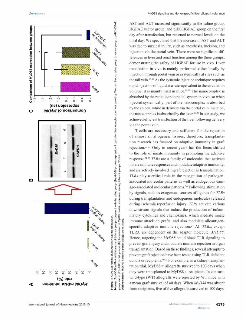
gene expression of MyD88 after liver transplantationAs shown in Figure 11, 5 days after liver transplantation, there
were no significant differences in MyD88 mRNA and protein
expression among the saline control, HGPAE vector con-
trol, and pHK/HGPAE control groups (P.0.05). However,
compared with the three control groups, the expression of
MyD88 mRNA and protein in the pMyD88/HGPAE group
was suppressed significantly (P,0.01).
expression of Il-2 and IFN-γ after liver transplantationAs shown in Figure 12, compared with the three control
groups, there was an obvious reduction in IL-2 and IFN-γ
concentrations in the pMyD88/HGPAE group (P,0.05).
In contrast, there were no significant differences in the IL-2
and IFN-γ concentrations among the three control groups
(P.0.05).
DiscussionThe availability of methods for efficient delivery of pDNA
into cells in vivo limits the use of RNA interference
technology both for research and clinical applications.13,14 In
our study, we designed and synthesized a type of HGPAE
nanovector, which was capable of complexing with an elec-
tronegative gene and promoting its release from the endo-
some and into the cytoplasm (Figure 13).15 Furthermore, we
showed that at a weight ratio of HGPAE to DNA of 80:1,
they formed electropositive spherical complexes, 50–150 nm
in dia meter, which were stable under electronegative condi-
tions. The HGPAE nanovectors were shown to be efficient
for transfection both in vitro and in vivo. In safety tests, both

International Journal of Nanomedicine 2015:10submit your manuscript | www.dovepress.com
Dovepress
Dovepress
4378
hu et al
Figure 9 The median survival time of recipients in different groups.Notes: The survival time of recipients is median survival time and comparisons were made using the Kaplan–Meier cumulative survival method.
Figure 10 Pathological changes of the donor liver after transplantation.Notes: (A) liver tissue sections of the saline group. (B) liver tissue sections of the hgPae vector group. (C) liver tissue sections of the phK/hgPae complex group. (D) liver tissue sections of the pMyD88/hgPae complex group.Abbreviation: hgPae, histidine-grafted poly(β-amino ester).
International Journal of Nanomedicine 2015:10 submit your manuscript | www.dovepress.com
Dovepress
Dovepress
4379
MyD88 signaling and donor-specific liver allograft tolerance
Figu
re 1
1 g
ene
expr
essi
on o
f MyD
88 a
fter
liver
tra
nspl
anta
tion.
Not
es: (
A) M
yD88
mr
Na
inhi
bitio
n ra
te o
f diff
eren
t gro
ups
com
pare
d w
ith th
e sa
line
grou
p. (B
) MyD
88 p
rote
in e
xpre
ssio
n 5
days
afte
r liv
er tr
ansp
lant
atio
n w
as te
sted
by
Wes
tern
blo
t (a:
sal
ine
grou
p, b
: vec
tor
grou
p, c
: ph
K/h
gPa
e gr
oup,
and
d: p
MyD
88/h
gPa
e gr
oup)
. (C
) c
ompa
riso
n of
MyD
88 p
rote
in e
xpre
ssio
n am
ong
diffe
rent
gro
ups.
*P,
0.01
.A
bbre
viat
ion:
hg
Pae,
his
tidin
e-gr
afte
d po
ly(β
-am
ino
este
r).
AST and ALT increased significantly in the saline group,
HGPAE vector group, and pHK/HGPAE group on the first
day after transfection, but returned to normal levels on the
third day. We speculated that the increase in AST and ALT
was due to surgical injury, such as anesthesia, incision, and
injection via the portal vein. There were no significant dif-
ferences in liver and renal function among the three groups,
demonstrating the safety of HGPAE for use in vivo. Liver
transfection in vivo is mainly performed either locally by
injection through portal vein or systemically at sites such as
the tail vein.16,17 As the systemic injection technique requires
rapid injection of liquid at a rate equivalent to the circulation
volume, it is mainly used in mice.18,19 The nanocomplex is
absorbed by the reticuloendothelial system in vivo, so when
injected systemically, part of the nanocomplex is absorbed
by the spleen, while in delivery via the portal vein injection,
the nanocomplex is absorbed by the liver.20,21 In our study, we
achieved efficient transfection of the liver following delivery
via the portal vein.
T-cells are necessary and sufficient for the rejection
of almost all allogeneic tissues; therefore, transplanta-
tion research has focused on adaptive immunity in graft
rejection.22,23 Only in recent years has the focus shifted
to the role of innate immunity in promoting the adaptive
response.24,25 TLRs are a family of molecules that activate
innate immune responses and modulate adaptive immunity,
and are actively involved in graft rejection in transplantation.
TLRs play a critical role in the recognition of pathogen-
associated molecular patterns as well as endogenous dam-
age-associated molecular patterns.26 Following stimulation
by signals, such as exogenous sources of ligands for TLRs
during transplantation and endogenous molecules released
during ischemia reperfusion injury, TLRs activate various
downstream signals that induce the production of inflam-
matory cytokines and chemokines, which mediate innate
immune attack on grafts, and also modulate alloantigen-
specific adaptive immune rejection.27 All TLRs, except
TLR3, are dependent on the adaptor molecule, MyD88.
Hence, targeting the MyD88 could block TLR signaling to
prevent graft injury and modulate immune rejection in organ
transplantation. Based on these findings, several attempts to
prevent graft rejection have been tested using TLR-deficient
donors or recipients.10,25 For example, in a kidney transplan-
tation trial, MyD88-/- allografts survived to 100 days when
they were transplanted to MyD88-/- recipients. In contrast,
wild-type (WT) allografts were rejected by WT mice with
a mean graft survival of 40 days. When MyD88 was absent
from recipients, five of five allografts survived to 100 days.

International Journal of Nanomedicine 2015:10submit your manuscript | www.dovepress.com
Dovepress
Dovepress
4380
hu et al
γ
Figure 12 expression of Il-2 and IFN-γ after liver transplantation.Notes: (A) The Il-2 serum concentration of different groups detected by elIsa. (B) IFN-γ concentration of different groups detected by elIsa. *P,0.05.Abbreviation: elIsa, enzyme-linked immunosorbent assay.
Figure 13 The functional mechanism of the pMyD88/hgPae complex.Notes: after being entrapped by the acidic endosome, the pMyD88/hgPae complex became swollen and disassembled. Then the pMyD88 escaped from the endosome and got into the nucleus to transcript sirNa of MyD88 for suppressing the expression of MyD88.Abbreviation: hgPae, histidine-grafted poly(β-amino ester).

International Journal of Nanomedicine 2015:10 submit your manuscript | www.dovepress.com
Dovepress
Dovepress
4381
MyD88 signaling and donor-specific liver allograft tolerance
When only the allograft was MyD88 deficient, survival was
modestly prolonged compared with that of the WT allograft.28
To explore the role of MyD88-dependent TLR signaling in
liver transplant rejection, we chose to knock down MyD88
expression in the donor liver using pMyD88/HGPAE, which
was then used in MHC fully mismatched allogeneic liver
transplantation. Our study demonstrated that knocking down
MyD88 reduced graft rejection and prolonged the survival
time of the recipient in a liver allograft model.
Graft rejection is initiated by recognition of donor graft
antigens by the recipient’s T-cells; this recognition occurs via
direct and indirect pathways. In the direct pathway, the recipi-
ent’s T-cells recognize intact allo-MHC molecules presented by
donor APCs, while in the indirect pathway, recipient’s T-cells
recognize processed alloantigen presented by the recipient’s
APCs. TLRs are expressed primarily on macrophages and den-
dritic cells (DCs) and control the activation of these APCs. In
DCs, TLR signaling triggers a maturation program that includes
upregulation of MHC and costimulatory molecules and expres-
sion of proinflammatory cytokines, such as TNF-α, IL-1, and
IL-6. This DC maturation significantly increases their ability to
elicit the differentiation of naïve T-cells into mature effector and
memory T-cells, which secrete cytokines such as IL-2 and IFN-γ
to induce expression of class II MHC, adhesion molecules,
and costimulatory molecules by endothelial cells.29,30 These
molecules reinforce both the recognition pathways, thereby
recruiting more T-cells and amplifying the rejection process.
Therefore, blockade of donor TLR signaling by inhibition of
MyD88 prevents maturation of the donor APCs. As a result, the
direct pathway will be impaired, leading to reduced production
of class II MHC and cytokines, with a consequent reduction in
indirect pathway activity. Ultimately, the graft rejection will be
alleviated. In support of this hypothesis, our data demonstrate
that interruption of the TLR signaling in the donor liver by
inhibition of MyD88 reduces cytokine production and alleviates
graft rejection, leading to prolonged recipient survival.
ConclusionIn summary, pMyD88/HGPAE nanocomplexes, a new type
of gene delivery system, were prepared successfully for
inhibiting graft rejection and prolonging the survival time
of liver transplant recipients in a high-responder rat liver
transplantation model. We demonstrated that pMyD88/
HGPAE nanovectors can be used to deliver and release
the pMyD88 successfully both in vitro and in vivo. In liver
transplantation, pMyD88/HGPAE nanocomplexes efficiently
prevented MyD88 action with the therapeutic potential to
prevent allograft rejection. This study demonstrates that
pMyD88/HGPAE nanocomplexes with high transfection
efficiency represent an alternative strategy for preventing
allograft rejection in liver transplantation.
AcknowledgmentsThe authors gratefully acknowledge the National Natural Science
Foundation of China (81102246, 51373117, and 51303126),
Tianjin Natural Science Foundation (13JCZDJC33200 and
13JCQNJC11900), Doctoral Base Foundation of Educational
Ministry of China (20120032110027), and Tianjin Municipal
Education Commission Science Foundation (20090126).
DisclosureThe authors report no conflicts of interest in this work.
References 1. Benichou G, Tonsho M, Tocco G, Nadazdin O, Madsen JC. Innate
immunity and resistance to tolerogenesis in allotransplantation. Front Immunol. 2012;3:73.
2. Shin OS, Harris JB. Innate immunity and transplantation tolerance: the potential role of TLRs/NLRs in GVHD. Korean J Hematol. 2011; 46(2):69–79.
3. Miller DM, Rossini AA, Greiner DL. Role of innate immunity in transplantation tolerance. Crit Rev Immunol. 2008;28(5):403–439.
4. Kawasaki T, Kawai T. Toll-like receptor signaling pathways. Front Immunol. 2014;5:461.
5. Goldstein DR, Tesar BM, Akira S, Lakkis FG. Critical role of the Toll-like receptor signal adaptor protein MyD88 in acute allograft rejection. J Clin Invest. 2003;111(10):1571–1578.
6. Yamamoto M, Takeda K. Current views of toll-like receptor signaling pathways. Gastroenterol Res Pract. 2010;2010:240365.
7. Takeda K, Akira S. Toll-like receptors in innate immunity. Int Immunol. 2005;17(1):1–14.
8. Tesar BM, Zhang J, Li Q, Goldstein DR. Th1 immune responses to fully MHC mismatched allografts are diminished in the absence of MyD88, a toll-like receptor signal adaptor protein. Am J Transplant. 2004;4(9):1429–1439.
9. Ro H, Lee EW, Hong JH, et al. Roles of islet Toll-like receptors in pig to mouse islet xenotransplantation. Cell Transplant. 2013;22(9): 1709–1722.
10. Ro H, Hong J, Kim BS, et al. Roles of Toll-like receptors in allogeneic islet transplantation. Transplantation. 2012;94(10):1005–1012.
11. Kamada N, Calne RY. Orthotopic liver transplantation in the rat. Technique using cuff for portal vein anastomosis and biliary drainage. Transplantation. 1979;28(1):47–50.
12. [No authors listed]. Banff schema for grading liver allograft rejection: an international consensus document. Hepatology. 1997;25(3):658–663.
13. Borna H, Imani S, Iman M, Azimzadeh Jamalkandi S. Therapeutic face of RNAi: in vivo challenges. Expert Opin Biol Ther. 2015;15(2):269–285.
14. Colombo S, Zeng X, Ragelle H, Foged C. Complexity in the therapeutic delivery of RNAi medicines: an analytical challenge. Expert Opin Drug Deliv. 2014;11(9):1481–1495.
15. Cristiano RJ. Targeted, non-viral gene delivery for cancer gene therapy. Front Biosci. 1998;3:D1161–D1170.
16. Liu F, Song Y, Liu D. Hydrodynamics-based transfection in animals by systemic administration of plasmid DNA. Gene Ther. 1999;6(7): 1258–1266.
17. Budker V, Budker T, Zhang G, Subbotin V, Loomis A, Wolff JA. Hypothesis: naked plasmid DNA is taken up by cells in vivo by a receptor-mediated process. J Gene Med. 2000;2(2):76–88.
International Journal of Nanomedicine
Publish your work in this journal
Submit your manuscript here: http://www.dovepress.com/international-journal-of-nanomedicine-journal
The International Journal of Nanomedicine is an international, peer-reviewed journal focusing on the application of nanotechnology in diagnostics, therapeutics, and drug delivery systems throughout the biomedical field. This journal is indexed on PubMed Central, MedLine, CAS, SciSearch®, Current Contents®/Clinical Medicine,
Journal Citation Reports/Science Edition, EMBase, Scopus and the Elsevier Bibliographic databases. The manuscript management system is completely online and includes a very quick and fair peer-review system, which is all easy to use. Visit http://www.dovepress.com/testimonials.php to read real quotes from published authors.
International Journal of Nanomedicine 2015:10submit your manuscript | www.dovepress.com
Dovepress
Dovepress
Dovepress
4382
hu et al
18. Herrero MJ, Monleon D, Morales JM, Mata M, Serna E, Aliño SF. Analy-sis of metabolic and gene expression changes after hydrodynamic DNA injection into mouse liver. Biol Pharm Bull. 2011;34(1):167–172.
19. Shashidharamurthy R, Machiah D, Bozeman EN, et al. Hydrodynamic delivery of plasmid DNA encoding human FcγR-Ig dimers blocks immune-complex mediated inflammation in mice. Gene Ther. 2012; 19(9):877–885.
20. Nakamura Y, Kominami A, Tsujimoto Y, et al. Actin and Vimentin proteins with N-terminal deletion detected in tumor-bearing rat livers induced by intraportal-vein injection of Ha-ras-transfected rat liver cells. Int J Cancer. 2009;124(11):2512–2519.
21. Iimuro Y, Fujimoto J. Strategy of gene therapy for liver cirrohosis and hepatocellular carcinoma. J Hepatobiliary Pancreat Surg. 2003;10(1): 45–47.
22. Lo YC, Lee CF, Powell JD. Insight into the role of mTOR and metabo-lism in T cells reveals new potential approaches to preventing graft rejection. Curr Opin Organ Transplant. 2014;19(4):363–371.
23. Mehrotra A, Leventhal J, Purroy C, Cravedi P. Monitoring T cell alloreactivity. Transplant Rev (Orlando). 2015;29(2):53–59.
24. Ma Z, Zhang E, Yang D, Lu M. Contribution of Toll-like receptors to the control of hepatitis B virus infection by initiating antiviral innate responses and promoting specific adaptive immune responses. Cell Mol Immunol. Epub 2014 Nov 24.
25. Zhang X, Beduhn M, Zheng X, et al. Induction of alloimmune tolerance in heart transplantation through gene silencing of TLR adaptors. Am J Transplant. 2012;12(10):2675–2688.
26. Leventhal JS, Schröppel B. Toll-like receptors in transplantation: sens-ing and reacting to injury. Kidney Int. 2012;81(9):826–832.
27. Iwasaki A, Medzhitov R. Regulation of adaptive immunity by the innate immune system. Science. 2010;327(5963):291–295.
28. Wu H, Noordmans GA, O’Brien MR, et al. Absence of MyD88 signaling induces donor-specific kidney allograft tolerance. J Am Soc Nephrol. 2012;23(10):1701–1716.
29. Schnare M, Barton GM, Holt AC, Takeda K, Akira S, Medzhitov R. Toll-like receptors control activation of adaptive immune responses. Nat Immunol. 2001;2(10):947–950.
30. Ozato K, Tsujimura H, Tamura T. Toll-like receptor signaling and regulation of cytokine gene expression in the immune system. Biotech-niques. 2002;Suppl:66–68, 70, 72.