The design of nitric oxide donor drugs: s-nitrosothiol tDodSNO is a superior photoactivated donor in...
9
Cardiovascular pharmacology The design of nitric oxide donor drugs: s-nitrosothiol tDodSNO is a superior photoactivated donor in comparison to GSNO and SNAP Sweta Kumari, Ivan A. Sammut, Gregory I. Giles n Department of Pharmacology and Toxicology, University of Otago, P.O. Box 913, Dunedin, New Zealand article info Article history: Received 5 February 2014 Received in revised form 13 May 2014 Accepted 14 May 2014 Available online 22 May 2014 Keywords: Nitric oxide Drug design Cardiovascular S-nitrosothiol Vasodilation abstract We have recently developed tert-dodecane S-nitrosothiol (tDodSNO) as a photoactivated nitric oxide (NO) donor. We here compare the potency of tDodSNO to S-nitrosoglutathione (GSNO) and S-nitroso-N- acetylpenicillamine (SNAP), drugs which are also based upon the S-nitrosothiol functionality and have been extensively used for NO release studies. Photoactivation in vitro, at a clinically relevant light fluence rate (200 W/m 2 ), demonstrated that tDodSNO released much higher levels of NO than either GSNO or SNAP. When evaluated in an ex vivo aortic ring vasorelaxation assay, tDodSNO was also the only drug to exhibit a photodynamic response, with an 8 fold decrease in EC 50 (8.1–1.0 mM) upon irradiation. While both GSNO and SNAP induced NO dependent vasorelaxation at lower concentrations than tDodSNO (EC 50 's of 158 and 38 nM respectively), this activity was due to their rapid metabolic decomposition, and could not be modulated by photoactivation. Additionally, tDodSNO's photodynamic response allowed vascular tone to be directly regulated by light intensity. Molecular modeling of drug properties suggested that these differences in activity could be attributed to a combination of an increase in tDodSNO's hydrophobicity, and substantial steric shielding of molecule's S-nitrosothiol group from solvent interactions. In conclusion, our study demonstrates that tDodSNO is currently the most effective known s-nitrosothiol for phototherapeutic applications. & 2014 Elsevier B.V. All rights reserved. 1. Introduction There have been extensive efforts to develop drugs capable of delivering nitric oxide (NO) to tissue, as NO releasing donors have the potential to provide treatments for multiple patho- logies including: ischemic cardiovascular disease (Richardson and Benjamin, 2002), hypertension (Bath et al., 2001) and cancer (Huerta et al., 2008). At present clinically utilized NO based therapeutics require metabolic activation to release NO, restricting their applications due to the onset of hypotension, which can result in syncope (Thadani and Rodgers, 2006), and the rapid development of tolerance (Thadani, 1997). A promising strategy to improve upon these agents is photodynamic therapy (PDT), whereby NO release is controlled by an applied light stimulus (Ford, 2013). Several photolabile pharmacophores have been examined as potential NO donors, including: N-nitrosamines (Karaki et al., 2012; Namiki et al., 1999), S-nitrosothiols (SNTs, also known as thionitrites) (Sexton et al., 1994), caged diazenium- diolates (Makings and Tsien, 1994) and transition metal-nitrosyl complexes (Madhani et al., 2006), as well as nanoparticle formu- lations (Deniz et al., 2012). Of these photoactives the SNTs are potentially the most useful for a range of PDT applications, as they can be excited by light in both the UV (340 nm) and visible (560– 600 nm) regions of the spectrum (Szacilowski and Stasicka, 2001). However, the development of these molecules as viable agents for PDT has been hindered by their rapid metabolism and metal catalyzed decomposition (Singh et al., 1996). To circumvent the limitations associated with current SNTs, we have recently developed a NO donor, tert-dodecane S-nitrosothiol (tDodSNO, Fig. 1), that exhibits improved NO release character- istics (Giles et al., 2012). The rationale for the development of tDodSNO was to increase its hydrophobicity in order to partition the molecule into cell membranes, shielding it from the cellular metabolic pathways, metal ion interactions, and trans-nitrosation reactions that typically degrade SNT based drugs (Al-Sa'doni and Ferro, 2004). Such a strategy has previously been followed with the long acting beta agonists used in asthma management (Lotvall, 2001), and we hypothesized that similarly hydrophobic SNTs would display augmented stability and hence controllable NO release. The current paper examines the effects of tDodSNO on Contents lists available at ScienceDirect journal homepage: www.elsevier.com/locate/ejphar European Journal of Pharmacology http://dx.doi.org/10.1016/j.ejphar.2014.05.012 0014-2999/& 2014 Elsevier B.V. All rights reserved. Abbreviations: DTPA, diethylenetriaminepentaacetic acid; GSNO, S-nitrosogluta- thione; KPi, potassium phosphate; MbO 2 , oxy-myoglobin; NO, nitric oxide (nitro- gen monoxide); ODQ, 1H-[1,2,4]oxadiazolo[4,3-a] quinoxalin-1-one; PDT, photo- dynamic therapy; PE, phenylephrine; SNAP, S-nitroso-N-acetylpenicillamine; SNT, S-nitrosothiol; tDodSNO, tert-dodecane S-nitrosothiol n Corresponding author. Tel.: þ64 3 479 7322; fax: þ64 3 479 9140. E-mail address: [email protected] (G.I. Giles). European Journal of Pharmacology 737 (2014) 168–176
Transcript of The design of nitric oxide donor drugs: s-nitrosothiol tDodSNO is a superior photoactivated donor in...

Cardiovascular pharmacology
The design of nitric oxide donor drugs: s-nitrosothiol tDodSNOis a superior photoactivated donor in comparison to GSNO and SNAP
Sweta Kumari, Ivan A. Sammut, Gregory I. Giles n
Department of Pharmacology and Toxicology, University of Otago, P.O. Box 913, Dunedin, New Zealand
a r t i c l e i n f o
Article history:Received 5 February 2014Received in revised form13 May 2014Accepted 14 May 2014Available online 22 May 2014
Keywords:Nitric oxideDrug designCardiovascularS-nitrosothiolVasodilation
a b s t r a c t
We have recently developed tert-dodecane S-nitrosothiol (tDodSNO) as a photoactivated nitric oxide(NO) donor. We here compare the potency of tDodSNO to S-nitrosoglutathione (GSNO) and S-nitroso-N-acetylpenicillamine (SNAP), drugs which are also based upon the S-nitrosothiol functionality and havebeen extensively used for NO release studies. Photoactivation in vitro, at a clinically relevant light fluencerate (200 W/m2), demonstrated that tDodSNO released much higher levels of NO than either GSNO orSNAP. When evaluated in an ex vivo aortic ring vasorelaxation assay, tDodSNO was also the only drug toexhibit a photodynamic response, with an 8 fold decrease in EC50 (8.1–1.0 mM) upon irradiation. Whileboth GSNO and SNAP induced NO dependent vasorelaxation at lower concentrations than tDodSNO(EC50's of 158 and 38 nM respectively), this activity was due to their rapid metabolic decomposition, andcould not be modulated by photoactivation. Additionally, tDodSNO's photodynamic response allowedvascular tone to be directly regulated by light intensity. Molecular modeling of drug properties suggestedthat these differences in activity could be attributed to a combination of an increase in tDodSNO'shydrophobicity, and substantial steric shielding of molecule's S-nitrosothiol group from solventinteractions. In conclusion, our study demonstrates that tDodSNO is currently the most effective knowns-nitrosothiol for phototherapeutic applications.
& 2014 Elsevier B.V. All rights reserved.
1. Introduction
There have been extensive efforts to develop drugs capableof delivering nitric oxide (NO) to tissue, as NO releasing donorshave the potential to provide treatments for multiple patho-logies including: ischemic cardiovascular disease (Richardsonand Benjamin, 2002), hypertension (Bath et al., 2001) and cancer(Huerta et al., 2008). At present clinically utilized NO basedtherapeutics require metabolic activation to release NO, restrictingtheir applications due to the onset of hypotension, which canresult in syncope (Thadani and Rodgers, 2006), and the rapiddevelopment of tolerance (Thadani, 1997). A promising strategyto improve upon these agents is photodynamic therapy (PDT),whereby NO release is controlled by an applied light stimulus(Ford, 2013). Several photolabile pharmacophores have beenexamined as potential NO donors, including: N-nitrosamines
(Karaki et al., 2012; Namiki et al., 1999), S-nitrosothiols (SNTs,also known as thionitrites) (Sexton et al., 1994), caged diazenium-diolates (Makings and Tsien, 1994) and transition metal-nitrosylcomplexes (Madhani et al., 2006), as well as nanoparticle formu-lations (Deniz et al., 2012). Of these photoactives the SNTs arepotentially the most useful for a range of PDT applications, as theycan be excited by light in both the UV (340 nm) and visible (560–600 nm) regions of the spectrum (Szacilowski and Stasicka, 2001).However, the development of these molecules as viable agents forPDT has been hindered by their rapid metabolism and metalcatalyzed decomposition (Singh et al., 1996).
To circumvent the limitations associated with current SNTs, wehave recently developed a NO donor, tert-dodecane S-nitrosothiol(tDodSNO, Fig. 1), that exhibits improved NO release character-istics (Giles et al., 2012). The rationale for the development oftDodSNO was to increase its hydrophobicity in order to partitionthe molecule into cell membranes, shielding it from the cellularmetabolic pathways, metal ion interactions, and trans-nitrosationreactions that typically degrade SNT based drugs (Al-Sa'doniand Ferro, 2004). Such a strategy has previously been followedwith the long acting beta agonists used in asthma management(Lotvall, 2001), and we hypothesized that similarly hydrophobicSNTs would display augmented stability and hence controllableNO release. The current paper examines the effects of tDodSNO on
Contents lists available at ScienceDirect
journal homepage: www.elsevier.com/locate/ejphar
European Journal of Pharmacology
http://dx.doi.org/10.1016/j.ejphar.2014.05.0120014-2999/& 2014 Elsevier B.V. All rights reserved.
Abbreviations: DTPA, diethylenetriaminepentaacetic acid; GSNO, S-nitrosogluta-thione; KPi, potassium phosphate; MbO2, oxy-myoglobin; NO, nitric oxide (nitro-gen monoxide); ODQ, 1H-[1,2,4]oxadiazolo[4,3-a] quinoxalin-1-one; PDT, photo-dynamic therapy; PE, phenylephrine; SNAP, S-nitroso-N-acetylpenicillamine; SNT,S-nitrosothiol; tDodSNO, tert-dodecane S-nitrosothiol
n Corresponding author. Tel.: þ64 3 479 7322; fax: þ64 3 479 9140.E-mail address: [email protected] (G.I. Giles).
European Journal of Pharmacology 737 (2014) 168–176

vascular tone in comparison to the commonly employed SNTsS-nitrosoglutathione (GSNO, Fig. 1) and S-nitroso-N-acetylpenicill-amine (SNAP, Fig. 1). Within vascular tissue, tDodSNO was the onlymolecule to act as a photoactive agent, indicating that tDodSNO isa superior candidate for PDT based applications.
2. Materials and methods
2.1. Materials
S-nitrosoglutathione (GSNO), horse heart myoglobin, pheny-lephrine (PE), indomethacin and 1H-[1,2,4]oxadiazolo[4,3-a]quinoxalin-1-one (ODQ) were purchased from Sigma-Aldrich(Auckland, NZ). S-nitroso-N-acetylpenicillamine (SNAP) was pur-chased from Invitrogen (Auckland, NZ). tDodSNO was synthesizedas previously described (Giles et al., 2012). All other chemicalswere of analytical grade.
2.2. Molecular modeling
Quantum mechanical derivations of molecular structure werecalculated via the MOPAC simulation program applying the AM1semiempirical Hamiltonian force field (Chem3D Pro v11.0, Cam-bridgeSoft Corporation, Cambridge, MA, USA).
2.3. Determination of SNT concentration
The concentration of stock solutions of SNTs was determinedby their characteristic absorbance at λ¼340 nm. SNTs were eitherdissolved in DMSO (tDodSNO ε340¼675 M–1 cm–1 (Giles et al.,2012) and SNAP ε340¼1168 M–1 cm–1 (Megson et al., 1999)) orMilliQ water (GSNO ε340¼922 M–1 cm–1 (Hart, 1985)).
2.4. Photoactivation
Irradiation was regulated using a 100 W halogen cold lightsource with variable power output (LG-PS2; Olympus, Auckland,New Zealand). Fluence rate was recorded using a P-9710 opt-ometer fitted with an RW-3705 detector head (Gigahertz-Optik,Munich, Germany).
2.5. SNT NO release kinetics
The release of NO from SNTs was quantified using a free radicalanalyzer equipped with a polarographic NO sensing electrode(ISO-NOP, WPI, Sarasota, FL, USA). NO release measurements wereconducted in a stirred, sealed incubation chamber, with no head-space, at 37 1C in KPi buffer (100 mM KPi, 2 mM DTPA, pH 7.4) aspreviously described (Giles et al., 2012). The electrode responsewas calibrated via the chemical generation of NO from sodiumnitrite (Hummel et al., 2006; Zhang, 2004).
2.6. Oxy-myoglobin preparation
Commercially available horse heart myoglobin consists of amixture of oxy (MbO2) and met (met-Mb) myoglobin. This mixturewas reduced to Mb via the addition of excess sodium dithionitein KPi buffer (pH 7.4, 100 mM KPi, 2 mM DTPA). To re-oxygenateand purify MbO2, the solution was then passed through a PD10desalting column (Sephadex G-25M, GE Healthcare, Auckland, NZ)(Zhang and Hogg, 2002). The MbO2 concentration in the elutedsolution was then determined at λ¼542 nm (ε542¼13, 900 M�1
cm�1) (Feelisch and Stamler, 1996).
2.7. Preparation of aortic rings
All animal experiments were conducted in accordance with theregulations of the University of Otago Committee on Ethics in theCare and Use of Laboratory Animals, and the NIH Guide for theCare and Use of Laboratory Animals. Male Sprague-Dawley rats(2-3 months old, 250–300 g) were obtained from the University ofOtago Animal Resource Unit. The rats were housed at 25 1C undera controlled 12-h day/night cycle in standard rat cages and fed adlibitum on a standard rat diet before experimentation. The ratswere killed by decapitation and the thoracic aortas immediatelydissected into 4 mm wide transverse rings. The aortic rings werethenwashed in ice-cold Krebs Henseleit (KH) buffer (118 mM NaCl,4.7 mM KCl, 1.66 mM MgSO4, 24.88 mM NaHCO3, 1.18 mM KH2PO4,11 mM glucose, 0.5 mM EDTA, 3 mM CaCl2, pH 7.4) before use.
2.8. Vasorelaxation assay
Endothelium intact aortic rings were placed between twostainless-steel stirrups and equilibrated under a resting tensionof 2 g in a 10 ml organ bath containing KH buffer at 37 1C, gassedwith 95% O2–5% CO2 (Resende et al., 2004). Indomethacin (10 mM)was included in the buffer to exclude prostaglandins as potentialmodulators of vascular tone. Vascular tone was recorded using anisometric force transducer (FT03, Grass) coupled to a PowerLab2/26 recorder (ADInstruments, Dunedin, New Zealand). Duringthis interval, the aortic rings were repeatedly washed in KH bufferand the applied force readjusted until the resting tone wasmaintained. The rings were then pre-constricted with phenylephr-ine (PE, 0.1 mM) to generate a reproducible, maintained level ofvascular tone, and concentration–responses to SNTs elicited fromthis PE-induced contraction. To confirm the involvement ofNO-mediated pathways in the SNT vasorelaxant response, aorticresponses to each of the SNTs were also studied in the presence ofoxymyoglobin (MbO2, 5 mM), a rapidly acting NO scavenger(Feelisch and Stamler, 1996; Zhang and Hogg, 2002), and excess1H-[1,2,4]Oxadiazolo[4,3-a]quinoxalin-1-one (ODQ, 10 mM), a selec-tive inhibitor of soluble guanylate cyclase activity (Garthwaite et al.,1995; Moro et al., 1996).
Fig. 1. Structures of SNTs.
S. Kumari et al. / European Journal of Pharmacology 737 (2014) 168–176 169

2.9. Statistical analysis
All values are expressed as means7standard error of themean (S.E.M). The concentrations of SNT that induced 50% ofthe maximum vasodilatory response (EC50) were interpolatedfrom semilogarithimic plots of individual concentration–responsecurves. Statistical analyses were performed using OriginPro 8 soft-ware (OriginLab Corporation, Northampton, MA). All data weresubjected to a paired t-test unless otherwise stated. Statisticalsignificance was accepted when Po0.05.
3. Results
3.1. Structural comparison of SNT physicochemical properties
Comparison of the MOPAC energy minimized structures for all3 SNTs (Table 1) demonstrated minimal differences in the calcu-lated C–S–N and S–N–O bond angles, which varied between 102–1061 and 120–1211 respectively. The dihedral C–S–N–O angle ofapproximately 1801, and the S–N bond length of 1.7 Å, were alsosimilar between all 3 molecules. Major differences were apparentin the relative octanol:water partition coefficients, with tDodSNO(c log P þ5.31) over 5 orders of magnitude more hydrophobic thanSNAP (c log P þ0.08), which in turn was 2 orders of magnitudemore lipid soluble than the hydrophilic GSNO (c log P �2.70).In addition to changes in hydrophobicity, for tDodSNO the stericbulk of the alkyl backbone also resulted in a decrease in theavailability of the S–N bond to solvent interactions (Fig. 2), whilethe primary SNT GSNO and the tertiary SNT SNAP were compara-tively less shielded.
3.2. in vitro tDodSNO is superior to SNAP and GSNO as aphotoactivated NO donor
Photoactivation of tDodSNO (100 mM) resulted in a rapidrelease of NO, which peaked within 3 min to produce a maximumconcentration of 8.5170.24 mM NO within the chamber (Fig. 3).To compare the efficacy of NO release between SNTs, we thencompared these NO release levels to those of SNAP and GSNO.At equal concentrations, neither of these SNTs were as effective astDodSNO as photoinducible donors. GSNO produced a maximumNO concentration of 2.4970.18 mM, while SNAP generated lowerNO levels of 0.3270.05 mM. NO release was not observed fromany of the SNTs in the absence of direct irradiation; room lightafforded a background fluence rate of 0.5 W/m2 which wasinsufficient to induce decomposition.
3.3. Ex vivo tDodSNO is superior to SNAP and GSNO as aphotoactivated NO donor
The vasorelaxant properties of each of the SNTs were thenassessed using a pre-contracted aortic ring, with concentration–response curves generated for each compound in the presence andabsence of photoactivation (Fig. 4). Without photoactivation, SNAPdisplayed the lowest EC50, in the low nM range (38711 nM),
while GSNO was approximately 5 fold less potent (158720 nM).tDodSNO (EC50 8.170.4 mM) was 50 fold less potent than GSNOand 215 fold less potent than SNAP. However, under photoactiva-tion, key differences in the ex vivo NO release properties of theSNTs emerged (Fig. 4). At 200 W/m2, photoactivation did notproduce any significant change in the EC50 response of eitherSNAP or GSNO from control (Table 2). For tDodSNO, photoactiva-tion produced a pronounced leftward shift in the concentration–response curve (Fig. 4), with a significant (Po0.05) 8 fold decreasein EC50 (8.170.4 to 170.1 mM, Table 2). Hence, while the overallEC50 value for photoactivated tDodSNO was higher than that ofeither SNAP or GSNO, it was the only SNT to exhibit a photo-dynamic response at this fluence rate.
3.4. Variable photoactivation of tDodSNO modulatesvasoconstriction
To test the pharmacological responsiveness of the SNTs tophotoactivation, we then compared SNT-mediated vasodilationunder fluence modulation. The calculated photoactivation EC50
concentrations for each SNT were first used to vasodilate thePE-contracted aortic rings to 50% of maximal contraction (Fig. 5),and we then investigated how the tissue would respond tocessation of the photostimulus. For tDodSNO, this decrease inlight intensity returned vascular tone to its original constrictedstate. This observation was in agreement with our earlier datawhich showed that, in the absence of photostimulation, 1 mMtDodSNO administration did not evoke a vasorelaxant effect(Fig. 4). In line with our observations that neither SNAP nor GSNOwere photoactive under these conditions, modulating fluence ratedid not alter contractile responses at the EC50 concentrations ofeither of these compounds (Fig. 5).
To investigate the potential toxicity of using PDT to induce NOrelease from tDodSNO, we then measured vascular responsivenesspost photoactivation. The aortic rings were first constricted withPE, then relaxed and re-constricted by adding tDodSNO andmodulating photoactivation. Following these PDT induced changesto vascular tone, all drugs were washed out and PE reapplied.Following washout, PE restored the aortic contractile response toits original tension, demonstrating that there were no long termeffects on tissue function as a result of the combination of drugexposure and photoactivation (See Supporting information).
3.5. Mechanism of photoactivation – tDodSNO releases NO andsignals through sGC
To confirm the involvement of NO in the vasorelaxationmechanism of each SNT under these photoactivation conditions,we used MbO2, a rapid NO scavenger (Zhang and Hogg, 2002),and ODQ, an inhibitor of sGC (Garthwaite et al., 1995), to block theNO signaling pathway. When administered individually, bothMbO2 and ODQ increased the basal extent of vasoconstriction(by 9.570.8% and 23.175.5% respectively, Po0.05), due toattenuation of endogenous cGMP production (Moro et al., 1996)(Figs. 6 and 7). When administered after each SNTs EC50 vasor-elaxant response, MbO2 resulted in a statistically significantincrease in vascular contractile tone, similar to the vasoconstric-tion response obtained with MbO2 alone (Fig. 6), indicating theinvolvement of NO in the relaxation mechanism for all SNTs.
The vasodilation response to the SNTs was also completelyblocked by the inhibition of sGC enzymatic activity via ODQadministration, which resulted in a significant elevation in aortictone above that obtained with PE, confirming a cGMP dependentvasorelaxation mechanism (Fig. 7). To confirm that the responserecorded following ODQ administration was a result of blockingdrug effects, and not due to the inhibition of endogenous NO
Table 1Calculated physicochemical parameters characterizing SNTs. θ: bond angle; B: bondlength; P: octanol:water partition coefficient.
SNT θ (C–S–N)(deg)
θ (S–N–O)(deg)
θ (C–S–N–O)(deg)
B (S–N)(Å)
c log P
SNAP 102.9 121.0 179.8 1.69 þ 0.08GSNO 102.3 120.7 180.0 1.70 �2.70tDodSNO 105.3 120.4 179.9 1.69 þ5.31
S. Kumari et al. / European Journal of Pharmacology 737 (2014) 168–176170
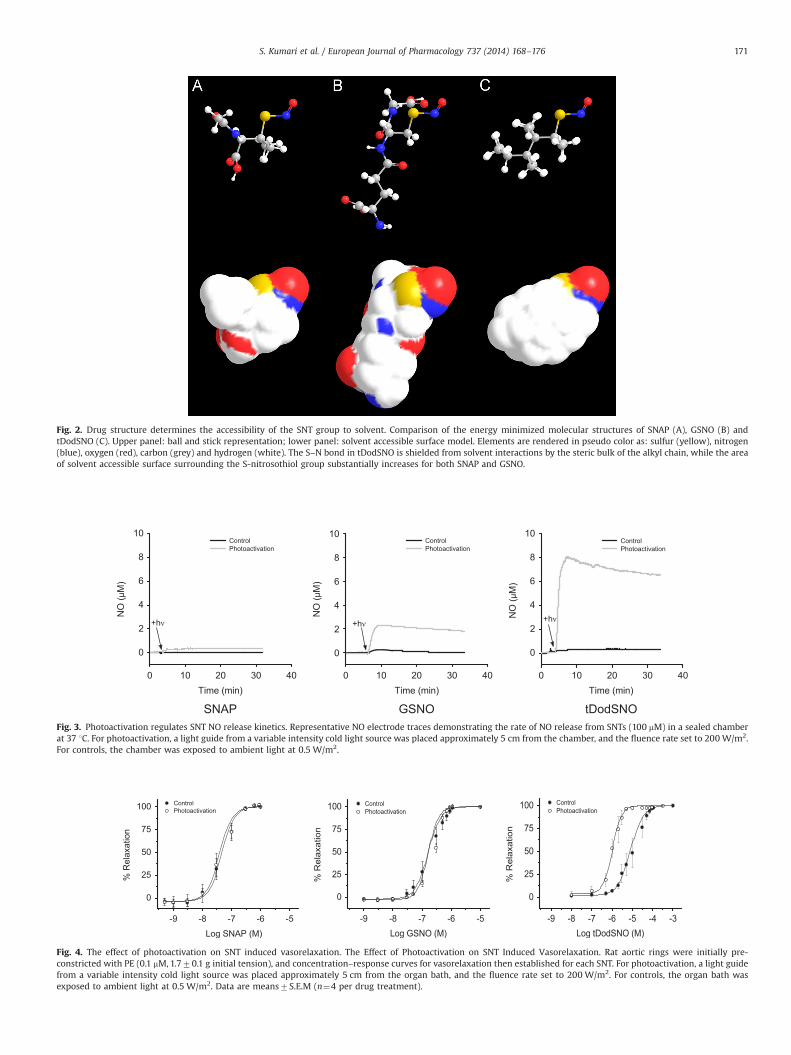
Fig. 2. Drug structure determines the accessibility of the SNT group to solvent. Comparison of the energy minimized molecular structures of SNAP (A), GSNO (B) andtDodSNO (C). Upper panel: ball and stick representation; lower panel: solvent accessible surface model. Elements are rendered in pseudo color as: sulfur (yellow), nitrogen(blue), oxygen (red), carbon (grey) and hydrogen (white). The S–N bond in tDodSNO is shielded from solvent interactions by the steric bulk of the alkyl chain, while the areaof solvent accessible surface surrounding the S-nitrosothiol group substantially increases for both SNAP and GSNO.
Fig. 3. Photoactivation regulates SNT NO release kinetics. Representative NO electrode traces demonstrating the rate of NO release from SNTs (100 mM) in a sealed chamberat 37 1C. For photoactivation, a light guide from a variable intensity cold light source was placed approximately 5 cm from the chamber, and the fluence rate set to 200 W/m2.For controls, the chamber was exposed to ambient light at 0.5 W/m2.
Fig. 4. The effect of photoactivation on SNT induced vasorelaxation. The Effect of Photoactivation on SNT Induced Vasorelaxation. Rat aortic rings were initially pre-constricted with PE (0.1 mM, 1.770.1 g initial tension), and concentration–response curves for vasorelaxation then established for each SNT. For photoactivation, a light guidefrom a variable intensity cold light source was placed approximately 5 cm from the organ bath, and the fluence rate set to 200 W/m2. For controls, the organ bath wasexposed to ambient light at 0.5 W/m2. Data are means7S.E.M (n¼4 per drug treatment).
S. Kumari et al. / European Journal of Pharmacology 737 (2014) 168–176 171

signaling, the experiment was also conducted with ODQ addedprior to tDodSNO. With ODQ initially present in the organ bathbuffer, photoactivation of tDodSNO did not induce vasodilation(See Supporting information).
3.6. The hydrophobicity of tDodSNO results in the SNT partitioninginto tissue
tDodSNO exhibited enhanced metabolic stability, as evidencedby its EC50 value without photoactivation being 50 and 215 foldhigher than GSNO and SNAP. To provide support for our hypothesisthat this stability of tDodSNO could be attributed to the partition-ing of the SNT into cellular membranes, we examined the extent towhich tDodSNO activity was retained in the vascular tissue postwashout. For these experiments, the tissue initially underwent acycle of photoactivation modulated relaxation and contraction toestablish the baseline activity of tDodSNO (Fig. 8). All drugs werethen washed out, and the aortic ring re-constricted with PE to itsoriginal tension. Without the addition of tDodSNO, the aorticpreparation was then photoactivated, and the extent of vasodila-tion as a result of residual tDodSNO evaluated. The data demon-strated that the tissue retained substantial tDodSNO activity postwashout, with photoactivation inducing 24.279.6% vasorelaxa-tion, which was not significantly different (P40.05) to the original48.771.1% vasorelaxation observed pre-washout (Fig. 8). To con-firm that this tissue uptake was specific to tDodSNO, the experi-ment was repeated with GSNO, the second most stable SNT based
on the EC50 values (Table 2). Following washout, no residual GSNOactivity was detected in the preparation (Fig. 8), demonstratingthat more hydrophilic SNTs were not partitioned into tissue.
4. Discussion
The ability to selectively target and focally release NO at func-tional vasoactive concentrations within the body has the potentialto provide a range of novel therapeutic treatment modalities.Towards this goal, we have designed and tested a photoactivatedNO donor compound, tDodSNO, and compared its calculatedmolecular properties and NO release characteristics with otherSNT based drugs. The results complement and extend our findingsover earlier data obtained in physiological buffers and in a cellculture model (Giles et al., 2012), where tDodSNO displayedpromising NO donor properties for PDT.
For photoactivation studies on vascular tissue, light withwavelengths from the UV through to the visible regions of thespectrum is known to cause vasorelaxation (Furchgott, 1991;Furchgott et al., 1961; Oplander et al., 2013) due to the release ofNO from endogenous photolabile derivatives within the tissue,including nitrite and protein SNTs (Matsunaga and Furchgott,1989; Oplander et al., 2013). As the magnitude of this light inducedvasorelaxation is proportional to light intensity (Furchgottet al., 1961), we used a standard fluence rate of 200 W/m2, underwhich conditions the light stimulus alone was insufficient to causevasorelaxation. This fluence rate is also in the mid-range oftherapeutic light doses, and should therefore give an indicationas to the levels of NO that can be achieved under clinically relevantconditions. While SNTs including GSNO have previously beentrialled as photoactivated NO donors, the other molecules in thisclass typically require much higher light intensities than tDodSNOto achieve therapeutically relevant NO release rates (Sexton et al.,1994). This was confirmed in the present study by comparing theextent of NO release in vitro from tDodSNO with SNAP and GSNOunder identical conditions. Both of these SNTs were clearly lesseffective as light activated NO donors, with GSNO approximately
Table 2The effect of photoactivation upon SNT induced vasorelaxation. The EC50 values forvasorelaxation for each SNT under both control (0.5 W/m2) and photoactivation(200 W/m2) conditions. Data are means7S.E.M (n¼4).
SNT EC50 control EC50 photoactivation Fold change
tDodSNO 8.170.4 mM 1.070.1 mM 8.1a
SNAP 45715 nM 38711 nM 1.2GSNO 158720 nM 160720 nM 1.0
a Indicates a significant difference between EC50 values with and withoutphotoactivation (Po0.05).
Fig. 5. tDodSNO is the only SNT that allows photoactivation to modulate vascular tone. Aortic rings were pre-constricted with PE (0.1 mM, 1.770.1 g initial tension),photoactivated (þhv, 200 W/m2), and then relaxed to 50% of maximum contraction using each SNTs EC50 (Table 1). Following relaxation, the photostimulus was removed(�hv, 0.5 W/m2) and the resulting change in contractile force evaluated. Top panels: representative isometric tension trace recordings. Bottom panels: mean data7S.E.M(n¼4 per drug treatment). ***indicates a significant difference between contractile force with and without photostimulus (Po0.001).
S. Kumari et al. / European Journal of Pharmacology 737 (2014) 168–176172

4 fold less potent than tDodSNO, while SNAP afforded minimal NOrelease in vitro.
We then extended this comparison into an ex vivo organpreparation where, under control ambient light conditions, SNAPwas by far the most potent vasodilator (EC50 38 nM), followedby GSNO (EC50 160 nM), while tDodSNO was only vasoactive atmuch higher concentrations (EC50 8.1 mM). Such activity was notpredictable from the NO electrode data, which indicated that, inbuffer, none of the SNTs released NO under control conditions(Fig. 3). Therefore we attributed the observed vasodilation tothe metabolic activation of the drugs within the tissue, as haspreviously been reported (Al-Sa'doni et al., 1997). However, upon
photoactivation, key distinctions in the pharmacological activity ofthe SNTs became apparent. Neither SNAP nor GSNO exhibited anysignificant changes to their vasorelaxation EC50 values uponirradiation indicating that, at this fluence rate, drug decompositiondue to photoactivation was insufficient to out-compete metabolicpathways for NO release. In the case of tDodSNO we observeda pronounced 8 fold change in EC50 (Table 2), demonstratingthat, unlike the other SNTs studied, in tissue it was possible tomodulate the levels of NO derived from this SNT.
These results are in accord with our original concept for thedesign of tDodSNO and demonstrate a clear functional differencebetween this compound and other SNTs. The limitation of the SNT
Fig. 6. All SNTs induce vasorelaxation via NO release. Aortic rings were pre-constricted with PE (0.1 mM, 1.770.1 g initial tension), photoactivated (þhv, 200 W/m2) and thenrelaxed to 50% of maximum contraction using each SNTs EC50 (Table 1). The NO trap MbO2 (5 mM) was then added and the resulting change in contractile force evaluated. Toppanels: representative isometric tension trace recordings. Bottom panels: mean data7S.E.M (n¼4 per drug treatment). ***indicates a significant difference betweencontractile force after the addition of MbO2 (Po0.001).
Fig. 7. SNT vasorelaxation is mediated via cGMP. Aortic rings were pre-constricted with PE (0.1 mM, 1.870.1 g initial tension), photoactivated (þhv, 200 W/m2) and thenrelaxed to 50% of maximum contraction using each SNTs EC50 (Table 1). The sGC inhibitor ODQ (10 mM) was then added and the resulting change in contractile forceevaluated. Top panels: representative isometric tension trace recordings. Bottom panels: mean data7S.E.M (n¼4 per drug treatment). ***indicates a significant differencebetween contractile force after the addition of ODQ (Po0.001).
S. Kumari et al. / European Journal of Pharmacology 737 (2014) 168–176 173

functionality is that it is readily degraded, either by metabolicenzymes or metal ions, resulting in the uncontrolled release of NO(Singh et al., 1996). Therefore, when designing tDodSNO, wetailored the compound's structural properties by incorporatingan extended alky chain backbone (Fig. 1) (Giles et al., 2012).The rationale behind this concept was to selectively partition thedrug into cellular membranes and shield it from the metal ionsand enzymes found in the cytosol. A molecular comparisonbetween SNTs (Fig. 2 and Table 1) confirms that the alkyl backboneof tDodSNO does not change the geometry of the SNT groupcompared to SNAP or GSNO, but does provide major increases inhydrophobicity and steric shielding. The functional effects of thesealterations to the molecule's properties are readily apparent fromthe aortic ring data. In the absence of a photostimulus, SNAP andGSNO released sufficient NO to activate sGC (between 1 and100 nM NO (Condorelli and George, 2001)) at orders of magnitudelower concentrations than tDodSNO. tDodSNO's alkyl chain wastherefore highly effective at preventing cellular decompositionpathways releasing NO from the molecule.
The extent to which the SNT drugs were shielded from meta-bolic breakdown could also explain the pronounced differences intheir efficacy as photoactive drugs. While the NO electrode datademonstrated that tDodSNO was the most potent NO donorfollowing photoactivation (Fig. 3), both GSNO, and to a lesserextent SNAP, did exhibit moderate to low NO release rates. Incontrast to this in vitro photoactivation ranking order, irradiationdid not produce a shift in the ex vivo vasorelaxation concentra-tion–response curves for either GSNO or SNAP (Table 2). Takentogether with their very low EC50 values, we conclude that, evenunder photoactivation, metabolic breakdown was still the ratedetermining step for NO release. This is consistent with observa-tions that the relaxation of smooth muscle tissue by SNAPand GSNO is the result of a Cu(I)-dependent catalytic reaction(Al-Sa'doni et al., 1997). In contrast tDodSNO displayed distinctphotoactive properties, which we suggest can be attributed to itslocalization to hydrophobic regions of the cell, with the concomi-tant retardation in the rate of SNT metabolism allowing the drugto function effectively as a PDT agent. Such hydrophobic partition-ing was confirmed by drug washout studies (Fig. 8), whichdemonstrated substantial accumulation of tDodSNO within the
vascular tissue. The lack of competing breakdown mechanismswas also evident from the transient nature of the vasodilationinduced by tDodSNO, as the tissue re-constricted upon cessation ofthe photostimulus. Hence tDodSNO was the only SNT that enabledvascular tone to be modulated by applied light intensity.
Independently of the requirement for photoactivation in themechanism by which the SNTs decomposed, all drug inducedvasorelaxation could be reversed by either MbO2 or ODQ, indicat-ing that the agents acted via the classical sGC mediated pathway.Recent studies have suggested alternative pathways by which SNTbased drugs could potentially cause vasodilation. Firstly, by theformation of intracellular dinitrosyl iron complexes (DNIC's),followed by direct transfer of NO from the DNIC to sGC resultingin activation of the enzyme (Severina et al., 2003). Secondly, bytrans-nitrosation reactions between SNTs and protein cysteineresidues inducing the modification of signaling molecules involvedin the vasorelaxation response, such as PKG (Burgoyne and Eaton,2009). However, for the SNTs studied, our data argues againstthese non-classical mechanisms, as vasodilation was completelyblocked by the addition of extracellular MbO2. Extracellular MbO2
is an effective scavenger of intracellular NO, due to NO's highdiffusion coefficient and membrane solubility resulting in themolecule being in equilibrium between the cell and the surround-ing extracellular environment (Espey et al., 2000; Ignarro et al.,1987; Kerwin et al., 1995). DNIC's, however, are constrained by theiron ligands, and cannot readily diffuse from the cell (Bosworthet al., 2009). Therefore, extracellular MbO2 is not an effective blockfor intracellular DNIC action, although this does not precludetransient DNIC formation in the mechanism of NO release fromthe SNTs. Similarly the inhibition of vasodilation by both ODQ andMbO2 precludes a significant contribution from SNT modifiedproteins.
The key pharmacological distinction between the SNTs wastherefore that tDodSNO was the only drug whose activity could beregulated via photoactivation. In a wider context, the directcomparison of tDodSNO with other non-SNT based photoactivatedNO donor molecules reported in the literature is problematical, asthe properties of these compounds are recorded using differentlight sources and typically acquired at higher photoactivationfluence rates. However, while there are currently no commercially
Fig. 8. The hydrophobicity of tDodSNO results in selective partitioning into tissue. Aortic rings were pre-constricted with PE (0.1 mM, 1.670.1 g initial tension),photoactivated (þhv, 200 W/m2) and then relaxed to approximately 50% of maximum contraction. The photostimulus was then removed (�hv), the tissue washed out, andthen re-constricted with PE (0.1 mM, 1.670.1 g initial tension). The extent of NO release from residual SNTs post-washout was then measured via photoactivation (þhv,200 W/m2). Top panels: representative isometric tension trace recordings. Bottom panels: mean data7S.E.M (n¼3 per drug treatment). **indicates a significant differencebetween vasorelaxation pre and post washout (Po0.01).
S. Kumari et al. / European Journal of Pharmacology 737 (2014) 168–176174

available NO donors that operate via a PDT mechanism, tDodSNO'sPDT properties induced vasorelaxation at levels consistent withthose reported for several metal complexes currently underdevelopment (Ford, 2013; Madhani et al., 2006; Ostrowskiet al., 2010). In terms of future drug development, it is likelythat both the metal and organic based classes of NO donors willfind distinct therapeutic applications, as they possess vastlydifferent pharmacokinetic profiles. While the development oforganic NO donors has been hindered by their metabolicdecomposition, the drawback to metal based therapeutics hasbeen their toxicity, which can arise from either the metal centeror the bound ligands (Al-Sa'doni and Ferro, 2000; Madhani etal., 2006). Recent advances have therefore focused on relativelynon-toxic metals, such as ruthenium. However such complexescan still modulate cell growth and display cytotoxicity at nMlevels (Carneiro et al., 2011). In comparison, we have showntDodSNO only displays significant cytotoxicity at concentrationsgreater than 50 mM (Giles et al., 2012), and the drug levelsrequired to induce vasorelaxation in the current study did notaffect the long term viability of the aortic preparation (SeeSupporting information). tDodSNO would therefore seem anexciting candidate for future translational development in dis-ease models, as well as providing a useful tool for NO research.
For future drug development, tDodSNO could find potentialapplications to treat a range of pathologies that can be targetedvia NO delivery, including: ischemic cardiovascular disease(Kita et al., 1995; Richardson and Benjamin, 2002), hyperten-sion (Bath et al., 2001), cancer (Huerta et al., 2008), asthma(Gaston et al., 1993), anal fissure (Lindsey et al., 2004) andbacterial and viral infection (MacMicking et al., 1997;Richardson and Benjamin, 2002). Although the pharmacoki-netics of tDodSNO administration have yet to be established, itis possible that the drug's hydrophobicity may result in loworal bioavailability, requiring drug delivery by injection ortopical administration. The utility of PDT is also limited bythe characteristics of the light source used. Most light emittingdevices have found applications in PDT, ranging in cost fromincandescent and arc lamps, through to light emitting diodesand lasers (Calin and Parasca, 2009). The selection of lightsource is determined by the therapeutic application, withoptimal wavelength, light intensity and tissue penetrationbeing key parameters. For example blue light has high energybut displays low tissue penetration (E100 mm), while red andinfrared wavelengths have lower energies but penetrate moredeeply into tissue (410 mm) (Agostinis et al., 2011). To over-come the limitations imposed by the light source, technologieshave also been developed to deliver light to inaccessible bodycompartments via the use of devices such as fiber optic cablesand implanted light sources (Huang, 2005). These technologiescould potentially be combined with tDodSNO to control thedrug's spatial and temporal NO release characteristics in vivo.
In conclusion, tDodSNO's metabolic stability and photoinduci-ble NO release characteristics demonstrate that this molecule isthe most effective known SNT for PDT applications. Additionally,tDodSNO is currently the only example of this drug class thatcan be activated using a therapeutically relevant fluence rate.Further development of tDodSNO could therefore realize new PDTapproaches towards disease treatment.
Acknowledgments
We are grateful for financial support provided by a LaurensonAward from the Otago Medical Research Foundation (LA 299) anda Dean's Bequest Grant (P6626R) from the Otago School of MedicalSciences, University of Otago. SK was supported by a University of
Otago Ph.D. Scholarship. IAS is supported by a New Zealand MBIE(PROP-29776-HVMSTR).
Appendix A. Supporting information
Supplementary data associated with this article can be found inthe online version at http://dx.doi.org/10.1016/j.ejphar.2014.05.012.
References
Agostinis, P., Berg, K., Cengel, K.A., Foster, T.H., Girotti, A.W., Gollnick, S.O., Hahn, S.M.,Hamblin, M.R., Juzeniene, A., Kessel, D., Korbelik, M., Moan, J., Mroz, P., Nowis, D.,Piette, J., Wilson, B.C., Golab, J., 2011. Photodynamic therapy of cancer: an update.CA Cancer J. Clin. 61 (4), 250–281.
Al-Sa'doni, H., Ferro, A., 2000. S-Nitrosothiols: a class of nitric oxide-donor drugs.Clin. Sci. (Lond.) 98 (5), 507–520.
Al-Sa'doni, H.H., Ferro, A., 2004. S-nitrosothiols as nitric oxide-donors: chemistry,biology and possible future therapeutic applications. Curr. Med. Chem. 11 (20),2679–2690.
Al-Sa'doni, H.H., Megson, I.L., Bisland, S., Butler, A.R., Flitney, F.W., 1997. Neocu-proine, a selective Cu(I) chelator, and the relaxation of rat vascular smoothmuscle by S-nitrosothiols. Br. J. Pharmacol. 121 (6), 1047–1050.
Bath, P.M., Pathansali, R., Iddenden, R., Bath, F.J., 2001. The effect of transdermalglyceryl trinitrate, a nitric oxide donor, on blood pressure and platelet functionin acute stroke. Cerebrovasc. Dis. 11 (3), 265–272.
Bosworth, C.A., Toledo Jr., J.C., Zmijewski, J.W., Li, Q., Lancaster Jr., J.R., 2009.Dinitrosyliron complexes and the mechanism(s) of cellular protein nitrosothiolformation from nitric oxide. Proc. Natl. Acad. Sci. USA 106 (12), 4671–4676.
Burgoyne, J.R., Eaton, P., 2009. Transnitrosylating nitric oxide species directlyactivate type I protein kinase A, providing a novel adenylate cyclase-indepen-dent cross-talk to beta-adrenergic-like signaling. J. Biol. Chem. 284 (43),29260–29268.
Calin, M.A., Parasca, S.V., 2009. Light sources for photodynamic inactivation ofbacteria. Lasers Med. Sci. 24 (3), 453–460.
Carneiro, Z.A., de Moraes, J.C., Rodrigues, F.P., de Lima, R.G., Curti, C., da Rocha, Z.N.,Paulo, M., Bendhack, L.M., Tedesco, A.C., Formiga, A.L., da Silva, R.S., 2011.Photocytotoxic activity of a nitrosyl phthalocyanine ruthenium complex—asystem capable of producing nitric oxide and singlet oxygen. J. Inorg. Biochem.105 (8), 1035–1043.
Condorelli, P., George, S.C., 2001. in vivo control of soluble guanylate cyclaseactivation by nitric oxide: a kinetic analysis. Biophys. J. 80 (5), 2110–2119.
Deniz, E., Kandoth, N., Fraix, A., Cardile, V., Graziano, A.C., Lo Furno, D., Gref, R.,Raymo, F.M., Sortino, S., 2012. Photoinduced fluorescence activation and nitricoxide release with biocompatible polymer nanoparticles. Chemistry 18 (49),15782–15787.
Espey, M.G., Miranda, K.M., Pluta, R.M., Wink, D.A., 2000. Nitrosative capacity ofmacrophages is dependent on nitric-oxide synthase induction signals. J. Biol.Chem. 275 (15), 11341–11347.
Feelisch, M., Stamler, J.S., 1996. Methods in Nitric Oxide Research. J. Wiley,Chichester.
Ford, P.C., 2013. Photochemical delivery of nitric oxide. Nitric Oxide 34, 56–64.Furchgott, R.F., 1991. Endothelium-dependent relaxation, endothelium-derived
relaxing factor and photorelaxation of blood vessels. Semin. Perinatol. 15 (1),11–15.
Furchgott, R.F., Ehrreich, S.J., Greenblatt, E., 1961. The photoactivated relaxation ofsmooth muscle of rabbit aorta. J. Gen. Physiol. 44, 499–519.
Garthwaite, J., Southam, E., Boulton, C.L., Nielsen, E.B., Schmidt, K., Mayer, B., 1995.Potent and selective inhibition of nitric oxide-sensitive guanylyl cyclase by 1H-[1,2,4]oxadiazolo[4,3-a]quinoxalin-1-one. Mol. Pharmacol. 48 (2), 184–188.
Gaston, B., Reilly, J., Drazen, J.M., Fackler, J., Ramdev, P., Arnelle, D., Mullins, M.E.,Sugarbaker, D.J., Chee, C., Singel, D.J., et al., 1993. Endogenous nitrogen oxidesand bronchodilator S-nitrosothiols in human airways. Proc. Natl. Acad. Sci. USA90 (23), 10957–10961.
Giles, N.M., Kumari, S., Gang, B.P., Yuen, C.W., Billaud, E.M., Giles, G.I., 2012. Themolecular design of s-nitrosothiols as photodynamic agents for controllednitric oxide release. Chem. Biol. Drug Des. 80 (3), 471–478.
Hart, T.W., 1985. Some observations concerning the S-nitroso and S-phenylsulfonylderivatives of L-cysteine and glutathione. Tetrahedron Lett. 26 (16), 2013–2016.
Huang, Z., 2005. A review of progress in clinical photodynamic therapy. Technol.Cancer Res. Treat. 4 (3), 283–293.
Huerta, S., Chilka, S., Bonavida, B., 2008. Nitric oxide donors: novel cancertherapeutics (review). Int. J. Oncol. 33 (5), 909–927.
Hummel, S.G., Fischer, A.J., Martin, S.M., Schafer, F.Q., Buettner, G.R., 2006. Nitricoxide as a cellular antioxidant: a little goes a long way. Free Radic. Biol. Med.40 (3), 501–506.
Ignarro, L.J., Byrns, R.E., Buga, G.M., Wood, K.S., 1987. Endothelium-derived relaxingfactor from pulmonary artery and vein possesses pharmacologic and chemicalproperties identical to those of nitric oxide radical. Circ. Res. 61 (6), 866–879.
Karaki, F., Kabasawa, Y., Yanagimoto, T., Umeda, N., Firman, Urano Y, Nagano, T.,Otani, Y., Ohwada, T., 2012. Visible-light-triggered release of nitric oxide fromN-pyramidal nitrosamines. Chemistry 18 (4), 1127–1141.
S. Kumari et al. / European Journal of Pharmacology 737 (2014) 168–176 175

Kerwin Jr., J.F., Lancaster Jr., J.R., Feldman, P.L., 1995. Nitric oxide: a new paradigmfor second messengers. J. Med. Chem. 38 (22), 4343–4362.
Kita, Y., Ohkubo, K., Hirasawa, Y., Katayama, Y., Ohno, M., Nishino, S., Kato, M.,Yoshida, K., 1995. FR144420, a novel, slow, nitric oxide-releasing agent. Eur.J. Pharmacol. 275 (2), 125–130.
Lindsey, I., Jones, O.M., Cunningham, C., Mortensen, N.J., 2004. Chronic anal fissure.Br. J. Surg. 91 (3), 270–279.
Lotvall, J., 2001. Pharmacological similarities and differences between beta2-agonists. Respir. Med. 95 (Suppl. B), S7–11.
MacMicking, J., Xie, Q.W., Nathan, C., 1997. Nitric oxide and macrophage function.Annu. Rev. Immunol. 15, 323–350.
Madhani, M., Patra, A.K., Miller, T.W., Eroy-Reveles, A.A., Hobbs, A.J., Fukuto, J.M.,Mascharak, P.K., 2006. Biological activity of designed photolabile metal nitro-syls: light-dependent activation of soluble guanylate cyclase and vasorelaxantproperties in rat aorta. J. Med. Chem. 49 (25), 7325–7330.
Makings, L.R., Tsien, R.Y., 1994. Caged nitric oxide. Stable organic molecules fromwhich nitric oxide can be photoreleased. J. Biol. Chem. 269 (9), 6282–6285.
Matsunaga, K., Furchgott, R.F., 1989. Interactions of light and sodium nitrite inproducing relaxation of rabbit aorta. J. Pharmacol. Exp. Ther. 248 (2), 687–695.
Megson, I.L., Morton, S., Greig, I.R., Mazzei, F.A., Field, R.A., Butler, A.R., Caron, G.,Gasco, A., Fruttero, R., Webb, D.J., 1999. N-Substituted analogues of S-nitroso-N-acetyl-D,L-penicillamine: chemical stability and prolonged nitric oxidemediated vasodilatation in isolated rat femoral arteries. Br. J. Pharmacol. 126 (3),639–648.
Moro, M.A., Russel, R.J., Cellek, S., Lizasoain, I., Su, Y., Darley-Usmar, V.M., Radomski,M.W., Moncada, S., 1996. cGMP mediates the vascular and platelet actions ofnitric oxide: confirmation using an inhibitor of the soluble guanylyl cyclase.Proc. Natl. Acad. Sci. USA 93 (4), 1480–1485.
Namiki, S., Kaneda, F., Ikegami, M., Arai, T., Fujimori, K., Asada, S., Hama, H., Kasuya,Y., Goto, K., 1999. Bis-N-nitroso-caged nitric oxides: photochemistry andbiological performance test by rat aorta vasorelaxation. Bioorg. Med. Chem. 7 (8),1695–1702.
Oplander, C., Deck, A., Volkmar, C.M., Kirsch, M., Liebmann, J., Born, M., vanAbeelen, F., van Faassen, E.E., Kroncke, K.D., Windolf, J., Suschek, C.V., 2013.Mechanism and biological relevance of blue-light (420–453 nm)-induced
nonenzymatic nitric oxide generation from photolabile nitric oxide derivatesin human skin in vitro and in vivo. Free Radic. Biol. Med. 65, 1363–1377.
Ostrowski, A.D., Deakin, S.J., Azhar, B., Miller, T.W., Franco, N., Cherney, M.M., Lee, A.J., Burstyn, J.N., Fukuto, J.M., Megson, I.L., Ford, P.C., 2010. Nitric oxidephotogeneration from trans-Cr(cyclam)(ONO)(2)(þ) in a reducing environ-ment. activation of soluble guanylyl cyclase and arterial vasorelaxation. J. Med.Chem. 53 (2), 715–722.
Resende, A.C., Tabellion, A., Nadaud, S., Lartaud, I., Bagrel, D., Faure, S., Atkinson, J.,Capdeville-Atkinson, C., 2004. Incubation of rat aortic rings produces a specificreduction in agonist-evoked contraction: effect of age of donor. Life Sci. 76 (1),9–20.
Richardson, G., Benjamin, N., 2002. Potential therapeutic uses for S-nitrosothiols.Clin. Sci. (Lond.) 102 (1), 99–105.
Severina, I.S., Bussygina, O.G., Pyatakova, N.V., Malenkova, I.V., Vanin, A.F., 2003.Activation of soluble guanylate cyclase by NO donors—S-nitrosothiols, anddinitrosyl-iron complexes with thiol-containing ligands. Nitric Oxide 8 (3),155–163.
Sexton, D.J., Muruganandam, A., McKenney, D.J., Mutus, B., 1994. Visible lightphotochemical release of nitric oxide from S-nitrosoglutathione: potentialphotochemotherapeutic applications. Photochem. Photobiol. 59 (4), 463–467.
Singh, R.J., Hogg, N., Joseph, J., Kalyanaraman, B., 1996. Mechanism of nitric oxiderelease from S-nitrosothiols. J. Biol. Chem. 271 (31), 18596–18603.
Szacilowski, K., Stasicka, Z., 2001. S-nitrosothiols: materials, reactivity and mechan-isms. Prog. React. Kinet. 26 (1), 1–58.
Thadani, U., 1997. Nitrate tolerance, rebound, and their clinical relevance in stableangina pectoris, unstable angina, and heart failure. Cardiovasc. Drugs Ther. 10 (6),735–742.
Thadani, U., Rodgers, T., 2006. Side effects of using nitrates to treat angina. ExpertOpin. Drug Saf. 5 (5), 667–674.
Zhang, X., 2004. Real time and in vivo monitoring of nitric oxide by electrochemicalsensors—from dream to reality. Front. Biosci. 9, 3434–3446.
Zhang, Y., Hogg, N., 2002. Mixing artifacts from the bolus addition of nitric oxide tooxymyoglobin: implications for S-nitrosothiol formation. Free Radic. Biol. Med.32 (11), 1212–1219.
S. Kumari et al. / European Journal of Pharmacology 737 (2014) 168–176176