Management of Split Thickness Skin Graft Donor Sites ...
233
Considering the evidence: What counts as the best evidence for the Post Harvest Management of Split Thickness Skin Graft Donor Sites? Richard John Wiechula, RN, BA, BN, MNSc This portfolio is submitted as partial requirement for the award of the Doctor of Nursing Department of Clinical Nursing The University of Adelaide May ?OO4
-
Upload
khangminh22 -
Category
Documents
-
view
0 -
download
0
Transcript of Management of Split Thickness Skin Graft Donor Sites ...

Considering the evidence: What counts as the
best evidence for the Post Harvest
Management of Split Thickness Skin Graft
Donor Sites?
Richard John Wiechula, RN, BA, BN, MNSc
This portfolio is submitted as partial requirement for the
award of the Doctor of Nursing
Department of Clinical Nursing
The University of Adelaide
May ?OO4

Contents
List of Tables
List of Figures
Abstract
Statement
Acknowledgments
Part 1: Portfolio Overview
Chapter 1 . Portfolio lntroduction ....................
Portfolio aim
Portfolio theme
Chapter 6. Systematic Review Objectives
Chapter 7. Systematic Review Methods
Criteria for considering studies in this review.............
Search strategy for identification of studies
Assessment of methodological quality.......
VI
IX
....xilt
XIV
XV
2
2
2
Portfolio structure...
Chapter 2. Portfolio Background 4
Chapter 3. Portfolio Objectives and Methods 7
0bjectives..
Methods 7
Part ?: Post Harvest Management of Split Thickness Skin Graft Donor Sites: A
Systematic Review 10
Chapter 4. Systematic Review Executive Summary........ 11
Chapter 5. Systematic Review lntroduction ..16
2
7
20
19
21
22
20
Data extraction 23

lnterventions relating to the post harvest management of STSG donor sites. -....'.28
lnterventions relating to the management of the infected STSG donor site...........99
lnterventions relating to the management of the STSG donor site following
epithelial cover......... 100
Chapter 9. Systematic Review Discussion and Recommendations ..'...'....101
Part 3: An Economic Evaluation of Alternate lnterventions for the Post Harvest
Management of Split Thickness Skin Graft Donor Sites. ......."' 1 09
Data analysis
Chapter 8. Systematic Review Results
Chapter 10. Economic Evaluation Abstract
Chapter 1 1. Economic Evaluation lntroduction......
Study aims....................
Decision making in healthcare......
Evidence in relation to STSG donor site management.......'
Chapter 1 2. Economic Evaluation Background...'......'.'..
Evidence-based health care ..........
The current role of economic evaluations in EBHC
STSG donor sites.................
Summary...
Chapter 13. Economic Evaluation Methodology
Economic evaluation in health care
Chapter 14. Economic Evaluation Methods.......
Study perspective
............1 1 0
............1 1 1
............1 1 1
112
Potential methods of incorporating economic evaluation into EBHC.......'.......'......1 18
117
113
116
116
129
121
122
._123
123
129
129
...129Data collection and preliminary analysis..........'.'.
ilt
Chapter 1 5. Economic Evaluation Results..
Scope of condition at study sites............
Chapter 16. Economic Evaluation Discussion
Recommendations for practice.
Recommendations for research
Parl 4: Portfolio lntegrating the Findings
Chapter 18. Portfolio Discussion and Conclusions
lntegration of studies
Conclusions....
References
Appendix 1 - Publications Arising from Research Projects"...........
1.1 Systematic Review Report......
Versions)
1.4 Abstraction of Best Practice lnformation Sheet..
Comparison 1 : Hydrocolloid dressings vs Paraffin gauze dressings: A cost
138
138
139effectiveness analysis...
Comparison 2: Hydrocolloid, calcium alginate and retention dressing regimens: A
cost minimisation analysis ...1 45
...1 53
Limitations 153
Comparison 1: Hydrocolloids versus paraffin gauze dressings.. .."....'..'155
Comparison 2: Multiple moist wound healing products/strategies compared........157
Combining clinical and cost effectiveness results 159
Summary of discussion .................. 160
Chapter 17. Economic Evaluation Conclusions 161
161
162
163
170
172
185
164
164
187
188
190
186
1.2 Peer Reviewed Article from the Systematic Review.
1.3 Joanna Briggs lnstitute Best Practice lnformation Sheet (English and ltalian
189
Appendix 2 - List of Portfolio Related Presentations..'....""...
IV
Appendix 3 - STSG Donor Site Products/Treatments used in this Review...........'....191
Appendix 4 - lnclusion Criteria: Split Skin Graft Donors...'...'..'.... ...'.....'....193
Appendix 5 - RCT/ll Critical Appraisal Form: Split Skin Graft Donors.........................194
Appendix 6 - References Excluded from Analysis ....................195
Appendix 7 - Ðata Extraction Form: Split Skin Graft Donors 197
Appendix 8 - Level of Evidence Ratings for the Systematic Review 200
Appendix 9 - Costing Tables for lnterventions............'... ..?01
Appendix 10 - lCD10 Codes Report 213
V

List of Tables
Table 1: Moist compared to non-moist wound healing dressings for STSG donor
sites: Days to complete healing, studies included in meta-analysis. ........29
Moist compared to non-moist wound healing dressings for STSG donor
sites: Donor sites not healed at day 8, studies included in meta-analysis....32
Table 3 Moist compared to non-moist wound healing dressings for STSG donor
sites: Donor sites not healed at day 10, studies included in meta-analysis..33
Table 4: Moist compared to non-moist wound healing dressings for STSG donorsites: Donor sites not healed at day 12, studies included in meta-analysis.
34
Table 5 Moist compared to non-moist wound healing dressings for STSG donorsites: Dressings rated more painful than alternate dressing, studiesincluded in meta-analysis. ................
Moist compared to non-moist wound healing dressings for STSG donor
sites: Overall pain VAS rated 1-1 0, (multiple ratings combined) studiesincluded in meta-analysis. ......... ........'..'39
Moist compared to non-moist wound healing dressings for STSG donorsites: Pain present at day 1, studies included ¡n meta-analysis"'..........".39
Moist compared to non-moist wound healing dressings for STSG donorsites: Presence of clinical infection, studies included in meta-analysis. ...41
Calcium alginate compared to other non-moist, non-biological dressings
for STSG donor sites: Donor not healed at day 10, studies included in
meta-analysis. 44
Table 10: Calcium Alginate compared to non-moist dressings for STSG donor sites:46
54
Table 2
Table 6:
Table 7:
Table 8:
Table 9:
Table 15:
Table 16:
Table 17:
36
Clinical infection present, studies included in meta-analysis
Table 1 1: Hydrocolloids compared to non-moist dressings for STSG donor sites:Days to complete healing, studies included in meta-analysis.'..................48
Table 12: Hydrocolloids compared to non-moist dressings for STSG donor sites:Donor not healed at day 8, studies included in meta-analysis.....'....'.......50
Table 13: Hydrocolloids compared to non-moist dressings for STSG donor sites:Donor not healed at day 12, studies included in meta-analysis'.'....'...---..52
Table 14: Hydrocolloids compared to non-moist dressings for STSG donor sites:
Clinical infection present, studies included in meta-analysis'
Hydrocolloids compared to other moist dressings for STSG donor sites:Days to complete healing, studies included in meta-analysis'..'.....'....'.....56
Hydrocolloids compared to other moist dressings for STSG donor sites:
Clinical infection present, studies included in meta-analysis'.............'..'...58
Polyurethane films compared to non-moist dressings for STSG donorsites: Days to complete healing, studies included in meta-analysis. "...'..62
VI

Table 18:
Table 19:
Table 20:
Table 21:
f ab],e 24:
Table 25:
Table 26:
Table 27:
Table 28:
Table 29:
Table 30:
Íable 22: Growth factor compared to control for STSG donor sites: Days tocomplete healing, studies included in meta-analysis'
Table 23: Biobrane compared to Scarlet Red for STSG donor sites: Days tocomplete healing, studies included in meta-analysis
Polyurethane films compared to non-moist dressings for STSG donor
sites: Clinical infection present, studies included in meta-analysis. .......... 66
Polyurethane films compared to other moist dressings for STSG donor
sites: Days to complete healing, studies included in meta-analysis. ..'.....67
Scarlet Red compared to moist wound dressings for STSG donor sites:
Donor not healed at 10 days, studies included in meta-analysis....'.'.......77
Porcine or bovine dressings compared to non-moist dressings for STSG
donor sites: Days to complete healing, studies included in meta-analysis....79
85
89
Summary of lCD10 Codes relating to the excision of STSG................'...136
Joanna Briggs lnstitute Levels of Evidence Hierarchy'.' '........166
Joanna Briggs lnstitute Grade of Recommendation Hierarchy 168
Example levels of evidence and grades of recommendation........".........169
Categories of products treatments used in the review
References excluded from ana|ysis................
Costing table, hydrocolloid dressing intervention, initial dressing at
191
195
excision, day 0 post-op, small donor site........ 201
Table 31: Costing table, hydrocolloid dressing intervention, initial dressing atexcision, day 0 post-excision, large donor site. .....'..'..... 201
Table 32: Costing table, hydrocolloid dressing intervention, inspection/redress, day
3 post-excision, small donor site..... 202
Table 33: Costing table, hydrocolloid dressing intervention, inspection/redress, day
3 post-excision, large donor site..... 202
Table 34: Costing table, hydrocolloid dressing intervention, removal of dressings, atcomplete healing, small and large donor sites.. 202
Table 35: Costing table, hydrocolloid dressing intervention, treatment day 3, 4 and
5 post-excision, small infected donor site.....'.. 203
Table 36: Costing table, hydrocolloid dressing intervention, treatment day 3,4 and
5 post-excision, large infected donor site. ?03
Table 37: Costing table, hydrocolloid dressing intervention, infected donor site,
redress following treatment for infection, small infected donor site.....204
Table 38: Costing table, hydrocolloid dressing intervention, infected donor site,
redress following treatment for infection, large infected donor site. ...'204
Table 39: Costing table, paraffin gauze dressing intervention, initial dressing atexcision, day 0 post-op, small donor site.
Table 40: Costing table, paraffin gauze dressing intervention, initial dressing atexcision, day 0 post-excision, large donor site. '.'....." .....'...'.205
20s
vlt

Table 41: Costing table, paraffin gauze dressing intervention, inspection/redress,day 3 post-excision, small donor site............. 205
Table 42 Costing table, paraffin gauze dressing intervention, inspection/redress,day 3 post-excision, large donor site.....'.... 206
Table 43: Costing table, paraffin gauze dressing intervention, removal of dressings,
at complete healing, small and large donor sites 206
Table 44 Costing table, paraffin gauze dressing intervention, treatmenl day 3,4and 5 post-excision, small infected donor site.... 207
Table 45: Costing table, paraffin gauze dressing intervention, treatmenl day 3,4and 5 post-excision, large infected donor site..... 207
Table 46: Costing table, paraffin gauze dressing intervention, infected donor site,
redress following treatment for infection, small infected donor site.....208
Costing table, paraffin gauze dressing intervent¡on, infected donor site,
redress following treatment for infection, large infected donor site. ....208
Costing table, hydrocolloid dressing intervention, inspection, primary
dressing remains intact, small and large donor sites ........."..209
Costing table, calcium alginate dressing intervention, initial dressing at
Table 47:
Table 48:
Table 49:excision, day 0 post-op, small donor site....' 209
Table 50: Costing table, calcium alginate dressing intervention, initial dressing atexcision, day 0 post-op, large donor site.. 209
Table 51: Costing table, calcium alginate dressing intervention, inspection/redress,
day 3 post-excis¡on, small donor site............ 210
Table 52: Costing table, calcium alginate dressing intervention, inspection/redress,
day 3 post-excision, large donor site 210
Table 53: Costing table, calcium alginate dressing intervention, removal ofdressings, at complete healing, small and large donor sites 210
Table 54: Costing table, calcium alginate dressing intervention, inspection, primary
dressing remains intact, small donor sites' ...211
Table 55: Costing table, calcium alginate dressing intervention, inspection, primary
dressing remains intact, large donor sites 211
Table 56: Costing table, retention tape dressing intervention, initial dressing atexcision, day 0 post-op, small donor site.....".." 211
Table 57: Costing table, retention tape dressing intervention, initial dressing atexcision, day 0 post-op, large donor site 212
Table 58: Costing table, retention tape dressing intervention, inspection, primary
dressing remains intact, small and large donor sites ............-212
Table 59: Costing table, retention tape dressing intervention, removal of dressings,
at complete healing, small and large donor sites......".. .........212
Table 60: Complete episodes recorded for the period July 2001 To June 2002--?13
vilt

List of Figures
Figure 1. Comparison: Moist Vs non-moist wound dressings. Outcome: Days tocomplete healing 29
Figure 2 comparison: Moist Vs non-moist dressings. outcome: Not healed by day
7 31
Figure 3. Comparison: Moist Vs non-moist dressings. Outcome: Not healed at day 832
Figure 4. Comparison: Moist Vs non-moist dressings. Outcome: Not healed at day 9a)JL
Figure 5. Comparison: Moist Vs non-moist dressings. Outcome: Not healed atday10 .. 33
Figure 6. Comparison: Moist Vs non-moist dressings. Outcome: Not healed atdayl? 34
Figure 7. Comparison: Moistmore painful .........
Vs non-moist dressings. Outcome: Dressings rated as
35
Figure 8. Comparison: Moist Vs non-moist dressings. Outcome: Overall pain rated
1-5,5 is most painful
Figure 9. Comparison: Moist Vs non-moist dressings. Outcome: Overall pain rated
1-1 0, 1 0 is most painful
Figure 10. Comparison: Moist Vs non-moist dressings. Outcome: Overall pain rated
1-100, 100 is most painful..
..36
37
Figure 1 1.
Figure 12.
Figure 13.
Figure 14.
Figure 15.
Figure 16.
Figure 17.
Figure 18.
1
Comparison: Moist Vs non-moist dressings. Outcome: Overall pain rated
1-10, 10 is most painful (multiple ratings combined) ....'.....'..38
Comparison: Moist Vs non-moist dressings. Outcome: Pain present at day39
Comparison: Moistpresent
Vs non-moist dressings. Outcome: Clinical
Comparison: Calcium alginates Vs non-moist dressings. Outcome: Not
healed at day 8.....,.......... .....44
comparison: calcium alginates Vs non-moist dressings. outcome: Not
healed at day 10 44
Comparison: Calcium alginates Vs non-moist dressings. Outcome: Patient
38
lnfection40
45
46
48
ot healed49
required analgesia
Comparison: Calcium alginates Vs non-moist dressings' Outcome:
Dressings rated as more painfu|.,............' ."......'.....45
Comparison: Calcium alginates Vs non-moist dressings. Outcome: Clinical
infection present
Figure 19. Comparison: Hydrocolloids Vs non-moist dressings. Outcome: days tocomplete healing.......
Comparison:al day 7 ......
Figure 20. Hydrocolloids Vs non-moist dressings. Outcome: N
IX
Figure 21. Comparison: Hydrocolloids Vs non-moist dressings. Outcome: Not healed
at day 8
Figure 22 Comparison: Hydrocolloids Vs non-moist dressings. Outcome: Not healed
50
51at day 9
Figure 23 Comparison: Hydrocolloids Vs non-moist dressings. Outcome: Not healed
at day 1 0...................... 51
Figure 24 Comparison: Hydrocolloids Vs non-moist dressings. Outcome: Not healed
atday 12 52
Figure 25.
Figure 26.
Figure 27.
Figure 28.
Figure 29.
Figure 30.
Figure 31 .
Figure 32.
Figure 33.
Figure 34.
Figure 35.
Figure 36.
Figure 37.
Figure 38.
Figure 39.
Comparison: Hydrocolloids Vs non-moist dressings. Outcome: Dressing
rated as more painfu|................ 52
Outcome: Overall pain
53
Comparison: Hydrocolloids Vs non-moist dressings. Outcome: Clinical
infection present......
Comparison: Hydrocolloids Vs other moist dressings. Outcome: Days tocomplete healing 56
Comparison: Hydrocolloids Vs other moist dressings. Outcome: Donors
sites not healed by day 10 57
Comparison: Hydrocolloids Vs other moist dressings. Outcome: Clinical
infection present..........
comparison: varihesive/DuoDERM Vs Comfeel Thin. outcome: Days tocomplete healing.....
Comparison: DuoDERM Vs Sureskin. Outcome: Days to complete healing
Comparison: Varihesive/DuoDERM Vs Comfeel Thin. Outcome: Clinical
infection present.
Comparison: Polyurethane films Vs non-moist dressings. Outcome
to complete healing
Comparison: Polyurethane films Vs non-moist dressings. Outcome: Donor
sites not healed at day 10..................
Comparison: Polyurethane films Vs non-moist dressings. Outcome:Dressing rated as more Painful 63
pain rated 1-100,100 is most painful.
Comparison: Polyurethane films Vs non-moist dressings. Outcome: Patient
requiring analgesia
Figure 40. Comparison: Polyurethane films Vs non-moist dressings. Outcome: Pain
reported at ?4hrs
Comparison: Polyu
lnfection present..
6s
rethane films Vs non-moist dressings. Outcome: Clinical66
Comparison: Hydrocolloids Vs non-moist dressings.
rated 1-5, 5 is most painfu|...............
54
s9
58
60
60
: Days62
63
Comparison: Polyurethane films Vs non-moist dressings. Qutcome: Days
to complete healing '.'........'..64
Comparison: Polyurethane films Vs non-moist dressings. Outcome: Overall64
X
Figure 41.

Figure 42.
Figure 43.
Figure 44.
Figure 45.
Figure 49.
Figure 50.
Figure 51 .
Figure 52.
Figure 53.
Figure 54.
Figure 55.
Figure 56.
Figure 57.
Figure 58.
Comparison: Polyurethane films Vs other moist dressings. Outcome: Days
to complete healing ......... 67
Comparison: Tegaderm Vs Opsite. Outcome: Overall pain rated 1-10, 10
is most painful...
Comparison: Polyurethane foams Vs non-moist dressings. Outcome: Days
69
to complete healing
Comparison: Polyurethane foams Vs non-moist dressings. Outcome:
Donor sites not healed at day 8................
Figure 46. Comparison: Polyurethane foams Vs non-moist dressings. Outcome
70
Donor sites not healed at day 10............'....
Figure 47 Comparison: Polyurethane foams Vs non-moist dressings. Outcome: Pain
present initially........ 72
Figure 48 Comparison: Polyurethane foams Vs non-moist dressings. Outcome: Pain
present at day 8.... 72
Comparison: Polyurethane foams Vs non-moist dressings. Outcome: Pain
71
71
present at day 10................ 73
Comparison: Hydrogel sheet dressing Vs hydrocollloid. Outcome: Not
healed at day 10.............. .....75
Comparison: Scarlet Red Vs other non-moist dressings. Outcome: Days tocomplete healing 76
Comparison: Scarlet Red Vs moist dressings. Outcome: Not healed at day77
Comparison: Scarlet Red Vs moist dressings. Outcome: Dressing rated as
10
more painful
Comparison: Porcine/bovine dressings Vs non-moist dressings. Outcome
78
79
Outcome:81
Days to complete healing
Comparison: Porcine/bovine dressings Vs non-moist dressings. Outcome:
Dressing rated as more painful ..80
Comparison: Porcine/bovine dressiClinical infection present
ngs Vs non-moist dressings
Comparison: Porcine/bovine dressings Vs moist dressings. Outcome: Days
to complete healing .................,., 82
Comparison: Porcine/bovine dressings Vs non-moist dressings. Outcome:
Not healed at day 7....... 82
Figure 59. Comparison: Porcine/bovine dressings Vs moist dressings. Outcome:
Clinical infection present 83
Figure 60. Comparison: rHGH Q.Zm1/kg/day Vs placebo. Outcome: Days tocomplete healing
Figure 61. Comparison: bFGF Vs placebo. Outcome: Days to complete healing '......86
Figure 62. Comparison: Cultured epidermal allografts vs non-biological dressing'
85
X
outcome: Days to complete healing

Figure 63. Comparison: Cultured a
to complete healing.....llogeneic keratinocyte Vs OPs
Figure 64 Comparison: Biobrane Vs Scarlet Red. Outcome: Days to complete healing89
Figure 65 Comparison: Antimicrobials Vs non-moist dressings' Outcome: Days tocomplete hea|in9................
Figure 66. Comparison: Antimicrobials Vs non-moist dressings. Outcome: Not healed
atday 10 93
Figure 67. Comparison: Hyaluronic acid Vs placebo. Outcome: Days to complete
healing ..96
Figure 68.
Figure 69.
Figure 70.
Comparison: Live yeast cell derivative Vs placebo. Outcome: Days tocomplete healing..... 97
Meta-view graph; Comparison: Hydrocolloids Vs Non-moist dressings,
Outcome: Clinical infection present...........
Decision Tree: Comparison - Hydrocolloid Vs Traditional non-moistregrmen
Figure 71. Decision Tree: Comparison - Multiple moist wound healing regimens --..147
ite. Outcome: DaYs
............' 88
92
131
xll

Abstract
This program of research comprises two studies conducted as the research
component of the Doctor of Nursing degree. The first study involved the systematic
review of evidence in relation to the post harvest management of split thickness skin
graft (STSG) donor sites. This review was in relation to evidence of clinical
effectiveness. The results of the review indicate that traditional paraffin gauze
dressings, still in use in some practice settings, should be abandoned in favour of
moist wound healing dressing products. The review failed to demonstrate a
significant difference in the effectiveness of specific moist wound products. The
second study in the series examined the cost effectiveness of a range of alternative
dressing products and strategies. The results indicate that potential savings could be
made by avoiding the routine removal of dressings for inspection early in the healing
process without any clinical indication to do so. The clinical effectiveness of this
strategy has yet to be rigorously tested in comparative clinical trials.
ln addition to the results of the individual projects a range of issues in relation to
emerging trends in evidence based practice were explored.
xilt

Statement
This work contains no material which has been accepted for the award of any other
degree or diploma in any university or other tertiary institution and, to the best of
my knowledge and belief, contains no material previously published or written by
another person, except where due reference has been made in the text.
I give consent to this copy of my thesis, when deposited in the University Library,
being available for loan and photocopying.
Signature of candidat" ... Date z /7/ e/
XIV

Acknowledgments
No doctoral research is done in isolation. A great many have contributed to this work
in a variety of ways and for that I am eternally grateful. For the individual projects I
have provided specific acknowledgements but many more require recognition.
Throughout this research I have been working with and being supported by the staff
of the Joanna Briggs lnstitute. The support has been direct in terms of advice and
also resources. To conduct a systematic rev¡ew in an organisation whose core
business is evidence review is an obvious boon. To work in an organisation that is
taking evidence review to another level is a privilege. ln particular I would
acknowledge the assistance of Dr David Evans and Brent Hodgkinson, both formally
of JBI for advice on review methods. Thanks to Anthea Court for advice and
assistance in formatting presentation of the reports. Thanks to Craig Lockwood
particularly for sitting through a Health Economics short course with me. ln relation
to the systemat¡c review my gratitude goes to Andrew Jull, Editor, Cochrane Wounds
Group particularly f or assistance with meta-analysis, Ms Pam Sando,
Coordinator/Educator, Wound Management Course, Royal Adelaide Hospital and
Professor Rodney Cooter, Head of Unit, Plastic and Reconstructive Surgery, Royal
Adelaide Hospital for their clinical expertise. ln relation to the economic evaluation
special thanks to Dr Eddie Rawinski, formally of the Department of Human Services
for advice on economic modelling and Julie Gardner of the Department of Human
Services for her kind assistance with ICD-10 reports. Many thanks also goes to the
staff of the three clinical sites where additional clinical and costing data was
collected and in particular the clinical staff, materials management staff and the
coders who provided assistance without reward or favour.
Thanks to staff of the Department of Clinical Nursing for providing a study
environment that not only encouraged academic excellence but also made it
thoroughly enjoyable. ln particular thanks to Professor Mary Fitzgerald and Professor
Ken Walsh for supervision, support and friendship.
Many thanks to my fellow doctoral candidates, Tina Jones, Judy Magarey and
Amanda Garlic, it's always nice to know you are not alone.
XV

Special thanks to my family, my wife Monika and my children, Caitlin, Samuel,
Madeleine and Laura. I promise I won't do it again.
Finally thanks and gratitude goes to Professor AIan Pearson. Over the course of this
doctorate Alan has fulfilled many roles in relation to my studies. When I began Alan
was the Head of the Department of Clinical Nursing and the Director of the Joanna
Briggs lnstitute. At various times he has been my teacher, supervisor and boss. More
than this he has been my mentor and my friend. Above all he has been my
inspiration. This relates not only to the ground breaking work he is doing in evidence
based practice but to his motivation in doing this work which is ultimately about
improving nursing practice. Thank you Alan.
XVI

Part 1: Portfolio Overview

Chapter 1. Portfolio lntroduction
The report that is presented here completes the requirements for the award of
Doctor of Nursing. The award comprised course work and research. This report in the
form of a portfolio documents the research activities conducted by the candidate.
Portfolio aim
The fundamental aim of the Doctor of Nursing program is to provide nurses with the
opportunity to develop skills that will enhance their role as leaders in the practice of
nursing. These skills include, knowledge translation through the conduct of
systematic review, knowledge generation through the conduct of primary research
and knowledge dissemination through publication and presentation of the research in
a variety of forums.
Portfolio theme
The program of research focussed on a specific area of clinical nursing. The candidate
has spent a good deal of his practice working in the area of wound management.
Although a multi-disciplinary field, nurses have considerable responsibilities in this
area of health care. The intervention at the core of this program of research is the
post harvest management of STSG donor sites. The topic was chosen primarily as it is
an area that has considerable practice variability. The program commenced with a
systematic review of the literature followed by an economic evaluation.
Portfolio structure
Unlike a doctoral thesis arising from a single study the portfolio report comprises the
results of multiple studies. lt is however more than a presentation of the individual
studies. The studies conducted are part of a cohesive program of research around a
specific theme. The studies are linked both in terms of the area of clinical practice in
which the studies were conducted but also because of the complementary nature of
the studies outcomes. This presents a challenge in terms of reporting the overall
program and the individual studies within it. The approach taken has been to treat
the portfolio as the report for the overall program of research. The individual studies
therefore have been presented here as the results of the program. The studies
themselves are presented intact as two results chapters, each with its background,
methodology, study description, results and conclusions. Part of the reason for this is
2

that although the two studies are complementary they are quite different in terms of
the style in which they are conventionally reported. Also the studies were conducted
consecutively. The first, a systematic review has been published both as a review
report (Wiechula,2001) and abstracted in various forms in diverse publications
(Joanna Briggs lnstitute, ?002; Joanna Briggs lnstitute, 2004a; Wiechula, 2003;
World of lrish Nursing, 2003). lt is presented here as it was first published with the
content intact although some minor formatting has been necessary for incorporation
into the portfolio. The second study is an economic evaluation and is presented in a
conventional style. Presenting the studies in this way allows the reader to view each
discreetly and to judge the studies on their own merits. However because the studies
are so closely related this does result in some repetition particularly in relation to the
clinical background to the studies. The overall results of the program integrating the
two studies are presented in the discussion chapter. Finally the dissemination of the
results of the studies by way of publications and presentations are detailed in the
appendices.
3

Chapter 2. Portfolio Background
ln planning this program of research the decisions made about the conduct of the
program were subject to a variety of influences. Exploring these influences provides
an opportunity to demonstrate both the motivation and justification for conducting
the research. Some of these influences relate directly to the professional experience
of the candidate such as previous clinical experience and current professional
activities. Other influences relate to what was and is occurring in health care in a
more general sense, particularly in relation to the evidence based practice movement.
Firstly, the clinical focus of the program, was naturally drawn from the candidate's
previous clinical and professional experience. The candidate has worked for many
years as a plastic surgical nurse caring for those with wounds. Not only has the
candidate held senior clinical positions, but this interest in wound management has
also led to participation in many representative bodies such as the Australian Wound
Management Assoc¡ation. lt is logical therefore that the theme for the program of
research would be drawn from the area of wound management. ln focussing the
portfolio theme more specifically toward STSG donor site management the candidate
has drawn not only on his professional experience but also on the extant literature
that demonstrates this is an area of practice that despite considerable high quality
research remains characterised by considerable practice variability. The moist wound
healing approach has increased in popularity over the last three or more decades. The
seminal work of Winter (1962) was the touchstone that demonstrated the potential
of moist wound healing. Since then its adoption has been strongly advocated. The
advantages of dressings using this approach are well documented. They prevent
desiccation and the deepening of wounds, reduce the risk of mechanical damage of
healing tissue at removal and provide an environment that results in more rapid
healing (Hermans, 1995). Considering the advantages of using this approach it is
surprising that there is still some hesitancy in adopting this approach to wound
management (Maclellan, 1993). Also surprising is that in managing STSG donor sites,
non-moist wound healing methods are still advocated as an alternative (Ablove &
Howell, 1997; Fowler & Dempsey, 1998; McCain & Sutherland, 1998). The
justification for focusing on this area of pract¡ce was deemed obvious and logical.
At the time of the commencement of the Doctor of Nursing program the candidate
was (and still is) working with the Joanna Briggs lnstitute. This has proved a strong
4

influence on the conduct of the research program in a number of ways. The
candidates work has naturally exposed him to what has been occurring in the
evidence based practice movement. Evidence-based health care (EBHC) has been
promoted as an approach that organisations and professionals may use to inform
decisions about the way in which health care is delivered. All health care organisations
are faced with alternative interventions and strategies that have different
implications for the organisation and the individuals that health care is provided for.
ln deciding which alternatives are used, the best available evidence must be sought
to ¡ncrease confidence in the decisions to be made. But what type of evidence is
required?
Until now the output from evidence-based researchers has been focussed on clinical
effectiveness derived from research trials (Hamer & Collinson, 1999). Certainly it is
important to ascertain whether an intervention is clinically effective or more effective
than an alternative. lt is also important to determ¡ne the implications of utilising that
intervention (Øvretve¡t, 1998). There will certainly be implications for the patient. A
positive health outcome may be counter-balanced by pain, or unpleasantness, in
delivering the intervention. There may be social implications that are not seen in the
artificial confines of a randomised controlled trial. Health care organisations also have
to resource the intervention which although effective clinically, may have significant
resource implications for the health care provider.
ln February of 2000 Pearson addressed the Znd Australasian Colloquium on Evidence-
Based Nursing in which these issues were raised. He spoke of the EBHC approach
incorporating evidence of effectiveness balanced with the evidence of
appropriateness and feasibility. This is a departure from the predominant view of
EBHC that has largely ignored research that examines the 'experience' of being the
recipient of health care. lnterpretive research provides evidence that increases our
understanding of the context in which the patient is situated. Evidence of feasibility
addresses the structural and organisational issues relating to an intervention. Pearson
suggested action research might provide evidence of how the feasibility of an
intervention might be studied. Additionally economic evaluation may be useful in
determining the financial feasibility of utilising a specific strategy or intervention. The
objective would be to combine these types of evidence in a meaningful way without
lending undue weight to one form of evidence. This pluralistic approach to evidence
has resulted in a number of innovations being developed by Pearson and his
5

associates. ln particular the FAME scale addresses the issue of the need to consider
multiple types of evidence in making clinical decisions. The FAME scale arose from a
project to develop a methodology and associated tools to review evidence from
interpretive and critical research. Under the leadership of Pearson the Qualitative
Assessment and Review lnstrument (QARI) development group developed a scale
that was intended to define levels of evidence for practice including evidence of
feasibility, appropriateness, meaningfulness and effectiveness (Pearson, 2004). This
work continues and recently the Joanna Briggs lnstitute has proposed both a
hierarchy of evidence and grading of recommendations based on the FAME scale
(Joanna Briggs lnstitute, 2004).
Working within this environment it was logical that the candidate would consider a
program of research that involves not only a systematic review of the evidence of
effectiveness but also the exploration of other forms of evidence in relation to STSG
donor sites. To this end the economic evaluation was also conducted.
6

Chapter 3. Portfolio Objectives and Methods
Objectives
As with any research degree studies the purpose in conducting doctoral research is
multifaceted. On one level it is about gaining the knowledge and experience in
conducting research. ln this instance the aim was to develop skills and abilities in
conducting research using different designs. At the doctoral level there is of course
another imperative, the research must result in generating new knowledge. The
University of Adelaide doctoral program also stresses that the research must be
applied and achieve a result that informs clinical practice. To this end the output from
the studies is required to be published and disseminated. Appendix 1 documents the
publications arising from the research conducted and appendix 2 the presentations
given in relation to the portfolio program.
The overall aim of the program was to explore issues relating to the integration of
the varying types of evidence required to make clinical decisions within a framework
of evidence based health care. The systematic review established the best available
evidence of clinical effectiveness in relation to the post harvest management of STSG
donor sites. The economic evaluation was a primary research exercise that developed
new evidence of economic effectiveness in relat¡on to donor sites. Although they
stand as individual works the results are combined to inform practice.
Methods
A brief overview of the methods used in conducting this research program is of use in
introducing the portfolio. For a detailed description of the methods used for the
individual projects please refer to the specific project reports.
Project 1 Systematic rev¡ew
The first component of the research program was a systematic review. This review
was conducted as an orthodox review of the evidence of clinical effectiveness. The
candidate received training and was co-supervised by staff of the Joanna Briggs
lnstitute. Although the candidate was working within the Joanna Briggs lnstitute his
role did not involve the conduct of systematic reviews. ln conducting a review as a
student the candidate was precluded from using a secondary reviewer for critical
appraisal and data extraction of identified studies. This was not because of any
7

restriction placed on the review in relation to the rules of the award but due to the
lack of availability of a volunteer second reviewer.
The systematic review was then submitted and accepted for publication as a full
report by the Joanna Briggs lnstitute (Wiechula,2001). Fortunately the review
identified a large number of studies across many different interventions. The review
was then further abstracted and published as a supplement in the lnternational
Journal of Nursing Practice (Wiechula, 2003) and as a Best Practice lnformation
Sheet (Joanna Briggs lnstitute, 2002). This publication has been additionally
translated into ltalian (Joanna Briggs lnstitute, 2OO4a) and also abstracted in the
World of lrish Nursing (World of lrish Nursing, 2003). These additional publications are
provided in appendix 1. The review has also been listed and critiqued in the DARE
database. The results of the review have been presented in a range of forums and
these are listed in appendix 2.
Project 2 Economic evaluat¡on
The second project was an economic evaluation. Although there was the option
available to conduct a primary clinical research project identified from gaps in the
research following the systematic review this option was not taken. Certainly there
were gaps in the clinical effectiveness evidence identified by the systematic review.
ln particular it was highlighted that there was very little evidence in relation to
comparisons between moist wound healing products. The decision to conduct an
economic evaluation arose principally from the notion that clinical decision making
should take into account a broad range of evidence and not be restricted to clinical
effectiveness. ln conducting the review the issue of cost was raised in a number of
publications. A search of the literature found no suitable economic evaluations in
relation to STSG donor management. At the time of making the decision about the
second project there was some growing interest within the evidence based
movement about what place economic evaluation may have in evidence review. ln
particular there was some debate about the use of clinical data from met-analysis
being used for economic modelling. These issues are explored in detail in the
background of the economic evaluation report. The decision was then made to
conduct an economic evaluation using the data extracted and synthesised from the
systematic review. Again at the time the candidate had limited experience in this
type of research. A range of strategies were undertaken to equip the candidate with
I

the necessary guidance to conduct such a study. ln the first instance a short course
in economic evaluation was undertaken at the University of Western Australia. This
was followed by accessing a number of health economists at the Department of
Human Services, South Australia. Fortunately the scope of the study, basically a
micro-economic evaluation, was at the level that did not require formal training in
health economics.
Program integration
The final step in the program was to integrate the results of the two studies. This
was done by considering the combined results of the studies using the framework of
the FAME scale (Joanna Briggs lnstitute, ?OO4b).
9

Part 2= Post Harvest Management of Split Thickness
Skin Graft Donor Sites: A Systematic Review
10

Chapter 4. Systematic Review Executive Summary
The use of the split skin graft as reconstructive technique is commonplace. This
process involves the creation of a superficial wound that is the donor site.
The focus of this review is the post harvest management of the Split Thickness Skin
Graft (STSG) donor site. The aim of donor management is to maintain an environment
that promotes optimal healing and prevents morbidity that may include, pain,
infection and ultimately delayed healing.
The focus of research in this area relates mainly to the type of dressings used.
The recent developments in dressing technology, the continued variability in practice
and persistent recommendations to use non-moist wound healing methods as an
alternative demonstrate the need for a systematic review in this area of care.
Objectives
To conduct a systematic review to determine the best available evidence related to
the post harvest management of STSG donor sites. Specific review questions
addressed; interventions/dressings used in the management of the STSG donor site,
interventions/dressings used in managing infected STSG donor sites, and
interventions managing the healed split skin donor site.
Criteria for considering studies in this review
This review considered all studies that included patients of any age and that related
to the objectives of the review. Outcomes included measures of healing, infection
rate, and pain scores
The review primarily considered any intra-individual trials (llTs) and prospective
randomised controlled trials (RCTs) relating to the management of STSG donors but
also considered other studies when RCTs and llTs were not identified.
Search strategy for identification of studies
The search sought to find published and unpublished studies. The databases searched
included; CINAHL, Medline, Pre-Medline, Cochrane Library, Current Contents,
Healthstar, Embase, Expanded Academic lndex, and Dissertation Abstracts
lnternational. Studies were additionally identified from reference list of all studies
retrieved.
11

Assessment and data extraction
All studies were checked for methodological quality, and data extracted using a data
extraction tool.
Results
lnterventions relating to the post harvest management of SISG donor
sites.
The objectives in managing a STSG donor site are, to achieve healing as rapidly as
possible, without complication, maximising patient comfort and at a cost effective
price. Treatment regimes vary considerably in terms of their ability to achieve these
objectives and cost in particular can be a significant factor. The circumstances of the
patient will dictate which of these objectives have priority.
Moist verses non-moist wound healing products
The analyses for this comparison revealed with a strong degree of confidence based
on many acceptable RCT/llTs that moist wound healing products are significantly
superior to non-moist products in terms of healing, infection rates and pain/comfort'
Calcium alginates
There were insufficient studies of sufficient quality to make any judgement between
the performance of calcium alg¡nates and other moist wound healing products or
between specific products within the calcium alginate group. Well designed clinical
trials should be conducted to compare calcium alginates with other moist wound
healing products.
Hydrocolloids
Hydrocolloids were found to be superior to non-moist wound products in relation to
healing, pain, and infection. The studies comparing hydrocolloids with other non-moist
products in relation to healing are insuffic¡ent to indicate that they are superior to
other moist wound healing products. The results for the outcomes of pain and rates
of infection suggest that hydrocolloids are not superior to other moist products.
The overall cost of any of the treatments used in wound management is affected by
frequency of dressing changes. lt has been suggested that when hydrocolloids leak
that reinforcement rather than changing the dressing outright is appropriate and has
12

no greater risk of morbidity. Further research is required to determine if hydrocolloids
have any clinical advantage over other moist wound products.
Polyurethane semipermeable transparent films
The results for polyurethane films relating to healing in comparison to non-moist
products are mixed. Polyurethane films faired better with regard to pain and infection
suggesting they are superior to non-moist products. When compared to other moist
wound products on balance there is no strong evidence to suggest one group is
superior to another for any of the outcome categories. Polyurethane films can be
recommended for use in the management of STSG donors and it can be suggested
that polyurethane films are more suited to wounds with light to moderate amounts of
exudate.
Polyurethane foams
Whilst no recommendations can be made with regard to polyurethane foams and the
management of STSG donors it is recommended that these products be subjected to
further clinical trials in comparison to other moist wound products.
Hydrogels
As these products are designed for wounds with only a low level of exudate these
products would not be recommended for use in the management of STSG donors
when alternative moist products are available.
Scarlet Red
This particular product was analysed separately to other non-moist wound products.
Of all the non-moist products analysed the results relating to Scarlet Red, although
not convincing, did hold some promise. Further clinical studies may clarify the
potential of this product and this should be considered in light of its level of use.
Porcine or bovine derived dressings
These products are not recommended for use in the management of STSG donors'
13

Growth factors
Results suggest that rHGH is most promising in relation to improving healing times for
STSG donors, however as an emerging technology the cost/benefit of these products
is a major concern and should be further investigated.
Cultured epidermal allografts
These technologies are not being suggested for routine use but in cases where
conventional therapy is inadequate. ln these circumstances there may be a valid
argument for their use despite their cost. Cost utility analysis should be conducted to
more accurately determine the overall effectiveness of these products.
Biobrane
ln view of the fact that more cost effective alternatives exist it would be difficult to
recommend their use above moist wound healing products.
Meshed split skin graft, retent¡on tape dressings, beeswax, Phenytoin,
Asiaticoside, amniotic membrane, live yeast cell derivative, and
Nobecutane spray.
Due to the lack of evidence relating to these treatments no recommendations can be
made.
Interventions relating to the management of the infected SISG donor
site
Extrapolating the evidence relating to antimicrobials and their use in managing
infected superficial burns it can be recommended that certain topical antimicrobials
may be used when clinical infection is confirmed. Silversulphadiazine and lodine based
treatments are recommended with suitable precautions.
lnterventions relating to the management of the SISG donor site
following epíthelial cover
Patient education and specific interventions should include the use of moisturisers
applied frequently, the avoidance of UV exposure and the use of strong sunscreens.
This seems not to be a priority for research but considering the cost of many of
these products and their extensive use clinical trials should be attempted.
14

Conclusions
Moist wound healing products have a distinct clinical advantage over non-moist
products in the management of STSG donors. There is a strong case for head to head
studies comparing products within the moist wound care group. Wounds with light to
moderate exudate may best be managed with polyurethane films, wounds with
moderate exudate with hydrocolloids, and heavily exuding wounds with calcium
alginates. This has yet to be tested rigorously but should be considered.
15
Chapter 5. Systematic Review lntroduction
The use of the split skin graft as reconstructive technique is commonplace. lt
involves the harvesting of a sheet of skin comprising epidermis and varying thickness
of dermis. Naturally this process involves the creation of a superficial wound that is
the donor site (Fowler & Dempsey, 1998).
The technique was used by the ancient Egyptians and was seen in lndia up to 3000
years ago. Skin grafting prior to this century was unsophisticated and was used oflen
as a last resort. During the last 70 years improvements in technique have seen the
procedure become precise and widely accepted (Ablove & Howell, 1997).
Considered one of the basic tools of plastic surgery the method is now being widely
used by other surgical specialties. Although it is a surgical technique nursing
involvement in the process is considerable. The nurse has a pre-operative role
including physical preparation of donor and recipient sites, and also
education/psychological support. Post operatively the nurse manages the patient to
ensure 'take' of the graft at the recipient site and prevention of morbidity of the
donor site.
The focus of this review is the post harvest management of the Split Thickness Skin
Graft (STSG) donor site. The resultant wound is essentially a superficial to partial
thickness wound depending on the depth of the graft. The donor heals by a process
of re-epithelialisation. Epithelial cells migrate across the wound surface from the rim
of the wound and the edges of various structures in the dermal layer, such as
sebaceous glands and hair follicles. This process results in an epithelial cover usually
within 7-14 days (McCain & Sutherland, 1998). The rate of healing is quite variable
and is affected by factors such as depth, site, size and the age of the patient (Fowler
& Dempsey, 1998). The aim of donor management is to maintain an environment
that promotes opt¡mal healing and prevents morbidity that may include, pain and
infection and ultimately delayed healing.
ln considering wound management generally, there has been a revolution in
approaches to treatment particularly in terms of dressing selection. These
developments have revolved around the introduction of many new dressing
alternatives with the emphasis shifting to products that promote moist wound
healing (Flanagan, 1992). The seminal work of Winter (1962) demonstrated the
potential of the moist wound healing approach. Since then there has been the gradual
16

introduction into practice of many types of dressings that promote moist wound
healing. The advantages of these dressings are well documented. They prevent
desiccation and the deepening of wounds, reduce the risk of mechanical damage of
healing tissue at removal and provide an environment that results in more rapid
healing (Hermans, 1995). Considering the advantages of using this approach it is
surprising that there is still some hesitancy in adopting this approach to wound
management (Maclellan, 1 993).
Also surprising is that in managing STSG donor sites, non-moist wound healing
methods are still advocated as an alternative (Ablove & Howell, 1997; Fowler &
Dempsey, 1998; McCain & Sutherland, 1998). STSG donor management is aimed at
promoting optimal healing and preventing morbid¡ty. The focus of research in this
area relates mainly to the type of dressings used.
The dressings commonly used in the management of STSGs fall into a number of
generic categories. The major categories are listed below.
Mesh Gauze
There are a number of products in this category that are impregnated with various
substances such as paraffin, lanolin, Scarlet Red, petroleum jelly, etc. (McCain &
Sutherland, 1998). These dressings are then covered with layers of absorbent
dressings. The airflow through the dressings allows the exudate to dry and the
dressings usually form a hard crust. Removal of the dressing often results in
considerable pain and damage to new epithelium (Hermans, 1995).
Biol ogical/ b i ocomposite d ressi ngs
This group includes, Biobrane, a composite of silicone, nylon and porcine collagen
peptides, and xenografts of porcine or bovine origin (Ablove & Howell, 1997).
Polyurethane Semipermeable Transparent Films
These products are self adhesive vapour permeable polyurethane sheets. This type of
dressing has gained considerable clinical acceptance and utilises the principles of
moist wound healing (Thomas, 1997).
Hydrocolloids
Also utilising the principles of moist wound healing these products are occlusive
sheets of hydrocolloid polymer on a layer of polyurethane foam that form a gel like
layer at the wound surface (Tan, Roberts, & Sinclair, 1993).
17

Fibre dressings
Calcium alginate dressings represent the majority of products in this category
although there are now other fibre dressings coming on to the market as well as
composite dressings, which include alginates. These are highly absorbent and like
hydrocolloids form a gel surface when in contact with a moist wound (Steenfos &
Agren, 1998; Thomas, 1997; Vanstraelen, 1992). Many of these dressings have
haemostatic properties useful in the management of donor sites (Hollinworth, 1992).
This is not an exhaustive list and a number of additional dressings have been used in
the management of STSG donor sites. A summary table of all dressings/products
examined in this review are presented in appendix 3.
Rakel et al., (1998) have conducted a quantitative synthesis of the research relating
these dressing methods. They point out that all methods have both advantages and
drawbacks. They examined these dressing groups in terms of healing rates, quality of
healing, infection rates, pain, and cost. They concluded that moist wound healing
products are superior to non-moist wound healing products in the management of
STSG donor sites and that transparent films demonstrated advantages over
hydrocolloids and alginates particularly in relation to cost and healing quality. Whilst
this is a useful and comprehensive review the authors acknowledge that they
searched only Medline and CINAHL data bases up until 1996 with an additional search
of reference lists of the retr¡eved articles. Few of the individual dressing products had
studies that were replicated and standard deviation measures were unable to be used
in their analysis. Outcome measures were quite variable and many measures such as
healing quality were largely subjective. Since 1996 there has been considerable
development in moist wound healing products but particularly the category of
products now known as fibre dressings suggesting that a further review is timely
(Steenfos & Agren, 1998; Young & Fowler, 1998). The Rakel et al., (1998) review
examined infection rates of various dressing types but did not address the
management of the infected donor site. The management of the donor following re-
epithelialisation including protection and moisturisation has also not been dealt with.
The recent developments in dressing technology, the continued variability in practice
and persistent recommendations to use non-moist wound healing methods
demonstrate the need for a systematic review in this area of care,
18

Chapter 6. Systematic Review Objectives
To conduct a systematic review to determine the best available evidence related to
the post harvest management of STSG donor sites. The specific review questions
addressed were:
What interventions/dressings used in the management of the STSG donor site are
most effective;
. in reducing time to healing,
. in reducing rates of infection, and
. in reducing pain levels and promoting comfort?
What interventions/dressings are most effective in managing delayed
healing/infection in the split skin graft donor site?
What interventions are most effective in managing the healed split skin donor site?
19

Chapter 7. Systematic Review Methods
Criteria for considering studies in this review
Types of participants
This review considered all studies that included patients of any age with split
thickness skin graft donor sites.
Types of interventions
lnterventions of interest related to the post harvest management of the STSG donor
sites included:
. Primary wound dressings of any type
. Secondary dressings and compression therapy
. Dressing regimens
. Non dressing topical applications
lnterventions of interest related to the management of the delayed healing/infected
STSG donor site included:
. Wound dressings of any type
. Non dressing topical applications including antibiotics and antiseptics
lnterventions of interest related to the management of the healed split skin graft
donor site included:
' Types of moisturisers
. Cleansing and moisturising regimens
. Strategies to protect the donor site from UV radiation
Types of outcome measures
Primary outcomes: objective measures of healing such as; the proportion of donors
healed within the study period, time to complete healing, rate of infection, rate of
breakdown following complete healing and pain scores.
20

Types of studies
The review primarily considered any intra-individual trials (llTs) and prospective
randomised controlled trials (RCTs) that evaluated the effectiveness of
interventions/strategies relating to the management of the STSG donor site. ln the
absence of RCTs/llTs other study designs such as controlled clinical trials (CCTs)
were considered for inclusion. ln the absence of studies that provided objective
measures of healing and morbidity of STSG donor sites other studies were considered
for the purpose of a narrative summary of current approaches.
Search strategy for identification of studies
The search sought to establish what published and unpublished studies were available
relating to the review questions. The search strategy involved three phases:
. Phase 1: An initial search of Medline and CINAHL databases was undertaken to
identify key words contained in the t¡tle or abstract, and index terms used to
describe relevant articles.
. Phase 2'. An extensive search of a number of databases, as listed below, was
conducted using all identified key words and index terms. Unpublished and
additional published papers were sought from a number of appropriate, educational
and clinical units.
. Phase 3: Reference lists and bibliographies of all retrieved articles were searched
for additional studies.
lnitial search terms included:
' skin
. graft
. donor
The databases searched included:
. CINAHL
. Medline
. Pre-Medline
. Cochrane Library
21
. Current Contents
. Healthstar
. Embase
. Expanded Academic lndex
The search for unpublished studies included:
. Dissertation Abstracts lnternational
Searching of the databases commenced in May 1999 and was repeated at 4 months.
All studies identified during the database search were assessed for relevance to the
review based on the information provided in the title, abstract, and descriptor/MeSH
terms. A full report was retrieved for all studies that meet the inclusion criteria (see
appendix 4). Studies identified from reference list searches were assessed for
relevance based on the study title.
Assessm ent of methodological quality
Methodological quality was assessed using a checklist developed by the reviewer
based on the work of the Cochrane Collaborat¡on and Centre for Reviews and
Dissemination and further refined by the staff of Joanna Briggs lnstitute for Evidence
Based Nursing and Midwifery (see appendix 5). ln the absence of RCTs/llTs other
study designs such as controlled clinical trials (CCTs) were considered for inclusion. ln
the absence of studies that provided objective measures of healing and morbidity of
split skin graft donor sites other studies were considered for the purpose of a
narrative summary of current approaches. Due to the large number of studies and
limited resources only a small number of studies were able to be assessed by a
second reviewer.
It should be considered that there are a number of alternative treatments for STSG
donor sites. ln general dressing products are managed in a similar fashion in that
ideally the products are left in place until epithelial cover has been completed. This is
not always possible as dressings may be changed for a variety of reasons but
principally due to leakage of exudate. Studies where one or either dressing product
was changed due to leakage as would normally occur in practice were still included in
the analysis. Studies where products were removed prematurely for observation were
also included but only if both treatment and control groups were treated in the same
22

manner. ln these cases only outcomes data up until the first dressing change were
included.
A list of studies excluded on methodological grounds and the reasons for the
exclusion can be seen in appendix 6.
Data extraction
Data was extracted using a data extraction tool developed specifically for this review
(see appendix 7).
Data analysis
Where possible study results were pooled in statistical meta-analysis using Review
Manager software from the Cochrane Collaboration (Review Manager V 4.04). All
results were double entered. Odds ratio (for categorical outcome data) and weighted
mean differences (for continuous data) and their 95% confidence intervals were
calculated for analysis. ln cases where meta-analyses was used to combine studies of
broader categor¡es of dressings, such as moist wound healing products compared to
non moist, a random effects model was used. Otherwise the fixed effects model was
used. Heterogeneity was assessed using standard Chi-squared test with a significance
level of p<0.01. Where statistical pooling was not possible the findings are presented
in narrative form.
23

Chapter 8. Systematic Review Results
Based on the search strategies used 1 1 1 papers were identified that reported on
clinical trials comparing various treatments in the post harvest management of split
thickness skin graft donor sites. A breakdown of these studies is provided below:
1 10 Clinical Trials
1 lntegrative review
0 Meta-analyses
0 Systematic Reviews
Of the 1 10 studies reporting on clinical trials 23 were excluded, as they did not meet
the inclusion criteria that they were an RCT or ll trial.
Of the 83 RCTs and llTs that met the inclusion criteria 25 studies were excluded on
methodological grounds.
58 RCTs and llTs remained that met the inclusion criteria and were accepted on
methodological grounds. These studies were included in the analyses. Where there
were comparisons of clinical significance and there were no included studies, some
previously excluded studies were used in the narrative summary. These studies are
clearly delineated in the text of the results.
lnterventions relating to the post harvest
management of STSG donor s¡tes.
There are many alternative treatments for managing STSG donor sites. The vast
majority of studies relate to the alternative dressings available as primary treatment
for new donor sites. For the purpose of analysis the range of treatment alternatives
have been grouped in ways that are clinically relevant. Some groupings are broad and
respond to clinical concerns that are more general in nature such as the comparison
between moist and non-moist wound healing products. Other comparisons are more
focussed examining specific generic dressings products such as hydrocolloid
dressings, Analysis was attempted for the general outcomes of healing, pain, and
infection for all comparative groups. For each of these outcomes there were a variety
of outcome measures used in the studies and not all comparisons were able to have
all outcomes analysed.
24

The following groups of comparisons are provided:
1 . Moist wound healing products compared to non-moist wound healing
products.
2. Calcium alginates are compared with non-moist (non biological) wound healing
products
3. Calcium alginates are compared with other moist wound healing products
4. Comparison between calcium alginates
5. Hydrocolloids compared with non-moist wound healing products
6. Hydrocolloids compared with other moist wound healing products
7. Comparison between hydrocolloids
8. Polyurethane film dressings compared to non-moist wound healing products
9. Polyurethane film dressings compared to other moist wound healing products
10. Comparison between polyurethane film dressings
1 1. Polyurethane foam dressings compared to non-moist wound healing products
12. Polyurethane foam dressings compared to other moist wound healing
products
13. Comparison between polyurethane foam dressings
14. Hydrogels compared with non-moist wound healing products
15. Hydrogels compared with other moist wound healing products
16. Comparison between hydrogels
17. Scarlet Red compared with other non-moist wound healing products
18. Scarlet Red compared with moist wound healing products
19. Bovine or porcine based products compared with non-moist wound healing
products
20. Bovine or porcine based products compared with other moist wound healing
products
21. Comparison between bovine or porcine based products
25

22. Growth factors compared with Placebo control
?3. Cultured epidermal allografts compared to non-biological dressings
24. Cultured allogeneic keratinocyte sheet compared with moist wound healing
products
25. Biobrane compared with non-moist wound healing products
26. Biobrane compared with moist wound healing products
27. Antimicrobials compared to non-moist wound products
28. Retention tape dressings
29. Meshed skin compared to paraffin gauze
30. Beeswax compared with non-moist wound healing products
31 . Phenytoin compared with Opsite and compared with Soframycin
3?. Amniotic Membrane compared to Antibiotic lmpregnated Gauze
33. Asiaticoside compared to placebo
34. Hyaluronic acid compared to 100o/o glycerin
35. Live yeast cell derivative compared with placebo control
36. Nobecutane spray
37. Calcium alginate compared with calcium alginate and Bipuvicaine
lnterventions relat¡ng to the management of the
infected STSG donor site
No studies dealt specifically with the alternative treatments of infected donor sites. A
number of the studies included in the analysis examined anti-microbial products but
these were used on new donors and not on infected wounds.
26

lnterventions relating to the management of the
STSG donor site following epithelial cover
There were only two studies found that examined moisturisers used when epithelial
cover was achieved. The following comparison is provided:
38. Bepanthen compared to placebo
Meta-analysis was conducted where studies of treatments and outcomes could be
pooled. For certain outcomes there were insufficient studies for meta-analysis and
these single studies are presented graphically. Some studies provided inadequate
data for analysis and these studies are presented in narrative form as additions.
Note, all studies unless otherwise specified were RCTs or llTs, some with additional
randomisation.
?7

Interventions retating to the post harvest management of
SISG donor sifes.
The selection of the appropriate dressing product or topical application represents
only one facet in the successful management of STSG donor site. Other issues that
must be considered in the treatment regime are; strategies to maintain haemostasis,
additional interventions to maintain comfort; measures to prevent contamination,
timing and methods to remove the dressing without trauma and discomfort and cost
containment. Virtually all clinical trials in this study related to alternative wound
products rather than other aspects of treatment regimes. This results chapter is
therefore naturally focussed to product alternatives and this is where the highest
ranking of evidence exists. There were a number of studies that reported on aspects
of management although in general this evidence is of the level of expert opinion.
Where available and appropriate these issues are dealt with in the summary sections
of each comparison.
Comparison 1: Moist wound healing products
compared to non-mo¡st wound healing products in
the management of STSG donor sites;
Note, biological products have not been included in this comparison, non-moist
wound healing products, mainly gauze products (impregnated with ointment or
without) of various types have been included. Biological products will be dealt with in
specific comparisons later in the chapter. When a meta-analysis has been conducted
the specific dressing products are listed in table form below the graphed results.
Outcome 1A: Healing
ln comparing performance of moist and non-moist wound healing dressings in relation
to healing times, varying criteria were used including; Days to complete healing,
number of wounds not healed by day 7,8,9,10, or 12. Results relating to each of
these criteria are presented below.
28
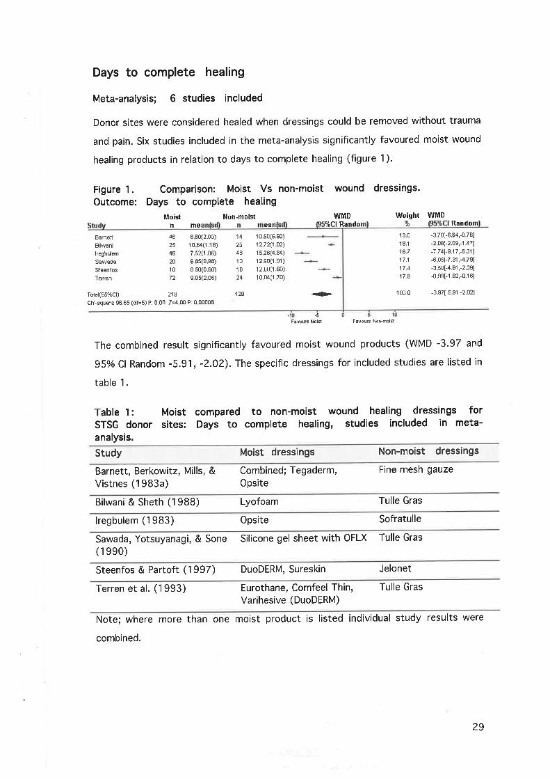
Days to complete healing
Meta-analysis; 6 studies included
Donor sites were considered healed when dressings could be removed without trauma
and pain. Six studies included in the meta-analysis significantly favoured moist wound
healing products in relation to days to complete healing (figure 1).
wound dressings.Figure 1.Outcome:
Bâïnett
E¡lwani
lregþulem
Saweda
SleenfosTerren
Table 1:STSG donoranalysis.
Comparison: Moist Vs non-moistDays to complete healing
Moist Non-moistnme ea
WMDItCl Rand
Weight WffiDlt Rando
46
46
2A
10
72
Ë 80(2.00)
1 0 64(1 .1 8)
7 52(r 06)
6.8s(o.se)
8,s0(0.80)s 05(2 05)
1 o 5o(5.50)
1272(1.OzJ
1 5 26(4.84)
12.S0(1 .91 )12.00(1 .60)
10 04(1 70)
14
46
10
10
24
'129
13018116.7
17.1
174178
-3 70[-6.64,-0 761
-2 08[-2.6S,-',1 47]
-7.74[-9.17,-6.31 I
-6 o5[-7.31 ,-4 791
-3 s0[-4.81 ,-2.39]-0 ss[-1 .82,-0 1 6l
100.0 -3.s7t-5 s1 ,-2.021Total(Ss%Cl) 21S
Chi-square 96 65 (df=S) P: 0 00 Z=4 00 P: 0 00006
+
-5FãvouÉ Èbil FavouË Non.moist
The combined result significantly favoured moist wound products (WMD -3.97 and
95o/oCl Random -5.91, -2.02). The specific dressings for included studies are listed in
table 1.
Moistsites:
compared to non-moist wound healing dressings forDays to complete healing, studies included in meta-
Study Moist dressings Non-moist dressings
Barnett, Berkowitz, Mills, &Vistnes (1983a)
Combined; Tegaderm,Opsite
Fine mesh gauze
Bilwani & Sheth (1988) Lyofoam Tulle Gras
lregbulem (1983) 0psite Sofratulle
Sawada, Yotsuyanagi, & Sone Silicone gel sheet with OFLX Tulle Gras
(1 ee0)
Steenfos & Partoft (1997) DuoDERM, Sureskin Jelonet
Terren et al. (1993) Eurothane, Comfeel Thin,Varihesive (DuoDERM)
Tulle Gras
Note; where more than one moist product is listed individual study results were
combined.
29
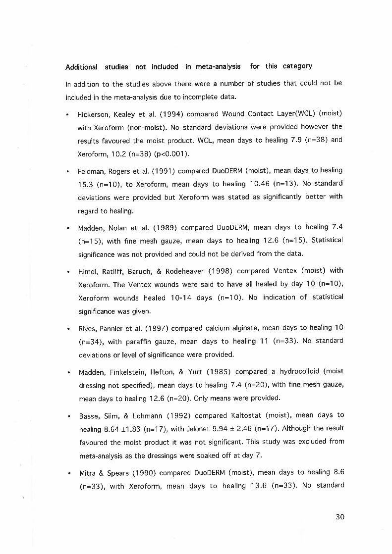
Additional studies not included in meta-analysis for this category
ln addition to the studies above there were a number of studies that could not be
included in the meta-analysis due to incomplete data.
. Hickerson, Kealey et al. (1994) compared Wound Contact Layer(WCL) (moist)
with Xeroform (non-moist). No standard deviations were provided however the
results favoured the moist product. WCL, mean days to healing 7.9 (n=38) and
Xeroform, 10.2 (n=38) (p<0.001).
. Feldman, Rogers et al. (1991) compared DuoDERM (moist), mean days to healing
1 5.3 (n=10), to Xeroform, mean days to healing 10.46 (n=13). No standard
deviations were provided but Xeroform was stated as significantly better w¡th
regard to healing.
. Madden, Nolan et al. (1989) compared DuoDERM, mean days to healing 7.4
(n=15), w¡th fine mesh gauze, mean days to healing 12.6 (n=15). Statistical
significance was not provided and could not be derived from the data.
. Himel, Ratliff, Baruch, & Rodeheaver (1998) compared Ventex (moist) with
Xeroform. The Ventex wounds were said to have all healed by day 10 (n=10),
Xeroform wounds healed 10-14 days (n=10). No indication of statistical
significance was given.
. Rives, Pannier et al. (1997) compared calcium alginate, mean days to healing 10
(n=34), with paraffin gauze, mean days to healing 1 1 (n=33). No standard
deviations or level of significance were provided.
. Madden, Finkelstein, Hefton, & Yurt (1985) compared a hydrocolloid (moist
dressing not specified), mean days to healing 7.4 (n=ZO), with fine mesh gauze,
mean days to healing 12.6 (n:20). Only means were provided.
. Basse, Siim, & Lohmann (1992) compared Kaltostat (moist), mean days to
healing 8.64 r1.83 (n=17), with Jelonet 9.94 t ?.46 (n=17). Although the result
favoured the moist product it was not significant. This study was excluded from
meta-analysis as the dressings were soaked off at day 7.
. Mitra & Spears (1990) compared DuoDERM (moist), mean days to healing 8.6
(n=33), with Xeroform, mean days to healing 13.6 (n=33). No standard
30

deviations were provided but DuoDERM was stated as significantly better with
regard to healing.
Foyatier (1992) compared DuoDERM (moist), mean days to healing 7.7 (n=34),
with Tulle Gras, mean days to healing 13.0 (n=34). No standard deviations were
provided but DuoDERM was stated as significantly better with regard to healing.
Donors not healed at Day 7
Meta-analysis not undertaken as only 1 study provided acceptable data
(figure 2)
Figure 2. Comparison: Moist Vs non-moist dressings. Outcome: Nothealed by day 7
Peto ORMoistn/H
Madden (2) 5 t20
Tstal(95%Cl) 5 f 20
Ch¡-square 0 00 (df=0) P: 1.00 Z=-4 84 P: <0.0t001
Non-moistn/H
2D t20
20 i 20
--ffi-+
.01 .tFãvouË l'!¡hist
Weightlt
1û0 0
100.û
Peto OR
0 û4[0.01 ,0.16]
0.04[0.01 ,0.1Ê]
Cl Fix
Fãvouß Non.moi5t
Madden, Finkelstein et al. (1985) compared a hydrocolloid (moist) with fine mesh
gauze (non-moist). Although this was only one small study, the result favoured the
moist product significantly for healing al day 7.
Additional studies not included in meta-analysis for this category
. Cadier and Clarke (1996) compared Dermasorb (moist) with Jelonet (non-moist).
Results presented were that Dermasorb (n=21) had significantly faster healing
times than Jelonet (n=21). Data was only provided in graphic form, significance
stated as (p<0.002).
Donors not healed at Day IMeta-analysis; 4 studies were combined for this category
The pooled result for donors not healed at day 8 favoured moist wound healing
products (Odds Ratio 0,16, 95%Cl Random 0.02, 1.51) (figure 3). The combined
result was not statistically significant. lt should also be noted the included studies
showed statistically significant heterogeneity (p<0.01). Review of the articles
revealed no clinical reason for this difference between studies.
31

Figure 3. Comparison: Moist Vs non-moist dressings. Outcome: Nothealed at day I
StuMoistn/H
Non-moistn/N
40/401Êi319i101s/16
ORCl Rand
Weightlt
ORY¡Cl Rando
0.01 [0 00,0 211
1 30[0.48,3.541
0.1 1[0.01 ,l .24]
0.17[0 02,1 .721
Ciharrtimur
Davenport
Dernetriades
Smith
20t4t18 /31
5i1010/14
21 .3
30823;l24.1
Totel(9s%Cl) 53 i 95
Chi-squerè 1 4 21 (df=s) P: û 00 Z=1.60 P: <0.00001
Table 2= MoistSTSG donor s¡tes:meta-analysis.
80/97
FavouE [,loist
compared to non-moistDonor s¡tes not healed
1 ot.o 0.16[0.02,1 .51]
60 1000
FavouË Non-moist
healing dressings8, studies included
woundat day
forin
Study Moist dressings Non-moist dressings
Cihantimur, Kahveci, & Ozcan Kaltostat(1 ee7)
Jelonet
Davenport, Dhooghe, &Yiacoumettis (1977)
Lyofoam Tulle Gras
Demetriades & Psaras (1992) Granuflex Tulle Gras
Smith, Thomson, Garner, &
Rodriguez (1994)Hydrocolloid Xeroform
Donors not healed at removal of dressing on Day 9
Meta-analysis not
(figure 4)
undertaken as only 1 study prov¡ded acceptable data
Figure 4. Comparison: Mo¡st Vs non-moist dressings. Outcome: Nothealed at day 9
Feto OR WeightltCl Fix
Peto OR
StuMoistn/H
Madden (2) o t20
Total($S%Cl) O !2oChi-squsre 0.00 (df=0) P: 1 U0 Z=-6.24 P: <0.00001
Hon-moistn/H
20 t2D
20 t20
Fixe
-ffi- 100 0 o o2[0.01 ,0.07]
10ù.û 0.02[0.01 ,o.o7l+
FãvouË hlo¡st Favouß Non-moist
Madden, Finkelstein et al, (1985) compared a hydrocolloid (moist) with fine mesh
gauze (non-moist). The result favoured the moist product significantly'
32

Donors not healed at removal of dressing on Day 10
Meta-analysis; 5 studies were included for this category
Three studies favoured non-moist dressings although not significantly. Pooled analysis
favoured moist dressings overall however not significantly (Odds Ratio 0.7, 9ío/oCl
Random 0.22,2.?0) (figure 5).
Figure 5. Comparison: Moist Vs non-moist dressings. Outcome: Nothealed at day10
Moistn/N
Non-moisln/N
OR Weight[¡¡[
21 .9
21 .S
16 3
2071S.1
OR
1 S7[0.70,5 s2l2 1S[0.78,6.141
1 04[0.18,6 12]
0 21[0.06,0 ¡11
0 1s[0.04,0.591
ltclDavenpoñ
LawrÊnce
Morris
o'Donohue
Tan. S.T. (2)
Table 3: MoistSTSG donor sites:meta-analysis.
1si3113/465t11s i 30
3 i 30
10 ¡31
7i464is
14 t2113i30
------1E-
----{-Total(S5%Cl) 45 / 148
Chi-squsre 17.14 (df=4) P: 0.00 Z=-0.61 F: <0 00001
48 i 137
Favouß híoist FavouÉ Non-mo¡l
compared to non-moist wound healing dressingsDonor sites not healed at day 10, studies included
100 0 o.7tlo22,2.2îl
forin
Study Moist dressings Non-moist dressings
Davenport et al. (1977) Lyofoam Tulle Gras
Lawrence & Blake (1991) Kaltostat Scarlet Red
Morris & Lamb (1990) Opsite Scarlet Red
O'Donoghue et al, (1997) Kaltostat with .?5o/o
BipuvicaineJelonet
Tan, Roberts, & Blake (1993) DuoDERM Scarlet Red
Additional studies not included in meta-analysis for this category
No additional studies were included for this outcome category.
33
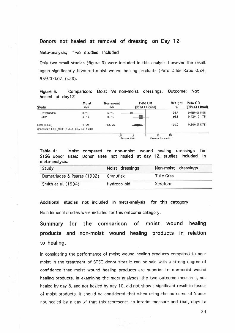
Donors not healed at removal of dressing on Day 1?
Meta-analysis; Two studies included
Only two small studies (figure 6) were included in this analysis however the result
again significantly favoured moist wound healing products (Peto Odds Ratio 0.24,
95o/oC10.07, 0.76).
Figure 6.healed at
Comparison:day"l2
Moistn/N
Moist Vs non-moist dressings. Outcome: Not
StuNon-moist
n/H
13 t26
Peto ORct Fi
Weight1t
34.7
É5.3
Peto ORltCI Fixe
o.o8t0.û1 ,0.s71
0.42t0.10,1 .781
DemEtriades
Smith
Total(S5%Cl) 4 124
Chi-square 1 .80 (df=1) P: 0.41 Z=-2.43 P: 0.01
0i104t14
s/108/1É
+- 100.0 0,2410 07,0.761
I fl0
F¡vouE ltloist F¡voup Non-moist
Table 4: Moist compared to non-moist wound healing dressingsSTSG donor sites: Donor sites not healed at day 1?, studies includedmeta-analysis.
Study Moist dressings Non-moist dressings
forin
Demetriades & Psaras (1992) Granuflex Tulle Gras
smith et al. (1994) Hydrocolloid Xeroform
Additional studies not included in meta-analysis for this category
No additional studies were included for this outcome category.
Summary for the comparison
products and non-mo¡st wound
to healing.
of mo¡st wound
healing products in
healing
relat¡on
ln considering the performance of moist wound healing products compared to non-
moist in the treatment of STSG donor sites it can be said with a strong degree of
confidence that moist wound healing products are superior to non-moist wound
healing products. ln examining the meta-analyses, the two outcome measures, not
healed by day 8, and not healed by day 10, did not show a significant result in favour
of moist products. lt should be considered that when using the outcome of 'donor
not healed by a day x' that this represents an interim measure and that, days to
34

complete healing is a more accurate indicator of performance. lt must also be stated
that for the outcome days to complete healing, the studies included in meta-analyses
lacked a high degree of homogeneity. The breadth of products used in the analysis
would explain this and for this reason a random effects model was used for meta-
analysis with the result still significantly favouring the moist wound products.
Outcome: I B Pain
ln comparing performance of moist and non-moist wound healing dressings in relation
to pain, varying criteria were used including; Dressings rated more painful than
alternate dressing, Overall pain rating using Visual Analogue Scales (VAS) of 1-5, 1-
1 0, 1-1 00 and pain present at day 1 . Results relating to each of these criteria are
presented below.
Dressings rated more painful than alternate dressing
Note, only llTs have been included in this analysis.
Meta-analysis; 3 studies have been included in the analysis
The pooled result for, rating the dressing as more painful, was in favour of the moist
products but not to statistical significance (Odds Ratio 0.80,9ío/oCl Random 0.01,
1.25) (figure 7). Chi-squared analysis revealed that the studies were not significantly
heterogenous. Products used are listed in table 5.
Figure 7.Dressings
Basse
Demetriades
lregbulem
Comparison: Moist Vs non-moist dressings. Outcome:rated as more painful
OR Weight ORlict lt
Moistn/H
Non-moistn/N
10t176 /8
32t50
7 t172t8
0 f 50
383328230
Cl Rand
0 4s[0 13,1 .921
0.11[0.01 ,1 .o7l
o.r1[0.00,0.10]
100.0 o oBt0.01 ,1 .251Totel(gs%Cl) si7s 4at7sChi-square 9.S5 (df=2) P: 0.01 Z=-1.80 P: <0.00001
#
FãvouE lïibist F¡vouE Hon'moi5t
35
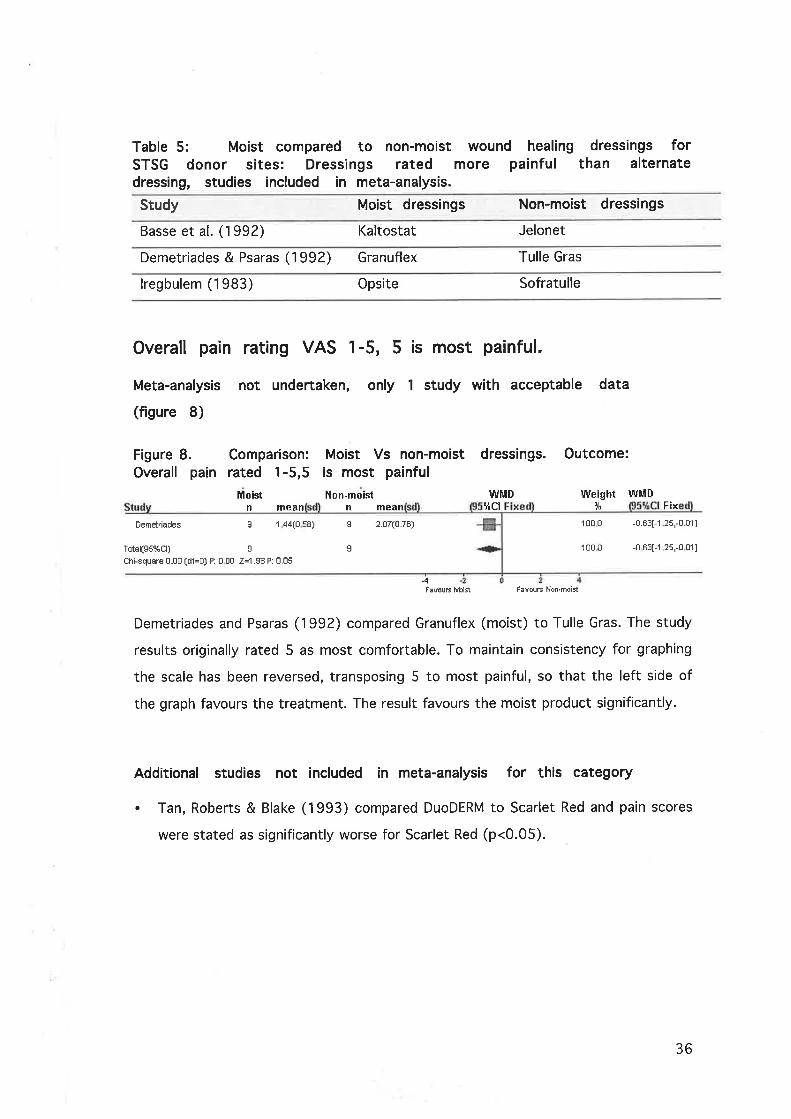
Table 5: Moist compared to non-moist wound healing dressings forSTSG donor sites: Dressings rated more painful than alternatedressing, studies included in meta-analysis.
Study Moist dressings Non-moist dressings
Basse et al. (1992) Kaltostat Jelonet
Demetriades & Psaras (1992) Granuflex Tulle Gras
lregbulem (1983) Opsite Sofratulle
Overall pain rat¡ng VAS 1-5, 5 is most painful.
Meta-analysis not undertaken, only 1 study with acceptable data
(figure 8)
Figure 8. Comparison:Overall pain rated 1-5,5
Moist
Moist Vs non-moist dressings.is most painful
Non-moist WMD
Outcome:
Weight9f
100 0
100 0
WMDFix
-0.Ê3[-1 25,-0 011
-0 63[-1 2s,-0 01 I
n mean
Demelriqdes s 1 44(0 s8)
Total(gs%Cl) s
Chi-square 0.00 (df=O) P: 0 00 Z=1.98 P: 0 05
n mean ltctI
s
2.07(0.76)
.4FãvouË h,loifi FãvouE Non.mo¡l
Demetriades and Psaras (1992) compared Granuflex (moist) to Tulle Gras. The study
results originally rated 5 as most comfortable. To maintain consistency for graphing
the scale has been reversed, transposing 5 to most painful, so that the left side of
the graph favours the treatment. The result favours the moist product significantly.
Additional studies not included in meta-analysis for this category
. Tan, Roberts & Blake (1993) compared DuoDERM to Scarlet Red and pain scores
were stated as significantly worse for Scarlet Red (p<0.05).
36

Overall pain
Meta-analysis
(figure 9).
Figure 9. Comparison:Overall pain rated 1-10,
Moist
Earnett 4E 1 60(0.80)
rotal(gs%Cl) 46
:hi-squere 0 00 (df=o) P: 0.00 Z=5.1 7 P: <0 00001
Moist Vs non-moist10 is most painful
Non-moist
rat¡ng VAS 1-10, 10 is most painful.
not undertaken as only 1 study provided acceptable data
dressings. Outcome:
WMDFix
Weight WMDn mean
4.7o(2.20)
Fixe
-3.1 0[-4 28,-1 .921
-3 10[-4.28,-1 .92]
14
14
#+
100.0
100 0
.10
Fãvouß Mo¡st F¡vouß Non.moil
Barnett, Berkowitz et al. (1983a) compared Tegaderm and Opsite, with the control,
fine mesh gauze. For this study the individual results of the two moist wound
products were combined. The result significantly favoured the moist wound healing
products.
Additional studies not included ¡n meta-analysis for this category
. Hickerson, Kealey et al. (1994) compared Wound Contact Layer, mean pain score
?.94 (n=38), with Xeroform, mean pain score 4.64, (n=38). No standard
deviations were given however the result was stated as significantly favouring the
moist product (p<0.001 ).
. Feldman, Rogers et al. (1991) compared DuoDERM, mean pain score 0.53 (n=10),
with Xeroform, mean pain score 2.41 (n=13). No standard deviations were given
however the result was stated as significantly favouring the moist product
(p=0.01).
37

Overall pain rating VAS 1-100, 100 is most painful.
Meta-analysis was not undertaken, only 1 study with acceptable data
(figure 10)
Figure 10.Overall pain
Mâddên 15 1430(400)
Tolsl(gs%Cl) 15
Chi-squarÊ 0 00 (df=o) P: 0.00 Z=8.87 P: <0.00001
Figure 1 1. Comparison:Overall pain rated 1-10,
Moistme
Comparison: Moist Vs non-moist dressings.rated 1-100, 100 is most painful
Moist Non-moist WMDmean n mea Fi
Outcome:
Weight WMD9t 9tCl Fixe
-10 8o[-13 1S,-8.41]15
15
100.0
100.0
2s 1 o(2.s0)
4.70(2.20)
4.14(1 .s2)
2.51(0.25)
+ -10.80[-13.1s,-8 41 I
Madden, Nolan et al. (1989) compared DuoDERM with fine mesh gauze. Although the
result significantly favours the moist product caution should be taken, as the sample
was relatively small (n=15).
Overall pa¡n rat¡ng VAS studies converted to the un¡form
scale of 1-10, 1 0 is most painful.
Meta-analysis: 3 studies included (figure 1 1)
For this meta-analysis the results of different visual analogue scales were converted
to achieve a single uniform scale of 1-10. The pooled result favoured the moist
products significantly, although there is still a lack of homogeneity for the combined
VAS 1-10 scale outcome (WMD -1.75 and 95% Cl Random -2.94, -0.56).
FavouE h,loist
F¡vouË lllo¡l
FãvouË Non-mo¡st
FavouE Non-mo¡l
BärnÉ:tt
Demetriades
Madden
46
I10
1.60(0,80)
2.88(1 .18)
1.40(0.40)
Moist Vs non-moist dressings. Outcome:10 is most painful (multiple ratings combined)Non-moil W|ìID Weight WMD
n Ra lt Rando
14 -3.10[-4.28,-1 921
-1 .26[-2.s1 ,-0 01 I
-1.111-1 40,-0 821
---s- 2SS
28.8
41 3
I10
33 +Total(gs%Cl) 65
Chi-squarB 1 0.37 (df=2) P: 0 01 Z=2 89 P: 0 004
100.0 -1 75[-2.94,-0.56]
38

Table 6: Moist compared to non-moist wound healing dressings forSTSG donor sites: Overall pain VAS rated 1-10, (multiple ratingscombined) studies included in meta-analysis.
Study Moist dressings Non-moist dressings
Barnett et al. (1983a) Tegaderm, Opsite Fine mesh gauze
Demetriades & Psaras (1992) Granuflex Tulle Gras
Madden et al. (1989) DuoDERM Fine mesh gauze
Pain present at day 1
Meta-analysis: 2 studies included (figure 12)
The two studies have a high degree of homogeneity and the pooled result
demonstrates a significant result in favour of the moist products in relation to pain
present at day 1 (Odds Ratio 0,00, 95o/o C10.00, 0.04).
Moist Vs non-moist dressings. Outcome: PainFigure 12.present at
Comparison:day 1
Moistn/N
0 /251i 10
StuHon-moist
n/H
25 t2510i10
ORRando
Weightlt
43256.8
ORlËCl Rando
0.00[0.r]0,0.021
0.01[0 00,0 211
Bilwani
Himel
+-<__#_slÞ-
FãvouE fufoi't
Tttal(ss%Cl) 1i 35
Chi-square 1 27 (dl=1) P: 0 26 Z=-4 13 P: 0 2
1oú.0 o oolo oo,0.o4]
1000
FavouE Non-moil
Table 7: Moist compared to non-moist wound healing dressings forSTSG donor sites: Pain present at day 1, studies included in meta-analysis.
Study Moist dressings Non-moist dressings
Bilwani & Sheth (1988) Lyofoam Tulle Gras
Himel et al. (1998) Ventex Xeroform
39
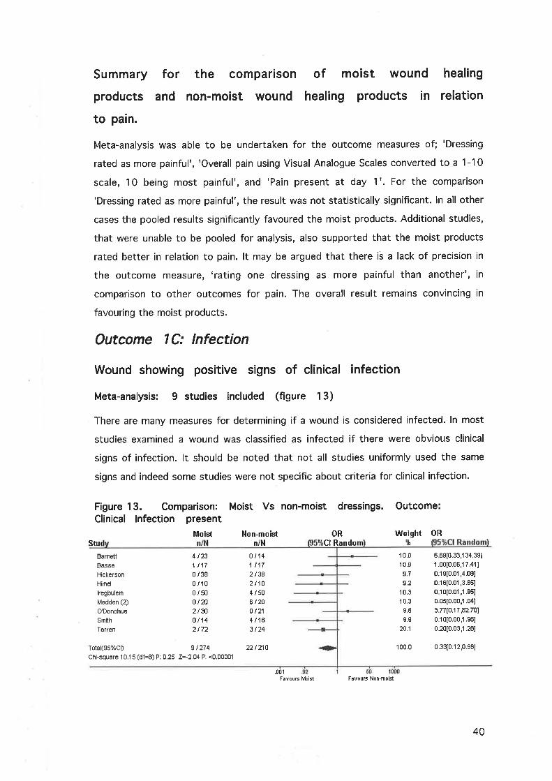
Summary
products
to pain.
Stu
Barnett
Basse
Hickerson
Himel
lregbulen
Madden (2)
O'Donohue
Smith
Teren
for the compar¡son
and non-mo¡st wound
of mo¡st wound
healing products in
healing
relation
Meta-analysis was able to be undertaken for the outcome measures of; 'Dressing
rated as more painful', 'Overall pain using Visual Analogue Scales converted to a 1-1 0
scale, 10 being most painful', and 'Pain present at day 1 '. For the comparison
'Dressing rated as more painful', the result was not statistically significant. ln all other
cases the pooled results significantly favoured the moist products. Additional studies,
that were unable to be pooled for analysis, also supported that the moist products
rated better in relation to pain. lt may be argued that there is a lack of precision in
the outcome measure, 'rating one dressing as more painful than another', in
comparison to other outcomes for pain. The overall result remains convincing in
favouring the moist products.
Outcome |C: lnfection
Wound showing pos¡t¡ve signs of clinical infection
Meta-analysis: 9 studies included (figure 13)
There are many measures for determining if a wound is considered infected. ln most
studies examined a wound was classified as infected if there were obvious clinical
signs of infection. lt should be noted that not all studies uniformly used the same
signs and indeed some studies were not specific about criteria for clinical infection.
Figure 13. Comparison:Clinical lnfection present
Moist
Moist Vs non-moist dressings. Outcome
OR
4 t231t170 /380 /100 /5uo t202 130
ûi142 t72
ot141r172 l3A2t104 i s0
Ê t2tot214/1Ë3 t24
l,lon-moistn/H
ORCl Rando
Weight5t
10.0
10.9
9.7
9.2
10.3
10.3
SE
s.s
20:l
100.0 0.33[0.12,0.s61
6 6S[0.33,134.3S1
1 .00[0.0Ê,1 7 411
0.1st0.01 ,4.081
0 16[0.01 ,3.851
0 10[0.01 ,'l .951
0.0s[0.00,1 .04]
3.77[0.17,82.701
0.10[0.00,1 .s6l
0.20[0.03,1 .28]
Total(gs%cl) 91274Chi-squarÊ 1û 15 (df =8) P: O.25 Z=-2.O4 P: <0.00001
.001
22 t210
Favouß h,'loist FavouË Non.moist
40
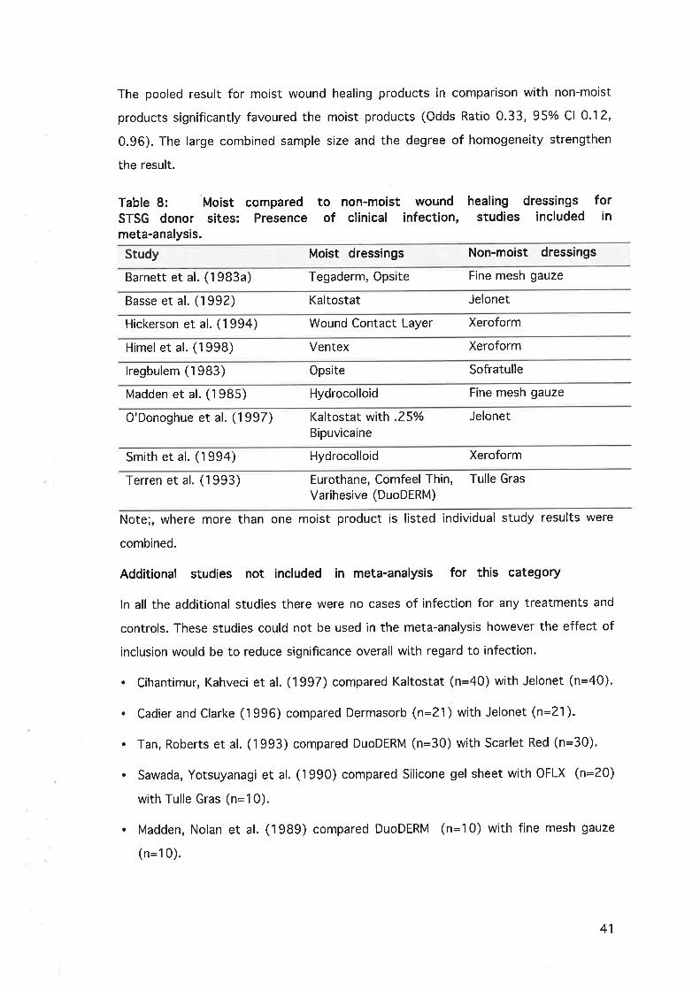
The pooled result for moist wound healing products in comparison with non-moist
products significantly favoured the moist products (Odds Ratio 0.33,95o/o Cl 0.12,
0.96). The large combined sample size and the degree of homogeneity strengthen
the result.
Table 8: Moist comparedSTSG donor sites: Presencemeta-analysis.
to non-moist woundof clinical infection,
healing dressingsstudies included
forin
Study Moist dressings Non-moist dressings
Barnett et al. (1983a) Tegaderm, Opsite Fine mesh gauze
Basse et al. (1992) Kaltostat Jelonet
Hickerson et al. (1994) Wound Contact Layer Xeroform
Himel et al. (1998) Ventex Xeroform
lregbulem (1983) Opsite Sofratulle
Madden et al. (1985) Hydrocolloid Fine mesh gauze
O'Donoghue et al. (1997) Kaltostat with .25%Bipuvicaine
Jelonet
Smith et al. (1994) Hydrocolloid Xeroform
Terren et al. (1993) Eurothane, Comfeel Thin, Tulle Gras
Varihesive (DuoDERM)
Note;, where more than one moist product is listed individual study results were
combined.
Additional studies not included in meta-analysis for this category
ln all the additional studies there were no cases of infection for any treatments and
controls. These studies could not be used in the meta-analysis however the effect of
inclusion would be to reduce significance overall with regard to infection.
. Cihantimur, Kahveci et al. (1997) compared Kaltostat (n=40) with Jelonet (n=40).
. Cadier and Clarke (1996) compared Dermasorb (n=21) with Jelonet (n=21).
. Tarì, Roberts et al. (1993) compared DuoDERM (n=30) with Scarlet Red (n=30).
. Sawada, Yotsuyanagi et al. (1990) compared Silicone gel sheet with OFLX (n=20)
with Tulle Gras (n=10).
. Madden, Nolan et al. (1989) compared DuoDERM (n=10) with fine mesh gauze
(n=10).
41
a Steenfos and Partoft (1997) compared DuoDERM combined with Sureskin (n=20),
with Jelonet (n=10).
Bilwani and Sheth (1988) compared Lyofoam (n=25) with Paraffin Gauze (n=25).
Overall summary for mo¡st wound healing products in
compar¡son to non- mo¡st wound healing products.
There were sufficient studies of adequate quality to conduct meta-analysis for all
outcomes for this comparison. The moist category of products was found to be
superior for the outcomes of healing time, pain, and infection rate. ln general the
studies lacked homogeneity but this could be expected when considering the variety
of products included within this broad category. To compensate for this lack of
homogeneity a random effects model was used in meta-analysis where a broad range
of moist wound healing products were pooled. Therefore the results are quite
convincing in suggesting that moist wound healing products are superior to
traditional non-moist products.
The following group of comparisons examine studies in which various
generic moist wound healing products are compared to other
treatments. This provides a more specific view of the individual
generic moist products than is the case when pooling all the moist
products together.
a
42

Comparison 2= Calcium alginates are compared with
non-moist (non-biological) wound healing products in
the management of STSG donor sites;
Note, biological products have not been included in this comparison, non-moist
wound healing products, mainly gauze products (impregnated with o¡ntment or
without) of various types have been included.
Outcome 2A: Healing
ln comparing performance of calcium alginates and non-moist wound healing
dressings in relation to healing times, varying criteria were used including; Days to
complete healing, number of wounds healed by day 8, or 10. Results relating to each
of these criteria are presented below.
Days to complete healing, all studies required complete
epithelial cover with dressing able to be removed without
trauma
Meta-analysis not conducted due to insufficient studies and incomplete
results
One study of acceptable design by Rives, Pannier et al. (1997) compared calcium
alginate, mean days to healing 10 (n=34), with paraffin gauze, mean days to healing
1 1 (n=33). No standard deviations were provided and the author stated that healing
times were 'comparable'. lt was noted that the calcium alginate donors were able to
be re-harvested earlier.
Donors not healed at removal of dressing on Day 8
Meta-analysis not conducted due to insufficient studies.
43

Figure 14. Comparison: Calcium alginates Vs non-moist dressings.Outcome: Not healed at day I
WeightE¡J!
Peto ORltCl Fixe
cihûniinur 20 i 4ú
Total(95%Cl) 20 l4tChi-square 0.00 (df=O) P: 1 00 Z=-5.13 P: <0 00001
Calc Alg Non-moistn/H n/H
4û /40
40i40
-#-+
1U0.0
10ú.0
55.S
44.1
Peto 0RF¡
0.07t0.03,û 201
0,07[0.03,0.201
Peto 0R
2.13[0 80,s.72]
0 23[0.08,0.71]
Favous Calc Ag Favous Non-moisl
Cihantimur, Kahveci et al. (1997) compared Kaltostat (CA) with Jelonet and although
only a single study the result was highly significant in favour of the calcium alginate.
Donors not healed at removal of dressing on Day 10
Meta-analysis 2 studies included (figure 1 5)
The studies used in the analysis were not homogenous and the result was not
significant. Little can be drawn from this meta-analysis (Peto Odds Ratio 0.81' 95%
Cl 0.39, 1.68) (figure 15).
Figure 15.outcome:
Vs non-moist dressings.
Pefo OR lllfeightnCl Fix F¡
Comparison: Calcium alginatesNot healed at day 10
Calc Alg Non-moistn/H n/H
LawrencÊO'Donohue
13/48s /30
7i46't4 t21
21 tÊ7
---F
Total(ss%Cl) 2217Ê
Chi-squûre 8.52 (df=1) P: 0 01 Z=-0.57 P: 0 6
F¡vouE C¡lc Ag
Table 9: Calcium alginate comparedbiological dressings for STSG donor sites:studies included in meta-analysis.
1000 u81[0.39,1 68]
FãvouE Non-moist
to other non-moist, non-Donor not healed at day 10,
Study Calcium alginates Non-moist dressings
Lawrence & Blake (1991) Kaltostat Scarlet Red
O'Donoghue et al. (1997) Kaltostat with .25%Bipuvicaine
Jelonet
44

Outcome 28: Pain
Patient requiring analgesia
Meta-analysis not conducted due to insufficient studies.
Vs non-moist dressings.Figure 16.outcome:
Comparison: Calcium alginatesPatient required analgesia
Calc Alg Hon-moistniH n/N
17 i 33 18i31Rives
Total(95%Cl) 17 i 33
Chi-square 0.00 (df=0) P: 1.00 Z=-0.52 P: 0.Ë
Figure 17.Outcome:
18 i 31
Peto ORCl Fixe
Feto ORCI F¡
WeightIlt!
Weight9t
Peto OR1ËCl Fixe
Peto ORlfCl Fixe
1û0.0 o 77[0.2s,2 os]
1t0 0 0.77t0.29,2.0s1
FãvouË Cãlc Ag FãvouÉ Non-moist
This single study by Rives, Pannier et al. (1997) showed no significant difference
between the calcium alginate and the paraffin gauze dressings.
Dressings rated more painful than alternate dressing, only
intra-individual studies included
Meta-analysis not conducted due to insufficient studies.
Vs non-moist dressings.Comparison: Calcium alginatesDressings rated as more painful
Calc Alg Non-moistn/H n/H
Basse 7 I 17
Totnl(ss%cl) 7 t 17
Chi-square 0.00 (df=0) P: 1.00 Z=l .01 F: 0.3
10i17 100.0 o.s0[0.1 3,1 .s0]
10/17 100.0 0.50[0.13,1 .901
Favous Calc Ag F¡vouE Non-moist
This single intra-individual study by Basse, Siim et al. (1992) whilst favouring the
Kaltostat (CA) did not achieve a significant result compared with Jelonet.
45

Outcome 2C: Infection
Wound showing positive signs of clinical infection
Meta-analysis 2 studies included
Neither of the two studies demonstrated a significant difference of infection rates
between treatments. The pooled analysis also failed to find a significant difference
(Peto Odds Ratio 2.36, 95% Cl 0.32, 17.45).
Figure 18.Outcome:
Basse
O'Donohue
Table 10:STSG donoranalysis.
Comparison: Calcium alginatesClinical infection present
Calc Alg Hon-moistn/H n/H
Vs non-moist dressings.
Weight¡¡t0
Peto ORFixe
s0.5
4S.5
Peto OR%Cl Fixe
1 .00[0.06,16.6S1g.Ê7[0.33,s7.4s1
111-l2 t30
1t17E t21
1/38 100.0 2 36[0.32,17.45]Total(95%Cl) 3l4fChi-squsre 0.72 (df=1) P: 0.70 Z=0.84 P: 0.4
F¿vouE Cälc Ag
Calcium Alginate compared tosites: Clinical infection present,
non-moist dressings forstudies included in meta-
FãvouE Non-mo¡Et
Study Calcium alginates Non-moist dressings
Basse et al. (1992) Kaltostat Jelonet
O'Donoghue et al. (1997) Kaltostat with .25%Bipuvicaine
Jelonet
Additional studies not included in meta-analysis for this category
. An additional study by Cihantimur, Kahveci et al. (1997) compared infection rates
between Kaltostat and Jelonet but was not able to be included in the meta-
analysis as neither treatment nor control had any infections.
Overall summary for calc¡um alginates in compar¡son to non-
mo¡st wound healing products.
ln comparing calcium alginates with non-moist wound healing products there were
very few studies of adequate quality for any of the outcome categor¡es. ln
considering healing there was only one outcome, healing at day 8, that demonstrated
a statistical significance between treatment and control groups and this was the
46

result of only one study. ln considering the outcomes of pain and infection no
conclusions can be drawn, as there are insufficient studies to make a judgement.
Comparison 3: Calcium alginates compared w¡th other
mo¡st wound healing products in the management of
STSG donor sites;
Note;, there were no studies comparing calcium alg¡nates with other
moist wound healing products that contained sufficient data for
analysis.
One study by Porter (1991) did compare a calcium alginate (Kaltostat), with another
moist wound healing product, a hydrocolloid (Granuflex E). Although the result
favoured the hydrocolloid with mean days to healing of 10.0 compared with the
calcium alginate 15.5 days. The results were unable to be used in analysis as part of
the calcium alginate group was also dressed with Tulle Gras after the first dressing
change. ln addition some of the patients were also autografted and ¡t is not clear if
these patients were also included in the results.
Comparison 4= Comparison between calcium alginates
in the management of STSG donor sites;
Note;, there were no studies of comparisons between calcium alginates
that contained sufficient data for analysis.
An additional attraction in using calcium alginates is the reported haemostatic
qualities. Although none of the studies investigating the use of these products w¡th
STSG donors specifically measured outcomes of haemostasis many authors support
this view (Cihantimur et al., 1997; Porter, 1991; Williams, 1994).
47
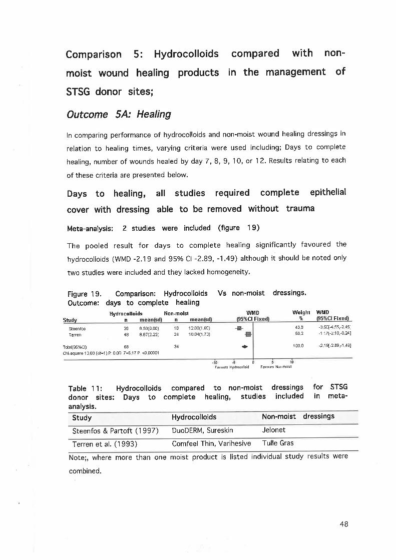
Comparison 5: Hydrocolloids
moist wound healing products
STSG donor sites;
Figure 19. Comparison: HydrocolloidsOutcome: days to complete healing
Hydrocolloids Non-moisiStu
compared w¡th non-
in the management of
Vs non-moist dressings.
WMD Weight WMDyICI F Fix
Outcome 5A: Healing
ln comparing performance of hydrocolloids and non-moist wound healing dressings in
relation to healing times, varying criteria were used including; Days to complete
healing, number of wounds healed by day 7, 8, 9,10, or 1 2. Results relating to each
of these criteria are presented below.
Days to healing, all studies requ¡red complete epithelial
cover w¡th dressing able to be removed w¡thout trauma
Meta-analysis: 2 studies were included (figure 19)
The pooled result for days to complete healing significantly favoured the
hydrocolloids (WMD -?.19 and 95% Cl -2.89, -1.49) although it should be noted only
two studies were included and they lacked homogeneity'
n liSteenfos
Tefien
20
48
10
24
I s0(0 80)
I 87(2.23)
1 2 00f 60)
1 0.04(1 70)
438562
100.0
dressingsincluded
-3 50[-4 55,-2 45]
-1 ,17f-210,-0.241
-2 1 9[-2.8S,-1 49]
-&-
FavousHydrcoolloid FavouÈNon'mo¡l
Total(gs%Cl) 68
Chi-square 1 0 E0 (df=1 ) P: 0 00 Z=6 1 7 P: <Û 00001
34 +
Table 1 1: Hydrocolloids compared to non-moistdonor sites: Days to complete healing, studiesanalysis.
forin
STSG
meta-
Study Hydrocolloids Non-moist dressings
Steenfos & Partoft (1997) DuoDERM, Sureskin Jelonet
Terren et al. (1993) Comfeel Thin, Varihesive Tulle Gras
Note;, where more than one moist product is listed individual study results were
combined.
48
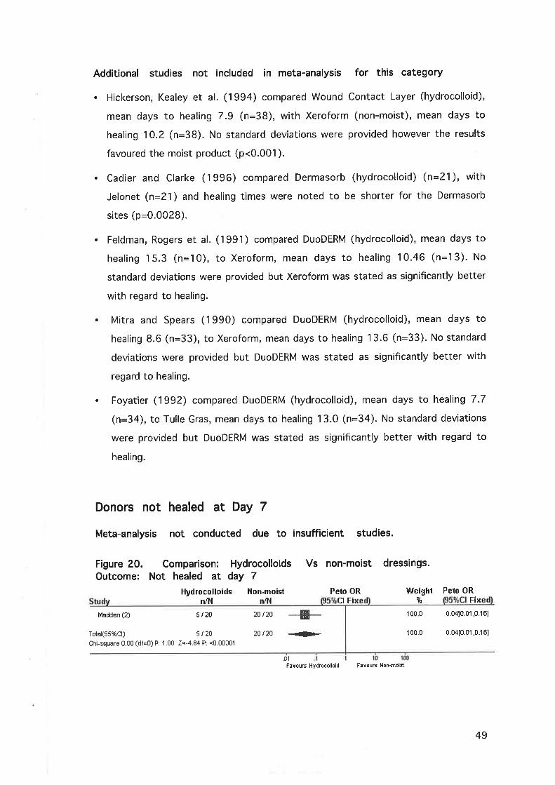
Additional studies not included in meta-analysis for this category
. Hickerson, Kealey et al. (1994) compared Wound Contact Layer (hydrocolloid),
mean days to healing 7.9 (n=38), with Xeroform (non-moist), mean days to
healing 10.2 (n=38). No standard deviations were provided however the results
favoured the moist product (p<0.001).
. Cadier and Clarke (1996) compared Dermasorb (hydrocolloid) (n=21), with
Jelonet (n=21) and healing times were noted to be shorter for the Dermasorb
sites (p:0.0028).
. Feldman, Rogers et al. (1991) compared DuoDERM (hydrocolloid), mean days to
healing 15.3 (n=10), to Xeroform, mean days to healing 10.46 (n=13). No
standard deviations were provided but Xeroform was stated as significantly better
with regard to healing.
. Mitra and Spears (1990) compared DuoDERM (hydrocolloid), mean days to
healing 8.6 (n=33), to Xeroform, mean days to healing 13.6 (n=33). No standard
deviations were provided but DuoDERM was stated as significantly better with
regard to healing.
. Foyatier (1992) compared DuoDERM (hydrocolloid), mean days to healing 7.7
(n=34), to Tulle Gras, mean days to healing 13.0 (n=34). No standard deviations
were provided but DuoDERM was stated as significantly better with regard to
healing.
Donors not healed at Day 7
Meta-analysis not conducted due to insufficient studies.
Figure 20.outcome:
Comparison: HydrocolloidsNot healed at day 7
Hydrocolloids Hon-moistn/N n/l'l
20 t20 --F2Ol2O {Þ-
.01
Vs non-moist dressings.
Peto OR
Madden (2) 5/20
Total(S5%Cl) 5 t 20
Chi-square 0.00 (df=O) P: 1.00 Z=-4.84 P: <0,00001
CI F¡
Peto ORFix
1oo.o o.o4[0.01 ,o.1El
100.0 0.04[0.01 ,0.16]
Weightlt
FavousHydrocolloid F¡vousNon'moist
49

Madden, Finkelstein et al. (1985) compared a non-specified hydrocolloid with fine
mesh gauze demonstrating a significant difference in donors healed at day 7
favouring the hydrocolloid.
Donors not healed at Day 8
Meta-analysis 2 studies included
Although neither study included in the meta-analysis provided a significant result
both favoured the hydrocolloid. The combined result however did achieve a
significant result in favour of the hydrocolloids (Peto Odds Ratio 0.19, 95% Cl 0.05,
0.70) (fisure 21).
Vs non-moist dressings.Figure 21
outcome:
Derne{riades 5i1010 i 14
Total(95%Cl) 15 124
Chi-square 0.04 (df=1 ) P: 0.38 Z=-2 48 P: 0 û1
Table 1?= Hydrocolloidsdonor sites: Donor notanalysis.
Comparison: HydrocolloidsNot healed at day 8
Hydrocolloids Hon-moistn1l'l niH
Smilh
Peto ORFix
Weight[¡,tr
50.7
4S.3
Feto ORCl Fix
0.16t0.03,1 .061
0 21[r] f13,1 .40I
s /1015i16
24 t26 + 100 0 0.1sto.0s,0.701
.0t .tFavourHydrocolloid FavoußNon-moist
to non-moist dressingsday 8, studies included
comparedhealed at
forin
STSG
meta-
Study Hydrocolloids Non-moist dressings
Demetriades & Psaras (1992) Granuflex Tulle Gras
smith et al. (1994) Hydrocolloid Xeroform
50

Donors not healed at Day 9
Meta-analysis not conducted due to ¡nsufficient studies.
Figure ??.outcome:
Comparison: HydrocolloidsNot healed at day 9
Hydrocolloids Non-moistn/H niN
2D Ì2O
20 tzt
Comparison: HydrocolloidsNot healed at day 10
Hydrocolloids Non-moistn/H n/H
3 /30 13 i 30
Vs non-moist dressings
Peto OR
Madden (2) E t2E
Total(ss%cl) 0 / 20
Chi-squnre 0 00 (df=O) P: 1.û0 Z=-6.24 P: <0.00001
-ffi-+
Fixe
Peto ORllCl Fix
Weightit
100.0
1f10.0
Weight9t
Peto ORlictFi
0 t?to.01 ,0.071
0 f12t0.01 ,0.071
Peto ORFixe
50
FBvouEHydmcolloid F¡vousNon.moist
Madden, Finkelstein et al. (1985) compared a non-specified hydrocolloid with fine
mesh gauze demonstrating a significant difference in donors healed at day 9
favouring the hydrocolloid.
Donors not healed at Day 10
Meta-analysis not conducted due to insufficient studies.
Vs non-mo¡st dressingsFigure 23.outcome:
Tan. S.T. (2)
Tstal(Ss%Cl) 3 i 30
Ch¡-square 0.00 (df=0) P: 1.00 Z=-2.89 P: 0.004
-Æ- 100.0 o.1s[0.06,0.581
100.0 0.1s[0.oE,0.sel+.01
FavousHydmoolloid F¡vousHon-moist
Tan, Roberts et al. (1993) compared a DuoDERM (hydrocolloid) with Scarlet Red
demonstrating a significant difference in donors healed at day 10 favouring the
hydrocolloid.
Donors not healed at Day 12
Meta-analysis 2 studies included
The meta-analysis favoured the hydrocolloids (Peto Odds Ratio 0.24, 95o/o Cl 0.07,
0.76). The result should be viewed with some caution, as the combined sample size
is still small.
13i30
51

Figure 24. Comparison: Hydrocolloids Vs non-moistOutcome: Not healed at day 12
Hydrocolloids Non-moist Peto ORniN n/H liCl Fix
DBmBtriades --------{-
+
FavousHydmcolloid F¡vousNon-mo¡st
34.7
65.3
100.0
non-mo¡st dressings12, stud¡es included
dressings.
Weight Peto ORCl Fixe
0.08[0.01 ,o.s7l0.42[o 10,1 781
0.24[0.07,0.76]
Il¡E
0i104i14
Total(gs%cl) 4 124
Chi-square 1 80 (df=1) P: 0.41 Z=-2.43 P: 0.û1
Smith
5 /108/16
13 /26
Table 13: Hydrocolloids compareddonor sites: Donor not healed atanalysis.
today
forin
STSGmeta-
Study Hydrocolloids Non-moist dressings
Demetriades & Psaras (1992) Granuflex Tulle Gras
Smith et al. (1994) Hydrocolloid Xeroform
Outcome 58: Pain
Dressing rated as more painful
Meta-analysis not conducted due to insufficient studies
Figure 25Outcome:
Demetriades
Comparison: Hydrocolloids VsDressing rated as more painful
Hydrocolloids Hon-moistn/N n/H
2t9 6/9
non-moist dressings.
Peto ORltct F
lllleight1ü
100.0
100.0
Peto ORct Fi
0.1810.03,1 .111
0.1rt0.03,1 .11 ITotal(gs%Cl) 2 t IChi-square 0.00 (df=o) P: 1.00 Z=-1 .84 P: 0.07
6is
l0FavousHydrcoolloid F¡vousNon-moist
Demetriades and Psaras (1992) compared a Granuflex (hydrocolloid) with Tulle Gras
demonstrating a significant difference in, dressing rated as more painful, favouring
the hydrocolloid. Please note, in this study one patient rated both dressings as equal.
52

Overall pain rated l -5, 5 ,s most painful
Meta-analysis not conducted due to insufficient studies
Figure 26. Comparison: Hydrocolloids Vs non-moistOutcome: Overall pain rated 1-5, 5 is most painful
Hydrocolloids Non-moisi WlìlDFix
dressings.
nmWeight
5t
WMDCl Fixe
DemBtriades s 1.44(0.s8) I 2.07(0.76) 100.0 -0 63[-1 .25,-0.01 I
Totäl(gs%Cl) S
Chi-squere 0 00 (df=0) P: 0 00 Z=1.98 P: 0 05
100 0 -o 63[-1.25,-o.o1l
FavouËHydrccolloid F¡vouENon-moist
ln the same study as the previous comparison when the 1-5 overall pain scale was
used in comparing Granuflex (hydrocolloid) and Tulle Gras the result significantly
favoured the hydrocolloid however this single study had only a small sample (n:9)
(Demetriades & Psaras, 1992).
Overall pa¡n rat¡ng VAS 1-100, 100 is most painful.
Meta-analysis was not undertaken, only 1 study with acceptable data
(figure 10)
Madden, Nolan et al. (1989) compared DuoDERM with fine mesh gauze. Although the
result significantly favours the moist product caution should be taken, as the sample
was quite small (n=15).
Additional studies not included in meta-analysis for the outcome pain
. Hickerson, Kealey et al. (1994) using a VAS of 1-10 with 10 most painful,
compared Wound Contact Layer (hydrocolloid), mean pain score 2.94 (n=38),
with Xeroform (non-moist), mean pain score 4.64 (n=38). No standard deviations
were provided however the results favoured the hydrocolloid (p<0.001).
. Tan, Roberts et al. (1993) compared DuoDERM to Scarlet Red and pain scores
were stated as significantly worse for Scarlet Red (p<0.05).
. Feldman, Rogers et al. (1991) compared DuoDERM, mean pain score 0.53 (n=10),
to Xeroform, mean pain score 2.41 (n=13) No standard deviations were given
however the result was stated as significantly favouring the hydrocolloid (p=0.01).
s
53

Outcome 5C: lnfection
Wound showing positive signs of clinical infection
Meta-analysis 4 studies included
Although 3 of the 4 studies included did not achieve a significant result the combined
result favoured the hydrocolloids significantly (Peto Odds Ratio 0.21, 95o/o Cl 0.07,
0.65) with a considerable degree of homogeneity.
Figwe 27. Comparison:Outcome: Clinical lnfection
Hydrocolloidsn/H
Hydrocolloidspresent
l{on-moistniN
0i132 i 38
6 t203 t24
Vs non-moist dressings
Peto ORFix
Feldmsn
Hickerson
Madden (2)
Terren
1t100i380/202t4A
7.S
15 S
42.2
33.9
dressingsincluded
100 0 0.21 [0.07,0.6s1
Weightit
Peto ORFixe
s.s7[0.19,51S 37]
0 13[0 01 ,2 1sl0.10[0.02,0 s6]
0 28[0.04,1 .S0l
---#-
Total(gs%cl) 3i116Chi-square 4.56 (df=3) P: û.34 Z=-2.71 P: 0 007
11 i 95 +
.001 .02
FavouËHydrccolloid FavouÉHon-moil
Table 14: Hydrocolloids compared to non-moistdonor sites: Clinical infection present, studiesanalysis.
forin
STSG
meta-
Study Hydrocolloids Non-moist dressings
Feldman et al. (1991) DuoDERM Xeroform
Hickerson et al. (1994) Wound Contact Layer Xeroform
Madden et al. (1985) DuoDERM Fine Mesh Gauze
Terren et al. (1993) Comfeel Thin, Varihesive Tulle Gras
Note;, where more than one moist product is listed individual study results were
combined.
Additional studies not included in meta-analysis for this category
One additional study was unable to be included in the meta-analysis. For this study
there were no reported infections.
. Steenfos and Partoft (1997) compared DuoDERM combined with Sureskin (n=20),
with Jelonet (n=10).
54
Overall summary for hydrocolloids in comparison to non-
moist wound healing products.
ln considering healing there were three outcome categories; days to healing, healed
at day 8, and healed at day 12 where meta-analysis was possible, although in each
case only 2 studies were combined. The outcome categories of healed al day 7, 9,
and 10 each had only one study of adequate quality. A statistically significant result
was demonstrated for all of the healing categories where meta-analysis was possible'
Only one study produced a result that significantly favoured the non-moist product
(Feldman et al., 1991). Despite the relatively small number of studies this indicates
with considerable confidence that hydrocolloids are superior to non-moist wound
healing products in relation to healing. ln considering the outcomes of pain it was not
possible to combine any studies for meta-analysis however a number of single studies
did significantly favour the hydrocolloids. Although the number of studies is small it
can be stated with some confidence that hydrocolloids out-perform non-moist
products in relation to pain. ln relation to infection the meta-analysis significantly
favoured the hydrocolloids.
Comparison 6: Hydrocolloids
mo¡st wound healing products
STSG donor s¡tes;
Outcome 6A: Healing
compared w¡th other
in the management of
ln comparing performance of hydrocolloids and other moist wound healing dressings
in relation to healing times, the following criteria were used; Days to complete healing
and number of wounds healed by day 10. Results relating to these criteria are
presented below.
55

Days to
epithelial
trauma
Figure 28.Outcome:
Leicht
Tefien
Table 15:donor sites:analysis.
complete healing all studies requ¡red complete
cover with dressing able to be removed w¡thout
Meta-analysis: 2 studies included
The pooled result in comparing healing of hydrocolloid treated donors and those with
other moist wound healing products significantly favoured the hydrocolloids (WMD -
1.45 and 95% Cl -2.17, -0.74) although it should be noted only two studies were
included (figure 28).
Vs other moist dressings.Comparison: HydrocolloidsDays to complete healing
Ïydrocolioid bther moist WlrlDmea
7 630 0E)
I B7(2 23)1 0 60(1 ,30)
e.41(1 .61)
Fi
dressingsincluded
Weight WNDìlr Cl Fix
37.6 -2.S7[-4.13,-1 811
62 4 -0.s4[-1 .44,0 36]
100,0 -1 .4s[-2 17,-0.741
for STSG
in meta-
48
ú
24
32
-t-
Total(S5%C¡) 5E
Chi-squaïe 1 0 48 (dl=1 ) F: 0.00 Z=4 00 P: 0.00006
+
5
FãvouÉ Hydrocollold Favouß OlhÊr moist
Hydrocolloids compared to other moistDays to complete healing, studies
Study Hydrocolloids Other moist dressings
Leicht, Siim, & Sorensen (1989) DuoDERM Omiderm
Terren et al. (1993) Comfeel Thin,Varihesive
Eurothane
Note;, where more than one moist product is listed individual study results were
combined.
Additional study not included in meta-analysis for this category
. Rohrich and Pittman (1991) also compared DuoDERM, mean days to complete
healing 11.78 (n=9), with Opsite, mean days to complete healing 8.23 (n=10).
s6
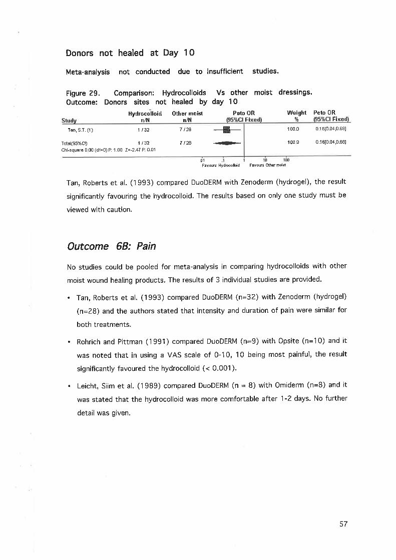
Donors not healed at Day 10
Meta-analysis not conducted due to insufficient studies.
Figure 29.Outcome:
Comparison: Hydrocolloids Vs other moist dressings.Donors sites not healed by day 10
Tan, S.T. (1) 1 t32
Total(Ss%Cl) 1 132
Ch¡-square 0 U0 (df=O) P: 1.00 Z=-2.47 P: 0.01
7 t28 --#7 ¡2Ê {Þ
Hydrocolloidn/H
Other moiln/N
Peto ORCI F¡
Weightlf
100.0
1rl0 0
Peto ORFixe
0.16[0.04,0.68]
0.16[0.04,0.68]
.0'l
FãvouEHydrocolloid FãvoußOthermoist
Tan, Roberts et al. (1993) compared DuoDERM with Zenoderm (hydrogel), the result
significantly favouring the hydrocolloid. The results based on only one study must be
viewed with caution.
Outcome 68: Pain
No studies could be pooled for meta-analysis in comparing hydrocolloids with other
moist wound healing products. The results of 3 individual studies are provided.
. Tan, Roberts et al. (1993) compared DuoDERM (n=32) with Zenoderm (hydrogel)
(n:28) and the authors stated that intensity and duration of pain were similar for
both treatments.
. Rohrich and Pittman (1991) compared DuoDERM (n=9) with Opsite (n=10) and it
was noted that in using a VAS scale of 0-10, 10 being most painful, the result
significantly favoured the hydrocolloid (< 0.001).
. Leicht, Siim et al. (1989) compared DuoDERM (n = 8) with Omiderm (n=8) and it
was stated that the hydrocolloid was more comfortable after 1-2 days. No further
detail was given.
57
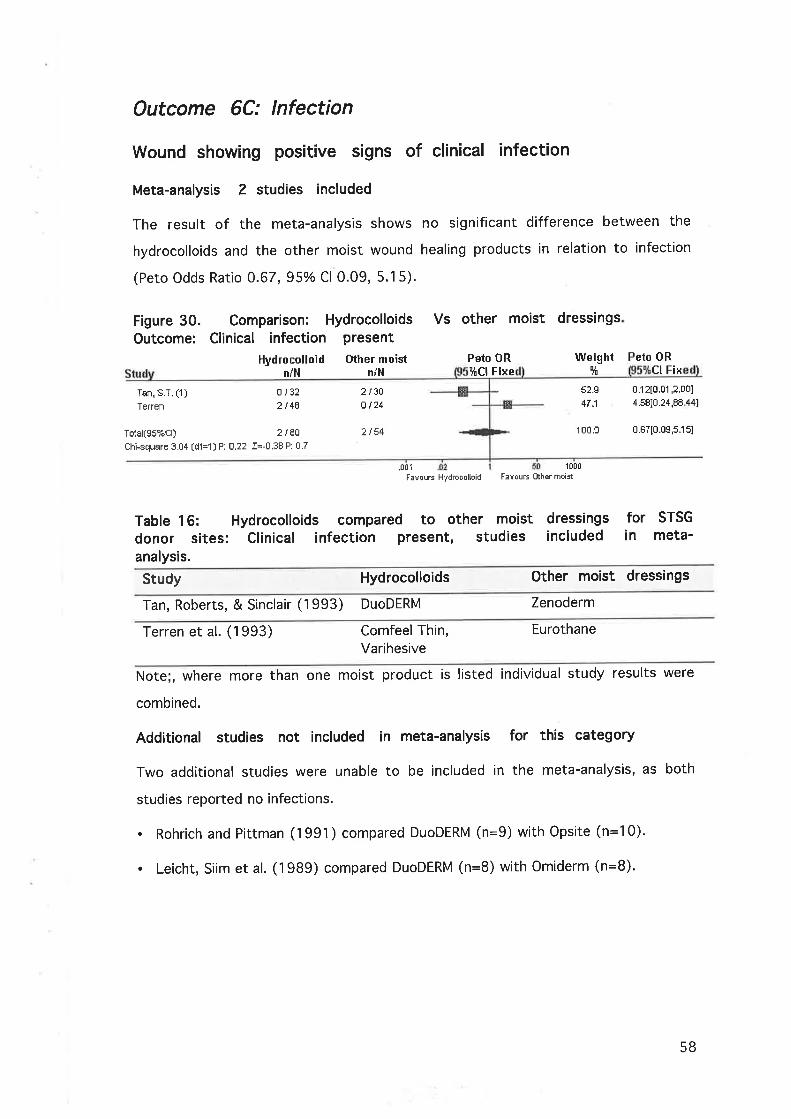
Outcome 6C: lnfection
Wound showing positive signs of clinical infection
Meta-analysis 2 studies included
The result of the meta-analysis shows no significant difference between the
hydrocolloids and the other moist wound healing products in relation to infection
(Peto Odds Ratio 0.67, 95% Cl 0.09, 5.15).
Figure 30.outcome:
other moist dressings
Tan, S.T. (1)
Teffen
Tstf,l(gs%cl) 2 t 8E
Chi-squere 3.04 (df =1) P: 0.22 Z=-0.38 F: 0.7
Comparison: Hydrocolloids Vs
Clinical infection present
Hydrocolloid Othermoistn/H n/H
0/322t 48
2 /3ûo t24
2¡54
Peto ORlfCl Fixe
Weightci
52.S
47.1
Peto ORCl Fix
0.12tt.01 ,2.001
4.58[0.24,88 441
for STSG
in meta-
100.0 0.67[0.09,s.1s]
.00fFavous Hydrooollo¡d F¡vúuÉ Olher moisl
Hydrocolloids compared to other moistClinical infection present, studies
1000
Table 16:donor sites:analysis.
dressingsincluded
Study Hydrocolloids Other rnoist dressings
Tan, Roberts, & Sinclair (1993) DuoDERM Zenoderm
Terren et al. (1993) Comfeel Thin,Varihesive
Eurothane
Note;, where more than one moist product is listed individual study resu Its were
combined.
Additional studies not included in meta-analysis for this category
Two additional studies were unable to be included in the meta-analysis, as both
studies reported no infections.
. Rohrich and Pittman (1991) compared DuoDERM (n=9) with Qpsite (n=10).
. Leicht, Siim et al. (1989) compared DuoDERM (n=8) with Omiderm (n=8).
58

Overall summary for hydrocolloids in comparison to other
moist wound healing products
The meta-analysis for days to healing included two studies that compared
hydrocolloids with other moist products. ln both studies the other products were
polyurethane film sheets. The result significantly favoured the hydrocolloids although
this result must be viewed with some caution. Only one study was examined for the
category of 'numbers of donors healed by day 10' and this study significantly
favoured the hydrocolloid over a hydrogel product. Although these results suggest
that hydrocolloids are superior to polyurethane films and hydrogels in terms of
healing this must be balanced against the limited number of studies available.
ln considering the outcomes of pain and infection there is no evidence to suggest
that hydrocolloids perform any better than other moist products.
Compariso n
hydrocolloids
sites;
Figure 31.Outcome:
7 = Compar¡son between different
in the management of STSG donor
Outcome 7A: Healing
Days to healing all studies requ¡red complete epithelial cover
with dressing able to be removed without trauma
Varihesive compared to Comfeel Thin
Meta-analysis not conducted due to insufficient studies.
Comparison: Varihesive/DuoDERMDays to complete healing
Varihesiven mean
Comfeel Thin
24 7.4s(1 .25) 24 10.2s(2.11) -{-24+
Vs Comfeel Thin.
WMD Weight5t
100.0
WMD
-2.A4[-3.82,-1 .86]
-2 84[-3.82,-1.861
nm F
Teren
Tûlal(gs%Cl) 24
Chi-square 0.00 (df=0) P: 0 00 Z=5.67 P: <0.00001
.4FavouE \àñhes F¡vouF Comfeel
100.0
59

This study by Terren, Serna et al. (1993) compared Varihesive (DuoDERM) with
Comfeel Thin. The result significantly favoured Varihesive however the sample size
was quite small.
DuoDERM compared to Sureskin
Meta-analys¡s not conducted due to insufficient studies.
Figure 32. Comparison: DuoDERM
complete healingDUoDERM Sureskin
Vs Sureskin. Outcome: DaYs to
FixeIÀ'MD
F
Weight WMD
1 00 0 0.00[-0.70,0 701
1 oú.0 0.00[-0.70,0.701
Steenfos 10 I 50(0.80)
Total(gs%Cl) 10
Chi-squere 0.00 (df=o) P: 0 00 Z=0.00 P: (null)
Terren Et24
Totâl(Ss%Cl) t t 24
Chi-square 0.00 (df=O) P: 1 .00 Z=l 43 P: 0.15
10
10
8.50(0.80)
l0
This study by Steenfos and Partoft (1997) compared the two hydrocolloids,
DuoDERM, and Sureskin. The results show no significant difference in healing between
the two products.
Outcome 78: Pain
No studies were found that dealt specifically with the outcome of pain in relation to
comparisons between hydrocolloids
Outcome 7C: lnfection
Wound showing pos¡t¡ve signs of clinical infection
Meta-analysis not conducted due to insufficient studies.
Figure 33.Outcome:
Comparison: Varihesive/DuoDERM Vs Comfeel Thin.
F¡vouE Duo
Peto 0RFixe
F¡vous Sueskin
50
Weightlt
Peto ORlËCt Fi
Clinical infection present
Varihesive ComfeelThinn/N niN
2 t24
2 ¡24
100.0 0 1310.01 ,2.131
1r0.0 0.13[0.01 ,2 13]
F¡vour \ä¡ihes Favous Êomfeel
60

This study by Terren, Serna et al. (1993) comparing Varihesive (DuoDERM) with
Comfeel Thin showed no significant difference between the two products in relation
to infection.
Overall summary of compar¡sons between different
hydrocolloid products.
There are a very limited number of studies comparing different hydrocolloids.
Although one study revealed a significant result favouring Varihesive (DuoDERM) in
terms of healing, no firm conclusions can be made about this comparison. Therefore
there is little evidence to indicate that one hydrocolloid product is superior to
another.
ln considering the use of hydrocolloids a number of authors indicated that leakage
and the need for additional dressings was a problem that would impact on treatment
choice (Doherty, Lynch, & Noble, 1986; Rakel et al., 1998; Tan, Roberts, & Blake,
1993; Tan, Roberts, & Sinclair, 1993). ln response to this problem several authors
have indicted that they took the strategy of avoiding changing the hydrocolloids and
reinforced them until healing resulting in use of only the one primary dressing
(Foyatier, 1992; Madden et al., 1985; Madden et al., 1989).
61

Comparison 8: Polyurethane films compared to non-
moist products in the management of STSG donor
sites;
Outcome 8A: Healing
Days to healing all studies required complete epithelial cover
with dressing able to be removed without trauma
Meta-analysis 3 studies included
The three studies in this group lacked homogeneity although the combined result did
significantly favour the polyurethane films (WMD -2.82 and 95% Cl -3.58, -2.07).
Figure 34.Outcome:
Table 17=STSG donoranalysis.
Comparison: Polyurethane films Vs non-moist dressings.Days to complete healing
lms Non-moist WIìTD Itlleight WMDme n Vt lfcr
Earnett
Teffen
ly Fin
Po
46
46
24
14
50
24
1 0 s0(5 s0)
1s.26(4.84)
1 0 04(1 .70)
---*-
F¡vous Films
1Ë
6529663S
-3 70[-6.64,-0.76]
-7 741-9.12,-8.361
-0.63[-1 s7,0.311
100.0 -2.93[-3.68,-2.191
lregbulem
6 80(2 00)
7 52(1 06)
941f 61)
Total(gs%Cl) 116
chi-square 70.38 (df=2) P: 0.00 Z=7.68 P: <0,00001
8B +
l0FavouE Non.moist
Polyurethanesites: Days
films compared to non-moist dressings forto complete healing, studies included in meta-
Study Polyurethane films Non-moist dressings
Barnett et al. (1983a) Tegaderm, Opsite Fine Mesh Gauze
lregbulem (1983) Opsite Sofratulle
Terren et al. (1993) Eurothane Tulle Gras
Note;, where more than one moist product is listed individual study results werecombined.
Additional studies not included in meta-analysis for this category
. Himel, Ratliff et al. (1998) compared Ventex (polyurethane film) with Xeroform.
The Ventex treated wounds were said to have all healed by day 10 (n=10) and
Xeroform wounds healed 10-14 days (n=10). No indication of statistical
significance was given.
62

Donors not healed at Day 10
Meta-analysis not conducted due to insufficient studies.
Figure 35. Comparison: Polyurethane films Vs non-moist dressings.
Outcome: Donor sites not healed at day 10
Comparison:Dressing rated
Poly Filmsn/H
Polyurethane filmsas more painful
Non-moistn/H
4ts
4 f s
Vs non-moist dressings.
Weightlt
Peto ORct Fi
1ûr.fr 1 .62[0 28,S.321
Peto ORCl Fix
100.0 0.30[0.0s,1 .ssl
100.0 0,30[0.05,1 .9s]
Poly FilmsniN
Non-moisin/H
3/S
Peto ORFix
Weightlt
3is 100.0 1 .Ë2[0.28,S.321
F¡vouE F¡lms FevouE Non.moist
ln relation to healing, Morris and Lamb (1990) compared Opsite to Scarlet Red and
found no significant difference between dressings.
Outcome 8B: Pain
Dressing rated
Morris 5 i 11
Total(Ss%Cl) 5 i 11
Chi-squâre 0.0û (df=O) P: 1.00 I=0.54 P: 0.6
Meta-analysis not
as more painful
conducted due to insufficient studies.
Figure 36.Outcome:
Peto ORFixeStu
Morris 2 ¡ 11
Total(Ss%Cl) 2111Chi-square 0.00 (df =0) P: 1 ,00 Z=l .24 Pi O.2
't .lFavouE Films FavouÉ Non-moiEt
Morris and Lamb (1990) also compared Opsite to Scarlet Red in relation to pain. No
significant difference was found.
63

Overall pain rated 1-10, 10 is most painful
Meta-analysis not conducted due to insufficient studies
Figure 37. Comparison: PolyurethaneOutcome: Days to complete healing
Comparison: 07 Polyurethane Films Vs Non-moist dressingsOutcome: 04 Overall pain rated f -10, 10 is mos{ painful
Poly Films l{on-moist
films Vs non-moist dressings.
Stu mean 9tct
Barnetl 4E 1.60(0.80)
Total(Ss%Cl) 46
Chi-square 0.00 (df=0) P: 0.00 Z=5.17 P: <0.00001
14
14
#4.7O(2.28)
WMD
0
WeightIt
Fevouß Non.moist
FavouE Non-mo¡5l
WMDct
100.0 -3 1 0l-4.28,-1 ,921
100.0 -3 1 0[-4.28,-1 921
Fi
+
-10
Barnett, Berkowitz et al. (1983a) compared two polyurethane films, Tegaderm and
Opsite, with fine mesh gauze achieving a significant result in favour of the film
dressings.
Overall pa¡n rated 1-100, 100 is most painful
Meta-analysis not conducted due to insufficient studies.
Madden, Nolan et al. (1989) compared Opsite with fine mesh gauze achieving a
significant result in favour of the film dressing (figure 38).
FãvouE Fllms
FãvouE F¡lms
Figure 38. Comparison: PolyurethaneOutcome: Overall pain rated 1-100,100
Poly Filmsn mean
l,lon-moist
Madden 10 1430(400)
Totâl(gs%cl) 10
Chi-square 0.00 (df=o) P: 0.00 Z=7.24 P: <0.00001
1o 25 10(2.s0)
10
films Vs non-moist dressings.is most painful
WiJtD Weight WMDFix
t
100 0
100.0
-1 0 80[-1 3.72,-7 881
-10 801-13.72,-7.Ë81
64

Patient requiring analgesia
Meta-analysis not conducted due to insufficient studies.
Figure 39. Comparison: Polyurethane filmsOutcome: Patient requiring analgesia
Poly Films Non-moistn/N
Vs non-moist dressings.
Stu n/N
lreghulen 0 i 50 32 /50
Total(Ss%Cl) 0/50 32i50Chi-square 0.ú0 (df=01 P: 1.f10 Z=-8.83 P: <Û.00001
-ffi-+
Peto ORF¡
ltCl Fix
Weightlt
llVeighlry!
Peto ORltCl Fixe
Feto ORct Fi
100.0 0.0sf0.02,0.131
100,0 0.0st0.02,0.131
achieving a
100
F¡vours Films FãvouE Non-moist
This study by lregbulem (1983) compared Opsite with Sofratulle,
significant result in favour of the film dressing.
Pain reported at 24hs
Meta-analysis not conducted due to insufficient studies.
Figure 40outcome:
Comparison:Pain reported
Poly FilmsniN
1/10
Polyurethane films Vs non-moist dressings.at 24hrs
Peto 0RNon-moistniN
10/10
10/10
-#- 1fto.0 0.03[0.01 ,0.1 8lHinel
Total(Ss%Cl) 1/10Chi-square 0.00 (df=o) P: 1.00 Z=-3 34 P: 0.Û0008
+ 100.0 0.03[ü.01 ,0.181
FavouÉ F¡lms
50 lt00FBVouß Hon-moist
This study by Himel, Ratliff et al. (1998) compared Ventex (polyurethane film) with
Xeroform, achieving a significant result in favour of the film dressing.
Outcome 8C: lnfection
Wound showing pos¡t¡ve signs of clinical infection
Meta-analysis: 4 studies included
The meta-analysis combined the results of four studies and demonstrated a
significant result in favour of polyurethane films (Peto Odds Ratio 0.?8, 95o/o Cl 0.09,
0.91). The studies lacked strong homogeneity, however examining the individual
studies gave no indication as to why this would be the case. The confidence intervals
for the pooled result were narrow and the combined sample size large.
65
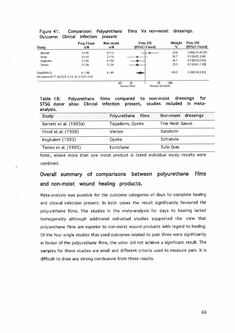
Figure 41
Outcome:
Barnett
Himel
lregbulem
Terren
Tdal(Ss%Cl) 4 i 130
Chi-square 6.1 7 (df=3) P: 0.1 3 Z=-2 12 F: 0.03
Comparison: PolyurethaneClinical lnfection present
Poly Films llon-moistn/H n/l'l
4i460i1D0 i 5û
o ¡24
I i s8
films Vs non-moist dressings.
Peto ORlÈCl Fix
Weightct
Peto ORCI Fi
B ¡142 t104 /503 t24
23S18.7
34.1
3 Ss[0 37,42.63]
0.12[0.01 ,2.09]
0 13[0.02,0.931
0.121û.01 ,1 zsl
1 o0.o 0.28[0.0s,0 911
F¡uous Films
I t00FevouE Non-moist
Table 18:STSG donoranalysis.
Polyurethanesites: Clinical
films compared to non-moist dressings forinfection present, studies included in meta-
Study Polyurethane films Non-moist dressings
Barnett et al. (1983a) Tegaderm, Opsite Fine Mesh Gauze
Himel et al. (1998) Ventex Xeroform
lregbulem (1983) Opsite Sofratulle
Terren et al. (1993) Eurothane Tulle Gras
Note;, where more than one moist product is listed individual study results were
combined.
Overall summary of compar¡sons between polyurethane films
and non-mo¡st wound healing products.
Meta-analysis was possible for the outcome categories of days to complete healing
and clinical infection present. ln both cases the result significantly favoured the
polyurethane films. The studies in the meta-analysis for days to healing lacked
homogeneity although additional individual studies supported the view that
polyurethane films are superior to non-moist wound products with regard to healing.
Of the four single studies that used outcomes related to pain three were significantly
in favour of the polyurethane films, the other did not achieve a significant result. The
samples for these studies are small and different criteria used to measure pain. lt is
difficult to draw any strong conclusions from these results.
66

Comparison 9: Polyurethane films compared to other
moist products in the management of STSG donor
sites;
Outcome 9A: Healing
Days to healing all studies required complete epithelial cover
with dressing able to be removed without trauma
Meta-analysis 2 studies included
The studies in this meta-analysis lacked homogeneity although no clear reason can be
derived from the study reports. The pooled result did favour the other moist wound
products (WMD 1.453 and 95o/o C|0.740,2.165).
Figure 42. Comparison: PolyurethaneOutcome: Days to cornplete healing
Comparison: 0B Polyurethane Films Vs Other moist dressingsOutcome: 01 Daysto complete healing
Polyurethane Films Oiher moist
films Vs other moist dressings
n mean 9tct F Fixe
Leichl
Terren
I24
1 0 60(1 .30)
9.41(1 .61)
7 63(1 06)
I 87(2 23)
men
I48
56
WND
+
Weight$i
I,1'MD
37.6
62.4
2 97[1 81 ,4.13]
0 54[-0.36,1 ,44]
100.0 1 .4s[0 74,2.17]Total(95%Cl) 32
Chi-squâre 1 0 48 (df=l ) P: 0.00 Z=4 00 Pr 0.00006
F¡voun Films
Table 19: Polyurethane films compared toSTSG donor sites: Days to complete healing,analysis.
other moist dressings forstudies included in meta-
l0FãvouE [hher mai5t
Study Polyurethane films Other moist dressings
Leicht et al. (1989) Omiderm DUoDERM
Terren et al. (1993) Eurothane Varihesive, Comfeel Thin
Note;, where more than one moist product is listed individual study results were
combined.
Additional study not included in meta-analysis for this category
. This study by Rohrich and Pittman (1991) also compared Opsite, mean days to
complete healing 8.23 (n = 10), with DuoDERM, mean days to complete healing
11 .78 (n = 9).
67

Outcome 98: Pain
Two individual studies compared polyurethane films against other moist wound
healing products. lnsufficient data was presented to combine these studies.
. Rohrich and Pittman (1991) compared Opsite (film) (n:10) with DuoDERM (n:9)
and it was noted that in using a VAS scale of 0-10, 10 being most painful, the
result significantly favoured the other moist product (< 0.001).
. Leicht, Siim et al. (1989) compared Omiderm (n=8) with DUoDERM (n=B) and it
was stated that the other moist product (DuoDERM) was more comfortable after
1-2 days. No further detail is given.
Outcome 9C: lnfection
Two individual studies compared polyurethane films against other moist wound
healing products. ln both studies there were no cases of infection.
. Rohrich and Pittman (1991) compared Opsite (n=10) with DuoDERM (n=9).
. Leicht, Siim et al. (1989) compared Omiderm (n=8) with DUoDERM (n=8)'
Overall summary of comparisons between polyurethane films
and other mo¡st wound healing products.
Meta-analysis was only possible for the outcome category of days to complete
healing, the result significantly favouring other moist products. lt should be noted
that only two studies were included in the meta-analysis and additional studies did
not strongly support the case for or against polyurethane films. As a result it is
difficult to make any conclusions with regard to healing. No meta-analysis was
possible for the outcomes of pain and infection. The single studies examined have
small samples and it is difficult to draw any conclusions from these results.
68

Comparison
polyurethane
sites;
10:
films
Comparisons between different
in the management of STSG donor
I'lfMD Weight l¡VMD
23 1.60(0.80)
23
Outcome 10A: Healing
Meta-analysis not conducted due to insufficient studies.
. One study by Barnett, Berkowitz et al. (1983a) compared two different
polyurethane films, Tegaderm, mean days to complete healing 6.7 (n=23), and
Opsite, mean days to complete healing 6.9 (n=23). No standard deviations were
given and no comment was made with regard to significance'
Outcome 108: Pain
Overall pain rated 'l-10, 10 is most painful
Meta-analysis not conducted due to ¡nsufficient studies.
Figure 43. Comparison: Tegaderm Vs Opsite.rated 1-10, 10 is most Painful
Tegaderm OPsite
Outcome: Overall Pain
n $ct Fix
Barnetl 23 1 Bo(0.80) 100.0 0.00[-0.46,0.461
100.0 0 00[-0.46,0 4Ê]Total(gs%cl) 23
Chi-square 0 00 (df=o) P: 0 00 Z=0.00 P: (null)
F¡vouÉ Tegãdem FavouÉ Opsite
Barnett, Berkowitz et al. (1983a) compared Tegaderm (n = 23) to Opsite (n = 23).
Scores for the two products of this small trial were identical.
Outcome 10C: lnfection
Meta-analysis not conducted due to insufficient studies'
ln the study by Barnett, Berkowitz et al. (1983a), Tegaderm (n = 23) was compared
to Opsite (n = 23). There were two cases of reported infection in both groups.
69
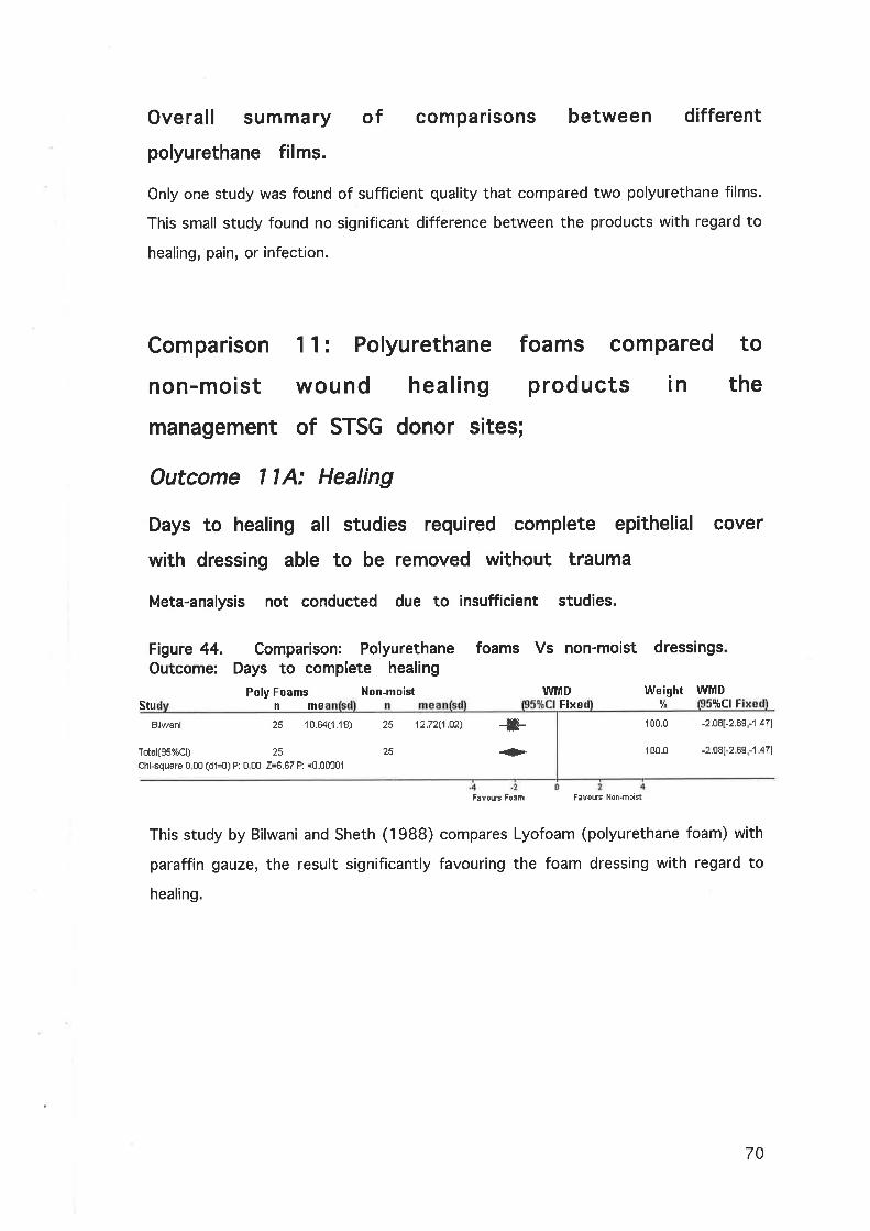
Overall summary of comparisons between different
polyurethane films.
Only one study was found of sufficient quality that compared two polyurethane films.
This small study found no significant difference between the products with regard to
healing, pain, or infection.
Comparison 1 1 : Polyurethane foams compared to
non-mo¡st wound healing products in the
management of STSG donor sites;
Outcome llA: Healing
Days to healing all studies required complete ep¡thel¡al cover
with dressing able to be removed without trauma
Meta-analysis not conducted due to insufficient studies.
Figure 44. Comparison: PolyurethaneOutcome: Days to comp¡ete healing
Poly Foams Non-moist
foams Vs non-moist dressings
Fixe [¡¡¡ ltct FWMD
Stu nmeBilwani 25 10.64(1 .18)
Total(95%Cl) 2sChi-square 0 00 (df=o) P: 0 00 2=8.67 P: <0.00001
12.72(1.02) -F+
FavouE Fo¡m FavouÉ Non.moist
Weight WMD
1 00.0 -2.08[-2.69,-'1 ,471
180.0 -2.08[-2 69,-1 471
25
25
This study by Bilwani and Sheth (1988) compares Lyofoam (polyurethane foam) with
paraffin gauze, the result significantly favouring the foam dressing with regard to
healing.
70

Donors not healed at Day 8
Meta-analysis not conducted due to insufficient studies.
Figure 45outcome:
Comparison: Polyurethane foams Vs non-moist dressings.Donor sites not healed at day I
Poly Foams Non-moistn/H n/H
Peto ORFixe
Peto ORCl Fixe
Weightlt
10
Peto ORCl Fixe
Davenpod 18 /31 16/31 10û.0 1 .2S[0 48,3.491
Total(S5%Cl) 18 / 31
Chi-square 0.00 (df=o) P: 1 .00 Z=0.51 P: 0 6
1E /31 100.0 1 .2st0.48,3 491
Favouß Fo¡m510
F¡vouE Non-moist
This study by Davenport, Dhooghe et al. (1977) compared Lyofoam with Tulle Gras
showing no significant difference in healing at day 8.
Donors not healed at Day 10
Meta-analysis not conducted due to insufficient studies.
Figure 46. Comparison: Polyurethane foams Vs non-moist dressings.Outcome: Donor s¡tes not healed at day 10
StuPoly Foams
niHHon-moist
n/N
12 ¡31
12t31
Weighllt
Peto ORCI F¡
100 0 1 47[0.54,3.3S1
100.0 1 47[0.s4,3.99]
Dâvenport 15i31
Total(gs%Cl) 15 / 31
Chi-square 0.00 (d1=0) P: 1 .00 Z=0.76 P: 0.4
FãvouE Foem F¡vouË Non.moist
The study detailed in the previous comparison also compared healing rates at day 10
showing no significant difference between Lyofoam and Tulle Gras (Davenport et al.,
1977).
71

Outcome I 1B: Pain
Pain present initially
Meta-analysis not conducted due to insufficient studies
foams Vs non-moist dressings.Figure 47.outcome:
Stu
Bilwani 0 / 25
Total(9s%Cl) 0 i 25
Chi-square 0.00 (df=0) P: 1.00 Z=-7.00 F: <0.00001
n/H
DâvenpoÉ 22],31
Total(S5%Cl) 22 131
Chi-square 0.00 (df=0) P: 1 .00 Z=-0.88 P: 0 4
Comparison: PolyurethanePain present ¡nit¡ally
Poly Foams Non-moistn/N n/N
25 t25
25 t2s
n/N
Æ-.+
Peto ORlÈCIF¡
Peto ORCl Fixe
WeightIl¡d
100.0
100.t
Peto ORliCl Fix
o o2[0.û1 ,0.061
0.ù2[0.01 ,0.06]
Peto ORlf ct Fi
o.E0[0.1s,1 881
0.60t0 19,1 .Ê81
.û2
FãvouË Fo¡m FavouE Non'móist
This study by Bilwani and Sheth (1988) compared Lyofoam with paraffin gauze, the
result significantly favouring the foam dressing with regard to pain.
Pain present at day 8
Meta-analysis not conducted due to insufficient studies.
Figure 48. Comparison: PolyurethaneOutcome: Pain present at day 8
Poly Foams Hon-moist
foams Vs non-moist dressings.
25i31
25 !31
Weightlf
100.0
10t.0
FatÌouB Foem
5 t0F¡vouß Non.moìst
Davenport, Dhooghe et al. (1977) compared Lyofoam with Tulle Gras showing no
significant difference in pain at day 8.
72

Pain present at day 10
Meta-analysis not conducted due to insufficient studies.
Figure 49. Comparison: PolyurethaneOutcome: Pain present at day 10
Poly Foams Hon-moist
foams Vs non-moist dressings.
Peto ORïrcl Fi
Weightry!
Peto ORItCl Fixn/H n/H
Davenporl 15 131 12 t31 100.0 1.47[o s4,3.SSl
Total(ss%cl) 15 /31Chi-square 0.00 (df=o) P: 1.00 Z=0 76 P: 0 4
12t31 100.0 1 .47[0.s4,3.S9]
FavouÉ Foãm510
F¡vouE Non-moist
The study by Davenport, Dhooghe et al. (1977) comparing Lyofoam with Tulle Gras
also showed no significant difference in pain at day 10.
Outcome llC: lnfection
Meta-analysis not conducted due to insufficient studies.
Bilwani and Sheth (1988) compared Lyofoam (n=25) with Paraffin Gauze (n=25).
There were no cases of reported infection in either group.
Overall summary of compar¡sons between polyurethane
foams and non-mo¡st wound healing products.
Only two studies were found of sufficient quality that compared two polyurethane
foams and non-moist wound healing products. No meta-analyses were able to be
performed for this comparison and the small number of studies and sample sizes
prevent any strong conclusions, however there is an indication that in terms of days
to complete healing and with regard to initial pain the foam dressings are favoured.
73
No studies of sufficient quality were found comparing polyurethane
foam dressings with other moist products.
Compar¡son 12: Polyurethane
compared to other mo¡st wound
management of STSG donor sites;
foam dressings
products in the
between different
the management of
Comparison 1 3:
polyurethane foam
STSG donor sites;
Comparison
dressings in
No studies were found comparing different polyurethane foam
dressings in the management of split skin graft donor sites'
Comparison 14= Hydrogel sheet dressings compared
to non-mo¡st wound products in the management of
STSG donor sites;
No studies of sufficient quality were found comparing hydrogel sheet
dressings with non-moist wound products.
74

Comparison 15: Hydrogel sheet dressings compared
to other moist wound products in the management of
STSG donor sites;
Outcome 15A: Healing
Donors not healed at Day 10
Meta-analysis not conducted due to insufficient studies.
Figure 50outcome:
Comparison: Hydrogel sheet dressing Vs hydrocollloid.Not healed at day 10
Hydrogel Sheet Hydrocolloid Feto OR Weightlt
Feto ORctn/H CI F¡
Tan, S.T. (1) --Æ- 100.0 6.43[1 .47,28.2s]
Total(S5%cl) {Þ 1000 843[1 .47,28.2s1
Chi-squâre 0,00 (df=o) P: 1.00 Z=2.47 P: 0.01
FavoursHydmgel F¡vouËHydmcolloid
Only one study involving hydrogel sheets was found. This study by Tan, Roberts et al.
(1993) compared Zenoderm (hydrogel) and DuoDERM (other moist). ln relation to
healing of split skin donors at day 10 the result significantly favoured the other moist
product.
The study d¡d not provide sufficient data to make a comparison
relating to pain or infection.
7 Ì28
7 l2A
1 t32
1 Í32
Comparison 1 6:
hydrogel dressings
sites;
Comparison between different
in the management of STSG donor
There were no studies found comparing different hydrogel products in
the treatment of split skin graft donor sites
75
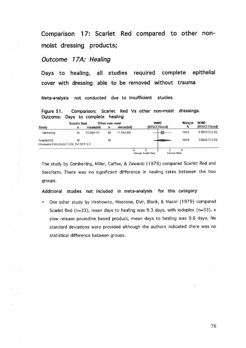
Comparison 17= Scarlet Red compared to other non-
moist dressing products;
Outcome 17A: Healing
Days to healing, all studies required complete epithelial
cover with dressing able to be removed without trauma
Meta-analysis not conducted due to insufficient studies.
Figure 51. Comparison: Scarlet Red Vs other non-moistOutcome: Days to complete healing
Scarlet Red Other non-most W¡lD
dressings.
Weightlt
W]UD
mean
50 12s0(4r7)
nm F¡
Gemberling 50
50
11 70(3.s8) 100 0
100,0
0.80[-0 72,2.32]
0 .80[-0 .72 ,2 .321Total(Ss%Cl) 50
Chi-square 0.00 (df=0) P: 0.00 Z=1.03 P: 0.3
FavouE Sc¡det Red FãvouË Olher
The study by Gemberling, Miller, Caffee, & Zawacki (1976) compared Scarlet Red and
Xeroform. There was no significant difference in healing rates between the two
groups.
Additional studies not included in meta-analysis for this category
. One other study by Hirshowitz, Moscona, Dvir, Blank, & Mazor (1979) compared
Scarlet Red (n=33), mean days to healing was 9.3 days, with lodoplex (n=33), a
slow release poviodine based product, mean days to healing was 9.6 days. No
standard deviations were provided although the authors indicated there was no
statistical difference between groups.
76

Comparison 18: Scarlet Red compared to moist
wound healing products;
Outcome l8A: Healing
Donors not healed at Day 10
Meta-analysis: 3 studies included.
The pooled result for the three studies concluded no significant difference between
Scarlet Red and the moist products. The studies lacked homogeneity and it should be
considered that no studies provided the outcome of days to complete healing which
is a stronger indicator of performance.
Figure 52. Comparison: Scarlet Red Vs moist dressings. Outcome: Nothealed at day 10
LswrenÊe
Morris
Tan. S.T. (2)
Table 20:donor sitesanalysis.
Scarlet Redn/N
7i463 i I
13 /30
Moistn/H
13i465 /113 i 30
21 tAl
comparedhealed at
Pelo ORFixe
WeightIl¡¡
48.3
15 3
36.4
Peto ORCI F¡
0.47[0.17,1 .2sl
0.62[0.11 ,3.s7]5.34[1 .72,16 631---F
Totál(Ss%Cl) 23 i 8s
Chi-square 10.70 (df=2) P: 0.01 2=0.49 P: 0.Ë
F¡vous Sc¡rlet Red
to moist1 0 days,
F3vouÈ hlhisl
wound dressingsstudies included
100 0 1 .1 S[0.60,2 35]
for STSG
in meta-ScarletDonor
Rednot
Study Scarlet Red Moist dressings
Lawrence & Blake (1991) Scarlet Red Kaltostat
Morris & Lamb (1990) Scarlet Red Opsite
Tan, Roberts, & Blake (1993) Scarlet Red DUoDERM
77

Outcome 188: Pain
Dressing rated
Meta-analysis not
Scarlet Redn/N
Morr¡s 4 i S
Total(ss%Cl) 4 / S
Chi-square 0.00 (df=0) P: 1.00 Z=1 .24P:82
as more painful
conducted due to insuff¡c¡ent studies.
Pelo 0R
Figure 53. Comparison: Scarlet Red Vs moist dressings. Outcome:Dressing rated as more painful
It¡loistn/H Fixe
llVeight1Ë
100.0
100.0
Peto ORct Fi
3.28[0.s0,21 .371
3.28[0.50,21 .371
2t11
2t11
.01 .1
FavouË Scarlel RÊd FavouB lùloist
This very small study by Morris and Lamb (1990) compared Scarlet Red with Opsite,
the result showing no significant difference between the treatment and control
groups.
Overall summary of compar¡sons between Scarlet Red,
compared to other wound dressings.
Due to the small number of studies all with quite small samples it is difficult to draw
any firm conclusions about the comparison between Scarlet Red and non-moist or
moist wound healing products.
78

Comparison 1 9: Bovine
compared to non-moist
the management of STSG
Outcome l9A: Healing
or Porcine based dressings
wound healing products in
donor sites;
dressings Vs non-moisthealing
WMD Weightlt
WMD
Days to healing, all studies required complete epithelial
cover with dressing able to be removed without trauma
Meta-analysis 2 studies included.
The two studies included in this meta-analysis had a high degree of homogeneity. The
pooled result shows a statistically significant difference between the two groups
favouring the porcine/bovine products (WMD -4.08 and 95o/o Cl -4.75, -3.42) (figure
s4).
Figure 54dressings,
Comparison: Porcine./bovineOutcome: Days to completeForcine/bovine Hon-moist
n
Gr swa dGao 12
I1 0 30(0 70)
7 e0(1 58)
1 4 sof 00)
I 0 60(2 92)
12
I91.7
83-4 20[-4.89,-3 s1l
-2 80[-s 1 0,-0 501
Total(Ss%Cl) 20
Chi-square 1,30 (df=1)P. O25 Z=12.10 P: <0 00001
2E
Table 2"1= Porcine or bovinedressings for STSG donor sites:included in meta-analysis.
compared to non-moistcomplete healing, studies
+ 100.0 -4.08[-4.75,-3.421
F¡vouß Porc/Bov
dressingsDays to
FãvouE Non-moist
Study Porcine./ Bovine dressings dressings
Gao, Hao, Li, lm, & Spence Porcine collagen sheet(1 9e2)
Jelonet
Griswold et al. (1995) SkinTemp (bovine) Xeroform
Additíonal studies not included in meta-analysis for this category
. Sagi, Walter et al. (1986) compared Dermodress (bovine collagen sheet), healed
between 1 5 and 20 days (n=10), with Furacin Gauze healed between 10 and 15
days (n=10). Statistical significance was not provided.
79

a
a
Breach, Davies, & Yiacoumettis (1979) compared lyophilised porcine epidermis
(Corethium), and lyophilised bovine dermis with a control of Tulle Gras. No
significant difference in healing was detected.
Salisbury, Wilmore, Silverstein, & Pruitt (1973) compared porcine xenograft to fine
mesh gauze and exposure. No significant difference in healing was detected.
Outcome 198: Pain
Dressing rated as more painful
Meta-analysis not conducted due to insufficient studies
Figure 55dressings.
Comparison: Porcine/bovine dressingsOutcome: Dressing rated as more painful
Porcine/bovine Hon-moist Peto ORniN n/N Cl Fixe
1i I 7ta --ffi-7!8
Vs non-moist
100.0
1ûù t
Weight Peto 0RStu Fixe
o.06[0.01 ,0.40]
0 û6[0.01 ,0.40]
Griswald
Total(Ss%Cl) 1 i IChi-square 0.00 (df=0) P: 1.00 Z=-2.90 P: 0.004
{{Þ
.00t .02
FEvruß Porc/Bov Favoum Non-moist
This study by Griswold, Cepica et al. (1995) compared SkinTemp (bovine dressing)
with Xeroform, the result significantly favouring SkinTemp.
Additional stud¡es not included in meta-analysis for this category
. Lyophilised porcine epidermis (Corethium), and lyophilised bovine dermis were
compared with Tulle Gras in the study by Breach, Davies et al. (1979). Nine out of
twenty patients complained of severe irritation under the bovine dressings and in
some cases this irritation persisted for 8 weeks.
80

Outcome 19C: lnfection
Wound showing positive
Meta-analysis not conducted
signs of clinical infection
due to insufficient studies.
Figure 56.dressings.
Comparison: Porcine/bovineOutcome: Clinical infection
Porcine/hovine Non-moistn/H n/N
dressingspresent
Peto ORltct Fi
.ût .1
F¡vouE Poro/Bov
Vs non-moist
Weight1ò
100
FBvouË Non-mÒist
Stu
Peto ORltCl Fixe
Sagi
Total(85%cl)
Chi-square 0.00 (df=0) P: 1 00 Z=-0.61 P: 8.5
1/10
1/1û
2 /10
2 i 10
1oo.o ù.47[0.04,s.191
1oû.0 t 47[0.04,5.191
This study by Sagi, Walter et al. (1986) compared Dermodress (bovine dressing) with
Furacin Gauze, the result showed no significant difference between products.
Additional studies not included in meta-analysis for this category
. Gao, Hao et al. (1992) compared porcine collagen sheet (n=12) and Jelonet
(n=12) with no infection in either group.
. The study by Griswold, Cepica et al. (1995) compared SkinTemp (bovine) (n=8)
and Xeroform (n=8) with no infection in either group.
Overall summary of comparisons between porcine or bovine dressings
compared to non-moist dressings.
Two studies combined in meta-analysis did demonstrate superior performance related
to healing in comparison with non-moist products. This was in part balanced by a
number of individual studies that showed no significant difference between the
product groups. ln one study there were a considerable number of patients had
severe and persistent irritation where a bovine product was used. Another small
study indicated bovine dressings are less painful than non-moist dressings.
81

Comparison 2O= Bovine or porcine
compared to moist wound healing
management of STSG donor sites;
based dressings
products in the
cover
* 1oo.o
Outcome 20A: Healing
Days to healing all studies required complete epithelial
with dressing able to be removed without trauma
Meta-analysis not conducted due to insufficient studies.
Figure 57. Comparison: Porcine/bovine dressings Vs moist dressings.Outcome: Days to complete healing
Porcine/Eovine Moist WMD Weight WMDn lt
Sawada 1o 13 30(f.1 s)
Total(Ss%Cl) 10
Ch¡-square 0 00 (df=o) P: 0 00 Z=1 5.1 I P: <0.00001
Stu
Comparison: Porcine/bovineNot heafed at day 7
Porcine/Bovine Moistn/H n/ll
10t20
10 /20
20
20
6 8s(0.98)
FavouË Porc/8ov
+ 100.0
t0FãvouË hioil
6.4s[s.62,7 281
6.45[s 62,7.281
Pefo OR
This study by Sawada, Yotsuyanagi et al. (1990) compared collagen sheet
(unspecified) with a Silicone Gel Sheet with OFLX (Orfloxacin). Although only one
small study the result significantly favoured the moist wound dressing.
Donors not healed at Day 7
Meta-analysis not conducted due to insufficient studies.
Figure 58.Outcome:
dressings Vs moist dressings.
Feto OR Weight5È Cl Fix
Vanstraelen 20 t20 +- 100.0 1s.46t3.23,ss.3sl
Total(gs%Cl) 2Ê tZEChi-square 0.00 (df=0) P: 1.00 Z=3.61 P: 0.0003
+ 100.0 13.46[328,55.331
Favours P0É/Bov F¡vouË h¡loil
This single study by Vanstraelen (1992) comparedEZ Derm porcine xenograft with a
calcium alginate, Kaltostat (moist). Although only one small study the result
significantly favoured the moist wound dressing.
Fixe
82
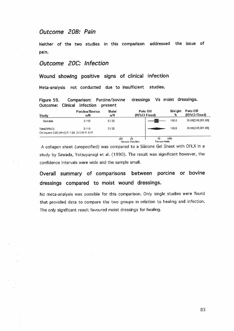
Outcome 208: Pain
Neither of the two studies in this comparison addressed the issue of
pain.
Outcome 20C: lnfection
Wound showing positive signs of clinical infection
Meta-analysis not conducted due to insufficient studies.
Figure 59.Outcome:
dressings Vs moist dressings.Comparison: Porcine./bovineClinical infection present
Porcinei Bovine Moistn/N n/l{
o r2a
0i20
Æ- 100 0 2s.DB[2.08,s01 .881
<>- 100.0 25.08[2.08,301 .88]
Peto ORClFix
Weightlt
Peto OR9tCI Fixe
Sawada
Tstal(ss%Cl)
3 i 10
3/10Chi-square 0.00 (df=0) P: 1 .00 Z=2.54 P: 0.01
F¡vouE PoB/Bov Favoum luloist
A collagen sheet (unspecified) was compared to a Silicone Gel Sheet with OFLX in a
study by Sawada, Yotsuyanagi et al. (1990). The result was significant however, the
confidence intervals were wide and the sample small.
Overall summary of compar¡sons between porc¡ne or bovine
dressings compared to mo¡st wound dressings.
No meta-analysis was possible for this comparison. Only single studies were found
that provided data to compare the two groups in relation to healing and infection.
The only significant result favoured moist dressings for healing.
83
Comparison 2"1= Comparison
or porcine based products
STSG donor sites;
different bovine
management of
between
in the
Outcome 21A: Healing
Days to healing all studies required complete epithelial cover
with dressing able to be removed w¡thout trauma
Meta-analysis not conducted due to insufficient studies.
. The only study found of sufficient quality compared lyophilised porcine epidermis
(Corethium) and lyophilised bovine dermis. The study by Breach, Davies et al.
(1979) showed no significant difference in healing. No other details were provided.
Comparison 22= Growth factors compared to placebo
in the management of STSG donor sites;
Outcome 22A: Healing
Days to healing all studies required complete epithelial cover
with dressing able to be removed without trauma
Recombinant human growth hormone rHGH O.Zmglkg/day compared to
placebo
Meta-analysis two studies included
Three almost identical studies comparing rHGH with placebo were pooled in this
meta-analysis to demonstrate a significantly better healing rate for the growth factor
group (WMD -1.847 and 95% Cl -2.520, 1.174) (figure 60).
84

Figure 60. Comparison: rHGH O.Zmglkg/day Vs placebo. Outcome:Days to complete healing
rHctl Placebo WMD Weight Will¡
Gilpin
Herndon
Herndon(2)
dl¡!
34428S367
---s-
-#----#-
46
10
Ê
mea
6 80(1.50)
7.40(1 .s8)
5 60(0 70)
mean
I 50(2 30)
9.10(1 .64)
7 70(1 20)
18
17
6
41
-1 70[-2 8s,-0.ssl-1 70[-2.es,-o 45]
-2.1 0[-3 21 ,-0 SSI
100 0 -1 .8s[-2.52,-1 .171Tûtal(Ss%Cl) Ê2
Chi-squere 0 32 (df=Z) P: 0 85 Z=5 38 P: <0 00001
{Þ
.10
F¡vouE rllGH F¡vouß Flacebo
Table 22= Growth factor compared to control for STSG donor s¡tes:Days to complete healing, studies included in meta-analysis.
Study Growth Factor Control
Gilpin, Barrow, Rutan,Broemeling, & Herndon (1994)
rHGH O.2mglkglday with Placebo with Scarlet Red
Scarlet Red dressing
Herndon, Barrow, Kunkel,Broemeling, & Rutan (1990)
rHGH 0.2mglkglday with Placebo with Scarlet Red
Scarlet Red dressing
Herndon et al. (1995) rHGH O.2mglkglday with Placebo with Scarlet Red
Scarlet Red dressing
rHGH 10 mg SC/day compared to Placebo
Meta-analysis not conducted due to insufficient studies.
ln the 1989 study by Shernan, Demling et al. (1989) rHGH was administered daily to
the treatment group, with a mean time to healing of 8t2.82 days (n=8), compared
to placebo, mean time to healing of 10t0.0 days (n=9). This was a significantly
better healing rate ¡n favour of the growth hormone with p < 0.01.
Basic Fibroblast Growth Factor (bFGF) compared to Placebo
Meta-analysis not conducted due to insufficient studies.
85

Figure 61. Comparison: bFGF Vs placebo.healing
Comparison: 16 hFGF Vs PlaceboOutcome: 01 Days to complete healing
hFGF Placebo
outcome: Days to complete
IÀIMD Weight WMDme E'
¿¡ Fix
ereênhalgh 11 12.30(3.s0) 12 20(5.s0) 100,0 0 .¡0[-3 .28 ,4 .88]
o .¡o[-3 ,28 ,4 .681Total(95%Cl) 11
Chi-squäre 0.00 (dJ=O) P: 0 00 Z=0.34 P: 0.7
'100.0
F¡vouË bFGF51
levouß Pl¡cebo
ln a study by Greenhalgh and Rieman (1994) bFGF applied daily was compared to
placebo with no significant difference between the two groups.
Recombinant human Epidermal Growth Factor (rEGF) compared to
Placebo
Meta-analysis not conducted due to insufficient studies.
One study by Brown, Nanney et al. (1989) compared healing rates of rEGF applied
daily in a vehicle of Silversulphadiazine with the control group receiving
Silversulphadiazine only. The growth factor decreased healing time by 1.5 days
compared to control (p<0.02).
ct
11
11
86
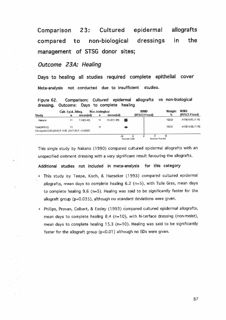
Comparison 23: Cultured epidermal
compared to non-biological dressings
management of STSG donor sites;
Outcome 23A: Healing
allografts
in the
Weight l¡lfMD
100.01s.271 .4s) ti+
-8.09[-9 00,-7.181
-E 0s[-9.00,-7.181
Days to healing all studies required complete epithelial cover
Meta-analys¡s not conducted due to insufficient studies
Figure 62. Comparison: Cultured epidermaldressing. Outcome: Days to complete healing
Cult. Epid. Allog. Non-biological
allografts vs non-biological
n n Fix 1È
WMD
Näkano 1'l 7.18(0.40)
Total(gs%Cl) 11
Chì-square 0.00 (df=0) P: 0.00 Z=17.50 P: <0.00001
11
'11 100.0
FãvouE CEA Favous Non-biol
This single study by Nakano (1990) compared cultured epidermal allografts with an
unspecified ointment dressing with a very significant result favouring the allografts.
Additional studies not included in meta-analysis for this category
. This study by Teepe, Koch, & Haeseker (1993) compared cultured epidermal
allografts, mean days to complete healing 6.2 (n=5), with Tulle Gras, mean days
to complete healing 9.6 (n=5). Healing was said to be significantly faster for the
allograft group (p=0.035), although no standard deviations were given.
. Phillips, Provan, Colbert, & Easley (1993) compared cultured epidermal allografts,
mean days to complete healing 8.4 (n=10), with N-terface dressing (non-moist),
mean days to complete healing 1 5.3 (n=10). Healing was said to be significantly
faster for the allograft group (p<0.01) although no SDs were given'
87

Comparison 24= Cultured allogeneic keratinocyte
sheet compared to Opsite;
Outcome 24A: Healing
Days to healing all studies required complete epithelial
cover
Meta-analysis not conducted due to insufficient studies.
allogeneic keratinocyte Vs OpsiteFigure 63. Comparison: CulturedOutcome: Days to complete healing
Cuìt. Heratinocytes Opsiten n
WMD Weight WMDFixe $
100 0Duinslâger 15
15
Ê 7s(2.34) f5
l5
13.60(3.04) -fi- -6.8¡[-8.81 ,-4.S31
-6.8¡[-8.81,-4 S3]Totàl(S5%Cl)
Chi-squäre U 00 (df=0) P: 0 00 I=Ê.94 P: <0 00001
+
Fsvous ËK
100 0
F¡vouF opshe
This single study by Duinslaeger, Verbeken, Vanhalle, & Vanderkelen (1997)
compared cultured allogeneic keratinocyte sheets with Opsite, with a very significant
result favouring the keratinocyte sheets.
Comparison 25: Biobrane compared to non-moist
wound dressings;
Outcome 25A: Healing
Days to healing all studies requ¡red complete epithelial cover
w¡th dressing able to be removed without trauma
Meta-analysis: 2 studies included.
The two studies used in the meta-analysis are obviously not homogenous. The
subjects in both studies were similar and the treatments administered in similar
fashion. lt is difficult from the study reports to explain the disparity between the two
studies and so despite the strong result in favour of the Biobrane dressing this must
be viewed with caution (figure 64).
88

Figure 64.complete
Comparison:healing
Biobrane
Biobrane Vs Scarlet Red. Outcome: Days to
Scarlet Red WIITD Weight WMD
mean FixStu n m
1 0 05(3 40)
I80(0.40)8.48(1 70)
1 2 40(0 40)
Fixe
1 5 1 5r[-0.06,3.201
98.s -2.80[-2 80,-2.40]
100.0 -2.s41-2;t4,-2.341
Prâsad
Zepata-Servent s+
21
31
2',1
31
Totâl(gs%Cl) 52
Chi-square 24 90 (df=1) P: 0,00 Z=25 17 P: <0.00001
52
0Fãvouß BìobEne F¡vouE ScãrlÊ1 Red
Table 23: Biobrane compared to Scarlet Red for STSG donor s¡tes:
Days to complete healing, studies included in meta-analysis.
Study Biobrane Non-moist
Prasad, Feller, & Thomson (1987) Biobrane Scarlet Red
Tapata Sirvent, Hansbrough, Carroll,Johnson, & Wakimoto (1985)
Biobrane Scarlet Red
Additional study not included in meta-analysis for this category
. The study by Feldman, Rogers et al. (1991) compared Biobrane, mean days to
complete healing 19 (n=7), with Xeroform, mean days to complete healing 10.46
(n=13). Healing was said to be significantly faster for the Xeroform group
(p=0.023) although no SDs were given.
Outcome 258: Pain
Meta-analysis not conducted due to insufficient studies.
Additional studies not included in meta-analysis for this category
. ln the study by Prasad, Feller et al. (1987) comparing Biobrane and Scarlet Red,
11/21 patients stated that Biobrane was the preferred dressing with regard to
pain, 5/21 patients preferred the Scarlet Red dressing and 5/21 were undecided.
. Although specific rates of pain are not provided, the study by Zapala Sirvent,
Hansbrough et al. (1985) indicated that the Biobrane dressing (n=31) was
significantly more comfortable than the Scarlet Red control (n=31) (p<0.05).
. The study by Feldman, Rogers et al. (1991) compared Biobrane, mean pain score
1 .44 (VAS 1-10, 10 most painful, n=7) with Xeroform, mean pain score 2.41
(n=13), although no standard deviations or indications of significance levels were
grven.
89

Outcome 25C: lnfection
Meta-analysis not conducted due to insufficient studies.
Additional studies not included in meta-analysis for this category
. The study by Feldman, Rogers et al. (1991) compared Biobrane, with an infection
rate of 2/7 (n=7), with no infection in the Xeroform group (n=13). No indication
of significance was given.
Overall summary of compar¡sons between Biobrane compared
to non-moist wound dressings.
Due to the small number of studies all with quite small samples it is difficult to draw
any firm conclusions about the comparison between Biobrane and non-moist
products. There is a suggestion that Biobrane is superior in relation to pain although
no meta-analysis was possible.
Comparison 26= Biobrane compared to mo¡st wound
dressings;
Outcome 26A: Healing
Days to healing all studies requ¡red complete epithelial cover
with dressing able to be removed without trauma
Meta-analysis not conducted due to insufficient studies.
Additional studies not included in meta-analysis for this category
. The study by Feldman, Rogers et al. (1991) compared Biobrane, mean days to
complete healing 19 (n=7), with DuoDERM, mean days to complete healing 15.3
(n=10). No SDs were given and there was no indication the result was significant.
90
Outcome 268: Pain
Meta-analysis not conducted due to insufficient studies.
Additional studies not included in meta-analysis for this category
. The study by Feldman, Rogers et al. (1991) compared Biobrane, mean pain score
1 .44, VAS 1-10, 10 most painful (n=7), with DuoDERM, mean pain score 0.53
(n=10), although no SD or indications of significance levels were given.
Outcome 26C: lnfection
Meta-analysis not conducted due to insufficient studies.
Additional studies not included in meta-analysis for this category
. The study by Feldman, Rogers et al. (1991) compared Biobrane, infection rate of
2/7,with DuoDERM infection rate of 1/10, no indication of significance was given.
Overall summary of compar¡sons between Biobrane compared
to moist wound dressings.
Due to the inclusion of only one small study it is difficult to draw any firm conclusions
about the comparison between Biobrane and moist products.
91

Comparison 27 = Antimicrobials compared to non-
moist wound healing products;
Outcome 27A: Healing
Days to healing all studies required complete epithelial cover
with dressing able to be removed without trauma
Meta-analysis not conducted due to insufficient studies.
Figure 65. Comparison: Antimicrobials Vs non-moist dressings.Outcome: Days to complete healing
Antimicrobial Non-moist Weight5t
100.0
100 0
wtilDFixe
0.3s[-1 .07,1 7¡]
0.3s[-1 .07,1 .771
n
WMDFixe
F¡vour AÉimiorc. FavouE Non-moist
Stern(2) 18 10.8s(266)
Total(gs%Cl) 18
Chi-squere 0.00 (df=0) P: 0 00 Z=0.48 P: 0.6
18
18
10 s0(1 .s3)
0
Stern (1989) compared Silversulphadiazine to Tulle Gras with no significant
difference in healing between treatment and control.
Additional studies not included in meta-analysis for this category
. This study by Johansen and Sorensen (1972) compared Fucidin Gauze (Fucidin
Sodium 2o/o impregnated gauze), n=93, mean days to healing was 10.6 days, with
a range of non-moist wound products including;
Trex (silicone treated gauze, n=15, mean days to healing was 1 2.7 days), Haemodan
(Andrenone impregnated gauz€, rì=16, mean days to healing was 12.9 days),
Carbonet (polyethylene glycole impregnated gauze, n=21, mean days to healing was
13.2 days), Jelonet (n=18, mean days to healing was 10.1 days), Furazin Gauze
(Nitrofurazon O.2o/o impregnated gauze, n=11, mean days to healing was 12.6 days)'
and exposure without dressing (n=12, mean days to healing was 1 2.3 days).
The result was that there was no significant difference in healing between Fucidin
and Jelonet.
. One other study by Hirshowitz, et al. (1979) compared lodoplex (n=33), a slow
release poviodine based product, mean days to healing was 9.6 days, with Scarlet
Red (n=33), mean days to healing was 9.3 days. No standard deviations were
92

provided although the authors indicated there was no statistical difference
between groups.
Donors not healed at Day 10
Meta-analysis not conducted due to insufficient studies.
Figure 66.Outcome:
Vs non-moist dressings.
Strnley
Totaì(ss%Cl) 5 / 20
Chi-squ8re 0.00 (df=û) P: 1 .00 Z=-0.35 P: 0.7
Comparison: AntimicrobialsNot healed at day 10
Antimicrobial Non-moistn/il n/H
5i20 6 /20
6 t20
Peto ORcl Fi
Weightlt
100.0
100.0
Peto ORjtCl Fixe
0.78[û.20,3.081
0.78[0.20,3.081
F¡vous Frnimicm.51
F¡vours Non-moist
The study by Stanley, Emerson, & Daley (1988) compared Jelonet and Whitehead's
Varnish (compound iodoform paint) with plain Jelonet. There was no significant
difference between treatment and control.
Comparison 28= Retention Tape Dressings;
Meta-analysis not conducted due to insufficient studies.
Only one paper was found that provided specific information regarding the use of
retention tape dressings such as Mefix, Fixomull, and Hypafix in the management of
STSG donors. No trial was reported but Giele (1997) indicated that they found
retention tape dressings to be inexpensive and satisfactory in terms of comfort. No
information about healing times was provided.
93

Comparison 29= Meshed split skin graft compared to
paraffin gauzei
Outcome 29A: Healing
Days to healing all studies required complete epithelial cover
with dressing able to be removed without trauma
Meta-analysis not conducted due to insufficient studies.
One study by Fatah and Ward (1984) compared healing of donors in 20 patients
where one half of the donor was grafted with meshed skin and the other treated with
paraffin impregnated gauze. All grafted areas were healed by day 10 whilst the
control areas experienced significant delays in healing.
Comparison 3 0: Beeswax compared to non-moist
wound healing products;
Outcome 30A: Healing
Days to complete healing
Meta-analysis not conducted due to insufficient studies.
This single study by Robinson, Cawthorne et al. (1983), that was excluded on
methodological grounds, divided a group of 50 patients to receive either a paraffin
gauze (n=25) donor dressing or a dressing of 15% beeswax/ïSo/o liquid paraffin
dressing (n=25). There was no indication that any randomisation was used and there
was no intra-individual control. No difference was found in healing rates.
94
Comparison 31 : Phenytoin compared with Opsite and
compared with Soframycin;
Outcome 31 A: Healing
Days to complete healing
Meta-analysis not conducted due to insufficient studies'
This single study by Yadav, Singhvi, Kumar, & Garg (1993), that was excluded on
methodological grounds, of 60 patients, divided the sample into three groups based
on 'matching wound size and age'. No other information was provided with regard to
assignment. One group was treated with topical Phenytoin and dressed with paraffin
gauze (n=30), with no indication of dosage given. The two other groups had either
Opsite (n=15) or Soframycin (n=15). The Phenytoin group had a mean time to
healing of 6.211.6, the Opsite group, 8.6!2.2, and the Soframycin group 1?.6!3.4.
The result was stated as significantly favouring the Phenytoin group p<0.001.
Comparison 32= Amniotic membrane compared w¡th
ant¡b¡ot¡c impregnated gauzei
Outcome 32A: Healing
Meta-analysis not conducted due to insufficient studies.
This study by Waikakul, Chumniprasas, Setasubun, & Vajaradul (1990), excluded on
methodological grounds, compared freeze-dried amniotic membrane (n=65), with an
unspecified antibiotic impregnated gauze (n=65). The author stated there was no
significant difference between the products in relation to healing, p>O-z-
9s

Comparison 33: Asiaticoside compared with placebo;
Outcome 33A: Healing
Meta-analysis not conducted due to insuff¡c¡ent studies.
A single study by O'Keeffe (1974), excluded on methodological grounds, compared
2o/o Asiaticoside powder (n=26) with a placebo powder of magnesium stearate and
talc (n=26). The rate of healing in both groups was said to be identical.
Comparison 34= Hyaluronic ac¡d compared w¡th l OOo/o
glycerin;
Outcome 34A: Healing
Days to 95/o healing
Meta-analysis not conducted due to insufficient studies.
Figure 67.complete
Comparison: Hyaluronichealing
Hyaluronic Acid Placebo
acíd Vs placebo. Outcome: DaYs to
WIìJ!D Weight5f
WMD1Ë 1ÈCl Fixe
100.0 1 .20[-0.3'1 ,2.711
100.0 1 .20[-0.31 ,2.711
FavouE Hyãlur F¡vouE FlecÊbo
Stu
Eetlinger 11 1 0 30(2.00)
Total(gs%Cl) 1'l
Chi-square 0.00 (df=O) P: 0,00 Z=1 55P:812
n me
11
11
I 10(1 .60)
Bettinger, Mast, & Gore (1996) compared the topical application of 0.5m1 of 1.5o/o
hyaluronic acid covered with Tegaderm (n=1 1), and 10Oo/o Glycerin and Tegaderm
(n=1 1) as placebo. The authors stated that hyaluronic acid significantly impeded
healing compared to the control.
96

Comparison 3 5: Live yeast cell derivative compared
to placebo;
Outcome 35A: Healing
studies required complete ep¡thel¡al coverDays to healing all
with dressing able removed without trauma
due to insufficient studies.
cell derivative Vs placebo.
WMD
to be
Meta-analysis not conducted
Figure 68. Comparison: Live yeastOutcome: Days to complete healing
LYCD Placebon mean n mean F
Kaplan 16 Ë 3¡(1.20)
Total(95%Cl) 16
Chi-square000(dl=0)P: 0.00 Z=f .48P: 014
16
16
7,000 .21)
Weight WMD
100 0
100.0
F
-0 63[-1.4Ì,0 211
-0,63[-1 .47,0.211
FavauE LY CD F¡vouË Pl¡cebo
Only one study was found for this comparison and it did not find a significant
difference between the treatment and control group (Kaplan, 1984).
Comparison 36: Nobecutane spray;
Outcome 36A: Healing
Meta-analysis not conducted due to insuff¡cient studies.
The study by Brodovsky, Dagan, & Bassatt (1986), that was excluded as it did not
meet the inclusion criteria, was conducted on 50 patients without comparison.
Nobecutane spray (containing an acrylic resin in an organic solvent) was applied to
the donors forming an elastic film. The authors stated that healing was achieved in 7-
10 days without complication.
97

Comparison 37 = Calcium alginate compared with
calcium alginate and Bipuvicaine;
Outcome 37A: Pain
Pain at day 1, 2, and 3
Meta-analysis not conducted due to insufficient studies.
A single study by Butler, Eadie, Lawlor, Edwards, & McHugh (1993) compared calcium
alginate (n=1 5), with calcium alginate soaked in Bipuvicaine 0.5% (n=13). Using a
pain scale of 0-10 with 10 the most painful, pain was said to be significantly lower at
days 1 and 2 for the Bipuvicaine group, p<0.04. At day three there was no significant
difference in pain levels.
98

lnterventions relating to the management of the infected SISG
donor site
No studies of clinical trials that dealt specifically with the alternative treatments of
infected donor sites were found. A number of the studies included in the analysis
examined anti-microbial products but these were used on new donors and not on
infected wounds.
A wider search revealed a number of papers dealing with the use of antimicrobials
and burns. lt is logical that the evidence relating to antimicrobials and their use in
managing infected superficial burns has been extrapolated to donor sites. No formal
analysis has been done of these results as this was beyond the objectives of this
review, however the following brief summary is provided as background informat¡on.
Many antimicrobials have been used in the management of burns and infected donors,
examples include Silver Nitrate, acetic acid, Sulfamylon, Betadine, Gentamycin,
Furacin, and Silversulphadiazine (Fox, 1977; Hollinworth, 1997; Sinha, Agarwal, &
Agarwal, 1997; Steen, 1993). Although the management of clinical infection should
be based on accurate diagnosis and consideration of the individual needs of the
patient, Silversulphadiazine is often the treatment of choice with the ability to reduce
bacterial load, particularly in the case of Pseudomonas aeruginosa (Ballin, 1974;
Clarke, 1975; Fox, 1977;Hermans, 1984; Hollinworth, 1997; Koo, Zhen, Then, Shi, &
Xiang, 1989).
99

lnterventions relating to the management of the SISG donor
site following epithelial cover
Very few articles were found that dealt with the management of the STSG donor site
post healing. Management is directed at protecting the new epithelium from
dehydration, physical trauma and UV damage. Patient education generally includes
the use of moisturisers applied frequently, the avoidance of UV exposure and the use
of strong sunscreens (Coull, 1991;Fowler & Dempsey, 1998). There were only two
studies found that examined moisturisers used when epithelial cover was achieved.
No trial reports were found that related to any other aspects of management and the
evidence provided is at the level of expert opinion. The following comparison is
provided:
Comparison 38: Bepanthen compared to placebo;
Outcome 38A: Healing
Two studies were reported comparing Bepanthen on newly epithelialised split skin
donor sites with its vehicle. Both studies were double-blinded intra-individual designs
with an area of unaffected skin providing control measures. The first study by Girard,
Goujon, Violin, & Foyatier (1997) (n=5), used subjective measures of surgeons
clinical assessment and patient satisfaction supported by objective measures of
cutaneous micro-circulation and temperature as indicators of healing activity at day
14. All measures were said to favour the treatment group although the authors
cautioned that a larger study was required.
The follow up study by Girard, Beraud et al. (1998) used a similar design (n=33) with
measures taken at day 7 and day 14. Outcomes included; hydration of the skin
microcirculation and temperature, suppleness, roughness, rigidity, thickness, pruritus
and tonicity. At day 7 the treatment wounds were significantly more hydrated and
whilst most other measures favoured this group these results were not significant. At
day 14 there appeared to be no significant difference between the two groups.
100
Chapter 9. Systematic Review Discussion and
Recommendations
Levels of evidence
Throughout this section the evidence referred to is ranked from I to lV. The
classification system for these levels can be found in appendix 8.
lnterventions relating to the post harvest management of
SISG donor sifes.
The objectives in managing a STSG donor site are, to achieve healing as rapidly as
possible, without complication, maximising patient comfort and at a cost effective
price. Treatment regimes vary considerably ¡n terms of their ability to achieve these
objectives and cost in particular can be a significant factor. The circumstances of the
patient will dictate which of these objectives have priority. Rapid healing and the
ability to re-crop from existing donor sites may be a high priority in the patient with
extensive thermal injuries. High cost alternatives to achieve a more rapid healing rate
may be a viable alternative. ln the case of a single grafting event such as the repair
of minor trauma or reconstruction following excision of lesion, comfort and cost may
take a greater consideration.
Outcome category healing
The majority of studies defined healing as achieving complete epithelial cover. For
many dressings this can be problematic. lt can be argued that under adherent
dressings the donor may be healed but the removal of dressings may damage the
fragile epithelium. Most studies therefore judged the donor site to be healed when
the dressings were able to be removed without trauma and undue discomfort. These
studies provided data that allowed the outcome measure of 'days to complete
healing'. This measure whilst being the most clinically relevant did rely on the skill of
the clinician in removing the dressing thus introducing the possibility of operator error
and bias. The alternative approach was to remove the dressings at regular intervals
resulting in the outcome measure of 'donors not healed by day X'. This would more
101
accurately map the progress of healing over time but would also artificially interrupt
the normal healing process. There is greater accuracy in this method of outcome
measure but it is clinically less relevant.
Moist verses non-moist wound healing products
The first major comparison provided in the results compares the performance of
moist wound healing products collectively against so called non-moist products. This
is an important clinical comparison as the adoption of moist wound healing products
has dominated wound management literature since the 1960s. This not only relates
to STSG donor sites but also has implications for wound management generally. ln
the early 90s Maclellan (1993) noted with some dismay the delays in adopting moist
wound healing approaches. A recent survey suggests that in relation to STSG donors
there is still a problem in accepting moist wound healing methods (Lyall & Sinclair,
1999). There is some irony here in that the work by Winter (1962) most often cited
as seminal in promoting this approach, used an experimental model most closely
resembling the STSG donor.
The analyses for this comparison revealed with a strong degree of confidence based
on many acceptable RCT/IlTs that moist wound healing products are significantly
superior to non-moist products in terms of healing, infection rates and pain/comfort.
This information was based on level ll evidence.
Naturally this result is tempered by the fact that not all moist wound products
perform to the same level and there is variation in the performance of the non-moist
products also. This is a possible explanation for the lack of homogeneity in the
studies included in this category. For this reason, where possible, further analyses
were conducted to examine individual generic groups of products within these broad
categories.
Calcium alginates
The next group of comparisons examined calcium alginates. Considering the
widespread adoption of these products particularly in Australia (Lyall & Sinclair,
1999) it was surprising to have so few studies of acceptable quality that were able
to be used for analysis.
102

ln comparison to non-moist wound products only one study of moderate size
provided a significant result for the outcome category of 'donor not healed at day 8'
representing level ll evidence. ln relation to pain and infection rate the limited studies
provided indicate no significant difference between calcium alginates and non-moist
products, representing level ll evidence.
There were insufficient studies of sufficient quality to make any judgement between
the performance of calcium alginates and other moist wound healing products or
between specific products within the calcium alginate group.
Considering the level of acceptance of these products it would be highly
recommended that well designed clinical trials are conducted to compare calcium
alginates with other moist wound healing products and in particular with
hydrocolloids.
Hydrocolloids
0f all the generic products within the moist wound healing category hydrocolloids
were the most widely studied. The hydrocolloids were found to be superior to non-
moist wound products in relation to healing, pain, and infection, level of evidence ll
for all outcomes.
The picture is less clear in comparing hydrocolloids with other moist wound products.
The studies comparing hydrocolloids with other non-moist products included
polyurethane films in all but one study that made the comparison with a hydrogel.
The results relating to healing are insufficient to indicate with any confidence that
hydrocolloids are superior to other moist wound healing products. The results for the
outcomes of pain and rates of infection suggest that hydrocolloids are not superior
to other moist products, level of evidence ll.
The hydrocolloids; DuoDERM/Varihesive, Sureskin, and Comfeel Thin were included in
this comparison. The only result that showed a significant difference between
products was a single study that showed Varihesive (DuoDERM) to be superior in
terms of healing when compared with Comfeel Thin, level of evidence ll. This result is
not too surprising. Comfeel Thin, as the name suggests, is a thinner version of the
standard Comfeel and is not designed to have the fluid handling capacity of Comfeel
regular or DuoDERM.
103
One aspect about the management of donors using hydrocolloids mentioned in a
number of papers is the number of dressing changes required due to leakage of
excess exudate. The overall cost of any of the treatments used in wound
management is greatly affected by frequency of dressing changes. lt has been
suggested that when hydrocolloids leak that reinforcement, rather than changing the
dressing outright is appropriate and has no greater risk of morbidity, level of evidence
lV. There have been no RCTs or llTs that have specifically addressed this issue.
The evidence suggests that hydrocolloids are to be recommended for use in the
management of STSG donors particularly above non-moist products. Further research
is required to determine if hydrocolloids have any clinical advantage over other moist
wound products. Clinical trials should also be conducted to determine the clinical
effectiveness of reinforcing hydrocolloids as compared to changing dressings
throughout the healing episode. This will have considerable cost implications and so
these studies should incorporate a cost effectiveness component.
Polyurethane semipermeable transparent films
The results for polyurethane films relating to healing in comparison to non-moist
products are mixed. For the outcome 'days to complete healing' the result
significantly favoured the film dressings however the studies lacked homogeneity.
The result then must be viewed with caution. Polyurethane films faired better with
regard to pain and infection rates with level ll evidence suggesting they are superior
to non-moist products.
When compared to other moist wound products on balance there is no strong
evidence to suggest one group is superior to another for any of the outcome
categories.
Only one study allowed a comparison between different polyurethane films and for all
outcomes there was no significant difference between the products Opsite and
Tegaderm, level of evidence ll.
Based on these results polyurethane films can be recommended for use in the
management of STSG donors in preference to non-moist products. Concerns have
been expressed about films in managing heavily exuding wounds (Rakel et al., 1998).
It can be suggested that polyurethane films are more suited to wounds with light to
moderate amounts of exudate, level of evidence lV.
104
Polyurethane foams
Only two studies of sufficient quality were found that dealt with polyurethane foams
in the management of STSG donors. Both studies compared the same product
Lyofoam with paraffin impregnated gauze. While one study favoured the foam
product the other showed no significant difference between Lyofoam and the paraffin
gauze product. These studies differed in that one used the outcome of 'days to
complete healing' and the other 'healed at day 10'. The first outcome is more
clinically relevant and this therefore suggests the foam dressing is in fact superior to
the non-moist product, level of evidence ll.
Whilst no strong recommendations can be made with regard to polyurethane foams
and the management of STSG donors it is recommended that these products be
subjected to further clinical trials in comparison to other moist wound products.
Hydrogels
Only one study was found of sufficient quality relating to the use of hydrogels and
this compared the hydrogel, Zenoderm with the hydrocolloid, DuoDERM. The result
significantly favoured DuoDERM, level of evidence ll. As these products are designed
for wounds with only a low level of exudate (Thomas, 1997), these products would
not be recommended for use in the management of STSG donors when alternative
moist products are available.
Scarlet Red
This particular product was analysed separately to other non-moist wound products.
This was as a result of the Australian and New Tealand survey of Plastic Surgeons
that revealed the popularity of Scarlet Red in New Zealand (Lyall & Sinclair, 1999). ln
comparing Scarlet Red with other non-moist products there were insufficient studies
of appropr¡ate quality to perform a meta-analysis however the results of one
moderately sized study did find there was no significant difference between Scarlet
Red and a Xeroform, level of evidence ll. The comparison of Scarlet Red and moist
wound products is less clear as the outcome measure of donors not healed at day 10
was the only indicator of performance with regard to healing. The studies lacked
homogeneity and the result was not significant when pooled. Of all the non-moist
products analysed the results relating to Scarlet Red, although not convincing, did
10s
hold some promise. Further clinical studies may clarify the potential of this product
and this should be considered in light of its level of use.
Porcine or bovine derived dressings
Only one study showed a significant result in favour of porcine or bovine dressings in
relation to pain when comparing these products against non-moist products. This
result was balanced against another study that reported severe irritation for 9 out of
20 patients with a bovine product used. Overall the evidence rated level ll suggests
no significant difference between these products and non-moist products.
When comparing this group to moist wound products the porcine/bovine dressings
were found to be inferior in relation to healing and infection, level of evidence ll.
These products should not be recommended for use in the management of STSG
donors.
Growth Factors
Meta-analysis was conducted on three homogenous studies that compared
recombinant Human Growth Hormone O.Zmg/kg/day and placebo, Results
significantly favoured the treatment groups, level of evidence ll. ln addition a study
comparing rHGH at 1)mg/day to placebo also significantly favoured the treatment.
The dressing used with the controls and treatment groups was Scarlet Red. As the
carrier for the rHGH may have had some additional effect on healing no conclusions
can be drawn about how the treatment might compare to non-moist wound products.
No comparisons have been performed to compare rHGH with moist wound healing
products.
Another study comparing basic Fibroblast Growth Factor to placebo showed no
significant difference in healing between groups, level of evidence ll.
From these results it is suggested that rHGH is most promising in relation to
improving healing times for STSG donors, however as an emerging technology the
cost/benefit of these products is a major concern and should be further investigated.
Cultured epidermal allografts
The three studies that compared cultured epidermal allografts with a range of non-
moist products significantly favoured the treatment groups, level of evidence ll. Like
the previous group of comparisons this technology is relatively expensive. lt should
106

be considered however that these new technologies are not being suggested for
routine use but in cases where conventional therapy would provide insufficient skin
within the required time frame and result in grave risk to the patient. ln these
circumstances there may be a valid argument for their use despite their cost. Cost
utility analysis should be conducted to more accurately determine the overall
effectiveness of these products.
Biobrane
This product, although used considerably in the management of burns, had very few
studies of sufficient quality relating to the management of STSG donors. Those
studies of acceptable quality provided only ambiguous and conflicting results.
ln view of the fact that more cost-effective alternatives exist it would be difficult to
recommend their use above moist wound healing products.
Meshed split skin graft, retention tape dressings,
beeswax, Phenytoin, Asiaticoside, amniotic
membrane, live yeast cell derivative, and Nobecutane
spray.
Due to the lack of evidence relating to these treatments no recommendations can be
made.
lnterventions relating to the management of the
infected STSG donor site
As no studies of clinical trials that dealt specifically with the alternative treatments of
infected donor sites were found it ¡s difficult to make confident recommendat¡ons
about treatment. Extrapolating the evidence relating to antimicrobials and their use in
managing infected superficial burns it can be recommended that certain topical
antimicrobials may be used when clinical infection is confirmed. Silversulphadiazine
and lodine based treatments are recommended with suitable precautions, level of
evidence lV. Although some clinical studies were of adequate quality and type this
107

level of evidence rating was allocated as the studies did not directly relate to STSG
donors.
Interventions relat¡ng to the management of the
STSG donor site following ep¡thel¡al cover
Patient education and specific interventions should include the use of moisturisers
applied frequently, the avoidance of UV exposure and the use of strong sun screens,
level of evidence lV. As no head to head trials of moisturisers were found, no
recommendations about specific moisturisers could be made.
This area seems not to be a priority for research, but considering the cost of many of
these products and their extensive use, clinical trials should be attempted.
Summary
Clearly moist wound healing products have a distinct clinical advantage over non-
moist products in the management of STSG donors. This advantage relates to
healing, pain,/comfort and infection rates. ln differentiating between moist products
there is a strong case for head to head studies comparing products within this group.
ln designing these studies it should be considered that products within the moist
group best manage wounds with differing levels of exudate. lt is logical that wounds
with light to moderate exudate may best be managed with polyurethane films,
wounds with moderate exudate with hydrocolloids, and heavily exuding wounds with
calcium alginates. This has yet to be tested rigorously but should be considered.
Alternatively existing or new moist products may be developed to deal with wounds
over a wider exudate range as the prediction of the amount of exudate from STSG is
often difficult.
It should also be noted that this review specifically looked at post harvest
management of the STSG donor site. The candidate recognises that there are a
number of interventions such as the use of haemostatics and topical analgesics that
will impact on the post harvest care of the skin graft donor. The scope of this review
prevented the candidate from examining these interventions also.
108
Part 3: An Economic Evaluation of Alternate
lnterventions for the Post Harvest Management of
Split Thickness Skin Graft Donor Sites.
109

Chapter 10. Economic Evaluation Abstract
The purpose of this study was to examine the economic effectiveness of a range of
alternative products and strategies for the post harvest management of split
thickness skin graft donor sites.
This area of practice has long been subject to considerable practice variation.
Traditionally paraffin gauze covered with bulky absorbent dressings was used to
manage these wounds but in the last thirty years there has been a shift toward moist
wound healing products. The shift is not complete and there are still those using the
traditional paraffin gauze. The number of moist wound healing alternatives also being
used further compounds practice variability.
ln an attempt to determine what is best practice in this area two studies have been
conducted. Previously a systematic review was undertaken to determine clinical
effectiveness in this area and this economic evaluation builds on this initial work in an
attempt to identify evidence of cost effectiveness.
The source of the clinical effectiveness data for the economic evaluation was meta-
analyses in the systematic review. This resulted in two comparisons that were
subjected to economic modelling. The first was a comparison between traditional
paraffin gauze dressings and hydrocolloid sheet dressings. Due mainly to the impact
of a differential infection rate the hydrocolloid dressings were determ¡ned to be more
cost effective. The second comparison examined a number of moist wound dressing
product alternatives and a variety of strategies. The result was that no product
and/or strategy could be deemed as being more cost effective than another. This
was due to the lack of specific clinical effectiveness data particularly in relation to
mandatory changing of dressings early in the healing process. This raises some
important issues about the use of data from meta-analysis in economic modelling.
While the orthodox approach would be to establish the clinical effectiveness of
alternative interventions first and use this for economic modelling this study suggests
that economic modelling may drive further clinical effectiveness research where
potential cost savings exist.
110

Chapter 11. Economic Evaluation lntroduction
The information that is required to make decisions in health care includes objective
evidence such as the clinical efficacy of alternative interventions and subjective or
contextual information such as the specific characteristics of an individual or group
that receive the care. The evidence based approach dictates that this information
(evidence) from a variety of sources should be considered within a framework that
balances these often competing sources.
This study is the second of the portfolio of studies undertaken as part of the
requirements for the of the Doctor of Nursing. The portfolio is aimed at exploring the
integration of various types of evidence required to make decisions about the post
harvest management of split thickness skin graft (STSG) donor sites within a
framework of evidence-based health care (EBHC). To do this, the studies within the
portfolio firstly determined what objective evidence exists to inform the decision
making process. ln addition the portfolio examines how the objective evidence may
be combined with contextual evidence to decide how patients with STSG donor sites
may be managed.
The first study of the doctoral portfolio was a systematic review that examined the
clinical effectiveness of interventions used in the post harvest management of STSG
donor sites (Wiechula, 2001). This second study is an economic evaluation that
incorporates and builds on results of the systematic review.
Study aims
There are a number aims in conducting this study, some broad and some specific to a
particular area of practice. The primary objective of this economic evaluation is to
determine the economic effectiveness of a range of alternative interventions relating
to the management of the STSG donor site. ln itself this outcome may have
considerable value to those who care for such patients. The study also provides an
additional opportunity in investigating the way in which this type of evaluation be
conducted. The other major objectives of this study are therefore to consider the
process of developing an economic model that may be adapted to other areas of
similar practice and specifically to consider the use of clinical effectiveness data
derived from systematic review and meta-analysis as the clinical outcome measures
in this model.
111

Decision making in healthcare
Arguably there has never been a time when health professionals have been so
pressured to consider how they conduct their practice. The evidence based
movement has provided methods to establish the evidence that supports decision
making to achieve best practice. Some of the criticisms of the movement are that
the focus has been on establishing the evidence and not on supporting health
professionals to incorporate the evidence into pract¡ce. More specifically there has
been criticism that the focus of the evidence has been mainly on clinical
effectiveness, ignoring other important elements that must be considered in clinical
decision making. There has therefore been an increasing demand from health
professionals for evidence that is more inclusive. Although there has been some
recognition from evidence based researchers that this is a reasonable demand the
delivery of this evidence is problematic. lt must be considered that w¡thin the
evidence based movement the focus on clinical effectiveness has meant that the
methods used to identify, appraise and synthesise that particular type of evidence
are highly developed. This is not the case for other forms of evidence. The methods
used in the examination of interpretive and critical research are in their infancy
however, the work of a number of groups such as the Joanna Briggs lnstitute, the
Cochrane Qualitative Methods Group and the Campbell Collaboration are beginning to
address this (Popay & Roen, 2003). Likewise there has been some considerable
development in the EB review methods relating to economic effectiveness data but
this work is characterised by debate on the broad approaches to be used rather than
refinement of established processes. The number of systematic reviews that have
been conducted on economic effectiveness data is understandably small and of
questionable quality (Jefferson, Demicheli, & Vale, ?002). There is some argument to
suggest that systematic reviews of this type are inappropriate due to the highly
contextual nature of economic evaluation. The question must therefore be asked, 'lf
systematic reviews of economic effectiveness are not readily available what can the
evidence based movement provide to clinicians to assist them in decision making?'
Economic evaluation of healthcare interventions
ln the current climate of increasing demands on healthcare systems and scarce
resources to provide this care the pressure to consider the costs involved is
inescapable. lt is also apparent that healthcare systems are complex and that in
112

examin¡ng the costs of healthcare the methods used must reflect this complexity and
be both valid in the approach used and rigorous in their execution.
Economic evaluation is a broad term that encompasses a variety of methods used to
establish the cost and cost effectiveness of health care interventions. The methods
used are often limited by the data available to the investigator. For an intervention
where the clinical outcome is not known or cannot be accurately measured only a
simple costing exercise is possible. When clinical outcomes are known and alternative
interventions can be compared, a more complex economic evaluation can be
undertaken. Conducting an economic evaluation of any type requires drawing data
from a variety of sources. ln the past clinical outcomes data for economic evaluations
were often derived from prospective clinical studies by the investigators or
retrospectively from previously published clinical trials. The thrust of evidence-based
practice has been in developing systematic reviews of clinical effectiveness that
report the clinical outcomes relating to alternative treatments. Utilising systematic
reviews enables economic evaluation to be conducted with the clinical efficacy
already determined. ln addition, when available, meta-analysis provides estimates of
treatment effect with greater precision and confidence than single trials alone (Pang,
Drummond, & Song, 1999). There is now a significant body of clinical outcomes data
available in a number of published systematic reviews. The output from evidence-
based researchers conducting systematic reviews of clínical effectiveness provides a
potentially rich and readily available source of clinical outcomes data for input into
economic evaluation. This approach has been used for this study but as the following
chapter illustrates, the use of this method is not without its critics.
Evidence in relation fo SISG donor site management
Although an important aspect of this study is to examine the issues in relation to the
use of clinical effectiveness data from systemat¡c review in economic modelling, the
primary objective of the study is to provide health professionals with evidence to
assist in clinical decision making. A fundamental principal of the evidence based
approach to practice is to reduce practice variability and improve patient outcomes.
This was the rationale for conducting the initial systematic review of the evidence of
clinical effectiveness in relation to the management of STSG donor sites. lt is an area
of practice that is characterised by a large number of alternate therapies, some of
which have the potent¡al for significant adverse outcomes for the pat¡ents involved
113

(Wiechula, 2001). Although it was always intended that economic effectiveness
evidence would be identified in relation to this area of practice the results of the
systematic review reinforced the necessity to make this evidence available to
clinicians. A literature search did not identify any primary economic effectiveness
studies so a systematic review was not possible and this supported the need for a
primary economic evaluation to be conducted.
Despite the debate about the method used in this study, the results of the previously
conducted systematic review do provide clinical effectiveness data on a variety of
alternative approaches available to manage the split thickness skin graft (STSG)
donor site. lt is, however, not the purpose of this study to determine what is the
'best' method to be used in managing STSG donor sites. lt should be clear from the
outset that the results of this study alone do not provide sufficient evidence to
determine which alternative approach or intervention should be used in practice. This
is but one piece of the jigsaw. This piece relates to the economic aspects of
managing STSG donor sites and although in the current economic climate many might
suggest this factor is of the highest importance, the results must be considered
along with other objective evidence available and the context in which the care of
these pat¡ents will be provided.
ln summary this report provides evidence of the economic effectiveness in relation to
the management of STSG donor sites. ln doing so it also provides a model that may
be adapted to other similar interventions andlor contexts. Finally it also explores
issues around the use of data derived from systematic reviews of clinical
effectiveness as inputs to primary economic effectiveness studies.
ln reviewing the literature relating to this study the purpose is not only to provide a
background that informs the reader about the context of the study but also to justify
the decisions in relation to the conduct of the study. This is particularly important in
an area of research that is subject to considerable debate. The examination of the
literature has therefore been divided into two chapters. The following chapter deals
with a number of issues both broad and specific. The refocus of the evidence based
movement and in part¡cular the increasing interest in economic effectiveness will be
detailed. The issues around how this evidence should be used in clinical decision
making will be explored. Finally the decision to examine the evidence in relation to
STSG donor sites will be justified. Chapter thirteen deals quite specifically with the
issues around the methods and overall approach used in conducting this study. The
114

chapter is titled 'Methodology' and although this may seem somewhat inappropriate
it is quite deliberate. ln conducting this economic evaluation the methods used are
subject to some considerable debate particularly in relation to the use of systematic
review output. While much of the debate is technical in nature there are also
elements that are philosophical. This debate will be examined.
Chapter fourteen describes in detail the methods used in this study. An economic
model by its very nature combines objective data within a specific context. ln
construct¡ng the model many decisions are made about the contextual elements that
were included in the study. These are detailed in this chapter.
The results chapter follows and details the outcomes of the two comparisons
conducted in the study including the results of the sensitivity analysis.
The final chapters discuss the findings of the study and detail the implications for
practice and research in relation to the management of STSG donor sites.
115

Chapter 12. Economic Evaluation Background
ln the introductory chapter it was stated that the study intended to examine the
cost effectiveness of alternatives for the post harvest management of STSG donor
sites. Although this area of practice is quite specialised it provides a useful vehicle to
address some broader issues about EBHC and the use of economic evaluation within
this approach. This chapter therefore examines the available literature in relation to a
range of issues on the EB approach to clinical decision making including the pluralistic
approach to evidence. This includes the current role of economic evaluation in EBHC
and potential methods that are the subject of some debate. ln addition the chapter
will detail the specific literature available in terms of STSG including issues of concern
in relation to decision making and what evidence of clinical and economic
effectiveness are available. The intention here is to demonstrate both the need to
identify specific information to be used in the management of STSG donor sites and
to validate the approach taken by the ¡nvestigator to obtain this information.
Evidence-based health care
Evidence-based health care (EBHC) has been promoted as an approach that
organisations and professionals may use to inform decisions about the way in which
health care will be conducted. Every health care organisation is faced with alternative
interventions and strategies that will have different implications for the organisation
and the individual that health care is provided for. ln deciding which alternatives will
be used, the best available evidence must be sought to increase confidence in the
decisions to be made.
Systematic reviews are produced and disseminated by an increasing number of
organisations such as the Cochrane Collaboration, the Centre for Reviews and
Dissemination in the UK and the Joanna Briggs lnstitute. Although there is a recent
trend to broaden the focus of these reviews the output from evidence-based
researchers has been largely focussed on clinical effectiveness derived from clinical
trials research (Hamer & Collinson, 1999). The first study in this portfolio is such a
review (Wiechula, 2001). Despite this apparent focus of EBHC on clinical
effectiveness, it was always intended that other forms of evidence would be an
integral part of the approach. Sackett and Rosenberg (1995) suggested that clinical
and other health care decisions should be based on the best patient population and
116

laboratory based evidence and should involve the application of epidemiological,
economic and biostatistical principles.
Certainly it is important to ascertain whether an intervention is clinically effective or
more effective than an alternative. lt is also important to determine the broader
implications of utilising a particular intervention. ln considering the implications for
the patient, a positive health outcome may be counter-balanced by pain, or
unpleasantness, in delivering the intervention. There may be social implications that
are not readily seen in the artificial confines of a randomised controlled trial. The
organisation will also have to resource the intervention which although effective
clinically, may have significant resource implications for the health care provider. The
cost effectiveness of alternative interventions has become increasingly important in
an era of cost containment and reducing resources in the health sector (Øvretveit,
1 998).
The current role of economic evaluations in EBHC
The notion of using economic evaluation to inform practice using an EB approach is
not new. The link between EBHC and Health Technology Assessment (HTA) also
supports the importance of economic evaluation. Health Technology Assessment is a
broad examination of the safety, efficacy and feasibility of implementing particular
health technologies, including cost-effectiveness, and grew out of the concern for
emerging technologies and the corresponding expansion of expenditure in health. The
principle here is that clinical effectiveness evidence must be placed within a context
and other forms of evidence construct that context. The Australian Health
Technology Advisory Committee (AHTAC), a subcommittee of the National Health
and Medical Research Council (NHMRC), was formed to provide advice on emerging
technologies because of these concerns. lt should be noted that to assist AHTAC the
Australasian Cochrane Centre was established by NHMRC. The intention was to bring
together evidence of effectiveness and other relevant evidence including economic
data to determine the overall value of a proposed health technology (Kearney &
Willis, 1997).
There have been efforts by evidence-based research groups to make economic
evidence available, The Department of Health in the UK commissioned the NHS
Centre for Reviews and Dissemination to establish and maintain a database of critical
abstracts of economic evaluations. The NHS Economic Evaluation Database (NHS
117
EED) is designed to compliment the Database of Abstracts of Reviews of
Effectiveness (DARE) and can be accessed at the URL:
http://agatha.vork.ac.uklnhsdhp.html. As with DARE, the NHS EED contains reports
subjected to a process of determining suitability for abstraction and critique of
quality (Vanoli, Drummond, & Sheldon, 1996). Other similar databases include Health
Economic Evaluations Database (HEED) a subscriber service available at URL:
http;//w-wr'v=ohehced"c-cm and CDC-WONDER produced by the Center for Disease
Control in the US at URL: httn://wonder.cdc.gov/.
To have these databases available is a vital step in incorporating economic evidence
into practice. How widely these resources are used and how useful they may be
remains to be seen, CRD have committed to undertake impact evaluations of the NHS
EED, to first determine the potential use of the information provided for decision
making and to investigate the usefulness of the information in practice (Vanoli et al.,
1 996).
Potential methods of incorporating economic evaluation into
EBHC
Given that to this point, economic aspects of EBHC have not been given due
consideration (Hamer & Collinson, 1999), there is considerable potential to
incorporate this type of evidence into the EBHC approach. There are a number of
ways in which economic evaluations may be used although each has certain
methodological and practical considerations that should be addressed'
Using econom¡c evaluation to prioritise EBHC
activities
As mentioned previously, as health care costs rise and technological advances lead to
higher capital expenditure for health care providers, such providers need to make
informed decisions about how best to spend their limited resources. Economic
evaluation has always been an important feature of priority setting in health care if
not always universally used. The process of improving practice itself is not without
cost. To undertake a systematic review or any other EBHC activity is usually a
resource intensive exercise. lt should be noted that in this discussion the reference to
EBHC relates not just to evidence review but the chain of processes including
knowledge translation (review of the evidence), knowledge transfer (dissemination of
118

the evidence) and knowledge utilisation (real and sustained change in practice). As a
general principal the target areas for practice improvement should be activities and
interventions that involve high volume and/or known poor levels of performance and
in the current economic environment those of high cost (Gates, 1995). This principle
may be applied by organisations that are deciding which clinical interventions should
be reviewed using evidence-based data sources, or by evidence-based researchers in
determining the subject and scope of systematic review programs. The type and
depth of economic evaluation will vary considerably and care should be taken to
ensure the evaluation is valid and applicable. The health economics literature refers to
a number of types of economic evaluations.
Needs analysis
A common type of economic evaluation that can be used in priority setting is a needs
analysis. This type of assessment can be conducted in two ways. Firstly the
assessment can be based on illness where the 'amount' of illness within a given
population is calculated as incidence or prevalence (Mooney, 1994). This approach is
reliant on the ability to accurately measure pat¡ent outcomes. To use an example,
consider an initiative to introduce evidenced-based information on pressure ulcer
prevention and treatment. A recent Australian study was conducted using a before
and after design measuring the change in pressure ulcer prevalence at three different
sites following the introduction of evidence based information. The study did not see
a significant reduction in prevalence at any of the sites involved. The overall
prevalence rate was 4.7o/o for the first survey and 4.60/o on the second survey (p>
0.05) (Pearson, Wiechula, Nay, Mitchell, & Hodgkinson, 2000). Whilst no statistically
significant change occurred it might be argued that the initial prevalence rate was
satisfactory and therefore another focus area should have taken priority. For this to
occur there would have to have been data available to determine the existing
prevalence rate. This data was not available prior to commencement of the project.
The other approach to needs analysis is to consider the 'capacity to benefit'. Some
assessment is made of the potential for current technology to cure or reduce the
impact of the disease or condition. lt may well be that for a disease or condition with
a high prevalence, current technology may not provide an intervention to deal with it.
This effectively reduces the priority for dealing with this disease over others that can
be more easily managed with existing technology (Mooney, 1994). Both of these
119
approaches as described, provide only the most rudimentary assessment and alone
are insufficient to rank a range of focus areas.
A more common approach in setting priorities is the use of cost of illness studies.
These are used frequently and would be familiar to most readers. Studies normally
would include the cost of treating the illness plus costs arising as a result of the
illness (Mooney, 1994). The scope may range from a community level through to a
national or global level. Too often only the cost of treating the illness or condition is
provided. For example, a 1982 study in the UK reported that nationally the cost of
treating patients with pressure ulcers was É150,000,000 annually (Scales, 1982).
Figures from the US are even more impressive in the order of billions of dollars
annually (Bergstrom, Bennett, Carlson, & à1., 1994)' These figures attract the
attention of the reader and indicate to some degree the gravity of the problem, that
is their purpose, however they do not provide sufficient information to determine
priority. Mooney argues that this approach ignores the cost of illness in a broader
sense, which may be considerable and not readily measured in dollar terms (Mooney,
1994). This approach also frequently disregards the cost of alternatives including
methods of prevention. ln some cases the cost of preventing an illness may be as
significant as the treatment for it (Pieper, Sugrue, Weiland, Sprague, & Heimann,
1997). lf economic evaluations are to be considered when prioritising evidence-based
activities then ideally they should be at a level of complexity above that which is
often undertaken at present. Unfortunately this would entail resources and a level of
expertise that may not be readily available. More realistically, priority setting using
economic evaluation should in the least consider the limitations of the current
approaches taken.
Using economic evaluation in clinical decision making
ln order to illustrate some of these issues about EBHC and the use of economic
evaluation it is worth exploring these issues in relation to a specific area of practice,
the post harvest management of STSG donor sites. The following discussion serves
the dual purposes of considering broadly the use of economic evaluation in informing
clinical decision making but also justifies the conduct of this study which is about an
area where despite good clinical effectiveness evidence there still exists considerable
variation in practice.
120

SISG donor sifes
The use of the split skin graft as reconstructive technique is commonplace. This
process involves the harvesting of skin resulting in the creation of a superficial to
partial thickness wound that is the donor site (Fowler & Dempsey, 1998). The donor
site wound heals by a process of re-epithelialisation. This process results in an
epithelial cover usually within 7-14 days (McCain & Sutherland, 1998). The aim of
donor management is to maintain an environment that promotes optimal healing and
prevents morbidity that may include, pain and infection and ultimately delayed
healing.
ln considering wound management generally, there has been a revolution in
approaches to treatment particularly in terms of dressing selection. The seminal work
of Winter (1962) demonstrated the potential of the moist wound healing approach.
Since then there has been the gradual introduction into practice of many types of
dressings that promote moist wound healing. The advantages of these dressings are
well documented. They prevent desiccation and the deepening of wounds, reduce the
risk of mechanical damage of healing tissue at removal and provide an environment
that results in more rapid healing (Hermans, 1995). Considering the advantages of
using this approach it is surprising that there is still some hesitancy in adopting this
approach to wound management (Maclellan, 1993). Also surprising is that in
managing STSG donor sites, non moist wound healing methods are still advocated as
an alternative (Ablove & Howell, 1997; Fowler & Dempsey, 1998; McCain &
Sutherland, 1998). As a result the investigator conducted a systematic review, which
forms part of this doctoral portfolio into the effectiveness of various alternative
dressings for the management of STSG donor sites (Wiechula, 2001).
The results of the systematic review indicate the clinical effectiveness of various
wound management products. Moist wound healing products were found to be
superior to traditional products in terms of clinical effectiveness. There is however
little evidence to suggest that one moist wound healing product is more clinically
effective than another in the management of STSG donor sites (Wiechula, 2001). ln
considering what products should therefore be recommended for the management of
STSG donor sites further information is required. ln particular any evidence relating to
cost should be taken into account. lncidental findings from this review suggested
there are potentially significant cost differentials between moist wound healing
products and various protocols that may be used for each product.
121
The investigator conducted an extensive literature search and no cost effectiveness
studies specific to this area of pract¡ce were identified. lt is timely then to explore
the cost effectiveness of this particular area of wound management as this
information would be valuable in determining decisions about alternative therapies for
STSG donor site management.
Summary
This chapter has briefly explored the necessity to incorporate evidence of economic
effectiveness in clinical decision making and practice improvement. ln relation to this
particular study it supports the case for determining the cost effectiveness of a
range of possible alternative interventions for the post harvest management of STSG
donor sites. Although this would be a useful outcome in itself, the study has much
greater potential. As indicated previously the incorporation of economic evaluation in
EBHC is at a stage of development where not only the methods used are the subject
of considerable debate but also how the results of economic evaluation articulate
with other forms of evidence is also being considered. The debate is multi faceted
and is on the one hand mechanistic in terms of what methods are currently or
potentially possible, on the other hand it is philosophical, particularly in relation to the
emerging pluralistic approach to what counts as evidence to inform practice, ln
conducting the study a particular approach has been taken. The results of the study
and the ensuing discussion I believe contribute to this debate from both a
mechanistic and philosophical perspective. lt is therefore important to provide an
overview of these issues. The following chapter is titled methodology in that it not
only supports the reasons for the methods used within the study but also examines
the broader debate that is occurring in the EBHC movement about different types of
evidence and how they can be incorporated into practice.
122
Chapter 1 3. Economic Evaluation Methodology
Many organisations have made, or claim to have made, some commitment to
undertake practice based on the principles of EBHC. There are many reasons why an
organisation might make the decision to take this approach, but in a time of limited
resources organisations need to feel confident that interventions being used are not
only clinically effective, but cost effective also.
This chapter examines specific issues relating to the use of economic evaluation in an
evidence-based approach to health care. Economic evaluation will be defined in
relation to health care and the varying types of evaluations will be briefly described.
The various ways in which the use of economic evaluation may be expanded within an
EBHC framework will be discussed in light of some of the methodological issues
currently being debated. ln part¡cular the use of clinical effectiveness data derived
from existing systematic reviews will be examined.
Economic evaluation in health care
The purpose of an economic evaluatíon is to determine the value of an intervention,
strategy or program. To achieve this a comparison must be made between
alternatives, considering not only the costs, but also importantly, the consequences.
Economic evaluations can be conducted in a number of ways, differing in how they
assess costs and the consequences of care, and what aspects of these are included
or excluded from analysis (Øvretveit, 1998). The following are common types of
evaluations used in health care.
Cost description evaluations: This is the simplest form of economic evaluation
and can be the measurement of only the cost of an intervention. This is
considered a partial evaluation and becomes a full evaluation when the cost of
one intervention is compared to another (Bowling, 1997).
Cost minimisation evaluations; Similar to cost description evaluations, the
purpose of the analysis is to compare costs between alternatives to determine
which has the lower cost. Th¡s is based on the assumption that the outcome of
both interventions is not significantly different (Øvretveit, 1998).
Cost effectiveness evaluations; These evaluations compare the cost of an
intervention with a specific measure of clinical outcome. Alternate interventions
123

can be compared where the outcomes are assumed to be the same (Udvarhelyi,
Colditz, Rai, & Epstein, 1992).
Cost-utility evaluations.' More complex than cost effectiveness evaluations, the
end result of the intervention is quantified and compared to the cost. QALYs
(quality adjusted life years) is a common measure used (Øvretveit, 1998).
Cost-benefit evaluations: As with cost effectiveness studies the objective is to
compare the cost against the outcome. The outcome however is also converted
to monetary terms resulting in an overall monetary value for the intervention
(Udvarhelyi et al., 1992).
The type of economic evaluation undertaken is determined by a number of factors.
Primarily the decision relates to the availability of outcomes data and the degree of
confidence in that data. For instance, cost minimisation studies rely on the
assumption that the outcomes from alternative interventions are the same. Unless
there is a high level of confidence that this is the case then cost minimisation studies
should not be undertaken (Bowling, 1997). The level of confidence in outcomes data
is important in considering how economic evaluations fit with EBHC. Systematic
reviews, the cornerstone of evidence-based research, are concerned with determining
the efficacy of clinical interventions based on a given level of confidence.
Using appropr¡ate clinical data for econom¡c
evaluation
Economic evaluations differ from simple costing exercises in that they take into
account the outcomes of alternative practices. The thrust of evidence-based practice
has been in developing databases of clinical effectiveness that report the clinical
outcomes relating to alternative treatments. lf economic evaluation is to be included
as part of the evidence to support decisions about alternative health care practices
then how this economic evidence relates to evidence of clinical effectiveness should
be considered.
The principles of evidence-based practice dictate that the evidence used should be of
sufficient rigour and this should be determined by critical analysis. ln the same way
that criteria have been developed and used to conduct systematic reviews of clinical
studies a similar set of criteria exist to judge economic evaluations. lt is worth
124

examining these criteria as they provide valuable clues as to the part clinical
effectiveness data may play in economic evaluation .
The ten point checklist developed by Drummond, O'Brien, Stoddart, & Torrance,
(1997) p28 to critically assess economic evaluations includes the following criteria:
1. Was a well defined question posed in answerable form?
2. Was a comprehensive description of competing alternatives given?
3. Was the effectiveness of the alternatives established?
4. Were all the important and relevant costs and consequences for each
alternative identified?
5. Were the costs and consequences measured accurately in appropriate
physical units?
6. Were costs and consequences valued credibly?
7. Were costs and consequences adjusted for different¡al timing?
8. Was an incremental analysis of costs and consequences of alternatives
performed?
9. Was allowance made for uncertainty in the estimates of costs and
consequences?
10. Did the presentation of results include all issues of concern to users?
ln a number of the criteria listed aspects of the consequences (outcomes) of the
program/intervention are considered. ln particular criteria no. 3 specifically examines
evidence of effectiveness determined by clinical outcomes. This seems to suggest
that clinical trials would provide an obvious source of outcomes data for economic
evaluation. The alternative would be to use real time data from actual practice.
Clinical outcomes data from pr¡mary clinical research
studies of experimental design
There are a number of ways clinical research data may be used in economic
evaluation. ln the first instance it has been suggested that clinical trials include
economic analysis as part of the primary data collection. The advantage of this is
that economic and clinical data are collected under identical environmental conditions
with the same population sample resulting in a stronger relationship between the two
125

sets of data (Adams, McCall, Gray,Orza, & Chalmers,1992). The counter argument is
that collecting additional economic data significantly increases both the cost and
complexity of clinical trials. lf clinical efficacy is established first this would then
justify the addit¡onal use of resources for economic evaluation (O'Brien, Drummond,
Labelle, & Willan, 1994).
Clinical outcomes data from systemat¡c rev¡ews and
meta-analysis
Another method of utilising clinical trials data is to aggregate results of clinical trials
from systemat¡c reviews that have already established the efficacy of alternatives.
Not only would economic evaluation be conducted with the efficacy already
determined but meta-analysis would provide estimates of treatment effect with
greater precision and confidence than single trials alone (Pang et al., 1999). Where
the result of the systematic review identified no significant difference in outcomes
between alternative interventions, and the cost of the outcome can be accurately
determined, then a cost effectiveness analysis can be conducted. lf the cost of the
outcome can not be determined then a cost minimisation analysis could still be
conducted (Udvarhelyi et al., 1992). The advantage in using this method is that there
is now a significant body of clinical outcomes data available in a number of
systematic review databases. The databases are readily accessible and relatively
inexpensive to use. There are however, a number of criticisms to this approach that
must be considered. lt has been argued that the strict conditions under which some
clinical trials, and in particular randomised controlled trials, are conducted give rise to
questions of external validity and generalisability, as these conditions are not likely to
be found in the natural setting where practice actually occurs (Adams et al., 1992).
The RCT design is used to eliminate or neutralise the effect of variables other than
the interventions being investigated. As a result it allows prediction with some
certainty as to how the intervention will impact on a patient within a given set of
circumstances (Bowling, 1997). These circumstances (confounding variables), often
have considerable social and economic impact that must be determined by other
research designs (Bowling, 1997). As a result economists may feel that clinical trials
of non-RCT data may be more realistic for use in economic evaluation. When primary
studies are aggregated in a systematic review there are concerns that the resulting
data is retrospective and so has a greater potential for bias (Pang et al., 1999). The
126

quality of the systematic review itself must also be carefully considered. ln the same
way that individual studies are assessed for quality, systematic reviews must also
have their quality assessed critically. Although the DARE is a reliable source of
evidence evaluating systematic reviews, not all published systematic reviews are
listed in the database and many listed are still awaiting review. Where this is the case,
those using systemat¡c reviews for economic evaluation must have the knowledge
and skill to make their own judgments about the quality of the reviews.
Caution also needs to be taken to ensure that the review based on a particular clinical
focus has not excluded studies or outcomes data that would be important from the
perspective of the economic evaluation (Pang et al., 1999). The candidate recently
completed a systematic review on split skin graft donor sites (Wiechula, 2001). The
outcome measures extracted from the studies reviewed were, healing times, pain and
infection. All of these outcomes have both clinical and economic implications. Data
relating to the frequency of dressing changes was not extracted from studies for the
review. This data was only available for a small number of studies and it did not fit
with the specific clinical objectives of the review, which related to clinical efficacy.
This data, if extracted, would have had considerable implications from an economic
perspective.
Recognising the limitations of using meta-analysis of RCTs in economic evaluation, a
possible solution is to incorporate this data with that of other experimental designs
providing a fuller picture of how an intervention may behave in the natural setting.
This development ¡n meta-analysis is called cross-design synthesis. lt is in the early
stages of development but may prove to be more useful and palatable for the health
economist (Pang et al., 1999).
Approaches to combining results of economic
evaluation studies
The other development where the EBHC research methods may assist economic
evaluation is in the use of systematic review and application of meta-analysis to
existing economic evaluations. Economic evaluations like any research are costly, may
be time consuming and require a level of expertise. lf suitable economic evaluations
exist for a specific clinical intervention or program then the systematic review
methodology would assist in accessing these, preventing repetition. Additionally, if a
number of economic evaluations with sufficient homogeneity could be combined with
127

meta-analysis, this would increase the confidence in decisions made arising from the
evaluations (Pang et al., 1999). This may also address the concerns of economists
about the cross-national differences in economic evaluations where combining studies
from a wide variety of settings increases generalisability (Drummond et al., 1992).
Pang et al. point out this type of meta-analysis is in the very early stages of
development and many that have been conducted have had serious methodological
deficiencies (Pang et al., 1999). Significantly, Vanoli et al. (1996) in reviewing the
NHS EED stated that although many studies had been assessed for quality no quality
scoring method had been validated.
The lack of expertise and resources for conducting economic evaluation means that
many organisations would benefit from being able to access this evidence, as they do
with evidence of clinical effectiveness. Unfortunately it appears that the lack of
expertise and resources is restricting the development of economic evaluation as part
of the EBHC approach. This does not preclude the use of the resources that are
currently available, but due caution should be exercised.
128
Chapter 14. Economic Evaluation Methods
Study overview
This study was essentially a micro-economic evaluation of a range of alternatives for
the post harvest management of STSG donor sites. Due to the clinical data available
two methods of economic evaluation were utilised. ln the comparison between the
hydrocolloids and the traditional paraffin gauze style dressings, a cost effectiveness
evaluation was used. This was possible as the two interventions have different
infection rates as determined by the candidate's previously conducted systematic
review (Wiechula, 2001). The second comparison used a cost minimisation
evaluation. This was conducted on the comparison between interventions using a
range of moist wound healing products including hydrocolloids, calcium alginates and
retention tape dressings. This chapter will outline the processes used to conduct the
evaluations and detail the rationale for the decisions made about the methods used
and the inputs for the study.
Study perspective
ln the complexity of the modern health care system even the simplest of clinical
interventions will have a broad range of cost and benefit implications for all those
involved. The first decision to be made in conducting this type of study is to
determine from whose perspective the study is conducted. For the purpose of this
study the evaluation was conducted from the perspective of the health service and
the health professionals providing the interventions used to manage donor sites. This
is not intended to diminish the significant potential costs and benefits to patients and
their families. ln terms of the traditional paraffin gauze donors the clinical evidence
alone indicates considerable costs to the patient in terms of pain and discomfort and
this cannot not be ignored. The perspective chosen was deemed to be appropriate as
the overall study program aims are related to exploring the decision making process
employed by health professionals.
Data collection and preliminary analysis
ln terms of wound management the STSG donor site is a relatively simple wound. lt is
created in a controlled environment in a consistent manner. The modern dermatome
is very accurate and although there are patient specific characteristics that may
129

z
influence management, in general these wounds are able to be created and managed
in a uniform manner. Despite this there is considerable variation in practice. The
systematic review previously conducted by the candidate supports this and indicates
that consequently there is also variation in the outcomes of these interventions
(Wiechula, 2001). This adds a complexity to the economic evaluation that
necessitates a stepped process of initial data collection and analysis to allow
decisions to be made in proceeding to further data collection and analysis. ln
determining what interventions would be compared consideration was given to what
clinical effectiveness data was available, what data could be collected from the sites
available to the investigator and what would be of interest to clinicians who manage
these wounds, not only locally, but as widely as possible. As a result the evaluation
occurred in a stepwise fashion with each phase determining the subsequent direction
in data collection and analysis. The following is a detailed description of each
consecutive phase of the economic evaluation.
Phase 1: Decision analys¡s and preparation for phase
The first task undertaken was to determine which alternative interventions for the
post harvest management of STSG donor sites were to be used for the evaluation.
This was done by considering the results of the previously completed systematic
review (Wiechula, 2001). Specifically, the process sought to determine which
interventions were commonly used and what evidence existed in relation to the
clinical effectiveness of these alternatives. Outcomes were examined to identify
which were likely to have an economic impact and to what degree this would differ
between alternate interventions. Outcomes of interest that were considered initially
included healing and infection rates. Both had the potential to impact on length of
stay, incurring additional treatment costs.
The systematic review identified clinical trials relating to an extremely large number
of interventions (Wiechula, 2001). Many of these, such as live yeast cell derivative
and asiaticoside, a plant extract, are clearly not commonly used and were not
considered for economic evaluation. Many of these interventions and others were
likewise excluded, as there was little evidence of their clinical effectiveness. This was
either due to a lack of suitable studies or, where studies were identified, there was
130

inadequate reporting of results. Using the criteria that the interventions were
currently in use and there was evidence of their clinical effectiveness, the following
intervent¡ons were chosen for evaluation.
The comparison of hydrocolloids against traditional paraffin gauze was selected
because the evidence from meta-analysis indicated that infection rates were higher
for the traditional dressings and because paraffin gauze dressings are still being used
in some areas (fig. 69).
Figure 69.dressings,
FeldmanH¡ckersonMadden(2)Terren
Meta-view graph; Comparison: Hydrocolloids Vs Non-moistOutcome: Clinical infection present.
Hydrocolloids Non-molst Peto OR Welght Peto OR
1 t100/38ol202148
0/132t386t203t24
1t/95
7.915.942.233.9
{-9.97[0.19,519.97]0.13[0.01 ,2.1 s]0.10[0.02,0.56]0.28[0.04,I .s0]
Total(9s%Cl) 3/116Chi-square 4.56 (d1:3\P : 0.U Z=-2.7 1 P: 0.007
.+ 100,0 0.21[0.07,0.651
FavouFHydrocdloid FavoußNon-moist
It may be argued that the promotion of moist wound healing principles has largely
removed this method from practice, however incidental findings from the review
suggest sadly this is not the case (Wiechula, 2001).
The next group of interventions that were chosen, employ moist wound healing
methods. The clinical effectiveness evidence indicates that these dressings, when
used appropriately, achieve similar outcomes particularly in terms of infection
(Wiechula, 2001). Calcium alginates represent an interesting case. The review
identified that most of the trials of calcium alginates specific to donor sites used
traditional paraffin gauze dressings as controls. There were only a small number of
trials where calcium alginates were compared head to head against other moist
wound healing products in the management of donor sites. The decision to include
calcium alginates related specifically to their wide use, particularly in Australia, where
the evaluation was conducted. Hydrocolloids were included for their common usage
and evidence of their clinical effectiveness. Polyurethane semi-permeable films were
initially considered but withdrawn from the study due to the lack of additional clinical
information, as they were no longer used on any of the sites where data was
collected. The final major dressing type to be included for evaluation was the
retention tape dressings. At the time of the systematic review there were few
comparative clinical trials identified that examined these dressings. Despite this they
131

have been included for a number of reasons. lronically a well run clinical trial
evaluating retention tape dressings was reported at the World Wound Congress in
2000 in Melbourne in the session immediately following the presentation of the donor
site review. The article reporting this study has only recently been published
(Hormbrey, Pandya, & Giele, 2003). Coincidentally this study was presented at the
same session in which the candidate's systematic review was reported. ln addition
the candidate is aware of a number of plastic and burn surgery units around Australia
where these dressings are commonly used. One of the sites used to collect additional
clinical data for this study had recently adopted retention tape dressings as their
primary approach to STSG donor management.
Phase 2= Data collection: additional clinical
information
The clinical information derived from the systematic review was drawn princípally
from randomised controlled and intra-individual trials. The information from the trials
included types of interventions, descriptions of the interventions and most
importantly comparisons of the clinical outcomes from the various alternatives.
Although the interventions within the trials were well described, they did not include
all the required clinical information to determine the economic effectiveness of the
alternatives. Additional clinical data was required to determine specific aspects of the
alternative interventions for the purposes of comparative costing. After deciding on
the alternative dressings to be compared it was necessary to find sufficient sites that
employed the various alternatives. There were ultimately three hospitals involved in
the study, all within the public health system. Each had plastic and reconstructive
surgical wards/units that performed STSG for a variety of purposes. The hospitals
chosen were a purposive sample. These sites were chosen, as they are known to the
investigator to have units that performed STSG in sufficient numbers to allow for
adequate data collection in the time required. Some of these hospitals had dedicated
plastic surgery units others had wards with composite units that included plastics and
other clinical specialist units such as orthopaedics. The number of hospitals and units
within these hospitals that are observed was not a critical issue. The purpose of the
data collection was not to quantify the variation in practice but to document a
sufficiently broad range of alternate interventions. lnitially it was intended to access
sites in both the public and private health sectors and if possible include both adult
132

and paediatric settings. For a variety of reasons this was not feasible and ultimately
only public hospitals w¡th adult patients were used. Recruitment of sites for this
study in many cases was difficult. The collection of some data involved mater¡al that
was classified as 'commercial in confidence' and although guarantees were given that
sites would remain anonymous and confidential information would be managed in a
secure manner, clearly this was a cause of concern for some sites. ln addition all sites
required approval from their local research and ethics committees. The protocol
proformas and the processes of the individual committees varied considerably
between sites. Even though a number of sites had a common uniform proforma the
interpretation of the stated requirements differed considerably. ln one case approval
was achieved for the study within two weeks of submission. At a number of sites it
took 10 months of submission and resubmission. At some sites the attempt to get
ethics approval was abandoned due to time, resources or shear frustration. Ultimately
this has not greatly impacted on the results of the study as there was sufficient
variation in practice at the recruited sites to provide a broad range of interventions
that were modelled.
To determine how the interventions were conducted and what resources were used
data was collected from a variety of sources. Where they existed, any protocols
prescribing donor site management were collected. Discussions with nursing and
medical staff were conducted to verify the accuracy and currency of these protocols.
ln addition patient documentation, including operation records and progress records
were examined. Where possible actual care was observed. When data collection
involved clinical observation, no patient specific data was collected, as it was not
required for the study. lnformed written consent, however, was obtained from each
patient prior to observation of any intervention and/or accessing of patient records.
The involvement of patients and staff was voluntary and participation could be
withdrawn without consequence at any time during the study.
Data collected included details of resources used in the post harvest management of
STSG donor sites that impacted on direct and overhead costs of specific
interventions and induced costs particularly where negative outcomes occurred. Data
collected included the type and number of consumables used such as dressings,
tapes and cleansing solutions. Data on staff time and level of staff typically used for
the specific procedures was also collected by direct observation with validation from
discussion with staff.
133

Phase 3: Data collection: costing information
ln determining the costs involved in the post harvest management of the STSG donor
sites a number of issues must be highlighted. Firstly the primary procedure and the
major focus of care in such circumstances is the wound that receives the grafted
skin, not the donor site. The progress of the grafted wound determines the length of
stay and is not usually effected by the healing of the STSG donor site. lt is possible
that complications arising from the donor site can impact on length of stay but staff
at all sites indicated this was very rare. For this reason hospitality costs and other
overheads were not included in the analysis as they would have been equivalent for
each of the alternative therapies.
During the period of observation it became obvious that many of the consumables,
particularly the primary dressings came in a range of sizes and that as expected
different size dressings would be used for different size wounds. lt was decided that
all analysis would be conducted on two donor wound models, a 7 .5 x 7.5 cm and a 1 5
x 15 cm donor model. The majority of the dressing products were available in a 10 x
10 cm (t1 cm) size, which was mostly the minimum size used. A single primary
dressing would accommodate a wound of 7.5 x 7.5 cm. For the larger donor model
not all types of dressings had a size that would cover the wound with one dressing
and this had a significant impact on the overall evaluation.
Costing data was collected on all resources used for alternative interventions of STSG
donor management from sources including the materials management departments,
pharmacy supply departments and central sterilising services of the participating
hospitals. All costs were in Australian dollars and collected during the period of July
to September 2002. The cost of some resources such as staff time was uniform
across all sites. All hospitals were in the public sector and permanent nursing staff
were employed under the same award. Transient staff, such as agency nurses, were
not considered in the analysis. When it came to the cost of consumables there was
some variation for the same product between sites. For this reason it was decided to
obtain costs on all consumables at each of the sites. Where significant variations
existed (greater than 2Oo/o) the costs were verified again with the materials
management staff. ln some cases the products were costed by the box or carton
that would contain multiple units. Particular care was taken to ensure the unit cost
was accurate. For certain products there was a significant cost variation but this was
134

usually due to discounts provided because of larger volumes being purchased. Only
the mean costs of the consumables are provided in this report. This accommodates
the price variation and protects the confidentiality of the organisations involved.
Details of the alternative interventions and the corresponding costing tables are
provided in appendix 9.
As the economic evaluation was taken from the perspective of the organisation
providing care it was decided to determine the number of patients who for a given
period would have a STSG donor site requiring care. Within these organisations there
are a number of databases that could potentially provide this information. At each
site nurses used a computerised care plan system called Excelcare. The system had
components known as a 'Units of Care' (UoC) that indicated what interventions were
performed for the individual patient. Although each site had a UoC for STSG donor
sites at one site the UoC was inactive and staff were instructed not to use this
particular UoC. Staff at all sites questioned the accuracy of this system in
documenting care. The next attempt at extracting the data required was to access
theatre utilisation systems. These systems were not uniform across sites and did not
include procedures performed in outpatient departments or on wards where small
grafts may be harvested. Finally it was decided that the data would be retrieved from
case mix data, which was collected uniformly at all sites. This was particularly useful
as the reports for all sites could be generated centrally. The report was based on the
following lCDl0 codes (table 24).
13s

Table 24: Summary of lCD10 Codes relating to the excision of STSG
lCDl0 Code numbers lntervention/procedure description
45400-01, 45403-01 SSG to granulating burn site
45400-00, 45403-00 SSG to other granulating area
45421-00 to 45421-OB SSG to burn
45406-00, 45409-00, 45412-00, SSG to burn45415-00, 45418-00
45448-00 to 45448-08, 45439-00 SSG, small
45442-OO SSG, extensive
45445-00 SSG, inlay
*All the above codes are inclusive of 'excision of skin for graft'
90669-00 Harvesting of skin, grafting separate episode
* lt should be noted, that in the majority of cases donor skin would be harvested
(excised) and laid on the primary wound at the same time. Although this would
essentially be two procedures, they achieve one code. These codes are those in the
left part of table 26. ln some cases skin may be harvested to be laid at another time,
which is why the final code in the table was also included. Please note that the full
descriptions of the lCD10 codes and the details of the number of patients
corresponding to these codes for the participating sites during the study period are
provided in appendix 10 (table 57).
Phase 4= Cost effectiveness analysis
The results of the investigators previous systematic review and additional analysis
from phase one provided the necessary clinical effectiveness data. Phases two and
three provided costing data and detailed protocols of alternate interventions for
managing STSG donors. The available data was used for two types of analysis. The
comparison of hydrocolloids and the traditional paraffin gauze dressings was a 'cost
effectiveness' evaluation. The systematic review previously conducted provided a
differential infection rate between the two interventions. A decision tree was built
based on the available data. This incorporated both large and small STSG donor sites.
The proportion of large to small donor sites was estimated from the ICD-10 reports
for the three participating sites for the year 2OO1-2002 (appendix 10). The second
type of analysis conducted was a 'cost minimisation' evaluation comparing a wide
range of interventions involving the use of moist wound healing products. ln both
136

cases the analysis was conducted from the perspective of the health care provider to
determine which of the alternative interventions is most cost effective. All costing
data was incorporated into costing tables in Excel 2000 for Macintosh. Mean costs
were used for all consumables and staffing time was based on an RN level 1, year 3,
South Australian Public Service Nursing Award rates at July 2002.
The post harvest management of STSG donor sites is episodic and typically occurs at
intervals dictated by both prevailing clinical factors and personal preference. These
episodes of care are easily documented in table form the descriptions of the
alternative ¡nterventions are therefore presented in this manner in appendix 9.
137

Chapter 1 5. Economic Evaluation Results
As stated previously, this study includes two types of economic evaluation. ln
comparing the use of hydrocolloid dressings (moist wound healing) and the traditional
paraffin gauze dressings (non-moist wound healing) a cost effectiveness analysis was
possible because the clinical data available indicated a difference between the two
approaches in relation to infection rates. When comparing dressings within the moist
wound healing category there was no clinical data available that demonstrated
different clinical outcomes so a cost minimisation analysis was conducted.
The chapter is therefore divided into two broad sections dealing with the two
evaluations separately. The chapter begins with some data that is common to both
evaluations particularly with respect to the number of patients within the study
settings for the previous financial year who were subject to the harvest of a STSG.
Scope of condition at study sifes
Determining how many patients have a STSG donor site for a given period at a
number of locations serves two major purposes. ln the first instance it provides
information about the significance of the issue under examination and addresses the
question, 'ls the scope of the issue sufficient to support the resources required to
investigate it and if required institute a change process to modify current practice'?
Next the scoping exercise provides real world examples that may provide useful
comparisons with similar organisations. Using the lCD10 codes provided a uniform and
accurate method of determining a number of factors from the participating cites.
Number of episodes of STSG donor sites.
The lCD10 code report (appendix 10, table 60) was conducted for the 12 month
period from July 2001 to June 2002. Overall for three participating sites there were
737 episodes in which patients had a STSG donor site created. At hospital 1 this
figure was 161, at hospital 2 there were 185 episodes and at hospital 3,391
episodes were recorded. lt should be noted this does not represent the number of
patients involved. Although the vast majority of these patients will only have a single
donor site, some patients would have had multiple procedures. lt does however,
represent the number of instances in which a donor site is created and this is the
appropriate figure for analysis.
138

Size of donor sites
As the init¡al stages of the investigation developed it became increasingly obvious
that it was important to determine, if possible, the size of the donor sites being
harvested. Fortunately the lCD10 codes do provide some detail about the size of the
grafted area thus providing an estimate of the size of the donor site required to
cover this area. The table in appendix 10 has been structured to present the data
categorised as either small or larger donor sites. From this report it was determined
that overall 81.1 5o/o of donor sites would be classed as small and for this analysis
would be considered for the 7.5cm x 7.5cm model. There was one code for excision
of skin graft that was not size specific and these episodes were divided proportionally
using the percentages for the other categories. As a result it was determined that
598 episodes would be classed as requiring the 7.5cm x 7.5cm model of intervention
and 139 episodes requiring the larger model of intervention. These resultant figures
were then used as a basis to provide an estimated of the comparative costs of a
range of interventions to manage STSG donor sites at the participating sites for the
12 month period.
Comparison 1: Hydrocolloid dressings vs Paraffin gauze
dressings: A cost elfectiveness analysis
As previously discussed, the clinical effectiveness data derived from the systematic
review indicated there was a significant difference between the infection rates of
donor sites in which hydrocolloids were used as compared to the traditional paraffin
gauze dressings. As a result it could be assumed that a given proportion of patients
for each intervention will sustain a donor site infection. These patients would then be
managed differently incurring induced costs to treat the infected donor site. Fig. 70
details a decision tree that models what would occur given the two alternative
interventions based, on the 2OO1-2002 figures for the total number of episodes for
the three participating sites.
139
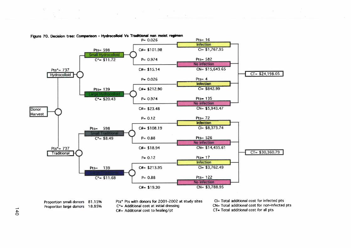
Fþure 70. tþctsbn tree: Co¡T¡rbon - lltrcolgil Ys Tr¡dtiotC nqr mobt rcthãtP= 0.026
Prs= 598
Pts*= 737
C#= î23.48
P= O.12
Pt= 598
737
c#= $19.30
16
CN= S5,943.47
Pts= 72
Proportion small donorsProportion large donors
81.15%18.85%
Pts* Pts wtth donors for 2OO1-2O02 at study sitesC*= Additional cost at in'itial dressirqC#= Additional cost to healirig/pt
Cl= Total additional cost for infected ptsCN= Total additkrnal cost for non-infected ptsCT= Total additional cost for all ptsJ
èo
Harvest
c#= 3212.90
P: 0.974
.99
Prs= 1 35
C1=Prs= 1 39
't 'l
15,643.65cN=
4
C#= $15.14
P= 0.026
C#= $101.98
P= 0.974
ct= '1,767.95
Pts= 582
IÏiÈrvrrreEreil
P= 0.88
c#: $2r 3.9s
Vß= 122
Pts= 1
cN= 14,435.61
Pts= I 7
C#= $18.94
P= O.12
c#= s108.r9
P= 0.88 Pts= 526
r@@

Description of alternative interventions
The following description gives an overview of the alternative interventions based on
the decision to use either hydrocolloids or the traditional paraffin gauze dressings in
the management of STSG donor sites.
Hydrocolloids
As detailed in fig. 70, when 737 cases are allocated to have hydrocolloids as the
therapy of choice, 598 cases (81.15%) would have a small dressing applied and 139
(18.85%) would have the larger dressing applied. ln determining costs for this
episode of care materials included the primary dressing, secondary dressings and the
tapes used to secure these items. Staff time to conduct the ¡ntervention was also
calculated. lt should be noted that overheads and the cost of the primary procedure,
that of skin grafting the primary wound, was not calculated as this cost would have
been equal regardless of which alternative was used in the management of the donor
site. The details of the costings for this component of the intervention are found in
appendix 9, table 30 for the smaller dressing model, and table 31 for the larger
dressing model. The cost for the smaller dressing model was $11.7? per episode and
$20.43 for the larger dressing model. The primary dressing (hydrocolloid) closest to
the skin is self-adhesive and would not necessarily require any additional fixation. At
the sites used to collect additional clinical information however, the primary dressing
was additionally secured with a border of fabric retention tape. For this reason this
item was also included in the costing. The primary dressing would then routinely be
covered by a secondary dressing of combine and crepe bandage to manage any
unforseen leakage from the primary dressing. From the systematic review previously
conducted it can be assumed that for those cases with the hydrocolloid dressing
regimen 97.4o/o would not get infected.
Non-infected cases
For the purpose of this analysis it has been assumed that for the non-infected group
there would be a dressing change at day 3, post excision. At the study sites using
hydrocolloids this occurred frequently as a result of leakage and or for inspection.
The costing details for this component are detailed in appendix 9, table 32 for the
small donor model and table 33 for the larger model. The next component of the
141
intervention would be the removal of the dressing at time of complete healing.
Details of this final component are provided in appendix 9, table 34 for the small
donor model for the larger model. The cost of the removal of the small and large
dressings were determined to be the same as the size difference made no
appreciable difference in the time taken to remove the dressings the healed donor
was not redressed after complete healing had occurred.
lnfected cases
ln the model used for this analysis it was determined that if all cases used the
hydrocolloids 2.60/o of cases (16 small donors and 4 large donors) would become
infected. At the participating sites the management of infected donors was quite
uniform. lnfected donors were treated with silversulphadiazine cream. For the
purpose of analysis the model assumes that treatment was daily for three days. The
details of the costs are provided in appendix 9, table 35 for the small donor model
and table 36 for the larger model. The application of silversulphadiazine cream was
facilitated by impregnating thin porous cloth sheets by hand and applying these
sheets directly to the infected donor site. This was then covered with combine
dressing and crepe bandage and secured with elastic tape. Following successful
treatment of the infection the donor site would be redressed with the hydrocolloid
dressing (appendix 9, table 37 for the small donor model and table 38 for the larger).
The final component of the intervention is identical to the non-infected donors as
detailed in table 34.
Based on 737 cases that represents the situation for the 3 participating sites for a
12 month period, the additional costs of managing the STSG donor sites with
hydrocolloids is $24,198. This includes both small and large donors and those
infected and without infection.
Paraffin gauze
ln fig.70,in the lower half of the decision tree, when737 cases are allocated to use
the traditional paraffin gauze dressings as the therapy of choice, 598 cases
(81.15%) would have a small dressing applied and 139 (18.85%) would have the
larger dressing applied. ln determining costs for this episode of care materials
included the primary dressing, secondary dressings and the tapes used to secure
these items. Staff time to conduct the intervention was also calculated. As with the
hydrocolloid regimen overheads and the cost of the primary procedure, that of skin
142
grafting the primary wound, were not calculated as this cost would have been equal
regardless of which alternative was used in the management of the donor site. The
details of the costings for this component of the intervention are found in appendix
9, table 39 for the smaller dressing model, and table 40 for the larger dressing
model. The cost for the smaller dressing model was $8.49 per episode and $11.68
for the larger dressing model. At this point the cost comparison, prior to any
infection, is considerably less for the paraffin gauze regimen. However, when the
infection rate derived from the meta-analysis of the systematic review (Wiechula,
2001) is applied it can be assumed that 89 cases (12o/o) would become infected.
Non-infected cases
For the purpose of this analysis it has been assumed that for the non-infected group
there would be a dressing change at day 3, post excision. This would not involve the
complete removal of dressings but would entail the removal of tapes and outer
dressings that would then be replaced. The traditional STSG donor site dressing
was/is multi-layered, consisting of paraffin gauze, closest to the wound, covered
then with a thick layer of cotton gauze, combine dressing and crepe bandage. The
bulky dressing was designed to absorb and draw away from the wound the large
amounts of haemoserous exudate so as to dry the wound out. The dressings were
then secured with a binding of tape to secure the dressings even when the patient
was able to ambulate. The traditional donor site dressings is no longer used at any of
the study sites however many senior staff were able to describe the regimen in
elaborate detail. They indicated that at some point usually within the first three days
post excision the amount of exudate would strike through the outer dressings and
would facilitate the need to change the outer dressings of combine and crepe. The
costing details for this component are detailed in appendix 9, table 41 for the small
donor model and table 42 for the larger donor.
For the non-infected cases the next component of the intervent¡on would be the
removal of the dressing at time of complete healing. Details of this final component
are provided in appendix 9, table 43 for the small donor model for the larger model.
As with the hydrocolloid regimen the cost of the removal of the small and large
dressings were determined to be the same as the size difference made no
appreciable difference in the time taken to remove the dressings the healed donor
was not redressed after complete healing had occurred.
143
lnfected cases
ln the model used for this analysis it was determined that if all cases were managed
with the paraffin gauze regimen 12o/o of cases (72 small donors and 17 large donors)
would become infected. At the participating sites the management of infected
donors was quite uniform and in fact was the same as for the hydrocolloid example.
lnfected donors were treated with silversulphadiazine cream. For the purpose of
analysis the model assumes that treatment was daily for three days. The details of
the costs are provided in appendix 9, table 44 for the small donor model and table 45
for the larger model. Following successful treatment of the infection the donor site
would be redressed with the paraffin gauze dressing (appendix 9, table 46 for the
small donor model and table 47 for the larger model). The final component of the
intervention is identical to the non-infected donors as detailed in table 43.
Based on 737 cases that represents the situat¡on for the 3 participating sites for a
12 month period, the additional costs of managing the STSG donor sites with the
traditional paraffin gaaze regimen is $30,360. This includes both small and large
donors and those infected and without infection.
Sensitivity analys¡s
One of the difficulties in determining the infection rate using data from the meta-
analysis in the systematic review is that is that the rate did not account for the
weighting of the studies combined in the meta-analysis. lt may be argued that this
would then have the potential to inflate the infection rate of the paraffin gauze
regimen. lt was therefore decided to conduct a sensitivity analysis to determine what
the result of the analysis would be if it were assumed that the infection rate for
cases managed with paraffin gauze was reduced. Assuming an infection rate of only
60/o (paraffin gauze regimen) and not lhe 12o/o as previously described would still
result in the hydrocolloid being more cost effective with albeit a small potential
saving of $1,337. At the point where the infection rates were assumed to be
equivalent for the two alternative regimens the analysis would indicate the traditional
regimen to be most cost effective but only by $t,397. This would only represent a
saving of effectively less than $2.00 per patient.
144

Summary of findings
ln considering the two alternative interventions of, hydrocolloid regimen and the
traditional paraffin gauze regimen for the management of STSG donor sites, the cost
effectiveness analysis establishes that the hydrocolloid regimen is the most cost
effective. ln the model described in fig. 70, should the three study sites have chosen
to use the hydrocolloid regimen for all the 737 cases the cost of managing these
cases would have been $24,198. The cost of choosing the paraffin gauze alternative
would have been $30,360. Utilising the hydrocolloids provides a potential saving of
$6,162.
Comparison 2: Hydrocolloid, calcium alginate and retention
dressing regimens: A cost minimisation analysis
As with the previous cost effectiveness analysis the intention is not to describe every
possible alternative dressing regimen but to cover a range of alternatives that were
commonly seen in the study units. The three generic dressings that are considered
are hydrocolloids, calcium alginates and fabric retention dressings. The main
assumption used in the analysis is that the clinical outcome in terms of healing and
infection were equal for all regimens described.
Descriptions of alternative interventions
The following descriptions give an overview of the alternative interventions based on
the decision to use a particular moist wound healing dressing as the primary dressing
in the management of STSG donor sites. The details of the analysis are depicted
graphically in fig.71,in the section following. As with the previous analysis the model
describes the number of cases that occurred over a 12 month period with cases
divided proportionally between small and large STSG donor sites. lt should also be
noted that the analysis deals with the issue of an early dressing change/inspection.
Moist wound healing dressings are designed to remain intact until healing is complete.
ln the case of STSG donor sites this is the ideal circumstance and is frequently
achieved. There are however, a number of reasons why a dressing may be removed
prior to healing. These dressings have a potential to absorb a certain amount of fluid.
When this potential is exceeded then the dressing will leak this excess fluid, requiring
in the least a change of the secondary dressings and most often a primary dressing
change. ln addition, as routine there were a number of plastic surgeons that required
145

dressings to be removed prior to healing, as their preference was to conduct a visual
inspection of the wound. For one of the regimens described, the primary dressing is
changed to an alternative at this inspection stage.
146

Fig¡re 71. tÞcisbn ücc: Comparisorl - l{tdtHc mdst tflomd hGalng nìgiæhsRenpralFist
c#= 98
cT=
CT=
353.52
4 728.59
10.91
7 .60
C#=
Pts* pts with dorprs for 2O0l-2003 at study sites CR= Addtional cost at removal of üessingC*= Adrltiond cost at initid üessirE CT= Totd additional cost lncluding removal
C#= Adclitional cost at firct change of dressirB CTC= Total add¡tional cost inclucfing removal cornbined (S+L)
srzes
CTC= 163.36
$16,1 13.10
CTC= ,472
CTC=
CTC= 14,1
CTC= 14,628.88
Proportion snn[ donorsProportion large donors
81.15%18.85%J
5N
737 Pls
IEEl:lt-
Pts
598 Pts
737 Pts
IEE|ilt-
fts
Pts
Pts*= 737
Donor Harvest
cT=I 39 Pts
598 fts
737 Pts-f:IfilIEr,rf,-
Hydrocolloids
For this group of regimens hydrocolloid dressings are the primary dressings used
throughout. For each regimen the care is described for both small and large donors.
The regimen is illustrated by using the number of cases that had occurred during the
12 month period at the three participating sites. lt should be stressed that this is a
model of what would have occurred if a particular regimen were used for all these
cases. lt is not a record of what actually occurred.
ln fig.71, when 737 cases are allocated to have hydrocolloids as the therapy of
choice, 598 cases (81.15%) would have a small dressing applied and 139 (18.85%)
would have the larger dressing applied. ln determining costs for this episode of care
materials included the primary dressing, secondary dressings and the tapes used to
secure these items. Staff time to conduct the intervention was also calculated. lt
should be noted that overheads and the cost of the primary procedure, that of skin
grafting the primary wound, was not calculated as this cost would have been equal
regardless of which alternative was used in the management of the donor site. As the
care for this regimen is identical to that described in the previous cost effectiveness
analysis the details of the costings for this component of the intervention are found
in appendix 9, table 30 for the smaller dressing model, and table 31 for the larger
dressing model.
Primary dressing changed once pr¡or to final removal
For this group the analysis assumes a change of dressing at day 3 post excision. The
timing of this dressing change is not critical and does not effectively impact on the
cost. The costing details for this component are identical to the hydrocolloid regimen
used in the cost effectiveness analysis and are therefore detailed in appendix 9, table
32 for the small donor model and table 33 for the larger model. The next component
of the intervention would be the removal of the dressing at time of complete healing.
Details of this final component are provided in appendix 9, table 34 for the small
donor model and for the larger model.
Primary dressing ¡ntact unt¡l final removal
The variation for the hydrocolloid regimen involves inspection of the dressings but
with leaving the primary dressing intact. The cost of the initial dressings remains the
same but there is a cost reduction if at inspection the primary dressing remains
148

intact. The costing details of this component of the regimen are presented in table
48. The cost of removal of the dressing once healed is the same for all hydrocolloid
examples (see table 34).
Summary for the hydrocolloid reg¡mens
The analysis indicates that if there were 737 cases and all cases used a hydrocolloid
regimen that included a change of primary dressing prior to healing the total
additional cost would be $22,163 (see fig.71). This does not take into account
infected donor sites as the model assumes no difference in infection rates between
the moist wound healing regimens. lf all cases (737) were subjected to a hydrocolloid
regimen that had the primary dressing remain intact the total additional cost would
be $16,1 13 (see fil.71).
Calcium alginates
For this group of regimens calcium alginates are the primary dressings used
throughout the intervention until the donor is healed.
ln determining costs for this episode of care materials included the primary dressing,
secondary dressings and the tapes used to secure these items. Staff time to conduct
the intervention was also calculated. As with the hydrocolloids two regimens are
described one with the primary dressing removed and redressed and alternatively
with the primary dressing remaining intact throughout the management of the STSG
donor site. Calcium alginates are highly absorbent dressings that can manage wounds
with high exudate levels. The calcium alginate is applied directly to the wound and
reinforced with further absorbent layers of gauze, combine dressings crepe bandage
and elastic tape. The details of the additional cost for the ¡nitial calcium alginate
donor site dressings are provided in the costing tables; table 49 (small donors) and
table 50 (large donors).
Primary dressing changed once pr¡or to final removal
For this group the analysis assumes a change of dressing at day 3 post excision. The
costing details for this component are detailed in appendix 9, table 51 for the small
donor model and table 52 for the larger model. The next component of the
intervention would be the removal of the dressing at time of complete healing.
Details of this final component are provided in appendix 9, table 53 for the small
donor model and for the larger model.
149

Primary dressing intact until final removal
The variation for the calcium alginate regimen involves inspection of the dressings
but with leaving the primary dressing intact. The cost of the initial dressings remains
the same but there is a cost reduction if at inspection the primary dressing remains
intact. The costing details of this component of the regimen are presented in table
54 for small donor sites and table 55 for large donor sites. The cost of removal of
the dressing once healed is the same for all calcium alginate examples (see table 53).
Primary and secondary dressings intact unt¡l final removal
A further variation for the calcium alginate regimen involves leaving both the primary
and secondary dressings intact. No additional costs are incurred in the interim period.
Summary for the calcium alginate reg¡mens
The analysis indicates that if there were 737 cases and all cases used a calcium
alginate dressing regimen that included a change of primary dressing prior to healing
the total additional cost would be $?4,472 (see fig.71). This again does not take
into account infected donor sites as the model assumes no difference in infection
rates between all the moist wound healing regimens. lf all cases (737) were
subjected to a calcium alginate regimen that had the primary dressing remain intact
the total additional cost would be $19,565 (see fig. 71). For the third variation
where all dressings remain intact until healing is achieved the total add¡tional costs
would be $1 4,194 (see fig. 71).
Calcium alginate/hydrocolloid combination
At two of the study sites a regimen was regularly used in which the primary dressing
was changed at the early inspection stage from a calcium alginate to a hydrocolloid
dressing. The staff involved provided the rational that, when the donor site was newly
excised the calcium alginate provided the additional absorption required. When the
donor had stabilised and was exuding only small to moderate amounts of fluid the
hydrocolloid provided adequate absorption and was flexible enough to increase
comfort when ambulating. This explanation was anecdotal and was not specifically
tested by this study. The costing tables previously used for calcium alginates (initial
dressings) and hydrocolloids (inspection/first change and removal) were used for this
regimen. The result was that for the example of 737 cases the total additional costs
for donor site management were $22,837 (see fig.71).
150

Retention tape dressings
The final group of dressing regimens concerns those with the primary dressing of
fabric retention tape. This dressing is gaining some popularity and was used almost
exclusively at one of the units at a participating site. The method involves applying
the adhesive tape directly to the wound. There are a number of brands available in a
variety of widths so that most wounds can be totally covered with an adequate
margin. The tape is porous and exudate escapes into the absorbent secondary
dressings. Once haemostasis has been achieved the outer dressings are removed and
the primary dressing can be wet and dried again allowing patients who are capable, to
shower. Due to the conformability of the dressing patient can ambulate with comfort.
Again it should be noted that this is was beyond the resources of this study to
quantifiably demonstrate these attributes of the retention dressings.
The costing tables that describe the in¡tial dressings for both wound sizes for
retention dressings are presented in tables 56 (small) and 57 (large) respectively.
The costings for the inspection with secondary dressings only removed are presented
in table 58 (both small and large donor sizes). The removal of the retention tape
dressings, are costed in table 59. The total additional costs, if all 737 cases were to
have the retention tape dressings, is $14,628.
Summary for the comparison of all mo¡st wound
healing reg¡mens
A wide range of alternative moist wound healing dressing interventions were analysed
to determine which regimen produced the minimal costs. Based on a 737 cases, 598
cases would represent wounds of approximately 7.5 x 7.5 cm or less and 139 cases
of larger donors up to approximately 15 x 15cm. The analysis detailed in fig. 71
indicates the total additional costs if all cases where to follow one particular regimen.
As these regimens essentially are a secondary procedure overhead costs such as
hospitality costs are not included in the analysis. The regimen with the least
additional costs is that where calcium alginates are used and all dressings are left
intact until complete healing has occurred ($t 4,192). The regimen that incurs the
greatest additional cost is also a calcium alginate regimen in which there is
replacement of the primary dressing during the period of healing ($24,47?). fhe
difference between these two strategies is over $10,000 or a mean of $13.94 per
151

case. The example using retention tape dressings ($14,628) is almost equivalent to
calcium alginate regime with all dressings intact. ln general for the regimens where
the primary dressing remains intact the cost difference is negligible. Further fine-
tuning would involve mixing regimens based on size of donor wound. Using the
number of cases previously specified if all patients with the smaller donor sites were
managed with calcium alginates and the cases with larger donors managed with
retention tape dressings the total additional costs would be $13,324.
152

Chapter 1 6. Economic Evaluation Discussion
ln regarding the results of this study the reader is reminded that the intention is to
compare the cost effectiveness of a range of interventions used in the post harvest
management of STSG donor sites. The analysis uses a number of regimens that
describe, in as much detail as possible, what would occur in actual practice. ln
describing these regimens there are two major inter-related issues that the reader
must consider. ln the first instance, as with any area of practice, there is the
potential for variability in practice. The regimens describe a range of interventions
that differ in the type of materials and the manner in which they are used. For
instance, in some cases one primary dressing is used and maintained until the
intervention is completed, in other circumstances the dressings are removed prior to
healing and the donor site redressed with either the same primary dressing or even
another type of primary dressing. Throughout the period of data collection in the
clinical atea a great deal of variation in practice was observed. This variation was
evident both between sites and between individuals at the same sites. ln some cases
variation was due to personal preference of the clinicians. ln other cases, as
expected, it was due to the specific circumstances of the individuals that were the
subject of the intervention. There was no intention to present all observed regimens
but to represent a reasonable cross-section to provide the reader with a range of
alternatives to consider.
The second issue that should be considered is that within the confines of the study
not all variables could be observed directly. ln the case of the traditional paraffin
gauze dressings, none of the sites reported using this dressing method although all
sites had previously done so. Clinicians were able to describe in detail this
intervention and 'mock' dressings were conducted to achieve reasonably accurate
timings.
Limitations
As with any research exercise this study was compromised in a number of ways that
ultimately limit the strength of the results. ln particular, economic evaluation is
criticised because it often includes assumptive elements in modelling that may be
judged as unrealistic or inaccurate. There have been assumptions made in conducting
this study that if adequate resources were available may have been more accurately
verified. The following sections deal with the limitations of the study and detail why
153

certain assumptions were made and in what way this may have impacted on the
strength of the results.
Accuracy of cost¡ngs
As detailed in the methods chapter, the cost of various items used in the analysis
were provided by materials management and other relevant departments at the three
participating sites. ln terms of staffing costs this was calculated at the rate for a
base level registered nurse at three years post registration. For the part¡cipating sites
the hourly rate for this level of staff would have been uniform as all sites were public
hospitals and subject to the same industrial award. lt may be argued that a more
junior staff member could have conducted the interventions. lf a junior staff member
was used in the modelling this would have some impact on the comparative cost of
the alternative regimens however the impact of time on overall costs was minimal
and this was not likely to alter the overall results.
The cost of the dressings and consumables did alter between sites and this occurred
for a variety of reasons. ln discussion with materials management staff, prices varied
in the first instance due to the volume purchased. This is particularly important for
wound products where there are not only viable alternatives between generic types
of dressings but also within generic dressing groups. These wound products are not
only used for donor sites but many other wound types also. Consequently depending
on the types of wounds (including donors) and the volumes to be treated, a
particular site may achieve a significant discount price for their dressing purchase. lt
was also apparent that companies who supplied a range of dressing types provided
discounts for 'bundled' purchases. For example, if a site required four or five different
dressing types, and these could all be supplied from the same company, a discount
would be offered to have the products supplied from the one source. There was no
way to determine what discounts m¡ght be applied for particular dressing products if
the volumes used in the analysis were applied. This would only represent a proportion
of the total product purchased. Given these limitations it was decided that averaging
the unit prices between sites was the only viable approach. The other issue that must
be addressed in relation to the cost of dressings is that within generic dressing
groups there may be several suppliers with dressings that are identical or have some
minor variation. ln what is a very competitive market there was often variation in cost
between products that clinicians would otherwise consider identical. Again, it was
154

decided that where two alternatives of the same dressing type were available an
average would be used in the analysis. These costs are detailed in the costing tables
in appendix 9. The specific costs of products provided by the individual sites are not
detailed in this report on instruction from the participating sites.
The use of clinical effectiveness data from
systemat¡c rev¡ew
A crucial element in any economic evaluation of practice is the integrity or
robustness of the clinical effectiveness data used in the analysis. As previously
indicated there are a number of alternative approaches to deriving this data. The
clinical effectiveness data used in this study were derived using meta-analysis of
previously conducted clinical trials. Using this approach means the study is to a
degree constrained by the data that is available. This constraint is twofold. ln the
first instance there is the level of confidence in the accuracy of the effectiveness
measures used in the analysis. ln the case of the infection rates for the comparison
between hydrocolloids and the tradit¡onal paraffin gauze dressings results were
determined from the meta-analysis of only a small number of studies with modest
sample sizes. The sensitivity analysis compensates for this limitation to a degree.
The other constraint that must be considered is the availability of head to head trials.
The retention tape dressings were included in the comparison because of the growing
interest in this approach to donor site management but clearly there are very few
well designed RCTs or llTs to substantiate their clinical effectiveness. These issues
will be specifically explored in the discussion that follows in relation to the results of
the two comparisons.
Comparison 1: Hydrocolloids versus paraffin gauze dressings
The decision tree (fig. 70) illustrates both the probable clinical outcomes for our
study population and the associated costs (excluding common costs) of the
alternative interventions. As a result of the increased infection rate for the traditional
paraffin dressings the hydrocolloid is determined to be the most cost effective of the
two alternatives. The result however is not absolute and the following questions
should be addressed. Firstly how confident are we in this outcome and then to what
degree is the result generalisable?
155

Addressing the first part of this question there are a range of issues that should be
considered. As previously discussed the clinical effectiveness data used in this
analysis is retrospective and derived from the meta-analysis of four studies. When
pooled the total number of subjects was only 211 patients (Wiechula,2001).
Although the result was statistically significant in favour of the hydrocolloids with
regard to infection rate this represents a modest sample size. This is somewhat
balanced by fact that the studies used were of high quality using the gold standard
of RCT/llT design. ln addition the sensitivity analysis demonstrated that after the
infection rate for the traditional donor dressings was reduced by 5Oo/o, the
hydrocolloid was still more cost effective. ln determining the costs of the alternate
interventions it should also be considered that only the hydrocolloid intervention was
observed in actual practice. As the traditional paraffin dressing was no longer used at
any of the study sites a mock dressing was performed to establish both the
consumables that would be used and the time to undertake the intervention. At all
sites there were clinicians that had previously performed this intervention and were
able to verify what consumables would be used for the dressing. The less reliable
element in the modelling was the time taken and thus the labour costs of the
intervention. The most accurate method of determining the time taken for the
procedure would have been to observe this care being undertaken on actual patients
in the clinical setting but this was simply not possible. Given these considerations it
can be argued that there is still a strong degree of confidence in the results of the
analysis.
The analysis was conducted within a given context; three large teaching hospitals in
one city in Australia, at a given time, 2001, with a particular population using
dressings that were managed in a specified manner. The purpose of conducting the
analysis was to inform clinicians not only in the study settings but also more broadly.
ln determining the generalisability of the findings, the following should be considered.
Using the output from meta-analysis has an advantage over prospective data from a
single study as the pooling of multiple study results increases their generalisability
(Pang et al., 1999). ln this case the studies used were from a variety of settings and
demographic types (Wiechula, 2001). The fact that the study sites for the additional
clinical data and cost data were large teaching hospitals should be considered. ln
terms of the feasibility of using hydrocolloids for STSG donor management in other
settings it should be noted that the intervention is not reliant on any specific
156

infrastructure and the post harvest care using hydrocolloids could easily be managed
in a variety of settings. The availability of hydrocolloids may be an issue, however as
evidenced from the systematic review, clinical studies were conducted in a number of
countries and this suggests they are widely available.
ln summary comparing the two interventions, the trad¡tional paraffin tulle donor
dressing regimen and the hydrocolloid regimen, without the confounding impact of
complications, the hydrocolloid is more costly. However, when the impact of infection
is taken into account, clearly the hydrocolloid regimen is more cost effective. These
results are sufficiently robust that from an economic perspective it is difficult to
support the contlnued use of the paraffin tulle regimen. Clinical decision making
however must consider issues other than cost. The results of the analysis should be
considered in conjunction with subjective evidence part¡cular to the individual and the
setting in which the clinical decision is made, but there is also some objective
evidence that should also be considered. The results of the systematic review
indicate that in addition to infection rates the hydrocolloid is superior in terms of
healing time and pain further supporting the case for the use of hydrocolloids
(Wiechula, 2001).
Comparison 2: Multiple moist wound healing
p ro d u cts/st rateg i es c o m p a red
The major issue to address when considering the results of the cost minimisation
analysis of the range of moist wound healing dressings/regimens is the assumpt¡on
that all the alternatives perform equally well in terms of clinical effectiveness. lt is
assumed that rates of infection, healing and pain levels will be the same for all
regimens. lt is important to note that this assumption is made not because we have
objective evidence that this is the case but because there is no strong evidence to
dispute this. As previously discussed, there are few head to head trials comparing the
clinical effectiveness of hydrocolloids and calcium alginates. There is even less
objective evidence with regard to retention tape dressing regimens (Wiechula, 2001).
ln most cases where moist wound healing products have been subjected to
comparative trial the control/comparator was the traditional paraffin gauze regimen.
Arguably, in the past this choice of control was appropriate, however with an
apparent shift in interest to moist wound healing regimens it is odd that more recent
studies are still being conducted with the traditional comparator. There is an
157
argument that suggests that without strong clinical effectiveness evidence the cost
minimisation analysis should not be conducted (Drummond et al., 1997). The
decision to conduct the analysis was based on the concern that despite the lack of
clinical trials these products are being used in practice. This does not supersede the
need to conduct head to head trials, in fact it further supports that their conduct is
imperative. The reader should then consider the results of the analysis knowing that
the head to head clinical effectiveness data is deficient and treat the results with due
caution.
ln the first comparison for this study only two alternate dressing regimens were
compared. ln the second comparison a wider variety of regimens were considered.
lmportantly the variation in the regimens is not only in the type of primary dressing
used but also in how the dressings are managed. The major variations in management
relate to the inspection and or replacement of the primary dressing. As stated in the
results chapter when the primary dressings are left intact the difference in cost
between the regimens is minimal. For instance the average cost per patient (total
excluding common costs) for the hydrocolloid regimen with the primary dressing
intact at the inspection stage is AUD$21.85. For the calcium alginate regimen, when
all dressings are intact until complete healing, the average cost is AUD$19.25.
Naturally, as indicated in fig.71, when the primary dressings are inspected and
changed the cost per patient increases considerably. This is a crucial consideration.
When the additional clinical data was being collected a variety of justifications were
given for both leaving the primary dressing intact or alternatively replacing it. Some
clinicians indicated that their preference was to remove the primary dressing, usually
at day three post operatively to definitively assess the wound. Others indicated that
they felt they could adequately assess the wound with the primary dressing intact
and that removing the primary dressing increased the risk of contamínation. The
problem is that these clinical opinions are anecdotal. One of the objectives of the
systematic review of clinical effectiveness was to identify studies where, for the
same primary dressing there was comparison of different management methods. No
studies examining the issue of leaving the primary dressing intact versus changing it
were identified (Wiechula, 2001). Again we have the situation where variation in
practice is occurring but has not been tested objectively with high quality clinical
comparative trials.
158
Combining clinical and cost eftectiveness results
One of the aims of this study was to compare the cost effectiveness of a range of
regimens for the post harvest management of STSG donor sites. Due largely to the
available clinical effectiveness data two comparisons using different techniques were
conducted. The first comparison between the traditional paraffin gauze dressings and
the more contemporary hydrocolloid dressings favours the hydrocolloids, that is the
hydrocolloid is the more cost effective treatment option. This result is given with a
strong level of confidence and is relatively generalisable. Combined with the available
clinical effectiveness data from the systematic review in relation to patient
pain/comfort levels and healing rates, the objective evidence suggests that paraffin
gauze dressings should not be used. The evidence does indicate that hydrocolloids
should be used but only as an alternative to the traditional style of dressings. This is
also in keeping with the principles of moist wound healing. The difficulty is that there
are many other options for the management of STSG donor sites, which the second
comparison attempts to address. The analysis for this comparison is based on the
assumption that all regimens are equally clinically effective. The major differences in
the regimens are the primary dressing product used and the necessity to visually
inspect the wound early in the intervention. This assumption is not based on
objective evidence but rather the absence of evidence to dispute this. lf the
assumptions about the product used and visual inspection are tested in rigorous
clinical trial the outcomes may impact on our analysis and subsequent
recommendation in a number of ways.
lf the assumption that there is no greater risk in leaving the dressings intact is
supported by clinical trial, regardless of the primary dressing products used, then the
clinical and economic evidence supports the case that unless otherwise clinically
indicated dressings should be left intact and the type of primary dressing used will
not significantly impact on cost.
lf by way of rigorous clinical trial it can be shown that mandatory early inspection,
regardless of dressing type, will result in significantly reduced infection rates then the
focus shifts to the type of primary dressing used. This is because the analysis
indicates that the additional cost of changing the primary dressings varies depending
on the dressing type used.
159

lf it can be demonstrated that depending on the type of primary dressing used
different clinical outcomes occur (particularly in relation to infection rates) then the
current analysis is redundant and no conclusions can be drawn from it.
Another element that has not been considered in either of the two comparisons
presented in the study is the cost of changing practice. Clearly from the data
collection at the three sites there is quite a deal of variation both in the primary
dressing product and the manner in which that product is used. ln the least there
would be some costs incurred for staff training when altering practice.
Summary of discussion
Due to the uniform nature of the creation of the STSG donor site it is reasonable to
assume that there is considerable opportunity for consistency in the management of
these wounds. Despite this we know that there is considerable practice variability
both in terms of products used and the manner in which they are used. The analysis
conducted in this study provides us with cost effectiveness results that when
combined with clinical effectiveness data provide evidence that supports a reduction
in this variability. The strength of this evidence is however variable largely due to the
confidence in the assumptions made in the economic modelling used in the analysis.
ln the case of the traditional paraffin gauze dressings there is strong evidence that
this intervention is not cost effective and considering adverse clinical outcomes
should be abandoned. The analysis does not however provide us with a single
definitive product and regimen that can be considered best practice. The evidence
suggests that hydrocolloids are a more cost and clinically effective alternative, but
there is lack of evidence to indicate that they are superior to other moist wound
healing alternatives. There is some evidence to suggest that calcium alginates or
retention tape dressings may be more cost effective but this is based on clinical
assumptions that are yet to be rigorously tested.
Although the results of this study may be seen as somewhat disappointing in that the
evidence is variable in strength and therefore limited in its ability to support and
direct a reduction in practice variability a number of recommendations for practice
and future research can be made. The final chapter will summarise these
recommendations.
160
Chapter 17. Economic Evaluation Conclusions
An inherent characteristic of clinical wound management is that in all cases the focus
of the care, the wound and off course the wounded, is variable. No two wounds are
identical. ln the case of traumatic and chronic wounds the number of intrinsic and
extrinsic factors that can impact on the wound and on the subsequent management
are considerable. There is at least one example that is an exception. ln the case of
STSG donor using modern harvesting methods wounds can be created relatively
uniformly. This is not to say that all donor sites are identical and that the individuals
with these wounds are also identical but compared with many other wound types
there is a high degree of uniformity in the wound itself. lt is this level of uniformity
that allowed Winter (1962) to conduct his seminal research comparing dry and moist
wound healing techniques on domestic pigs. lt is this uniformity that provides a
reasonable potential to reduce variability in the management of these wounds.
Despite this potential it is obvious there is practice variability both in the products
and strategies used in the management of STSG donor sites. To reduce this variability
will require individuals to change their practice. lnducing clinicians to change practice
is a complex issue that is the subject of considerable study and debate. The major
contribution of the evidence based movement to practice change is in providing
objective evidence to support which practice alternative should be considered best
practice. ln the case of STSG donor sites this study and the preceding systematic
review provide objective evidence of clinical and economic effectiveness. The task is
to consider what implications this evidence has for practice recommendations.
Recommendations for practice
From the outset it must be stated that the clinical and economic effectiveness
evidence does not provide us with a single unequivocal regimen that could be
considered best practice. The evidence does allow some recommendations about
practice to be made.
lf we consider the vast array of alternatives available for the post harvest
management of STSG donor sites our strongest evidence directs us against the use
of the traditional paraffin gauze type regimens. They should simply not be used in
practice. The use of hydrocolloid sheet dressings is considered to be superior.
However, there are a large number of moist wound healing alternatives to
161
hydrocolloids. Unfortunately there is no strong objective evidence to indicate which
of these alternatives are superior and should be considered best practice. Further
there are variations in the manner in which these dressings can be used. The cost
effectiveness analysis indicates that mandatory change of dressings can be a
significant factor when deciding on a particular dressing type but critically there is a
gap in the clinical effectiveness evidence to direct us with this recommendation.
Recommendations for research
It is often the case with evidence based researchers that when evidence synthesis
fails to provide definitive recommendations for practice then somewhat ironically it
leaves us with strong and definitive recommendations for research. This study is no
exception. ln terms of clinical trials the primary recommendation would be to cease
using the control/comparator of the traditional paraffin gauze dressing. lnstead it is
strongly recommended that head to head trials comparing alternative moist wound
healing products should be conducted and the focus should be on those most widely
used in the first instance. Perhaps the most important finding and subsequent
recommendation to come from this study relates to the issue of mandatory dressing
changes. This has previously not been explored in clinical trials. lt certainly has
potential cost implications but also may have considerable impact on patient comfort
and therefore should be investigated in future trials. This is a fundamental issue and
one that is wider than STSG donor management. The basis of moist wound healing is
that the dressings are able to maintain an optimum environment to best facilitate
healing. Unnecessary dressing changes have the potential to disrupt this environment
and retard wound healing. The STSG donor site does provide a relatively unique
opportunity where clinical trials can be conducted with a high level of control and
uniformity to explore this issue.
16?

Part 4: Portfolio lntegrating the Findings
163

Chapter 1 8. Portfolio Discussion and Conclusions
As stated previously the value of undertaking doctoral research is not isolated to the
study findings. ln being instructed and supported through a research project the
doctoral candidate develops skills that can then be used to conduct further research.
Since completing the systematic review as part of this program the candidate has
completed further reviews and is currently involved in teaching the review process to
others. ln the case of the Doctor of Nursing the candidate develops skills within a
number of research designs. This is particularly important for nursing, although
arguably the same can be said for healthcare generally, where the complexity of
practice demands that research be an integrated process using a variety of study
designs. This is perhaps the major appeal of the Doctor of Nursing where a program
of research is completed rather than a single study. This also fits well with what is
emerging in the evidence based practice movement. The recent and growing impetus
to review and incorporate evidence beyond the RCT into practice recommendations
(Pearson, 2OO4; Popay & Roen, 2003) is recognition that a pluralistic approach to
evidence is necessary. The structure of the Doctor of Nursing is also demonstrative
of the symbiosis of knowledge translation and knowledge generation. Nursing in
particular has been criticised for conducting small, isolated and often unnecessarily
duplicated clinical studies. lt is therefore logical to conduct a systematic review to
confirm with confidence the justification for conducting further primary research.
Perhaps the most significant limitation of this particular Doctoral portfolio is that it
was undertaken at a time when the methods of reviewing evidence other than clinical
effectiveness were only just emerging. The Cochrane Qualitative Methods Group, the
Campbell Collaboration and the Joanna Briggs lnstitute among others have been
examining the review of qualitative research (Popay & Roen, 2003). Pearson
(Pearson, 2004) is taking this a step further with the development of the SUMARI
software suite that will allow the conduct of the Comprehensive Systemat¡c Review.
This involves conducting an integrated review of multiple evidence types.
lntegration of studies
ln addition to providing the candidate with research skills the studies have also
provided evidence that informs practice. The results of the studies as detailed in the
individual reports are of value even when viewed in isolation however in keeping with
164

the notion of integrating evidence the following discussion will consider the results of
the two studies and how they may be combined to inform practice.
There is always the hope that in reviewing the evidence that, simple and direct
recommendations, without equivocation can be made. ln the review of the evidence
of clinical effectiveness no one intervention stood out as best practice. The review
established, with a great degree of confidence, that moist wound healing methods of
dressing STSG donor sites are superior to the traditional 'dry' methods in terms of
healing, infection and patient comfort. These traditional methods are still being used
in some practices and should be abandoned. When considering the range of moist
wound healing dressings the picture is less clear. The lack of head to head research
comparing moist wound healing dressings of different types means that no particular
generic group can be held above another. These dressings all function by attempting
to maintain a moist wound environment but they do not do this uniformly. Dressings
within one generic group such as semi-permeable films are designed to manage
wounds with a low level of exudate. At the other end of the spectrum calcium
alginates are able to manage wounds with high levels of exudate. This suggests that
clinicians have available to them a range of dressing products that can be tailored to
the individual circumstances that they may be presented with in the practice setting.
There was however no evidence identified that would confirm this conclusively,
leading to the recommendation that this research be conducted.
Based on the premise that evidence of clinical effectiveness is only one factor in
considering how to manage a STSG donor site other evidence that might additionally
direct practice was sought. The second project of the portfolio was the economic
evaluation. The purpose of conducting the economic evaluation was not only to
generate evidence of economic effectiveness in relation to the management of STSG
donor sites but to demonstrate how the clinical effectiveness data from meta-
analysis may be incorporated into this type of study. ln this way not only can the
results of the two studies be integrated but the conduct of the second study is
linked to the results of the review. The economic evaluation concluded that the
traditional dry methods of dressing STSG donor sites was less cost effective than
using hydrocolloids. This was due mainly to the increase in infection rate for the
traditional donor dressing as identified in the systematic review. When comparing a
variety of moist wound dressings the results were more complex and related to the
165

size of the wound and the frequency of dressing changes. lt is possible to consider
the results of the systematic review and the economic evaluation within a framework.
The tables (25 and 26) below are a further refinement by the JBI of the work done
by Pearson and the QARI development group on the FAME scale (Joanna Briggs
lnstitute, 2004b).
Table 25: Joanna Briggs lnstitute Levels of Evidence HierarchyLevel ofEvidence
Feasibility
F( 1 -4)
Appropriateness
A(1-4)
Meaningfulness
M(1-4)
Effectiveness
E(1 -4)
Economic Evidence
EE (1-4)
SR of researchwithunequivocalsynthesisedfindings
SR of researchwith unequivocalsynthesìsedfindings
SR of researchwith unequivocalsynthesisedfindings
SR (withhomogeneity) ofExperimental studies(eg. RCT withconcealed allocation)
Or 1 or more largeexperimental studieswith narrowconfidence intervals
SR (withhomogeneity) ofevaluations ofimportantalternative¡nterventionscomparing allclinically relevantoutcomes againstappropriate costmeasurement, andincluding a clinicallysensible sensitivìtyanalysis
2 SR of researchwith crediblesynthesisedfindings
SR of researchwith crediblesynthesisedfindings
SR of researchwith crediblesynthesisedfindings
Quasi-experimentalstudies (eg. withoutrandomisation)
Evaluation of¡mportantalternat¡veinterventionscomparing allclinically relevantoutcomes againstappropr¡ate costmeasurement, andincluding a clinicallysensible sensitiv¡tyanalysis
3 SR oftext/op¡n¡onwith crediblesynthesisedfindings
SR oftext/opinion withcrediblesynthesisedfindings
SR oftext/opinion withcrediblesynthesisedfindings
3a. Cohort studies(with control group)
3b. Case-controlled
3c Observationalstudies withoutcontrol groups
Evaluation ofimportantalternativeinterventionscomparing a limitednumber of outcomesagainst appropriatecost measurement,without a clinicallysensible sensitivityanalysis
4 Expert opinionwithout explicitcriticalappraisal
Expert opinionwithout explicitcrit¡cal appra¡sal
Expert opinionwithout explicitcritical appraisal
Expert opinion withoutexplicit criticalappraisal, or based onphysiology, benchresearch or consensus
Expert opin¡onwithout explicitcritical appraisal, orbased on economictheory
166

Table 25 is an evidence hierarchy table that accommodates the ranking of different
types of evidence. lt is intended that this table will undergo further refinement.
Evidence tables/hierarchies are the source of considerable debate. Many alternative
hierarchies exist and there is no consensus about which is most appropriate.
Fundamentally this table addresses the issue that for a given area of practice
clinicians can and should consider a range of evidence types and that these different
types of evidence can be considered in an integrated fashion.
Table 26 is a hierarchy of grades of recommendation. Again it is distinct in that it
attempts to accommodate the principle that recommendations can arise from a
variety of evidence types and that each can inform practice in a different way.
167

Table 26: Joanna BriggsHierarchy
lnstitute Grade of Recommendation
Grade ofRecommendation
Feasibility Appropriateness Meaningfulness Effectiveness
A lmmediatelypracticable
Ethically acceptableand justifiable
Provides a strongrationale forpractice change
Effectivenessestablished to a
degree thatmerits application
B Practicablewith limitedtraining and/ormodestadditionalresources
Ethical acceptance is
unclearProvides a
moderate rationalefor practice change
Effectivenessestablished to a
degree thatsuggestsapplication
c Practicablewith significantadditionaltraining and/orresources
Conflicts to someextent w¡th ethicalprincipals
Provides limitedrationale forpractice change
Effectivenessestablished to a
degree thatwarrantsconsideration ofapplying thefindings
D Practicablewith extensiveadditionaltraining and/orresources
Conflictsconsiderably withethical principals
Provides minimalrationale foradvocating change
Effectivenessestablished to a
limited degree
E lmpracticable Ethicallyunacceptable
There is norationale to supportpractice change
Effectiveness notestablished
To illustrate how these hierarchies may be used the results of the two projects will be
cons¡dered.
ln comparing the traditional paraffin gauze dressings with the hydrocolloids the
systematic review provided ev¡dence rated at E1 that the hydrocolloids were superior
in terms of clinical effectiveness. Assuming the economic evaluation is of sufficient
quality the results indicate that there is evidence rated at EEz that hydrocolloids are
more cost effective than the traditional donor dressings. ln both cases these are high
rankings without conflict. ln recommending hydrocolloids the grade of
recommendation would be the highest A in terms of effectiveness. The review did
not examine evidence of meaningfulness, appropriateness or feasibility although the
results of the economic evaluation indicate that hydrocolloids do not require any
additional resources and would receive an A rat¡ng for feasibility.
Comparing the different moist wound healing alternatives is quite a different matter.
The evidence of clinical effectiveness from the systematic review established that
hydrocolloids and calcium alginates are a better option than the traditional donor
168

dressing. The review failed to establish a clear leader in terms of the moist dressings.
A third classification of dressing was included in the economic evaluation, the
retention tape dressing. This was included in the evaluation as it is being used in a
number of practice settings. This is despite the fact that at the time of the
systematic review there were no clinical trials identified that assessed the
effectiveness of this dressing type. Taking the case of the retention tape dressing in
particular the level of evidence of clinical effectiveness is the lowest level, E4. The
level of economic effectiveness with regard this dressing was EE2. Further the
evidence from the economic evaluation indicated that although dressing type did
have some impact on cost it was the frequency of dressing change that was one of
the most important factors. The evidence regarding frequency of dressing change
was therefore rated EE2 but only rated E4 as the clinical effectiveness of this
strategy has not been determined in clinical trial. The recommendation that dressing
changes for routine inspection early in the healing phase of a donor not occur, would
have a grade of recommendation of A for feasibility and E for effectiveness.
Table ?7: Example levels of evidence and grades of recommendation
E
Routine earlyinspection of donorsites should be
avoided
Routine earlyinspection ofSTSG donorsites D4
c3
B2
A,l
E
Retention tape maybe used
Retention tapeVs other moistfor STSG donorsites D4
c3
B2
A1
E
The use of paraffingauze should beabandoned
Hydrocolloid Vsparaffin gauzefor STSG donorsites D4
c3
B2
A,|
EMAFEEEMAF
Grade ofRecommendation
RecommendationLevel of Evidencelntervention/Comoarison
A number of issues arise from this classification process. Firstly although it would be
preferable to provide clinicians with direct and unequivocal recommendations for
practice this is not always going to be the case. ln considering the traditional paraffin
gauze dressings the evidence and subsequent recommendation is clear. Regarding
the moist wound healing alternatives, recommendations for practice are hampered by
169
not only by a lack of evidence but conflicting directions from different types of
evidence. This approach will naturally be the subject of considerable debate and is
highly likely to undergo many further refinements, however its value is in attempting
to provide a frame work that considers different types of evidence that inform an
area of pract¡ce and to consider the distinction between the resultant levels of
evidence and grades of recommendation.
Conclusions
This program of research was intended to achieve a number of aims. ln the first
instance it was an attempt to translate existing research and generate new research
to inform practice and this has certainly been the case. The evidence available to
inform practice varied for a variety of reasons not the least of which was the amount
and quality of evidence already in existence. Despite this some significant
recommendations for practice have been identified. The program additionally
encouraged the dissemination of the evidence for practice. The subsequent
publications and presentations of the studies conducted testify to the success of
that aim. The program also provided an opportunity to examine of a number of
trends emerging from the evidence based practice movement including the
recognition that multiple forms of evidence from a variety of research designs are
appropriate to inform practice and that these require a framework that can be used
to consider each with due weight.
The program of research also raised for the candidate a number of questions that
have prompted ongoing research. One of the fundamental issues that confronts
anyone researching healthcare, either by conducting primary research or through the
synthesis of research via systematic review, is that different types of evidence are
not uniformly valued. lt is clear that until recently many involved in the evidence
review held evidence of clinical effectiveness as having greater value than that of
other evidence. We can speculate as to why this is now changing. Clearly there are
now health professionals conducting systematic reviews that have come from
research traditions not dominated by quantitative research. Logically this has and will
continue to result in the dominant position of clinical effectiveness being challenged.
It is also reasonable to speculate that those who use the output from systematic
reviews might also exert some influence on the emerging approach to evidence
170

rev¡ew. With this in mind the candidate has commenced an ethnographic study to
examine some of these issues. lf as researchers we wish to provide evidence that
informs practice and leads to practice change then we must consider what value the
end users will place on the evidence. The study is examining a number of similar
practice settings where all had previously used the traditional paraffin dressing
routinely to manage STSG donor sites. All have abandoned the traditional dressings
but subsequently there is no consistency between units as to the dressings and
strategies used to manage donors. The question to be answered is what evidence
was used to change practice? How was this evidence processed? What value was
given to the different types of evidence? This research is due for completion in late
2004.
171
References
Ablove, R., & Howell, R. (1997). The Physiology and Technique of Skin Grafting. Hand
Clinics, 1 3(2), 1 63-173.
Adams, M. E., McCall, N. T., Gray, D. 1., Orza, M. J., & Chalmers, T. C. (1992).
Economic analysis in randomized control trials. Medical Care, 30(3),231-243.
Attwood, A. l.(1989). Calcium alginate dressing accelerates split skin graft donor
site healing. British Journal of Plastic Surgery, 42(4),373-379.
Ballin, J.C.(1974). Evaluation of a new topical agent for burn therapy. Silver
sulfadiazine (silvadene). JAMA, 230(8), 1 184-1 185.
Barnett,4., Berkowitz, R. L., Mills, R., & Vistnes, L. M.(1983a). Comparison of
synthetic adhesive moisture vapor permeable and fine mesh gauze dressings for split-
thickness skin graft donor sites. American Journal of Surgery, 145(3), 379-381.
Barnett, 4., Berkowitz, R. L., Mills, R., & Vistnes, L. M. (1983b). Scalp as skin graft
donor site: rapid reuse with synthetic adhesive moisture vapor permeable dressings.
Journal of Trauma, 23(2), 1 48-1 51 .
Basse, P., Siim, E., & Lohmann, M. (1992). Treatment of donor sites-calcium alginate
versus paraffin gauze. Actaica Chirugie Plasticke, 34(2),92-98.
Bergstrom, N., Bennett, R., Carlson, C., & al., e. (199a). Treatment of Pressure
Ulcers. Rockville, MD: Agency for Health Care Policy and Research, Public Health
Service, US Department of Health and Human Services. Clinical Practice Guidelines,
Number 7S(AHCPR Publication No. 95-0652).
Bettinger, D., Gore, D., & Humphries, Y. (1995). Evaluation of calcium alginate for
skin graft donor sites. Journal of Burn Care and Rehabilitation, 16(1), 59-61.
Bettinger, D. 4., Mast, 8., & Gore, D. (1996). Hyaluronic acid impedes
reepithelialization of skin graft donor sites. Journal of Burn Care and Rehabilitation,
17(4),30?-304.
172

Biltz, H., Kiessling, M., & Keysel, H. (1985). Comparison of Hydrocolloid Dressing and
Saline Gauze in the Treatment of Skin Graft Donor Sites. ln: An environment for
healing: the role of occlusion. edited by Ryan T.J. ln T. J. Ryan (Ed.), Royal Society of
Medicine lnternational Congress and Symposium Series (Vol. 88, pp. 125-128).
London: Royal Society of Medicine.
Bilwani, P.K., & Sheth, H. (1988). The use of Lyofoam as a dressing for split skin
graft donor sites. /ndran Journal of Plastic Surgery, 21,38.
Birdsell, D. C., Hein, K.S., & Lindsay, R. L. (1979). The theoretically ideal donor site
dressing. Annals of Plastic Surgery,2(6), 535-537.
Blight, 4., Fatah, M. F., Datubo Brown, D. D., Mountford, E. M., & Cheshire, L M.
(1991). The treatment of donor sites with cultured epithelial grafts. British Journal of
Plastic Surgery, 44(1), 12-14.
Bowling, A. (1997). Research Methods in Health: lnvestigating Health and Health
Services. Buckingham: 0pen University Press.
Brady, S. C., Snelling, C. F., & Chow, G.(1980). Comparison of donor site dressings.
Annals of Plastic Surgery, 5(3),238-243.
Breach, N.M., Davies, D.M., & Yiacoumettis, A. (1979). Study of effects of porcine
skin and bovine dermis on the healing of split-skin graft donor sites in humans. Plastic
& Reconstructive Surgery, 63(4), 546-549.
Brodovsky, S., Dagan, R., & Bassatt, M. (1986). Nobecutane spray as a temporary
dressing of skin graft donor sites. Journal of Dermatology and Surgical Oncology,
1 2(4),386-388.
Brotherston, T.M., & Lawrence, J.C. (1993). Dressings for donor sites: a comparison
of a hydrocolloid dressing and non-medicated tulle gras in the treatment of split-
thickness skin graft donor sites. Journal of Wound Care, 2(2),84-88.
Brown, G. L., Nanney, L. 8., Griffen, J., Cramer, A. 8., Yancey, J. M., Curtsinger, L. J.
d., et al. (1989). Enhancement of wound healing by topical treatment with epidermal
growth factor. New England Journal of Medicine, 32'l(2),76-79.
Butler, P. E,, Eadie, P. 4., Lawlor, D., Edwards, G., & McHugh, M. (1993). Bupivacaine
and Kaltostat reduces post-operative donor site pain. British Journal of Plastic
Surgery, 46(6), 523-524.
173
Cadier, M.4., & Clarke, J. A. (1996). Dermasorb versus Jelonet in patients with burns
skin graft donor sites. Journal of Burn Care and Rehabilitation, 17(3),246-251.
Cihantimur,8., Kahveci, R., & Ozcan, M. (1997). Comparing Kaltostat with Jelonet in
the treatment of split-thickness skin graft donor sites. European Journal of Plastic
Surgery, 20(5), 260-263.
Clarke, A. M. (1975). Topical use of silver sulphadiazine and chlorhexidine in the
prevent¡on of infection in thermal injuries. Medical Journal of Australia, 1(13),413-
41 5.
Coull, A. (1991). Making sense of ... split skin graft donor sites. Nursing Times,
87(40),5?-54.
Davenport, P. J., Dhooghe, P. L., & Yiacoumettis, A. (1977). A prospective
comparison of two split-skin graft donor site dressings. Burns, 3(4), 225-228.
Demetriades, D., & Psaras, G. (1992). Occlusive versus semi-open dressings in the
management of skin graft donor sites. South African Journal of Surgery, 30(2), 40-
41.
Doherty, C., Lynch, G., & Noble, S. (1986). Granuflex hydrocolloid as a donor site
dressing. Care of the Critically lll, 2(5), 193-194.
Drummond, M., O'Brien,8., Stoddart, G., & Torrance, G. (1997). Methods for
Economic Evaluation of Health Care Programmes (2nd ed.). Oxford: Oxford University
Press.
Drummond, M. F., Bloom, B. S., Carrin, G., Hillman, A. L., Hutchings, H. C., Knill Jones,
R. P., et al. (1992). lssues in the cross-national assessment of health technology.
lnternational Journal of Technology Assessment in Health Care, 8(4),671-682.
Duinslaeger, L.A.Y., Verbeken, G., Vanhalle, S., & Vanderkelen, A. (1997). Cultured
allogenic keratinocyte sheets accelerate healing compared to Op-Site treatment of
donor sites in burns. Journal of Burn Care and Rehabilitation, l8(6), 545-551.
Fatah, M. F., & Ward, C. M. (1984). The morbidity of split-skin graft donor sites in the
elderly: the case for mesh-grafting the donor site. British Journal of Plastic Surgery,
37(2), 1 84-1 90.
174

Feldman, D.L., Rogers, A., & Karpinski, R. H. (1991). A prospective trial comparing
Biobrane, Duoderm and xeroform for skin graft donor sites. Surgery Gynaecology &
Obstetrics, 1 73(1), 1 -5.
Flanagan, M. (1992). Outside lnfluences. Nursing Times, 88(36), 72-78.
Fowler,4., & Dempsey, A. (1998). Split-thickness skin graft donor sites. Journal of
Wound Care, 7(8), 399-402.
Fox, C. L., Jr. (1977). Pharmacology and clinical use of silver sulfadiazine and related
topical antimicrobial agents. Pahlavi Medical Journal, 8(1), 45-64.
Foyatier, J. (1992). Skin Grafts: Healing of Donor Sites. ln; Going into the 90's, The
Pharmacist and Wound Care. Paris Eurosciences Communication.
Gao, Z. R., Hao, Z. Q., Li, Y., lm, M. J., & Spence, R. i. (1 992). Porcine dermal collagen
as a wound dressing for skin donor sites and deep partial skin thickness burns. Burns,
t 8(6),492-496.
Gates, P. E. (1995). Think globally, act locally: an approach to implementation of
clinical practice guidelines. Journal of Quality lmprovement, 21(2),71-84.
Gemberling, R.M., Miller, T.4., Caffee, H., & Zawacki, B. E. (1976). Dressing
comparison in the healing of donor sites. Journal of Trauma, 16(10), 812-814.
Genecov, D. G., Schneider, A. M., Morykwas, M. J., Parker, D., White, W. L., & Argenta,
L.C.(1998). A controlled subatmospheric pressure dressing increases the rate of
skin graft donor site reepithelialization. Annals of Plastic Surgery, 40(3), 219-225.
Giele, H. (1997). Retention dressings: a new option for donor site dressings.
Australasian Journal of Dermatology, 38(3), 166.
Gilpin, D. 4., Barrow, R. E., Rutan, R. L., Broemeling, L., & Herndon, D. N. (1994).
Recombinant human growth hormone accelerates wound healing in children with large
cutaneous burns. Annals of Surgery, 220(1), 19-24.
175

Girard, P., Beraud, A., Goujon, C., Sirvent, A., Foyatier, J. L., Alleaume, 8., et al.
(1998). Effect of Bepanthen registered Ointment on the graft-donor site wound-
healing model: Double-blind biometrological and clinical study, with assessment by the
patient, versus the vehicle. Nouvelles Dermatologiques, 1 7(9), 559-570.
Girard, P., Goujon, C., Violin, L. D. B. R., & Foyatier, J. L. (1997). A double-blind
biometrological, histological, clinical and patient evaluation pilot study of the effect
of Bepanthen (R) Ointment versus excipient using a skin graft donor site as a wound
healing model. Nouvelles Dermatologiques, 1 6(6), 266-275.
Greenhalgh, D.G., & Rieman, M. (1994). Effects of basic fibroblast growth factor on
the healing of partial-thickness donor sites: a prospective, randomized, double-blind
trial. Presented at the Combined Meeting of the European Tissue Repair Society and
the Wound Healing Society, Amsterdam, The Netherlands, August 22-25, 1993.
Wound Repair and Regeneration, 2(2), 113-121.
Griswold, J., Cepica, T., Rossi, L., Wimmer, J. S., Merrifield, H. H., Hester, C., et al.
(1995). A comparison of Xeroform and SkinTemp dressings in the healing of skin
graft donor sites [see comments]. Journal of Burn Care & Rehabilitation, 1 6, 136-
140.
Hamer, S., & Collinson, G. (Eds.). (1999). Evidence-Based Practice: A Handbook for
Practitioners. London: Bailliere Tindall.
Harris, D. R., Filarski, S. 4., Jr., & Hector, R. E. (1973). The effect of silastic sheet
dressings on the healing of split-skin graft donor sites. Plastic & Reconstructive
Surgery, 52(2), 1 89-1 90.
Hermans, M. (1995). An overview of physiological aspects of occlusive and non-
occlusive dressings. Primary lntention (May), 8-13.
Hermans, R. P. (1984). Topical treatment of serious infections with special reference
to the use of a mixture of silver sulphadiazine and cerium nitrate: two clinical studies.
Burns lncluding Thermal lnjury, 1l(1),59-62.
Herndon, D. N., Barrow, R. E., Kunkel, K. R., Broemeling, L., & Rutan, R. L. (1990).
Effects of recombinant human growth hormone on donor-site healing in severely
burned children. Annals of Surgery, 212(4), 424-429.
176

Herndon, D. N., Hawkins, H. K., Nguyen, T. T., Pierre, E., Cox, R., & Barrow, R. E.
(1995). Characterization of growth hormone enhanced donor site healing in patients
with large cutaneous burns. Annals of Surgery, 221(6),649-656.
Hickerson, W.L., Kealey, G.P., Smith, D.J., Jr., & Thomson, P.D.(1994). A
prospective comparison of a new, synthetic donor site dressing versus an
impregnated gauze dressing. Journal of Burn Care and Rehabilitation, l5(4), 359-
363.
Himel, H. N., Ratliff, C. R., Baruch, L. D., & Rodeheaver, G. T. (1998). Pilot study of a
unique film dressing for the treatment of donor site wounds. Journal of Burn Care and
Rehabilitation, 19(1 Part 1 ), 62-65.
Hirshowitz, 8., Moscona, R., Dvir, E., Blank, 1., & Mazor, Z. (1979). A new polymer-
iodine combination (iodoplex) for treatment of donor sites: a preliminary controlled
study. Annals of Plastic Surgery, 2(1), 84-88.
Hollinworth, H. (1992). Pathway to success. Nursing Times,88(36), 66-72.
Hollinworth, H. (1997). The management of infected wounds. Professional Nurse,
1 2(12), S8-1 1, S1 7-1 8.
Horch, R. E., & Stark, G. B. (1998). Comparison of the effect of a collagen dressing
and a polyurethane dressing on the healing of split thickness skin graft (STSG) donor
sites. Scandinavian Journal of Plastic & Reconstructive Surgery & Hand Surgery,
32(4),407-413.
Hormbrey, E., Pandya, H., & Giele, H. (2003). Adhesive retention dressings are more
comfortable than alginate dressings on split skin graft donor sites. Britrsh Journal of
Plastic Surgery, 56', 498-503.
Joanna Briggs lnstitute (2OOZ). Split Skin Graft Donor Site: Post Harvest
Management. Best Practice, 6(2).
Joanna Briggs lnstitute. (200aa). S¡to Di Prelievo Per lnnesto Cutaneo a Spessore
Variabile: Gestione Post-Preliovo. Accessed May 12,2004, from
Joanna Briggs lnstitute (2004b). Joanna Briggs lnstitute Proposed Evidence
Hierarchies. Accessed April 25, 2004, from
177

lregbulem, L. M. (1983). Use of a semi-permeable membrane dressing in donor sites
in Nigerians. Annals of Academy of Medicine Singapore, 72(2 Suppl),4?5-429.
Jefferson, T., Demicheli, V., & Vale, L. (2002). Quality of systematic reviews of
economic evaluations in health care. Journal of the American Medical Association,
287(21), 2809-2812.
Johansen,4.M., & Sorensen, B. (1972). Treatment of donor sites. A controlled trial
with fucidin gauze. Scandinavian Journal of Plastic & Reconstructive Surgery, 6(1),
47-50.
Jonkman, M.F., Bruin, P., Pennings, A. J., Coenen, J.M., & Klasen, H. J. (1989).
Poly(ether urethane) wound covering with high water vapour permeability compared
with conventional tulle gras on split-skin donor sites. Burns, 15(4),211-216.
Kaplan, J. Z. (1984). Acceleration of wound healing by a live yeast cell derivative.
Archives of Surgery, 1 19(9), 1005-1008.
Kearney,8., & Willis, E. (1997). Health Technology Assessment in Australia: The Role
of AHTAC. Australasian Physical & Engineering Sciences in Medicine, 20(4), 193-197.
Koo, D. S., Zhen, S., Zhen, Z. D., Shi, X. W., & Xiang, S. J. (1989). Assessment of
topical therapy of the burn wound with silver sulphadiazine after its use for 15 years
in a burn unit. Eurng 75(3), 193-196.
Lawrence, J. E., & Blake, G. B. (1991). A comparison of calcium alginate and scarlet
red dressings in the healing of split thickness skin graft donor sites [see comments].
British Journal of Plastic Surgery, 44(4),247-?49.
Leicht, P., Siim, E., & Sorensen, B. (1989). Treatment of donor sites--Duoderm or
Omiderm? Burns lncluding Thermal lnjury, 75(1), 7-10.
Lyall, P., & Sinclair, S. (1999). Australian Survey of Split Skin Graft Donor Site
Dressings. Australian & New Zealand Journal of Surgery, 69(Supplement), 471.
Maclellan, D. (1993). Wound Foundation of Australia. Primary lntention, T(1), 14-15.
Madden, M.R., Finkelstein, J. L., Hefton, J., & Yurt, R. W. (1985). Optimal Healing of
Donor Site Wounds with Hydrocolloid Dressings. Edited by Ryan T.J. ln R. T.J. (Ed.),
Royal Society of Medicine lnternational Congress and Symposium Series (Vol. 88, pp.
133-139): Royal Society of Medicine.
178
Madden, M. R., Nolan, E., Finkelstein, J. L., Yurt, R. W., Smeland, J.,.Goodwin, C. W., et
al. (1989). Comparison of an occlusive and a semi-occlusive dressing and the effect
of the wound exudate upon keratinocyte proliferation. Journal of Trauma, 29(7),
924-930.
McCain, D., & Sutherland, S. (1998). Nursing Essentials: Skin Grafts for Patients with
Burns. American Journal of Nursing, 98(7),34-38.
Mitra,4., & Spears, J. (1990). The Effects of DuoDerm CGF Dressing on Wound
Healing of Partial Thickness Donor Site Wound Defects. ln:. Second World Week of
Professional Updating in Surgery and in Surgical and Oncological Disciplines in the
University of Milan, Milan: University of Milan.
Mooney, G. (1994). Key lssues in Health Economics. London: Harvester Wheatsheaf.
Morris, W.T., & Lamb, A. M. (1990). Painless split skin donor sites: a controlled
double-blind trial of Opsite, scarlet red and bupivacaine. Australian & New Zealand
Journal of Surgery, 60(8), 617-620.
Myczkowski, T. (1975). Healing of the donor area after transplantation of splitted
grafts. Medizinische Klinik, 70(19), 861 -866.
Nakano, M. (1990). Clinical application of cultured autologous epithelium to donor
sites for split-thickness skin graft. Hokkaido lgaku Zasshi, 65(1), 56-66.
O'Brien, B. J., Drummond, M. F., Labelle, R. J., & Willan, A. (1994). ln search of power
and significance: issues in the design and analysis of stochastic cost-effectiveness
studies in health care. Medical Care, 32(2), 150-163.
O'Donoghue, J. M., O'Sullivan, S. T., Beausang, E. S., Panchal, J. 1., O'Shaughnessy, M.,
& O'Connor, T. P. (1997). Calcium alginate dressings promote healing of split skin
graft donor sites. Actaica Chirugie Plasticke, 39(2),53-55.
0'Keeffe, P. (1974). A trial of asiaticoside on skin graft donor areas. British Journal
of Plastic Surgery, 27(2), 194-195.
Øvretveit, J. (1998). An lntroduction to Evaluation of Treatments, Services, Policies,
and Organizational lnterventions. Buckingham: Open University Press.
Owen, T. D., & Dye, D. (1990). The value of topical lignocaine gel in pain relief on skin
graft donor sites. Britrsh Journal of Plastic Surgery, 43(4), 480-482.
179

Pang, F., Drummond, M., & Song, F. (1999). The Use of Meta-Analysis in Economic
Evaluation: NHS Centre for Reviews and Dissemination.
Pearson, A. (2004). Balancing the evidence: incorporating the synthesis of qualitative
data into systematic reviews. JBI Reports, 2(2).
Pearson, 4., Wiechula, R., Nay, R., Mitchell, 4., & Hodgkinson, B. (2000). Promoting
Best Practice in the Prevention and Treatment of Pressure Ulcers. Adelaide: Joanna
Briggs lnstitute for Evidence Based Nursing and Midwifery.
Perrot, J., Carsin, H., & Gilbaud, J. (1986). Use of Duoderm dressing in the healing of
graft donor sites in burned patients. Annales de Chirurgie Plastique et Esthetique,
31(3),279-282.
Phillips, T. J., Provan, 4., Colbert, D., & Easley, K. W. (1993). A randomized single-
blind controlled study of cultured epidermal allografts in the treatment of split-
thickness skin graft donor sites. ,4rchives of Dermatology, 129(7), 879-882.
Pieper, 8., Sugrue, M., Weiland, M., Sprague, K., & Heimann, C. (1997). Presence of
pressure ulcer prevention methods used among patients considered at risk versus
those considered not at risk. Journal of Wound Ostomy and Continence Nursing,
24(4), 1 91 -1 99.
Popay, J., & Roen, K. (2003). Synthesis of Evidence from Research using Diverse
Study Designs - A Review of Selected Methodological Work. Health Care Reports,
1(1), 1-27.
Porter, J. M. (1991). A comparative investigation of re-epithelialisation of split skin
graft donor areas after application of hydrocolloid and alginate dressings fsee
commentsl. British Journal of Plastic Surgery, 44(5),333-337.
Prasad, J. K., Feller, 1., & Thomson, P. D. (1987). A prospective controlled trial of
Biobrane versus scarlet red on skin graft donor areas. Journal of Burn Care &
Rehabilitation, 8(5), 384-386.
Rakel, B. 4., Bermel, M. 4., Abbôtt, L. 1., Baumler, S. K., Burger, M. R., Dawson, C. J.,
et al. (1998). Split-thickness skin graft donor site care: a quantitative synthesis of
the research. Applied Nursing Research, 11(4), 174-182.
Reig,4., Tejerina, C., Codina, J., Hidalgo, J., & Mirabet, V. (1991). Application of a
new cicatrization dressing in treating second-degree burns and donor siles. Annals of
the MBC, 4(3), 174-176.
180
Rives, J. M., Pannier, M., Castede, J. C., Martinot, V., Le Touze, 4., Romana, M. C., et
al. (1997). Calcium alginate versus paraffin gauze in the treatment of scalp graft
donor sites. l¡Vounds: A Compendium of Clinical Research and Practice, 9(6), 199-
205.
Roberts, W. L., McManus, W. F., Mason, A. D., & Pruitt, A. B. (1985). Duoderm in the
management of skin graft donor sites. ln: Hall, C.W. (ed) Surgical research: recent
developments. Paper presented at the 1st Annual Scientific Session of the Academy
of Surgical Research.
Robinson, L., Cawthorne, J., Parys, H., & Howells, B. (1983). The use of beeswax as a
dressing... on split skin graft donor sites. Nursing Mirror, 157(19), 25-26.
Rohrich, R., & Pittman, C. (1991). A Clinical Comparison of DuoDerm CGF and Op-Site
Donor Site Dressings. Wounds, 3(6), 221-2?6.
Sackett, D., & Rosenberg, W.(1995). On the need for evidence-based medicine.
Health Economics, 4, 249-254.
Sagi, A., Walter, P., Walter, M. H., Baruchin, A. M., Mahler, D., Rosenberg, L., et al.
(1986). Dermodress a New Temporary Skin Substitute Pilot Study On Donor Sites.
lnternational Journal of Tissue Reactions, 8(2), 153-1 56.
Salisbury, R. E., Wilmore, D. W., Silverstein, P., & Pruitt, B. 4., Jr. (1973). Biological
dressings for skin graft donor sites. ,Archives of Surgery, 106(5),705-706.
Sawada, Y., Yotsuyanagi, T., & Sone, K. (1990). A silicone gel sheet dressing
containing an antimicrobial agent for split thickness donor site wounds. British Journal
of Plastic Surgery, 43(1),88-93.
Scales, J. (1982). Vaperm patient-support system: a new general purpose hospital
support mattress. Lancet (November 20).
Shelanski, M. (1992). Double Blind Study Determining the lnfluence of Alternative
Wound Treatments on the Rates of Healing of Superficial, Uninfected Wounds.
Unpublished Clinical Report.
Shernan, S. K., Demling, R. H., Lalonde, C., & al., e. (1989). Growth hormone
enhances reepithelialization of human split skin graft donor sites. Surgical Forums,
40,37-39.
181

Sinha, R., Agarwal, R. K., & Agarwal, M. (1997). Povidone iodine plus neosporin in
superficial burns--a continuing study. Burns, 23(7-8), 626-628.
Smith, D. J., Jr., Thomson, P. D., Garner, W. L., & Rodriguez, J. L. (1994). Donor site
repair. American Journal of Surgery, 167(1a), 49s-51s.
Stanley, D., Emerson, D. J., & Daley, J. C. (1988). Whitehead's varnish and Jelonet--a
better dressing for skin graft donor sites than Jelonet alone. Annals of the Royal
College of Surgeons England, 70(6),369-371.
Steen, M. (1993). Review of the use of povidone-iodine (PVP-|) in the treatment of
burns. Postgraduate Medicine J, 69 Suppl 3,584-92.
Steenfos, H. H., & Agren, M. S. (1998). A fibre-free alginate dressing in the
treatment of split thickness skin graft donor sites. Journal of the European Academy
of Dermatology and Venereology, 1 1 (3), 252-256.
Steenfos, H. H., & Partoft, S. (1997). Comparison of Sure Skin, Duoderm, and Jelonet
gauze in split skin donor sites: a clinical and histological evaluation. Journal of the
European Academy of Dermatology and Venereology, 8(3), 18-22.
Stern, H. S. (1989). Silver sulphadiazine and the healing of partial thickness burns: a
prospective clinical trial. British Journal of Plastic Surgery, 42(5),581-585.
Tan, S. T., Roberts,R.H., & Blake, G. B. (1993). Comparing DuoDERM E with scarlet
red in the treatment of split skin graft donor sites. British Journal of Plastic Surgery,
46(1),79-81.
Tan, S. T., Roberts, R. H., & Sinclair, S. W. (1993). A comparison of Zenoderm with
DuoDERM E in the treatment of split skin graft donor sites. Eritish Journal of Plastic
Surgery, 46(1), 82-84.
Teepe, R. G., Koch, R., & Haeseker, B. (1993). Randomized trial comparing
cryopreserved cultured epidermal allografts with tulle-gras in the treatment of split-
thickness skin graft donor sites. Journal of Trauma, 35(6),850-854.
Terren, J., Serna, C., Tejerina, C., Reig,4., Codina, J., Baena, P., et al. (1993). A
comparative study of three new occlusive dressings for healing of graft donor sites
versus conventional therapy. European Journal of Plastic Surgery, 1 6(2),98-1 03.
Thomas, S. (1997). A guide to dressing selection. Journal of Wound Care,6(10),
479-482.
182
Udvarhelyi, l. S., Colditz, G. A., Rai, 4., & Epstein, A. M. (1992). Cost-effectiveness
and cost-benefit analyses in the medical literature. Are the methods being used
correctly? fsee comments]. Annals of lnternal Medicine, 1 16(3), 238-244.
Vanoli, 4., Drummond, M., & Sheldon, T. (1996). Making Cost-effectiveness
lnformation Accessible: The NHS Economic Evaluation Database Project (No. 6): NHS
Centre for Reviews and Dissemination, University of York.
Vanstraelen, P. (1992). Comparison of calcium sodium alginate (KALTOSTAT) and
porcine xenograft (E-Z DERM) in the healing of split-thickness skin graft donor sites.
Burns, I 8(2), 145-148.
Waikakul, S., Chumniprasas, K,, Setasubun, S., & Vajaradul, Y. (1990). Application of
freeze-dried amniotic membrane: a control trial at the donor site of split-thickness
skin grafting. Bulletin of the Hospital for Joint Diseases Orthopaedic lnstitute,50(1),
27-34.
Waymack, J. P., Nathan, P., Robb, E. C., Plessinger, R., Rapien, J., Krummel, R., et al.
(1986). An evaluation of Aquaphor Gauze dressing in burned children. Burns lncluding
Thermal lnjury, 12(6), 443-448.
Weber, R. S., Hankins, P., Limitone, E., Callender, D., Frankenthaler, R. M., Wolf, P., et
al. (1995). Split-thickness skin graft donor site management. A randomized
prospective trial comparing a hydrophilic polyurethane absorbent foam dressing with
a petrolatum gauze dressing. Archives of Otolaryngology Head and Neck Surgery,
121(10), 1 1 45-1149.
Wiechula, R. (2001). Post Harvest Management of Split Thickness Skin Graft Donor
Sites.' Systematic Revievv: Joanna Briggs lnst¡tute for Evidence Based Nursing and
Midwifery.
Wiechula, R. (2003). The use of moist wound-healing dressings in the management of
split-thickness skin graft donor sites: a systematic review. lnternational Journal of
Nursing Practice, 9(2), 39-17.
Williams, C. (1994). Kaltostat. British Journal of Nursing, 3(18), 965-967.
Winter, G. (1962). Formation of the scab and rate of epithelialisation of superficial
wounds in the skin of the young domestic pig. Nature, 193, 293-294.
World of lrish Nursing. (2003). Wound Care: Management of Skin Graft Donor Sites.
World of lrish Nursing, 11(4).
183

Yadav, J. K., Singhvi, A. M., Kumar, N., & Garg, S.(1993). Topical phenytoin in the
treatment of split-thickness skin autograft donor sites: a comparative study with
polyurethane membrane drape and conventional dressing. Burns, l9(4),306-310.
Young, T., & Fowler, A. (1998). Wound care management. Nursing management of
skin grafts and donor sites. Britrbh Journal of Nursing, 7(6),324,326,328.
Zapala Sirvent, R., Hansbrough, J. F., Carroll, W., Johnson, R., & Wakimoto, A. (1985).
Comparison of Biobrane and Scarlet Red dressings for treatment of donor site
wounds. Archives of Surgery, 120(6),743-745.
184

Appendix 1 - Publications Arising from Research
Projects
185

1.1 Systematic Review Report
186

Wiechula, R. (2001). Post harvest management of split thickness skin graft
donor sites. Adelaide: Joanna Briggs Institute for Evidence-Based Nursing and
Midwifery.
NOTE:
This publication is included in the print copy
of the thesis held in the University of Adelaide Library.

1.2 Peer Reviewed Article from the Systematic Review
187

Wiechula, R. (2003). The use of moist wound-healing dressings in the
management of split-thickness skin graft donor sites: a systematic review.
International journal of nursing practice, 9(2), S9-S17.
NOTE:
This publication is included in the print copy
of the thesis held in the University of Adelaide Library.
It is also available online to authorised users at: https://doi.org/10.1046/j.1322-7114.2003.00417.x

1.3 Joanna Briggs lnstitute Best Practice Information Sheet
(English and ltalian Versions)
188

Wiechula, R. J. (2002). Split thickness skin graft donor sites: post harvest
management. Best Practice, 6(2), 1-6.
Also the same article translated into Italian.
NOTE:
This publication is included in the print copy
of the thesis held in the University of Adelaide Library.

1.4 Abstraction of Best Practice lnformation Sheet
189

Wiechula, R. J. (2003). Management of skin graft donor sites. The World of
Irish Nursing, [3p.].
NOTE:
This publication is included in the print copy
of the thesis held in the University of Adelaide Library.
Appendix 2 - List of Ponfolio Related Presentations
Wiechula, R. 'Post Harvest Management of the Split Thickness Skin Graft Donor Site:
A Systematic Review' at First World Wound Healing Congress, Melbourne, September
2000.
Wiechula, R. 'Post Harvest Management of the Split Thickness Skin Graft Donor Site:
A Systematic Review' at Generating and Using Research Findings to Achieve Best
Practice, Royal Adelaide Hospital, Adelaide, September 2001.
Wiechula, R. 'An economic evaluat¡on of alternate interventions for the post-harvest
management of split thickness skin graft donor sites.' 2nd Biennial lnternational
Conference on Nursing Best Practice Guidelines, Ontario. June 2003
Wiechula, R. 'The integration of evidence of clinical effectiveness, economic
effectiveness and context to assist decision making in wound management.' 2nd
Biennial lnternational Conference on Nursing Best Practice Guidelines, Ontario (poster
presentation). June 2003.
190

Appendix 3 - STSG Donor Site Products/Treatments
used in this Review
Table 28: Categories of products treatments used in the review
General Category Generic Group Trade Names
Non-moist Fine mesh gauze Aquaphor
lmpregnated gauze(non medicated)
Jelonet
Scarlet Red
Tulle Graz
Xeroform
lmpregnated gauze(medicated)
Aquaflo
Carbonet
Fucidin
Furacin
Haemodan
Sofratulle
Trex
Moist wound healing Calcium alginate Algiderm
Kaltostat
Hydrocolloid sheet Comfeel Thin
Dermasorb, (DuoDERM,
Granuflex, Varihesive)
Sureskin
Wound Contact Layer
Hydrogel sheet Zenoderm
Polyurethane foam Lyofoam
Polyurethane semi-permeablemembrane
Eurothane
Omiderm
Opsite
Tegaderm
Ventex
191

General Category Generic Group Trade Names
Biological Allograft (cultured epidermal)
Allograft (culturedkeratinocyte)
Allograft meshed
Amniotic membrane
Collagen sheet (human) Biobrane
Collagen sheet (bovine) Corethium
Dermodress
Skin Temp
Collagen sheet (porcine) Coethium
EZDerm
Growth factor (rHGH) Nutropin, Protopin
Growth factor (bFGF)
Growth factor (rEGF)
Miscellaneous Aerosol acrylic film Nobecutane
Anticonvulsant Phenytoin
Antimicrobial Silversulphadiazine
Betadine
lodoplex
Beeswax
Herbal Asiaticaside
Hyaluronic acid
Live yeast cell derivative
Non-adherent sheet N-terface
0intment Bepanthan
Retention tape dressing Fixomull
Hypafix
Mefix
Silicone sheet (with Orfloxacin)
19?
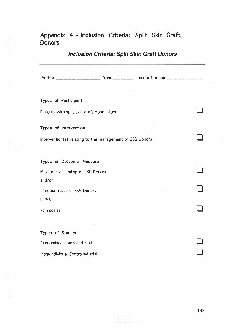
Appendix 4 - lnclusion Criteria: Split Skin GraftDonors
Inclusion Criteria: Split Skin Graft Donors
Author Year ---- Record Number
Types of Participant
Patients with split skin graft donor sites
Types of lntervention
lntervention(s) relating to the management of SSG Donors
Types of Outcome Measure
Measures of healing of SSG Donors
and/or
lnfection rates of SSG Donors
and/or
Pain scales
Types of Studies
Randomised controlled trial
lntra-lndividual Controlled trial
tr
tr
tr
tr
trE
tr
193

Apændix 5 - RCill Critical Appraisal Form: Split Skin Gnaft
Donors
RCT/ll Critical Appraisal Form: Split Skin Graft Donors
Author Year ---- Record Number
Questions 1-4 must be answered "yes" for study to be included in the meta-analysis
1. Were the recipients randomised to study groups or where intra-individual controlsused?
yes tr no tr not ctear tr2. Other than the research interventions were participants/sites in each group
treated the same?
yes tr no tr not clear
3. Were the outcomes measured in the same manner for all participants?
yes tr no tr not clear
4. Were the groups comparable at entry?
yes tr no not clear
Studies that answer no to questions 5,6 or 7 will only be included in thesystematic review if no other high quality studies are identified, however thismust be noted in the report.
5. For RCT only studies was randomisation of participants blinded?
yes tr no E not ctear tr
6. Were those assessing outcome blinded to treatment allocation (if outcome wasnot objective such as survival or length of hospitalisationf
yes tr no tr not ctear tr7. Was there adequate follow-up of participants?
no tr tr
tr
tr
trtr
yes tr(Less than 80% followed up)
not clear
194

Appendix 6 - References Excluded from Analysis
The following are references that were accepted as fulfilling the criteria for study
type but were unable to be used in analyses due to methodological issues'
Table 29: References excluded from analysis
Study Comparison Reason for exclusion
Attwood (1989) Calcium alginate Vs Jelonet Following phase I some treatmentareas were changed to becomecontrol areas for phase ll. Resultwere unable to be isolated for phase
I
Barnett, Berkowitz,Mills, & Vistnes(1 e83b)
Fine mesh gauze Vs OpsiteVs Tegaderm
Some dressings were altered toXeroform prior to healing
Bettinger, Gore, &Humphries (1995)
Calcium alginate Vs ScarletRed
Some patients had CA dressingsremoved daily others were left forlonger periods
Biltz, Kiessling, &Keysel (1 985)
Hydrocolloid Vs saline gauze Report did not indicate if groupswere randomised
Birdsell, Hein, &Lindsay (1979)
Opsite Vs Owens Silk Vs
Scarlet Red
No numerical results were provided
Blight, Fatah,Datubo Brown,Mountford, &Cheshire (1991)
"The treatment of donorsites with cultured epithelialgrafts."
Patients with delayed healingexcluded from analysis
Brady, Snelling, &Chow (1 980)
0psite, Vaseline PetroleumGauze (fine), Jelonet(course), Scarlet Red, &exposure.
Randomisation process wasinvalidated by local conditions
Brotherston &Lawrence (1993)
Hydrocolloid Vs Tulle Gras Treatment allocated on rotation
Cadier & Clarke(1 ee6)
DuoDERM Vs Tulle-gras, ln the treatment group the methodof dressing changes was alteredduring the study
Genecov et al
(1 ee8)A subatmospheric pressuredressing V.A.C. Vs Opsite
Over 30% 5/1 5 of the treatmentgroup dropped out of the study
Harris, Filarski, &Hector (1 973)
Silastic sheet Vs fine meshgauze
No detailed results of the studywere provided
195
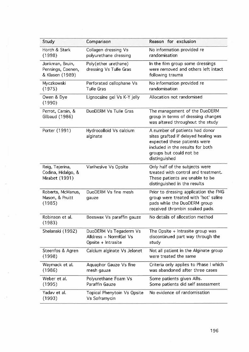
Study Comparison Reason for exclusion
Horch & Stark(1 ee8)
Collagen dressing Vspolyurethane dressing
No information provided rerandomisation
Jonkman, Bruin,Pennings, Coenen,& Klasen (1989)
Poly(ether urethane)dressing Vs Tulle Gras
ln the film group some dressingswere removed and others left intactfollowing trauma
Myczkowski(1 e7s)
Perforated cellophane VsTulle Gras
No information provided rerandomisation
Owen & Dye(1 eeo)
Lignocaine gel Vs K-Y jelly Allocation not randomised
Perrot, Carsin, &Gilbaud (1986)
DuoDERM Vs Tulle Gras The management of the DuoDERMgroup in terms of dressing changeswas altered throughout the study
Porter (1991) Hydrocolloid Vs calciumalginate
A number of patients had donorsites grafted if delayed healing wasexpected these patients wereincluded in the results for bothgroups but could not bedistinguished
Reig, Tejerina,Codina, Hidalgo, &Mirabet (1991)
Varihesive Vs Opsite Only half of the subjects weretreated with control and treatment.These patients are unable to be
distinguished in the results
Roberts, McManus,Mason, & Pruitt(1 e8s)
DuoDERM Vs fine meshgauze
Prior to dressing application the FMG
group were treated with 'hot' salinepads while the DuoDERM groupreceived thrombin soaked pads.
Robinson et al.(1 e83)
Beeswax Vs paraffin gauze No details of allocation method
Shelanski (1992) DuoDERM Vs Tegaderm VsAlldress + NormlGel VsOpsite + lntrasite
The Opsite + lntrasite group wasdiscontinued part way through thestudy
Steenfos & Agren Calcium alginate Vs Jelonet(1 ee8)
Not all patient in the Alginate groupwere treated the same
Waymack et al.(1 e86)
Aquaphor Gauze Vs finemesh gauze
Criteria only applies to Phase I whichwas abandoned after three cases
Weber et al.(1 ses)
Polyurethane Foam VsParaffin Gauze
Some pat¡ents given ABs.Some patients did self assessment
Yadav et al.(1 ee3)
Topical Phenytoin Vs Opsite No evidence of randomisationVs Soframycin
196

Appendix 7 - Data Extraction Form: Split Skin Graft
Donors
Data Extraction Form: Split Skin Graft Donors
Author
Journal
Year
Reviewer
Method
Setting
Part¡c¡pants
(male or female)
lntervention A
lntervention B
Record Number
Number of Participants
Group A
lnterventions
Group B Group C
lntervention C
197

outcome Measures
Definition of donor healing
Other Outcome Measures
Scale/measureOutcome Description
198

Results
Dichotomous Data
Control Group
number/total number
Treatment Group
number/total number
0utcome
Continuous Data
Control Group
mean & SD (number)
Treatment Group
mean & SD (number)
Outcome
Authors Conclusions
Comments
199

Appendix I - Level of Evidence Ratings for theSystematic Review
Level of Evidence Ratings
Studies were categorised according to the strength ofevidence based on the following classification system.
Level / Evidence obtained from a systematic review of all relevantrandomised controlled trials
Level tl Evidence obtained from at least one properly designed
randomised controlled trial
Level lll.l Evidence obtained from at least one well designed controlledtrial without randomisation
Level tll.Z Evidence obtained from well designed cohort or case controlanalytic studies preferably from more than one centre or research group
Level lll.3 Evidence obtained from multiple time series with or withoutintervention. Dramatic results in uncontrolled experiments
Level /V Opinion of respected authorities, based on clinical experience,descriptive studies, or reports of expert committees
200

Appendix
Comparison 1
paraffin gauze
Table 30: Costingdressing at excis¡on,
table, hydrocolloidday 0 post-op, small
dressing ¡ntervention,donor site.
9 - Costing Tables for lnterventions
Hydrocolloid dressing reg¡men verses
reg¡men
initial
Direct costs Materials Unit measure Quantity Unit cost Total cost
hydrocolloid sheet
retention tape
gauze squares
combine dressing
crepe bandage
Elastoplast tape
Staffing RN1 yr3
9.5 x 9.7 cm
2.5cmx10m
10x80cmx420x20cm
10cmx1.5m
2.5 cm x 3m
minutes
1
0.06
1
1
1
0.5
5
$3.80
$6.18
$2.60
$0.21
$0.92
$3.12
$0.4s
$3.80
$0.37
$2.60
$0.21
$0.92
$1.s6
$2.2s
Total $11 .72
Table 31: Costingdressing at excision,
table, hydrocolloidday 0 post-excision,
dressing intervent¡on,large donor site.
initial
Direct costs Materials Unit measure Quantity Unit cost Total cost
hydrocolloid sheet
retention tape
gauze squares
combine dressing
crepe bandage
Elastoplast tape
Staffing RNI yr3
18.9x19.5cm
2.5cmx10m
10x80cmx420 x20 cm
15cmxl.5m2.5cmx3mminutes
1
0.1
1
1
1
0.5
5
$1 1.90
$6.1 8
$2.60
$0.21
$1.29
$3.12
$0.4s
$1 1.90
$0.62
$2.60
$0.21
$1.29
$1.56
$2.2s
Total $20.43
?01
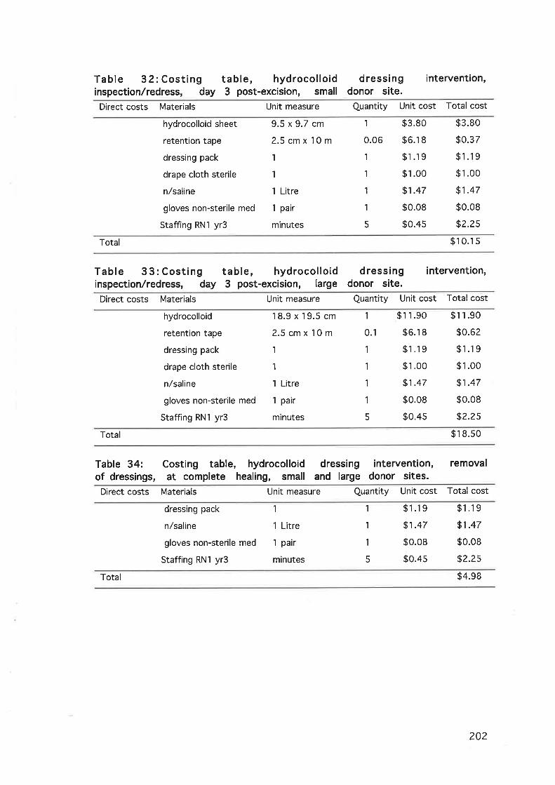
Table 32:Costing table, hydrocolloidinspection,/redress, day 3 post-excision, small
d ressi ngdonor site.
intervention,
Direct costs Materials Unit measure Quantity Unit cost Total cost
hydrocolloid sheet
retention tape
dressing pack
drape cloth sterile
n,/saline
gloves non-sterile med
Staffing RN1 yr3
9.5 x 9.7 cm
2.5cmxl0m1
1
1 Litre
1 pair
minutes
1
0.06
1
1
1
1
5
$3.80
$6.18
$1.19
$1.00
$1.47
$0.08
$0.4s
$3.80
$0.37
$1.19
$1.00
$1.47
$0.08
$2.2s
Total $10.1 s
Table 3 3: Costing table, hydrocolloidinspection./redress, day 3 post-excision, large
d ressi ngdonor site.
intervention,
Direct costs Materials Unit measure Quantity Unit cost Total cost
hydrocolloid
retention tape
dressing pack
drape cloth sterile
n/saline
gloves non-sterile med
Staffing RNI yr3
18.9x19.5cm
2.5cmx10m
1
1
1 Litre
1 pair
minutes
1
0.1
1
1
1
1
5
$1 1.90
$6.18
$1.19
$1.00
$1.47
$0.08
$0.4s
$11.90
$0.62
$1.19
$1.00
$1.47
$0.08
$2.2s
Total $18.s0
Table 34:of dressings,
Costing table, hydrocolloidat complete healing, small
intervention, removaldonor sites.
dressingand large
Direct costs Materials Unit measure Quantity Unit cost Total cost
dressing pack
n/saline
gloves non-sterile med
Staffing RN1 yr3
1
1 Litre
1 pair
minutes
1
1
1
5
$1.19
$1.47
$0.08
$0.45
$1.19
$1.47
$0.08
$2.25
Total $4.98
202

Table 3 5: Costing table, hydrocolloidtreatment day 3, 4 and 5 post-excision, small
dressing intervention,infected donor site.
Direct costs Materials Unit measure Quantity Unit cost Total cost
Silversulphadiazine
Daylee towels
gauze squares
combine dressing
crepe bandage
Elastoplast tape
n/saline
gloves sterile size 8
gown sterile
dressing pack
drape cloth sterile
Staffing RN1 yr3
1009m
34x60cmx210x80cmx4
?O x20 cm
10cmx1.5m
2.5cmx3m1 Litre
1 pair
1
1
1
minutes
1
1
1
1
1
0.5
1
1
1
1
1
10
$10.77
$0.87
$2.60
$0.21
$0.92
$3.12
$1.47
$0.70
$2.00
$1.19
$1.00
$0.4s
$10.77
$0.87
$2.60
$0.21
$0.92
$1.s6
$1.47
$0.70
$2.00
$1 .19
$1.00
$4.49
Sub Total s27.78
Total Treatment for 3 days $83.34
Table 36: Costing table, hydrocolloidtreatment day 3, 4 and 5 post-excision, large
dressing intervention,infected donor site.
Direct costs Materials Unit measure Quantity Unit cost Total cost
Silversulphadiazine
Daylee towels
gauze squares
combine dressing
crepe bandage
Elastoplast tape
n/saline
gloves sterile size 8
gown sterile
dressing pack
drape cloth sterile
Staffing RN1 yr3
500 gm
34 x 60 cmx2
10x80cmx4
20 x20 cm
15cmx1.5m
2.5cmx3m
1 Litre
1 pair
1
1
1
minutes
$42.9s
$0.87
$2.60
$0.21
$1.29
$3.12
$t.+t$0.70
$2.00
$1 .19
$1.00
$0.4s
$42.9s
$0.87
$2.60
$0.21
$1.29
$1.s6
$1.47
$0.70
$2.00
$1 .19
$1.00
$s.84
1
1
1
1
1
0.5
1
1
1
1
1
13
Sub Total $61.67
$18s.01Total Treatment for 3 days
203

Table 37:donor site,donor site.
Costingredress
table, hydrocolloid dressing ¡ntervention,following treatment for infection, small
infectedinfected
Direct costs Materials Unit measure Quantity Unit cost Total cost
hydrocolloid sheet
retention tape
dressing pack
gauze squares
drape cloth sterile
n/saline
gloves non-sterile med
Staffing RN 1 yr3
9.5 x 9.7 cm
2.5cmx10m
1
10x80cmx41
1 Litre
1 pair
minutes
1
0.06
1
1
1
1
1
7
$3.80
$6.18
$1.19
$2.60
$1.00
$1.47
$0.08
$0.4s
$3.80
$0.s7
$1.19
$2.60
$1.00
$1.47
$0.08
$3.14
Total $ 1 3.6s
Table 38:donor site,donor site.
Costingredress
table, hydrocolloid dressing ¡ntervent¡on,following treatment for infection, large
infectedinfected
Direct costs Materials Unit measure Quantity Unit cost Total cost
hydrocolloid sheet
retention tape
dressing pack
gauze squares
drape cloth sterile
n/saline
gloves non-sterile med
Staffing RNI yr3
18.9x19.5cm
2.5cmxl0m1
l0x80cmx41
1 Litre
1 pair
minutes
1
0.1
1
1
1
1
1
9
$11.90
$6.1 8
$'l .19
$2.60
$1.00
$1.47
$0.08
$0.4s
$11.90
$0.62
$1.19
$2.60
$1.00
$1.47
$0.08
$4.04
Total $22.90
204

Table 39: Costingdressing at excision,
gavze dressing intervention, initialsmall donor site.
table, paraffinday 0 post-op,
Direct costs Materials Unit measure Quantity Unit cost Total cost
paraffin gauze
gauze squares
combine dressing
crepe bandage
Elastoplast tape
Staffing RN1 yr3
10x10cm
10x80cmx420 x20 cm
10cmx1.5m
2.5cmx3mm¡nutes
2
1
1
1
0.5
5
$o.47
$2.60
$0.21
$0.92
$3.12
$0.4s
$0.94
$2.60
$0.21
$0.92
$1.s6
s2.2s
Total $8.49
Table 40: Costingdressing at excision,
table, paraffin gauze dressing ¡ntervention,day 0 post-exc¡sion, large donor site.
initial
Direct costs Materials Unit measure Quantity Unit cost Total cost
paraffin gauze
gauze squares
combine dressing
crepe bandage
Elastoplast tape
Staffing RN1 yr3
10x10cm
10x80cmx420 x20 cm
15cmx1.5m
2.5cmx3mminutes
I1
1
1
0.5
5
$0.47
$2.60
$0.21
$1.29
$3.12
$0.4s
$3.77
$2.60
$0.21
$1.29
$1.s6
$z.zs
Total $1 1.68
Table 41: Costing table, paraff¡ninspection/redress, day 3 post-excision,
gauze dressingsmall donor site.
intervention,
Direct costs Materials Unit measure Quantity Unit cost Total cost
combine dressing
crepe bandage
Elastoplast tape
dressing pack
drape cloth sterile
gloves non-sterile med
Staffing RN1 yr3
20x20cm
10cmx1.5m
2.5cmx3m
1
1
1 pair
minutes
1
1
0.5
1
1
1
5
$0.21
$0.92
$3.12
$1.19
$1.00
$0.08
$0.4s
$0.21
$0.92
$1.s6
$1.19
$1.00
$0.08
$?.2s
Total $7.22
205

Table 42= Costing table, paraffininspection/redress, day 3 post-excision,
gauze dressing intervention,large donor slte.
Direct costs Materials Unit measure Quantity Unit cost Total cost
combine dressing
crepe bandage
Elastoplast tape
dressing pack
drape cloth sterile
gloves non-sterile med
Staffing RN1 yr3
20 x20 cm
15cmxl.5m2.5cmx3mI
1
1 pair
minutes
1
1
0.5
I
1
1
5
$0.21
$1.29
$3.12
$1 .19
$1.00
$0.08
$0.4s
$0.21
$1.29
$1.s6
$1.19
$1.00
$0.08
$2.2s
Total $7.s8
Table 43= Costingremoval of dressings,
table, paraffinat complete healing
dressing intervent¡on,and large donor sites.
gauzesmall
Direct costs Materials Unit measure Quantity Unit cost Total cost
dressing pack
gloves non-sterile med
n/saline
Staffing RN1 yr3
1
1 pair
1 Litre
minutes 20
$1 .19
$0.08
$1.47
$0.4s
$1.19
$0.08
$1.47
$8.98
Total $11.72
206

Table 44= Costing table, paraff intreatment day 3, 4 and 5 post-excision,
gauze dressing ¡ntervent¡on,small infected donor site.
Direct costs Materials Unit measure Quantity Unit cost Total cost
Silversulphadiazine
Daylee towels
9auze squares
combine dressing
crepe bandage
Elastoplast tape
n/saline
gloves sterile size 8
gown sterile
dressing pack
drape cloth sterile
Staffing RN1 yr3
100 gm
34x60cmx2
10x80cmx420x20cm
10cmxl.5m2.5cmx3m
I Litre
1 pair
1
1
1
minutes
1
1
1
1
1
0.5
1
1
1
1
1
10
$10.77
$0.87
$2.60
$0.21
$0.92
$3.12
$1.47
$0.70
$2.00
$1.19
$1.00
$0.4s
$10.77
$0.87
$2.60
$0.21
$0.92
$1.s6
$1.47
$0.70
$2.00
$1.19
$1.00
$4.49
Sub Total $27.78
Total Treatment for 3 days $83.34
Table 45: Costing table, paraffintreatment day 3, 4 and 5 post-excision,
gauze dressing intervention,large infected donor site.
Direct costs Materials Unit measure Quantity Unit cost Total cost
Silversulphadiazine
Daylee towels
gauze squares
combine dressing
crepe bandage
Elastoplast tape
n/saline
gloves sterile size Igown sterile
dressing pack
drape cloth sterile
Staffing RN1 yr3
500 gm
34 x60 cmxZ
10x80cmx4
20 x20 cm
l5cmx1.5m2.5cmx3m
1 Litre
1 pair
1
1
1
minutes
1
1
1
1
1
0.5
1
1
1
1
I
13
$42.9s
$0.87
$2.60
$0.21
$1.29
$3.1 2
$1.47
$0.70
$2.00
$1.19
$1.00
$0.4s
$42.9s
$0.87
$2.60
$0.21
$1.29
$1.s6
$1.47
$0.70
$2.00
$1.19
$1.00
$s.84
Sub Total $61.67
$18s.01Total Treatment for 3 days
207

Table 46= Costinginfected donor site,infected donor site.
table,redress
paraffinfollowing
gauze dressing intervention,treatment for infection, small
Direct costs Materials Unit measure Quantity Unit cost Total cost
paraffin gauze
9auze squares
combine dressing
crepe bandage
Elastoplast tape
dressing pack
drape cloth sterile
n/saline
gloves non-sterile med
Staffing RN1 yr3
10x10cm
10x80cmx420 x20 cm
10cmxl.5m2.5cmx3m
1
1
1 Litre
1 pair
minutes
2
1
1
1
0.5
1
1
1
1
7
so.47
$2.60
$0.21
$0.92
$3.12
$1.19
$1.00
$1.47
$0.08
$0.4s
$0.94
$2.60
$0.21
$0.92
$1.s6
$1.19
$1.00
$1.47
$0.08
$3.14
Total $13.13
Table 47= Costinginfected donor site,infected donor site.
table,redress
paraff infollowing
gauze dressing intervention,treatment for infection, large
Direct costs Materials Unit measure Quantity Unit cost Total cost
paraffin gauze
gauze squares
combine dressing
crepe bandage
Elastoplast tape
dressing pack
drape cloth sterile
n/saline
gloves non-sterile med
Staffing RN1 yr3
10x10cm
10x80cmx420x20cm
15cmx1.5m
2.5 cm x 3m
1
1
1 Litre
1 pair
minutes
I1
1
1
$o.47
$2.60
$0.21
$1.29
$3.12
$1.19
$1.00
$1.47
$0.08
$0.4s
$3.77
$2,60
$0.21
$1.29
$1.s6
$1.19
$1.00
$1.47
$0.08
$4.04
0.5
9
Total $17.22
208
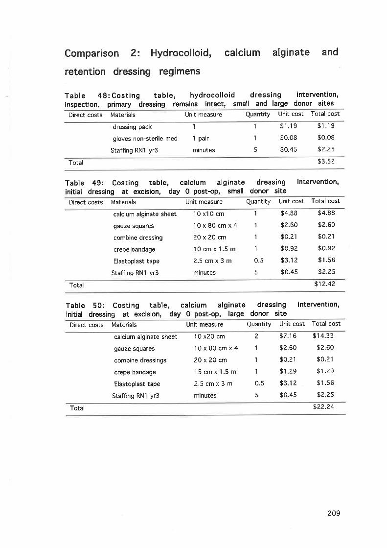
Comparison 2= Hydrocolloid, calcium alginate and
retention dressing regimens
Table 48: Costing table, hydrocolloid dressinginspection, primary dressing remains ¡ntact, small and large
intervention,donor sites
Direct costs Materials Unit measure Quantity Unit cost Total cost
dressing pack
gloves non-sterile med
Staffing RN1 yr3
1
1 pair
minutes 5
$1.19
$0.08
$0.4s
$1.19
$0.08
$2.2s
Total $3.s2
Table 49= Costing table, calcium alginateinitial dressing at exc¡s¡on, day 0 post-op, small
dressing intervent¡on,donor site
Direct costs Materials Unit measure Quantity Unit cost Total cost
calcium alginate sheet
gauze squares
combine dressing
crepe bandage
Elastoplast tape
Staffing RNl yr3
10x10cm
10x80cmx420x20cm
10cmx1.5m
2.5cmx3mminutes
1
1
1
1
0.5
5
$4.88
$2.60
$0.21
$0.92
$3.12
$0.4s
$4.88
$2.60
$0.21
$0.92
$1.56
$2.2s
Total $12.42
Table 50: Costing table, calcium alginateinitial dressing at excision, day 0 post-op, large
dressing intervention,donor s¡te
Direct costs Materials Unit measure Quantity Unit cost Total cost
calcium alginate sheet
gauze squares
combine dressings
crepe bandage
Elastoplast tape
Staffing RN1 yr3
10x20cm
10x80cmx420 x20 cm
15cmx1.5m
2.5cmx3mminutes
2
1
1
1
0.5
5
$7.16
$2.60
$0.21
$1.29
$3.12
$0.4s
$14.33
$2.60
$0.21
$1.29
$1.s6
$2.2s
Total $22.24
209

Table 51 : Costing table, calciuminspection./redress, day 3 post-excision,
alginate dressing intervention,small donor site
Direct costs Materials Unit measure Quantity Unit cost Total cost
calcium alginate sheet
combine dressing
crepe bandage
Elastoplast tape
dressing pack
drape cloth sterile
gloves non-sterile med
Staffing RNl yr3
10 x10 cm
2O x20 cm
10cmx1.5m
2.5cmx3m1
1
1 pair
minutes
1
1
1
0.5
1
1
1
5
$4.88
$0.21
$0.92
$3.1 2
$1.19
$1.00
$0.08
$0.4s
$4.88
$0.21
$0.92
$1.s6
$1.19
$1.00
$0.08
$2.2s
Total $12.10
Table 52= Costing table, calciuminspection/redress, day 3 post-excision,
alginate dressing intervention,large donor site
Direct costs Materials Unit measure Quantity Unit cost Total cost
calcium alginate sheet
combine dressing
crepe bandage
Elastoplast tape
dressing pack
drape cloth sterile
gloves non-sterile med
Staffing RN1 yr3
10x20cm
20 x20 cm
15cmx1.5m
2.5cmx3m'l
1
1
minutes
2
1
1
0.5
1
1
1
5
$7.16
$0.21
$1.29
$3.12
$1.19
$1.00
$0.08
$0.4s
$14.33
$0.21
$1.29
$1.s6
$1.19
$1.00
$0.08
$2.2s
Total $21.91
Table 53: Costingremoval of dressings,
table, calcium alginateat complete healing, small
dressing intervention,and large donor s¡tes
Direct costs Materials Unit measure Quantity Unit cost Total cost
dressing pack
gloves non-sterile med
n/saline
Staffing RN1 yr3
'l
1 pair
1 Litre
minutes
1
1
1
5
$1.19
$0.08
sl.47
$0.4s
$'t.19
$0.08
$1.47
$2.2s
Total $4.98
210

Table 54: Costing table,inspection, primary dressing
calcium alginateremains intact, small
dressing intervention,donor sites
Direct costs Materials Unit measure Quantity Unit cost Total cost
combine dressing
crepe bandage
Elastoplast tape
dressing pack
drape cloth sterile
gloves non-sterile med
Staffing RN1 yr3
20x20cm
10cmx1.5m
2.5cmx3m'l
1
1 pair
minutes
1
1
0.5
1
1
1
5
$0.21
$0.92
$3.12
$1.19
$1.00
$0.08
$0.4s
$0.21
$0.92
$1.s6
$1.19
$1.00
$0.08
$2.2s
Total $7.22
Table 55:inspection,
Costing table,primary dressing
calcium alginateremains intact, large
dressing intervention,donor sites
Direct costs Materials Unit measure Quantity Unit cost Total cost
combine dressing
crepe bandage
Elastoplast tape
dressing pack
drape cloth sterile
gloves non-sterile med
Staffing RN1 yr3
20 x20 cm
l5cmx1.5m2.5cmx3m1
1
1 pair
minutes
1
1
0.5
1
1
1
5
$0.21
$1.29
$3.1 2
$1.19
$1.00
$0.08
$0.4s
$0.21
$1.29
$1.s6
$1.19
$1.00
$0.08
$2.2s
Total $7.s8
Table 56: Costingdressing at excision,
table, retention tape dressing intervention, initialday 0 post-op, small donor site
Direct costs Materials Unit measure Quantity Unit cost Total cost
retention tape
9auze squares
combine dressing
crepe bandage
Elastoplast tape
Staffing RNI yr3
10x10cm
10x80cmx4
20x20cm
10cmx1.5m
2.5cmx3mminutes
1
1
1
1
0.5
5
$0.66
$2.60
$0.21
$0.92
$3.12
$0.4s
$0.66
$2.60
$0.21
$0.92
$1.s6
$2.25
Total $8.21
211

Table 57: Costing table, retent¡on tape dressing ¡ntervent¡on, initialdressing at excision, day 0 post-op, large donor site
Direct costs Materials Unit measure Quantity Unit cost Total cost
retent¡on tape
gauze squares
combine dressing
crepe bandage
Elastoplast tape
Staffing RNI yr3
15x40cm
10x80cmx420 x20 cm
15 cm x '1.5 m
2.5cmx3mminutes
1
1
1
1
0.s
5
$1.67
$2.60
$0.21
$1.29
$3.12
$0.4s
$1.67
$2.60
$0.21
$1.29
$1.s6
$2.2s
Total $9.s8
Table 5 8:inspection,
Costing table, retention tapeprimary dressing remains intact, small
dressingand large
intervention,donor sites
Direct costs Materials Unit measure Quantity Unit cost Total cost
dressing pack
gloves non-sterile med
Staffing RN1 yr3
1
1 pair
minutes 5
$1.19
$0.08
$0.4s
$1.19
$0.08
$2.2s
Total $3.s2
Table 59: Costingremoval of dressings,
table, retent¡onat complete healing,
dressing intervention,and large donor sites
tapesmall
Direct costs Materials Unit measure Quantity Unit cost Total cost
dressing pack
gloves non-sterile med
Zoff adhesive remover
n/saline
Staffing RN1 yr3
1
1 pair
250m1
1 Litre
minutes
1
1
0.5
1
5
$1.19
$0.08
$s.76
$1.47
$0.4s
$1.1 9
$0.08
$2.88
$1.47
$2.2s
Total $7.86
212

Appendix 10 - lCD10 Codes Report
Table 60: Complete episodes recorded lor the period July 2001 ToJune 2002
Note; the pink area relates to donor sites in the smaller range, the grey area is of
donor sites in the larger range"
TOTAL All categories 1 61 1 85 391 737
90669-00 Excision of skin for graft 2 0 19 21
Sub-total 40 ?1 74 135
45403-OO
45403{1
45409-00
45412-00
454r 5-00
454't8-00
45442-OO
SSG of extensive granulating area
SSG extensive granulating bum site >=396 BSA
SSG burn other sites inv >*396 & <6% BSA
SSG burn other sites inv >=69ó & <9% BSA
SSG burn other sites inv >=996 & <12% BSA
SSG to burn other sites inv >=12% BSA
Extensive split skin graft of any site
3
o
0 13 1/t
26 16 34 76
I 3
2
1
Sub-total I 19 16¡+ 298 581
45400-00
45400-01
4s406-00
45421 -08
45439-00
4544s-00
45448-00
45448-08
Split skin graft of small granulating area
SSG small granulating burn site <3% BSA
SSG to burn of other sites inv <370 BSA
Split skin graft to burn of genitals
Small split skin graft of other site
Split skin graft as an inlay graft
Small split skin graft of eyelid
Small split skin graft of genitals
1 ?
1
1
2
1
64
5
2
70
2
487
I
13
I
0
5
0 0 2
111 156 220
0
1
0
4
0
,|
1
0
8
lCDl0 code Descriptor Hosp 1 Hosp 2 Hosp 3 Totals
213