Mutation at the phenylalanine hydroxylase gene (PAH) and its ...
Upload
independentCategory
view
1download
0
1
2
4
5
6
7
8 Q1
9
101112
1 4
15161718
19202122232425262728
2 9
4849
50
51
52
53
54
55
56
57
58
59
European Journal of Pharmaceutics and Biopharmaceutics xxx (2014) xxx–xxx
EJPB 11731 No. of Pages 10, Model 5G
27 October 2014
Contents lists available at ScienceDirect
European Journal of Pharmaceutics and Biopharmaceutics
journal homepage: www.elsevier .com/locate /e jpb
Research paper
Cell permeable peptide conjugated nanoerythrosomes of fasudil prolongpulmonary arterial vasodilation in PAH rats
http://dx.doi.org/10.1016/j.ejpb.2014.10.0120939-6411/� 2014 Published by Elsevier B.V.
Abbreviations: ALP, alkaline phosphatase; BAL, bronchoalveolar lavage; CAR,CARSKNKDC; 5-HT, 5-hydroxytryptamine; LDH, lactate dehydrogenase; LPA,lysophosphatidic acid; LTI, lung targeting indices; MCT, monocrotaline; MPAP, meanpulmonary arterial pressure; MSAP, mean systemic arterial pressure; NERs, nano-erythrosomes; OD, optical density; PAH, pulmonary arterial hypertension; PASM,pulmonary arterial smooth muscle; PBS, phosphate buffered saline; ROCK,rho-kinase; SD, Sprague Dawley; SDS, sodium dodecyl sulfate; SPDP, N-succinimidyl3-[2-pyridyldithio]-propionate; TGF-b, tissue growth factor-beta.⇑ Corresponding author at: Department of Pharmaceutical Sciences, School of
Pharmacy, Texas Tech University Health Sciences Center, 1300 Coulter Drive,Amarillo, TX 79106, USA. Tel.: +1 806 414 9235; fax: +1 806 356 4034.
E-mail address: [email protected] (F. Ahsan).
Please cite this article in press as: N. Gupta et al., Cell permeable peptide conjugated nanoerythrosomes of fasudil prolong pulmonary arterial vasodin PAH rats, Eur. J. Pharm. Biopharm. (2014), http://dx.doi.org/10.1016/j.ejpb.2014.10.012
Nilesh Gupta, Brijeshkumar Patel, Kamrun Nahar, Fakhrul Ahsan ⇑Department of Pharmaceutical Sciences, School of Pharmacy, Texas Tech University Health Sciences Center, Amarillo, USA
a r t i c l e i n f o
3031323334353637383940414243
Article history:Received 30 May 2014Accepted in revised form 16 October 2014Available online xxxx
Keywords:Targeted deliveryRho-kinaseFasudilNanoerythrosomesControlled releasePulmonary arterial hypertensionPulmonary deliverySafety
4445
a b s t r a c t
In this study, we tested the hypothesis that a cell permeable peptide, CARSKNKDC (CAR), conjugatednanoerythrosomes (NERs) containing fasudil, a rho-kinase (ROCK) inhibitor, produces prolonged pulmon-ary preferential vasodilation. CAR conjugated NERs containing fasudil were prepared by hypotonic lysisand extrusion method, optimized for various physicochemical properties in-vitro. The formulations werethen used to study the hemodynamic efficacy in a monocrotaline-induced rodent model of pulmonaryarterial hypertension (PAH). CAR-NERs-Fasudil was spherical in shape with an average vesicle size andentrapment efficiency of 161.3 ± 1.37 nm and 48.81 ± 1.96%, respectively. Formulations were stable for�3 weeks when stored at 4 �C and the drug was released in a controlled fashion for >48 h. The uptakeof CAR-NERs-Fasudil by TGF-b activated pulmonary arterial smooth muscle cell was �1.5-fold greaterthan the uptake of NERs-Fasudil. CAR-NERs-Fasudil inhibited ROCK activity and 5-hydroxytryptamineinduced cell proliferation. In terms of reduction of pulmonary arterial pressure, intratrachealadministration of CAR-NERs-Fasudil was �2-fold more specific to the lungs compared with plain fasudil.Overall, CAR peptide grafted nanoerythrosomes offers a new platform for improving the therapeuticefficacy of a rho-kinase inhibitor, fasudil, without affecting peripheral vasodilation.
� 2014 Published by Elsevier B.V.
46
47
60
1. Introduction dilates pulmonary arteries and arterioles, and reduces arterial 6162
63
64
65
66
67
68
69
70
71
The RhoA/Rho-kinase signaling pathway influences the patho-genesis of a number of diseases related to cardiovascular, nervousand metabolic systems [1]. Endogenous vasoconstrictors, includingendothelin-1, angiotensin, and 5-hydroxy tryptamine, activateRho-kinase (ROCK) and downstream effector molecules. ROCK, apotent downstream effector of RhoA, inhibits myosin phosphataseand causes the cells to differentiate, proliferate, adhere, migrateand contract. ROCK mediated vasoconstriction and vascularremodeling play a major role in the development and progressionof PAH, a deadly chronic disorder. Inhibition of ROCK activity
72
73
74
75
76
77
78
79
80
81
82
83
remodeling [2]. Of the ROCK inhibitors, fasudil (HA-1077) amelio-rates the chief PAH symptom, elevated pulmonary arterial pres-sure, both in animals and human patients. But lack of pulmonaryvascular selectivity, drug induced peripheral vasodilation, anddrug’s short biological life are major deterrents for fasudil’s transi-tion from an investigational drug to a clinically viable therapeuticagent [3]. The drawback of short half-life can be addressed by putt-ing the drug in carriers with an ability to modulate drug releaseand extend plasma circulation time. The problem of pulmonaryselectivity and peripheral vasodilation can be overcome by meansof homing device-empowered carriers that can position the formu-lation over the lung vasculature.
The approaches to extend the biological half-life and deploydrugs at a disease site include the use of polymeric, lipidic, inor-ganic, and cellular micro- and nano-sized chariots equipped withdifferent homing devices such as antibodies, aptamers, enzymes,and peptides [4,5]. Particulate drug chariots armed with homingdevices have been used to deliver drugs for the treatment of manydisorders localized in the respiratory system. Liposomes modifiedwith an epidermal growth factor receptor binding peptide, forexample, enhanced the accumulation of doxorubicin in lung tumortissue by 2.2-fold compared with unmodified liposomes [6].Modified nanoparticles prolonged the residence time of
ilation
84
85
86
87
88
89
90
91
92
93
94
95
96
97
98
99
100
101
102
103
104
105
106
107
108
109
110
111
112
113
114
115
116
117
118
119
120
121
122
123
124
125
126
127
128
129
130
131
132
133
134
135
136
137
138
139
140
141
142
143
144
145
146
147
148
149
150
151
152
153
154
155
156
157
158
159
160
161
162
163
164
165
166
167
168
169
170
171
172
173
174
175
176
2 N. Gupta et al. / European Journal of Pharmaceutics and Biopharmaceutics xxx (2014) xxx–xxx
EJPB 11731 No. of Pages 10, Model 5G
27 October 2014
dexamethasone in deep lung tissue and helped overcome steroidresistance in asthma [7], and promoted the localization of nanopar-ticles in respiratory syncytial viral infection site [8].
Similar to the above approaches, we and others have used nano-particles to deliver anti-PAH drugs such as fasudil, iloprost, pita-vastatin and adrenomedullin directly to the lung [9–11]. Wehave shown that nanoerythrosomes (NERs), an erythrocyte basedbiomimetic system, extend the biological half-life of fasudil andare safe for intratracheal administration [12]. NERs exhibit reducedaggregation, minimal drug loss, biodegradability, longer circulationtime, and superior targeting efficiency [12]. Being natural in origin,NERs are endowed with biological and chemical senses; thus NERscan deliver enzymes, peptides, toxins, contrast agents. Proteins andsmall molecule drugs can also be conjugated to NERs surface [13].Importantly, unlike polymeric particles, NERs can be functionalizedwithout involving complex chemical modification [14]. However,these cell based carriers have not yet been used to deliver pharma-cologically active drug via inhalational route for the treatment of adebilitating respiratory disease, PAH.
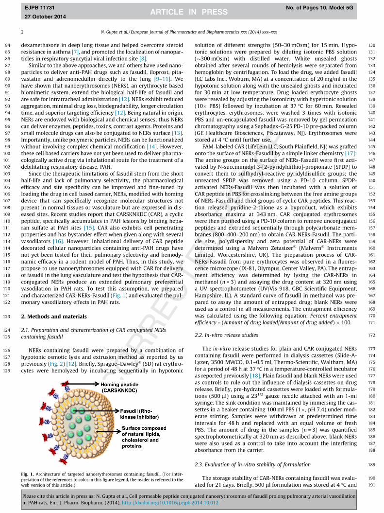
Since the therapeutic limitations of fasudil stem from the shorthalf-life and lack of pulmonary selectivity, the pharmacologicalefficacy and site specificity can be improved and fine-tuned byloading the drug in cell based carrier, NERs, modified with homingdevice that can specifically recognize molecular structures notpresent in normal tissues or vasculature but are expressed in dis-eased sites. Recent studies report that CARSKNKDC (CAR), a cyclicpeptide, specifically accumulates in PAH lesions by binding hepa-ran sulfate at PAH sites [15]. CAR also exhibits cell penetratingproperties and has bystander effect when given along with severalvasodilators [16]. However, inhalational delivery of CAR peptidedecorated cellular nanoparticles containing anti-PAH drugs havenot yet been tested for their pulmonary selectivity and hemody-namic efficacy in a rodent model of PAH. Thus, in this study, wepropose to use nanoerythrosomes equipped with CAR for deliveryof fasudil in the lung vasculature and test the hypothesis that CAR-conjugated NERs produce an extended pulmonary preferentialvasodilation in PAH rats. To test this assumption, we preparedand characterized CAR-NERs-Fasudil (Fig. 1) and evaluated the pul-monary vasodilatory effects in PAH rats.
2. Methods and materials
2.1. Preparation and characterization of CAR conjugated NERscontaining fasudil
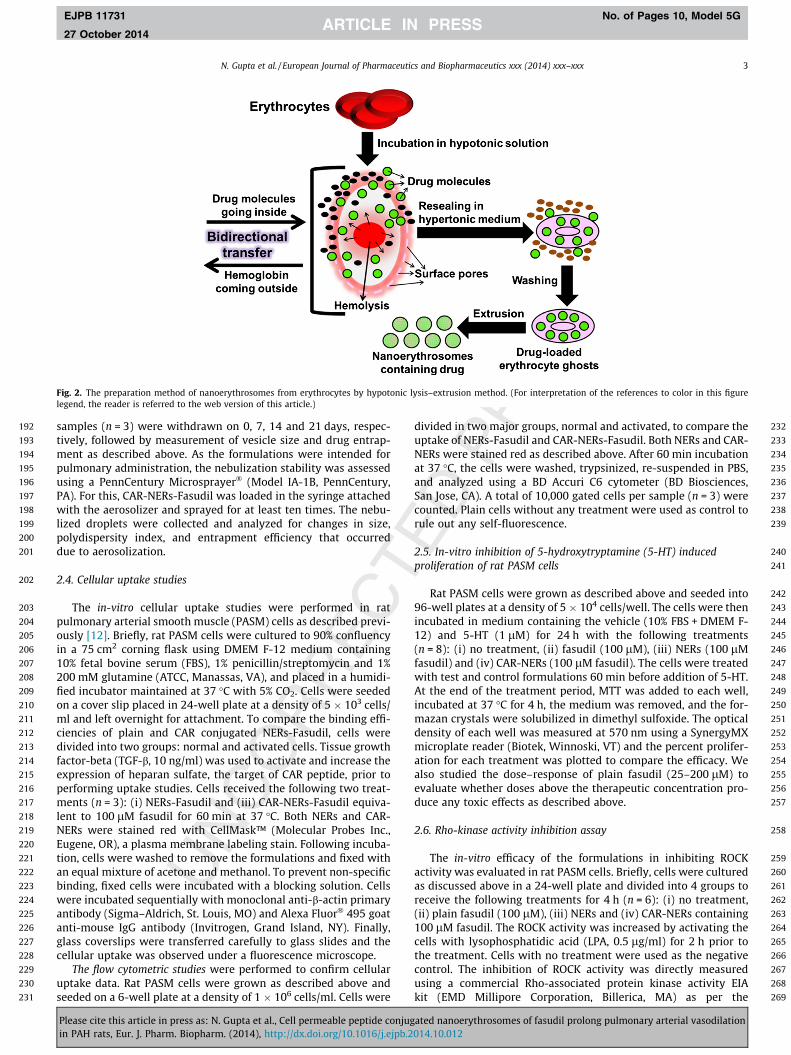
NERs containing fasudil were prepared by a combination ofhypotonic osmotic lysis and extrusion method as reported by uspreviously (Fig. 2) [12]. Briefly, Sprague–Dawley� (SD) rat erythro-cytes were hemolyzed by incubating sequentially in hypotonic
177
178
179
180
181
182
183
184
185
186
187
188
189
190
191
Fig. 1. Architecture of targeted nanoerythrosomes containing fasudil. (For inter-pretation of the references to color in this figure legend, the reader is referred to theweb version of this article.)
Please cite this article in press as: N. Gupta et al., Cell permeable peptide conjugin PAH rats, Eur. J. Pharm. Biopharm. (2014), http://dx.doi.org/10.1016/j.ejpb.2
solution of different strengths (50–30 mOsm) for 15 min. Hypo-tonic solutions were prepared by diluting isotonic PBS solution(�300 mOsm) with distilled water. White unsealed ghostsobtained after several rounds of hemolysis were separated fromhemoglobin by centrifugation. To load the drug, we added fasudil(LC Labs Inc., Woburn, MA) at a concentration of 20 mg/ml in thehypotonic solution along with the unsealed ghosts and incubatedfor 30 min at low temperature. Drug loaded erythrocyte ghostswere resealed by adjusting the isotonicity with hypertonic solution(10� PBS) followed by incubation at 37 �C for 60 min. Resealederythrocytes, erythrosomes, were washed 3 times with isotonicPBS and un-encapsulated fasudil was removed by gel permeationchromatography using a Sephadex-G-25 PD-10 pre-packed column(GE Healthcare Biosciences, Piscataway, NJ). Erythrosomes werestored at 4 �C until further use.
FAM-labeled CAR (LifeTein LLC, South Plainfield, NJ) was graftedonto the surface of NERs-Fasudil by a simple linker chemistry [17]:The amine groups on the surface of NERs-Fasudil were first acti-vated by N-succinimidyl 3-[2-pyridyldithio]-propionate (SPDP) toconvert them to sulfhydryl-reactive pyridyldisulfide groups; theunreacted SPDP was removed using a PD-10 column. SPDP-activated NERs-Fasudil was then incubated with a solution ofCAR peptide in PBS for crosslinking between the free amine groupsof NERs-Fasudil and thiol groups of cyclic CAR peptides. This reac-tion released pyridine-2-thione as a byproduct, which exhibitsabsorbance maxima at 343 nm. CAR conjugated erythrosomeswere then purified using a PD-10 column to remove unconjugatedpeptides and extruded sequentially through polycarbonate mem-branes (800–400–200 nm) to obtain CAR-NERs-Fasudil. The parti-cle size, polydispersity and zeta potential of CAR-NERs weredetermined using a Malvern Zetasizer� (Malvern� InstrumentsLimited, Worcestershire, UK). The preparation process of CAR-NERs-Fasudil from pure erythrocytes was observed in a fluores-cence microscope (IX-81, Olympus, Center Valley, PA). The entrap-ment efficiency was determined by lysing the CAR-NERs inmethanol (n = 3) and assaying the drug content at 320 nm usinga UV spectrophotometer (UV/Vis 918, GBC Scientific Equipment,Hampshire, IL). A standard curve of fasudil in methanol was pre-pared to assay the amount of entrapped drug; blank NERs wereused as a control in all measurements. The entrapment efficiencywas calculated using the following equation: Percent entrapmentefficiency = (Amount of drug loaded/Amount of drug added) � 100.
2.2. In-vitro release studies
The in-vitro release studies for plain and CAR conjugated NERscontaining fasudil were performed in dialysis cassettes (Slide-A-Lyzer, 3500 MWCO, 0.1–0.5 ml, Thermo-Scientific, Waltham, MA)for a period of 48 h at 37 �C in a temperature-controlled incubatoras reported previously [18]. Plain fasudil and blank NERs were usedas controls to rule out the influence of dialysis cassettes on drugrelease. Briefly, pre-hydrated cassettes were loaded with formula-tions (500 ll) using a 231/2 gauze needle attached with an 1-mlsyringe. The sink condition was maintained by immersing the cas-settes in a beaker containing 100 ml PBS (1�, pH 7.4) under mod-erate stirring. Samples were withdrawn at predetermined timeintervals for 48 h and replaced with an equal volume of freshPBS. The amount of drug in the samples (n = 3) was quantifiedspectrophotometrically at 320 nm as described above; blank NERswere also used as a control to take into account the interferingabsorbance from the carrier.
2.3. Evaluation of in-vitro stability of formulation
The storage stability of CAR-NERs containing fasudil was evalu-ated for 21 days. Briefly, 500 ll formulation was stored at 4 �C and
ated nanoerythrosomes of fasudil prolong pulmonary arterial vasodilation014.10.012
192
193
194
195
196
197
198
199
200
201
202
203
204
205
206
207
208
209
210
211
212
213
214
215
216
217
218
219
220
221
222
223
224
225
226
227
228
229
230
231
232
233
234
235
236
237
238
239
240
241
242
243
244
245
246
247
248
249
250
251
252
253
254
255
256
257
258
259
260
261
262
263
264
265
266
267
268
269
Fig. 2. The preparation method of nanoerythrosomes from erythrocytes by hypotonic lysis–extrusion method. (For interpretation of the references to color in this figurelegend, the reader is referred to the web version of this article.)
N. Gupta et al. / European Journal of Pharmaceutics and Biopharmaceutics xxx (2014) xxx–xxx 3
EJPB 11731 No. of Pages 10, Model 5G
27 October 2014
samples (n = 3) were withdrawn on 0, 7, 14 and 21 days, respec-tively, followed by measurement of vesicle size and drug entrap-ment as described above. As the formulations were intended forpulmonary administration, the nebulization stability was assessedusing a PennCentury Microsprayer� (Model IA-1B, PennCentury,PA). For this, CAR-NERs-Fasudil was loaded in the syringe attachedwith the aerosolizer and sprayed for at least ten times. The nebu-lized droplets were collected and analyzed for changes in size,polydispersity index, and entrapment efficiency that occurreddue to aerosolization.
2.4. Cellular uptake studies
The in-vitro cellular uptake studies were performed in ratpulmonary arterial smooth muscle (PASM) cells as described previ-ously [12]. Briefly, rat PASM cells were cultured to 90% confluencyin a 75 cm2 corning flask using DMEM F-12 medium containing10% fetal bovine serum (FBS), 1% penicillin/streptomycin and 1%200 mM glutamine (ATCC, Manassas, VA), and placed in a humidi-fied incubator maintained at 37 �C with 5% CO2. Cells were seededon a cover slip placed in 24-well plate at a density of 5 � 103 cells/ml and left overnight for attachment. To compare the binding effi-ciencies of plain and CAR conjugated NERs-Fasudil, cells weredivided into two groups: normal and activated cells. Tissue growthfactor-beta (TGF-b, 10 ng/ml) was used to activate and increase theexpression of heparan sulfate, the target of CAR peptide, prior toperforming uptake studies. Cells received the following two treat-ments (n = 3): (i) NERs-Fasudil and (iii) CAR-NERs-Fasudil equiva-lent to 100 lM fasudil for 60 min at 37 �C. Both NERs and CAR-NERs were stained red with CellMask™ (Molecular Probes Inc.,Eugene, OR), a plasma membrane labeling stain. Following incuba-tion, cells were washed to remove the formulations and fixed withan equal mixture of acetone and methanol. To prevent non-specificbinding, fixed cells were incubated with a blocking solution. Cellswere incubated sequentially with monoclonal anti-b-actin primaryantibody (Sigma–Aldrich, St. Louis, MO) and Alexa Fluor� 495 goatanti-mouse IgG antibody (Invitrogen, Grand Island, NY). Finally,glass coverslips were transferred carefully to glass slides and thecellular uptake was observed under a fluorescence microscope.
The flow cytometric studies were performed to confirm cellularuptake data. Rat PASM cells were grown as described above andseeded on a 6-well plate at a density of 1 � 106 cells/ml. Cells were
Please cite this article in press as: N. Gupta et al., Cell permeable peptide conjugin PAH rats, Eur. J. Pharm. Biopharm. (2014), http://dx.doi.org/10.1016/j.ejpb.2
divided in two major groups, normal and activated, to compare theuptake of NERs-Fasudil and CAR-NERs-Fasudil. Both NERs and CAR-NERs were stained red as described above. After 60 min incubationat 37 �C, the cells were washed, trypsinized, re-suspended in PBS,and analyzed using a BD Accuri C6 cytometer (BD Biosciences,San Jose, CA). A total of 10,000 gated cells per sample (n = 3) werecounted. Plain cells without any treatment were used as control torule out any self-fluorescence.
2.5. In-vitro inhibition of 5-hydroxytryptamine (5-HT) inducedproliferation of rat PASM cells
Rat PASM cells were grown as described above and seeded into96-well plates at a density of 5 � 104 cells/well. The cells were thenincubated in medium containing the vehicle (10% FBS + DMEM F-12) and 5-HT (1 lM) for 24 h with the following treatments(n = 8): (i) no treatment, (ii) fasudil (100 lM), (iii) NERs (100 lMfasudil) and (iv) CAR-NERs (100 lM fasudil). The cells were treatedwith test and control formulations 60 min before addition of 5-HT.At the end of the treatment period, MTT was added to each well,incubated at 37 �C for 4 h, the medium was removed, and the for-mazan crystals were solubilized in dimethyl sulfoxide. The opticaldensity of each well was measured at 570 nm using a SynergyMXmicroplate reader (Biotek, Winnoski, VT) and the percent prolifer-ation for each treatment was plotted to compare the efficacy. Wealso studied the dose–response of plain fasudil (25–200 lM) toevaluate whether doses above the therapeutic concentration pro-duce any toxic effects as described above.
2.6. Rho-kinase activity inhibition assay
The in-vitro efficacy of the formulations in inhibiting ROCKactivity was evaluated in rat PASM cells. Briefly, cells were culturedas discussed above in a 24-well plate and divided into 4 groups toreceive the following treatments for 4 h (n = 6): (i) no treatment,(ii) plain fasudil (100 lM), (iii) NERs and (iv) CAR-NERs containing100 lM fasudil. The ROCK activity was increased by activating thecells with lysophosphatidic acid (LPA, 0.5 lg/ml) for 2 h prior tothe treatment. Cells with no treatment were used as the negativecontrol. The inhibition of ROCK activity was directly measuredusing a commercial Rho-associated protein kinase activity EIAkit (EMD Millipore Corporation, Billerica, MA) as per the
ated nanoerythrosomes of fasudil prolong pulmonary arterial vasodilation014.10.012

270
271
272
273
274
275
276
277
278
279
280
281
282
283
284
285
286
287
288
289
290
291
292
293
294
295
296
297
298
299
300
301
302
303
304
305
306
307
308
309
310
311
312
313
314
315
316
317
318
319
320
321
322
323
324
325
326
327
328
329
330
331
332
333
334
335
336
337
338
339
340
341
342
343
344
345
346
347
348
349
350
351
352
353
354
355
356
357
358
359
360
361
362
363
364
365
366
367
368
369
370
371
372
373
374
375
376
377
378
379
380
381
382
383
384
385
386
387
388
389
390
4 N. Gupta et al. / European Journal of Pharmaceutics and Biopharmaceutics xxx (2014) xxx–xxx
EJPB 11731 No. of Pages 10, Model 5G
27 October 2014
manufacturer’s instructions. In brief, following the treatment per-iod, the cells were lysed and diluted with the assay buffer. An ali-quot of lysed cells was then added to the wells pre-coated withrecombinant myosin phosphatase target subunit-1 (rMYPT1) andincubated for 30 min. Next, horseradish peroxidase (HRP) conju-gated primary and secondary antibodies for MYPT1 were addedsequentially followed by 1 h incubation. The amount of phosphory-lation of the substrate was determined by addition of a chromo-genic substrate, tetramethylbenzidine (TMB), which turns thesolution color to blue. Finally the stop solution was added thatturned the solution to yellow. The relative amount of ROCK activityin the lysed cells was determined by measuring the absorbance at450 nm.
2.7. The acute hemodynamic efficacy studies in monocrotaline (MCT)-induced PAH rats
The in-vivo efficacy of the formulations was evaluated in MCT-induced PAH rats as reported previously [18,19]. Briefly, male SDrats (250–300 g) were injected a single subcutaneous injection ofMCT (50 mg/kg body weight, Sigma–Aldrich Inc., St. Louis, MO)and housed for 28 days for development of PAH. After 4 weeks,PAH rats were anesthetized by an intramuscular injection of a cock-tail of ketamine (90 mg/kg) and xylazine (10 mg/kg) and two sepa-rate catheters were placed in the pulmonary (via jugular vein andright ventricle) and right carotid artery. The mean pulmonary(MPAP) and systemic arterial pressure (MSAP) were measured withMemscap SP844 physiological pressure transducers (Memscap AS,Scoppum, Norway) and bridge amplifiers connected to a PowerLab16/30 system with LabChart Pro 7.0 software (AD Instruments, Inc.,Colorado Springs, CO). Rats were divided into four groups (n = 6–8)to receive 3 mg/kg fasudil as (i) plain intravenous (IV) fasudil, (ii)plain intratracheal (IT) fasudil, (iii) NERs containing fasudil, and(iv) CAR-NERs containing fasudil via the IT route. IV formulationswere administered into the penile vein and IT administration wasperformed using the PennCentury Microsprayer�. The animalswere maintained under anesthesia throughout the procedure, andMPAP and MSAP were recorded for at least 6 h. At the end of theexperiment, rats were sacrificed by exsanguination.
All animal studies were performed in accordance with NIHGuidelines for the care and use of Laboratory Animals under aprotocol approved by TTUHSC Animal Care and Use Committee(AM-10012).
2.8. Safety studies
The cell viability assay was performed to determine the detri-mental effects of the formulations on the native cells of bronchialrespiratory system. For this, we performed an MTT assay in twodifferent cell lines, human airway epithelial (Calu-3) and rat PASMcells, as reported previously. Briefly, cells were seeded in a 96-wellplate at a density of 5 � 104 cells/well and left overnight for adher-ence. Then, cells were treated with (n = 12): (i) saline (negativecontrol), (ii) 0.1% sodium dodecyl sulfate (SDS, positive control),(iii) plain fasudil (100 lM), and (iv) CAR-NERs (equivalent to100 lM fasudil) for 24 h. Post treatment, cells were washed twicewith saline and incubated with 100 ll MTT (0.5 mg/ml in sterilePBS) for 4 h. The formazan crystals were solubilized in dimethylsulfoxide along by moderate shaking for 1 h, and the absorbancewas measured at 570 nm. The cell viability was calculated usingthe following equation: % Cell viability = (ODsample � ODblank)/(ODcontrol � ODblank) � 100; OD is optical density, sample is testcompound, control is saline and blank is no treatment.
Further, the safety of the formulations was evaluated in-vivo bymeasuring the levels of injury markers in bronchoalveolar lavage(BAL) fluid after the pulmonary administration. Briefly, male SD
Please cite this article in press as: N. Gupta et al., Cell permeable peptide conjugin PAH rats, Eur. J. Pharm. Biopharm. (2014), http://dx.doi.org/10.1016/j.ejpb.2
rats (250–300 g) were anesthetized using a cocktail of ketamineand xylazine and divided into three groups (n = 4) to receive fol-lowing treatments via the intratracheal route: (i) saline (negativecontrol), (ii) CAR-NERs (equivalent to 6 mg/kg fasudil), (iii) 0.1%SDS (positive control). 12 h post-administration, animal weightswere recorded, lungs were surgically removed, and weighed sepa-rately. The wet lung weight was reported as g/100 g body weight.The BAL procedure was performed and fluid was collected to mea-sure the total protein concentration (mg/ml) by a Bradford assay.The levels of two lung injury markers, lactate dehydrogenase(LDH) and alkaline phosphatase (ALP), in BAL fluid were measuredusing commercial kits (Pointe Scientific, Canton, MI) and reportedin IU/L.
2.9. Data analysis and statistics
The data are presented as mean ± standard deviation and wereanalyzed by one-way ANOVA followed by Tukey post hoc analysisusing GraphPad Prism 6.0 software (GraphPad Software, San Diego,CA). p value 6 0.05 was considered as statistically significant.
3. Results and discussion
In this study, we have developed a targeted biomimetic systemfor inhalational delivery of fasudil. Previously, we have shown thatit is possible to encapsulate fasudil, a small molecular weight rho-kinase inhibitor, in erythrocyte based systems [12]. As a continua-tion to our published study, we conjugated a homing peptide, CAR,onto the surface of NERs-Fasudil, performed a series of in-vitrostudies, and evaluated the efficacy in a rodent model of PAH.
3.1. Preparation and characterization of CAR-NERs containing fasudil
Fasudil loaded NERs were prepared according to our previouslyestablished procedure to achieve a maximum entrapment andfavorable physiochemical properties. The transition of erythro-somes from erythrocytes was captured by microscopy. NERs-Fasu-dil was completely resealed and was spherical with smooth textureas opposed to biconcave discoidal erythrocytes. Formulationsshowed no aggregation over time.
A major drawback of the current PAH therapy is the lack of sitespecificity, and thus causes systemic vasodilation. We aimed toaddress this issue by conjugating a homing peptide onto the NERSsurface. CAR peptide was grafted on NERs surface utilizing theSPDP linker chemistry (Fig. 3A); the absence of peak at 343 nmbefore addition of CAR peptide but appearance of a characteristicpeak after addition of CAR suggest conjugation of peptide withactivated amine groups of NERs-Fasudil (Fig. 3B). CAR-NERs-Fasu-dil showed green fluorescent peptide (FAM-labeled CAR) under afluorescent microscope, suggesting that CAR was grafted ontoNER surface (Fig. 4). The average size of CAR-NERs-Fasudil was161.3 ± 1.37, slightly higher than that of plain NERs-Fasudil asreported earlier. The slight increase in size may be the result ofpeptide accumulation over the erythrosome surface. Publishedreports suggest that particles less than 250 nm can escape lungs’natural clearance mechanisms and stay longer in the lungs [20].The zeta potential of the formulation was �19.83 ± 1.66 mV thatis optimal for colloidal stability of erythrosomes. Some studies sug-gest negatively charged particles accumulate more efficiently inthe lungs than positively charged ones [21]. CAR-NERs-Fasudilexhibited homogenous size distribution with a PDI of 0.159 ±0.21. The drug entrapment was 48.81 ± 1.96% and the extrusionprocess did not reduce the drug entrapment, indicating thatNERs-Fasudil is flexible and can travel via the small and distal pul-monary vasculature. This also confers stability to the formulation
ated nanoerythrosomes of fasudil prolong pulmonary arterial vasodilation014.10.012

391
392
393
394
395
396
397
398
399
400
401
402
403
404
405
406
407
408
409
410
411
412
413
414
415
416
Fig. 3. (A) Conjugation of CAR peptide on the surface of nanoerythrosomes (NERs) utilizing linker chemistry. (B) UV spectrophotometric monitoring for release of pyridine-2-thione at 343 nm before and after conjugation. (For interpretation of the references to color in this figure legend, the reader is referred to the web version of this article.)
N. Gupta et al. / European Journal of Pharmaceutics and Biopharmaceutics xxx (2014) xxx–xxx 5
EJPB 11731 No. of Pages 10, Model 5G
27 October 2014
because a slight change in the flexibility of erythrocytes may leadto their clearance by macrophages. Our previously published stud-ies suggest that NERs-Fasudil prevents the premature release offasudil, and consequently increases drug’s biological half-life.CAR peptide has a biological half-life of �27 h and thus we antici-pate that it will guide the carrier to accumulate on PAH sites andproduce localized action for an extended period of time.
417
418
419
420
421
422
423
424
425
426
427
428
429
430
3.2. In-vitro release studies of NERs-Fasudil containing fasudil
The in-vitro release studies of fasudil from NERs and CAR-NERswere performed for 48 h in PBS buffer at 37 �C (Fig. 5A). About100% of the plain drug was released within 2 h which suggestedthat cassettes were not the rate limiting factors for drug release.In contrast, only 33.9 ± 1.71% and 28.9 ± 1.92% of drug was releasedfrom plain NERs and CAR-NERs, respectively, which showed thecontrolled release property of the formulations. Also, a smallamount of drug (10–12%) was released in 15 min from both formu-lations, which may be due to disruption of some cells caused bystirring or release of surface associated drug. A slower and pro-longed release behavior may stem from the compact structure ofNERS surface that is composed of natural lipids, cholesterol and
Fig. 4. The fluorescent microscopic images of erythrocytes (inherently red colored due themolysis) and CAR conjugated erythrosomes (green color is due to FAM-labeled CAcontaining fasudil after extrusion and purification. PDI here refers to polydispersity inreferences to color in this figure legend, the reader is referred to the web version of thi
Please cite this article in press as: N. Gupta et al., Cell permeable peptide conjugin PAH rats, Eur. J. Pharm. Biopharm. (2014), http://dx.doi.org/10.1016/j.ejpb.2
surface proteins [22]. The in-vitro release data strongly suggest thatCAR-NERs could be used as controlled and continuous release car-riers for delivery of fasudil. However, this release data may notreflect the drug release in the lung, since lung fluid is composedof surfactants and various ions.
3.3. In-vitro stability of CAR-NERs-Fasudil
The storage stability of CAR-NERs-Fasudil was evaluated for aperiod of three weeks at 4 �C. The drug loaded CAR-NERs wereresistant to aggregation and showed no significant changes in ves-icle size (Fig. 5B). As NERs are composed of endogenous phospho-lipids, proteins and cholesterol, and have a very compact structure,drug leakage from NER was minimal (�6%). Large negative zetapotential also contributed to the stability of formulations becauseoppositely charged NERs created repulsion among the particles,reduced aggregation, and minimized drug loss. Based on this shortterm stability studies, we expect that CAR-NERs-Fasudil would besuitable for long term use. However, NERs are not stable at low pH[22], thus storage at 4 �C in a neutral pH is recommended.Alternatively, NERS surface should be modified with polyethyleneglycol to prevent premature degradation.
o hemoglobin), erythrosomes (colorless erythrocyte ghosts resealed after completeR peptide). The lower panel shows the physicochemical properties of CAR-NERsdex. Data represent mean ± standard deviation (n = 3). (For interpretation of the
s article.)
ated nanoerythrosomes of fasudil prolong pulmonary arterial vasodilation014.10.012

431
432
433
434
435
436
437
438
439
440
441
442
443
444
445
446
447
448
449
450
451
452
453
454
455
456
457
458
459
460
461
462
463
464
465
466
467
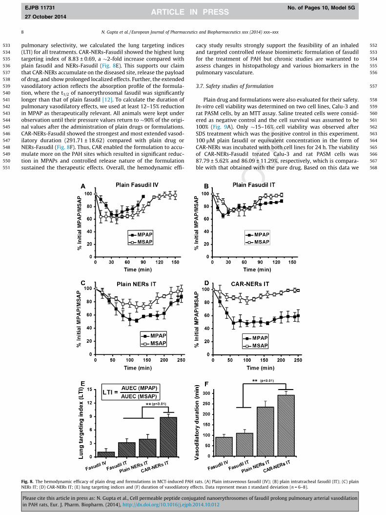
468
469
470
471
472
473
474
475
476
477
478
479
480
481
482
483
484
485
486
487
488
489
490
491
492
493
494
495
496
497
498
499
500
501
502
Fig. 5. (A) The in-vitro release profiles of plain fasudil and formulations in PBS (pH7.4) at 37 �C. (B) The storage stability of the formulations at 4 �C for 3 weeks. (C)Changes in vesicle size and entrapment efficiency of the formulations before andafter nebulization through the microsprayer. Data represent mean ± standarddeviation (n = 3).
6 N. Gupta et al. / European Journal of Pharmaceutics and Biopharmaceutics xxx (2014) xxx–xxx
EJPB 11731 No. of Pages 10, Model 5G
27 October 2014
We have used a microsprayer to spray instill the formulationsto the animals’ lungs. The aerosolizer we have used has an atom-izer at the tip that spray the solution into smaller and uniformdroplets; but such spraying may rupture the formulation and pre-maturely release the drug. This prompted us to evaluate the effectof nebulization on the stability and integrity of CAR-NERs-Fasudil.No significant differences in the vesicle size, homogeneity, andentrapment efficiency before and after nebulization of the CAR-NERs (Fig. 5C) were observed, indicating that formulations canwithstand the force of nebulization.
503
504
505
506
507
508
3.4. Cellular uptake of the formulations
Fasudil causes smooth muscle relaxation by inhibiting ROCKand reduces mean pulmonary arterial pressure. We expect thatour formulations will produce the intended therapeutic effects onarterial smooth muscle cells after crossing the air–blood barrier.So we have performed cellular uptake studies in normal and acti-
Please cite this article in press as: N. Gupta et al., Cell permeable peptide conjugin PAH rats, Eur. J. Pharm. Biopharm. (2014), http://dx.doi.org/10.1016/j.ejpb.2
vated PASM cells and compared the binding efficiencies of plaindrug with that of CAR conjugated NERs-Fasudil. Cells were acti-vated with TGF-b because it is a potent modulator of heparan sul-fate expression, which is mediated by an increase in steady statelevels of mRNA for heparan sulfate proteoglycan [23]. In agreementwith the published report, CAR-NERs-Fasudil (red color, TRITCchannel) was internalized more efficiently by activated PASM cells(green color, FITC channel) compared with plain NERs-Fasudil thatshowed moderate internalization (Fig. 6A). Enhanced uptake canbe explained by cell penetrating property of the peptide, which isevident from the enhanced cytoplasmic and nuclear localizationof CAR-NERs-Fasudil in 60 min.
In addition to qualitative microscopic studies, we performedflow cytometry to quantitate the cellular uptake. Compared withplain NERs-Fasudil, cellular uptake of CAR-NERs-Fasudil washigher in activated cells (Fig. 6B). Mean fluorescence intensity(Fig. 6C) shows that CAR-NERs-Fasudil was internalized �1.5-foldmore than plain NERs-Fasudil by TGF-b activated cells. This agreeswith the microscopy data which show enhanced localization ofpeptide conjugated formulation in the cells. Also, CAR peptidehas cell penetrating properties which, we believe, is also contribut-ing to enhance the cellular uptake. Further transport studies usingco-culture of cells lining the pulmonary vasculature would providemore information regarding the transport mechanism of CAR-NERs-Fasudil from respiratory epithelial side to the vascular side.
3.5. Inhibition of 5-HT induced proliferation and rho-kinase activity ofrat PASM cells
5-HT, a potent vasoconstrictor, is mitogenic for PASM cells andtheir proliferation influences the pulmonary arterial remodelingin PAH [24]. We examined the effect of our formulation in inhibiting5-HT induced proliferation of PASM cells (Fig. 7A). Pretreating thecells with fasudil or formulations reduced the proliferation of PASMcells. Although the formulations produced similar effect as that ofplain fasudil, we believe that the controlled release property andtargeting potential of the formulation would be beneficial for PAHtherapy. Furthermore, we also performed dose–response studiesfor fasudil to differentiate drug’s pharmacological effect from toxiceffects. A dose-dependent decrease in 5-HT induced cell prolifera-tion was observed but no cytotoxicity was observed when the drugwas applied at 200 lM concentration (Fig. 7B). Based on this obser-vation, we assume that accumulation of drug in lungs during multi-ple dosing would not adversely affect the viability of lung cells.
Further, we tested the efficacy of formulations in inhibitingROCK activity by an antibody capture ELISA assay. ROCK inacti-vates myosin phosphatase, increases phosphorylation of myosinlight chain that induces smooth muscle cell contraction. Rho-kinase mediated vasoconstriction and smooth muscle proliferationplay an important role in the pathogenesis of PAH [25]. In thisexperiment, we exposed cells to LPA to increase ROCK activity[26] and treated with either plain fasudil or formulations (NERs-Fasudil or CAR-NERs-Fasudil). No drug treatment exhibited highlevels of ROCK, as evidenced by near zero percent inhibition(Fig. 7C). In contrast, plain fasudil inhibited the ROCK activity by65.57 ± 7.89% and in case of NERs-Fasudil and CAR-NERs-Fasudil,the percent inhibition was 53.39 ± 8.99% and 58.76 ± 10.04%,respectively. A reduced level ROCK inhibition by the formulationsmay have resulted from slow release property of the formulation,which restricted the amount of drug available to act against ROCK.
3.6. Acute hemodynamic efficacy of formulations
The pharmacological efficacy of the formulations was tested in awidely accepted model, MCT-induced PAH [27]. The basal MPAP inMCT induced PAH rats was 45.17 ± 4.23 mmHg, which was
ated nanoerythrosomes of fasudil prolong pulmonary arterial vasodilation014.10.012

509
510
511
512
513
514
515
516
517
518
519
520
521
522
523
524
525
526
527
528
529
530
531
532
Fig. 7. In-vitro inhibition of 5-hydroxy tryptamine induced cell proliferation (n = 8)by (A) same dose of plain drug and formulations, and (B) different doses of plainfasudil. (C) Rho-kinase inhibition by plain fasudil and formulations (n = 6). Datarepresent mean ± standard deviation.
Fig. 6. Comparative cellular uptake of plain and CAR conjugated NERs (stained redwith plasma membrane dye CellMask) by normal and TGF-b activated rat PASMcells (actin stained green). (A) Fluorescent microscopic images. (B) Flow cytometricanalysis. (C) Quantitative analysis of uptake of formulations. Data representmean ± standard deviation (n = 3). (For interpretation of the references to color inthis figure legend, the reader is referred to the web version of this article.)
N. Gupta et al. / European Journal of Pharmaceutics and Biopharmaceutics xxx (2014) xxx–xxx 7
EJPB 11731 No. of Pages 10, Model 5G
27 October 2014
�12–16 mmHg in healthy rats. In agreement with the previouslypublished studies, single IV fasudil (3 mg/kg) produced a33.47 ± 7.95% reduction in MPAP, which peaked within 30–40 minbut the vasodilatory effect disappeared within 60–80 min(Fig. 8A). But MSAP started to go down within 10–20 min becauseof the availability of free drug in the circulation. When plain fasudilwas administered via IT, MPAP level fell rapidly within 10–20 min,but lasted a little longer than IV fasudil (Fig. 8B). MSAP did not fallimmediately after the administration perhaps because the drug wasadministered as an aerosol. However, plain NERs-Fasudil and CAR-NERs-Fasudil produced vasodilatory effect for >200 min; the maxi-mal reduction in MPAP produced by NERs and CAR-NERs was49.32 ± 2.52% and 52.46 ± 2.37%, respectively, which was �1.5-fold
Please cite this article in press as: N. Gupta et al., Cell permeable peptide conjugin PAH rats, Eur. J. Pharm. Biopharm. (2014), http://dx.doi.org/10.1016/j.ejpb.2
higher than that produced by plain fasudil administered as IT or IVbolus (Fig. 8C and D). MSAP was minimally affected by CAR-NERs-Fasudil (�17%), whereas plain NERs-Fasudil reduced the MSAP to28.68 ± 7.82%. A point-by-point comparison of hemodynamic datasuggests that the reduction in MSAP produced by aerosolizedCAR-NERs was consistently lower than that produced by plainfasudil or NERs administered via the either route (Fig. 8). Fasudilencapsulated in CAR-NERs is likely to reduce the frequency ofadministration and minimize the fluctuations in MSAP due to IVbolus or continuous infusion that leads to serious side effectsincluding syncope and cardiovascular collapse. To further validate
ated nanoerythrosomes of fasudil prolong pulmonary arterial vasodilation014.10.012
533
534
535
536
537
538
539
540
541
542
543
544
545
546
547
548
549
550
551
552
553
554
555
556
557
558
559
560
561
562
563
564
565
566
567
568
8 N. Gupta et al. / European Journal of Pharmaceutics and Biopharmaceutics xxx (2014) xxx–xxx
EJPB 11731 No. of Pages 10, Model 5G
27 October 2014
pulmonary selectivity, we calculated the lung targeting indices(LTI) for all treatments. CAR-NERs-Fasudil showed the highest lungtargeting index of 8.83 ± 0.69, a �2-fold increase compared withplain fasudil and NERs-Fasudil (Fig. 8E). This supports our claimthat CAR-NERs accumulate on the diseased site, release the payloadof drug, and show prolonged localized effects. Further, the extendedvasodilatory action reflects the absorption profile of the formula-tion, where the t1/2 of nanoerythrosomal fasudil was significantlylonger than that of plain fasudil [12]. To calculate the duration ofpulmonary vasodilatory effects, we used at least 12–15% reductionin MPAP as therapeutically relevant. All animals were kept underobservation until their pressure values return to �90% of the origi-nal values after the administration of plain drugs or formulations.CAR-NERs-Fasudil showed the strongest and most extended vasod-ilatory duration (291.71 ± 18.62) compared with plain drug orNERs-Fasudil (Fig. 8F). Thus, CAR enabled the formulation to accu-mulate more on the PAH sites which resulted in significant reduc-tion in MPAPs and controlled release nature of the formulationsustained the therapeutic effects. Overall, the hemodynamic effi-
Fig. 8. The hemodynamic efficacy of plain drug and formulations in MCT-induced PAHNERs IT; (D) CAR-NERs IT; (E) lung targeting indices and (F) duration of vasodilatory eff
Please cite this article in press as: N. Gupta et al., Cell permeable peptide conjugin PAH rats, Eur. J. Pharm. Biopharm. (2014), http://dx.doi.org/10.1016/j.ejpb.2
cacy study results strongly support the feasibility of an inhaledand targeted controlled release biomimetic formulation of fasudilfor the treatment of PAH but chronic studies are warranted toassess changes in histopathology and various biomarkers in thepulmonary vasculature.
3.7. Safety studies of formulation
Plain drug and formulations were also evaluated for their safety.In-vitro cell viability was determined on two cell lines, Calu-3 andrat PASM cells, by an MTT assay. Saline treated cells were consid-ered as negative control and the cell survival was assumed to be100% (Fig. 9A). Only �15–16% cell viability was observed afterSDS treatment which was the positive control in this experiment.100 lM plain fasudil or equivalent concentration in the form ofCAR-NERs was incubated with both cell lines for 24 h. The viabilityof CAR-NERs-Fasudil treated Calu-3 and rat PASM cells was87.79 ± 5.62% and 86.09 ± 11.29%, respectively, which is compara-ble with that obtained with the pure drug. Based on this data we
rats. (A) Plain intravenous fasudil (IV); (B) plain intratracheal fasudil (IT); (C) plainects. Data represent mean ± standard deviation (n = 6–8).
ated nanoerythrosomes of fasudil prolong pulmonary arterial vasodilation014.10.012

569
570
571
572
573
574
575
576
577
578
579
580
581
582
583
584
585
586
587
588
589
590
591
592
593
594
595
596
597
598
599
600
601
602
603
604
605
606
607
608
609
610
611
612
613
614
615
616
617
618
619Q2Q3
620
621622623624625626
Fig. 9. Safety of the formulations: (A) viability of Calu-3 and rat PASM cells upon incubation with plain drug or CAR-NERs for 24 h (n = 12). Effect of the formulation on (B) wetlung weight; (C) total protein content, and (D) levels of injury markers in bronchoalveolar lavage fluid (n = 4). Data represent mean ± standard deviation.
N. Gupta et al. / European Journal of Pharmaceutics and Biopharmaceutics xxx (2014) xxx–xxx 9
EJPB 11731 No. of Pages 10, Model 5G
27 October 2014
assume that formulation is not toxic to the cells of pulmonary vas-culature and bronchio-respiratory system. This is consistent withthe earlier study that NERs-Fasudil composed of endogenous mate-rials such as phospholipids and cholesterol, the main constituentsof lung tissue and surfactant pool, are unlikely to cause undesirableside effects; CAR peptide has been reported to exhibit no preclini-cal side-effects [12]. Recently, inhaled liposomal amphotericin B(AmBisome), amikacin (ARIKACE), and ciprofloxacin (Lipoquinand Pulmaquin), have been tested in humans with no report of sig-nificant adverse effects even after one month of multiple adminis-trations [28]. Since NERs resemble liposomes in composition, weanticipate that our formulation will be safe to administer via thepulmonary route.
The in-vivo safety was evaluated by determining wet lungweight and analyzing BAL fluid after intratracheal administration.Wet lung weight (g/100 g body wt.) was determined prior to per-forming lavage. Lungs of CAR-NERs-Fasudil treated rats showed alittle higher weight (0.41 ± 0.09) than that of saline treated ones(0.38 ± 0.07) but significantly lower than 0.1% SDS treated rat lungs(0.72 ± 0.03). The formulation did not cause any extensive lunginjury (Fig. 9B). The total protein content in BAL fluid of CAR-NERs-Fasudil treated rats did not change much, suggesting mini-mal inflammation in the lungs (Fig. 9C). Neither LDH nor ALP levelsincreased in CAR-NERs-Fasudil treated rats (Fig. 9D). But total pro-tein content, ALP, and LDH levels went up in SDS treated rats. Thecyclic structure of CAR peptide with disulfide connection offersresistance to degradation by proteases and the pulmonary routehas reduced enzymatic activity compared with other routes ofadministration [16]. While this acute safety data are encouraging,evaluation of pathological changes after repeated administrationsmay provide more useful data regarding the long-term safety ofthe formulations.
Please cite this article in press as: N. Gupta et al., Cell permeable peptide conjugin PAH rats, Eur. J. Pharm. Biopharm. (2014), http://dx.doi.org/10.1016/j.ejpb.2
4. Conclusions
In this study, we have evaluated CAR conjugated NERs as inha-lable carriers of fasudil for the treatment of PAH. In-vitro studiessuch as cellular uptake, rho-kinase inhibition and 5-HT inducedproliferation showed favorable results which was translatable toin-vivo efficacy. We demonstrated that CAR-NERs-Fasudil wereeffective in pulmonary selective vasodilation with little effect onMSAP. This study suggests that CAR-NERs based formulation offasudil would be clinically significant because they prolong vasodi-lation in the pulmonary circuit. Chronic efficacy studies are under-way to evaluate the long term effects of formulation in alleviatingthe symptoms of PAH.
Conflict of interest
No conflict of interest.
Acknowledgments
The authors acknowledge Drs. E. Nozik-Grayck and K. Stenmarkat the University of Colorado, Denver for providing PASM cell lines.This work was supported in part by an American Recovery andReinvestment Act Fund, NIH 1R15HL103431, to F. Ahsan.
References
[1] R. Guan, X. Xu, M. Chen, H. Hu, H. Ge, S. Wen, S. Zhou, R. Pi, Advances in thestudies of roles of Rho/Rho-kinase in diseases and the development of itsinhibitors, Eur. J. Med. Chem. 70 (2013) 613–622.
[2] S.A. Barman, S. Zhu, R.E. White, RhoA/Rho-kinase signaling: a therapeutictarget in pulmonary hypertension, Vasc. Health Risk Manage. 5 (2009) 663–671.
ated nanoerythrosomes of fasudil prolong pulmonary arterial vasodilation014.10.012
627628629630631632633634635636637638639640641642643644645646647648649650651652653654655656657658659660661662663664665666
667668669670671672673674675676677678679680681682683684685686687688689690691692693694695696697698699700701702703704705706
10 N. Gupta et al. / European Journal of Pharmaceutics and Biopharmaceutics xxx (2014) xxx–xxx
EJPB 11731 No. of Pages 10, Model 5G
27 October 2014
[3] D.O. Schwenke, J.T. Pearson, T. Sonobe, H. Ishibashi-Ueda, A. Shimouchi, K.Kangawa, K. Umetani, M. Shirai, Role of Rho-kinase signaling and endothelialdysfunction in modulating blood flow distribution in pulmonary hypertension,J. Appl. Physiol. 110 (2011) (1985) 901–908.
[4] S. Majumdar, T.J. Siahaan, Peptide-mediated targeted drug delivery, Med. Res.Rev. 32 (2012) 637–658.
[5] J.P. Dassie, P.H. Giangrande, Current progress on aptamer-targetedoligonucleotide therapeutics, Ther. Deliv. 4 (2013) 1527–1546.
[6] L. Cheng, F.Z. Huang, L.F. Cheng, Y.Q. Zhu, Q. Hu, L. Li, L. Wei, D.W. Chen, GE11-modified liposomes for non-small cell lung cancer targeting: preparation, exvitro and in vivo evaluation, Int. J. Nanomedicine 9 (2014) 921–935.
[7] H. Kim, H.T. Park, Y.M. Tae, W.H. Kong, D.K. Sung, B.W. Hwang, K.S. Kim, Y.K.Kim, S.K. Hahn, Bioimaging and pulmonary applications of self-assembled Flt1peptide-hyaluronic acid conjugate nanoparticles, Biomaterials 34 (2013)8478–8490.
[8] R.A. Tripp, R. Alvarez, B. Anderson, L. Jones, C. Weeks, W. Chen, Bioconjugatednanoparticle detection of respiratory syncytial virus infection, Int. J.Nanomedicine 2 (2007) 117–124.
[9] E. Kleemann, T. Schmehl, T. Gessler, U. Bakowsky, T. Kissel, W. Seeger, Iloprost-containing liposomes for aerosol application in pulmonary arterialhypertension: formulation aspects and stability, Pharm. Res. 24 (2007) 277–287.
[10] W. Mosgoeller, R. Prassl, A. Zimmer, Nanoparticle-mediated treatment ofpulmonary arterial hypertension, Methods Enzymol. 508 (2012) 325–354.
[11] K. Wernig, M. Griesbacher, F. Andreae, F. Hajos, J. Wagner, W. Mosgoeller, A.Zimmer, Depot formulation of vasoactive intestinal peptide by protamine-based biodegradable nanoparticles, J. Control. Release 130 (2008) 192–198.
[12] N. Gupta, B. Patel, F. Ahsan, Nano-engineered erythrocyte ghosts asinhalational carriers for delivery of fasudil: preparation and characterization,Pharm. Res. (2014).
[13] C. Gutierrez Millan, C.I. Colino Gandarillas, M.L. Sayalero Marinero, J.M. Lanao,Cell-based drug-delivery platforms, Ther. Deliv. 3 (2012) 25–41.
[14] M.N. Ravi Kumar, Nano and microparticles as controlled drug delivery devices,J. Pharm. Pharm. Sci. 3 (2000) 234–258.
[15] T. Urakami, T.A. Jarvinen, M. Toba, J. Sawada, N. Ambalavanan, D. Mann, I.McMurtry, M. Oka, E. Ruoslahti, M. Komatsu, Peptide-directed highly selectivetargeting of pulmonary arterial hypertension, Am. J. Pathol. 178 (2011) 2489–2495.
[16] M. Toba, A. Alzoubi, K. O’Neill, K. Abe, T. Urakami, M. Komatsu, D. Alvarez, T.A.Jarvinen, D. Mann, E. Ruoslahti, I.F. McMurtry, M. Oka, A novel vascular homing
707
Please cite this article in press as: N. Gupta et al., Cell permeable peptide conjugin PAH rats, Eur. J. Pharm. Biopharm. (2014), http://dx.doi.org/10.1016/j.ejpb.2
peptide strategy to selectively enhance pulmonary drug efficacy in pulmonaryarterial hypertension, Am. J. Pathol. 184 (2014) 369–375.
[17] M.K. Yu, J. Park, S. Jon, Targeting strategies for multifunctional nanoparticles incancer imaging and therapy, Theranostics 2 (2012) 3–44.
[18] V. Gupta, N. Gupta, I.H. Shaik, R. Mehvar, I.F. McMurtry, M. Oka, E. Nozik-Grayck, M. Komatsu, F. Ahsan, Liposomal fasudil, a rho-kinase inhibitor, forprolonged pulmonary preferential vasodilation in pulmonary arterialhypertension, J. Control. Release 167 (2013) 189–199.
[19] V. Gupta, N. Gupta, I.H. Shaik, R. Mehvar, E. Nozik-Grayck, I.F. McMurtry, M.Oka, M. Komatsu, F. Ahsan, Inhaled PLGA particles of prostaglandin E(1)ameliorate symptoms and progression of pulmonary hypertension at areduced dosing frequency, Mol. Pharm. 10 (2013) 1655–1667.
[20] S. Chono, T. Tanino, T. Seki, K. Morimoto, Uptake characteristics of liposomesby rat alveolar macrophages: influence of particle size and surface mannosemodification, J. Pharm. Pharmacol. 59 (2007) 75–80.
[21] I.J. Fidler, A. Raz, W.E. Fogler, R. Kirsh, P. Bugelski, G. Poste, Design of liposomesto improve delivery of macrophage-augmenting agents to alveolarmacrophages, Cancer Res. 40 (1980) 4460–4466.
[22] R. Pouliot, A. Saint-Laurent, C. Chypre, R. Audet, I. Vitte-Mony, R.C. Gaudreault,M. Auger, Spectroscopic characterization of nanoErythrosomes in the absenceand presence of conjugated polyethyleneglycols: an FTIR and (31)P NMRstudy, Biochim. Biophys. Acta 1564 (2002) 317–324.
[23] G.R. Dodge, I. Kovalszky, J.R. Hassell, R.V. Iozzo, Transforming growth factorbeta alters the expression of heparan sulfate proteoglycan in human coloncarcinoma cells, J. Biol. Chem. 265 (1990) 18023–18029.
[24] D. Song, H.L. Wang, S. Wang, X.H. Zhang, 5-Hydroxytryptamine-inducedproliferation of pulmonary artery smooth muscle cells are extracellular signal-regulated kinase pathway dependent, Acta Pharmacol. Sin. 26 (2005) 563–567.
[25] B. Kolozsvari, E. Bako, B. Becsi, A. Kiss, A. Czikora, A. Toth, G. Vamosi, P. Gergely,F. Erdodi, Calcineurin regulates endothelial barrier function by interactionwith and dephosphorylation of myosin phosphatase, Cardiovasc. Res. 96(2012) 494–503.
[26] G.P. van Nieuw Amerongen, M.A. Vermeer, V.W. Van Hinsbergh, Role of RhoAand Rho kinase in lysophosphatidic acid-induced endothelial barrierdysfunction, Arterioscler. Thromb. Vasc. Biol. 20 (2000) E127–E133.
[27] J.G. Gomez-Arroyo, L. Farkas, A.A. Alhussaini, D. Farkas, D. Kraskauskas, N.F.Voelkel, H.J. Bogaard, The monocrotaline model of pulmonary hypertension inperspective, Am. J. Physiol. Lung Cell. Mol. Physiol. 302 (2012) L363–L369.
[28] D. Cipolla, I. Gonda, H.K. Chan, Liposomal formulations for inhalation, Ther.Deliv. 4 (2013) 1047–1072.
ated nanoerythrosomes of fasudil prolong pulmonary arterial vasodilation014.10.012
Copyright © 2022 FDOKUMEN