mTORC1-mediated translational elongation limits intestinal tumour initiation and growth
Upload
independentCategory
view
3download
0
Experimental Hematology 2010;38:666–676
Inhibition of mTORC1 by RAD001 (everolimus)potentiates the effects of 1,25-dihydroxyvitamin D3 to induce
growth arrest and differentiation of AML cells in vitro and in vivo
Jing Yanga, Takayuki Ikezoea, Chie Nishiokaa, Lei Nia, H. Phillip Koefflerb, and Akihito Yokoyamaa
aDepartment of Hematology and Respiratory Medicine, Kochi Medical School, Kochi University, Nankoku, Kochi, Japan;bDivision of Hematology and Oncology, Cedars-Sinai Medical Center, UCLA School of Medicine, Los Angeles, Calif., USA
(Received 25 December 2009; revised 8 March 2010; accepted 30 March 2010)
Offprint requests to
and Respiratory Med
Japan; E-mail: ikezoe
0301-472X/$ - see fro
doi: 10.1016/j.exph
Objective. Differentiation-inducing therapy by agents such as 1,25-dihydroxyvitamin D3
(1,25(OH)2D3) represents a useful approach for the treatment of acute myelogenous leukemia(AML). We previously showed that Gemini-23-yne-26,27-hexafluoro-D3 inhibited the prolifer-ation of MCF-7 breast cancer cells in association with inhibition of the mammalian target ofrapamycin (mTOR) signaling. This study explored the drug interaction of 1,25(OH)2D3 andrapamycin analog RAD001 (everolimus) in AML cells.
Materials and Methods. Effects of RAD001 and 1,25-(OH)2D3 on the proliferation and differ-entiation of U937 cells were assessed by colony-forming assay and quantification of CD11b cellsurface antigens and their endocytic capability, respectively. Effects of RAD001 and 1,25-(OH)2D3 on Akt/mTOR complex-1 (mTORC1) signaling and cell-cycleLrelated moleculeswere explored by Western blot analysis. The reporter gene and chromatin immunoprecipita-tion assays were employed to examine the effects of RAD001 and 1,25-(OH)2D3 on thepromoter of the p21waf1 gene. U937 murine xenograft model was utilized to explore the effectsof RAD001 and 1,25-(OH)2D3 in vivo.
Results. RAD001 potentiated the ability of 1,25(OH)2D3 to induce growth arrest and differen-tiation of AML cells in parallel with downregulation of the levels of p-S6K and p-4E-BP1,substrates of mTORC1. In addition, RAD001 significantly enhanced 1,25(OH)2D3-mediatedtranscriptional activity of p21waf1 in association with increased levels of the acetylated formsof histone H3 and vitamin D receptor bound to the p21waf1 promoter in U937 cells. Moreover,RAD001 (3 mg/kg, every another day) significantly enhanced 1,25(OH)2D3-induced growthinhibition of U937 tumor xenografts in nude mice without adverse effects.
Conclusions. Concomitant administration of 1,25(OH)2D3 and the mTORC1 inhibitor maybe a promising treatment strategy for individuals with AML. � 2010 ISEH - Society forHematology and Stem Cells. All rights reserved.
1,25-dihydroxyvitamin D3 (1,25(OH)2D3) is a member ofthe seco-steroid hormone family, which controls calciumhomeostasis and bone metabolism [1]. In addition,1,25(OH)2D3 negatively regulates proliferation of a widerange of cancer cells, including breast [2], prostate [3],and colon cancers [4], as well as acute myelogenousleukemia (AML) [5] cells in conjunction with accumulationof cells in the G0/G1 phase of the cell cycle. 1,25(OH)2D3
: Takayuki Ikezoe, M.D., Department of Hematology
icine, Kochi University, Nankoku, Kochi 783-8505,
@kochi-u.ac.jp
nt matter. Copyright � 2010 ISEH - Society for Hematol
em.2010.03.020
binds to the vitamin D receptor (VDR) to form hetero-dimers with the retinoid receptor (RXR) and regulatesgene transcription by binding to the specific responseelements in the promoter region of the target genes, suchas cyclin-dependent kinase inhibitor p21waf1 [5,6]. Wepreviously showed that exposure of AML cells to1,25(OH)2D3 or its analog potently induced growth arrestand differentiation of these cells in association withupregulation of p21waf1 [6–9].
Recent studies found that the prosurvival signal path-ways also played a role in 1,25(OH)2D3-mediated growtharrest and differentiation of cancer cells. For example,exposure of HL60 cells to 1,25(OH)2D3 downregulated
ogy and Stem Cells. All rights reserved.

667J. Yang et al./ Experimental Hematology 2010;38:666–676
levels of Akt and activated Raf/mitogen-activated proteinkinase/extracellular signal-regulated kinase signaling whilethey underwent differentiation [10]. We also showed thatGemini-23-yne-26,27-hexafluoro-D3 inhibited proliferationof breast cancer cells in parallel with inactivation of theAkt/mammalian target of rapamycin (mTOR) [11].
Although 1,25(OH)2D3 shows promise in counteractingtumors in vitro, supraphysiological concentration of thiscompound, required for antineoplastic effects, is notachievable in patients because of hypercalcemia. Thislimits the application of this compound for the clinic. Toreduce the risk of 1,25(OH)2D3-induced hypercalcemia,development of a combination therapy with low doses of1,25(OH)2D3 and other agents is warranted.
The serine/threonine kinase Akt was activated in varioustypes of cancers and was implicated in carcinogenesis andtumor progression [12]. Akt activity stimulates mTORsignaling by phosphorylation of the mTOR inhibitor,tuberous sclerosis 2 [13]. mTOR exists in two distinctmultiprotein complexes: mTORC1and mTORC2. ThemTORC1 positively regulates protein synthesis by activatingthe ribosomal protein p70S6 kinase (S6K), which enhancesthe translation of messenger RNAs (mRNAs) that contain50 terminal oligopyrimidine tract structure. In addition,mTORC1 augments translation of mRNAs with significant50UTR secondary structure, such as cyclin D1 and c-Myc,by inhibition of 4E-BP1, which binds to the initiation factoreIF4E [14,15]. The role of mTORC2 in regulating cellularprocesses is not well-understood. However, mTORC2 regu-lates assembly of the actin cytoskeleton in response to mito-genic signals through phosphorylation of protein kinaseC�a, a member of the AGC family of serine-threoninekinases [16,17]. The mTORC2 also activates Akt [18].Preclinical studies showed that blockade of mTORC1 byrapamycin caused downregulation of cyclin D1 and c-Mycin association with accumulation of cells in the G1 phase ofthe cell cycle [19,20]. Rapamycin or its analog RAD001(everolimus) have been shown to be active against varioustypes of solid tumors, as well as hematological malignancies,and are now under evaluation in clinical trials [11,21,22].This study examined the drug interaction of 1,25(OH)2D3
and the mTORC1 inhibitor RAD001 in AML, as well asbreast cancer cells in vitro and in vivo.
Materials and methods
ReagentsRAD001 was provided by Novartis (Basel, Switzerland) and wasdissolved in 100% dimethyl sulfoxide to a stock concentration of10�2 M and stored at �80�C. 1,25(OH)2D3 (Chugai Pharmaceu-tical, Tokyo, Japan) was dissolved in 100% ethanol to a stockconcentration of 10�3 M and stored at �20�C. Control small inter-fering RNA (siRNA) and an siRNA against p21waf1 werepurchased from Santa Cruz Biotechnology (Santa Cruz, CA,USA) and Sigma (St Louis, MO, USA), respectively.
CellsU937 cells were obtained from American Type Culture Collection(Manassas, VA, USA) and grown in RPMI-1640 medium with10% heat-inactivated fetal bovine serum.
Colony-forming assaySingle-cell suspensions of cells were plated in 24-wellflat-bottomed plates with a total of 300 cells/well in a volume of400 mL/well using a two-layer soft agar system as describedpreviously [23]. All experiments were done in triplicate platesper experimental point.
Assays for differentiation of leukemia cellsThe U937 cells were seeded in 12-well plates and were cultured withvarious concentrations of RAD001 and/or 1,25(OH)2D3. After2 days, expression of CD11b on their cell surface was measuredby staining with fluorescein isothiocyante (FITC)�conjugatedanti-CD11b (Dako, Glostrup, Denmark) antibody using flowcytometry.
Analysis of endocytic capabilityFor the analysis of endocytic activity, 5 � 105 cells were incubatedwith the 1 microliter FITC-dextran (40,000 molecular weight,molecular probes; Invitrogen, Karlsruhe, Germany) for 1 hour at37�C. The isotype control was FITC-labeled mouse immunoglob-ulin G1 (eBioscience, San Diego, CA, USA). Cells were washedthree times and immediately analyzed on a FACSCaliburcytometer.
Cell-cycle analysis by flow cytometryCell-cycle analysis was performed using U937 cells incubatedwith various concentrations of RAD001 and/or 1,25(OH)2D3, asdescribed previously [24].
Western blot analysisImmunoblotting was done as described previously [24]. Anti�p-Akt(Cell Signaling Technology, Beverly, MA, USA), -Akt (CellSignaling Technology), -p-S6K (Cell Signaling Technology), -S6K(Cell Signaling Technology), -p-4E-BP1 (Cell Signaling Tech-nology), -p21waf1 (Santa Cruz Biotechnology), -p27kip1 (SantaCruz Biotechnology), -c-Myc (Santa Cruz Biotechnology), and-glyceraldehyde 3-phosphate dehydrogenase (Abcam, Cambridge,MA, USA) antibodies were used.
RNA isolation and real-time RT-PCRRNA isolation and complementary DNA preparation were done asdescribed previously [25]. We measured expression of 18s fornormalization as described previously [25]. Real-time polymerasechain reaction (PCR) used Power SYBR Green PCR Master Mix(Applied Biosystems, Marrington, UK). Primers for PCR areshown in Table 1. PCR conditions for all genes were as follows:95�C initial activation for 10 minutes followed by 45 cycles of95�C for 15 seconds and 60�C for 1 minute, and fluorescencedetermination at the melting temperature of the product for20 seconds on an ABI PRISM 7000 (Applied Biosystems).
TransfectionsU937 cells were transiently transfected with either control orp21waf1 siRNA by Amaxa electroporator Nucleofector II (WakoPure Chemical Industries, Ltd. Osaka, Japan), using the Nucleo-fector Kit C (program W-001). The preliminary experiments using
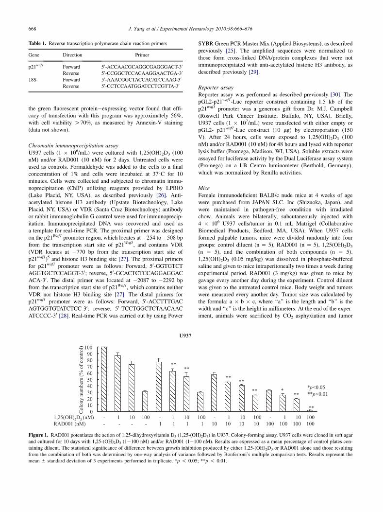
Table 1. Reverse transcription polymerase chain reaction primers
Gene Direction Primer
p21waf1 Forward 50-ACCAACGCAGGCGAGGGACT-30
Reverse 50-CCGGCTCCACAAGGAACTGA-30
18S Forward 50-AAACGGCTACCACATCCAAG-30
Reverse 50-CCTCCAATGGATCCTCGTTA-30
668 J. Yang et al./ Experimental Hematology 2010;38:666–676
the green fluorescent protein�expressing vector found that effi-cacy of transfection with this program was approximately 56%,with cell viability O70%, as measured by Annexin-V staining(data not shown).
Chromatin immunoprecipitation assayU937 cells (1 � 106/mL) were cultured with 1,25(OH)2D3 (100nM) and/or RAD001 (10 nM) for 2 days. Untreated cells wereused as controls. Formaldehyde was added to the cells to a finalconcentration of 1% and cells were incubated at 37�C for 10minutes. Cells were collected and subjected to chromatin immu-noprecipitation (ChIP) utilizing reagents provided by LPBIO(Lake Placid, NY, USA), as described previously [26]. Anti-acetylated histone H3 antibody (Upstate Biotechnology, LakePlacid, NY, USA) or VDR (Santa Cruz Biotechnology) antibodyor rabbit immunoglobulin G control were used for immunoprecip-itation. Immunoprecipitated DNA was recovered and used asa template for real-time PCR. The proximal primer was designedon the p21Waf1 promoter region, which locates at �254 to �508 bpfrom the transcription start site of p21Waf1, and contains VDR(VDR locates at �770 bp from the transcription start site ofp21waf1)5 and histone H3 binding site [27]. The proximal primersfor p21waf1 promoter were as follows: Forward, 50-GGTGTCTAGGTGCTCCAGGT-30; reverse, 50-GCACTCTCCAGGAGGACACA-30. The distal primer was located at �2087 to �2292 bpfrom the transcription start site of p21Waf1, which contains neitherVDR nor histone H3 binding site [27]. The distal primers forp21waf1 promoter were as follows: Forward, 50-ACCTTTGACAGTGGTGTATCTCC-30; reverse, 50-TCCTGGCTCTAACAACATCCCC-30 [28]. Real-time PCR was carried out by using Power
Figure 1. RAD001 potentiates the action of 1,25-dihydroxyvitamin D3 (1,25-(OH
and cultured for 10 days with 1,25-(OH)2D3 (1�100 nM) and/or RAD001 (1�1
taining diluent. The statistical significance of difference between growth inhibiti
from the combination of both was determined by one-way analysis of variance
mean 6 standard deviation of 3 experiments performed in triplicate. *p ! 0.05
SYBR Green PCR Master Mix (Applied Biosystems), as describedpreviously [25]. The amplified sequences were normalized tothose form cross-linked DNA/protein complexes that were notimmunoprecipitated with anti-acetylated histone H3 antibody, asdescribed previously [29].
Reporter assayReporter assay was performed as described previously [30]. ThepGL2-p21waf1-Luc reporter construct containing 1.5 kb of thep21waf1 promoter was a generous gift from Dr. M.J. Campbell(Roswell Park Cancer Institute, Buffalo, NY, USA). Briefly,U937 cells (1 � 107/mL) were transfected with either empty orpGL2- p21waf1-Luc construct (10 mg) by electroporation (150V). After 24 hours, cells were exposed to 1,25(OH)2D3 (100nM) and/or RAD001 (10 nM) for 48 hours and lysed with reporterlysis buffer (Promega, Madison, WI, USA). Soluble extracts wereassayed for luciferase activity by the Dual Luciferase assay system(Promega) on a LB Centro luminometer (Berthold, Germany),which was normalized by Renilla activities.
MiceFemale immunodeficient BALB/c nude mice at 4 weeks of agewere purchased from JAPAN SLC. Inc (Shizuoka, Japan), andwere maintained in pathogen-free condition with irradiatedchow. Animals were bilaterally, subcutaneously injected with4 � 106 U937 cells/tumor in 0.1 mL Matrigel (CollaborativeBiomedical Products, Bedford, MA, USA). When U937 cellsformed palpable tumors, mice were divided randomly into fourgroups: control diluent (n 5 5), RAD001 (n 5 5), 1,25(OH)2D3
(n 5 5), and the combination of both compounds (n 5 5).1,25(OH)2D3 (0.05 mg/kg) was dissolved in phosphate-bufferedsaline and given to mice intraperitoneally two times a week duringexperimental period. RAD001 (3 mg/kg) was given to mice bygavage every another day during the experiment. Control diluentwas given to the untreated control mice. Body weight and tumorswere measured every another day. Tumor size was calculated bythe formula: a � b � c, where ‘‘a’’ is the length and ‘‘b’’ is thewidth and ‘‘c’’ is the height in millimeters. At the end of the exper-iment, animals were sacrificed by CO2 asphyxiation and tumor
)2D3) in U937. Colony-forming assay. U937 cells were cloned in soft agar
00 nM). Results are expressed as a mean percentage of control plates con-
on produced by either 1,25-(OH)2D3 or RAD001 alone and those resulting
followed by Bonferroni’s multiple comparison tests. Results represent the
; **p ! 0.01.

669J. Yang et al./ Experimental Hematology 2010;38:666–676
weights were measured after their careful resection. Tumor tissuewas collected for analysis. These experiments were approved bythe Review Board of Kochi University (approved # B-00011).
Statistical analysisStatistical analysis was performed to assess the difference betweentwo groups under multiple conditions by one-way analysis of vari-ance followed by Bonferroni’s multiple comparison tests usingPRISM statistical analysis software (GraphPad Software, Inc.,San Diego, CA, USA). When comparing two groups, Student’st-test was used.
Results
Blockade of mTOR signaling inhibited the growthof U937 cellsWe explored the drug interaction of 1,25(OH)2D3
and RAD001 in AML cells by colony-forming assay.1,25(OH)2D3 (1-100 nM) alone inhibited the colony-forming ability of U937 cells in a dose-dependent manner,and RAD001 (10�100 nM) apparently potentiated theeffects of 1,25(OH)2D3 (Fig. 1). For example, either1,25(OH)2D3 (100 nM) or RAD001 (100 nM) alone inhibited
Figure 2. Cell-cycle analysis. U937 cells were cultured with 1,25-dihydroxyvit
days, the cell-cycle distribution of these cells was analyzed. Results represent o
U937 clonogenic growth by 31% 6 2% and 33% 6 1%,respectively. When both were combined at the same concen-tration, colony formation was almost completely inhibited(Fig. 1).
Effects of 1,25(OH)2D3 and RAD001 on the cell-cycledistribution of U937 cellsWe next examined whether RAD001 potentiated the abilityof 1,25(OH)2D3 to accumulate cells in the G0/G1 phase ofthe cell cycle. Either 1,25(OH)2D3 (100 nM, 72 hours) orRAD001 (10 nM, 72 hours) caused an accumulation of61% 6 2% and 63% 6 0.3% (mean 6 standard deviation)of U937 cells in the G0/G1 phase of the cell cycle, respec-tively. When these cells were exposed to both of thesecompounds at the same concentration, the proportion ofcells in the G0/G1 phase of the cell cycle increased to67% 6 0.6% (Fig. 2).
RAD001 enhanced 1,25(OH)2D3-induced differentiationof U937 cellsWe next explored the effect of RAD001 on the ability of1,25(OH)2D3 to induce differentiation of U937 cells by
amin D3 (1,25-(OH)2D3) (10�100 nM) and/or RAD001 (10 nM). After 3
ne of the three experiments performed in duplicate.

Figure 3. Effect of RAD001 and 1,25-dihydroxyvitamin D3 (1,25-
(OH)2D3) on Akt/mammalian target of rapamycin complex-1 (mTORC1)
signaling. Western blot analysis. U937 cells were cultured with
1,25(OH)2D3 (10�100 nM) and/or RAD001(10 nM) for 3 hours. Lysates
were made and Western blot analysis was performed to measure the levels
of the indicated proteins. Each lane was loaded with 30 mg protein. The
figure represents one of three experiments performed independently with
similar results.
Figure 4. Effect of RAD001 and 1,25-dihydroxyvitamin D3 (1,25-
(OH)2D3) on cell-cycle�related molecules. Western blot analysis. U937
cells were cultured with 1,25(OH)2D3 (10�100 nM) and/or RAD001 (10
nM) for 48 hours. Western blot analysis was utilized to monitor levels
of p21waf1, p27 kip1, c-Myc, and glyceraldehyde 3-phosphate dehydroge-
nase (GAPDH) proteins. Each lane was loaded with 30 mg protein. The
figure represents one of the three experiments performed independently
with similar results.
670 J. Yang et al./ Experimental Hematology 2010;38:666–676
quantifying the population of cells expressing CD11b,a marker of differentiation of myeloid cells by flow cytom-etry. Exposure of U937 cells to 1,25(OH)2D3 modestlyinduced differentiation of these cells in a dose-dependentmanner. For example, either 1,25(OH)2D3 (10 nM) or1,25(OH)2D3 (100 nM) alone induced approximately 18%or 22% of U937 cells, respectively, to express CD11bantigen. Interestingly, RAD001 (10 nM) potently enhanced1,25(OH)2D3 (10 or 100 nM)-induced differentiation ofU937 cells. For example, either 1,25(OH)2D3 (100 nM)or RAD001 (10 nM) alone induced approximately 22% or5% of U937 cells, respectively, to express CD11b antigen.When these cells were exposed to both of these compoundsat the same concentration, a mean of 40% of U937 cellswere induced to express CD11b on their surface (figurenot shown). The effect of RAD001 on the ability of1,25(OH)2D3 to induce differentiation of U937 cells wasfurther explored by monitoring the endocytic capability ofthe cells. Either 1,25(OH)2D3 (100 nM, 72 hours) orRAD001 (10 nM, 72 hours) alone induced approximately8% or 4% of U937 cells, respectively, to internalizeFITC-labeled dextran. However, when 1,25(OH)2D3 (100nM, 72 hours) was combined with RAD001 (10 nM, 72hours), a mean of 22% of U937 cells internalized theFITC-labeled dextran (figure not shown).
Effect of 1,25(OH)2D3 and RAD001 on Akt/mTORsignaling in U937 cellsEither 1,25(OH)2D3 (10 or 100 nM, 3 hours) or RAD001(10 nM, 3 hours) alone potently downregulated the levelsof p-Akt, as well as p-S6K, without affecting the totalamount of these proteins in U937 cells (Fig. 3). On theother hand, these agents alone only slightly decreased thelevels of p-4E-BP1 in U937 cells (Fig. 3). Interestingly,1,25(OH)2D3 (10 or 100 nM, 3 hours) in combinationwith RAD001 (10 or 100 nM, 3 hours) apparently inhibitedexpression of the phosphorylated forms of these proteins inU937 (Fig. 3).
Combination of 1,25(OH)2D3 and RAD001 modulatedlevels of p21waf1, p27kip1 and c-Myc proteinsFurther studies explored whether 1,25(OH)2D3 and/orRAD001 modulated levels of the cell-cycle�related mole-cules in U937 cells by Western blot analysis. Either1,25(OH)2D3 (100 nM) or RAD001 (10 nM) alone slightlyinduced expression of cyclin-dependent kinase inhibitorsp21waf1 and p27kip1in U937 cells (Fig. 4). When U937 cellswere exposed to the combination of both at the sameconcentration, levels of these proteins were markedly upre-gulated (Fig. 4). In addition, exposure of U937 cells toeither 1,25(OH)2D3 (10 or 100 nM) or RAD001 (10 nM)alone downregulated levels of c-Myc. Interestingly, expo-sure of these cells to the combination of both compoundsat the same concentration almost completely inhibitedexpression of c-Myc (Fig. 4).
Induction of p21Waf1 was essential for the 1,25(OH)2D3-mediated growth arrest of U937 cellsWe examined whether p21Waf1 was essential to combinationof 1,25(OH)2D3 and RAD001-mediated growth arrest anddifferentiation of U937 cells. We downregulated levels of

Figure 5. Repression of p21waf1 blunts 1,25-dihydroxyvitamin D3 (1,25-(OH)2D3)�induced cell-cycle arrest. U937 cells were transiently transfected with
either control or p21waf1 small interfering RNA (siRNA). After 24 hours, cells were exposed to either 1,25(OH)2D3 (100 nM) and/or RAD001 (10 nM) for 48
hours, and subjected to fluorescein-activated cell sorting to quantify the population of cells expressing p21waf1 (A) or CD11b (B). Simultaneously, the
cell-cycle distribution (C) of these cells were measured by FACScan. Results represent the mean 6 SD of 3 experiments performed in duplicate, *p!0.05.
671J. Yang et al./ Experimental Hematology 2010;38:666–676
p21Waf1 by siRNA. After 24 hours, these cells were exposedto 1,25(OH)2D3 and/or RAD001 for 48 hours and subjectedto fluorescein-activated cells sorting to quantify the levelsof p21Waf1 and CD11b. The siRNA against p21Waf1 almostcompletely blocked combination of 1,25(OH)2D3 andRAD001-induced expression of p21Waf1 in U937 cells(Fig. 5A). The 1,25(OH)2D3-induced expression of CD11bwas not blunted in the p21Waf1 siRNA transfected U937cells (Fig. 5B). On the other hand, the 1,25(OH)2D3-induced G0/G1 cell-cycle arrest was significantly attenuatedafter depletion of p21Waf1 in U937 cells (Fig. 5C). Theseobservations suggested that p21Waf1 was required, at leastin part, for the 1,25(OH)2D3-induced growth arrest ofU937 cells.
RAD001 enhanced 1,25(OH)2D3-mediated expression ofp21Waf1 at transcriptional levelsReal-time reverse transcription PCR found that the induc-tion of p21Waf1 after exposure of U937 cells to1,25(OH)2D3 and RAD001 was at the mRNA level(Fig. 6A). Further studies utilizing the p21Waf1-Luc reporterconstruct found that either 1,25(OH)2D3 (100 nM, 48hours) or RAD001(10 nM, 48 hours) alone slightlyincreased transcriptional activity of p21Waf1. Combination
of compounds at the same concentration further stimulatedthis activity (Fig. 6B).
RAD001 and 1,25(OH)2D3 increased levels of theacetylated forms of histone H3 and VDR on the p21Waf1
promoter in U937 cellsWe explored the mechanisms by which RAD001 enhanced1,25(OH)2D3-stimulated transcriptional activity of p21Waf1.We first measured nuclear levels of the acetylated forms ofhistone H3 (acetyl-histone H3) in U937 cells after exposureto either 1,25(OH)2D3 or RAD001 because the transcrip-tional activity of genes usually increases as histone H3becomes acetylated in the promoter region of the gene[31]. U937 cells were cultured in the presence of1,25(OH)2D3 (10 nM, 100 nM) and/or RAD001 (10 nM)for 48 hours. Western blot analyses showed that the effectof 1,25(OH)2D3 or RAD001 on the levels of acetyl-histone H3 was negligible, however, combination of bothcompounds at the same concentration strikingly increasedthe level of acetyl-histone H3 in their nuclei (Fig. 7A).Similarly, the effect of 1,25(OH)2D3 or RAD001 on thelevels of VDR was negligible in U937 cells. Importantly,the combination of both compounds at the same concentra-tion significantly increased the nuclear levels of VDR

Figure 6. Effect of RAD001 on 1,25(OH)2D3-induced expression of p21waf1 in U937 cells. (A) Real-time reverse transcription polymerase chain reaction
(RT-PCR). U937 cells were cultured with 1,25-dihydroxyvitamin D3 (1,25-(OH)2D3) (10�100 nM) and/or RAD001 (10 nM) for 24 hours. Cells were
harvested and RNA was isolated and subjected to real-time RT-PCR. Results represent the mean 6 standard deviation of three experiments performed in
duplicate. Statistical significance was determined by analysis of variance (ANOVA) followed by Bonferroni’s multiple comparison tests. (B) p21waf1 lucif-
erase reporter assay. The schema of the pGL2-p21waf1-Luc construct was shown at the top. U937 cells were transiently transfected with either empty or pGL2-
p21waf1-Luc construct. After 24 hours, cells were exposed to RAD001 (10 nM, 48 hours) and/or 1,25(OH)2D3 (100 nM, 48 hours). pRL-SV40-Luciferase
(Renilla luciferase) vector was cotransfected for normalization. Results represent the mean 6 standard deviation of three experiments carried out in duplicate.
Statistical significance was determined by ANOVA followed by Bonferroni’s multiple comparison tests. Results represent the mean 6 SD of 3 experiments
performed in duplicate, *p!0.05.
672 J. Yang et al./ Experimental Hematology 2010;38:666–676
(Fig. 7A). We next examined the effect of combination of1,25(OH)2D3 and RAD001 on acetyl-histone H3 andVDR in the p21Waf1 promoter by ChIP assay. To quantifythe DNA precisely, we employed real-time PCR, asdescribed previously [28,29]. Exposure of U937 cells tothe combination of 1,25(OH)2D3 (100 nM) and RAD001(10 nM) for 48 hours significantly increased levels ofacetyl-histone H3 and VDR bound on the p21Waf1 promoterin these cells (Fig. 7B, C).
RAD001 enhanced the ability of 1,25(OH)2D3 to inhibitthe proliferation of human U937 leukemic xenograftsin vivoWe further evaluated the antiproliferative activity of1,25(OH)2D3 combined with RAD001 in vivo. 1,25(OH)2D3
(0.05 mg/kg) or RAD001 (3 mg/kg) alone inhibited prolifer-ation of U937 xenografts by approximately 40% or 50%,respectively, compared with tumors in vehicle control mice(Fig. 8A). When mice were treated with both compounds,tumor growth was inhibited by nearly 75% (Fig. 8A), result-ing in decreased weights of U937 tumors at autopsy(Fig. 8B). None of the treated mice showed signs of illness(data not shown) or significant weight loss (Fig. 8C).
DiscussionConsiderable preclinical data indicated the activity of1,25(OH)2D3 against some types of cancer, as well as
leukemia cells [2–5]. However, clinical studies of1,25(OH)2D3 as an antitumor agent were hampered bythe adverse effect mediated by hypercalcemia. Therefore,several highly potent analogs of 1,25(OH)2D3 with littlecalcemic activity have been synthesized and tested in vitroand in vivo [32–34]. For example, EB 1089 inducedapoptosis in B-cell chronic lymphocytic leukemia cellsvia a p53-independent pathway in association with suppres-sion of extracellular signal-regulated kinase activity andactivation of the p38 mitogen-activated protein kinasepathway [32]. Also, calcipotriol (MC 903), a relativelynoncalcemic analog of 1,25(OH)2D3, inhibited proliferationof non-Hodgkin lymphoma SU-DHL4 and SU-DUL5 cellsand induced their differentiation [33]. In addition,1a(OH)D3 inhibited proliferation of follicular lymphomain rats, although hypercalcemia was induced [33]. A trialwith oral paricalcitol [19-nor-1,25(OH)2D2] for myelodys-plastic syndrome included 12 individuals with a poor prog-nosis. They were given an average dose of 16 mg/dayparicalcitol (range, 4�56 mg/day) during 4 months. Notoxicity was observed and one patient had a partialresponse, with his platelet count rising from 50,000 mL�1
to the normal range [34]. Recent phase 2 trial exploringthe effects of high-dose (10,000 IU/day) 1,25(OH)2D3 inbreast cancer patients with bone metastases regions showedthat this dose of 1,25(OH)2D3 for 4 months was tolerated[35]. There was a significant reduction in the number of

Figure 7. RAD001 enhances 1,25-dihydroxyvitamin D3 (1,25-(OH)2D3)�induced acetyl-histone H3. (A) Western blotting. U937 cells were cultured in the
presence of RAD001 (10 nM) and/or 1,25(OH)2D3 (10 or 100 nM). After 48 hours, cells were harvested, and nuclear proteins were prepared and subjected to
Western blot analysis. The membrane was sequentially reprobed with anti�Ac-histone H3, -vitamin D receptor (VDR), and - glyceraldehyde 3-phosphate
dehydrogenase (GAPDH) antibodies. Ac-histone H3 5 acetylated histone H3. Band intensities were measured by ImageJ software (Wayne Rasband, National
Institutes of Health, Bethesda, MD). (B, C) Chromatin immunoprecipitation (ChIP) assay. Levels of the acetylated forms of histone H3 (B) and VDR (C) on
the p21waf1 promoter were analyzed by ChIP assay. U937 cells were cultured with RAD001 (10 nM) and/or 1,25(OH)2D3 (10 or 100 nM). After 48 hours,
cells were harvested and subjected to ChIP followed by real-time PCR by utilizing either the proximal (black rectangular box) or distal primer set (white
rectangular box). The amplified sequences were normalized to the input DNA (the cross-linked DNA/protein complexes, which were not immunoprecipitated
with either anti�acetyl-histone H3 or VDR antibody). The statistical significance was determined by analysis of variance followed by Bonferroni’s multiple
comparison tests. Results represent the mean of two experiments performed in duplicate; *p ! 0.05, with respect to control. Ac-histone H3 5 acetylated
histone H3.
673J. Yang et al./ Experimental Hematology 2010;38:666–676
sites of pain, nonetheless 1,25(OH)2D3 did not inhibit boneresorption [35].
A number of types of anticancer agents were utilized incombination with 1,25(OH)2D3 to potentiate anti-tumoreffect of 1,25(OH)2D3 [36,37]. The present study demon-strated for the first time that inhibition of mTORC1 byRAD001 significantly potentiated the ability of 1,25(OH)2D3
to induce growth arrest and differentiation of U937 cellsin conjunction with upregulation of p21waf1 and p27kip1
and downregulation of c-Myc (Figs. 1–4). We also foundthat RAD001 potentiated the ability of 1,25(OH)2D3 toinhibit proliferation and induce differentiation of freshlyisolated leukemia cells from individuals with AML(n 5 2) in conjunction with upregulation of p21waf1 andinduction of CD11b cell surface antigen (data not shown).Moreover, we found that induction of p21waf1 was essen-tial in 1,25(OH)2D3-mediated growth arrest of U937 cells,as assessed by p21waf1 knockdown study (Fig. 5). Notably,inhibition of mTORC1 by RAD001 dramatically enhanced
1,25(OH)2D3-stimulated expression of p21waf1 via epige-netic modification. RAD001 enhanced 1,25(OH)2D3-induced acetylation of histone H3 on the p21Waf1
promoter, as measured by ChIP assay (Fig. 7B). Theseobservations are reminiscent of our recent studies. Wehave shown that RAD001 potentiated the ability ofa histone deacetylase inhibitor MS-275 to induce differen-tiation of AML cells in association with upregulation ofCCAAT/enhancer binding protein�e (C/EBPe), the crit-ical nuclear transcription factor in induction of myeloiddifferentiation [19]. RAD001 dramatically enhanced acet-ylation of histone H3 on the promoter of C/EBPe [19].Similarly, a previous study found that induction of mono-cytic differentiation of HL-60 cells by auranofin, a lipophilicgold compound, and 1,25(OH)2D3, increased acetylation ofhistone H3 at the p21waf1 promoter [38].
We have very recently found that RAD001 potentiatedthe ability of all-trans retinoic acid (ATRA) to inducegrowth arrest and differentiation of HL60 and NB4 AML

Figure 8. Effect of coadministration of 1,25-dihydroxyvitamin D3 (1,25-(OH)2D3) and RAD001 on proliferation of U937 cells in a murine xenograft model.
(A) U937 cells were injected bilaterally subcutaneously into BALB/c nude mice, forming two tumors per mouse. When U937 cells formed palpable tumors
(40 mm3), mice were randomized into four groups (n 5 5) and treatment was initiated. 1,25(OH)2D3 (0.05 mg/kg) was administered to mice intraperitoneally
twice a week. RAD001 (3 mg/kg) was given to mice every other day. During the experimental period, tumor volumes were measured twice a week. Each
point represents mean 6 standard deviation of 10 tumors. (B) Tumor weights at autopsy. After 2 weeks of treatment, tumors were removed and weighed.
Results represent the mean 6 standard deviation of tumor weights. Statistical significance was determined by analysis of variance (ANOVA) followed by
Bonferroni’s multiple comparison tests. Bars 5 standard deviation. (C) Body weight. Body weights of mice were measured twice. Control 5 diluent control.
Statistical significance was determined by ANOVA followed by Bonferroni’s multiple comparison tests.
674 J. Yang et al./ Experimental Hematology 2010;38:666–676

675J. Yang et al./ Experimental Hematology 2010;38:666–676
cells [20]. RAD001 augmented ATRA-induced expressionof C/EBPe. Both ATRA and RAD001 induced acetylationof histone H3 in AML cells; however, the combination ofboth did not further increase the levels of acetylated histoneH3 compared to either compound alone [20].
1,25(OH)2D3, in combination with RAD001 potentlyincreased levels of VDR in U937 cells (Fig. 7A), whichalso contributed to enhanced expression of p21Waf1afterexposure to a combination of 1,25(OH)2D3 and RAD001.RAD001 could augment 1,25(OH)2D3-induced expressionof VDR via epigenetic mechanism. Further experimentsare required to clarify this hypothesis.
Akt/mTOR regulates levels of c-Myc by several differentmechanisms. For example, Akt/mTOR stabilizes c-Myc tran-scripts, as well as c-Myc protein, resulting in increasedexpression of c-Myc [39,40]. Other studies showed thatAkt/mTOR stimulated translation of c-Myc mRNA [41].Very recently, rapamycin has been shown to decrease levelsof c-Myc by inhibiting c-Myc mRNA translation in murinepromyelocytes, leading to terminal differentiation [42]. Wehave very recently found that RAD001 augmented ATRA-induced differentiation of HL60 and NB4 AML cells inassociation with upregulation of expression of C/EBPe anddownregulation of levels of c-Myc [20]. The present studyfound that RAD001 downregulated levels of c-Myc protein,and 1,25(OH)2D3 further decreased c-Myc in the presence ofRAD001 (Fig. 4A). Consistent with these observations, otherinvestigators also showed that 1,25(OH)2D3 downregulatedlevels of c-Myc in cancer cells [43–45]. The augmenteddownregulation of c-Myc was probably mediated by inacti-vation of mTORC1 by a combination of these compounds.This may be associated with induction of differentiation ofAML cells, as noted in this study.
RAD001 has been tested in phase 1 and 2 trials forhematologic malignancies, including AML, myelodysplas-tic syndrome, B-cell chronic lymphocytic leukemia, andmantle cell lymphoma. Investigators found that RAD001was well-tolerated at a daily dose of 10 mg and showedactivity in patients with myelodysplastic syndrome [46].Another phase 2 study of the oral single-agent RAD001(10 mg/day) for recurrent/refractory indolent lymphoidmalignancies also showed the activity of RAD001 againstchronic lymphocytic leukemia [47].
Taken together, concomitant administration of1,25(OH)2D3 and RAD001 may be a promising treatmentstrategy for individuals with AML. Further studies are war-ranted to evaluate the efficiency of this combination ina clinical setting.
AcknowledgmentsThis work was supported in part by Grant-in-Aid from theMinistry of Education, Culture Sports, Science, and Technologyof Japan (Chiyoda-ku, Tokyo, Japan) (to T.I.), The Kochi Univer-sity President’s Discretionary Grant (Nankoku, Kochi, Japan) (toT.I.), Takeda Science Foundation (Osaka, Japan) (to T.I.), and
Sagawa Foundation for Promotion of Cancer Research (Kyoto,Japan) (to T.I.), as well as NIH RO1 CA026038-30 (H.P.K.).
Conflict of Interest DisclosureThe authors have no conflict of interest to declare.
References1. Takashi K, Lee YS, Hughes SV, et al. 19-Nor-1,25(OH)2D2 (a novel,
noncalcemic vitamin D analogue), combined with arsenic trioxide, has
potent antitumor activity against myeloid leukemia. Cancer Res. 2005;
65:2489–2497.
2. Colstone KW, Chander SK, Mackay AG, Coombes RC. Effects of
synthetic vitamin D analogues on breast cancer cell proliferation in
vitro and in vivo. Biochemical Pharm. 1992;44:693–702.
3. Shabahang M, Buras RR, Davoodi F, Schumaker LM, Nauta RJ, Evans
SRT. 1,25-Dihydroxyvitamin D3 receptor as a marker of human colon
carcinoma cell line differentiation and growth inhibition. Cancer Res.
1993;53:3712–3718.
4. Eisman JA, Barkla DH, Tutton PJM. Suppression of in vivo growth of
human cancer solid tumor xenografts by 1,25-dihydroxyvitamin D3.
Cancer Res. 1987;47:21–25.
5. Liu M, Lee MH, Cohen M, Bommakanti M, Freedman LP. Transcrip-
tional activation of the Cdk inhibitor p21 by vitamin D3 leads to the
induced differentiation of the myelomonocytic cell line U937. Genes
Dev. 1996;10:142–153.
6. Rots NY, Iavarone A, Bromleigh V, Freedman LP. Induced differenti-
ation of U937 cells by 1,25-dihydroxyvitamin D3 involves cell cycle
arrest in G1 that is preceded by a transient proliferative burst and an
increase in cyclin expression. Blood. 1999;93:2721–2729.
7. Elstner E, Campbell MJ, Munker R, et al. Novel 20-epi-vitamin D3
analog combined with 9-cis-retinoic acid markedly inhibits colony
growth of prostate cancer cells. Prostate. 1999;40:141–149.
8. Muto A, Kizaki M, Yamato K, et al. 1,25-Dihydroxyvitamin D3 induces
differentiation of a retinoic acid-resistant acute promyelocytic leukemia
cell line (UF-1) associated with expression of p21(WAF1/CIP1) and
p27(KIP1). Blood. 1999;93:2225–2233.
9. Munker R, Kobayashi T, Elstner E, et al. A new series of vitamin D
analogs is highly active for clonal inhibition, differentiation, and
induction of WAF1 in myeloid leukemia. Blood. 1996;88:2201–2209.
10. Wang JR, Zhao Y, Kauss MA, Spindel S, Lian H. Akt regulates vitamin
D3-induced leukemia cell functional differentiation via Raf/MEK/ERK
MAPK signaling. Eur J Cell Biol. 2009;88:103–115.
11. O’Kelly J, Uskokovic M, Lemp N, Vadgama J, Koeffler HP. Novel
Gemini-vitamin D3 analog inhibits tumor cell growth and modulates
the Akt/mTOR signaling pathway. J Steroid Biochem Mol Biol.
2000;100:107–116.
12. Vivanco I, Sawyers CL. The phosphatidylinositol 3-kinase-Akt
pathway in human cancer. Nat Rev Cancer. 2002;2:489–501.
13. Inoki K, Li K, Zhu T, Wu J, Guan KL. TSC2 is phosphorylated and
inhibited by Akt and suppresses mTOR signaling. Nat Cell Biol.
2002;4:648–657.
14. Bjornsti MA, Houghton PJ. Lost in translation: dysregulation of
cap-dependent translation and cancer. Cancer Cell. 2004;5:519–523.
15. Hay N, Sonenberg N. Upstream and downstream of mTOR. Genes
Dev. 2004;18:1926–1945.
16. Jacinto E, Loewith R, Schmidt A, et al. Mammalian TOR complex
2 controls the actin cytoskeleton and is rapamycin insensitive. Nat
Cell Biol. 2004;6:1122–1128.
17. Sarbassov DD, Ali SM, Kim DH, et al. Rictor, a novel binding partner
of mTOR, defines a rapamycin-insensitive and raptor-independent
pathway that regulates the cytoskeleton. Curr Biol. 2004;14:1296–
1302.
676 J. Yang et al./ Experimental Hematology 2010;38:666–676
18. Sarbassov DD, Guertin DA, Ali SM, Sabatini DM. Phosphorylation
and regulation of Akt/PKB by the rictor-mTOR complex. Science.
2005;307:1098–1101.
19. Nishioka C, Ikezoe T, Yang J, Koeffler HP, Yokoyama A. Blockade of
mTOR signaling potentiates the ability of histone deacetylase inhibitor
to induce growth arrest and differentiation of acute myelogenous
leukemia cells. Leukemia. 2008;22:2159–2168.
20. Nishioka C, Ikezoe T, Yang J, Gery S, Koeffler HP, Yokoyama A. Inhi-
bition of mammalian target of rapamycin signaling potentiates the
effects of all-trans retinoic acid to induce growth arrest and differen-
tiation of human acute myelogenous leukemia cells. Int J Cancer.
2009;125:1710–1720.
21. Chan S. Targeting the mammalian target of rapamycin(mTOR): a new
approach to treating cancer. Br J Cancer. 2004;91:1420–1424.
22. Haritunians T, Mori A, O’Kelly J, Luong QT, Giles FJ, Koeffler HP.
Antiproliferative activity of RAD001(everolimus) as a single agent
and combined with other agents in mantle cell lymphoma. Leukemia.
2007;21:333–339.
23. Munker P, Kobayashi T, Elstner E, et al. A new series of vitamin D
analogs is highly active for clonal inhibition, differentiation, and
induction of WAF1 in myeloid leukemia. Blood. 1996;88:2201–
2209.
24. Ikezoe T, Nishioka C, Tasaka T, et al. The antitumor effects of suniti-
nib (formerly SU11248) against a variety of human hematologic
malignancies: enhancement of growth inhibition via inhibition of
mammalian target of rapamycin signaling. Mol Cancer Ther. 2006;
5:2522–2530.
25. Ikezoe T, Tanosaki S, Krug U, et al. Insulin-like growth factor binding
protein-3 antagonizes the effects of retinoids in myeloid leukemia
cells. Blood. 2004;104:237–242.
26. Nishioka C, Ikezoe T, Yang J, Koeffler HP, Yokoyama A. Inhibition of
MEK/ERK signaling synergistically potentiates histone deacetylase
inhibitor-induced growth arrest, apoptosis and acetylation of histone
H3 on p21(waf1) promoter in acute myelogenous leukemia cell.
Leukemia. 2008;22:1449–1452.
27. Richon VM, Sandhoff TW, Rifkind RA, Marks PA. Histone deacetylase
inhibitor selectively induces p21WAF1 expression and gene-associated
histone acetylation. Proc Natl Acad Sci U S A. 2000;97:10014–10019.
28. Nishio H, Walsh MJ. CCAAT displacement protein/cut homolog
recruits G9a histone lysine methyltransferase to repress transcription.
Proc Natl Acad Sci U S A. 2004;101:11257–11262.
29. Nagaki K, Talbert PB, Zhong CX, Dawe RK, Henikoff S, Jiang J.
Chromatin immunoprecipitation reveals that the 180-bp satellite
repeat is the key functional DNA element of Arabidopsis thaliana
centromeres. Genetics. 2003;163:1221–1225.
30. Ikezoe T, Yang Y, Bandobashi K, et al. Oridonin, a diterpenoid
purified from Rabdosia rubescens, inhibits the proliferation of cells
from lymphoid malignancies in association with blockade of the
NF-kappa B signal pathways. Mol Cancer Ther. 2005;4:578–586.
31. Archer SY, Hodin RA. Histone acetylation and cancer. Curr Opin
Genet Dev. 1999;9:171–174.
32. Pepper C, Thomas A, Hoy A, Milligan D, Bentley P, Fegan C. The
vitamin D3 analog EB1089 induces apoptosis via a p53-independent
mechanism involving p38 MAP kinase activation and suppression of
ERK activity in B-cell chronic lymphocytic leukemia cells in vitro.
Blood. 2003;101:24554–24560.
33. Hickish T, Cunningham D, Colston K, et al. The effect of 1,25-dihy-
droxyvitamin D3 on lymphoma cell lines and expression of the
vitamin D receptor in lymphoma. Br J Cancer. 1993;68:668–672.
34. Koeffler HP, Aslanian N, O’Kelly J. Vitamin D2 analog (Paricalcitol;
Zemplar) for treatment of myelodysplastic syndrome. Leukemia Res.
2005;29:1259–1262.
35. Amir E, Simmons CE, Freedman OC, et al. A phase 2 trial exploring
the effects of high-dose (10,000 IU/day) vitamin D(3) in breast cancer
patients with bone metastases. Cancer. 2010;116:284–291.
36. Wietrzyk J, Nevozhay D, Filip B, Milczarek M, Kutner A. The anti-
tumor effect of lowered doses of cytostatics combined with new
analogs of vitamin D in mice. Anticancer Res. 2007;27:3387–3398.
37. Chiang KC, Persons KS, Istfan NW, Holick MF, Chen TC. Fish oil
enhances the antiproliferative effect of 1alpha, 25-dihydroxyvitamin
D3 on liver cancer cells. Anticancer Res. 2009;29:3591–3596.
38. Park SJ, Kim M, Kim NH, et al. Auranofin promotes retinoic acid- or
dihydroxyvitamin D3-mediated cell differentiation of promyelocytic
leukaemia cells by increasing histone acetylation. Br J Pharmacol.
2008;154:1196–1205.
39. Marderosian M, Sharma A, Funk AP, et al. Tristetraprolin regulates
Cyclin D1 and c-Myc mRNA stability in response to rapamycin in
an Akt-dependent manner via p38 MAPK signaling. Oncogene.
2006;25:6277–6290.
40. Gregory MA, Qi Y, Hann SR. Phosphorylation by glycogen sythase
kinase-3 controls c-Myc proteolysis and subnuclear localization.
J Biol Chem. 2003;278:51606–51612.
41. Gera JF, Mellingghoff IK, Shi Y, et al. AKTactivity determines sensitivity
to mammalian target of rapamycin (mTOR) inhibitors by regulating
cyclin D1 and c-Myc expression. J Biol Chem. 2004;279:2737–2746.
42. Wall M, Poortinga G, Hannan KM, Pearson RB, Hannan RD, McArthur
GA. Translational control of c-MYC by rapamycin promotes terminal
myeloid differentiation. Blood. 2008;112:2305–2317.
43. Polek TC, Stewart LV, Ryu EJ, Cohen MB, Allegretto EA, Weigel NL.
p53 is required for 1,25-dihydroxyvitamin D3-inducedG0 arrest but is
not required for G1 accumulation or apoptosis of LNCaP prostate
cancer cells. Endocrinology. 2003;144:50–60.
44. Malone JM, Deppe G. Inhibition of c- myc in breast and ovarian
carcinoma cells by 1, 25-dihydroxyvitamin D3, retinoic acid and
dexamethasone. Anticancer Drugs. 1993;4:201–208.
45. Tong WM, Kallay E, Hofer H, et al. Growth regulation of human
colon cancer cells by epidermal growth factor and 1, 25-dihydroxyvi-
tamin D3 is mediated by mutual modulation of receptor expression.
Eur J Cancer. 1998;34:2119–2125.
46. Yee KW, Zeng Z, Konopleva M, et al. Phase I/II study of the mamma-
lian target of rapamycin inhibitor everolimus (RAD001) in patients
with relapsed or refractory hematologic malignancies. Clin Cancer
Res. 2006;12:5165–5173.
47. Zent CS, Laplant BR, Johnston PB, et al. The treatment of recurren-
t/refractory chronic lymphocytic leukemia/small lymphocytic
lymphoma (CLL) with everolimus results in clinical responses and
mobilization of CLL cells into the circulation. Cancer. 2010;116:
2201–2207.
Copyright © 2022 FDOKUMEN