The mTOR signaling pathway mediates control of ribosomal protein mRNA translation in rat liver
ORIGINAL ARTICLE
AKT induces senescence in human cells via mTORC1 and p53 in the
absence of DNA damage: implications for targeting mTOR during
malignancy
MV Astle1, KM Hannan1, PY Ng1, RS Lee1, AJ George1,2,3, AK Hsu4, Y Haupt1,3,6,RD Hannan1,5,6,7 and RB Pearson1,5,6,7
1Growth Control and Differentiation Program, Trescowthick Research Laboratories, Peter MacCallum Cancer Centre, Melbourne,Victoria, Australia; 2School of Biomedical Sciences, University of Queensland, Brisbane, Queensland, Australia; 3Department ofPathology, University of Melbourne, Melbourne, Victoria, Australia; 4Hematology Immunology Translational Research Laboratory,Trescowthick Research Laboratories, Peter MacCallum Cancer Centre, Melbourne, Victoria, Australia; 5Department of Biochemistryand Molecular Biology, Monash University, Clayton, Victoria, Australia and 6Department of Biochemistry and Molecular Biology,University of Melbourne, Melbourne, Victoria, Australia
The phosphatidylinositol 3-kinase (PI3K)/AKT and RASoncogenic signalling modules are frequently mutated insporadic human cancer. Although each of these pathwayshas been shown to play critical roles in driving tumourgrowth and proliferation, their activation in normal humancells can also promote cell senescence. Although themechanisms mediating RAS-induced senescence have beenwell characterised, those controlling PI3K/AKT-inducedsenescence are poorly understood. Here we show thatPI3K/AKT pathway activation in response to phosphataseand tensin homolog (PTEN) knockdown, mutant PI3K,catalytic, a polypeptide (PIK3CA) or activated AKTexpression, promotes accumulation of p53 and p21,increases cell size and induces senescence-associatedb-galactosidase activity. We demonstrate that AKT-induced senescence is p53-dependent and is characterisedby mTORC1-dependent regulation of p53 translation andstabilisation of p53 protein following nucleolar localisa-tion and inactivation of MDM2. The underlying mechan-isms of RAS and AKT-induced senescence appear to bedistinct, demonstrating that different mediators of senes-cence may be deregulated during transformation byspecific oncogenes. Unlike RAS, AKT promotes rapidproliferative arrest in the absence of a hyperproliferativephase or DNA damage, indicating that inactivation of thesenescence response is critical at the early stages of PI3K/AKT-driven tumourigenesis. Furthermore, our data implythat chronic activation of AKT signalling providesselective pressure for the loss of p53 function, consistentwith observations that PTEN or PIK3CA mutations aresignificantly associated with p53 mutation in a number ofhuman tumour types. Importantly, the demonstration thatmTORC1 is an essential mediator of AKT-inducedsenescence raises the possibility that targeting mTORC1
in tumours with activated PI3K/AKT signalling may exertunexpected detrimental effects due to inactivation of asenescence brake on potential cancer-initiating cells.Oncogene (2012) 31, 1949–1962; doi:10.1038/onc.2011.394;published online 12 September 2011
Keywords: PI3K; AKT; stress; senescence; p53; mTOR
Introduction
Activating mutations in the gene encoding a catalyticsubunit of phosphatidylinositol 3-kinase (PI3K), PI3K,catalytic, a polypeptide (PIK3CA), or inactivating muta-tions in the negative regulator of PI3K signalling,phosphatase and tensin homolog (PTEN), accountfor approximately 30% of human sporadic tumours(Luo et al., 2003; Campbell et al., 2004). Furthermore,hyperactivation of an important mediator of PI3Ksignalling, AKT, is observed in a high proportion ofhuman cancers, including 20–55% of breast cancers,B60% of lung cancers, B50% of prostate cancers and40–70% of melanomas (Altomare and Testa, 2005). Inaddition to its role in PI3K/PTEN mutation-drivencancer, AKT acts as a central node in pathways activatedby oncogenic mutation in genes, including those encodingEGFR, PDGFR, RAS and LKB1 (Hynes and MacDo-nald, 2009; Solomon and Pearson, 2009). Three homo-logous AKT isoforms, AKT1/PKBa, AKT2/PKBb andAKT3/PKBg, have distinct and overlapping roles in adiverse range of fundamental cellular functions consideredhallmarks of cancer when dysregulated, including cellgrowth and proliferation, cell survival, migration andangiogenesis (Hanahan and Weinberg, 2000; Manningand Cantley, 2007). AKT-dependent control of cellgrowth and proliferation is in large part mediated bymTORC1 regulation of ribosome biogenesis and proteintranslation (Shaw and Cantley, 2006; Hannan et al., 2011).
Like the other prototypical oncogenes, RAS andMYC, PI3K is an important driver of malignant
Received 23 January 2011; revised 6 July 2011; accepted 1 August 2011;published online 12 September 2011
Correspondence: Associate Professor RB Pearson, Protein ChemistryLaboratory, Peter MacCallum Cancer Centre, Locked Bag 1, A’BeckettStreet, Melbourne, Victoria 8006, Australia.E-mail: [email protected] authors contributed equally to this work.
Oncogene (2012) 31, 1949–1962& 2012 Macmillan Publishers Limited All rights reserved 0950-9232/12
www.nature.com/onc

transformation; however, paradoxically, when hyperac-tivated in normal cells, all three can result in celldeath, cell cycle arrest or senescence, depending oncellular context (Lowe et al., 2004; Gorgoulis andHalazonetis, 2010). Well-established in vitro markersof oncogene-induced senescence have now been detectedin vivo in humans, including in melanocytic naevi(Gray-Schopfer et al., 2006) and neurofibromas(Courtois-Cox et al., 2006). Clearly, oncogene-inducedsenescence must be overcome to enable tumour forma-tion and pharmacological induction of senescence oftumour cells is being increasingly seen as a mechanismfor therapeutic intervention (Gewirtz et al., 2008;Ewald et al., 2010; Nardella et al., 2011). Thus,understanding the basis of senescence induced byindividual oncogenes in normal cells and how thisprocess is subverted in cancer cells is critical to ourunderstanding of malignant transformation and how itcan be effectively targeted.
The paradigm for oncogene-induced senescence wasestablished by examining the growth arrest of non-transformed human and mouse cells in response toactivated alleles of the RAS superfamily (Serrano et al.,1997). In the case of activated RAS, cells undergo aninitial hyperproliferative phase (Tremain et al., 2000)that is later followed by an irreversible proliferationarrest (senescence) associated with the accumulation ofcell cycle inhibitors p53, p21 and p16 and largerflattened cell morphology (Serrano et al., 1997). Themechanisms contributing to the RAS-induced accumu-lation of p53 include increased protein stability follow-ing activation of DNA damage response (Di Miccoet al., 2006; Mallette et al., 2007), p38MAPK (Wanget al., 2002) and accumulation of promyelocyticleukemia protein (PML) (Ferbeyre et al., 2000).Elevated p53 translation also contributes to the senes-cence phenotype induced by RAS (Bellodi et al., 2010).Deletion of p53 in mouse cells is sufficient to rescueRAS-induced proliferation arrest (Serrano et al., 1997);however; this is not the case in human cells where thep16 pathway is the prominent effector for proliferationarrest (Brookes et al., 2002; Huot et al., 2002).
Multiple lines of evidence indicate that increasedPI3K/AKT signalling also induces cell senescence. Lossof PTEN, the major negative regulator of the PI3K/AKT pathway, induces senescence in mouse embryonicfibroblasts (Chen et al., 2005) and mouse prostateepithelium (Chen et al., 2005; Alimonti et al., 2010a).PTEN deletion also induces loss of mouse hematopoie-tic stem cells by induction of the senescence effectorsp53, p21 and p19Arf (Yilmaz et al., 2006; Zhang et al.,2006). Expression of constitutively active AKT inducessenescence in human endothelial cells (Miyauchi et al.,2004), mouse embryonic fibroblasts (Chen et al., 2005;Mavrakis et al., 2008; Nogueira et al., 2008) and mouseprimary keratinocytes (Moral et al., 2009). The mechan-isms underlying induction of senescence by AKT arepoorly defined, although the accumulation of reactiveoxygen species has been implicated as playing a role(Nogueira et al., 2008). A better understanding of themechanisms of PI3K/AKT-induced senescence will be
crucial for understanding the role of dysregulated PI3K/AKT signalling in cancer.
To this end, we have analysed PI3K/AKT pathwayactivation-induced senescence in a normal humanfibroblast model. We show that this senescence requiresmTORC1-dependent accumulation of p53 involvingincreased p53 synthesis and stabilisation mediated byinactivation of MDM2. We have identified importantdifferences between senescence induced by the AKT andRAS oncogenic modules. Activated PI3K/AKT signal-ling rapidly induces senescence independent of thehyperproliferative phase required for the effect of H-RASV12. Consistent with this finding, PI3K/AKT-induced senescence is independent of the DNA damageresponse that is required by H-RASV12.
Results
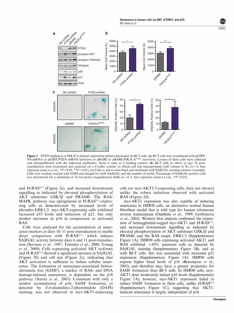
Hyperactivation of the PI3K/AKT pathway inducessenescence in BJ-T cellsWe utilised the well-characterised BJ human fibroblastsimmortalised with human telomerase reverse transcrip-tase (BJ-T cells) (Hahn et al., 1999) as a model to studythe mechanisms of PI3K-induced responses in non-transformed human fibroblasts. BJ-T cells were trans-duced with expression constructs encoding PTEN shorthairpin RNA (shRNA) or the PI3K catalytic subunitmutant PIK3CAE545K (Zhao et al., 2005), which iscommonly detected in multiple cancer types andenhances PI3K/AKT pathway signalling (Campbellet al., 2004; Samuels et al., 2005). Cells with depletedPTEN protein levels or expressing PIK3CAE545K exhib-ited increased levels of phospho-AKT and phospho-rylation of the AKT substrate PRAS40 (proline-richAkt substrate of 40 kDa) demonstrating PI3K pathwayactivation and consistent with a previous report (Kimet al., 2007) accumulation of cell cycle inhibitors p53and its target p21 (Figure 1a). Cells expressing PTENshRNA or PIK3CAE545K also exhibited a significantincrease in cell size (Figure 1b) and senescence-associated b-galactosidase activity (SAbGAL) (Figure 1c).These results indicate that activation of PI3K activityinduces multiple markers of senescence in the BJ-Thuman fibroblasts.
AKT is a major effector of the PI3K signallingpathway in cancer (Altomare and Testa, 2005; Franke,2008). To examine its role in the pro-senescenceresponse, we expressed constitutively active myristoy-lated AKT isoforms, myr-AKT1, myr-AKT2 and myr-AKT3, in BJ-T cells to test whether activation of AKTalone is sufficient to drive senescence. Each AKTisoform was examined as they exhibit overlapping butdistinct roles in specific tumour types (Altomare andTesta, 2005; Cristiano et al., 2006). BJ-T cells werealso transduced with H-RASV12, to allow direct compar-ison with the prototypical oncogene that activatessenescence pathways in non-transformed cells (Serranoet al., 1997). Western blot analysis confirmed theexpression of hemagglutinin-tagged myr-AKT isoforms
Senescence in human cells via AKT, mTORC1 and p53MV Astle et al
1950
Oncogene
and H-RASV12 (Figure 2a), and increased downstreamsignalling as indicated by elevated phosphorylation ofAKT substrates GSK3b and PRAS40. The RAS/MAPK pathway was upregulated in H-RASV12-expres-sing cells as demonstrated by increased levels ofphospho-ERK1/2. myr-AKT-expressing cells exhibitedincreased p53 levels and induction of p21, but onlymodest increases in p16 in comparison to activatedRAS.
Cells were analysed for the accumulation of senes-cence markers at days 10–11 post-transduction to enabledirect comparison with H-RASV12, which inducesSAbGAL activity between days 6 and 11 post-transduc-tion (Serrano et al., 1997; Tremain et al., 2000; Younget al., 2009). Cells expressing activated AKT isoformsand H-RASV12 showed a significant increase in SAbGAL(Figure 2b) and cell size (Figure 2c), indicating thatAKT activation is sufficient to induce cellular senes-cence. The formation of senescence-associated hetero-chromatic foci (SAHF), a marker of RAS- and DNAdamage-induced senescence, is dependent on the p16pathway (Narita et al., 2003). Consistent with only amodest accumulation of p16, SAHF formation, asdetected by 40,6-diamidino-2-phenylindole (DAPI)staining, was not observed in myr-AKT1-expressing
cells (or myr-AKT2/3-expressing cells, data not shown)unlike the robust induction observed with activatedRAS (Figure 2d).
myr-AKT1 expression was also capable of inducingsenescence in IMR90 cells, an alternative normal humanfibroblast model that is wild type for human telomerasereverse transcriptase (Ouellette et al., 1999; Gorbunovaet al., 2002). Western blot analysis confirmed the expres-sion of hemagglutinin-tagged myr-AKT1 and H-RASV12,and increased downstream signalling as indicated byelevated phosphorylation of AKT substrates GSK3b andPRAS40, and the RAS target, ERK1/2 (SupplementaryFigure 1A). IMR90 cells expressing activated AKT1 andRAS exhibited B60% senescent cells as detected bySAbGAL staining (Supplementary Figure 1B), and aswith BJ-T cells, this was associated with increased p21expression (Supplementary Figure 1A). IMR90 cellsexpress higher basal levels of p16 (Beausejour et al.,2003), and therefore may have a greater propensity forSAHF formation than BJ-T cells. In IMR90 cells, myr-AKT1 does moderately induce p16 levels (SupplementaryFigure 1A); however, myr-AKT1 expression failed toinduce SAHF formation in these cells, unlike H-RASV12
(Supplementary Figure 1C), suggesting that AKT1-induced senescence is largely independent of p16.
NS shRNA
PTEN shRNA
pBABE
PIK3C
AE54
5K
PTEN
phospho-AKT
Actin
PRAS40
phospho-PRAS40
0
1000
50
40
30
20
10
0
50
40
30
20
10
0
2000
3000
4000
5000
6000
p21
p53
NS shRNA PTEN shRNA
SA
βGA
LS
AβG
AL
PIK3CAE545KpBABE
**
Mea
n C
ell V
olum
e (f
L)
pBABE
PIK3C
AE54
5K
*
pBABE
PIK3C
AE54
5K
SA
βGA
L P
ositi
ve C
ells
(%
)
*
Mea
n C
ell V
olum
e (f
L)
NS shRNA
PTEN shRNA
*
NS shRNA
PTEN shRNA
SA
βGA
L P
ositi
ve C
ells
(%
)
0
1000
2000
3000
4000
5000
Figure 1 PTEN depletion or PIK3CAmutant expression induces senescence in BJ-T cells. (a) BJ-T cells were transduced with pGIPZ-NS-shRNA or pGIPZ-PTEN-shRNA lentivirus or pBABE or pBABE-PIK3CAE545K retrovirus. Lysates of these cells were collectedand immunoblotted with the indicated antibodies. Actin is used as a loading control. (b) BJ-T cells as above at day 10 post-transduction were trypsinised and analysed on a Coulter counter to obtain cell size measurements (cell volume in fL) (n¼ 4, barsrepresent mean±s.e.m., *Po0.05, **Po0.01). (c) Cells as above were fixed and incubated with SAbGAL staining solution overnight.Cells were washed, stained with DAPI and imaged for both SAbGAL and the number of nuclei. Percentage of SAbGAL-positive cellswas determined for a minimum of 10 low-power magnification fields (n¼ 4–5, bars represent mean±s.e.m., *Po0.05).
Senescence in human cells via AKT, mTORC1 and p53MV Astle et al
1951
Oncogene

To further compare AKT- and RAS-induced senes-cence, we examined the time courses of the response ofBJ-T cells to the expression of myr-AKT1 and H-RASV12. Analysis of cell proliferation from days 4 to 9post-transduction, directly following puromycin selec-tion, revealed that myr-AKT1-expressing cells exhibitedmarkedly reduced proliferation at these early timepoints (Figure 2e) as did myr-AKT2/3-expressing cells(data not shown). H-RASV12-expressing cells exhibitedcomparable proliferation to pBABE control cells at alltime points.
We next compared the SAbGAL levels and the p53,p21 and p16 responses to the expression of myr-AKT1and H-RASV12 at multiple time points (SupplementaryFigure 2). myr-AKT1 expression induced a more rapidand complete induction of senescence than H-RASV12 upto 11 days post-retroviral infection (SupplementaryFigure 2A). At early time points following transduction(days 5 and 8), p21 expression increased rapidly inAKT-expressing cells (Supplementary Figure 2B),whereas no increase was observed in RAS-expressingcells until day 10 (Figure 2a) and day 12 (SupplementaryFigure 2B). Despite the incomplete senescence responseof the H-RASV12-expressing population (30–40% of cellspositive for SAbGAL activity), we observed robust p16induction at days 10–12 in these cells; however, we didnot detect major p16 increases in the myr-AKT-expressing BJ-T cells.
The late induction of p21 and p16 upon expression ofH-RASV12 is consistent with RAS-induced senescencebeing associated with an initial proliferative phase andconsequent DNA damage resulting in senescence(Tremain et al., 2000; Di Micco et al., 2006). Conversely,we found that AKT rapidly induced proliferativearrest (Figure 2e) and did not require an initialproliferative phase to promote senescence (Supplemen-tary Figure 2A), and therefore we examined whetherexpression of activated AKT-induced markers of DNAdamage (Figure 3). Activated AKT expression did notaffect the percentage of cells exhibiting phospho-gH2A.X foci (Figure 3a) and phosphorylation of p53at serine 15, an indicator of ATM activation anddownstream CHK2-mediated p53 stabilisation (Baninet al., 1998; Hirao et al., 2000) (Figure 3b). On the otherhand, H-RASV12 expression significantly increased bothmarkers of the DNA damage response. Taken together,these data identify clear differences between classicalRAS-induced senescence and that mediated by activatedAKT that is rapid and not associated with SAHFformation or DNA damage.
myr-AKT induces a senescence-associated secretoryphenotypeTo further characterise the AKT-induced senescencephenotype, we examined whether, like H-RASV12
(Acosta et al., 2008; Coppe et al., 2008), constitutively
myr-A
KT1
myr-A
KT2
myr-A
KT3
pBABE
H-RAS
V12
Mea
n C
ell V
olum
e (f
L)
**** **
***
HA-tagged myr-AKT
H-RAS
phospho-GSK3β
phospho-PRAS40
PRAS40
phospho-ERK1/2
myr-A
KT1
myr-A
KT2
myr-A
KT3
pBABE
H-RAS
V12
Actin
p53
p21
p16
Cel
l Num
ber
Days Post Transduction
myr-A
KT1
myr-A
KT2
myr-A
KT3
pBABE
H-RAS
V12
SA
βGA
L P
ositi
ve C
ells
(%
)
***
***** **
myr-AKT1 H-RASV12pBABE
myr-AKT1pBABE myr-AKT3 H-RASV12
DA
PI
SA
βGA
L
myr-AKT1
pBABEH-RASV12
8000
6000
4000
2000
0
100000
0
20
40
60
80
100
10000
10005 6 7 8 9
myr-AKT2
Figure 2 Activated AKT isoforms induce markers of senescence and proliferation arrest in BJ-T cells. (a) BJ-T cells were transducedwith pBABE, pBABE-myr-AKT isoforms or pBABE-H-RASV12. At day 10 post-transduction, cells were harvested and lysatesimmunoblotted with the indicated antibodies to demonstrate construct expression, activation of downstream signalling pathways andaccumulation of senescence markers. Actin is used as a loading control. Black arrowheads indicate nonspecific bands from the 12CA5anti-HA antibody. (b) Cells prepared as in (a) were fixed, incubated with SAbGAL staining solution overnight, and then stained withDAPI to visualise non-senescent cells and quantitate total cell number. The percentage of senescent cells was quantitated manually(n¼ 4, bars represent mean±s.e.m., **Po0.05, ***Po0.001). (c) BJ-T cells as above were trypsinised and analysed on a Coultercounter to determine cell size measurements (fL) (n¼ 6, bars represent mean±s.e.m., *Po0.01, **Po0.05, ***Po0.001). (d) Tovisualise SAHFs, BJ-T cells expressing myr-AKT1 of H-RASV12 at day 10 post-transduction were stained with DAPI and imaged usingconfocal microscopy. Scale bar represents 10 mm. (e) Proliferation of BJ-T cells expressing myr-AKT1 or H-RASV12. At 4 days post-transduction, cells were plated at equal cell number and harvested daily for cell counting on a Coulter counter over a period of 5 days(n¼ 5, error bars represent mean±s.e.m.).
Senescence in human cells via AKT, mTORC1 and p53MV Astle et al
1952
Oncogene

active AKT could initiate a ‘senescence-associatedsecretory phenotype’ or ‘SASP’, where cells markedlyincrease the secretion of a number of proinflammatorycytokines. The role of this SASP has not been welldelineated; however, it may function in an autocrinemanner to reinforce growth arrest and also promoteimmune system-mediated clearance of senescent cells(Freund et al., 2010). Most commonly associated withthe SASP is the upregulation of interleukin-6 (IL-6),IL-8 (Acosta et al., 2008; Coppe et al., 2008; Rodieret al., 2009), and importantly IL-1a, which can mediatea positive feedback loop, resulting in the induction ofitself, IL-6 and IL-8 (Orjalo et al., 2009).
As all three AKT isoforms potently induced senes-cence, continuing analyses focused on the most widelyexpressed AKT1 isoform. Real-time PCR (RT–PCR) ofknown SASP factors confirmed that expression of IL-1aand IL-1b was upregulated in cells expressing myr-AKT1 (Figure 4a). Expression of H-RASV12 alsoinduced IL-1a and IL-1b as described previously (Coppeet al., 2008), although to different levels than thatobserved with AKT. Surprisingly, IL-6 mRNA wasreduced in cells expressing myr-AKT1 or H-RASV12,whereas IL-8 was only significantly upregulated in H-RASV12-expressing cells. To further investigate this lackof IL-6 induction, we determined the levels of secretedIL-1a, IL-1b, IL-6 and IL-8 released into the media fromcells expressing myr-AKT1 or H-RASV12 at day 10 post-transduction. The relative levels of secreted IL-1a, IL-1band IL-8 induced by myr-AKT1 and H-RASV12
(Figure 4b) reflected the mRNA expression data(Figure 4a). Furthermore, despite the paradoxically
decreased IL-6 mRNA levels detected following myr-AKT1 or H-RASV12 expression, IL-6 protein levelssecreted into the media were elevated fourfold, consis-tent with published data showing that secreted IL-6is a major contributor to SASP (Coppe et al., 2008).Although the basis of this discrepancy is unclear, IL-6expression has been reported to be subjected to post-transcriptional regulation, including via miR-365- andmTOR-dependent regulation of IL-6 translation (Naritaet al., 2011; Xu et al., 2011). Thus, AKT-inducedsenescence is characterised by an SASP, with increasedsecretion of IL-1a, IL-1b, IL-6 and IL-8.
myr-AKT-induced senescence is p53-dependentIn human fibroblasts, the proliferation arrest induced byactivated RAS cannot be rescued by the inhibition ofp53 activity alone (Serrano et al., 1997); however, it isdependent on the presence of functional p16 (Brookeset al., 2002; Huot et al., 2002). Given the absence ofsignificant p16 pathway activation and the cleardifferences observed between H-RASV12- and myr-AKT1-induced senescence, we examined whetherAKT-induced senescence was specifically dependent onthe p53 pathway.
To determine the contribution of p53 to AKT-induced senescence, we examined the induction ofsenescence markers in BJ-T-myr-AKT cells stablyexpressing p53 shRNA or a control shRNA. myr-AKT failed to induce p21 expression in p53 knockdowncells (Figure 5a) or increase the cell doubling timeand mean cell volume, compared with control cells
myr-AKT1pBABE H-RASV12
phos
pho-
γH2A
.XD
AP
I
H-RAS
V12
pBABE
myr
-AKT1
phos
pho-
γH2A
.X P
ositi
veN
ucle
i (%
)
***
H-RAS
V12
myr
-AKT1
A549
Contro
lphospho-p53(Ser15)
p53
pBABE
40
30
20
10
0
Figure 3 myr-AKT1 fails to induce markers of DNA damage. (a) BJ-T cells transduced with pBABE, pBABE-myr-AKT1 or pBABE-H-RASV12 were immunostained with phospho-Ser139-gH2A.X. Cells were imaged and the percentage that exhibited nuclear phospho-Ser139-gH2A.X foci was quantitated for a minimum of 200 cells per experiment (n¼ 3, bars represent mean±s.e.m., ***Po0.001).(b) Lysates of BJ-T cells expressing pBABE, pBABE-myr-AKT1 or H-RASV12 were immunoblotted with phospho-p53(Ser15) and p53antibodies. Lysate of g-irradiated A549 cells were included as a positive control.
Senescence in human cells via AKT, mTORC1 and p53MV Astle et al
1953
Oncogene

expressing myr-AKT and the control shRNA(Figures 5b and c). SAbGAL positivity was significantlyreduced in BJ-T-myr-AKT/p53 stable knockdowncells as compared with control BJ-T-myr-AKT cells(Figure 5d). We also determined that AKT could notinduce p21 or SAbGAL activity in BJ-T cells expressingSV40 large T antigen (Hahn et al., 1999), consistent with
the ability of SV40 large T antigen to repress p53 andretinoblastoma activity (data not shown).
We also examined the effect of acute p53 knockdownduring induction of myr-AKT1 expression. BJ-T cellsco-expressing p53 shRNA and myr-AKT1 were ana-lysed for proliferation from day 5 post-transduction,and for apoptosis markers and the percentage of cells in
IL-1βIL-1α
myr
-AKT1
pBABE
H-RAS
V12
myr
-AKT1
pBABE
H-RAS
V12
IL-6
myr
-AKT1
pBABE
H-RAS
V12
IL-8
myr
-AKT1
pBABE
H-RAS
V12
GAPDH
myr
-AKT1
pBABE
H-RAS
V12
*
Rel
ativ
e m
RN
AE
xpre
ssio
n**
*** *
myr
-AKT1
pBABE
H-RAS
V12
myr
-AKT1
pBABE
H-RAS
V12
myr
-AKT1
pBABE
H-RAS
V12
myr
-AKT1
pBABE
H-RAS
V12
Rel
ativ
e P
rote
inS
ecre
tion
IL-1βIL-1α IL-6
100 15
10
5
0
1.5
1.0
0.5
0.0
1.0
0.5
0.0
25
20
10
5
0
15
80
60
40
20
40
30
10
10
6
4
2
0
8 6
4
2
0
8
6
4
2
0
8
20
0
0
*
IL-8
Figure 4 myr-AKT1 expression induces upregulation of senescence-associated secretory factors. (a) RT–PCR analysis of IL-1a,IL-1b, IL-6, IL-8 and GAPDH expression in BJ-T cells expressing pBABE, myr-AKT1 or H-RASV12 at 10–11 days post-transduction(n¼ 4–6, bars represent mean±s.e.m., *Po0.05, **Po0.01). (b) CBA analysis of secreted IL-1a, IL-1b, IL-6 and IL-8 in the conditionedmedia from BJ-T cells expressing pBABE, myr-AKT1 or H-RASV12 at 10 days post-transduction (n¼ 3, bars represent mean±s.e.m.).
p21
myr
-AK
T
MS
CV
MS
CV
myr
-AK
T
p53
ControlshRNA
p53shRNA
Dou
blin
g T
ime
(hou
rs)
myr
-AKT
MSCV
MSCV
myr
-AKT
Control shRNA p53 shRNA
myr
-AK
T
* **50
40
30
20
10
0
MS
CV
Mea
n C
ell V
olum
e (f
L)
myr
-AKT
MSCV
MSCV
myr
-AKT
Control shRNA
p53 shRNA
Control shRNA
p53 shRNA
SA
βGA
L P
ositi
ve C
ells
(%
)
myr
-AKT
MSCV
MSCV
myr
-AKT
*** ***
*** **
80
60
40
20
0
8000
6000
4000
2000
0
Figure 5 AKT-induced senescence is p53-dependent. (a) BJ-T cells were transduced with retrovirus encoding control or p53 shRNA.These cell lines were then transduced with MSCV or MSCV-myr-AKT. Lysates were harvested at day 10 post-transduction andimmunoblotted with p53 and p21 antibodies. (b) Control or p53 shRNA expressing BJ-T cell lines transduced with MSCV or MSCV-myr-AKT. Cells were plated at equal cell number and harvested 3 days later. Cell numbers were used to determine population doublingtime (n¼ 4, bars represent mean±s.e.m., *Po0.05, **Po0.01). (c) Cells as in (b) were analysed on the Coulter counter to obtain cellsize measurements (n¼ 3, bars represent mean±s.e.m., **Po0.01, ***Po0.001). (d) Cells as in (b) were analysed for SAbGALactivity. Cells were imaged under brightfield for SAbGAL- and DAPI-stained nuclei, and the cellular autofluorescence was imaged toindicate cell morphology. The percentage of cells that stained positive for SAbGAL was determined (n¼ 4, bars representmean±s.e.m., ***Po0.001).
Senescence in human cells via AKT, mTORC1 and p53MV Astle et al
1954
Oncogene
the S phase at day 10 post-transduction (SupplementaryFigure 3). Acute p53 knockdown, confirmed byimmunoblot analysis (Supplementary Figure 3A), res-cued the reduced proliferation of myr-AKT1-expressingcells to that of control cells (Supplementary Figure 3B).The major effect of p53 knockdown was to increase thepercentage of cells in the S phase (Supplementary Figure3C), with minimal effect on apoptosis observed byAnnexin V and propidium iodide positivity (Supple-mentary Figure 3D).
Taken together, these results indicate that p53 knock-down specifically regulates the induction of senescencemarkers by activated AKT and, unlike the case of H-RASV12 (Serrano et al., 1997), is sufficient to rescue cellproliferation. These results are intriguing given thatprevious reports demonstrate that AKT promotes theactivity of the E3 ubiquitin ligase MDM2 (Ogawaraet al., 2002), the important negative regulator of p53(Haupt et al., 1997; Kubbutat et al., 1997). Post-translational modifications, protein–protein interactionsand/or alterations to cellular localisation of either p53or MDM2 can block the p53:MDM2 interactionresulting in p53 stabilisation (Meek and Knippschild,2003).
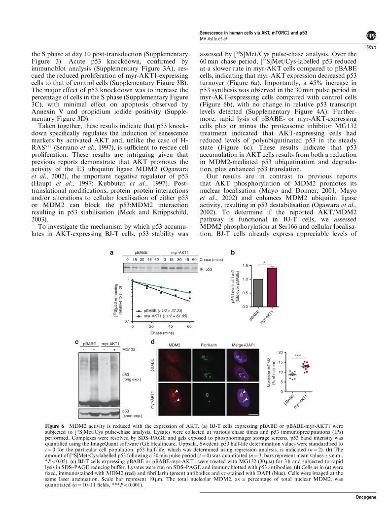
To investigate the mechanism by which p53 accumu-lates in AKT-expressing BJ-T cells, p53 stability was
assessed by [35S]Met/Cys pulse-chase analysis. Over the60min chase period, [35S]Met/Cys-labelled p53 reducedat a slower rate in myr-AKT cells compared to pBABEcells, indicating that myr-AKT expression decreased p53turnover (Figure 6a). Importantly, a 45% increase inp53 synthesis was observed in the 30min pulse period inmyr-AKT-expressing cells compared with control cells(Figure 6b), with no change in relative p53 transcriptlevels detected (Supplementary Figure 4A). Further-more, rapid lysis of pBABE- or myr-AKT-expressingcells plus or minus the proteasome inhibitor MG132treatment indicated that AKT-expressing cells hadreduced levels of polyubiquitinated p53 in the steadystate (Figure 6c). These results indicate that p53accumulation in AKT cells results from both a reductionin MDM2-mediated p53 ubiquitination and degrada-tion, plus enhanced p53 translation.
Our results are in contrast to previous reportsthat AKT phosphorylation of MDM2 promotes itsnuclear localisation (Mayo and Donner, 2001; Mayoet al., 2002) and enhances MDM2 ubiquitin ligaseactivity, resulting in p53 destabilisation (Ogawara et al.,2002). To determine if the reported AKT/MDM2pathway is functional in BJ-T cells, we assessedMDM2 phosphorylation at Ser166 and cellular localisa-tion. BJ-T cells already express appreciable levels of
MG132pBABE myr-AKT1
p53(long exp.)
p53(short exp.)
pBABE myr-AKT1
0 15 30 45 60 0 15 30 45 60
IP: p53
Chase (mins)
Chase (mins)
pBABE (t 1/2 = 37.23)myr-AKT1 (t 1/2 = 61.95)[35
S]p
53 r
emai
ning
(rel
ativ
e to
t =
0)
myr
-AKT1
pBABE
p53
Leve
ls a
t t =
0(f
old
over
pB
AB
E)
*
pBA
BE
myr
-AK
T1
pBABE
myr
-AKT1
Nuc
leol
ar M
DM
2(%
of n
ucle
ar)
***
0.10 20 40 60
0.0
20
15
10
5
0
0.5
1.0
1.5
1
- -+ +MDM2 Fibrillarin Merge+DAPI
Figure 6 MDM2 activity is reduced with the expression of AKT. (a) BJ-T cells expressing pBABE or pBABE-myr-AKT1 weresubjected to [35S]Met/Cys pulse-chase analysis. Lysates were collected at various chase times and p53 immunoprecipitations (IPs)performed. Complexes were resolved by SDS–PAGE and gels exposed to phosphorimager storage screens. p53 band intensity wasquantified using the ImageQuant software (GE Healthcare, Uppsala, Sweden). p53 half-life determination values were standardised tot¼ 0 for the particular cell population. p53 half-life, which was determined using regression analysis, is indicated (n¼ 2). (b) Theamount of [35S]Met/Cys-labelled p53 following a 30min pulse period (t¼ 0) was quantitated (n¼ 3, bars represent mean values±s.e.m.,*Po0.05). (c) BJ-T cells expressing pBABE or pBABE-myr-AKT1 were treated with MG132 (30mM) for 3 h and subjected to rapidlysis in SDS–PAGE reducing buffer. Lysates were run on SDS–PAGE and immunoblotted with p53 antibodies. (d) Cells as in (a) werefixed, immunostained with MDM2 (red) and fibrillarin (green) antibodies and co-stained with DAPI (blue). Cells were imaged at thesame laser attenuation. Scale bar represent 10mm. The total nucleolar MDM2, as a percentage of total nuclear MDM2, wasquantitated (n¼ 10–11 fields, ***Po0.001).
Senescence in human cells via AKT, mTORC1 and p53MV Astle et al
1955
Oncogene
phospho-Ser166-MDM2, but enforced expression ofmyr-AKT did not increase these levels any further(Supplementary Figure 4B). The migration of phospho-Ser166-MDM2 was altered, suggesting that AKTmay promote alternative post-translational modifica-tions of MDM2. Fractionation experiments demon-strated that AKT expression induced a slight increase innuclear and cytoplasmic MDM2 levels (SupplementaryFigure 4C).
To investigate why p53 ubiquitination was markedlyreduced despite slightly elevated nuclear MDM2 levels,we examined MDM2 subnuclear localisation. Nucleolaraccumulation of MDM2 has been reported to beassociated with reduced MDM2 activity (Weber et al.,1999; Ashcroft et al., 2000), due to its sequestration intoinactive complexes. Confocal analysis revealed a robustincrease in the accumulation of MDM2 in the nucleolusof AKT-expressing cells (Figure 6d). Next, we examinedif induction of the tumour suppressor ARF contributesto MDM2 nucleolar localisation. ARF expression canbe induced by oncogenic insult and mediates p53stabilisation by sequestration of MDM2 in the nucleolus(Weber et al., 1999). ARF protein could not be detectedin myr-AKT-expressing cells (Supplementary Figure4D), although it was observed in response to MYC, aknown activator of ARF (Zindy et al., 1998), indicatingthat MDM2 nucleolar sequestration is ARF-indepen-dent. Accumulation of the PML protein has been shownto contribute to p53 stabilisation (Ferbeyre et al., 2000)via its nucleolar sequestration (Bernardi et al., 2004).However, we found no change in the expression levels ofPML or any increase in PML nucleolar localisation inBJ-T cells expressing AKT as compared with controlcells (data not shown). Taken together, these resultsindicate that AKT induces DNA damage-independentp53 stabilisation via ARF- and PML-independentnucleolar sequestration of MDM2.
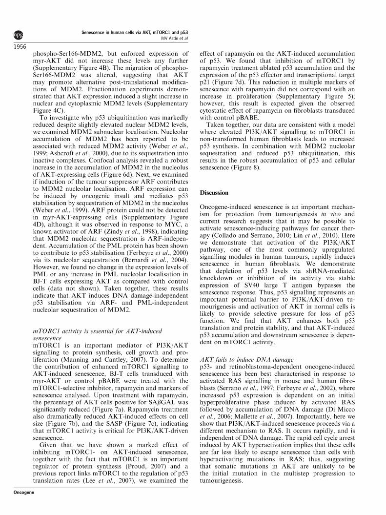
mTORC1 activity is essential for AKT-inducedsenescencemTORC1 is an important mediator of PI3K/AKTsignalling to protein synthesis, cell growth and pro-liferation (Manning and Cantley, 2007). To determinethe contribution of enhanced mTORC1 signalling toAKT-induced senescence, BJ-T cells transduced withmyr-AKT or control pBABE were treated with themTORC1-selective inhibitor, rapamycin and markers ofsenescence analysed. Upon treatment with rapamycin,the percentage of AKT cells positive for SAbGAL wassignificantly reduced (Figure 7a). Rapamycin treatmentalso dramatically reduced AKT-induced effects on cellsize (Figure 7b), and the SASP (Figure 7c), indicatingthat mTORC1 activity is critical for PI3K/AKT-drivensenescence.
Given that we have shown a marked effect ofinhibiting mTORC1- on AKT-induced senescence,together with the fact that mTORC1 is an importantregulator of protein synthesis (Proud, 2007) and aprevious report links mTORC1 to the regulation of p53translation rates (Lee et al., 2007), we examined the
effect of rapamycin on the AKT-induced accumulationof p53. We found that inhibition of mTORC1 byrapamycin treatment ablated p53 accumulation and theexpression of the p53 effector and transcriptional targetp21 (Figure 7d). This reduction in multiple markers ofsenescence with rapamycin did not correspond with anincrease in proliferation (Supplementary Figure 5);however, this result is expected given the observedcytostatic effect of rapamycin on fibroblasts transducedwith control pBABE.
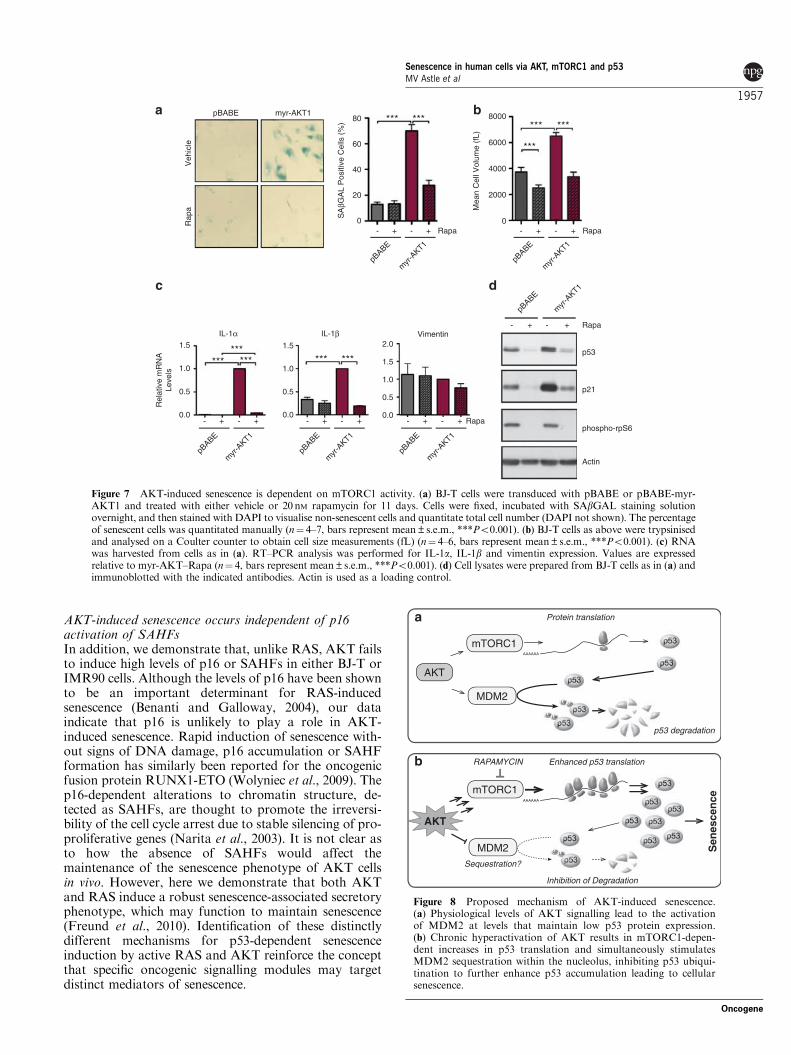
Taken together, our data are consistent with a modelwhere elevated PI3K/AKT signalling to mTORC1 innon-transformed human fibroblasts leads to increasedp53 synthesis. In combination with MDM2 nucleolarsequestration and reduced p53 ubiquitination, thisresults in the robust accumulation of p53 and cellularsenescence (Figure 8).
Discussion
Oncogene-induced senescence is an important mechan-ism for protection from tumourigenesis in vivo andcurrent research suggests that it may be possible toactivate senescence-inducing pathways for cancer ther-apy (Collado and Serrano, 2010; Lin et al., 2010). Herewe demonstrate that activation of the PI3K/AKTpathway, one of the most commonly upregulatedsignalling modules in human tumours, rapidly inducessenescence in human fibroblasts. We demonstratethat depletion of p53 levels via shRNA-mediatedknockdown or inhibition of its activity via stableexpression of SV40 large T antigen bypasses thesenescence response. Thus, p53 signalling represents animportant potential barrier to PI3K/AKT-driven tu-mourigenesis and activation of AKT in normal cells islikely to provide selective pressure for loss of p53function. We find that AKT enhances both p53translation and protein stability, and that AKT-inducedp53 accumulation and downstream senescence is depen-dent on mTORC1 activity.
AKT fails to induce DNA damagep53- and retinoblastoma-dependent oncogene-inducedsenescence has been best characterised in response toactivated RAS signalling in mouse and human fibro-blasts (Serrano et al., 1997; Ferbeyre et al., 2002), whereincreased p53 expression is dependent on an initialhyperproliferative phase induced by activated RASfollowed by accumulation of DNA damage (Di Miccoet al., 2006; Mallette et al., 2007). Importantly, here weshow that PI3K/AKT-induced senescence proceeds via adifferent mechanism to RAS. It occurs rapidly, and isindependent of DNA damage. The rapid cell cycle arrestinduced by AKT hyperactivation implies that these cellsare far less likely to escape senescence than cells withhyperactivating mutations in RAS; thus, suggestingthat somatic mutations in AKT are unlikely to bethe initial mutation in the multistep progression totumourigenesis.
Senescence in human cells via AKT, mTORC1 and p53MV Astle et al
1956
Oncogene
AKT-induced senescence occurs independent of p16activation of SAHFsIn addition, we demonstrate that, unlike RAS, AKT failsto induce high levels of p16 or SAHFs in either BJ-T orIMR90 cells. Although the levels of p16 have been shownto be an important determinant for RAS-inducedsenescence (Benanti and Galloway, 2004), our dataindicate that p16 is unlikely to play a role in AKT-induced senescence. Rapid induction of senescence with-out signs of DNA damage, p16 accumulation or SAHFformation has similarly been reported for the oncogenicfusion protein RUNX1-ETO (Wolyniec et al., 2009). Thep16-dependent alterations to chromatin structure, de-tected as SAHFs, are thought to promote the irreversi-bility of the cell cycle arrest due to stable silencing of pro-proliferative genes (Narita et al., 2003). It is not clear asto how the absence of SAHFs would affect themaintenance of the senescence phenotype of AKT cellsin vivo. However, here we demonstrate that both AKTand RAS induce a robust senescence-associated secretoryphenotype, which may function to maintain senescence(Freund et al., 2010). Identification of these distinctlydifferent mechanisms for p53-dependent senescenceinduction by active RAS and AKT reinforce the conceptthat specific oncogenic signalling modules may targetdistinct mediators of senescence.
Rap
aV
ehic
le
pBABE myr-AKT1
myr
-AKT1
pBABE
SA
βGA
L P
ositi
ve C
ells
(%
)
Rapa
Mea
n C
ell V
olum
e (f
L)
myr
-AKT1
pBABE
Rapa
***
*** ****** ***
IL-1α
- -+ + - -+ + - -+ +
myr
-AKT1
pBABE
IL-1β
myr
-AKT1
pBABE
Vimentin
myr
-AKT1
pBABE
Rapa
Rel
ativ
e m
RN
ALe
vels
********* *** ***
-- + Rapa
p21
phospho-rpS6
Actin
myr
-AKT1
pBABE
p53
80
60
40
20
0
1.5
1.0
0.5
0.0
1.5
1.0
0.5
0.0
2.0
1.5
1.0
0.0
0.5
8000
6000
4000
2000
0- - ++ - - ++
+
Figure 7 AKT-induced senescence is dependent on mTORC1 activity. (a) BJ-T cells were transduced with pBABE or pBABE-myr-AKT1 and treated with either vehicle or 20nM rapamycin for 11 days. Cells were fixed, incubated with SAbGAL staining solutionovernight, and then stained with DAPI to visualise non-senescent cells and quantitate total cell number (DAPI not shown). The percentageof senescent cells was quantitated manually (n¼ 4–7, bars represent mean±s.e.m., ***Po0.001). (b) BJ-T cells as above were trypsinisedand analysed on a Coulter counter to obtain cell size measurements (fL) (n¼ 4–6, bars represent mean±s.e.m., ***Po0.001). (c) RNAwas harvested from cells as in (a). RT–PCR analysis was performed for IL-1a, IL-1b and vimentin expression. Values are expressedrelative to myr-AKT–Rapa (n¼ 4, bars represent mean±s.e.m., ***Po0.001). (d) Cell lysates were prepared from BJ-T cells as in (a) andimmunoblotted with the indicated antibodies. Actin is used as a loading control.
AKT
MDM2
mTORC1
Protein translation
p53 degradation
Enhanced p53 translation
Inhibition of Degradation
Sen
esce
nce
MDM2
mTORC1
AKT
RAPAMYCIN
Sequestration?
a
b
Figure 8 Proposed mechanism of AKT-induced senescence.(a) Physiological levels of AKT signalling lead to the activationof MDM2 at levels that maintain low p53 protein expression.(b) Chronic hyperactivation of AKT results in mTORC1-depen-dent increases in p53 translation and simultaneously stimulatesMDM2 sequestration within the nucleolus, inhibiting p53 ubiqui-tination to further enhance p53 accumulation leading to cellularsenescence.
Senescence in human cells via AKT, mTORC1 and p53MV Astle et al
1957
Oncogene

AKT induces senescence via post-transcriptionalregulation of p53Activated PI3K/AKT signalling results in markedstabilisation of p53 protein. This was unexpected giventhat AKT has been reported to promote the activity ofthe E3 ubiquitin ligase MDM2, the important negativeregulator of p53 (Haupt et al., 1997; Kubbutat et al.,1997). However, in human fibroblasts, we found thatactive AKT signalling reduces p53 ubiquitination andthis corresponds to increased nucleolar sequestration,and hence inactivation of MDM2. Our data indicatethat this nucleolar sequestration is independent of ARFand PML, and thus is likely to involve binding to otherproteins, which localise to the nucleolus either constitu-tively or under conditions of stress such as nucleophos-min (Kurki et al., 2004) and multiple ribosomalproteins, including RPL5, RPL11 and RPL23 (Lohrumet al., 2003; Bhat et al., 2004; Dai and Lu, 2004; Daiet al., 2004). These binding events are invariablyassociated with p53 stabilisation (Zhang and Lu,2009). It is tempting to hypothesise that while transientactivation of AKT by growth factors can lead to activeMDM2, p53 degradation and cell survival, chronicactivation of the pathway, such as that followingoncogenic mutation within the signalling network,results in nucleolar sequestration and inactivation ofMDM2, and accumulation of p53 with a subsequentinduction of senescence.
PI3K/AKT pathway activation and p53 alterationscooperate in human cancerConsistent with p53-induced senescence acting as amajor ‘brake’ for PI3K/AKT-driven human cancer,mutational analysis of primary human tumours hasdemonstrated significant associations between PTEN orPIK3CA, and p53 alterations in colorectal cancer(Nosho et al., 2008), gastric cancer (Oki et al., 2005),non-small-cell lung cancer (Andjelkovic et al., 2011) andbladder cancer (Puzio-Kuter et al., 2009). Furthermore,concomitant alterations in these pathways often result inpoor patient outcome (Catasus et al., 2009; Puzio-Kuteret al., 2009; Andjelkovic et al., 2011). These reports,together with our data, raise concerns for the rationaldesign of pro-senescence therapies to combat cancersdriven by activating mutations in the PI3K/AKTsignalling network, as aggressive tumours will com-monly have previously subverted p53 signalling.In this context, therapeutic activation of p53-indepen-dent pro-senescence pathways will thus be required,such as targeting the E3 ligase Skp2, which acts viaregulation of p21, p27 and Atf4 (Lin et al., 2010), orinducing p16.
mTORC1 inhibitors prevent AKT-induced senescence:implications for cancer therapyImportantly, our results demonstrate that mTORC1 isthe critical mediator of induction of p53-dependentsenescence by AKT in non-transformed human fibro-blasts. Treatment of cells expressing active AKT withthe allosteric mTORC1 inhibitor rapamycin reduced
p53 and p21 to control levels and prevented senescence.Previous reports indicate that mTORC1 activationdownstream of AKT will promote p53 accumulationvia regulation of p53 translation as in PTEN�/�
(Alimonti et al., 2010b) and Tsc1�/� mouse embryonicfibroblasts, (Lee et al., 2007). Our results in human cellsare also consistent with the emerging idea that the levelsof mTORC1 activity determine the outcome of p53activation in cells, where low mTORC1 activity in thecontext of genotoxic stress or MDM2 activation favourscellular quiescence over senescence (Korotchkina et al.,2010; Leontieva and Blagosklonny, 2010).
The dependence of AKT-induced senescence onmTORC1 activity has direct implications for the useof mTORC1 inhibitors as anticancer agents. Inhibitorssuch as RAD001 and CCI-779 have delivered limitedtherapeutic benefit as single agents (Brachmann et al.,2009). This lack of effect has been attributed, atleast in part, to the inactivation of mTORC1-dependentnegative feedback of PI3K/AKT signalling (O’Reillyet al., 2006) and/or the activity of the RAS pathway(Carracedo et al., 2008). Our data provide anotherlevel of potential concern where mTORC1 inhibitionmay relieve the senescence ‘brake’ in p53 wild-typecells and promote PI3K-dependent transformation.Targeted therapies are now being designed and testedthat will alleviate the pro-tumourigenic effects ofmTORC1 inhibition. These include mTOR active siteinhibitors that will target both mTORC2-dependentAKT activation and mTORC1-dependent growth (Feld-man et al., 2009; Dowling et al., 2010; Hsieh et al.,2010), dual PI3K/mTOR inhibitors (Bhatt et al., 2010)or the use of the MDM2 antagonist Nutlin-3a incombination with mTOR inhibition (Alimonti et al.,2010b).
In summary, we demonstrate that constitutive PI3K/AKT pathway activation in normal human cells resultsin the accumulation of p53 and rapid induction ofcellular senescence. Consequently, inactivation of thesenescence response is likely to be critical at the earlystages of PI3K/AKT-driven tumourigenesis, and con-sistent with this model, PTEN or PIK3CA mutationsare significantly associated with p53 mutation in humantumours. Importantly, we demonstrate that mTORC1 isan essential mediator of AKT-induced senescence,providing further insight to aid in the rational designof cancer therapeutic strategies to target tumors withdysregulated PI3K/AKT signalling.
Materials and methods
Plasmid constructsMurine stem cell virus (MSCV) plasmids encoding myr-AKTisoforms are as described previously (Chan et al., 2011).pBABE-puro, pBABE-puro-myr-AKT2, pBABE-puro-myr-AKT3 and pBABE-puro-PIK3CAE545K were from AddgeneInc. (Cambridge, MA, USA). pBABE-myr-AKT1 was directlysubcloned from MSCV-myr-AKT1. pBABE-puro-H-RASV12
was a gift from Patrick Humbert (Peter MacCallum CancerCentre, Melbourne, VIC, Australia). pGIPZ-PTEN shRNA and
Senescence in human cells via AKT, mTORC1 and p53MV Astle et al
1958
Oncogene

pGIPZ-NS shRNA were from Open Biosystems (Huntsville,AL, USA).
Cell linesBJ-T cells, a gift from Robert Weinberg, and derivatives werecultured in Dulbecco’s modified Eagle’s medium:M199 mediaat a ratio of 4:1 (v/v) with 15% fetal calf serum and 2mM
L-glutamine (Hahn et al., 1999, 2002) and maintained in 5%CO2 at 37 1C. Cells expressing pBABE or MSCV constructswere enriched by puromycin selection (1 mg/ml) (Sigma-Aldrich, St Louis, MO, USA) or fluorescence-activated cellsorter, respectively. Rapamycin (20 nM) (Calbiochem, Gibbs-town, NJ, USA) was added 1 day following infection andreplaced every two days.
Western blotting and antibodiesCells were lysed in western solubilisation buffer (0.5mM
EDTA, 20mM HEPES (pH7.9), 2% sodium dodecyl sulfate(SDS)). Lysates were mechanically sheared and a DC assay(BioRad, Hercules, CA, USA) utilised for protein concentra-tion determination. Proteins were visualised using horse radishperoxidase-conjugated secondary antibodies (BioRad) andWestern Lightening Chemiluminescence Plus (Perkin-Elmer,Covina, CA, USA). MDM2 (SMP14), p16, p21 (C-19), p53(DO-1) and H-RAS antibodies were from Santa CruzTechnology (Santa Cruz, CA, USA). Actin and phospho-Ser139-gH2A.X antibody were from Millipore (Billerica, MA,USA). Fibrillarin antibodies were from Abcam (Cambridge,MA, USA). The 12CA5 (HA-tag) antibody was purified in-house. All other antibodies were from Cell SignallingTechnology (Beverly, MA, USA).
SAbGAL stainingCells were stained for SAbGAL as described previously(Debacq-Chainiaux et al., 2009) and co-stained with DAPI(Molecular Probes, Invitrogen, Carlsbad, CA, USA), beforeimaging on a Olympus BX-51 or BX-61 microscope (OlympusAmerica Inc., Center Valley, PA, USA), or a Cellomicsimaging platform (Thermo Fisher Scientific, Waltham, MA,USA). SAbGAL cells and total nuclei were counted manuallyusing the public domain ImageJ software (NIH, Bethesda,MD, USA; Abramoff et al., 2004).
ImmunofluorescenceFor phospho-Ser139-gH2A.X, staining cells were fixed in 95%ethanol/5% acetic acid, permeabilised in 0.1% Tx-100/phosphate-buffered saline for 2min and blocked for 30minin 3% bovine serum albumin/phosphate-buffered saline. ForMDM2/fibrillarin co-staining, cells were fixed in methanol for5min at �20 1C and blocked with 5% goat serum/phosphate-buffered saline for 30min. Antibodies were diluted in blockingbuffer and incubated with cells for 1 h. Cells were imaged onan Olympus BX51 epifluorescence or Leica SP5 confocalmicroscope (Leica, Wetzlar, Germany). Quantitation ofrelative nucleolar MDM2 localisation was performed usingMetaMorph (Molecular Devices, Sunnyvale, CA, USA).
RNA extraction and RT–PCRRNA was extracted using an RNA extraction kit (Bioline,Alexandria, NSW, Australia). 32P-labelled riboprobe wasadded to samples before column purification to allow RNArecovery calculation. RNA was normalised for cell number forcDNA production using random hexamers primers (Promega,Madison, WI, USA) and Superscript III (Invitrogen). Primersequences are listed in Supplementary information.
Cytokine bead array for the analysis of IL secretionCells were plated at 50 000–100 000 in 150ml of media per wellof a 96-well plate. After 24 h, the media were removed, clearedby centrifugation and stored at �80 1C. The level of secretedIL-1a, IL-b, IL-6 and IL-8 was determined using a custom-made Millipore Milliplex Human Cytokine Multiplex Immu-noassay Kit following the manufacturer’s instructions.
p53 pulse-chase analysisCells were incubated in Cys- and Met-free Dulbecco’s modifiedEagle’s medium containing 15% fetal calf serum for 1 h, andthen 200 mCi Express 35S Protein Labelling Mix (Perkin-Elmer)was added for 30min. Cells were washed with media, andincubated for the designated periods and harvested (50mM
Tris (pH 8.0), 150mM NaCl, 5mM EDTA, 0.5% NP-40,complete protease inhibitors; Roche Applied Science, CastleHill, NSW, Australia). P53 was immunoprecipitated using p53antibody (0.3 mg) and Protein A-Sepharose (AmershamBiosciences, Piscataway, NJ, USA). Following washes, sam-ples were run on SDS–polyacrylamide gel electrophoresis(PAGE), gels dried, exposed to a Phosphorimager screen andimaged on a Typhoon (GE Healthcare).
Statistical analysisAll statistical tests were performed in GraphPad Prism(GraphPad Software Inc., La Jolla, CA, USA). The analysiswas performed using paired t-tests or repeated measuresanalysis of variance with Bonferroni post-test.
Conflict of interest
The authors declare no conflict of interest.
Acknowledgements
We acknowledge the Victorian Centre for Functional Geno-mics for the use of equipment and advice. This work wassupported by grants from the National Health and MedicalResearch Council (NHMRC) of Australia to RDH (NHMRCNos. 166908 and 251688) and to RBP (NHMRC Nos. 509087and 400116) and from Cancer Council Victoria to RBP. RDHand RBP are NHMRC Senior Research Fellows. We thankProfessor Stephen Jane and Loretta Cerruti for assistance withretrovirus production.
References
Abramoff M, Magelhaes P, Ram S. (2004). Image processing withimage J. Biophotonics Int 11: 36–42.
Acosta JC, O’Loghlen A, Banito A, Guijarro MV, Augert A, Raguz Set al. (2008). Chemokine signaling via the CXCR2 receptorreinforces senescence. Cell 133: 1006–1018.
Alimonti A, Carracedo A, Clohessy JG, Trotman LC, Nardella C,Egia A et al. (2010a). Subtle variations in Pten dose determinecancer susceptibility. Nat Genet 42: 454–458.
Alimonti A, Nardella C, Chen Z, Clohessy JG, Carracedo A, TrotmanLC et al. (2010b). A novel type of cellular senescence that can be
Senescence in human cells via AKT, mTORC1 and p53MV Astle et al
1959
Oncogene

enhanced in mouse models and human tumor xenografts to suppressprostate tumorigenesis. J Clin Invest 120: 681–693.
Altomare D, Testa J. (2005). Perturbations of the AKT signalingpathway in human cancer. Oncogene 24: 7455–7464.
Andjelkovic T, Bankovic J, Stojsic J, Milinkovic V, Podolski-Renic A,Ruzdijic S et al. (2011). Coalterations of p53 and PTEN tumorsuppressor genes in non-small cell lung carcinoma patients. Transl
Res 157: 19–28.Ashcroft M, Taya Y, Vousden KH. (2000). Stress signals
utilize multiple pathways to stabilize p53. Mol Cell Biol 20:3224–3233.
Banin S, Moyal L, Shieh S-Y, Taya Y, Anderson CW, Chessa L et al.(1998). Enhanced phosphorylation of p53 by ATM in response toDNA damage. Science 281: 1674–1677.
Beausejour C, Krtolica A, Galimi F, Narita M, Lowe S, Yaswen Pet al. (2003). Reversal of human cellular senescence: roles of the p53and p16 pathways. EMBO J 22: 4212–4222.
Bellodi C, Kopmar N, Ruggero D. (2010). Deregulation of oncogene-induced senescence and p53 translational control in X-linkeddyskeratosis congenita. EMBO J 29: 1865–1876.
Benanti JA, Galloway DA. (2004). Normal human fibroblastsare resistant to RAS-induced senescence. Mol Cell Biol 24:2842–2852.
Bernardi R, Scaglioni PP, Bergmann S, Horn HF, Vousden KH,Pandolfi PP. (2004). PML regulates p53 stability by sequesteringMdm2 to the nucleolus. Nat Cell Biol 6: 665–672.
Bhat KP, Itahana K, Jin A, Zhang Y. (2004). Essential role ofribosomal protein L11 in mediating growth inhibition-induced p53activation. EMBO J 23: 2402–2412.
Bhatt AP, Bhende PM, Sin S-H, Roy D, Dittmer DP, Damania B.(2010). Dual inhibition of PI3K and mTOR inhibits autocrine andparacrine proliferative loops in PI3K/Akt/mTOR-addicted lympho-mas. Blood 115: 4455–4463.
Brachmann S, Fritsch C, Maira S-M, GarcIa-EcheverrIa C. (2009).PI3K and mTOR inhibitors—a new generation of targeted antic-ancer agents. Curr Opin Cell Biol 21: 194–198.
Brookes S, Rowe J, Ruas M, Llanos S, Clark PA, Lomax M et al.(2002). INK4a-deficient human diploid fibroblasts are resistant toRAS-induced senescence. EMBO J 21: 2936–2945.
Campbell IG, Russell SE, Choong DYH, Montgomery KG, CiavarellaML, Hooi CSF et al. (2004). Mutation of the PIK3CA gene inovarian and breast cancer. Cancer Res 64: 7678–7681.
Carracedo A, Ma L, Teruya-Feldstein J, Rojo F, Salmena L, AlimontiA et al. (2008). Inhibition of mTORC1 leads to MAPK pathwayactivation through a PI3K-dependent feedback loop in humancancer. J Clin Invest 118: 3065–3074.
Catasus L, Gallardo A, Cuatrecasas M, Prat J. (2009). ConcomitantPI3K-AKT and p53 alterations in endometrial carcinomas areassociated with poor prognosis. Mod Pathol 22: 522–529.
Chan JC, Hannan KM, Riddell K, Ng PY, Peck A, Lee RS et al.(2011). AKT is a critical regulator of rRNA synthesis andcooperates with MYC to control ribosome biogenesis in cancer.Sci Signal 4: ra56.
Chen Z, Trotman L, Shaffer D, Lin H, Dotan Z, Niki M et al. (2005).Crucial role of p53-dependent cellular senescence in suppression ofPten-deficient tumorigenesis. Nature 436: 725–730.
Collado M, Serrano M. (2010). Senescence in tumours: evidence frommice and humans. Nat Rev Cancer 10: 51–57.
Coppe J-P, Patil CK, Rodier F, Sun Y, Munoz DP, Goldstein J et al.(2008). Senescence-associated secretory phenotypes reveal cell-nonautonomous functions of oncogenic RAS and the p53 tumorsuppressor. PLoS Biol 6: 2853–2868.
Courtois-Cox S, Genther Williams SM, Reczek EE, Johnson BW,McGillicuddy LT, Johannessen CM et al. (2006). A negativefeedback signaling network underlies oncogene-induced senescence.Cancer Cell 10: 459–472.
Cristiano BE, Chan JC, Hannan KM, Lundie NA, Marmy-Conus NJ,Campbell IG et al. (2006). A specific role for AKT3 in the genesis ofovarian cancer through modulation of G(2)–M phase transition.Cancer Res 66: 11718–11725.
Dai M-S, Lu H. (2004). Inhibition of MDM2-mediated p53ubiquitination and degradation by ribosomal protein L5. J Biol
Chem 279: 44475–44482.Dai M-S, Zeng SX, Jin Y, Sun X-X, David L, Lu H. (2004).
Ribosomal protein L23 activates p53 by inhibiting MDM2 functionin response to ribosomal perturbation but not to translationinhibition. Mol Cell Biol 24: 7654–7668.
Debacq-Chainiaux F, Erusalimsky JD, Campisi J, Toussaint O.(2009). Protocols to detect senescence-associated beta-galactosidase(SA-[beta]gal) activity, a biomarker of senescent cells in culture andin vivo. Nat Protocols 4: 1798–1806.
Di Micco R, Fumagalli M, Cicalese A, Piccinin S, Gasparini P,Luise C et al. (2006). Oncogene-induced senescence is a DNAdamage response triggered by DNA hyper-replication. Nature 444:638–642.
Dowling RJO, Topisirovic I, Fonseca BD, Sonenberg N. (2010).Dissecting the role of mTOR: lessons from mTOR inhibitors.Biochim et Biophys Acta 1804: 433–439.
Ewald JA, Desotelle JA, Wilding G, Jarrard DF. (2010). Therapy-induced senescence in cancer. J Natl Cancer Inst 102: 1536–1546.
Feldman ME, Apsel B, Uotila A, Loewith R, Knight ZA, Ruggero Det al. (2009). Active-site inhibitors of mTOR target rapamycin-resistant outputs of mTORC1 and mTORC2. PLoS Biol 7:e1000038.
Ferbeyre G, de Stanchina E, Lin A, Querido E, McCurrach M,Hannon G et al. (2002). Oncogenic ras and p53 cooperate to inducecellular senescence. Mol Cell Biol 22: 3497–3508.
Ferbeyre G, de Stanchina E, Querido E, Baptiste N, Prives C, LoweSW. (2000). PML is induced by oncogenic ras and promotespremature senescence. Genes Dev 14: 2015–2027.
Franke T. (2008). PI3K/Akt: getting it right matters. Oncogene 27:6473–6488.
Freund A, Orjalo AV, Desprez P-Y, Campisi J. (2010). Inflammatorynetworks during cellular senescence: causes and consequences.Trends Mol Med 16: 238–246.
Gewirtz DA, Holt SE, Elmore LW. (2008). Accelerated senescence: anemerging role in tumor cell response to chemotherapy andradiation. Biochem Pharmacol 76: 947–957.
Gorbunova V, Seluanov A, Pereira-Smith OM. (2002). Expression ofhuman telomerase (hTERT) does not prevent stress-inducedsenescence in normal human fibroblasts but protects the cellsfrom stress-induced apoptosis and necrosis. J Biol Chem 277:38540–38549.
Gorgoulis VG, Halazonetis TD. (2010). Oncogene-induced senescence:the bright and dark side of the response. Curr Opin Cell Biol 22:816–827.
Gray-Schopfer VC, Cheong SC, Chong H, Chow J, Moss T, Abdel-Malek ZA et al. (2006). Cellular senescence in naevi and immortalisa-tion in melanoma: a role for p16? Br J Cancer 95: 496–505.
Hahn WC, Counter CM, Lundberg AS, Beijersbergen RL, BrooksMW, Weinberg RA. (1999). Creation of human tumour cells withdefined genetic elements. Nature 400: 464–468.
Hahn WC, Dessain SK, Brooks MW, King JE, Elenbaas B, SabatiniDM et al. (2002). Enumeration of the simian virus 40 early regionelements necessary for human cell transformation. Mol Cell Biol 22:2111–2123.
Hanahan D, Weinberg RA. (2000). The hallmarks of cancer. Cell 100:57–70.
Hannan KM, Sanij E, Hein N, Hannan RD, Pearson RB. (2011).Signalling to the ribosome in cancer—it’s more that just mTORC1.IUBMB Life 63: 79–85.
Haupt Y, Maya R, Kazaz A, Oren M. (1997). Mdm2 promotes therapid degradation of p53. Nature 387: 296–299.
Hirao A, Kong Y-Y, Matsuoka S, Wakeham A, Ruland, Yoshida Het al. (2000). DNA damage-induced activation of p53 by thecheckpoint kinase Chk2. Science 287: 1824–1827.
Hsieh AC, Costa M, Zollo O, Davis C, Feldman ME, Testa JR et al.(2010). Genetic dissection of the oncogenic mTOR pathway revealsdruggable addiction to translational control via 4EBP-eIF4E.Cancer Cell 17: 249–261.
Senescence in human cells via AKT, mTORC1 and p53MV Astle et al
1960
Oncogene
Huot TJ, Rowe J, Harland M, Drayton S, Brookes S, Gooptu C et al.(2002). Biallelic mutations in p16(INK4a) confer resistance to Ras-and Ets-induced senescence in human diploid fibroblasts. Mol Cell
Biol 22: 8135–8143.Hynes NE, MacDonald G. (2009). ErbB receptors and signaling
pathways in cancer. Curr Opin Cell Biol 21: 177–184.Kim J-S, Lee C, Bonifant CL, Ressom H, Waldman T. (2007).
Activation of p53-dependent growth suppression in human cells bymutations in PTEN or PIK3CA. Mol Cell Biol 27: 662–677.
Korotchkina LG, Leontieva OV, Bukreeva EI, Demidenko ZN,Gudkov AV, Blagosklonny MV. (2010). The choice between p53-induced senescence and quiescence is determined in part by themTOR pathway. Aging 2: 344–352.
Kubbutat MHG, Jones SN, Vousden KH. (1997). Regulation of p53stability by Mdm2. Nature 387: 299–303.
Kurki S, Peltonen K, Latonen L, Kiviharju TM, Ojala Pi M, Meek Det al. (2004). Nucleolar protein NPM interacts with HDM2 andprotects tumor suppressor protein p53 from HDM2-mediateddegradation. Cancer Cell 5: 465–475.
Lee C-H, Inoki K, Karbowniczek M, Petroulakis E, Sonenberg N,Henske EP et al. (2007). Constitutive mTOR activation in TSCmutants sensitizes cells to energy starvation and genomic damagevia p53. EMBO J 26: 4812–4823.
Leontieva OV, Blagosklonny MV. (2010). DNA damaging agents andp53 do not cause senescence in quiescent cells, while consecutive re-activation of mTOR is associated with conversion to senescence.Aging 31: 31.
Lin H-K, Chen Z, Wang G, Nardella C, Lee S-W, Chan C-H et al.(2010). Skp2 targeting suppresses tumorigenesis by Arf–p53-independent cellular senescence. Nature 464: 374–379.
Lohrum MAE, Ludwig RL, Kubbutat MHG, Hanlon M, VousdenKH. (2003). Regulation of HDM2 activity by the ribosomal proteinL11. Cancer Cell 3: 577–587.
Lowe SW, Cepero E, Evan G. (2004). Intrinsic tumour suppression.Nature 432: 307–315.
Luo J, Manning B, Cantley L. (2003). Targeting the PI3K-Akt pathwayin human cancer: rationale and promise. Cancer Cell 4: 257–262.
Mallette F, Gaumont-Leclerc M, Ferbeyre G. (2007). The DNAdamage signaling pathway is a critical mediator of oncogene-induced senescence. Genes Dev 21: 43–48.
Manning BD, Cantley LC. (2007). AKT/PKB signaling: navigatingdownstream. Cell 129: 1261–1274.
Mavrakis KJ, Zhu H, Silva RLA, Mills JR, Teruya-Feldstein J, LoweSW et al. (2008). Tumorigenic activity and therapeutic inhibition ofRheb GTPase. Genes Dev 22: 2178–2188.
Mayo LD, Dixon JE, Durden DL, Tonks NK, Donner DB. (2002).PTEN protects p53 from Mdm2 and sensitizes cancer cells tochemotherapy. J Biol Chem 277: 5484–5489.
Mayo LD, Donner DB. (2001). A phosphatidylinositol 3-kinase/Aktpathway promotes translocation of Mdm2 from the cytoplasm tothe nucleus. Proc Natl Acad Sci USA 98: 11598–11603.
Meek DW, Knippschild U. (2003). Posttranslational modification ofMDM2. Mol Cancer Res 1: 1017–1026.
Miyauchi H, Minamino T, Tateno K, Kunieda T, Toko H, Komuro I.(2004). Akt negatively regulates the in vitro lifespan of humanendothelial cells via a p53/p21-dependent pathway. EMBO J 23:212–220.
Moral M, Segrelles C, Lara M, Martinez-Cruz A, Lorz C, Santos Met al. (2009). Akt activation synergizes with Trp53 loss in oralepithelium to produce a novel mouse model for head and necksquamous cell carcinoma. Cancer Res 69: 1099–1108.
Nardella C, Clohessy JG, Alimonti A, Pandolfi PP. (2011). Pro-senescence therapy for cancer treatment. Nat Rev Cancer 11: 503–511.
Narita M, NuOez S, Heard E, Narita M, Lin A, Hearn S et al. (2003).Rb-mediated heterochromatin formation and silencing of E2Ftarget genes during cellular senescence. Cell 113: 703–716.
Narita M, Young AR, Arakawa S, Samarajiwa SA, Nakashima T,Yoshida S et al. (2011). Spatial coupling of mTOR and autophagyaugments secretory phenotypes. Science 332: 966–970.
Nogueira V, Park Y, Chen C-C, Xu P-Z, Chen M-L, Tonic I et al.(2008). Akt determines replicative senescence and oxidative oroncogenic premature senescence and sensitizes cells to oxidativeapoptosis. Cancer Cell 14: 458–470.
Nosho K, Kawasaki T, Ohnishi M, Suemoto Y, Kirkner GJ,Zepf D et al. (2008). PIK3CA mutation in colorectal cancer:relationship with genetic and epigenetic alterations. Neoplasia 10:534–541.
O’Reilly KE, Rojo F, She Q-B, Solit D, Mills GB, Smith D et al.(2006). mTOR inhibition induces upstream receptor tyrosine kinasesignaling and activates Akt. Cancer Res 66: 1500–1508.
Ogawara Y, Kishishita S, Obata T, Isazawa Y, Suzuki T, Tanaka Ket al. (2002). Akt enhances Mdm2-mediated ubiquitination anddegradation of p53. J Biol Chem 277: 21843–21850.
Oki E, Tokunaga E, Nakamura T, Ueda N, Futatsugi M, Mashino Ket al. (2005). Genetic mutual relationship between PTEN and p53 ingastric cancer. Cancer Lett 227: 33–38.
Orjalo AV, Bhaumik D, Gengler BK, Scott GK, Campisi J. (2009).Cell surface-bound IL-1alpha is an upstream regulator of thesenescence-associated IL-6/IL-8 cytokine network. Proc Natl Acad
Sci USA 106: 17031–17036.Ouellette MM, Aisner DL, Savre-Train I, Wright WE, Shay JW.
(1999). Telomerase activity does not always imply telomeremaintenance. Biochem Biophys Res Commun 254: 795–803.
Proud CG. (2007). Signalling to translation: how signal transductionpathways control the protein synthetic machinery. Biochem J 403:217–234.
Puzio-Kuter AM, Castillo-Martin M, Kinkade CW, Wang X, ShenTH, Matos T et al. (2009). Inactivation of p53 and Pten promotesinvasive bladder cancer. Genes Dev 23: 675–680.
Rodier F, Coppe J-P, Patil CK, Hoeijmakers WAM, Munoz DP, RazaSR et al. (2009). Persistent DNA damage signalling triggerssenescence-associated inflammatory cytokine secretion. Nat Cell
Biol 11: 973–979.Samuels Y, Diaz LA, Schmidt-Kittler O, Cummins JM, DeLong L,
Cheong I et al. (2005). Mutant PIK3CA promotes cell growth andinvasion of human cancer cells. Cancer Cell 7: 561–573.
Serrano M, Lin AW, McCurrach ME, Beach D, Lowe SW. (1997).Oncogenic ras provokes premature cell senescence associated withaccumulation of p53 and p16INK4a. Cell 88: 593–602.
Shaw RJ, Cantley LC. (2006). Ras, PI(3)K and mTOR signallingcontrols tumour cell growth. Nature 441: 424–430.
Solomon B, Pearson RB. (2009). Class IA phosphatidylinositol 3-kinase signaling in non-small cell lung cancer. J Thorac Oncol 4:787–791.
Tremain R, Marko M, Kinnimulki V, Ueno H, Bottinger E, Glick A.(2000). Defects in TGF-beta signaling overcome senescence ofmouse keratinocytes expressing v-Ha-ras. Oncogene 19: 1698–1709.
Wang W, Chen JX, Liao R, Deng Q, Zhou JJ, Huang S et al. (2002).Sequential activation of the MEK-extracellular signal-regulatedkinase and MKK3/6-p38 mitogen-activated protein kinase path-ways mediates oncogenic ras-induced premature senescence. Mol
Cell Biol 22: 3389–3403.Weber JD, Taylor LJ, Roussel MF, Sherr CJ, Bar-Sagi D. (1999).
Nucleolar Arf sequesters Mdm2 and activates p53. Nat Cell Biol 1:20–26.
Wolyniec K, Wotton S, Kilbey A, Jenkins A, Terry A, Peters G et al.(2009). RUNX1 and its fusion oncoprotein derivative, RUNX1-ETO, induce senescence-like growth arrest independently ofreplicative stress. Oncogene 28: 2502–2512.
Xu Z, Xiao SB, Xu P, Xie Q, Cao L, Wang D et al. (2011). miR-365, anovel negative regulator of interleukin-6 gene expression, iscooperatively regulated by Sp1 and NF-\{kappa\}B. J Biol Chem
286: 21401–21412.Yilmaz OH, Valdez R, Theisen BK, Guo W, Ferguson DO,
Wu H et al. (2006). Pten dependence distinguishes haematopoieticstem cells from leukaemia-initiating cells. Nature 441: 475–482.
Young ARJ, Narita M, Ferreira M, Kirschner K, Sadaie M, Darot JFJet al. (2009). Autophagy mediates the mitotic senescence transition.Genes Dev 23: 798–803.
Senescence in human cells via AKT, mTORC1 and p53MV Astle et al
1961
Oncogene

Zhang J, Grindley JC, Yin T, Jayasinghe S, He XC, Ross JT et al.(2006). PTEN maintains haematopoietic stem cells andacts in lineage choice and leukaemia prevention. Nature 441:518–522.
Zhang Y, Lu H. (2009). Signaling to p53: ribosomal proteins find theirway. Cancer Cell 16: 369–377.
Zhao JJ, Liu Z, Wang L, Shin E, Loda MF, Roberts TM. (2005). Theoncogenic properties of mutant p110alpha and p110beta phospha-tidylinositol 3-kinases in human mammary epithelial cells. Proc NatlAcad Sci USA 102: 18443–18448.
Zindy F, Eischen CM, Randle DH, Kamijo T, Cleveland JL, Sherr CJet al. (1998). Myc signaling via the ARF tumor suppressorregulates p53-dependent apoptosis and immortalization. Genes
Dev 12: 2424–2433.
Thiswork is licensedunder theCreativeCommonsAttribution-NonCommercial-No Derivative
Works 3.0 Unported License. To view a copy of this license,visit http://creativecommons.org/licenses/by-nc-nd/3.0/
Supplementary Information accompanies the paper on the Oncogene website (http://www.nature.com/onc)
Senescence in human cells via AKT, mTORC1 and p53MV Astle et al
1962
Oncogene

Copyright of Oncogene is the property of Nature Publishing Group and its content may not be copied or
emailed to multiple sites or posted to a listserv without the copyright holder's express written permission.
However, users may print, download, or email articles for individual use.
Copyright © 2022 FDOKUMEN